Note: Descriptions are shown in the official language in which they were submitted.
CA 02674816 2009-06-23
WO 2008/082957
PCT/US2007/087944
ACCOMMODATING INTRAOCULAR LENS SYSTEM HAVING
SPHERICAL ABERRATION COMPENSATION AND METHOD
Field of the Invention
[0001] The present invention relates to intraocular lenses ("IOLs") having
optical
parameters that are changeable in-situ. More particularly, the invention has
application in
IOLs for in-capsule implantation for cataract patients or presbyopic patients,
wherein
movement of the lens capsule applies forces to a circumferentially supported
haptic to more
efficiently induce transfer of fluid media within the interior of the IOL to
alter an optical
power of the lens.
Background of the Invention
[0002] Cataracts are a major cause of blindness in the world and the most
prevalent ocular
disease. Visual disability from cataracts accounts for more than 8 million
physician office
visits per year. When the disability from cataracts affects or alters an
individual's activities
of daily living, surgical lens removal with intraocular lens (IOL)
implantation is the
preferred method of treating the functional limitations. In the United States,
about 2.5
million cataract surgical procedures are performed annually, making it the
most common
surgery for Americans over the age of 65. About 97 percent of cataract surgery
patients
receive intraocular lens implants, with the annual costs for cataract surgery
and associated
care in the United States being upwards of $4 billion.
[0003] A cataract is any opacity of a patient's lens, whether it is a
localized opacity or a
diffuse general loss of transparency. To be clinically significant, however,
the cataract must
cause a significant reduction in visual acuity or a functional impairment. A
cataract occurs
as a result of aging or secondary to hereditary factors, trauma, inflammation,
metabolic or
nutritional disorders, or radiation. Age related cataract conditions are the
most common.
[0004] In treating a cataract, the surgeon removes the crystalline lens matrix
from the lens
capsule and replaces it with an intraocular lens ("IOL") implant. The typical
IOL provides
a selected focal length that allows the patient to have fairly good distance
vision. Since the
lens can no longer accommodate, however, the patient typically needs glasses
for reading.
CA 02674816 2009-06-23
WO 2008/082957
PCT/US2007/087944
2
[0005] More specifically, the imaging properties of the human eye are
facilitated by several
optical interfaces. A healthy youthful human eye has a total power of
approximately 59
diopters, with the anterior surface of the cornea (e.g. the exterior surface,
including the tear
layer) providing about 48 diopters of power, while the posterior surface
provides about -4
diopters. The crystalline lens, which is situated posterior of the pupil in a
transparent elastic
capsule, also referred to herein as "capsular sac," supported by the ciliary
muscles via
zonules, provides about 15 diopters of power, and also performs the critical
function of
focusing images upon the retina. This focusing ability, referred to as
"accommodation,"
enables imaging of objects at various distances.
[0006] The power of the lens in a youthful eye can be adjusted from 15
diopters to about 29
diopters by adjusting the shape of the lens from a moderately convex shape to
a highly
convex shape. The mechanism generally accepted to cause this adjustment is
that ciliary
muscles supporting the capsule (and the lens contained therein) move between a
relaxed
state (corresponding to the moderately convex shape) and a contracted state
(corresponding
to the highly convex shape). Because the lens itself is composed of viscous,
gelatinous
transparent fibers, arranged in an "onion-like" layered structure, forces
applied to the
capsule by the ciliary muscles via the zonules cause the lens to change shape.
[0007] Isolated from the eye, the relaxed capsule and lens take on a more
spherical shape.
Within the eye, however, the capsule is connected around its circumference by
approximately 70 tiny ligament fibers to the ciliary muscles, which in turn
are attached to an
inner surface of the eyeball. The ciliary muscles that support the lens and
capsule therefore
are believed to act in a sphincter-muscular mode. Accordingly, when the
ciliary muscles
are relaxed, the capsule and lens are pulled about the circumference to a
larger diameter,
thereby flattening the lens, whereas when the ciliary muscles are contracted
the lens and
capsule relax somewhat and assume a smaller diameter that approaches a more
spherical
shape.
[0008] As noted above, the youthful eye has approximately 14 diopters of
accommodation.
As a person ages, the lens hardens and becomes less elastic, so that by about
age 45-50,
accommodation is reduced to about 2 diopters. At a later age the lens may be
considered to
be non-accommodating, a condition known as "presbyopia". Because the imaging
distance
is fixed, presbyopia typically entails the need for bi-focals to facilitate
near and far vision.
CA 02674816 2014-02-21
52723-26
3
100091 Apart from age-related loss of accommodation ability, such loss is
innate to the
placement of 10Ls for the treatment of cataracts. 10Ls are generally single
element lenses
made from a suitable polymer material, such as acrylics or silicones. After
placement,
accommodation is no longer possible, although this ability is typically
already lost for
persons receiving an IOL. There is significant need to provide for
accommodation in IOL
products so that IOL recipients will have accommodating ability.
[0010] Although previously known workers in the field of accommodating IOLs
have made
some progress, the relative complexity of the methods and apparatus developed
to date have
prevented widespread commercialization of such devices. Previously known
devices have
proved too complex to be practical to construct or have achieved only limited
success, due
to the inability to provide accommodation of more than 1-2 diopters.
10011] U.S. Patent No. 5,443,506 to Garabet describes an accommodating fluid-
filled lens
wherein electrical potentials generated by contraction of the ciliary muscles
cause changes
in the index of refraction of fluid carried within a central optic portion.
U.S. Patent No.
4,816,031 to Pfoff discloses an IOL with a hard poly methyl methacrylate
(PMMA) lens
separated by a single chamber from a flexible thin lens layer that uses
microfluid pumps to
vary a volume of fluid between the PMMA lens portion and the thin layer
portion and
provide accommodation. U.S. Patent No. 4,932,966 to Christie et al. discloses
an
intraocular lens comprising a thin flexible layer sealed along its periphery
to a support layer,
wherein forces applied to fluid reservoirs in the haptics vary a volume of
fluid between the
layers to provide accommodation.
[0012] Although fluid-actuated mechanisms such as described in the
aforementioned
patents have been investigated, currently available accommodating lenses
include the
Crystalens developed by Eyeonics, Inc. (formerly C&C Vision, Inc.) of Aliso
Viejo,
California. According to Eyeonics, redistribution of the ciliary mass upon
constriction
causes increased vitreous pressure resulting in forward movement of the lens.
[0013] Co-pending, commonly assigned U.S. Patent Application Publication No.
2005/0119740 to Esch et al.,
describes an intraocular lens in which forces applied by the lens capsule to a
haptic portion
of the lens to induce fluid transfer to and from an actuator disposed in
contact with a
dynamic surface of the lens.
CA 02674816 2009-06-23
WO 2008/082957
PCT/US2007/087944
4
[0014] Another disadvantage of previously known devices is that they
oftentimes create
spherical aberrations. As is well known in the art, lenses composed of
elements having
spherical surfaces are easy to manufacture but are not ideal for creating a
sharp image
because light passing through the elements does not focus on a single focal
point. In
particular, light that passes through a positive optical element close to the
optical axis
generally converges at a focal point that is further from the lens than a
focal point of light
passing through the peripheral portion of the lens, thereby creating under
corrected spherical
aberration. As a result of spherical aberration in an intraocular lens, all of
the light passing
through the lens does not focus on the retina resulting in an image that may
be blurred and
may have softened contrast.
[0015] Various devices have been used in optical systems to reduce the effect
of spherical
aberration. For example, an aperture may be used that limits the ability of
light to pass
through the peripheral portion of the lens. As a result, the light
contributing to the
aberration is prevented from passing through the lens. Such a device provides
an obvious
disadvantage that the amount of light allowed to pass through the lens is
reduced. Another
way to reduce the effect of spherical aberration is to combine two lenses, one
convex and
one concave. A still further method of reducing the effects of spherical
aberration is to use
an aspherical lens. However, such combined lenses and lenses having aspherical
profiles
are significantly more expensive to produce. In addition, combining lenses
requires
additional space to house the multiple lenses.
[0016] While the lens described in the foregoing Esch application is expected
to provide
significant benefits over previously-known accommodating lens designs, it
would be
desirable to provide methods and apparatus for further enhancing conversion of
lens capsule
movements into hydraulic forces, so as to improve modulation of the lens
actuator and
dynamic surface.
[0017] It also would be desirable to provide methods and apparatus to enhance
the
efficiency with which loads arising due to natural accommodating muscular
action are
converted to hydraulic forces.
[0018] It also would be desirable to provide methods and apparatus that reduce
spherical
aberration while maximizing the useful surface area of an accommodating lens
design.
CA 02674816 2014-02-21
52723-26
Summary of the Invention
[0019] In view of the foregoing, it is an object of some embodiments of the
present invention
to provide apparatus and methods that restore appropriate optical focusing
power action to the
human eye.
5 [0020] It is a further object of some embodiments of this invention to
provide methods and
apparatus wherein a dynamic lens surface may be hydraulically manipulated
responsive to
movement of the ciliary muscles and lens capsule.
[0021] It also is an object of some embodiments of the present invention to
provide methods
and apparatus for further enhancing conversion of lens capsule movements into
hydraulic
forces, so as to improve modulation of the lens actuator and dynamic surface.
[0022] It is another object of some embodiments of this invention to provide
methods and
apparatus to enhance the efficiency with which loads arising due to natural
accommodating
muscular action are converted to hydraulic forces.
[0023] It is another object of some embodiments of this invention to provide
methods and
apparatus for reducing spherical aberration in an accommodating intraocular
lens device.
[0024] Some embodiments of the present invention provide an intraocular lens
responsive to
forces communicated from the ciliary muscles through the zonules to the
capsular bag to
operate one or more actuators disposed within the IOL. The actuator is coupled
to a dynamic
surface of the IOL to deflect the dynamic surface, e.g., from a moderately
convex to a highly
convex shape, responsive to operation of the one or more actuators. In
accordance with the
principles of the present invention, the IOL includes at least one secondary
deflection
mechanism that is configured to further alter the curvature of the dynamic
surface to correct
for spherical aberration. The secondary deflection mechanism may be
alterations of the lens
such as varying thickness or inflection points, selection of the boundary
condition of the lens,
and secondary fluid-mediated actuators.
CA 02674816 2014-02-21
52723-26
5a
[0025] In an embodiment, the secondary deflection mechanism is a fluid-
mediated actuator
coupled to a fluid column disposed in at least one haptic of the IOL and a
sealed fluid cavity
filled with shaping fluid that is adjacent to the dynamic surface. Forces
applied to the haptic
by the capsular bag, responsive to movement of the ciliary muscles, cause the
transfer of fluid
between the fluid column and the actuator, which in turn deflects a dynamic
surface of the
lens.
100261 Deflection of the dynamic surface causes the shaping fluid in the
sealed fluid cavity to
redistribute which, in turn, alters the shape of the dynamic surface so that
it is aspherical.
CA 02674816 2009-06-23
WO 2008/082957
PCT/US2007/087944
6
By making the dynamic surface aspherical the total amount of travel required
by the
actuator may be reduced from approximately 300 microns for non-aspheric lenses
to 200
microns. As a result, a more efficient IOL may be produced that requires less
influence
from the lens capsule.
[0027] In a preferred embodiment, the intraocular lens comprises an optic
portion including
a fluid cavity containing a fixed volume of shaping fluid and a haptic (or non-
optic) portion.
The optic portion comprises a light transmissive substrate defining one or
more fluid
channels, at least one actuator coupled in fluid communication with the fluid
channels, and
anterior and posterior lens elements. At least one of the anterior and
posterior lens elements
includes a dynamic surface that is operatively coupled to the actuator to
cause deflection of
the dynamic surface. The other of the anterior or posterior lens elements may
be coupled to
the substrate or integrally formed therewith.
[0028] The haptic portion is disposed at the periphery of the optic portion
and comprises
one or more haptics that extend outward from the optic portion, each haptic
including a
fluid channel coupled in fluid communication with a fluid channel in the optic
portion. In
accordance with one aspect of the present invention, the haptics have a cross-
sectional
configuration selected so that the internal volume of the haptic is small in
an accommodated
state. The accommodated state of the haptic is selected to correspond to the
accommodated
state of the eye, when the ciliary muscles are contracted and
anterior/posterior compressive
forces applied by the capsular bag to the haptics are reduced.
[0029] When the ciliary muscles relax, the zonules pull the capsular sac taut
and apply
forces to the anterior and posterior faces of the haptic. The forces applied
by the capsular
sac cause the cross-sectional area of the haptic to increase thereby
increasing the internal
volume of the haptic. This action in turn causes fluid to be withdrawn from
the actuator
disposed in the optic portion, so that the dynamic surface of the IOL
transitions from an
accommodated state to an unaccommodated state. The fixed volume of shaping
fluid in the
sealed fluid cavity is redistributed in the cavity by movement of the dynamic
surface and
that redistribution causes the shape of the dynamic surface to be altered.
[0030] The actuator used in the optic portion of the IOL may be centrally
located within the
optic portion that, when filled with fluid, biases the dynamic surface of the
IOL to the
accommodated state. When the ciliary muscles are contracted, the zonules and
capsular bag
are less taut, and the haptics are unstressed. Relaxation of the ciliary
muscle causes the
CA 02674816 2014-11-20
52723-26
7
zonules to transition the capsule to less convex shape, which applies
compressive forces to the
haptic, thereby withdrawing fluid from the actuator and causing the lens to
transition to the
unaccommodated state. Alternatively, the actuator may comprise structures
disposed at the
periphery of the optic portion, so as to further minimize refractive effects
and optical
aberrations in the optic portion.
[0031] Methods of making and using the lens of the present invention also are
provided.
[0031a] Some embodiments disclosed herein relate to an intraocular lens
configured for
implantation in a capsular sac following extraction of a natural lens, the
intraocular lens
accommodating in response to movement of the capsular sac, the intraocular
lens comprising:
an optic portion including a deformable anterior element and a posterior
element; a
deformable haptic secured to the optic portion, the haptic having
unaccommodated
configuration with a cross section in which a longest linear dimension in the
anterior-to-
posterior direction is greater than a longest linear radial dimension and
having an interior
volume in fluid communication with the optic portion; a fluid disposed in the
optic portion
and the interior volume of the haptic; and wherein the deformable anterior
element is
configured to be deformed to change the power of the intraocular lens in
response to
movement of the fluid between the haptic and the optic portion.
CA 02674816 2014-02-21
52723-26
7a
Brief Description of the Drawings
100321 Further features of the invention, its nature and various advantages
will be more
apparent from the accompanying drawings and the following detailed description
of the
preferred embodiments, in which:
[0033] FIG. 1 is a sectional side view of a human eye;
[0034] FIGS. 2A and 2B are, respectively, sectional side views of the lens and
supporting
structures of FIG. 1 illustrating relaxed and contracted states of the ciliary
muscles;
[0035] FIG. 3 is another sectional side view of a human eye illustrating light
passing
through a spherical lens in the lens capsule;
[0036] FIGS. 4A-4C are, respectively, a perspective, exploded perspective and
plan view of
an exemplary intraocular lens which may be modified to implement the structure
and
methods of the present invention;
100371 FIG. 5 is a cross-sectional view of a haptic of the intraocular lens of
FIGS. 4;
[0038] FIG. 6 is a cross-sectional view of the assembled intraocular lens of
FIGS. 4;
[0039] FIGS. 7A, 7B and 7C are, respectively, cross-sectional views of an
intraocular lens
optic portion in unaccommodated (FIGS. 7A and 7B), and accommodated
configurations
(FIG. 7C);
100401 FIGS. 8A and 8B are, respectively, a perspective view and a cross-
sectional view of
an illustrative embodiment of the intraocular lens of the present invention;
[0041] FIGS. 9A and 9B are, respectively, a perspective view and a cross-
sectional view of
an alternative embodiment of the intraocular lens of the present invention;
[0042] FIGS. 10A and 10B are, respectively, a perspective view and a cross-
sectional view
of an alternative embodiment of the intraocular lens of the present invention;
[0043] FIGS. 11A and 11B are, respectively, a perspective view and a cross-
sectional view
of an alternative embodiment of the intraocular lens of the present invention;
and
CA 02674816 2009-06-23
WO 2008/082957 PCT/US2007/087944
8
[0044] FIG. 12 is a perspective view of an alternative embodiment of the
intraocular lens of
the present invention.
Detailed Description of the Invention
[0045] In accordance with the principles of the present invention, an
intraocular lens is
provided having a haptic portion and a light-transmissive optic portion. The
optic portion
contains one or more fluid-mediated actuators arranged to apply a deflecting
force on a
dynamic surface of the lens to provide accommodation. As used herein, the lens
is fully
"accommodated" when it assumes its most highly convex shape, and fully
"unaccommodated" when it assumes its most flattened, least convex state. The
lens of the
present invention is capable of dynamically assuming any desired degree of
accommodation
between the fully accommodated state and fully unaccommodated state responsive
to
movement of the ciliary muscles and lens capsule.
[0046] Furthermore, in accordance with the principles of the present invention
the optic
portion contains one or more secondary deflection mechanism that alters the
curvature of
the lens. For example, the secondary deflection mechanism may be sealed fluid
cavities
that are filled with a constant volume of shaping fluid that is redistributed
when the lens is
actuated between the accommodated and unaccommodated states. As will be
discussed in
further detail below, when a fluid-mediated actuator applies a deflecting
force on a portion
of the dynamic surface it causes a portion of a sealed fluid cavity to change
in volume.
However, because the volume of fluid is fixed, the change in volume in one
portion of the
cavity causes a complimentary change in volume of another portion, whereby one
portion of
the dynamic surface becomes more convex at a different rate than another
portion. The
secondary deflection mechanism also may be integrated into the lens, such as
varying
thickness or inflection points or areas. As a further alternative, the
secondary deflection
mechanism may be a boundary condition, i.e., characteristics of the connection
of the lens
to the remainder of the optic portion around the circumference. As a result of
the one or
more secondary deflection mechanism, the dynamic surface may be deflected into
an
aspheric profile, which may be used to correct spherical aberration.
[0047] Referring to FIGS. 1 and 2, the structure and operation of a human eye
are first
described as context for the present invention. Eye 10 includes cornea 11,
iris 12, ciliary
muscles 13, ligament fibers or zonules 14, capsule 15, lens 16 and retina 17.
Natural lens
16 is composed of viscous, gelatinous transparent fibers, arranged in an
"onion-like"
CA 02674816 2014-02-21
52723-26
9
layered structure, and is disposed in transparent elastic capsule 15. Capsule
15 is joined by
zonules 14 around its circumference to ciliary muscles 13, which are in turn
attached to the
inner surface of eye 10. Vitreous 18 is a highly viscous, transparent fluid
that fills the
center of eye 10.
[0048] Isolated from the eye, the relaxed capsule and lens take on a convex
shape.
However, when suspended within the eye by zonules 14, capsule 15 moves between
a
moderately convex shape (when the ciliary muscles are relaxed) and a highly
convex shape
(when the ciliary muscles are contracted). As depicted in FIG. 2A, when
ciliary muscles 13
relax, capsule 15 and lens 16 are pulled about the circumference, thereby
flattening the lens.
As depicted in FIG. 2B, when ciliary muscles 13 contract, capsule 15 and lens
16 relax and
become thicker. This allows the lens and capsule to assume a more convex
shape, thus
increasing the diopter power of the lens.
[0049] Additionally, various natural mechanisms affect the design requirements
of the
present invention. For example, during accommodation the pupil naturally stops
down (i.e.,
reduces in diameter) which reduces the area of the natural lens that transmits
light. In
addition, the eye will experience the Stiles-Crawford Effect which also
reduces the effective
area of the natural lens. In particular, the brightness of light rays incident
on cones in the
eye is dependent on the angle at which those rays are incident on the cones.
In particular,
light rays that strike the cones perpendicular to their surface appear
brighter than those that
do not. As a result, the light rays passing through the periphery of the lens
are less
significant for proper vision.
[0050] Accommodating lenses that are currently commercially available, such as
the
TM
Crystalens device developed by Eyeonics, Inc., Aliso Viejo, California,
typically involve
converting movements of the ciliary muscle into anterior and posterior
translation of an
optic portion of the JUL relative to the retina. Such devices do not employ
the natural
accommodation mechanisms described above with respect to FIGS. 1-2, but
instead rely
directly on changes in vitreous pressure to translate the lens.
[0051] Referring now to FIG. 3, a simplified schematic is provided of the
spherical
aberration effects of implanting spherical lens 19 within capsule 15 thereby
introducing
spherical aberrations. In particular, light rays L passing through a central
portion of
spherical lens 19, i.e., near the optical axis, converge at location A on
retina 17. However,
light rays L passing through the peripheral portion of spherical lens 19
converge at location
CA 02674816 2014-02-21
52723-26
B which is spaced from location A and retina 17. Because location B is spaced
from retina
17, when those light rays reach retina 17 they are dispersed. Although only
two focal points
are illustrated in FIG. 3, it should be appreciated that light rays passing
through lens 19 will
focus at many different focal points along the optical axis of the lens and
the distance of any
particular focal point from retina 17 depends on the radial location on lens
through which
the light rays pass.
100521 Referring now to FIGS. 4-6, an exemplary embodiment of an intraocular
lens
suitable for implementing the structure of the present invention is described,
such as is
described in the commonly assigned U.S. Patent Application No. 2005/0119740 to
Esch et
al. For completeness of disclosure, details of the
IOL described in that application are provided below.
[0053] IOL 20 comprises optic portion 21 and haptic portion 22. Optic portion
21 is
constructed of light transmissive materials, while haptic portion 22 is
disposed at the
periphery of the optic portion and does not participate in focusing light on
the retina of the
eye.
[0054] Optic portion 21 comprises anterior lens element 23 including actuator
24 (see FIG.
6), intermediate layer 25 and posterior lens element 27, also referred to
herein as
"substrate," all made of light-transmissive materials, such as silicone or
acrylic polymers or
other biocompatible materials as are known in the art of intraocular lenses.
Illustratively,
actuator 24 comprises a bellows structure that is integrally formed with
anterior lens
element 23. It will be appreciated that actuator 24 may alternatively be
integrally formed
with intermediate layer 25, if desired. Optic portion 21 is illustratively
described as
comprising three layers, although it will be apparent that other arrangements
may be
employed.
[0055] Anterior lens element 23, actuator 24 and intermediate layer 25 are
spaced from
each other and lens element 23 and intermediate layer 25 are sealably coupled
at their
circumferences to define cavity 34 therebetween. Cavity 34 is filled with a
fixed volume of
shaping fluid. The shaping fluid is light-transmissive fluid, preferably
silicone or acrylic oil
or another suitable biocompatible fluid, and is selected to have a refractive
index that
matches the materials of anterior lens element 23, actuator 24, intermediate
layer 25 and
posterior lens element 27. Furthermore, the viscosity of shaping fluid is
selected so that
CA 02674816 2009-06-23
WO 2008/082957
PCT/US2007/087944
11
shaping fluid may be easily distributed within cavity 34 in response to
relative motion
between anterior lens element 23, actuator 24 and intermediate layer 25.
[0056] Haptic portion 22 illustratively comprises haptics 28 and 29 that
extend from
substrate 26. Each of haptics 28 and 29 includes an interior volume 30 that
communicates
with channel 31 in substrate 26. Actuator 24 is disposed in well 32 formed in
intermediate
layer 25 and substrate 27, so that a lower end of the actuator seats within
well 32. Haptics
28 and 29 may include resilient support members 33 (see FIGS. 5 and 6) that
urge haptics
28, 29 radially outward to ensure that haptics 28, 29 seat against the
capsular equator and
ensure that optic portion 21 remains centered in capsule 15. It should be
appreciated that
support members 33 need not form a portion of the structure of haptics 28, 29,
but instead
may be separate components that primarily ensure that optic portion 21 remains
centered, as
will be described in further detail with reference to additional embodiments
below.
[0057] Although channel 31 and well 32 are depicted in FIG. 6 having their
side walls
disposed parallel to the optical axis of the lens, it is expected that all
such surfaces may be
arranged obliquely relative to the optical axis of IOL 20. Such an arrangement
is expected
to reduce the potential to create spurious reflections in light passing along
the optical axis of
the IOL. It should be understood that such arrangements may be beneficially
employed
throughout the IOLs described in this specification.
[0058] As depicted in FIG. 5, each of haptics 28, 29 has an undeformed state
and may be
transitioned to a deformed state (shown in dotted line in FIG. 5) by
application of
compressive forces (shown by arrows C) to the anterior and posterior surfaces
of haptic 28,
29. Haptics 28 and 29 are configured so that the interior volumes of the
haptics increase as
the haptics deform from the undeformed, unstressed state to the deformed
state. The
undeformed, unstressed state depicted by the solid lines in FIG. 5 corresponds
to a fully-
contracted state of the ciliary muscles, as described herein below.
[0059] Actuator 24 is disposed in well 31 of intermediate layer 25 and
substrate 27, and
preferably comprises a sturdy elastomeric material. Intermediate layer 25 and
actuator
isolate fluid in channel 31, well 32 and the interior of actuator 24 from the
shaping fluid
disposed in cavity 34. The fluid disposed within channel 31, well 32 and
actuator 24,
preferably comprises silicone or acrylic oil or another suitable biocompatible
fluid, and is
selected to have a refractive index that matches the materials of anterior
lens element 23,
actuator 24, intermediate layer 25 and posterior lens element 27.
CA 02674816 2009-06-23
WO 2008/082957
PCT/US2007/087944
12
[0060] Illustratively, actuator 24 comprises a bellows structure integrally
formed with
anterior lens element 23, and is configured to deflect anterior lens element
23 responsive to
fluid pressure applied within the bellows by haptics 28, 29. Alternatively,
actuator 24 may
be fabricated as a separate component and glued or otherwise bonded to
anterior lens
element 23 and intermediate layer 25.
[0061] Deflection of the anterior lens element resulting from movement of
actuator 24
causes the anterior lens element to transition between an accommodated state,
in which the
lens surface is more convex, to an unaccommodated state, in which the lens
surface is less
convex. As will of course be understood, optic portion could instead be
arranged so that
actuator 24 deflects posterior lens element 27. Still further, the actuator
may be configured
to induce a major deflection of one lens element and a minor deflection of the
other lens
element; the arrangement depicted in FIGS. 4 is intended to be illustrative
only.
[0062] The inner surface and thickness of anterior element 23 (relative to the
optical axis of
the lens) are selected so that the outer surface of anterior lens element 23
retains an optically
corrective shape throughout the entire range of motion of actuator 24, e.g.,
for
accommodations 0-10 diopters. It should of course be understood that the inner
surface and
thickness of anterior element 23 may be selected to provide an aspherical
outer surface in
combination with the deforming characteristics of the shaping fluid within
cavity 34 of the
present invention, as required for a desired degree of optical correction.
[0063] While IOL 20 includes a single actuator 24 located at the center of
optic portion 21,
the IOL alternatively may include an array of actuators spaced apart in any
predetermined
configuration on the posterior surface of the anterior lens element, as may be
required to
impose a desired pattern of localized deflection on the anterior lens element.
As will be
apparent to one of skill in the art, an annular structure may be substituted
for the individual
actuator depicted in FIG. 5, and the side walls of the actuator may be of any
suitable shape
other than a bellows structure. For example, the actuator may comprise a
polymer that had
been treated, such as by application of bi-axial stress, to pre-orient the
polymer to stretch
predominantly in a desired direction.
[0064] IOL 20 also may include coating 35 disposed on all interior fluid-
contacting surfaces
within IOL 20, such as fluid channel 31 and well 32 and the surfaces defining
cavity 34.
Coating 35 is configured to reduce or prevent diffusion of the index-matched
fluid used to
drive actuator 24, and within cavity 34, from diffusing into the polymer
matrix of the lens
CA 02674816 2009-06-23
WO 2008/082957
PCT/US2007/087944
13
components and/or to prevent inward diffusion of external fluids into the IOL.
The IOL
also may include coating 36, which comprises the same or a different material
than coating
35, disposed on the exterior surfaces of the lens. Coating 36 is intended to
serve as a barrier
to prevent the diffusion of fluids from the eye into the IOL or from the IOL
into the eye, and
may be disposed on the entire exterior surface of the optic portion and haptic
portion,
including the anterior and posterior lens elements and haptics.
[0065] Alternatively, both coatings 35 and 36 may be layered onto a single
surface to
prevent or reduce both ingress of bodily fluids into the IOL or fluid circuit,
and loss of
index-matched fluid from the interior spaces of the IOL. Coatings 35 and 36
preferably
comprise a suitable biocompatible polymer, perfluorinated hydrocarbon, such as
PTFE,
inorganic (e.g., silicone dioxide) or metallic layer (e.g., nickel-titanium)
applied by any of a
variety of methods known in the art.
[0066] Operation of IOL 20 of FIGS. 4-6 is now described. IOL 20 is implanted
within a
patient's capsule after extraction of the native lens using any suitable
technique. When
implanted, haptics 28 and 29 support the IOL so that optic portion 21 is
centered along the
central axis of eye. When the ciliary muscles are in a contracted state, the
zonules and
capsule are less taut, and the haptics 28 and 29 are in the undeformed state.
In this
condition, fluid pressure applied by the fluid in the haptics, channel 31 and
well 32 maintain
actuator 24 fully extended, so that anterior lens element 23 is deflected to
its accommodated
state.
[0067] When the ciliary muscles relax, the zonules pull the capsule taut,
thereby applying
compressive forces on the anterior and posterior surfaces of haptics 28, 29.
These forces
cause haptics 28, 29 to deform to the deformed state depicted by the dotted
lines in FIG. 5,
thereby increasing interior volume 30 of haptics 29, 30. Because there is only
a
predetermined amount of fluid contained within the interior of haptics 28, 29,
channel 31,
well 32 and actuator 24, the increased interior volume 30 in deformed haptics
28, 29 draws
fluid from within actuator 24. This in turn causes actuator 24 to shorten,
thereby deflecting
anterior lens element 23 to a less convex, unaccommodated state. Subsequent
contraction
and relaxation of the ciliary muscles causes the foregoing process to repeat,
thereby
providing a degree of lens accommodation that mimics the accommodating action
of the
natural lens.
CA 02674816 2009-06-23
WO 2008/082957 PCT/US2007/087944
14
[0068] As described above, spherical lenses may introduce spherical
aberrations. The inner
surface and thickness of anterior element 23 may be selected to provide an
aspherical outer
surface to lessen the spherical aberration through the lens. The present
invention is directed
to an IOL having another structural feature that alters the shape of the
dynamic lens surface
to further lessen the effects of spherical aberrations.
[0069] Referring to FIGS. 7A, 7B and 7C, an embodiment of optic portion 41 of
an IOL
constructed in accordance with the principles of the present invention is
described. Optic
portion 41 includes anterior lens element 43, intermediate layer 45, actuator
44 and
substrate 46. In the present embodiment, intermediate layer 45 is integral
with actuator 44.
Similar to the above-described embodiment, the components of optic portion 41
are made of
light-transmissive materials, such as silicone or acrylic polymers or other
biocompatible
materials as are known in the art of intraocular lenses.
[0070] Actuator 44 includes projection 47 and flexible wall 48 that
circumscribes projection
47. Wall 48 forms a generally annular undulation, or corrugation, and extends
between a
substantially stationary portion of intermediate layer 45 and projection 47.
Similar to the
above-described embodiment, actuator 44 is in fluid communication with
deformable
haptics (not shown) that are used to distribute a fluid between the haptics,
channel 51 in
substrate 46 and well 52 that is located adjacent actuator 44.
[0071] Deformation of the haptics by action of the ciliary muscles causes the
interior
volume of the haptics to change, which may either force fluid through channel
51 toward
well 52 or draw fluid through channel 51 from well 52. Forcing fluid into well
52 causes
the fluid pressure within well 52 to increase, which increases the force
placed on actuator
44. An increase in pressure in well 52 causes projection 47 to translate in an
anterior
direction. Conversely, when fluid is drawn from well 52, pressure within well
52 decreases
and projection 47 translates in a posterior direction. In the present
embodiment, translation
of projection 47 is permitted by flexing of the wall of actuator 44 adjacent
projection 47.
Projection 47 is coupled to anterior lens element 43 so that movement of
projection 47
causes anterior lens element 43 to deform.
[0072] Anterior lens element 43 and intermediate layer 45 are coupled to each
other at their
circumferences to provide a fluid seal 42 between the two components. As a
result of fluid
seal 42, fluid cavity 50 is formed between anterior lens element 43 and
intermediate layer
45. Anterior lens element 43 and intermediate layer 45 may be coupled by
adhering,
CA 02674816 2009-06-23
WO 2008/082957
PCT/US2007/087944
welding or any other technique recognized in the art for creating a fluid
seal. For example,
in an embodiment, an index-matched adhesive, such as an acrylic monomer,
couples
anterior lens element 43 and intermediate layer 45. However, it will be
appreciated that any
biocompatible adhesive may be employed.
[0073] Fluid cavity 50 is filled with a substantially fixed volume of shaping
fluid. Coatings
may be applied to the surfaces of cavity 50 to reduce or prevent diffusion of
the shaping
fluid from cavity 50.
[0074] For the purpose of further discussion, optic portion 41 will be
described with
reference to boundary zone 55, outer peripheral zone 56, inner peripheral zone
57 and
central zone 58. Boundary zone 55 is located the furthest radially outward
from the optical
axis of optic portion 41 and includes the sealed coupling between anterior
lens element 43
and intermediate layer 45. Boundary zone 55 includes a portion of cavity 50
located the
furthest radially outward from the optical axis and fluid seal 42.
[0075] Outer peripheral zone 56 is located adjacent and radially inward from
boundary zone
55. Outer peripheral zone 56 of optic portion 41 includes a large portion of
intermediate
layer 45 and cavity 50. In the present embodiment, anterior lens element 43
has a reduced
thickness and is flexible in outer peripheral zone 56. In addition, the
anterior surface of
intermediate layer 45 may be generally concave so that it curves away from
anterior lens
element 43, thereby forming an enlarged region of cavity 50 and an enlarged
space between
anterior lens element 43 and intermediate layer 45.
[0076] Inner peripheral zone 57 is located adjacent and radially inward from
outer
peripheral zone 56. Inner peripheral zone 57 includes a portion of cavity 50
that is located
between anterior lens element 43 and wall 48 of actuator 44.
[0077] Central zone 58 is located further radially inward from inner
peripheral zone 57.
The optical axis of optic portion 41 extends through central zone 58 and
central portion of
anterior lens element 43 and projection 47 are disposed within central zone
58.
[0078] As described above, deformation of the haptics by action of the ciliary
muscles and
capsule causes the interior volume of the haptics to change, thereby causing
actuator 44 and
anterior lens element 43 to move. The portions of cavity 50 within each of
boundary zone
55, outer peripheral zone 56, inner peripheral zone 57 and central zone 58
each have a first
volume when optic portion 41 is in the unaccommodated state shown in FIGS. 7A
and 7B.
When movement of actuator 44 and translation of projection 47 causes optic
portion 41 to
CA 02674816 2009-06-23
WO 2008/082957
PCT/US2007/087944
16
transition to the accommodated state, shown in FIG. 7C, there is a resultant
change in the
shape of cavity 50 and each of the portions of cavity 50 experiences a change
to a second
volume.
[0079] During the transition of optic portion 41 from the unaccommodated state
to the
accommodated state, the total volume of cavity 50 remains constant, but the
volume of
portions of cavity 50 may change. In particular, the volumes of the inner
peripheral and
central portions of cavity 50 generally increase as projection 47 translates
and forces
anterior lens element 43 anteriorly. The increase in volume of those portions
causes the
shaping fluid contained within cavity 50 to be drawn into that increased
volume from the
outer peripheral and boundary portions of cavity 50. As the shaping fluid is
drawn from
those outer portions, it reduces pressure in those outer portions of cavity
50, thus causing
the outer portions of anterior lens element 43 to be drawn toward intermediate
layer 45, as
shown in FIG. 7C, thereby reducing the volume of those portions.
[0080] The shape of cavity 50 and resulting changes in volume of the various
portions of
cavity 50 result in the central and inner peripheral portions of anterior lens
element 43 being
generally more convex than the boundary and outer peripheral portions of
cavity 50. It will
be appreciated that the boundary and outer peripheral portions of anterior
lens element 43
may be convex, concave or flat as desired because due to the stopping down of
the pupil
and/or the Stiles-Crawford Effect light passing through those portions may be
less
significant for proper vision.
[0081] It should be appreciated that the shape of cavity 50 may be selected by
creating
intermediate layer 45 and anterior lens element in any desired shape and
thickness. The
shapes and thicknesses of those components may be used to create any desired
changes in
the volumes of the various portions of the cavity 50 and to create any desired
pressure
changes during movement of actuator 44. Furthermore, the change in volume of
the various
portions of cavity 50 may be controlled by adjusting the elasticity of each of
the
corresponding portions of anterior lens element 43, intermediate layer 45 and
actuator 44.
[0082] It should also be appreciated that the boundary condition, i.e., the
configuration of
the interface of intermediate layer 45 and anterior lens element 43 may be
selected to create
relative motion between those components in boundary zone 55. For example, as
shown in
the present embodiment, anterior lens element 43 and intermediate layer 45 may
be rigidly
fixed so that there is no relative movement between the components at the
location of fluid
CA 02674816 2009-06-23
WO 2008/082957
PCT/US2007/087944
17
seal 42 between the parts. Alternatively, the sealed coupling between anterior
lens element
43 and intermediate layer 45 may be configured to allow limited relative
motion between
the parts. For example, fluid seal 42 may include a bellows or hinge member
that allows
relative motion.
[0083] Referring now to FIGS. 8A and 8B, an embodiment of an IOL constructed
in
accordance with the principles of the present invention is described. IOL 60
utilizes a
sealed cavity 70 and shaping fluid to create an aspherical accommodated lens.
Additionally,
IOL 60 includes backstops 73 for maximizing the hydraulic forces generated by
asymmetric
loads imposed during transition of the lens capsule between the accommodated
and
unaccommodated states. IOL 60 generally includes optic portion 61 and haptic
portion 62,
both of which are similar in construction to the corresponding portions of the
embodiment
of FIGS. 4-6. In particular, optic portion 61 includes anterior lens element
63, actuator 64,
intermediate layer 65 and substrate 66.
[0084] Haptic portion 62 includes haptics 68, 69, each of which defines
interior volume 67
that is in fluid communication with channel 71 and well 72 formed in substrate
66. Because
the structure of the components is substantially identical to the
corresponding structures of
IOL 20 described above, these components will not be described in further
detail.
[0085] In accordance with the principles of the present invention, IOL 60
further comprises
cavity 70 which is a fluidly sealed cavity defined by anterior lens element 63
and
intermediate layer 65. Cavity 70 contains a substantially fixed volume of
shaping fluid.
Which is distributed through cavity 70 when actuator 64 forces anterior lens
element 63 to
move under the influence of haptic portion 62.
[0086] In the present embodiment, intermediate layer 65 is a separate
component from
actuator 64 and as a result, a fluid seal is provided both between anterior
lens element 63
and intermediate layer at the periphery of optic portion 61 as well as between
intermediate
layer 65 and actuator 64 near the center of optic portion 61.
[0087] IOL 60 further comprises backstops 73 that rigidly support at least a
portion of the
circumference of each of haptics 68 and 69. Backstops 73 are coupled to a
portion of the
outer surface of each haptic 68, 69 and are cantilevered members that
generally follow the
substantially toroidal shape of haptics 68, 69.
CA 02674816 2009-06-23
WO 2008/082957
PCT/US2007/087944
18
[0088] The present embodiment combines the shaping fluid included in cavity 70
and
backstops 73 to more efficiently convert movement of a lens capsule into
hydraulic forces
in IOL 60 and to prevent or reduce resulting spherical aberration.
[0089] Referring now to FIGS. 9A and 9B, an alternative embodiment of an IOL
constructed in accordance with the principles of the present invention is
described. IOL 80
generally includes optic portion 81 and haptic portion 82, both of which are
similar in
construction to the corresponding portions of the embodiments described above.
In
particular, optic portion 81 includes anterior lens element 83, actuator 84,
intermediate layer
85 and substrate 86.
[0090] Haptic portion 82 includes haptics 88 and 89, each of which define
interior volume
87 that is in fluid communication with channels 91 and well 92 that are formed
in substrate
86. Because the structure of the components is substantially identical to the
corresponding
structures of the previously described embodiment these components will not be
described
in further detail.
[0091] IOL 80 also includes sealed cavity 90 that contains shaping fluid. As
described
above, movement of actuator 84 and anterior lens element 83 causes changes in
the volumes
of portions of cavity 90 which in turn causes the shaping fluid to be
redistributed within
cavity 90. The redistribution of the shaping fluid causes changes in pressure
within cavity
90 which causes further deflection of anterior lens element 83 generally to an
aspheric
shape.
[0092] Backstops 93 also are provided in IOL 80, and extend from optic portion
81 to
haptics 88 and 89. Backstops 93 are generally shaped as sections of a disk or
cone. Similar
to the backstops described with regard to the previous embodiment, backstops
93 provide
support to a portion of haptics 88, 89 so that movement of the lens capsule is
more
efficiently converted into deformation of haptics 88, 89 rather than into
translation of
haptics 88, 89.
[0093] Referring to FIGS. 10A and 10B, an additional embodiment of an IOL
constructed
in accordance with the principles of the present invention is described.
Similar to the
previously described embodiments, IOL 100 generally includes optic portion 101
and haptic
portion 102. Optic portion 101 includes anterior lens element 103, substrate
106 and
actuator 104 interposed therebetween. In the present embodiment, actuator 104
also forms
an intermediate layer and substrate 106 may function as a posterior lens
element.
CA 02674816 2009-06-23
WO 2008/082957 PCT/US2007/087944
19
[0094] In accordance with the present invention, IOL 100 includes a sealed
cavity 110 that
is formed between intermediate layer 105, actuator 104 and anterior lens
element 103.
Cavity 110 is filled with a substantially constant volume of shaping fluid
that is
redistributed through cavity 110 when actuator 104 moves anterior lens
element. Cavity
110 is fluidly sealed by a seal between anterior lens element 103 and
intermediate layer 105
formed by the circumferential coupling of those components.
[0095] Haptic portion 102 includes haptics 108 and 109, each of which defines
interior
volume 100 that is in fluid communication with channels (not shown) and well
101 that are
formed between actuator 104 and substrate 106. Each haptic 108, 109 is
integrated into
substrate 106 and extends backstop portion 113 of substrate 106. Backstop 113
is
configured to provide support over a posterior portion of haptics 108, 109. It
should be
appreciated that the dimensions of haptics 108 and 109 and backstop portion
113 are
selected so that backstop portion 113 is significantly more rigid than haptics
108, 109 so
that haptics are permitted to deform when acted upon by the lens capsule.
[0096] Additionally, load shelf 114 is provided on an anterior portion of each
haptic 108,
109 that is approximately diametrically opposed to backstop 113. Shelf 104
includes
anterior surface 115 that is configured to engage a portion of the anterior
wall of a lens
capsule. Anterior surface 115 provides a greater surface area upon which force
may be
exerted on haptic 108, 109 by the lens capsule. As a result, energy from
movement of the
capsular bag may be captured more efficiently and converted into deformation
of haptic
109, 98 and hydraulic forces within IOL 100.
[0097] The present embodiment also illustrates an alternative boundary
condition between
anterior lens element 103 and intermediate layer 105. In particular, anterior
lens element
103 includes an undulation similar to that of actuator 104 and a wall section
of anterior lens
element 103 that is oriented in the anterior/posterior direction is coupled to
intermediate
layer 105. As a result of that wall section, the peripheral portion of
anterior lens element
103 may be permitted to bend more freely when actuator 104 deforms anterior
lens element
103 and redistributes the shaping fluid within cavity 110.
[0098] Referring now to FIGS. 11A and 11B, an embodiment of an IOL constructed
in
accordance with the principles of the present invention is described. IOL 120
utilizes sealed
cavity 130 and shaping fluid to create an aspherical accommodated lens while
maximizing
the hydraulic forces generated by asymmetric loads imposed during transition
of the lens
CA 02674816 2009-06-23
WO 2008/082957
PCT/US2007/087944
capsule between the accommodated and unaccommodated configurations. IOL 120
generally includes optic portion 121 and haptic portion 122, both of which are
similar in
construction to the corresponding portions of the embodiments described above.
In
particular, optic portion 121 includes anterior lens element 123, actuator
124, intermediate
layer 125 and substrate 126.
[0099] Haptic portion 122 includes haptics 128, 129, each of which define
interior volume
127 that is in fluid communication with channels 131 and well 132 that are
formed in
substrate 126. Because the structure of the components is substantially
identical to the
corresponding structures of the embodiments described above, these components
will not be
described in further detail.
[00100] IOL 120 also includes capsule support members 135 that are located
external
of haptics 128, 129. Support members 135 are tab-shaped features that extend
radially
outward and are configured to engage the inner wall of a lens capsule so that
the capsule is
held in a more taut configuration so that engagement between haptics 128, 129
and the lens
capsule is maintained when the ciliary muscles are relaxed or contracted.
Maintaining that
engagement more efficiently converts movement of the lens capsule to
deformation of
haptics 128, 129. Support members 135 are preferably located adjacent to the
coupling of
haptics 128, 129 to optic portion 121, because deformation of that portion of
haptics 128,
129 is not relied upon for moving fluid in IOL 120. It should be appreciated
however that
support members 135 may be located anywhere that will not prevent haptics 128,
129 from
deforming sufficiently to transition optic portion 121 between the
accommodated and
unaccommodated configurations.
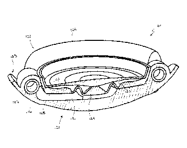
[00101] Referring now to FIG. 12, an embodiment of an IOL constructed in
accordance with the principles of the present invention is described. IOL 140
utilizes a
sealed cavity and shaping fluid to create an aspherical accommodated lens
while
maximizing the hydraulic forces generated by asymmetric loads imposed during
transition
of the lens capsule between the accommodated and unaccommodated
configurations. IOL
140 generally includes optic portion 141 and haptic portion 142, both of which
are similar
in construction to the corresponding portions of the previously described
embodiments.
[00102] The present embodiment illustrates an alternative construction of
support
members 145. Support members 145 are generally wires that circumscribe haptic
portion
142 radially outward from each of haptics 148, 149. Each support member 145 is
CA 02674816 2014-02-21
52723-26
21
preferably coupled to haptic portion 142 where each of haptics 148, 149 is
coupled to optic
portion 141.
[00103] Support members 145 are configured to engage the inner wall of
a lens
capsule so that the capsule is held in a more taut configuration so that
engagement between
haptics 148, 149 and the lens capsule is maintained when the ciliary muscles
are relaxed or
contracted. Maintaining that engagement more efficiently converts movement of
the lens
capsule to deformation of haptics 148, 149.
[00104] In addition to utilizing the sealed cavities containing a
fixed volume of
shaping fluid, the flexibilities and shapes of the components may be selected
to tailor the
influence of the shaping fluid. In particular, the thickness and material of
the anterior lens
component may be selected to provide an desired deflection. In addition, the
shape of the
sealed cavity may be selected by altering the shapes of the adjacent
components to provide
any desired change in volume for any portion of the cavity.
[00105] It should be appreciated that although each embodiment has
been described
having one sealed cavity, any number of sealed cavities containing shaping
fluid may be
included. For example, sealed cavities may be included adjacent to any desired
portion of
the lens element so that discrete portions of the lens element may be shaped
in a desired
fashion.
[00106] While preferred illustrative embodiments of the invention are
described
above, it will be apparent to one skilled in the art that various changes and
modifications
may be made therein without departing from the invention. The appended claims
are
intended to cover all such changes and modifications that fall within the
scope of the invention.