Note: Descriptions are shown in the official language in which they were submitted.
CA 02677903 2009-08-11
WO 2008/100856 PCT/US2008/053588
JOINT REVISION IMPLANT
FIELD
[001] A backing implant for joint revisions is provided. More
specifically, an osteoconductive backing implant for joint revisions is
provided.
BACKGROUND
[002] Overview of Bone Grafts
[003] The rapid and effective repair of bone defects caused by injury,
disease, wounds, or surgery has long been a goal of orthopaedic surgery.
Toward
this end, a number of compositions and materials have been used or proposed
for
use in the repair of bone defects. The biological, physical, and mechanical
properties of the compositions and materials are among the major factors
influencing their suitability and performance in various orthopaedic
applications.
[004] Much effort has been invested in the identification and
development of alternative bone graft materials. Urist has published seminal
articles on the theory of bone induction and a method for decalcifying bone,
i.e.,
making demineralized bone matrix (DBM). Urist M.R., Bone Formation by
Autoinduction, Science 1965; 150(698):893-9; Urist M.R. et al., The Bone
Induction Principle, Clin. Orthop. Rel. Res. 53:243-283, 1967. DBM is an
osteoinductive material, in that it induces bone growth when implanted in an
ectopic site of a rodent, owing to the osteoinductive factors contained within
the
DBM. Honsawek et al. (2000).
[005] DBM implants have been reported to be particularly useful (see,
for example, U.S. Patent Nos. 4,394,370, 4,440,750, 4,485,097, 4,678,470, and
4,743,259; Mulliken et al., Calcif Tissue Int. 33:71, 1981; Neigel et al.,
Opthal.
Plast. Reconstr. Surg. 12:108, 1996; Whiteman et al., J. Hand. Surg. 18B:487,
1993; Xiaobo et al., Clin. Orthop. 293:360, 1993, each of which is
incorporated
herein by reference). DBM typically is derived from cadavers. The bone is
removed aseptically and treated to kill any infectious agents. The bone is
particulated by milling or grinding, and then the mineral component is
extracted
CA 02677903 2009-08-11
WO 2008/100856 PCT/US2008/053588
by various methods, such as by soaking the bone in an acidic solution. The
remaining matrix is malleable and can be further processed and/or formed and
shaped for implantation into a particular site in the recipient. Demineralized
bone
prepared in this manner contains a variety of components including proteins,
glycoproteins, growth factors, and proteoglycans. Following implantation, the
presence of DBM induces cellular recruitment to the site of injury. The
recruited
cells may eventually differentiate into bone forming cells. Such recruitment
of
cells leads to an increase in the rate of wound healing and, therefore, to
faster
recovery for the patient.
[006] Overview of Total Hip Joint Replacement Arthroplasty
[007] Total hip joint replacement arthroplasty can provide a patient with
dramatically improved quality of life by relieving pain and offering increased
mobility. Total hip joint replacement arthroplasty is a surgical procedure
wherein
diseased portions of the hip joint are removed and replaced with artificial
prostheses, such as a femoral component and an acetabular cup. The acetabular
cup is fitted in or against the acetabulum. It is noted that the acetabulum
comprises the ilium, the ischium, and the pubis. These bones are fused at the
acetabulum. For ease of reference, the acetabulum will be discussed as a
single
structure. Successful replacement of deteriorated, arthritic, or severely
injured
hips has contributed to enhanced mobility and comfortable, independent living
for
many people who would otherwise be substantially disabled.
[008] There generally are two broad classes of joint arthroplasty
procedures: primary joint replacement arthroplasty and revision arthroplasty.
Primary joint replacement is when the original, biological joint is removed
and
replaced with an implant. Revision arthroplasty is when the primary joint
replacement fails and must be replaced.
[009] A failed prosthesis and/or dislocation of a total hip replacement
generally causes pain, reduces the ability to work, and necessitates a
revision
operation. Prosthesis failure and/or dislocations can result from a variety of
causes, such as soft tissue laxity, loosening of the implant, and impingement
of
the femoral neck with either the rim of an acetabular cup implant or the soft
tissue
-2-
CA 02677903 2009-08-11
WO 2008/100856 PCT/US2008/053588
or bone surrounding the implant. Loosening of the implant is often due to bone
loss around the implant, caused by adverse tissue reactions to wear particles.
[010] Revision arthroplasty involves additional challenges over primary
joint replacement because, in addition to placement of the revision implant,
the
primary implant must be removed. A common problem with revision arthroplasty
is a loss of bone stock associated with the removal of bone cement, or
osteolysis
due to wear debris and the body's reaction to it, or from stress shielding, or
a
combination of these. Further, in some instances, upon insertion into the
acetabulum of an implant, voids may remain between the back surface of the
implant and the pelvic bone remaining in the acetabulum. In cases where there
is
a defect in the area of the acetabulum, or behind the acetabulum, the surgeon
will
often wish to fill the defect in some way. Bone graft material is sometimes
applied to the acetabulum to encourage bone growth between the acetabulum and
the acetabular cup. Frequently, the bone graft material falls through voids in
the
acetabulum.
[011] Commonly, the acetabular cup prosthesis is manufactured of a
polymeric material, such as polyethylene. A backing is commonly placed
between the acetabular cup prosthesis and the acetabulum. In the past, metal
backings have been widely used, at least in part because a stiff backing was
believed to be mechanically favorable. It has more recently been determined
that
a stiff backing causes two problems: It generates higher stress peaks around
the
acetabular rim than those caused by full polyethylene cups, and it reduces the
stresses transferred to the dome of the acetabulum, causing stress shielding.
[012] It would be useful to provide a backing for an acetabular cup
prosthesis using bone graft materials such that the prosthesis aids in holding
graft
material in place, encourages bone apposition up to the implant or, in the
case of
an implant with a porous metallic coating, encourages ingrowth and biological
attachment to the implant.
-3-
CA 02677903 2009-08-11
WO 2008/100856 PCT/US2008/053588
BRIEF SUMMARY
[013] An osteoconductive backing implant for joint revisions is provided
that may enhance bone healing and, for cementless implants, bony integration
of
the implant.
[014] In one embodiment, the backing implant comprises a disc having
an inner hole and an outer edge, at least one slit extending from the inner
hole to
the outer edge. The disc may be formed from a coherent mass of elongate,
mechanically entangled demineralized bone particles.
[015] In another embodiment, the backing implant comprises a disc that
has at least one slit extending from an interior of the disk to the outer
edge. The
disc further includes at least one perforation by which the size or shape of
the disc
may be manipulated. The disc may be shaped into a cone.
[016] In a further embodiment, the backing implant comprises a sheet of
material having a center and a plurality of petals. The sheet of material may
be
partially folded in on itself by manipulating the petals toward one another.
[017] While multiple embodiments are disclosed, still other
embodiments of the present invention will become apparent to those skilled in
the
art from the following detailed description. As will be apparent, the
invention is
capable of modifications in various obvious aspects, all without departing
from
the spirit and scope of the present invention. Accordingly, the detailed
description is to be regarded as illustrative in nature and not restrictive.
BRIEF DESCRIPTION
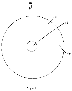
[018] Figure 1 illustrates a backing implant comprising a disc having a
center hole and an outer edge, and a slit extending from the center hole to
the
outer edge, in accordance with one embodiment.
[019] Figure 2 illustrates a backing implant comprising a disc having a
center hole and an outer edge, a slit extending from the center hole to the
outer
edge, and further comprising a series of generally concentric perforations, in
accordance with one embodiment.
-4-
CA 02677903 2009-08-11
WO 2008/100856 PCT/US2008/053588
[020] Figure 3 illustrates a backing implant comprising a disc having a
center hole and an outer edge, a slit extending from the center hole to the
outer
edge, and further comprising a series of wedges, in accordance with one
embodiment.
[021] Figure 4 illustrates a cone shaped from a backing implant in
accordance with one embodiment.
[022] Figure 5 illustrates a backing implant comprising four generally
curved petals in accordance with one embodiment.
[023] Figure 6 illustrates a backing implant comprising five generally
tipped petals in accordance with one embodiment.
[024] Figure 7 illustrates the backing implant of Figure 6 manipulated to
form a backing in accordance with one embodiment.
DEFINITIONS
[025] Biocompatible, as used herein, is intended to describe materials
that, upon administration in vivo, do not induce undesirable long-term
effects.
[026] Bone as used herein refers to bone that is cortical, cancellous or
cortico-cancellous, of autogenous, allogenic, xenogenic, or transgenic origin.
[027] Demineralized, as used herein, refers to any material generated by
removing mineral material from tissue, e.g., bone tissue. In certain
embodiments,
the demineralized compositions described herein include preparations
containing
less than 5% calcium and preferably less than 1% calcium by weight. Partially
demineralized bone (e.g., preparations with greater than 5% calcium by weight
but containing less than 100% of the original starting amount of calcium) is
also
considered within the scope of the invention. In some embodiments,
demineralized bone has less than 95% of its original mineral content.
Demineralized is intended to encompass such expressions as "substantially
demineralized," "partially demineralized," "fully demineralized," "surface
demineralized," etc.
[028] Demineralized bone matrix, as used herein, refers to any material
generated by removing mineral material from bone tissue. In some embodiments,
-5-
CA 02677903 2009-08-11
WO 2008/100856 PCT/US2008/053588
the DBM compositions as used herein include preparations containing less than
5% calcium and preferably less than 1% calcium by weight. Partially
demineralized bone (e.g., preparations with greater than 5% calcium by weight
but containing less than 100% of the original starting amount of calcium) are
also
considered within the scope of the invention.
[029] Osteoconductive is used herein to refer to the ability of a non-
osteoinductive substance to serve as a suitable template or substance along
which
bone may grow.
[030] Osteogenic is used herein to refer to the ability of an agent,
material, or implant to enhance or accelerate the growth of new bone tissue by
one or more mechanisms such as osteogenesis, osteoconduction, and/or
osteoinduction.
[031] Osteoimplant as used herein refers to any bone-derived implant
prepared in accordance with the embodiments of this invention and therefore is
intended to include expressions such as bone membrane, bone graft, etc.
[032] Osteoinductive, as used herein, refers to the quality of being able to
recruit cells from the host that have the potential to stimulate new bone
formation.
Any material that can induce the formation of ectopic bone in the soft tissue
of an
animal is considered osteoinductive. For example, most osteoinductive
materials
induce bone formation in athymic rats when assayed according to the method of
Edwards et al., "Osteoinduction of Human Demineralized Bone: Characterization
in a Rat Model," Clinical Orthopaedics & Rel. Res., 357:219-228, December
1998, incorporated herein by reference. In other instances, osteoinduction is
considered to occur through cellular recruitment and induction of the
recruited
cells to an osteogenic phenotype.
[033] Superficially demineralized as used herein refers to bone-derived
elements possessing at least about 90 weight percent of their original
inorganic
mineral content. The expression "partially demineralized" as used herein
refers to
bone-derived elements possessing from about 8 to about 90 weight percent of
their original inorganic mineral content and the expression "fully
demineralized"
-6-
CA 02677903 2009-08-11
WO 2008/100856 PCT/US2008/053588
as used herein refers to bone containing less than 8% of its original mineral
context.
DETAILED DESCRIPTION
[034] I. INTRODUCTION
[035] A backing implant for joint revisions is provided that may enhance
bone healing. The backing implant may generally be used in reconstruction of a
joint, including concave or convex joint surfaces. For the purposes of
description,
reference is made herein to replacement of the acetabulum. It is to be
appreciated,
however, that the backing implant described herein may be used in other joint
replacements such as replacement of a speroidal joint (also referred to as a
"ball
and socket joint"), an ellipsoid joint, a sellar joint (also referred to as a
"saddle
joint"), a bicondular joints, or any joint having a concavity with a defect
and a
mating convex surface. The present invention may be beneficially used in a
variety of joint configurations; see Williams, P. L. and Warwick, R., Gray's
Anatomy. 36th ed., Livingstone, Edinburgh (1980), which is hereby incorporated
by reference. In some embodiments, the backing implant may be placed in the
concavity of the joint.
[036] For cementless implants, such as cementless acetabular cups, the
backing implant may enhance bony integration of the implant. When used in a
concavity, such as in an acetabular cup, the backing implant accentuates the
concavity and offers a possibility of adhesion around the outer perimeter of
the
concavity. Generally, the backing implant may be fit to the joint surface such
that
it fills voids or defects in the joint surface. The backing implant may be
used with
bone, such as autograft bone or allograft bone. More specifically, bone may
placed between the backing implant and the implant, such as the acetabular
cup,
the backing implant substantially preventing bone graft from falling through
voids
or defects in the joint surface. After implantation, the backing implant may
be
remodeled and wholly or partially replaced by bone. Thus, in various
embodiments, the backing implant fulfills mechanical and bone forming
functions. As noted, the backing implant may be used in revision of any
suitable
-7-
CA 02677903 2009-08-11
WO 2008/100856 PCT/US2008/053588
joint articulations, such as the shoulder joint, for example being fitted in
the
glenoid cavity of the scapula. Thus, while this description refers
specifically ball-
and-socket joints, and more specifically to acetabular cups, one skilled in
the art
will be able to modify the backing implant to fit other joints.
[037] In accordance with some embodiments, the backing implant
comprises a sheet of material that may be shaped to generally form to the
joint
surface prior to implantation. The sheet may be flexible and/or conformable
such
that it may conform to irregularities in the bone, for example in the bone of
the
acetabulum. Such flexibility or conformability permits achieving an increased
degree of contact. In some embodiments, the backing implant may comprise a
material that is osteoconductive and, possibly, osteoinductive, several
examples of
which are described below.
[038] Generally, during the healing phase of total joint replacement
arthroplasty, the backing implant may not act as the sole weight bearing
component. Accordingly, the backing implant may be used with another implant.
For example, in a total hip replacement arthroplasty, the backing implant may
be
used with an acetabular cup, the acetabular cup contacting sufficient host
bone to
provide support.
[039] The backing implant may be press-fit into the joint, such as in the
concavity of a joint, or may be fit around an implant, such as an acetabular
cup,
prior to its implantation. When press-fit into the joint, it may be desirable
to
effect pressing using a trial implant or using an implant that will be
implanted. In
some embodiments, the backing implant may be treated to impart additional
stickiness to a portion of the backing implant coming into contact with the
joint.
The backing implant may be formed of a flexible material, may be formed of a
rigid material, a semi-rigid or semi-flexible material, or a material that is
rigid but
can be made flexible. If the backing implant is formed of a material that is
rigid
but can be made flexible, the backing implant may be fit in the joint in the
flexible
state and allowed to become rigid before implanting the implant.
Alternatively,
the backing implant may remain flexible when the implant is implanted.
-8-
CA 02677903 2009-08-11
WO 2008/100856 PCT/US2008/053588
[040] As previously noted, bone graft may be used with the backing
implant. For example, the bone may comprise morselized allograft, autograft,
or
other suitable bone material. In use, the backing implant may be placed in the
joint, such as in the acetabulum, for example via press fitting with an upper
surface of the backing implant being thus provided for receiving an implant.
Bone graft, for example morselized allograft, may be provided on the upper
surface of the backing implant, before or after placement of the backing
implant
in the joint. The backing implant substantially prevents the morselized
allograft
from penetrating into joint, for example from penetrating the joint, for
example, in
hip arthroplasty, from penetrating the acetabulum and the pelvis. More
specifically, the backing implant acts as a barrier to morselized allograft
from
falling into or through voids or defects in the joint surface. The backing
implant
works in conjunction with the morselized allograft through osteoinductive
and/or
osteoconductive properties to form new bone. In an alternative embodiment,
autograft bone may be provided on the upper surface of the backing implant
before or after placement of the backing implant in the joint. In alternative
embodiments, other materials may be provided on the upper surface of the
backing implant to aid in bone forming function.
[041] Acetabular cups used in total hip joint replacement arthroplasty
may be press-fit or may be cemented in place, for example using methacrylate
bone cement. Often, press-fit cups are preferred because of possible bone-to-
implant bonding. Using the backing implant provided herein, bone-to-implant
bonding is enhanced; the backing implant closely molding to both the
acetabulum
and the acetabular cup and being osteoconductive, and possibly osteoinductive.
Thus, the backing implant enables bone to be induced or conducted from the
acetabulum to the acetabular cup.
[042] II. IMPLANT SHAPES
[043] The backing implant may be initially formed as a flat sheet of
material The flat sheet of material may, in some embodiments, be formed of one
or more layers.. The backing implant may be preshaped, may be partially
preshaped (described below), or may be shaped by a surgeon for implantation.
-9-
CA 02677903 2009-08-11
WO 2008/100856 PCT/US2008/053588
Generally, the backing implant may be shaped such that, when formed, it is
generally complementary to the portion of the joint against which it will be
placed. In the embodiments of Figures 1-3, the backing implant is described as
having a generally conical shape. Thus, in the embodiments shown, the backing
implant includes surfaces extending from a base towards an apex. As shown, the
backing implant may not extend fully to the apex and thus may comprise a
frustum or truncated cone. Further, while the backing implant is shown as a
truncated right circular cone, the backing implant may have other conical
shapes,
such as an elliptical cone, an oblique cone, or other, or non-conical shapes,
as
appropriate for a given application.
[044] In a first embodiment, shown in Figure 1, the backing implant 10
comprises a generally circular sheet 12 having a hole 14. The size, shape, and
placement of the hole may be varied. For example, the hole may be centered or
may be eccentric, may be circular or may be ovoid, etc. The size, shape, and
placement of the hole may be determined based on, for example, concavity of
the
joint surface. In some embodiments, the hole facilitates folding or shaping
the
backing implant into a cone, described below. In these embodiments, the hole
is
sized for such use, generally reducing material that may need to be trimmed at
the
point of the cone and reducing the likelihood of the backing implant wrinkling
during folding. In the figures, the generally circular sheet 12 and the center
hole
14 are depicted as being round. It is to be understood, however, that, for the
embodiment of Figure 1 as well as all other embodiments described herein, any
suitable geometry may be used for the sheet 12, the hole 14, or both,
including
oval, etc., and, furthermore, that the hole 14 may be placed in any desired
location, and need not be at, or over, the center of the generally circular
sheet 12.
The shape of the implant thus may broadly be referred to as a disc. The sheet
12
and hole 14 may be of any suitable or desired dimensions. In one embodiment,
the sheet 12 has an outer diameter of approximately 70 mm and the hole 14 has
an
inner diameter of approximately 5 mm. Other sizes, for example outer diameters
of 30 mm, 45 mm, or 60 mm, may alternatively be provided. In addition, the
height or thickness of the implant, i.e., the distance between the upper
surface and
-10-
CA 02677903 2009-08-11
WO 2008/100856 PCT/US2008/053588
the lower surface, may be any desired dimension. A slit 16 extends from the
hole
to the outer edge. The slit 16 may be a straight, clean line and may be
perforated
or may be cut through. The slit 16 also may be curved, zig-zag, wavy, v-
shaped,
or any other desired configuration. Using the embodiment of Figure 1, the
surgeon may fold the sheet 12 into a cone shape, thereby correlating the sheet
12
with the interior surface of the acetabular cup. The cut ends of the backing
implant, corresponding with the slit 16, may be folded over one another, thus
permitting a wide variety of diameters of the cone shape. Further, the
overlapping
edges may be cut and removed.
[045] In a second embodiment, shown in Figure 2, the backing implant
20 comprises a generally circular sheet 22 having a hole 24 and a slit 26
extending from the hole to the outer edge, as in the embodiment of Figure 1.
The
slit 26 may be perforated or may be cut through. The backing implant 20 of
Figure 2 further includes a series of generally concentric perforations 28.
The
surgeon may select one of the generally concentric perforations 28
corresponding
to a desired outer diameter of the generally circular sheet 22. The surgeon
then
may tear or cut along the generally concentric perforation 28, removing
material
between the generally concentric perforation 28 and the outer edge, thereby
forming an implant of desired size. As with the embodiment of Figure 1, the
surgeon may then fold the sheet 22 into a cone shape. Further, the overlapping
edges may be cut and removed. While the generally concentric perforations 28
are depicted as being round, it is to be understood that any desired geometry
may
be used. In one embodiment, the perforations may be elliptical. Furthermore,
the
primary axes of the elliptical perforations may intersect, so that the
perforations of
the ellipses themselves intersect, thus allowing greater flexibility in the
removal
of shapes to be generated by the removal of pre-perforated sections. In a
variation
of the embodiment of Figure 2, the generally circular sheet 22 may be provided
without a hole 24, in which case the slit 26 extends into the interior of the
generally circular sheet 22.
[046] Figure 3 illustrates a further embodiment of a backing implant 30.
In the embodiment of Figure 3, the backing implant 30 comprises a generally
-11-
CA 02677903 2009-08-11
WO 2008/100856 PCT/US2008/053588
circular sheet 32 having a hole 34. A series of wedges 38 are formed between
the
center hole and the outer edge of the sheet. The wedges 38 may be perforated
or
may be cut through. In one embodiment, a relatively thin, breakable segment 39
is provided around the center hole 34 connecting the wedges 38. In a variation
of
the embodiment of Figure 3, the generally circular sheet 32 is provided
without a
hole 34. The surgeon may remove one or more wedges 38 to modify the sizing of
the backing implant 30 as formed into a cone shape. One or more wedges 38 may
be removed when a smaller cone shape is desired, thus less overlapping of the
sheet 32 may be required. The wedges 38 may be provided in any desired shape
or number. In further embodiments, the backing implant 30 may have both
concentric perforations 28 and wedges 38.
[047] Thus, the backing implant may be formed as a sheet of material
that may be folded and manipulated to form a backing for complementing an
acetabular cup. Figure 4 illustrates a cone 40 formed from a backing implant
as
provided in Figures 1-3. As shown, the hole 42 forms one end of the cone.
Where the implant of Figures 1-3 is provided without a hole 42, the cone may
have a point on its end. The outer diameter of the sheet forms the other end
44 of
the cone. The ends of the outer sheet corresponding to the slit overlap, or
abut, to
form a seam 46.
[048] Figures 5-7 illustrate a further embodiment of a backing implant.
Figures 5 and 6 illustrate the backing implant 60 and 62, respectively, in a
laid out
configuration. Figure 7 illustrates the backing implant 62 of Figure 6 in a
manipulated configuration to form a backing for complementing an acetabular
cup. As shown, the backing implant 60, 62 comprises a sheet of material having
a
center 66 and a plurality of petals or points 64. The center 66 may be a true
center of the backing implant or may merely be generally central to the petals
or
points 64. The petals or points 64 generally radiate from the center 66. The
petals or points 64 may be manipulated toward one another to partially fold
the
backing implant 60, 62 in on itself. The number and shape of the petals or
points
64 may be varied. In Figure 5, four generally curved petals 64 are provided.
In
Figure 6, five generally pointed petals 64 are provided. Any suitable number
may
-12-
CA 02677903 2009-08-11
WO 2008/100856 PCT/US2008/053588
be used, and they may be of any suitable shape or configuration. For use as a
backing for an acetabular cup, the petals or points 64 may be folded for
positioning towards the defect.
[049] In the embodiments of Figures 1-3, the backing implant may be
formed of a flat, flexible material that can be folded to shape.
Alternatively, the
backing implant may be formed of a flexible or more rigid material that is
premolded to a hemispherical shape that will fit an acetabular cup. The
backing
implants may be supplied in a single size or a small number of sizes where the
surgeon modifies or trims the backing implant to shape. Alternatively, the
backing implants may be provided in a wide variety of sizes that will satisfy
most
requirements without modifying or trimming.
[050] Thus, a backing implant is herein provided that comprises a
generally planar sheet form that may be shaped to conform generally to an
implant surface. While specific geometries are described for forming a sheet
into,
for example, a hemi-spherical shape, any suitable manner of doing so may be
used. Thus, generally, the backing implant may be provided as any planar
configuration that may be formed into a generally hemispherical shape. Such
configurations include, for example, those that have been developed in the
cartographic arts such as Cahill's butterfly, Waterman's butterfly,
pseudocylindrical projections of the hemisphere, pseudoconic projections of
the
hemisphere, sinusoidal projections of the hemisphere, dymaxion projections of
the hemisphere, other conic projections of the hemisphere, cyldinrical
projections
of the hemisphere, and other. As will be appreciated by one skilled in the
art, the
cartographic methods for converting a sphere to a planar surface may be
adapted
to developing a planar surface to fornn a hemisphere.
[051] In some embodiments, fixation elements may be provided for
fixing the backing implant to the acetabulum or other joint. For example, the
backing implant may be provided with tabs, overhangs, or other structure for
fixing to bone or other surface.
-13-
CA 02677903 2009-08-11
WO 2008/100856 PCT/US2008/053588
[052] III. IMPLANT MATERIALS
[053] The backing implant comprises a material that is formed into a
generally planar configuration For ease of reference, the generally planar
configuration is referred to as a sheet however such term is not intended to
be
limiting. The material may be osteoconductive, and also may be osteoinductive.
The backing implant may be formed of a flexible material. In a further
embodiment, the backing implant may be generally rigid but capable of becoming
flexible when exposed to liquids such as saline or body fluids. Alternatively,
other manners of providing flexibility to the material may be provided. For
example, the material may be generally rigid at room temperature but flexible
when heated. Alternatively, the material may comprise a reverse-phase
material,
such as Poloxamer 407, so that the implant is more flexible at cooler
temperatures, and then firms up to become less flexible when warmed to body
temperatures. In some embodiments, the backing implant may be rehydrated, for
example via exposure to saline, prior to use.
[054] To form the backing implant, the sheet may be cut using a cutting
machine, using cutting molds, or in any suitable manner. In some embodiments,
the material may be directly formed into the shape of the backing implant
without
cutting of a sheet.
[055] Bone Particles
[056] In one embodiment, the backing implant comprises bone matrix.
The bone matrix may be provided in a particulate form, wherein the particles
are
of any desired size and shape. The backing implant also may comprise a sheet
fabricated from, or including, elongate bone particles, such as disclosed in
U.S.
Patent No. 5,507,813 for Shaped Materials Derived from Elongate Bone
Particles,
herein incorporated by reference. The bone particles may be obtained from
cortical, cancellous and/or corticocancellous bone which may be of autogenous,
allogenic, transgenic, and/or xenogenic origin.
[057] In one embodiment, elongate bone particles used in forming the
backing implant may be generally characterized as having relatively high
median
length to median thickness ratios, e.g., about 50:1 or about 100:1 and,
similarly,
-14-
CA 02677903 2009-08-11
WO 2008/100856 PCT/US2008/053588
relatively high median length to median width ratios, e.g., about 10:1 or
about
50:1. Such particles can be readily obtained by any one of several methods,
e.g.,
by milling or shaving the surface of an entire bone or relatively large
section of
bone. Thereafter, the resulting elongate bone particles may be demineralized.
[058] Employing a milling technique, particles ranging in median length
from about 2 up to about 200 mm or more (as in the case of the long bones), in
median thickness from about 0.05 to about 2mm, and in median width from about
1 to about 20mm can be readily obtained. Depending on the procedure employed
for producing the elongate bone particles, one can obtain a mass of bone
particles
containing at least about 60 weight percent, at least about 70 weight percent,
or at
least about 80 weight percent of bone particles possessing a median length of
from about 2 to about 200 mm or more, or from about 10 to about 100 mm, a
median thickness of from about 0.05 to about 2 mm, or from about 0.2 to about
1
mm, and a median width of from about 1 mm to about 20 mm, or about 2 to about
mm. These bone particles may possess a median length to median thickness
ratio of 10:1, to 50:1, and up to about 500:1, or from about 10:1 to about
100:1,
and a median length to median width ratio of from about 10:1 to about 200:1,
or
from about 50:1 to about 100:1. The bone fibers or particles of the present
invention may be demineralized in any desired manner, and to any desired
extent.
[059] As descried more fully below, the bone particles can be admixed
with one or more substances such as adhesives, fillers, plasticizers,
flexibilizing
agents, biostatic/biocidal agents, surface active agents, binding and bonding
agents, fillers, and the like, prior to, during, or after shaping the
particles into a
desired configuration.
[060] To prepare the backing implant, a quantity of bone particles, for
example, demineralized, elongate bone particles, slurried in a suitable
liquid, e.g.,
water, organic protic solvent, aqueous solution such as physiological saline,
etc.,
and optionally containing one or more biocompatible ingredients such as a
carrier,
adhesives, fillers, plasticizers, flexibilizing agents, biostatic/biocidal
agents,
surface active agents, medically/surgically useful substances, etc. is applied
to a
form such as a flat sheet, mesh screen or three-dimensional mold and excess
-15-
CA 02677903 2009-08-11
WO 2008/100856 PCT/US2008/053588
liquid is removed, e.g., by being drained away. This procedure is referred to
herein as "wet-laying." For example, in the case of a sheet, the thickness of
the
layer of wetted bone particles can vary widely, e.g., from about 1 to about 40
mm.
Some particle entanglement results from the wet-laying operation. Further
particle entanglement, if necessary or desirable, can be effected by the use
of
water jets or other suitable mechanical entangling methods. Either before or
after
the wet-laying procedure, one or more additional substances can be added to
the
bone particles, e.g., thixotropic agents, therapeutic agents, and the like.
The wet
demineralized bone particles may then be dried, either in an oven or by
lyophilization. In an alternative embodiment, the bone particles can be
subjected
to a compressive force, e.g., of up to about 100 psi, during and/or after the
wet-
laying step and/or while the drained but still wet shaped article is being
dried.
The resulting sheet is rigid and relatively strong when dry and flexible and
pliable
when wetted or hydrated.
[061] In some embodiments, the sheet is formed by laying the DBM
solution on a sieve shaped to correspond to the shape of the backing implant.
Thus, for example, the sieve may be sized and shaped in a circle to correspond
with the backing implant of Figure 1.
[062] At the site of implantation, a backing implant formed of bone
particles may be employed in the dry state or, where site conformation is
desired,
in the hydrated state. The dry or hydrated article can be cut or sized if need
be to
conform to a site being repaired. The backing implant can be hydrated with a
suitable biocompatible liquid, e.g., water, saline solution, etc., for a
period of time
ranging from about 1 to about 120 minutes. After being hydrated, the backing
implant becomes flexible yet substantially retains its shape and much of its
strength. The backing implant may be packaged in either the dried or wet state
and stored for subsequent application. In some circumstances, the backing
implant may be packaged in the wet state so that it is ready for immediate use
at
the surgical site.
[063] Alternatively, the bone particles, including elongate bone particles,
may be formed into a sheet that remains flexible in the dry state, for example
-16-
CA 02677903 2009-08-11
WO 2008/100856 PCT/US2008/053588
through the addition of a plasticizer, or that is rigid and becomes flexible
upon
heating.
[064] In some embodiments, the backing implant may be formed of a
bone graft material having not greater than about 32% void volume formed at
least in part from elongate bone-derived elements optionally in combination
with
bone powder. U.S. Patent No. 6,332,779 for Method of Hard Tissue Repair
discusses such bone graft material and is hereby incorporated by reference.
[065] Other Materials
[066] In further embodiments, the backing implant may comprise other
or additional materials. For example, osteoinductive materials, such as
osteoinductive proteins, may be added to the backing implant. Any suitable
material may be used, but they should generally be biocompatible and lack
immunogenicity. The materials may be of biological origin, such as collagen
sponges, collagen fibers, etc., which may be cross-linked or otherwise
processed,
as desired. Other suitable biological materials, including those of allograft,
autograft (e.g., iliac crest or local bone), or xenograft origin, growth
factors and/or
bone morphogenic proteins (including on a carrier), gelatins, hydrogels, etc.,
also
may be used. Nonanimal biological material or synthetic materials also may be
used, as desired, including silk, cotton, linen, calcium phosphate- and
calcium
sulfate-based materials, etc. Polymers may be used, including in combination
with any of the above. Any suitable combination of the above materials may be
used.The implant also may comprise synthetic materials.
[067] In one embodiment, the backing implant comprises a polymer
sheet containing calcium phosphate particles. Examples of other suitable
materials include polymers, such as polyalkylenes (e.g., polyethylenes,
polypropylenes, etc.), polyamides, polyesters, polyurethanes, poly(lactic acid-
glycolic acid), poly(lactic acid), poly(glycolic acid), poly(glaxanone),
poly(orthoesters), poly(pyrolicacid), poly(phosphazenes), minerals, etc. These
may be resorbable, non-resorbable, or some of each. These materials may be
used
to form wicking materials, and may be synthetic, natural, etc. They may be
-17-
CA 02677903 2009-08-11
WO 2008/100856 PCT/US2008/053588
formed as a woven material, including a braid, a nonwoven matrix, axially
aligned, or in any other suitable manner.
[068] The osteoimplant may also comprise combinations of these and
other materials, and may further comprise bone, e.g., DBM fibers, DBM
particles,
combinations, etc.
[069] As previously described, the backing implant may be formed of a
flexible material. Such flexibility may be imparted wherein the backing
implant
includes a plasticizer such as glycerol. Alternatively, the backing implant
may be
constructed from a flexible polymer. In a further embodiment, the backing
implant may be generally rigid but capable of becoming flexible when exposed
to
liquids such as saline or body fluids.
[070] In alternative embodiments, the backing implant may be formed of
a relatively rigid material that is premolded to a hemispherical shape.
[071] Additives
[072] Regardless of the material used for forming the backing implant,
additional substances may be added to the material. The material used to form
the
backing implant may be admixed with one or more substances such as adhesives,
fillers, plasticizers, flexibilizing agents, biostatic/biocidal agents,
surface active
agents, binding and bonding agents, fillers, and the like, prior to, during,
or after
shaping the particles into a desired configuration. Suitable adhesives,
binding
agents and bonding agents include acrylic resins, cellulosics, bioresorbable
polymers such as polyglycolide, polylactide, glycolide-lactide copolymer, etc.
Suitable fillers include bone powder, demineralized bone powder,
hydroxyapatite,
etc. Suitable plasticizers and flexibilizing agents include liquid polyhydroxy
compounds such as glycerol, monacetin, diacetin, etc. Suitable
biostatic/biocidal
agents include antibiotics, povidone, sugars, etc. Suitable surface active
agents
include the biocompatible nonionic, cationic, anionic and amphoteric
surfactants.
[073] Any of a variety of medically and/or surgically useful substances
can be incorporated in, or associated with the material used to form the
backing
implant during or after fabrication of the backing implant. Thus, for example
when demineralized bone particles are used to form the material, one or more
of
-18-
CA 02677903 2009-08-11
WO 2008/100856 PCT/US2008/053588
such substances may be introduced into the demineralized bone particles, e.g.,
by
soaking or immersing the bone particles in a solution or dispersion of the
desired
substance(s).
[074] Radiopaque materials may be added to the material of the backing
implant for visualization. Such materials may comprise, for example,
nondemineralized bone, barium sulfate, iodine-containing compounds, titanium,
or other.
[075] Medically/surgically useful substances that can be readily
combined with the demineralized bone particles and/or osteogenic material
include, e.g., collagen, insoluble collagen derivatives, etc., and soluble
solids
and/or liquids dissolved therein, e.g., antiviricides, particularly those
effective
against HIV and hepatitis; antimicrobials and/or antibiotics such as
erythromycin,
bacitracin, neomycin, penicillin, polymyxin B, tetracyclines, viomycin,
chloromycetin and streptomycins, cefazolin, ampicillin, azactam, tobramycin,
clindamycin and gentamicin, etc.; biocidal/biostatic sugars such as dextroal,
glucose, etc.; amino acids, peptides, vitamins, inorganic elements, co-factors
for
protein synthesis; hormones; endocrine tissue or tissue fragments;
synthesizers;
enzymes such as collagenase, peptidases, oxidases, etc.; polymer cell
scaffolds
with parenchymal cells; angiogenic drugs and polymeric carriers containing
such
drugs; collagen lattices; antigenic agents; cytoskeletal agents; cartilage
fragments,
living cells such as chondrocytes, bone marrow cells, mesenchymal stem cells,
natural extracts, tissue transplants, bone, demineralized bone powder,
autogenous
tissues such blood, serum, soft tissue, bone marrow, etc.; bioadhesives, bone
morphogenic proteins (BMPs), angiogenic factors, transforming growth factor
(TGF-beta), insulin-like growth factor (IGF-1); growth hormones such as
somatotropin; bone digestors; antitumor agents; immuno-suppressants;
permeation enhancers, e.g., fatty acid esters such as laureate, myristate and
stearate monoesters of polyethylene glycol, enamine derivatives, alpha-keto
aldehydes, etc.; and, nucleic acids. The amounts of such optionally added
substances can vary widely with optimum levels being readily determined in a
specific case by routine experimentation.
-19-
CA 02677903 2009-08-11
WO 2008/100856 PCT/US2008/053588
[076] A number of endogenous factors that play important roles in the
development and/or repair of bone have been identified. BMPs such as BMP-2
and BMP-4 induce differentiation of mesenchymal cells towards cells of the
osteoblastic lineage, thereby increasing the pool of mature cells, and also
enhance
the functions characteristic of differentiated osteoblasts. Canalis et al.,
Endocrine
Rev. 24(2):218-235, 2003. In addition, BMPs induce endochondral ossification
and chondrogenesis. BMPs act by binding to specific receptors, which results
in
phosphorylation of a class of proteins referred to as SMADs. Activated SMADs
enter the nucleus, where they regulate transcription of particular target
genes.
BMPs also activate SMAD-independent pathways such as those involving
Ras/MAPK signaling. Unlike most BMPs such as BMP-2 and BMP-4, certain
BMPs (e.g., BMP-3) act as negative regulators (inhibitors) of osteogenesis. In
addition, BMP-1 is distinct both structurally and in terms of its mechanism of
action from other BMPs, which are members of the TGF-B superfamily. Unlike
certain other BMPs (e.g., BMP-2, BMP-4), BMP-1 is not osteoinductive. Instead,
BMP-1 is a collagenolytic protein that has also been shown to cleave chordin
(an
endogenous inhibitor of BMP-2 and BMP-4). Tolloid is a metalloprotease that is
structurally related to BMP-1 and has proteolytic activity towards chordin.
See
Canalis, supra, for further details regarding the activities of BMPs and their
roles
in osteogenesis and chondrogenesis.
[077] Further, other osteoinducing agents may be added to the material.
These agents may be added in an activated or non-activated form. These agents
may be added at anytime during the preparation of the inventive material. For
example, in the case of a DBM backing implant, the osteoinducing agent may be
added after the demineralization step and prior to the addition of the
stabilizing
agents so that the added osteoinducing agent is protected from exogenous
degrading enzymes once implanted. In some embodiments the DBM is
lyophilized in a solution containing the osteoinducing agent. In certain other
embodiments, the osteoinducing agents are adhered onto the hydrated
demineralized bone matrix and are not freely soluble. In other instances, the
-20-
CA 02677903 2009-08-11
WO 2008/100856 PCT/US2008/053588
osteoinducing agent is added after addition of a stabilizing agent so that the
osteoinducing agent is available immediately upon implantation of the DBM.
[078] Osteoinducing agents include any agent that leads to or enhances
the formation of bone. The osteoinducing agent may do this in any manner, for
example, the agent may lead to the recruitment of cells responsible for bone
formation, the agent may lead to the secretion of matrix which may
subsequently
undergo mineralization, the agent may lead to the decreased resorption of
bone,
etc. Suitable osteoinducing agents include bone morphogenic proteins (BMPs),
transforming growth factor (TGF-0), insulin-like growth factor (IGF-1),
parathyroid hormone (PTH), and angiogenic factors such as VEGF. In one
embodiment, the inducing agent is genetically engineered to comprise an amino
acid sequence which promotes the binding of the inducing agent to the DBM or
the carrier. Sebald et al., PCT/EPOO/00637, incorporated herein by reference,
describe the production of exemplary engineered growth factors suitable for
use
with DBM.
[079] Any of the implants made pursuant to the teachings herein may be
treated to impart, or to increase, osteoinductivity, as taught in U.S. Patent
Application Serial No. 11/555,606, filed November 1, 2006, hereby incorporated
by reference herein.
[080] IV. EXAMPLES
[081] Fibers are milled form human cortical shafts to a desired size
range. The milled fibers are demineralized, subjected to an ethylene oxide
soak
as a cleansing step, and introduced to a glycerol/water solution. The fibers
soak
in the glycerol/water solution for a predetermined period of time. The
solution
containing the fibers is poured through a sheet-forming sieve. Much of the
solution passes through the sieve, but the fibers and residual solution remain
in a
sheet-like form. The form is lyophilized, and the resulting DBM comprises a
flat
yet flexible consistency. A cutter is used to cut the flat form to the desired
shape,
such as a circular form having a diameter of 60 mm and a 5 mm hole center. The
shape may be formed into a generally conical shape.
-21-
CA 02677903 2009-08-11
WO 2008/100856 PCT/US2008/053588
[082] V. CONCLUSION
[083] A backing implant for joint replacement is thus provided.
Generally, the backing implant may be used in arthroplasty of speroidal joints
such as the hip and shoulder joints, ellipsoid joints such as the radiocarpal
joint,
sellar joints such as the carpometacarpal joint of the thumb or the talocrural
joint
of the ankle, or bicondular joints such as the knee, particularly in the
tibial area.
The backing implant may comprise a thin sheet of material, which may be
osteoconductive, and possibly osteoinductive, to thus enhance bone healing
and,
for cementless acetabular cups, bony integration of the acetabular cup. The
backing implant may be flexible and conformable such that it may conform to
irregularities in the bone of the joint and generally fill or act as a barrier
to voids
in the joint surface. After implantation, the backing implant may be remodeled
and wholly or partially replaced by bone.
[084] The backing implant may be used during original primary joint
replacement arthroplasty or revision arthroplasty.
[085] Although the invention has been described with reference to
preferred embodiments, persons skilled in the art will recognize that changes
may
be made in form and detail without departing from the spirit and scope of the
invention.
-22-