Note: Descriptions are shown in the official language in which they were submitted.
CA 02679413 2013-07-15
WO 2008/109800 PCT/US2008/056152
Attorney Docket: 1941/A68W0
Implantable Device with Removable Magnet
Technical Field
[0002] The present invention generally relates to an implantable device having
a
removable magnet. For example, this magnet may be located at the center of an
implanted
receiving coil for holding in place an external transmitter coil.
Background Art
[0003] Some implantable medical devices use magnets to hold internal and
external
pieces in proper position. For example, as shown in Fig. 1, an idealized
cochlear implant
system may include a receiving coil 108 located under the skin 103 and
embedded in or
just on top of the bone 104. A receiver magnet 106 is contained in the center
of the
receiving coil 108. An external transmitter housing 101 includes a transmitter
magnet 105
that is positioned over the receiver magnet 106 so that the external
transmitter housing
101 is held in place in an optimum position adjacent to the receiving coil
assembly 102.
When such an optimal position is maintained, an extemal transmitting coil 107
within the
transmitter housing 101 can use inductive coupling to transmit a
transcutaneous data
and/or power signal to the receiving coil 108.
[0004] The receiving coil 108 may, for example, be encapsulated within some
tissue-
compatible organic material such as silicone or epoxy, forming a receiving
coil assembly
102. In such an arrangement, the receiver coil assembly 102 is connected to
receiver
electronic circuits within a metal or ceramic case which is hermetically
sealed from the
surrounding tissue. Or, in another approach, the receiver magnet 106,
receiving coil 108
and the receiver electronic circuits are all contained within a common
hermetic case. In
any such arrangement, the receiver magnet 106 is a permanently integrated part
of the
implant structure.
-1-
CA 02679413 2009-08-28
WO 2008/109800 PCT/US2008/056152
[0005] One problem arises when the patient undergoes Magnetic Resonance
Imaging
(MRI) examination. Interactions occur between the receiver magnet and the
applied
external magnetic field for the MRI. As shown in Fig. 2, the external magnetic
field
lijfrom the MRI may create a torque f on an implanted receiver magnet 202,
which may
displace the receiver magnet 202 or the whole coil assembly 201 out of proper
position.
Among other things, this may damage the adjacent tissue in the patient. In
addition, the
CYJ
external magnetic field B from the MRI may reduce or remove the magnetization
a of
the receiver magnet 202. As a result, the demagnetized receiver magnet 202 may
no
CYJ
longer be strong enough after exposure to the external magnetic field B of the
MRI to
hold the external transmitter housing in proper position. The implanted
receiver magnet
202 may also cause imaging artifacts in the MRI image, there may be induced
voltages in
the receiving coil, and hearing artifacts due to the interaction of the
external magnetic
CYJ
field B of the MRI with the implanted device.
[0006] Therefore, implants with removable magnets have been developed. Fig. 3
shows a
portion of a typical implant system using magnets according to one approach
used in the
prior art. An external transmitter housing 301 includes transmitting coils 302
and an
external holding magnet 303. The external holding magnet 303 has a
conventional coin-
shape and north and south magnetic poles as shown which produce external
magnetic
field lines 304. Implanted under the patient's skin is a corresponding
receiver assembly
305 having similar receiving coils 306 and an internal holding magnet 307. The
internal
holding magnet 307 also has a coin-shape and north and south magnetic poles as
shown
which produce internal magnetic field lines 308. The internal receiver housing
305 is
surgically implanted and fixed in place within the patient's body. The
external transmitter
housing 301 is placed in proper position over the skin covering the internal
receiver
assembly 305 and held in place by interaction between the internal magnetic
field lines
308 and the external magnetic field lines 304. Rf signals from the transmitter
coils 302
couple data and/or power to the receiving coil 306 which is in communication
with an
implanted processor module (not shown).
[0007] The arrangement in Fig. 3 differs from the earlier prior art in that
the implant is
designed so that the internal holding magnet 307 is removable by a first pre-
MRI surgery.
This eliminates the problems of torque, demagnetization, and image artifacts
caused by
-2-
CA 02679413 2013-07-15
WO 2008/109800 PCT/US2008/056152
the magnet during the MRI procedure. Then, after the MRI, a second post-MRI
surgery is
necessary to replace the internal holding magnetic 307. While this arrangement
allows
implant users to receive MRI's when necessary, the requirement for two
surgeries raises
issues and problems of its own.
[0008] More recently, some MRI related problems have been addressed by using
an
implanted magnet structured to avoid producing torque in an MRI field. One
example of
such an arrangement is shown in Fig. 4, which is based on the disclosure of
U.S. Patent
Publication 20060244560.
The external transmitter housing 401 is basically the same as in Fig. 3, with
transmitting
coils 402 and an external holding magnet 403. The implanted receiver assembly
404 has
corresponding receiving coils 405 and an internal holding magnet 406, as well
as
connecting wiring 407 to a separate processor module. But in Fig. 4, the
internal holding
magnet 406 has a cylindrical or spherical shape. A ball-shaped welded case 408
(e.g., of
titanium or niobium) hermetically encapsulates and isolates the internal
holding magnet
406 from the body tissues (otherwise, it might rapidly corrode).
100091 As a result, the internal holding magnet 406 is able to rotate to re-
align itself to an
external MRI magnetic field without producing a torque, becoming demagnetized,
or
creating induced voltages, etc. This avoids many of the problems of their
earlier
arrangement shown in Fig. 3. Typically, a patient having an implant as shown
in Fig. 4
may undergo MRI without surgeries to remove and replace the internal holding
magnet
406. But even in this arrangement, there may still be imaging artifacts due to
the internal
holding magnet 406, especially in the nearby region adjacent to the magnet.
Summary of the Invention
100101 Embodiments of the present invention are directed to an implantable
device
having a receiver coil assembly including a magnet holding structure for
containing at
least one internal holding magnet. The internal holding magnet is reorientable
in
responsive alignment to a direction of an external magnetic field. The magnet
holding
structure is adapted for allowing removal and subsequent reinsertion the at
least one
magnet with respect to the receiver coil assembly.
[00111 In specific embodiments, the at least one internal holding magnet may
be
-3-
CA 02679413 2009-08-28
WO 2008/109800 PCT/US2008/056152
spherical, and in some embodiments, there may be multiple spherical magnets.
Or the
internal holding magnet may be cylindrical, and some embodiments may have
multiple
cylindrical magnets. The magnet holding structure may use a resilient material
to be
temporarily deformable, for example, a silicone-based material, for allowing
removal and
subsequent reinsertion of the magnet. In some embodiments, the magnet holding
structure
may protrude away from a side of the receiver coil assembly farthest from the
skin when
implanted. In some embodiments, the magnet may be held within a magnet holding
case.
[0012] The implantable device may also contain a signal processor module for
processing
at least one information signal associated with the implanted device. The
magnet holding
structure may include a layer of an anti-bacterial material such as a silicone
material over
at least a portion of an external surface of the magnet holding structure.
Brief Description of the Drawin2s
[0013] Fig. 1 shows a portion of a typical idealized cochlear implant
according to the
prior art.
[0014] Fig. 2 shows effects of an external magnetic field on an implanted
portion of a
prior art device.
[0015] Fig. 3 shows a portion of a typical implant system using magnets
according to the
prior art.
[0016] Fig. 4 shows a portion of a typical implant system using a low-torque
magnet
according to the prior art.
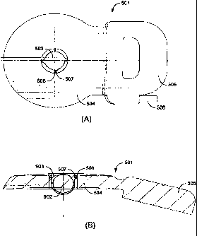
[0017] Fig. 5A shows a top plan view and Fig. 5B shows a cross-sectional view
of one
specific embodiment of an implanted device according to the present invention.
[0018] Fig. 6A-D show cross-sectional views of various other embodiments of an
implanted device coil assembly having an internal holding magnet contained
within
various differently shaped magnet cases.
-4-
CA 02679413 2009-08-28
WO 2008/109800 PCT/US2008/056152
[0019] Fig. 7A-B shows cross-section views of two other embodiments of an
implantable
device for a removable low-torque internal holding magnet case.
Detailed Description of Specific Embodiments
[0020] Embodiments of the present invention are directed to an implanted
device having
a low-torque internal magnet arrangement which allows for typical MRI
procedures that
otherwise require surgical removal and replacement of the magnet. But the
magnet
holding structure also is adapted to allow for easy removal and replace of the
internal
magnet for those MRI procedures where the magnet might produce unacceptable
imaging
artifacts if left in place; for example, for MRI' s of the tissue in the near
vicinity of the
implanted device.
[0021] More specifically, embodiments are directed to an implantable device
having a
receiver coil assembly including a magnet holding structure for containing at
least one
internal holding magnet. The internal holding magnet is reorientable in
responsive
alignment to a direction of an external magnetic field, as for example, during
an MRI
examination. And the magnet holding structure is adapted for allowing removal
and
subsequent reinsertion of the internal holding magnet, for example, to allow
for MRI
imaging. For example, the magnet holding structure may use a resilient
material such as a
silicone material to be temporarily deformable to allow for removal and
reinsertion.
[0022] Among the various considerations in specific embodiments of such an
arrangement are that the surgical removal and reinsertion operations should be
as
unproblematic as possible. In addition, the mechanical integrity of the
implanted
structures should not be compromised by the surgical procedures. And the
likelihood of
bacterial growth should be minimized as far as possible in all parts of the
implant, such as
for example, along the interfaces between the various structures.
[0023] Fig. 5A shows a top plan view and Fig. 5B shows a cross-sectional view
of one
specific embodiment of an implanted device 501 according to the present
invention. The
device includes an internal holding magnet 502 and located within a spherical
case
magnet holding structure 503, which in the embodiment shown, protrudes away
from a
side of the implanted device 501 farthest from the skin when implanted. In
this
embodiment, it is actually the metal case magnet holding structure 503
containing the
-5-
CA 02679413 2009-08-28
WO 2008/109800 PCT/US2008/056152
internal holding magnet 502 which is removable and replaceable.
[0024] In other specific embodiments, the internal holding magnet 502 may be
cylindrical, and some embodiments may have multiple cylindrical and/or
spherical
internal holding magnets 502. The implanted device 501 also includes a
receiving coil
assembly 504 and a signal processor housing 505 which cooperate to receive an
external
information and/or power signal and develop an electrode stimulation signal
for a
processor output 506 to an implanted prosthetic electrode carrier.
[0025] The magnet holding structure 503 also includes around its circumference
multiple
coupling projections 507, which cooperate with corresponding coupling
receptacles 508
in the receiving coil assembly 504 to hold the magnet holding structure 503 in
place. The
magnet holding structure 503 can be removed or replaced simply by rotating it
relative to
the receiving coil assembly 504. This rotation causes the coupling projections
507 and
their corresponding coupling receptacles 508 to engage against each
other¨temporarily
deforming one or both of them until they lock or unlock, depending on whether
the
magnet holding structure 503 is being replaced or removed.
[0026] Fig. 6A-D show cross-sectional views of various other embodiments of an
implanted device coil assembly 601 having an internal holding magnet 602
contained
within various differently shaped magnet cases 603. In such embodiments, all
or part of
the coil assembly 601 may use a resilient material to be temporarily
deformable as
required, for example, a bio-compatible plastic or a silicone-based material.
[0027] In Fig. 6A, the magnet case 603 is tapered to be wider at the top and
narrower at
the bottom and includes a flange head 604 as shown. Although the magnet case
603 is
normally securely positioned in the implanted device 601, it can be removed
simply by
prying under the flange head 604. This compresses part of the coil assembly
601,
temporarily deforming it so that it the case 603 can slide out and separate
from the
implanted device 601. This can be accomplished during relatively minor surgery
before
MRI testing. After the MRI testing, the magnet case 603 and its internal
holding magnet
602 can be replaced by a second minor surgery during which part of the coil
assembly
601 is again compressed to temporarily deform it to allow it to snap back into
place until
the flange head 604 is resting flush against the underside of the implanted
device 601.
-6-
CA 02679413 2009-08-28
WO 2008/109800 PCT/US2008/056152
Fig. 6B-D shows different similar variations on this concept and principle.
[0028] Fig. 7A-B shows cross-section views of two other embodiments of an
implantable
device 701 for a removable low-torque internal holding magnet case 702. In the
embodiment shown in Fig. 7A, the internal holding magnet case 702 is adapted
to be
press-fitted into a magnet holding socket 703 which as well as the coil
assembly includes
tapered ribs 704. The internal holding magnet case 702 is removable from the
implantable device 701 by applying force upward to deflect the tapered ribs
704 so that
they temporarily deform to allow the magnet to pop up and out of the socket
703.
Replacement of the internal holding magnet 702 is simply the opposite
operation,
pressing down on it to pop it back into the socket 703. In Fig. 7B, a coiled
holding spring
706 fits into corresponding threads in the side wall of the device, thereby
fixing the
internal holding magnet case 702 in place. In other embodiments, the internal
holding
magnet case 702 itself may include threads on its exterior surface so that it
can be
screwed into corresponding threads in implantable device 701.
[0029] Various embodiments such as the ones described above may also be
adapted so
that the internal magnet holding case may be removable and/or reinsertable
from the
other side of the device as shown in the figures. Thus, for example, the
embodiments
shown in Fig. 6A-D may be adapted to be removable and/or reinsertable from the
top side
closest to the patient's skin. In such embodiments, the internal magnet
holding case may
be centered within an opening in the center of the receiving coil and covered
by nearby
bone, securely holding it in place. Similarly, the embodiments shown in Fig 7A-
B may be
adapted to be removable and/or reinsertable from the underside farthest from
the patient's
skin.
[0030] Various specific embodiments may have some surfaces and/or structures
coated
with a therapeutic medicine such as an anti-bacterial agent to prevent or
minimize
bacterial growth which otherwise might be problematic, especially in the event
of
multiple surgeries such as the invention allows. Similarly, the interfaces
between the
various structural elements may be sealed, e.g. with a thin silicone layer, to
prevent or
minimize bacterial ingress into these interface regions. Thus, there may be a
thin sealing
layer between the internal magnet holding case and the coil assembly.
-7-
CA 02679413 2009-08-28
WO 2008/109800 PCT/US2008/056152
[0031] The described embodiments of the invention are intended to be merely
exemplary
and numerous variations and modifications will be apparent to those skilled in
the art.
All such variations and modifications are intended to be within the scope of
the present
invention as defined in the appended claims.
-8-