Note: Descriptions are shown in the official language in which they were submitted.
CA 02679707 2009-08-25
WO 2008/018933 PCT/US2007/010722
DETECTION OF PROTEASE AND PROTEASE ACTIVITY USING A
SINGLE NANOSCRESCENT SERS PROBE
CROSS-REFERENCE TO RELATED APPLICATIONS
[0001] This application claims benefit of and priority to USSN 60/797,525,
filed on
May 3, 2006, which is incorporated herein by reference in its entirety for all
purposes.
STATEMENT AS TO RIGHTS TO INVENTIONS MADE UNDER FEDERALLY
SPONSORED RESEARCH AND DEVELOPMENT
100021 This work was supported by DARPA, NIH Grant R1CA95393, UCSF
Prostate Cancer SPORE award (NIH Grant P50 CA89520), and P01 CA72006. This
work
was performed in part under the auspices of the U.S. Deptartment. of Energy,
at the
University of California/Lawrence Berkeley National Laboratory under Contract
no. DE-
AC02-05CH11231. The Government of the United States of America has certain
rights in
this invention.
FIELD OF THE INVENTION
100031 The present invention relates to the fields of Surface Enhanced Raman
Scattering (SERS) using nanoprobes for detection of proteases. The present
invention
relates specifically to the detection of Prostate Specific Antigen (PSA) and
proteolytically
active PSA for diagnostic applications in prostate cancer.
BACKGROUND OF THE INVENTION
100041 Prostate cancer is the most common cancer in men in Europe and North
America (Crawford (2003) Urology 62: 3-12; Gronberg et al. 92003) Lancet 361:
859-864;
Pienta et al. (2006) Urology 48: 676-683). One of the clinical diagnosis tools
for prostate
cancer is the measurement of plasma protein concentration of the prostate-
specific antigen
(PSA or hK3), which is a member of the large kallikrein (hK) protease family
(for reviews,
see, e.g., Yousef and Diamandis (2001) Endocr. Rev., 22: 184-204; Denmeade and
Isaacs
(2002) Nat. Rev. Cancer 2: 389-396; Denmeade and Isaacs (2004) BJUInt 93 Suppl
1: 10-
15) normally secreted from prostate luminal epithelial cells. Unlike other
kallikrein family
-1-
CA 02679707 2009-08-25
WO 2008/018933 PCT/US2007/010722
members, PSA is a chymotrypsin-like serine protease (Robert et al. (1997)
Biochemistry 36:
3811-3819). In prostate cancer, PSA, aided by the proteolytic activity, is
involved in tissue
remodeling against the extracellular matrix, contributing critical control
mechanisms to
tumor invasion or progression. Other proteases play similar roles in cancers
as well.
[0005] The introduction of plasma PSA screening since the 1980s has greatly
improved the diagnosis, staging, and management of prostate cancer (Denmeade
and Isaacs
(2002) Nat. Rev. Cancer 2: 389-396); however, measurement of plasma PSA
concentration
does not differentiate the prostate cancer patients from those with benign
prostatic
hyperplasia, leading to a high false positive rate, requirement for more
expensive biopsies,
and even unnecessary surgical procedures (Denmeade and Isaacs (2004) BJU Int
93 Suppl
1: 10-15; Robert et al. (1997) Biochemistry 36: 3811-3819). Efforts to enhance
the clinical
value of the PSA for early detection of prostate cancer have included the
characterization of
various molecular isoforms of PSA (Mikolajczyk et al. (2004) Clin. Chem., 50:
1017-1025;
Mikolajczyk and Rittenhouse (2003) Keio J. Med. 52: 86-91; Mikolajczyk et al.
(2004)
Clin. Biochem. 37: 519-528). Among those various isoforms, the proteolytically
active
subpopulation of PSA is accepted as a more useful tumor marker and malignancy
predictor
than the serum PSA concentration (Wu et al. (2004) Prostate 58: 345-353; Wu et
al. (2004)
Clin. Chem., 50: 125-129). Simple detection of the presence of PSA by a
traditional
immunostaining method can not reveal the proteolytic activity of PSA;
therefore, it is of
great importance to develop new methods to discriminate the proteolytically
active isoform.
Seminal fluid has been demonstrated to carry an abundance of proteolytically
active PSA
and is a biological source of PSA for protease activity assays (Brillard-
Bourdet et al. (2002)
Eur. J. Biochem., 269: 390-395; Rehault et al. (2002) Biochim. Biophys. Acta
1596: 55-62).
The concentration of proteolytically active PSA in seminal fluid is at 10-150
M (Rehault et
al. (2002) Biochim. Biophys. Acta 1596: 55-62), while its concentration in the
plasma is
much lower, from less than 0.1nM in healthy individuals to higher than 1nM in
patients
with prostate disease(Rittenhouse et al. (1998) Crit. Rev. Clin. Lab. Sci.,
35: 275-368).
However, an assay that measures the proteolytic activity of PSA in seminal
fluid or biopsy
samples from fine needle aspiration is still not widely accepted, due to the
quick decay of
the proteolytic activity, and the limited amount of seminal fluid available
from old patients
or biopsy samples.
-2-
CA 02679707 2009-08-25
WO 2008/018933 PCT/US2007/010722
[0006] The sensitivity of current detection methods reach subnanomolar
concentrations for PSA protein (Acevedo et al. (2002) Clin. Chim. Acta 317: 55-
63;
Charrier et al. (1999) Electrophoresis 20: 1075-108 1; Bjartell et al.
Prostate Cancer P D 2:
140-147) (mostly determined by the binding affinity of the antibody to PSA),
and relatively
large sample volume (milliliter) is required. However, the enzymatic assays
have not
enjoyed the same sensitivity enhancement.
SUMMARY OF THE INVENTION
[0007] In certain embodiments The present invention demonstrates the in vitro
detection of proteases using a single peptide-conjugate nanocrescent surface
enhanced
Raman scattering (SERS) indicator (probe) with at least nanomolar sensitivity.
This
indicator enables detection of proteolytic activity in extremely small
volumes. In certain
embodiments, the detection volume is less than about 80 femtoliters,
preferably less than
about 50 femtoliters, more preferably less than about 40 or 30 femtoliters,
and still more
preferably less than about 20 or 15 femtoliters. In certain embodiments, the
use of a highly
focused excitation source allows the detection volume to be only about 10
femtoliter. In
various embodiments the actual protease molecule number for the nanomolar
samples is
less than about 40 molecules, preferably less than about 40 molecules, more
preferably less
than about 30, 20, or 10 and in certain embodiments close to the single
molecule level.
Compared to other cancer biomarker detection assays, the present bioconjugated
nanocrescent allows the detection of nanomolar concentrations of
proteolytically active
protease molecules in femtoliter volumes, which is crucial especially for
cancer screening at
a single cancer cell level.
[0008] One of the major advantages and applications of the small volume
property
is that it is useful in detecting proteases such as prostate-specific antigen
(PSA) activity of
cancer cells at single cell level. The small volume requirement and
sensitivity level makes
it possible to detect PSA activity in captured circulating prostate cancer
cells for indications
of metastasis, which is not feasible with conventional techniques. In semen,
the PSA
concentration is 10-150 M, with approximately two thirds of the PSA
enzymatically
active. The sensitivity level achieved with the nanocrescent PSA probe
(nanomolar range)
is sufficient for a seminal fluid based assay, thus the nanocrescent SERS
platform described
herein is useful for clinical applications.
-3-
CA 02679707 2009-08-25
WO 2008/018933 PCT/US2007/010722
[0009] In certain embodiments the substrate is a nanocrescent surface enhanced
Raman scattering (SERS) probe. The surface enhanced Raman scattering (SERS)
probe is
comprised of a peptide conjugated to a nanocrescent core and shell, wherein
the probe
features a sequence, that can be specifically cleaved by a protease (e.g., a
protease
recognition site), linked to a Raman active tag. Thus, this peptide-conjugated
nanocrescent
can be used as a specific screening tool to provide information on the
presence,
concentration and proteolytic activity of one or more proteases including, but
not limited to
various cancer biomarkers, such as prostate-specific antigen (PSA) in a
biological sample.
100101 In one embodiment, the nanocrescents comprise a core and a shell,
having a
peptide conjugated or tethered to the surface of the nanocrescent. The
peptides comprise
substrates specifically recognized and cleaved by the corresponding proteases
to be
detected.
[0011] In certain embodiments other peptide substrates specific for the
protease can
be used for the nanoprobe. There might be circumstances that substrate peptide
with better
kinetic properties can be used to accelerate the detection process. In one
embodiment, other
peptides with better specificity to PSA can also be used to detect PSA with
better accuracy.
[0012] In various embodiments real-time reaction monitoring also provides
information on protease activity rather than just measuring the presence of
the protein.
Different Raman tag molecules can be used and are successfully utilized in the
Examples
thereby demonstrating that detection of two or more types of cancer-related
(or other)
proteases can be carried out by multiplexing the peptide-conjugated
nanocrescents. In
certain embodiments the core can comprise magnetic material to allow spatial
addressing of
individual nanoparticles.
[0013] In certain embodiments different peptide substrates orthogonal to each
other,
or with minor overlap in specificity, can be used to detect the corresponding
proteases. The
peptide library can be conjugated to the nanocrescent probes and spatially
separated in
either a random array or ordered microarray format. The multiplexed array of
the peptide-
nanocrescent hybrid probes can be used to detect multiple proteases
simultaneously.
100141 The nanocrescent(s) can also be manipulated by laser or magnetic fields
to
address at high accuracy spatially (Liu et al. (2006) Nat Mater 5: 27-32), so
that they can be
multiplexed as high density arrays (with sub-microliter volume). In addition,
the magnetic
-4-
CA 02679707 2009-08-25
WO 2008/018933 PCT/US2007/010722
or laser maneuverability allow biosensing at desired locations (Liu et al.
(2005) Adv. Mater.
17: 2683-2688), is useful for obtaining in situ measurements intracellularly.
100151 In certain embodiments this invention provides an indicator (probe) for
the
detection of an active protease. The indicator typically comprises a
nanocrescent attached
to a peptide (substrate), where the peptide comprises a recognition site for
the protease. In
certain embodiments the indicator further comprises a Raman label attached to
the peptide.
Suitable Raman labels include, but are not limited to fluorophore, a
chromophore, a
quantum dot, a fluorescent microsphere, biotin, and the like. In certain
embodiments the
Raman label comprises a Rhodamine, a fluoresceine, or an exogenous chemical
molecule.
In certain embodiments the Raman label comprises a moiety selected from the
group
consisting of TRIT (tetramethyl rhodamine isothiol), NBC (7-nitrobenz-2-oxa-
1,3-diazole),
Texas Red dye, phthalic acid, terephthalic acid, isophthalic acid, cresyl fast
violet, cresyl
blue violet, brilliant cresyl blue, para-aminobenzoic acid, erythrosine,
biotin, digoxigenin,
5-carboxy-4',5'-dichloro-2',7'-dimethoxy fluorescein, 5-carboxy-2',4',5',7'-
tetrachlorofluorescein, 5-carboxyfluorescein, 5-carboxy rhodamine, 6-
carboxyrhodamine, 6-
carboxytetramethyl amino phthalocyanines, 6-carboxy-X-rhodamine, azomethines,
cyanines, xanthines, succinylfluoresceins, aminoacridine, and cyanide (CN),
thiol (SH),
chlorine (Cl), bromine (Br), methyl, phorphorus (P), sulfur (S), SN, Al, Cd,
Eu, and Te.
The Raman label can be attached directly to the peptide or through a linker.
Similarly, the
peptide can be attached directly to the nanocrescent or through a linker. In
certain
embodiments the nanocrescent comprises a shell without a core. In certain
embodiments
the nanocrescent comprises a core and a shell. In various embodiments the core
comprises
a material that provides a constant Raman spectrum (e.g., a plastic (e.g.,
polystyrene), a
silica or other Group III, Group IV, or Group V material, a dextran, a
magnetic material,
and the like). In certain embodiments the nanocrescent comprises a metal
selected from the
group consisting of Ga, Au, Ag, Cu, Al, Ta, Ti, Ru, Ir, Pt, Pd, Os, Mn, Hf,
Zr, V, Nb, La, Y,
Gd, Sr, Ba, Cs, Cr, Co, Ni, Zn, Ga, In, Cd, Rh, Re, W, Mo, and oxides, and/or
alloys, and/or
mixtures, and/or nitrides, and/or sintered matrix thereof. In certain
embodiments the
nanocrescent has an outer radius that ranges from about 20 to about 800 nm. In
certain
embodiments the nanocrescent has an inner radius that ranges from about 10 nm
to about
500 nm. In certain embodiments the nanocrescent-is characterized by an inner
radius r, and
outer radius R, and a center to center distance between the center of the
circle defined by the
-5-
CA 02679707 2009-08-25
WO 2008/018933 PCT/US2007/010722
inner radius r and the outer radius R, where: r ranges from about 10 nm to
about 500 nm; R
ranges from about 20 nm to about 800 nm; and d ranges from about 5 nm to about
300 nm.
In certain embodiments the nanocrescent is characterized by an inner radius r,
and outer
radius R, and a center to center distance between the center of the the circle
defined by the
inner radius r and the outer radius R, where: r ranges from about 25 nm to
about 500 nm; R
ranges from about 20 nm to about 800 nm; and d ranges from about 5 nm to about
200 nm.
In various embodiments the peptide comprises a recognition site for a protease
selected
from the group consisting of a serine protease, a metalloprotease, a cysteine
protease, an
aspartic acid protease, and a glutamic acid protease. In certain embodiments
the peptide
comprises a recognition site for a protease in an apoptosis pathway (e.g., a
caspase). In
certain embodiments the peptide comprises a recognition site for a caspase
selected from the
group consisting of caspase-8, caspase-9, caspase-3, caspase-6, and caspase-7.
In certain
embodiments the peptide comprises a recognition site for a thrombin. In
certain
embodiments the peptide comprises a recognition site for a serine protease. In
certain
embodiments the peptide comprises a PSA recognition site (e.g., HSSKLQ (SEQ ID
NO: 1)). In various embodiments the peptide ranges in length from 2 amino
acids to 10, 20,
or 30 amino acids. In certain embodiments the peptide is attached to the
nanocrescent by a
thiol group. In certain embodiments two different substrates (e.g., peptides)
are attached to
the nanocresent. In certain embodiments more than two different peptides are
attached to
the nanocresent. In certain embodiments the indicator is a component of a
Raman active
substrate.
[0016] In certain embodiments, this invention provides an indicator for the
detection
of an active nuclease. These indicators are essentially the same as the
protease indicators
described above, except the peptide (substrate) is replaced with a nucleic
acid (e.g., a double
stranded or single stranded nucleic acid). In certain embodiments the nucleic
acid
comprises a one or more nuclease recognition/cleavage sites (e.g., restriction
sites).
[0017] In certain embodiments, the substrate (e.g., peptide, nucleic acid,
sugar,
carbohydrate, etc.) attached to the nanocrescent can comprise one or more
binding sites
(rather than cleavage sites) for the detection of, e.g., a cognate binding
partner (e.g.,
receptor, nucleic acid binding protein, ligand, etc.).
-6-
CA 02679707 2009-08-25
WO 2008/018933 PCT/US2007/010722
[00181 Also provided are methods of detecting or quantifying the presence,
amount,
or activity of at least one protease in a sample. The method involve
contacting the sample
with an indicator comprising a nanocrescent attached to a peptide comprising a
recognition
site for a protease (e.g., as above); and monitoring differences in spectral
characteristics of
detected surface- Raman scattering spectra, the differences being indicators
of the presence,
amount or activity of protease present in the sample. In various embodiments
the sample
comprises a material selected from the group consisting of whole blood, a
blood fraction,
lymph, cerebrospinal fluid, oral fluid, mucus, urine, feces, bronchial lavage,
ascites fluid,
seminal fluid, bone marrow aspirate, pleural effusion, urine, and tumor cells
or tissue. In
various embodiments the indicator further comprises a Raman label attached to
the peptide.
Suitable Raman labels include, but are not limited to fluorophore, a
chromophore, a
quantum dot, a fluorescent microsphere, biotin, and the like. In certain
embodiments the
Raman label comprises a rhodamine, a fluoresceine, or an exogenous chemical
molecule.
In certain embodiments the Raman label comprises a moiety selected from the
group
consisting of TRIT (tetramethyl rhodamine isothiol), NBC (7-nitrobenz-2-oxa-
1,3-diazole),
Texas Red dye, phthalic acid, terephthalic acid, isophthalic acid, cresyl fast
violet, cresyl
blue violet, brilliant cresyl blue, para-aminobenzoic acid, erythrosine,
biotin, digoxigenin,
5-carboxy-4',5'-dichloro-2',7'-dimethoxy fluorescein, 5-carboxy-2',4',5',7'-
tetrachlorofluorescein, 5-carboxyfluorescein, 5-carboxy rhodamine, 6-
carboxyrhodamine, 6-
carboxytetramethyl amino phthalocyanines, 6-Carboxy-X-rhodamine, azomethines,
cyanines, xanthines, succinylfluoresceins, aminoacridine, and cyanide (CN),
thiol (SH),
chlorine (Cl), bromine (Br), methyl, phorphorus (P), sulfur (S), SN, Al, Cd,
Eu, and Te.
The Raman label can be attached directly to the peptide or through a linker.
Similarly, the
peptide can be attached directly to the nanocrescent or through a linker. In
certain
embodiments the nanocrescent comprises a shell without a core. In certain
embodiments
the nanocrescent comprises a core and a shell. In various embodiments the core
comprises
a material that provides a constant Raman spectrum (e.g., a plastic (e.g.,
polystyrene), a
silica or other Group III, Group IV, or Group V material, a dextran, a
magnetic material,
and the like). In certain embodiments the nanocrescent comprises a metal
selected from the
group consisting of Ga, Au, Ag, Cu, Al, Ta, Ti, Ru, Ir, Pt, Pd, Os, Mn, Hf,
Zr, V, Nb, La, Y,
Gd, Sr, Ba, Cs, Cr, Co, Ni, Zn, Ga, In, Cd, Rh, Re, W, Mo, and oxides, and/or
alloys, and/or
mixtures, and/or nitrides, and/or sintered matrix thereof. In certain
embodiments the
-7-
CA 02679707 2009-08-25
WO 2008/018933 PCT/US2007/010722
nanocrescent has an outer radius that ranges from about 20 to about 800 nm. In
certain
embodiments the nanocrescent has an inner radius that ranges from about 10 nm
to about
500 nm. In certain embodiments the nanocrescent is characterized by an inner
radius r, and
outer radius R, and a center to center distance between the center of the the
circle defined by
the inner radius r and the outer radius R, where: r ranges from about 10 nm to
about 500
nm; R ranges from about 20 nm to about 800 nm; and d ranges from about 5 nm to
about
300 nm. In certain embodiments the nanocrescent is characterized by an inner
radius r, and
outer radius R, and a center to center distance between the center of the
circle defined by the
inner radius r and the outer radius R, where: r ranges from about 25 nm to
about 500 nm; R
ranges from about 20 nm to about 800 nm; and d ranges from about 5 nm to about
200 nm.
In various embodiments the peptide comprises a recognition site for a protease
selected
from the group consisting of a serine protease, a metalloprotease, a cysteine
protease, an
aspartic acid protease, and a glutamic acid protease. In certain embodiments
the peptide
comprises a recognition site for a protease in an apoptosis pathway (e.g., a
caspase). In
certain embodiments the peptide comprises a recognition site for a caspase
selected from the
group consisting of caspase-8, caspase-9, caspase-3, caspase-6, and caspase-7.
In certain
embodiments the peptide comprises a recognition site for a thrombin. In
certain
embodiments the peptide comprises a recognition site for a serine protease. In
certain
embodiments the peptide comprises a PSA recognition site (e.g., HSSKLQ, SEQ ID
NO:1).
In various embodiments the peptide ranges in length from 2 amino acids to 10,
20, or 30
amino acids. In certain embodiments the peptide is attached to the
nanocrescent by a thiol
group. In certain embodiments two different substrates (e.g., peptides) are
attached to the
nanocresent. In certain embodiments more than two different peptides are
attached to the
nanocresent. In certain embodiments the indicator is a component of a Raman
active
substrate.
[0019] Also provide are libraries for the detecting the presence or activity
of two or
more active proteases. The libraries typically comprise a plurality of
protease indicators the
indicators comprising a nanocrescent attached to a peptide, e.g., as described
above, where
the peptide comprises a recognition site for the protease; where different
nanocrescents have
attached thereto different peptides so that different nanocrescents detect
different proteases.
In various embodiments the library is spatially addressed so that protease
indicators specific
for different proteases are localized at different locations on a substrate.
In various
-8-
CA 02679707 2009-08-25
WO 2008/018933 PCT/US2007/010722
embodiments the library is optically addressed so that protease indicators
specific for
different proteases produce different signals. In certain embodiments the
library comprises
at least 3 or more, preferably at least 10 or more, more preferably at least
20, 40, 80, or 100
or more different protease indicators. In certain embodiments the
nanocrescents comprise
magnetic cores and spatial segregation of the indicators is provided by
magnetic fields. In
certain embodiments the indicators are ionically or chemically coupled and/or
adsorbed to a
substrate.
[0020] Kits for the detection of an active protease are also provided. The
kits
typically include a container containing a nanocrescent attached to a peptide
(e.g. as
described herein), where the peptide comprises a recognition site for the
protease. In certain
embodiments the kit further comprises a Raman label attached to the peptide.
In certain
embodiments the the kit further comprises instructional materials teaching the
use of the
indicator for the detection of the presence, concentration or activity of an
active protease
using surface enhanced Ramen scattering (SERS).
[0021] In certain embodiments this invention provides an indicator for the
detection
of an nuclease. The indicator comprises a nanocrescent (e.g., as described
herein) attached
to a single or double-stranded oligonucleotide, where the oligonucleotide
comprises a
recognition site for the nuclease. In certain embodiments the indicator
further comprises a
Raman label attached to the oligonucleotide.
[0022] Also provided are methods of detecting the presence or quantity of an
analyte. The methods typically involve contacting a sample comprising the
analyte to an
indicator, the indicator comprising a nanocrescent attached to a substrate
that is specifically
or preferentially bound by the analyte in the presence of a Raman-labeled
moiety that
competes with the analyte for binding to the substrate; and detecting the
Raman spectrum of
the indicator where a change in the Raman spectrum produced by dissociation of
the
Raman-labeled moiety from the substrate provides a measure of the presence or
quantity of
the analyte in the sample. In certain embodiments the substrate is a peptide
or a nucleic
acid.
100231 In certain embodiments, the peptide attached to the nanocrescent is not
an
antibody or an antibody fragment.
-9-
CA 02679707 2009-08-25
WO 2008/018933 PCT/US2007/010722
DEFINITIONS
[0024] The terms "polypeptide", "peptide" and "protein" are used
interchangeably
herein to refer to a polymer of amino acid residues. The terms apply to amino
acid
polymers in which one or more amino acid residues is an artificial chemical
analogue of a
corresponding naturally occurring amino acid, as well as to naturally
occurring amino acid
polymers.
[0025] The term "active protease" refers to a protease that is in a form
capable of
cleaving (hydrolyzing) a peptide bond in the substrate for that protease when
the protease is
contacted with the substrate under conditions that support activity of that
protease.
[0026] The term "nanocrescent" refers to nanoparticles whose cross-sectional
profile
resembles the crescent moon with sharp edges.
[0027] "Analyte," as used herein, is the substance to be detected in a test
sample
using the present invention. The analyte can be any substance, e.g. an enzyme
for which
there exists specific binding member e.g., a substrate or for which a specific
binding
member can be prepared, and the analyte can bind to one or more specific
binding members
in an assay. The term "analyte" also includes any enzymes, antigenic
substances, haptens,
antibodies, and combinations thereof. The analyte can include, but is not
limited to a
protein, a peptide, an amino acid, a carbohydrate, a hormone, asteroid, a
vitamin, a drug
including those administered for therapeutic purposes as well as those
administered for
illicit purposes, a bacterium, a virus, and metabolites of or antibodies to
any of the above
substances.
100281 "Radiation," as used herein, is an energy in the form of
electromagnetic
radiation which, when applied to a test mixture, causes a Raman spectrum to be
produced
by the Raman-active label therein, and also causes the metal surface to
support surface-
enhanced Raman light scattering by the Raman-active labels, which become
associated with
the particulate surface.
[0029] A "Raman label", "Raman tag", or "Raman active label" is a substance
that
produces a detectable Raman spectrum, which is distinguishable from the Raman
spectra of
other components present, when illuminated with a radiation of the proper
wavelength.
Other terms for a Raman-active label can include dye and reporter molecule.
-10-
CA 02679707 2009-08-25
WO 2008/018933 PCT/US2007/010722
[0030] "Specific binding member," as used herein, is a member of a specific
binding
pair, i.e., two different molecules where one of the molecules, through
chemical or physical
means, specifically binds to the second molecule. In addition to antigen and
antibody-
specific binding pairs, other specific binding pairs include biotin and
avidin, carbohydrates
and lectins, complementary nucleotide sequences (including probe and captured
nucleic
acid sequences used in DNA hybridization assays to detect a target nucleic
acid sequence),
complementary peptide sequences, effector and receptor molecules, enzyme
cofactors and
enzymes, enzyme inhibitors and enzymes, and the like. Furthermore, specific
binding pairs
can include members that are analogs of the original specific binding member.
For example
a derivative or fragment of the analyte, i.e., an analyte-analog, can be used
so long as it has
at least one epitope in common with the analyte. Immunoreactive specific
binding members
include antigens, haptens, antibodies, and complexes thereof including those
formed by
recombinant DNA methods or peptide synthesis.
[0031] The term "test mixture," refers to a mixture of the test sample and
other
substances used to apply the present invention for the detection of analyte in
the test sample.
Examples of these substances include: Specific binding members, ancillary
binding
members, analyte-analogs, Raman-active labels, buffers, diluents, and
particulates with a
surface capable of causing a surface-enhanced Raman spectroscopy, and others.
100321 The term "test sample," as used herein, means the sample containing the
analyte to be detected and assayed using the present invention. The test
sample can contain
other components besides the analyte, can have the physical attributes of a
liquid, or a solid,
and can be of any size or volume, including for example, a moving stream of
liquid. The
test sample can contain any substances other than the analyte as long as the
other substances
do not interfere with the specific binding of the specific binding member or
with the analyte
or the analyte-analog. Examples of test samples include, but are not limited
to: Serum,
plasma, sputum, seminal fluid, urine, other body fluids, tissue and cell
samples (e.g., tumor
samples, organ samples, and the like) and environmental samples such as ground
water or
waste water, soil extracts and pesticide residues.
BRIEF DESCRIPTION OF THE DRAWINGS
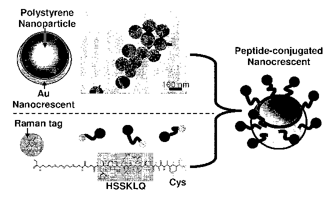
[0033] Figures 1A, 1B, 1C illustrate a peptide-conjugated nanocrescent for PSA
detection, a fabrication procedure, and detection. Figure 1 A: Fabrication
procedure. The
-11-
CA 02679707 2009-08-25
WO 2008/018933 PCT/US2007/010722
nanoscale Au layer was evaporated on polystyrene nanoparticles to form the Au
nanocrescent as shown in the TEM image, with the crescent tip showing lighter
density.
Peptides were synthesized with the specific PSA substrate sequence HSSKLQ (SEQ
ID
NO:1) and were terminated by a Raman tag molecule, biotin or R19 (not shown),
respectively, and cysteine for both versions of tagged peptides. The peptides
were
conjugated to the Au surface of the nanocrescents through an Au-S bond. Figure
1 B: PSA
detection scheme. Before the proteolytic reaction, the SERS spectrum of the
peptide-
conjugated nanocrescent contains the characteristic peaks from the Raman tag
molecules,
polystyrene nanoparticle, and the peptides; after the digestion reaction by
PSA, the peptide
is cleaved after Q. The cleavage fragment containing the Raman tag molecules
diffuses
away from the nanocrescent surface, while the other fragment remains on the
nanocrescent
surface. The SERS spectrum of the peptide becomes different and the
characteristic peaks
from the Raman tag molecule disappear. Figure 1 C: (1) Simulated local
electric field
amplitude enhancement by nanocrescent. The tip region of the nanocrescent has
an
electromagnetic enhancement factor of 100 fold. (2) Polar electric field
energy distribution
on the nanocrescent. Almost 100% energy is concentrated near the tip area
which accounts
for -1/6 of total area of the nanocrescent.
[0034] Figures 2A, 2B, and 2C illustrate Gold nanocrescent moons with sharp
edges. Fig. 2 A: Conceptual schematics of a nanocrescent moon SERS substrate.
The gold
surface can be functionalized with biomolecular linker to recognize specific
biomolecules.
The sharp edge of the nanocrescent moon can enhance the Raman scattering
intensity so that
the biomolecules on it can be detected. Fig. 2B: Geometrical schematics of a
nanocrescent
moon. A gold nanocrescent moon with sharp edges integrates the geometric
features of
nanoring and nanotips. Fig. 2 C: Transmission electron microscope images of
two
nanocrescent moons. Shown nanocrescent moons are both of 300 nm inner-
diameter, 100
nm-bottom-thickness, but with different orientations. The scale bars are 100
nm.
(0035] Figure 3 illustrates a fabrication procedure for nanocrescent moons.
(a)
Casting a monolayer of spherical polystyrene colloids on a photoresist coated
glass
substrates. (b) Coating a gold layer on the surfaces of polystyrene colloids
by electron beam
evaporation. The sample is kept rotating at a certain angle with respect to
the gold target
during deposition. The shape of the nanocrescent moons depends on the
deposition angle in
addition to the size of the polystyrene spheres. (c) Lift-off of the gold-
coated polystyrene
-12-
CA 02679707 2009-08-25
WO 2008/018933 PCT/US2007/010722
spheres from the substrate. (d) Scanning electron microscopy of gold
nanocrescent moons.
The dissolution of the colloidal particles releases the nanocrescent moons
into a suspension.
The nanocrescent moons are then collect and placed on a substrate. For the
convenience of
demonstration in SEM, the shown nanocrescent moons were not subject to
dilution in water
like the nanocrescent moons used in our optical experiments. The scale bar is
200 nm.
100361 Figure 4 illustrates the geometry of a nanocrescent moon where r is the
inner
radius, R is the outer radius, and d is the center-center distance as shown as
two partially
overlapping circles. .
100371 Figure 5 illustrates a SERS microspectroscopy system and nanocrescent
visualization. The peptide-conjugated nanocrescents are suspended in the
reaction buffer in
an enclosed transparent microchamber. The nanocrescents can be visualized
using the dark
field illumination from oblique angles as the bright dots shown in the inset
pictures. The
excitation laser is focused on the nanocrescents by a microscopy objective
lens. The SERS
signal is collected by the same objective lens and analyzed by a spectrometer.
[0038] Figures 6A and 6B show typical SERS spectra of peptide-conjugated
nanocrescents before and after PSA digestion reactions with biotin (Fig. 6A)
and R19 (Fig.
6B) as the Raman tag molecules respectively.
[0039] Figures 7A, 7B, 7C, and 7D show time-resolved SERS spectra in PSA
digestion reactions. Figure 7A: SERS spectra in the peptide digestion by 420
nM PSA
with biotin as the Raman tag molecule. Figure 7 B: SERS spectra in the peptide
digestion
by 420 nM PSA with R19 as the Raman tag molecule. Figure 7C: SERS spectra in
the
peptide digestion by 420 nM PSA in the presence of inhibitor with R19 as the
Raman tag
molecule. Figure 7D: SERS spectra in the peptide digestion by 420 nM Granzyme
B with
R19 as the Raman tag molecule.
[0040] Figures 8A and 8B show time-dependent Raman peak intensities in PSA
digestion reactions. Figure 8A: Raman peak intensities of biotin at 525 cm-1
in the
digestion reactions with 0 M (buffer solution), 4.2 nM, 42 nM and 420 nM PSA,
respectively. Figure 8 B: Raman peak intensities of R19 at 1183 cm-1 in the
digestion
reactions with 420 nM PSA, 420 nM PSA with inhibitor, and 420 nM Granzyme B,
respectively.
-13-
CA 02679707 2009-08-25
WO 2008/018933 PCT/US2007/010722
DETAILED DESCRIPTION
[0041) In various embodiments, this invention pertains to novel indicators
that
provide a measure of the presence and/or quantity and/or activity of one or
more proteases
in a sample. In certain embodiments the indicators comprise one or more
nanocrescent
structures attached to substrate(s) (e.g., polypeptide(s)) for one or more
protease molecules
see, e.g., Figure lA). The indicators can, optionally, further comprise one or
more Raman
tags attached to the substrate. The indicators function as extremely sensitive
probes for
Raman scattering detection systems (e.g. surface enhanced Raman scattering
(SERS)
probes).
[0042] In certain embodiments the present invention pertains to the in vitro,
in situ,
or, in certain instances in vivo, detection of proteolytically active
biological molecules using
a peptide-conjugate nanocrescent surface enhanced Raman scattering (SERS)
probe. The
probes described herein can achieve at least nanomolar sensitivity, thereby
enabling
detection of proteolytic (or other biological) activity in extremely low
concentrations (e.g.,
one or several molecules) and/or in extremely small volumes (e.g., femtoliter
volumes). In
various embodiments, the nanoscale dimension of the indicator(s) and the high
local
electromagnetic field enhancement of the indicator (Figure 1 C) enables high-
sensitivity
optical detection of biomolecular reactions on its surface.
100431 In certain preferred embodiments, the indicator comprises a
nanocrescent
surface enhanced Raman scattering (SERS) probe. The surface enhanced Raman
scattering
(SERS) probe is can comprise a peptide conjugated to a nanocrescent strcutre
(e.g., a
nanocrescent core and shell), where the peptide contains a specific amino acid
sequence that
is recognized and cleaved by a protease (a protease recognition site). In
various
embodiments the peptide is attached to a Raman tag. Cleavage of the peptide by
the
"target" protease provides a strong change in the Raman spectrum that is
readily detected.
Thus, the peptide-conjugated nanocrescent can be used as a specific screening
tool to
provide information on the presence, concentration and proteolytic activity of
the one or
more proteases, e.g., cancer biomarkers, such as prostate-specific antigen
(PSA) in a
biological sample.
-14-
CA 02679707 2009-08-25
WO 2008/018933 PCT/US2007/010722
Nanocrescent composition and fabrication.
[0044] The indicators of the present invention typically comprise one or more
nanocrescents coupled to a biological molecule, preferably a peptide. In
certain
embodiments the nanocrescents can comprise a core and a shell. When present,
the core
can be comprised of a plastic (e.g., polystyrene), silica or other mineral, or
other Group III,
Group IV, or Group V material, a dextran, a magnetic material, or any other
materials with
a substantially constant Raman spectra.
[0045] The nanocrescent "moon" structures have features of both nanotips and
nanorings which allows local electromagnetic field enhancement (Figure 2A). In
cross-
sectional view, the shape of the nanocrescent moon resembles a crescent
nanomoon with sharp
tips, so the sharp edge of the nanocrescent moon has the rotational analogy to
a sharp tip and
it expands the SERS "hot site" from a tip to a circular line (i.e., a group of
nanotips) as
shown in Figure 2B. From a top view the shape of the nanocrescent moon
resembles a
nanoring with a higher sharpness than previously demonstrated nanorings
(Aizpurua et al.
(2003) Phys. Rev. Lett. 90: 057401), so the circular sharp edge of the
nanocrescent moon
can have a stronger field emitting or "antenna" effect.
[0046] In various embodiments the gold nanophotonic crescent moons have sub-10
nm sharp edges as shown in Figure 2C.
[0047] In certain embodiments the nanocrescents can be characterized by a
geometry as illustrated in Figure 4, where r is the inner radius, R is the
outer radius, and d is
the center-center distance as shown as two partially overlapping circles. In
various
embodiments R ranges from about 20nm to about 800 nm, preferably from about 40
nm to
about 600 nm, more preferably from about 50 nm to about 500 or 400 nm, and
most
preferably from about 100 nm to about 200 nm 300 nm. In various embodiments, r
ranges
from about 10 nm to about 500 nm, preferably from about 20 nm to about 400 nm,
more
preferably from about 50 nm to about 300 nm, and most preferably from about
100 nm to
about 200 nm. In various embodiments d ranges from about 10 to about 400 nm,
preferably
from about 20 nm to about 200 nm or 300 nm, more preferably from about 30 nm,
40 nm or
50 nm to about 100 or 150 nm.
[0048] The nanocrescent shell can be comprised of a metal (e.g., gold, silver,
tungsten, platinum, titanium, iron, manganese, and the like, or oxides or
alloys thereof), a
-15-
CA 02679707 2009-08-25
WO 2008/018933 PCT/US2007/010722
semiconductor material, multi-layers of metals, a metal oxide, an alloy, a
polymer, carbon
nanomaterials, and the like. In certain embodiments the nanocrescent shell
comprises one
or more of the following: tungsten, tantalum, and niobium, Ga, Au, Ag, Cu, Al,
Ta, Ti, Ru,
Ir, Pt, Pd, Os, Mn, Hf, Zr, V, Nb, La, Y, Gd, Sr, Ba, Cs, Cr, Co, Ni, Zn, Ga,
In, Cd, Rh, Re,
W, Mo, and oxides, alloys, mixtures, and/or nitrides thereof.
[0049] In various embodiments the core ranges from about 30 nm to about 500
nm,
preferably about 50 nm to about 200 nm, 300 nm, or 400 nm, more preferably
from about
50 nm to about 100 nm or 150 nm in diameter, and the shell is preferably 3 nm
to about 80
nm, more preferably about 5 nm to about 50 nm, still more preferably about 8
nm to about
20 or 30 nm, and most preferably about 10 nm to about 20 nm or 25 nm. By
choosing
different core size and shell thickness, the plasmon resonance wavelength and
the surface
enhancement factor can be tuned to match various applications.
[0050] Figure 1 A schematically illustrates one embodiment of a nanocrescent
indicator of the present invention and provides an electron micrograph
thereof. In certain
embodiments the nanocrescents are preferably fabricated by angled deposition
of the
nanocrescent material(s) (e.g., silver, gold, etc.) on a rotating nanoparticle
(e.g., polystyrene
nanoparticle template) as described by Lu et al. (2005) Nano Lett 5, 119-124,
which is
incorporated herein by reference. The fabrication procedure is schematically
illustrated in
Figure 3. As shown in Figure 3, this method involves casting a monolayer of
spherical core
materials (e.g., polystyrene colloids) on photoresist-coated substrates (e.g.,
glass substrates).
The nanocrescent shell material(s) (e.g., gold, silver, etc.) are coated on
the surfaces of the cores
by electron beam evaporation. The sample is kept rotating at a certain angle
with respect to
the gold (or other material) target during deposition. The shape of the
nanocrescent moons
depends on the deposition angle in addition to the size of the core structures
(e.g.,
polystyrene spheres). The coated nanocrescents can be lifted from the
substrate using an
appropriate solvent (e.g., acetone). The cores can, optionally, be removed
from the
nanocrescents, by the use of appropriate solvent(s) (e.g., toluene). The
nanocrescent moons
can then be collected and placed on a substrate.
[0051] In one illustrative embodiments, the nanocrescents comprise a 100 nm
polystyrene core and a 10-20 nm gold crescent shell. The nanoscale Au layer is
evaporated
on polystyrene nanoparticles to form the Au nanocrescent as shown in the TEM
image in
-16-
CA 02679707 2009-08-25
WO 2008/018933 PCT/US2007/010722
Figure 1 C, with the crescent tip showing lighter density. In certain
embodiments the
nanoparticle core is not removed and serves as the internal control in the
SERS detections.
[0052] This fabrication procedure is illustrative and not limiting. Using the
teachings provided herein, variations of the present protocols and other
nanocrescent
fabrication methods will be recognized by one of skill in the art.
Protase substrates
100531 The nanocrescent indicators described herein can utilize polypeptide
sequences comprising one or more recognition site(s) for any protease(s) it is
desired to
detect. Proteases (proteolytic activity) are not only required for maintenance
of normal
cellular functions but are also central to the pathogenesis of a variety of
human diseases.
Parasitic (for example schistosomiasis and malaria), fungal (such as C.
albicans) and viral
infections (for example HIV, herpes and hepatitis), and also cancer,
inflammatory,
respiratory, cardiovascular and neurodegenerative diseases, including
Alzheimer's, require
proteolytic activity for progress. Detection of protease presence, quantity,
or activity is thus
useful as a diagnostic/prognostic marker for the presence or likelihood of
disease. In
addition, detection of protease activity (or the inhibition thereof) is useful
in screening for
protease inhibitor therapeutics for the treatment of a number of pathologies.
[0054] A "protease" that can be detected and/or quantified according to the
invention is an enzyme that typically hydrolyzes a peptide bond between a pair
of amino
acids located in a polypeptide chain, also called an endoprotease. Proteases
are typically
defined by reference to the nucleophile in the catalytic center of the enzyme.
The most
common nucleophiles arise from the side chains of serine, aspartic acid, and
cysteine,
resulting in families of proteases, such as serine proteases (Paetzel et al.
(1997) Trends
Biochem. Sci. 22: 28-31), aspartyl proteases (Spinelli et al. (1991) Biochemie
73: 1391-
1396), and cysteine proteases (Altschuh et al. (1994) Prot. Eng. 7: 769-75,
1994).
Metalloproteases usually contain a zinc catalytic metal ion at the catalytic
site (Klimpel et
al. (1994) Mol. Microbiol. 13: 1093-1100). Illustrative examples of members of
each of
these protease families are provided in Table 1.
-17-
CA 02679707 2009-08-25
WO 2008/018933 PCT/US2007/010722
[0055] Table 1. Illustrative proteases and protease recognition sites (*
indicates the
peptide bond being hydrolyzed).
Protease Family Protease Protease Recognition SEQ
Sites ID
NO
serine factor Xa Ile-Gly-Gly-Ar * 2
serine trypsin Lys*, Arg*
serine chymotrypsin Tyr*, Phe*, Leu*, Ile*, Val*,
Trp*, and His* at high pH
serine thrombin Arg*
serine PSA 3
serine and cysteine peanut mottle Glul-Xaa-Xaa-Tyr- 4
variants polyvirus Nla protease Gln*(Ser/Gly)
cysteine papaine Arg*, Lys*, Phe*
cysteine bromelaine Lys*, Ala*, Tyr*, Gly*
cysteine cathepsin B Arg*Arg, 5
Phe*Arg 6
cysteine cathepsin L Phe*Arg 6
aspartyl HIV protease Phe*Pro 7
aspartyl S. cerevisiae yapsin 2 Lys*, Arg*
aspartyl cathepsin D Phe*Phe 8
Phe*Lys 9
Leu*Phe 10
Leu*Tyr 11
metallo- thermolysin *Tyr, *Phe, *Leu, *Ile, *Val,
*Trp, and *His
metallo- peptidyl-Lys Xaa*Lys 12
metalloendo e tidase
metallo- peptidyl-Asp Xaa*Asp 13
metallodndopeptidase Xaa*Glu 14
Xaa*Cys 15
metallo- coccolysin *Leu, *Phe, *Tyr, *Ala
metallo- autolysin Leu-Trp-Met*Arg-Phe-Ala 16
metallo- gelatinase A (MMP-2) Pro-Gln-Gly*Ile-Ala-Gl -Gln 17
metallo- human neutrophil Gly-Leu-Ser-Ser-Asn-Pro * Ile- 18
collagenase (MMP-8) Gln-Pro
[0056] A "protease recognition site" is a contiguous sequence of amino acids
connected by peptide bonds that contains a pair of amino acids which is
connected by a
peptide bond that is hydrolyzed by a particular protease. Optionally, a
protease recognition
site can include one or more amino acids on either side of the peptide bond to
be
hydrolyzed, to which the catalytic site of the protease also binds (Schecter
and Berger,
(1967) Biochem. Biophys. Res. Commun. 27: 157-62), or the recognition site and
cleavage
-18-
CA 02679707 2009-08-25
WO 2008/018933 PCT/US2007/010722
site on the protease substrate can be two different sites that are separated
by one or more
(e.g., two to four) amino acids.
[0057] The specific sequence of amino acids in the protease recognition site
typically depends on the catalytic mechanism of the protease, which is defined
by the nature
of the functional group at the protease's active site. For example, trypsin
hydrolyzes peptide
bonds whose carbonyl function is donated by either a lysine or an arginine
residue,
regardless of the length or amino acid sequence of the polypeptide chain.
Factor Xa,
however, recognizes the specific sequence Ile-Glu-Gly-Arg (SEQ ID NO: 19) and
hydrolyzes peptide bonds on the C-terminal side of the Arg.
[0058] Thus, in various embodiments, a protease recognition site can comprise
at
least 2, 3, 4, 5, 6, 7, 8, 9, or 10 or more amino acids. Optionally,
additional amino acids can
be present at the N-terminus and/or C-terminus of the recognition site. A
protease
recognition site according to the invention also can be a variant of a
recognition site of a
known protease as long as it is recognized/cleaved by the protease.
[0059] Various preferred protease recognition sites include, but are not
limited to
protease recognition sites for proteases from the serine protease family, or
for
metalloproteases, or for a protease from the cysteine protease family, and/or
the aspartic
acid protease family, and/or the glutamic acid protease family. In certain
embodiments
preferred serine proteases recognition sites include, but are not limited to
recognition sites
for chymotrypsin-like proteases, and/or subtilisin-like proteases, and/or
alpha/beta
hydrolases, and/or signal peptidases. In certain embodiments preferred
metalloprotease
recognition sites include, but are not limited to recognition sites for
metallocarboxypeptidases or metalloendopeptidases.
[0060] Protease recognition sites are well known to those of skill in the art.
Recognition sites have been identified for essentially every known protease.
Thus, for
example, recognition sites (peptide substrates) for the caspases are described
by Earnshaw
et al. (1999) Annu. Rev. Biochem., 68: 383-424, which is incorporated herein
by reference
(see also Table 2).
-19-
CA 02679707 2009-08-25
WO 2008/018933 PCT/US2007/010722
[0061] Table 2. Illustrative peptide substrates for caspases (* indicates the
peptide
bond being hydrolyzed).
Name Peptide Substrate SEQ ID NO
Caspase I YEVD*X 20
(ICE) WEHD*X 21
Caspase 2 VDVAD*X 22
(Ich-IL) DEHD*X 23
Caspase 3 DMQD*X 24
(CPP32,.Apopain) DEVD*X 25
Caspase 4 LEVD*X 26
(Icefeill Tx, Ich-2) (W/L)EHD*X 27
Caspase 5 (W/L)EHD*X 28
(ICErelIII, Ty)
Caspase 6 VEID*N 29
(Mch2) VEHD*X 30
Caspase 7 DEVD*X 31
(Mch3, CMH-1, ICE-
LAP3)
Caspase 8 IETD*X 32
LED*X 33
Caspase 9 LEHD*X 34
Caspase 10 IEAD*X 35
[0062] In one illustrative embodiment to detect PSA, the peptide design
incorporates
the amino acid sequence of the active site of PSA-specific peptides with
serine residues and
flanking sequences that can be recognized by PSA. Thus, for example, in one
embodiment,
the peptide contains the sequence HSSKLQ-LAAAC (SEQ ID NO:36) which has been
shown to have very high specificity for proteolytically active PSA. (see,
e.g., Denmeade, et
al. (1997) Cancer Res 57: 4924-4930). It has been shown that HSSKLQ-L (SEQ ID
NO:37) is cleaved by PSA but not by any other proteases in vivo in a mouse
mode
(Denmeade et al. (2003) J. Natl. Cancer Inst. 95: 990-1000). Thus, in another
embodiment,
multiple peptides can be generated, each having a random or known sequence
portion, so
long as each incorporates the specific sequence of HSSKLQ-LAAAC (SEQ ID NO:36)
or
HSSKLQ-L (SEQ ID NO:37).
[0063] In one illustrative embodiment, the PSA digestion site is between the
Glutamine (Q) and Leucine (L) residues in the peptide HSSKLQ-LAAAC (SEQ ID
NO:36).
The peptides are digested into 2 fragments, HSSKLQ (SEQ ID NO: 1) and LAAAC
(SEQ
ID NO:38). The peptide is preferably attached to the nanocrescent surface,
such that the
-20-
CA 02679707 2009-08-25
WO 2008/018933 PCT/US2007/010722
peptide is not sterically hindered from the PSA enzyme and thereby optimally
accessible. It
is contemplated that an additional spacer positioned between the substrate
peptide sequence
HSSKLQ-LAAAC (SEQ ID NO:36) and the Cys (C) residue, can improve the
presentation
of PSA substrate peptide HSSKLQ (SEQ ID NO: 1) on the surface and thereby
increase the
detection sensitivity. Although by doing so the distance of the Raman tag
molecules could
be farther from the nanocrescent surface resulting in a lower Raman intensity
level.
However, the coil-like short peptide structure resuts in a large probability
of the distal
Raman tag molecule to contact the nanocrescent surface.
[0064] In certain embodiments the peptide comprises at least one protease
recognition site. In various embodiments the peptide can comprise two, three
or more
protease recognition sites. The sites can be for the same protease and have
different motifs
all of which are recognized by that protease. In certain embodiments the sites
can be
identical. In certain embodiments the peptide can comprise multiple
recognition sites, each
for a different protease thereby allowing detection or quantification of the
presence or
activity of any one of several proteases.
[0065) Typically, the peptide will be of sufficient length to incorporate the
desired
protease recognition site(s). In certain embodiments the peptide will be
longer than the
protease recognition sites and contain additional amino acid residues, e.g.,
ot act as spacers
and/or facilitate recognition by the protease. Typically, the peptide will
range in length
from any of about 2, 3, 4, 5, 6, 8, or 10 amino acids to any of about 20, 30,
50, 80, or 100
amino acids. In certain embodiments the substrate peptide is an oligopeptide
about 3-12, or
about 4-12, or about 6-12, or about 8-12, or about 10-12 amino acid residues
in length.
However, in certain embodiments the peptide can be as short as 4 amino acid
residues, and
as long as 100 amino acids.
Raman Tags
[0066] In various embodiments, one or more Raman labels (Raman tags) can be
attached to the substrate (e.g., polypeptide) that is attached to the
nanocrescent(s). The
presence of such Raman tags can enhance the change in Raman signal produced by
cleavage of the peptide.
-21-
CA 02679707 2009-08-25
WO 2008/018933 PCT/US2007/010722
100671 A variety of Raman labels are known in the art (e.g., U.S. Pat. Nos.
5,306,403; 6,002,471; 6,174,677, which are incorporated herein by reference)
and any such
known Raman label(s) can be used. The labels typically have characteristic
(e.g., unique)
and highly visible/detectable optical signatures. Non-limiting examples of tag
molecules
include TRIT (tetramethyl rhodamine isothiol), NBC (7-nitrobenz-2-oxa-1,3-
diazole),
Texas Red dye, phthalic acid, terephthalic acid, isophthalic acid, cresyl fast
violet, cresyl
blue violet, brilliant cresyl blue, para-aminobenzoic acid, erythrosine,
biotin, digoxigenin,
5-carboxy-4',5'-dichloro-2',7'-dimethoxy fluorescein, 5-carboxy-2',4',5',7'-
tetrachlorofluorescein, 5-carboxyfluorescein, 5-carboxy rhodamine, 6-
carboxyrhodamine, 6-
carboxytetramethyl amino phthalocyanines, 6-carboxy-X-rhodamine, azomethines,
cyanines, xanthines, succinylfluoresceins, aminoacridine, and cyanide (CN),
thiol (SH),
chlorine (C1), bromine (Br), methyl, phorphorus (P), sulfur (S), SN, Al, Cd,
Eu, Te, and
compounds containing such moieties. . In certain embodiments, carbon
nanotubes,
quantum dots (see, e.g., Evident Technologies, Troy N.Y.; Invitrogen/Molecular
Probes,
etc.), or microspheres (e.g. fluorescent microspheres (see, e.g.
Transfluosphres from
Invitrogen/Molecular Probes) can be used as Raman tags.
100681 Many Raman labels are commercially available (e.g., from
Invitrogen/Molecular probes) and are often provided attached to linkers,
and/or derivatized
with one or more functional groups to facilitate coupling to other moieties.
Coupling of substrate to nanocrescent
[0069] The peptide (protease substrate) and/or when present the Raman label(s)
can
be coupled to each other by any of a number of methods known to those of skill
in the art.
The peptide (or other substrate) can be coupled directly to the
nanocrescent(s), e.g., through
a reactive group on the substrate (peptide) and/or the nanocrescent(s). or the
peptide (or
other substrate) can be attached to the nanocrescent(s) through a linker.
[0070] Similarly, when present, the Raman label(s) can be attached to the
peptide
(or other substrate) directly (e.g., through a functional group) or through a
linker as well.
[0071] For example, in certain embodiments the substrate peptide is tethered
onto
the surface of a gold nanocrescent shell using the cysteine group at the
carboxyl terminus of
the peptide to attach the peptide to the gold surface, relying on the gold-
thiol reaction to
-22-
CA 02679707 2009-08-25
WO 2008/018933 PCT/US2007/010722
form a covalent bond. In various embodiments the nanocrescent (e.g., Au)
surface and/or
the substrate (e.g., protease substrate) can derivatized with, for example,
amine, carboxyl
groups, alkyl groups, alkyene groups, hydroxyl groups, or other functional
groups so the
peptide (or other substrate) can be linked directly to the nanocrescent
surface and/or Raman
label(s) or coupled through a linker. . In another embodiment, the
nanoparticles can be
coated with, e.g. silica shells with amine, carboxyl, or other functional
groups for
attachment to the peptide (or other substrate).
[0072] Suitable linkers include, but are not limited to hetero- or homo-
bifunctional
molecules that contain two or more reactive sites that may each form a
covalent bond with
the respective binding partner (i.e., Raman tag, peptide (or other substrate),
nanocrescent
surface or functional group thereon, etc.). Linkers suitable for joining such
moieties are
well known to those of skill in the art. For example, a protein molecule can
readily be
linked by any of a variety of linkers including, but not limited to a peptide
linker, a straight
or branched chain carbon chain linker, or by a heterocyclic carbon linker.
Heterobifunctional cross-linking reagents such as active esters of N-
ethylmaleimide have
been widely used to link proteins to other moieties (see, e.g., Lerner et al.
(1981) Proc. Nat.
Acad. Sci. (USA), 78: 3403-3407; Kitagawa et al. (1976) J. Biochem., 79: 233-
236; Birch
and Lennox (1995) Chapter 4 in Monoclonal Antibodies: Principles and
Applications,
Wiley-Liss, N.Y., and the like).
[0073] In certain embodiment, the nanocrescent and/or the Raman label can be
joined to the peptide (or other substrate) utilizing a biotin/avidin
interaction. In certain
embodiments biotin or avidin, e.g. with a photolabile protecting group can be
affixed to the
nanocrescent. Irradiation of the nanocrescent in the presence of the desired
moiety bearing
the corresponding avidin or streptavidin, or biotin, results in coupling of
the moiety to the
nanocrescent.
[0074] Where one or more moieties (e.g., the nanocrescent, the peptide (or
other
substrate, and/or the Raman label) bear reactive groups or are derivatized to
bear reactive
groups numerous coupling methods are readily available. Thus, for example, a
free amino
group is amenable to acylation reactions with a wide variety of carboxyl
activated linker
extensions that are well known to those skilled in the art. Linker extension
can performed at
this stage to generate terminal activated groups such as active esters,
isocyanates,
-23-
CA 02679707 2009-08-25
WO 2008/018933 PCT/US2007/010722
maleimides, and the like. For example, reaction of the peptide or amino-
derivatized
nanocrscent with one end of homobifunctional N-hydroxysuccinimide esters of
bis-
carboxylic acids such as terephthalic acid will generate stable N-
hydroxysuccinimide ester
terminated linker adducts that useful for conjugation to amines. Linker
extension can also
be accomplished with heterobifunctional reagents such as maleimido alkanoic
acid N-
hydroxysuccinimide esters to generate terminal maleimido groups for subsequent
conjugation to thiol groups. An amino-terminated linker can be extended with a
heterobifunctional thiolating reagent that reacts to form an amide bond at one
end and a free
or protected thiol at the other end. Some examples of thiolating reagents of
this type which
are well known in the art are 2-iminothiolane (2-IT), succinimidyl
acetylthiopropionate
(SATP) and succinimido 2-pyridyldithiopropionate (SPDP). The incipient thiol
group is
then available, after deprotection, to form thiol ethers with maleimido or
bromoacetylated
moieties or to interact directly with a gold surface. In various embodiments
the amino
group, e.g., of an amino-terminated linker can be converted a diazonium group
and hence
the substance into a diazonium salt, for example, by reaction with an alkali
metal nitrite in
the presence of acid, which is then reactive with a suitable nucleophilic
moiety, such as, but
not limited to, the tyrosine residues of peptides, and the like. Examples of
suitable amino-
terminated linkers for conversion to such diazonium salts include, but are not
limited to
aromatic amines (anilines), and may also include the aminocaproates and
similar substances
referred to above. Such anilines can readily be obtained by substituting into
the coupling
reaction between the an available hydroxyl group and an N-protected amino
acid, as
discussed above, the corresponding amino acid wherein the amino group is
comprised of an
aromatic amine, that is, an aniline, with the amine suitably protected, for
example, as an N-
acetyl or N-trifluoroacetyl group, which is then deprotected using methods
well-known in
the art. Other suitable amine precursors to diazonium salts will be suggested
to one skilled
in the art of organic synthesis.
[0075] Another favored type of heterobifunctional linker is a mixed active
ester/acid
chloride such as succinimido-oxycarbonyl-butyryl chloride. The more reactive
acid
chloride end of the linker preferentially acylates amino or hydroxyl groups,
e.g., on the
peptide to give N-hydroxysuccinimidyl ester linker adducts directly.
100761 Yet another type of terminal activated group useful in the present
invention
is an aldehyde group. Aldehyde groups may be generated by coupling a free
hydroxyl (e.g.
-24-
CA 02679707 2009-08-25
WO 2008/018933 PCT/US2007/010722
on a peptide or derivatized nanocrescent) with an alkyl or aryl acid
substituted at the omega
position (the distal end) with a masked aldehyde group such as an acetal
group, such as 1,3-
dioxolan-2-yl or 1,3-dioxan-2-yl moieties, followed by unmasking of the group
using
methods well-known in the art. In various embodiments alkyl or aryl carboxylic
acids
substituted at the omega position with a protected hydroxy, such as, for
example, an acetoxy
moiety, may be used in coupling reactions, followed by deprotection of the
hydroxy and
mild oxidation with a reagent such as pyridinium dichromate in a suitable
solvent,
preferably methylene chloride, to give the corresponding aldehyde. Other
methods of
generating aldehyde-terminated substances will be apparent to those skilled in
the art.
100771 In certain embodiments, multiple peptides are conjugated to the surface
of
the nanoscrescent, each being the same or different. In various embodiments
approximately
5 to 500, more preferably about 10 to about 400, still more preferably about
20, 30, or 40 to
about 200, 250, or 300, and most preferably about 50 to about 150 substrate
molecules (e.g.
peptides) are attached to the nanocrescent. In one embodiment, about 100
peptides are
conjugated to the nanocrescent with direct reaction between Au and the thiol
group on the
peptide
[0078] In various embodiments the substrate, e.g., a peptide that can be
specifically
cleaved by a proteolytically active protease is conjugated or tethered on the
surface of the
nanocrescent. In a preferred embodiment, the substrate peptide is an
oligopeptide about
10-12 amino acid residues in length. However, In various embodiments the
peptide can be
as short as 4 amino acid residues, and as long as 100 amino acids. In various
embodiments
the peptides comprise substrates specifically recognized and cleaved by the
corresponding
proteases. The peptide can be synthesized and obtained commercially or the
peptides can
be made according to the methods described in Example 1. In certain
embodiments at the
amino terminus of the peptide, Raman active molecules such as biotin (Figure 1
A) or
Rhodamine 6G (R19) (Figure lA) are preferably grafted through a short
polyethyleneglycol
or aminovaleric acid linker.
[0079] The foregoing coupling methods are meant to be illustrative and not
limiting.
Using the teaching provided herein numerous methods of coupling the substrate
to the
nanocrescent, and optionally the Raman label to the substrate, will be
recognized by one of
skill in the art.
-25-
CA 02679707 2009-08-25
WO 2008/018933 PCT/US2007/010722
Detection of the Raman indicator.
[00801 A variety of detection units of potential use in Raman spectroscopy are
known in the art and any known Raman detection unit may be used. A non-
limiting
example of a Raman detection unit is disclosed in U.S. Pat. No. 6,002,471. In
this example,
the excitation beam is generated by either a Nd:YAG laser at 532 nm
(nanometer)
wavelength or a Ti:sapphire laser at 365 nm wavelength. Pulsed laser beams or
continuous
laser beams may be used. The excitation beam passes through confocal optics
and a
microscope objective, and may be focused onto a substrate containing attached
biomolecule
targets. Raman emission light target(s) can be collected by the microscope
objective and
the confocal optics, coupled to a monochromator for spectral dissociation. The
confocal
optics can include a combination of dichroic filters, barrier filters,
confocal pinholes, lenses,
and mirrors for reducing the background signal. Standard full field optics can
be used as
well as confocal optics.
[0081] The Raman emission signal can be detected by a Raman detector. The
detector can include an avalanche photodiode interfaced with a computer for
counting and
digitization of the signal. Where arrays of target(s) are to be analyzed, the
optical detection
system may be designed to detect and localize Raman signals to specific
locations on a chip
or grid. For example, emitted light may be channeled to a CCD (charge coupled
device)
camera or other detector that is capable of simultaneously measuring light
emission from
multiple pixels or groups of pixels within a detection field.
[0082] Other examples of Raman detection units are disclosed, for example, in
U.S.
Pat. No. 5,306,403, including a Spex Model 1403 double-grating
spectrophotometer
equipped with a gallium-arsenide photomultiplier tube (RCA Model C31034 or
Burle
Industries Model C3103402) operated in the single-photon counting mode. The
excitation
source is a 514.5 mn line argon-ion laser from SpectraPhysics, Model 166, and
a 647.1 nm
line of a krypton-ion laser (Innova 70, Coherent).
[0083] Various excitation sources include, but are not limited to, a nitrogen
laser
(Laser Science Inc.) at 337 nm and a helium-cadmium laser (Liconox) at 325 nm
(U.S. Pat.
No. 6,174,677). The excitation beam can be spectrally purified with a bandpass
filter
(Corion) and may be focused on a substrate 140 using a 6× objective lens
(Newport,
Model L6X). The objective lens can be used to both excite the indicator(s) and
to collect
-26-
CA 02679707 2009-08-25
WO 2008/018933 PCT/US2007/010722
the Raman signal, by using a holographic beam splitter (Kaiser Optical
Systems, Inc.,
Model HB 647-26N 18) to produce a right-angle geometry for the excitation beam
and the
emitted Raman signal. A holographic notch filter (Kaiser Optical Systems,
Inc.) can be
used to reduce Rayleigh scattered radiation. Alternative Raman detectors
include, but are
not limited to, an ISA HR-320 spectrograph equipped with a red-enhanced
intensified
charge-coupled device (RE-ICCD) detection system (Princeton Instruments).
Other types of
detectors may be used, such as charged injection devices, photodiode arrays or
phototransistor arrays.
[0084] One typical experimental system configuration shown in Figure 5,
comprising a microscopy system with Raman spectrometer was used to acquire
Raman
scattering spectra from single nanocrescents. In one embodiment, the system is
comprised
of inverted microscope such as the Carl Zeiss Axiovert 200 (Carl Zeiss,
Germany),
equipped with a digital camera and a monochromator with a spectrograph CCD
camera, a
laser source and an optical lens. In various embodiments the laser wavelength
can be in the
visible and near infrared region. In one preferred embodiment, a 785 nm
semiconductor
laser is used as the excitation source of Raman scattering, and the laser beam
is focused by a
40X objective lens on the nanocrescent(s). The 785 nm or other near infrared
light source
can assure less absorption by biological tissue in the sample, and lower
fluorescence
background. For certain applications, however, lower wavelength excitation
light might be
more advantageous, and even UV light excitation can be used for applications.
The
excitation power can also be measured by a photometer to insure, in certain
embodiments,
an output of -0.5 to 1.0 mW. The Raman scattering light can collected through
the same
optical pathway through a long-pass filter and analyzed by the spectrometer.
[0085] In various embodiments the protease presence, and/or concentration,
and/or
activity is determined in a biological sample. The biological sample can
include essentially
any biomaterial that it is desired to assay. Such biomaterials include, but
are not limited to
biofluids such as blood or blood fractions, lymph, cerebrospinal fluid,
seminal fluid, urine,
oral fluid and the like, tissue samples, cell samples, tissue or organ
biopsies or aspirates,
histological specimens, and the like.
[0086] In various embodiments peptide-conjugated nanocrescents are incubated
with a sample suspected of containing protease molecules, preferably in a
closed transparent
-27-
CA 02679707 2009-08-25
WO 2008/018933 PCT/US2007/010722
microchamber. The microchamber is mounted on a thermal plate (e.g., at 37 C)
on an
inverted Raman microscope with darkfield illumination for nanoparticle
visualization. The
nanocrescents are visualized using the darkfield illumination from oblique
angles as the
bright dots shown in the inset pictures. The excitation laser is focused on
the nanocrescents
by a microscopy objective lens. The SERS signal is collected by the same
objective lens
and analyzed by a spectrometer. The inset pictures show the -0.8 mW excitation
laser spot
focusing on a single nanocrescent.
[0087] Real-time detection of digestion reactions can occur within 30 minutes.
However, In certain embodiments the incubation can be as short as 1 to 5
minutes and as
long as 24 hours, or longer, if the application needs longer incubation time.
After initial
centrifugal fractionation, the soluble content in crude cell lysate, urine
sample, seminal
fluid, cerebrospinal fluid, blood, or other sample materials can be directly
incubated with
the probes. The concentration of the probes is not critical because only one
or few probes
are examined every time. To specifically inhibit the protease-mediated
proteolysis of the
conjugated peptides, protease inhibitors can be introduced prior to the
addition of the
protease. For example, the peptide digestion by PSA is more than 90%
suppressed after the
addition of inhibitors given the same experimental conditions.
100881 One detection scheme for protease presence, concentration and activity
is
shown in Figure 1 B. In this method, the peptide-conjugated SERS probe is
provided to a
solution or sample. Before the proteolytic reaction, the SERS spectrum of the
peptide-
conjugated nanocrescent contains the characteristic peaks from the Raman tag
molecules,
polystyrene nanoparticle, and the peptides. The digestion reaction by the
protease should
cleave the peptide at a predetermined cleavage site. For example, during the
digestion
reaction by PSA, the peptide HSSKLQ-L (SEQ ID NO:37) is cleaved between the Q
and L
residues, here denoted by a dashed line. The SERS spectra of the artificial
peptides change
after cleavage by the protease because the cleavage fragment containing the
Raman tag
molecules diffuse away from the nanocrescent surface, while the other
fragments remain on
the nanocrescent surface. The characteristic SERS peaks of the molecular
moieties with the
Raman active tag disappear due to the diffusive dislocation of the tag
molecules from the
nanocrescent surface into the solution after peptide digestion; therefore the
existence and
concentration of the proteolytically active PSA in solution can be probed by
monitoring the
SERS spectra of the peptide-conjugated nanocrescents. The Raman scattering
signal of the
-28-
CA 02679707 2009-08-25
WO 2008/018933 PCT/US2007/010722
attached peptide is then amplified by the nanocrescent and detected by a
microscopy system
as described comprising a Raman spectrometer to acquire Raman scattering
spectra from
single nanocrescents. The Raman spectrometer is preferably linked to a
computer whereby
the spectrometer can be controlled and the spectra can be obtained and a
spectrograph can
be observed.
[0089] The digestion reaction dynamics can be monitored by time-resolved SERS
spectra acquisitions. For example, peaks from the Raman tag molecules seen in
the
spectrograph, such as the peaks at 525 cm"' from biotin in Figure 6A and 1183
cm"' from
R19 in Figure 6B, which almost completely disappear after the digestion
reaction is finished
(Figure 6A and B). As shown in the time-lapse SERS spectra in Figure 6A, the
digestion of
the peptides on each nanocrescent, as monitored by the disappearance of the
biotin peak at
525 cm"', takes -30 min at a PSA concentration of 420 nM. For the peptide with
R19 as the
Raman tag molecule, the disappearance of the R19 peak at 1183 cm'' can be also
observed
after digestion by 420 nM PSA (Figure 6B). Thus, the digestion of the peptide
can be
confirmed by the release of the Raman tag molecule and the disappearance of
its Raman
peaks. The temporal resolution of the real-time measurement can be around a
few seconds
and the reaction usually lasts for 10-20 minutes. The spectral detection can
be done with
ordinary spectral polychrometer and cooled CCD camera. The monitored
wavenumbers of
Raman peaks range from 400 cm"' to 2000 cm'.
[0090] In certain embodiments, the time-lapse intensities of the Raman peak of
the
Raman active tag in the nanocrescent SERS probe in the digestion reaction is
obtained with
the protease, the protease with inhibitor, and a negative control,
respectively. All the peak
intensity values are normalized to the internal control peak (e.g., the peak
intensity
measured for the polystyrene core is 1003 cm"') and the initial peak intensity
at the
wavenumber of either the positive or negative control. The negative control
can be a
nanocrescent-peptide hybrid, in which the peptide is not a substrate of the
protease(s) of
interest and would not be cleaved by the protease(s) being studied. The
results should
indicate that the peptides are efficiently and specifically cleaved by PSA by
the gradual
disappearance of the peak intensity of the Raman active tag.
[0091] The nanocrescent particle serves as the Raman signal amplifier and the
detected Raman signal comes from all the peptides tethered on the surface of
nanocrescent
-29-
CA 02679707 2009-08-25
WO 2008/018933 PCT/US2007/010722
particle. In certain embodiments at most 100 peptide molecules are attached
per
nanocrescent, it is likely that the nanocrescent surface with the highest SERS
signal is not
fully taken advantage of, and only a small percentage of the peptides are
attached to the
region that provides the greatest enhancement in electromagnetic field (Figure
1 C). The
numerical simulation (Figure 1 C) indicates the amplitude of the local
electric field can be
enhanced by close to 20 dB (100 fold) especially around the sharp edge. Due to
the fourth
power relation between the electric field amplitude and the Raman enhancement
factor, the
peptide Raman signal could be amplified 108 times by the nanocrescent.
[0092] Furthermore, because tens to hundreds of peptides are used in the
conjugation reaction for each nanocrescent on average, the disappearance of
the
characteristic Raman peaks from the tag molecules is not abrupt. Since most of
the
enhanced field is concentrated around the tip area, which accounts for -1/6 of
total area of
the nanocrescent, the actual molecule number contributing to the Raman
scattering signal in
this high enhancement area is less than 20, even if assuming the conjugation
efficiency is
100%.
[0093] In certain embodiments the intensities of the Raman peak for the
positive
control as a function of PSA digestion time for various protease (e.g., PSA)
concentrations
are obtained before detection of protease (e.g., PSA) presence or activity in
a sample. The
typical SERS spectra of the peptide-conjugated nanocrescents with positive
controls biotin
and R19 Raman tag molecules for PSA conjugates are shown in Figures 6A and 6B,
respectively. By comparing the SERS spectra before and 2 hours after the
peptide digestion
experiments, the Raman peaks from the nanocrescent core (e.g., polystyrene
core, e.g. 1003
cm'1) remain constant, and thus can also serve as an internal control. The
digestion rate is
related to the PSA concentration and PSA activity is typically observed in 30
min for a
concentration 1 nM (with -50% reduction in biotin signal intensity, data not
shown). Some
Raman peaks from the partial amino acid chain remaining on the nanocrescent
surface after
digestion may still appear in the spectra, although the peak positions have
slight changes
and the peak intensities decrease due to. possible conformational changes upon
peptide
cleavage.
[0094] In certain embodiments, a negative control is run to show that the
peptides
are specifically cleaved by protease present in the sample. Example 1 shows
the specificity
-30-
CA 02679707 2009-08-25
WO 2008/018933 PCT/US2007/010722
of the conjugated peptides to PSA using other serine proteases such as
Granzyme B, which
can serve as a negative control. Figures 7C and 7D show the time-lapse SERS
spectra of
PSA-conjugated nanocrescents with R19 tag molecules in the two control
experiments with
the PSA inhibitor and the serine protease Granzyme B, which has orthogonal
substrate
specificity to PSA, respectively. In the control experiment of peptide
digestion by 420 nM
Granzyme B, the reaction rate showed no statistically significant difference
from the
inhibitor-treated reaction. The inability for Granzyme B to cleave the peptide
is also
expected as PSA has been shown to be the only protease for the HSSKLQ-LAAAC
(SEQ
ID NO:36) sequence in vivo.
[0095] In various embodiments the peptide-conjugated nanocrescent can be used
as
a specific screening tool to provide information on the concentration and
proteolytic activity
of the protease cancer biomarkers such as PSA, and others in biological
samples obtained
from patients in a clinical setting.
[0096] It is contemplated that one application of the indicators described
herein is
the incorporation of nanocrescents particle into microfluidic devices that can
automate and
facilitate sample delivery and washing process. The nanocrescent particles can
be also
delivered in real-time or immobilized in the device.
[0097] Other applications include the introduction of the nanocrescent
particles into
live cells or tissues, so that protease activity can be measured within the
cells or tissues in
real-time.
[0098] These examples are intended to be illustrative and not limiting. Using
the
teachings proved herein other uses and assays will be available to one of
skill in the art.
Other indicators.
[0099] While the foregoing discussion pertained to the use of nanocrescent-
peptide
conjugates (indicators) to detect active proteases, it will be appreciated
that the same
approach can be used to detect the presence of other hydrolytic biological
molecules. Thus,
for example the peptide protease substrate can be replaced with single or
double-stranded
nucleic acids (RNA or DNA), and the indicator can detect and/or quantify the
presence of
active nucleases. In such instances, the nucleic acid substrate will typically
comprise one or
more recognition sites for nucleases (e.g. restriction endonucleases). The
nuclease
-31-
CA 02679707 2009-08-25
WO 2008/018933 PCT/US2007/010722
recognition sites typically range in length from about 3 bp, 4 bp, 5 bp, 6 bp,
7 bp, 8 bp, 9 bp
or 10 bp to about 15 bp, 20 bp, 25 bp, or 30 bp. In various embodiments the
nucleic acid
can range in length from about 3 bp to about 200 bp, preferably from about 4
bp to about
100 bp, more preferably from about 6, 8, 10, 16, or 20 bp to about 80, 60, 40,
or 30 bp.
[0100] The indicators of the present invention are also not limited simply to
the
detection of hydrolytic/proteolytic activities. The indicators can also be
used to detect
and/or quantify binding interactions (e.g., protein/protein interactions,
protein/DNA
interactions, antibody/antigen interactions, receptor/ligand interactions, and
the like).
[0101] Thus, for example, a protein, and/or sugar, and/or complex
carbohydrate,
and/or lipid, and/or nucleic acid "substrate" can be provided coupled to one
or more
nanocrescents. When the substrate is recognized and bound by a cognate binding
partner
the Raman spectrum will be changed and the interaction is detected.
[0102] Thus, for example a nucleic acid substrate can be provided attached to
the
nanocrescent(s) where the nucleic acid comprises one or more recognition sites
for, e.g., a
DNA binding protein. Binding of the nucleic acid by the DNA binding protein
alters the
Raman spectrum thereby producing a detectable signal. In certain embodiments
the
substrate further bears one or more Raman labels as described above. While the
Raman
label may not be cleaved in a simple binding interaction, the increased steric
hindrance
introduced by the bound moiety decreases association of the Raman label(s)
with the
nanocrescent(s) thereby substantially changing the Raman spectrum.
[0103] Other embodiments, utilize a "competitive" assay format for binding
assays.
In such assays, the analyte competes for the binding site(s) on the
nanocrescent attached
substrate(s) with a similar moiety bearing a Raman label. Displacement of the
Raman-
labeled moiety from its bound position on the substrate by the target analyte
in the sample
being assayed provides a detectable change in the Raman spectrum that is a
measure of the
amount of analyte present in the sample.
[0104] These assays are intended to be illustrative and not limiting. Using
the
teachings provided herein, other assay formats will be available to one of
skill in the art.
-32-
CA 02679707 2009-08-25
WO 2008/018933 PCT/US2007/010722
Kits.
[0105] In another embodiment this invention provides kits for practice of the
methods described herein. The kits typically comprise a container containing
nanocrescents
as described herein. The kits can additionally contain one or more substrates
(e.g., protease
substrates, nucleic acid substrates, etc.). The substrates can be provided in
separate
container(s) for subsequent conjugation to the nanocrescents or they can be
provided as a
nanocrescent conjugate. The kits can additionally comprise one or more Raman
labels. The
labels can be provided separately or as a component of the substrate-
nanocrescent
conjugate.
[0106] In various embodiments the kits, optionally, can include or more
control
reagents (e.g., a nanocrescent conjugated to a non-cleavable substrate) as
described herein.
101071 In various embodiments the kits, optionally include devices (e.g.,
syringe,
swab, etc.) and or reagents (e.g., diluents and/or buffers) for the collection
and/or processing
of a biological sample.
[0108] In addition, the kits optionally include labeling and/or instructional
materials
providing directions (i.e., protocols) for the practice of the methods
described herein. In
certain embodiments the instructional materials describe the use of one or
more indicators
of this invention to detect and/or quantify the presence or activity of a
protease. In various
embodiments the instructional materials teach the use of the indicator and
SERS detection
scheme to detect nucleolytic and hydrolysis reactions. The presence,
concentration and
activity of various enzymes such as nuclease and hydrolase can be detected.
[0109] While the instructional materials typically comprise written or printed
materials they are not limited to such. Any medium capable of storing such
instructions and
communicating them to an end user is contemplated by this invention. Such
media include,
but are not limited to electronic storage media (e.g., magnetic discs, tapes,
cartridges,
chips), optical media (e.g., CD ROM), and the like. Such media may include
addresses to
internet sites that provide such instructional materials.
EXAMPLES
[0018] The following examples are offered to illustrate, but not to limit the
claimed
invention.
-33-
CA 02679707 2009-08-25
WO 2008/018933 PCT/US2007/010722
Example 1
Peptide-Nanocrescent Hybrid SERS Probe for Optical Detection of Protease
[0110] Real-time in situ detection of protease enzymes is crucial for early-
stage
cancer screening and cell signaling pathway studies. Such detection, however,
is difficult to
realized using fluorescence or radioactive probes at small volumes (e.g.,
below 1nL). In
this example, we demonstrate the use of a hybrid optical probe that
incorporates a
nanocrescent particle peptides with artificial tag molecules. We performed a
proof-of-
concept study using PSA, one of the most prominent prostate cancer markers,
and a serine
protease present in patients' seminal fluid and serum. The Raman spectral
signal from the
tag molecules was enhanced by the nanocrescent and the signal was monitored as
an
indicator of peptide cleavage in a femtoliter reaction volume, at levels close
to a single
proteolytically active PSA molecule. The high reaction specificity of the
peptide and the
monitored Raman signal also minimized the false detection of other serine
proteases and
background Raman signal, which resulted in a high-fidelity and high-signal-to-
noise-ratio
cancer nanoprobe that can be easily incorporated into nano/microfluidic
devices.
[0111J Here we introduce a new optical spectroscopic detection method for PSA
proteolytic activity based on a PSA specific substrate peptide (Robert et al.
(1997)
Biochemistry 36: 3811-3819; Wu et al. (2004) Clin. Chem., 50: 125-129;
Brillard-Bourdet
et al. (2002) Eur. J. Biochem., 269: 390-395; Rehault et al. (2002) Biochim.
Biophys. Acta
1596: 55-62; Malm et al. (2000) Prostate 45: 132-139) conjugated crescent-
shaped Raman
nanoprobe, which can be used on minute sample volumes (femtoliters),
integrated into
microfluidics, or introduced intracellularly, and be used to optically monitor
PSA
proteolytic reactions in real time. The nanocrescent can serve as an
individual surface
enhanced Raman scattering (SERS) substrate (Lu et al. (2005) Nano Lett. 5: 119-
124).
Raman is a spectroscopic detection method for probing biochemical composition
with
abundant atomic level information without fluorophore labeling (Raman (1928)
Nature 121:
619-619), however the Raman signal intensity (scattering cross-section) is
much lower than
fluorescence. Various SERS substrates have been developed to enhance the weak
Raman
scattering signals in chemical and biomolecule detections on the substrate
surface over
several orders of magnitude (Lu et al. (2005) Nano Lett. 5: 119-124; Lu et al.
(2005) Nano
Lett. 5: 119-124; Lu et al. (2005) Nano Lett. 5: 5-9; Haes et al. (2005) J.
Am. Chem. Soc.
127: 2264-2271; Jackson and Halas(2004) Proc. Natl. Acad. Sci., USA, 101:
17930-17935;
-34-
CA 02679707 2009-08-25
WO 2008/018933 PCT/US2007/010722
Nie and Emory 91997) Science 275: 1102-1106; Liu and Lee (2005) Appl. Phys.
Lett. 87:
074101).
101121 The nanocrescents consist of a 100 nm polystyrene core and a 10-20 nm
gold crescent shell. Figure 1 A shows the schematics and transmission electron
micrograph
of the nanocrescent. The nanocrescents are fabricated by angled Au deposition
on the
rotating polystyrene nanoparticle template (Lu et al. (2005) Nano Lett. 5: 119-
124). The
fabrication details were described previously (Id.). In this example, the
polystyrene
nanoparticle core is not removed and it serves as the internal control in the
SERS detections.
We then tether on the surface of the Au nanocrescent a substrate peptide that
can be
specifically cleaved by proteolytically active PSA. The peptides contain the
sequence of
HSSKLQ (SEQ ID NO:37) which has been shown to have very high specificity for
proteolytically active PSA (Denmeade et al. (1997 Cancer Res 57, 4924-4930).
It has been
shown that HSSKLQ-L (SEQ ID NO:37) is cleaved by PSA but not by any other
proteases
in vivo in a mouse model (Denmeade et al. (2003) J. Natl. Cancer Inst. 95: 990-
1000). A
cysteine group at the carboxyl terminus of the peptide is used to attach the
peptide to the Au
surface, relying on the Au-thiol reaction to form a covalent bond. At the
amino terminus of
the peptide, Raman active molecules such as biotin (Figure 1A) or rhodamine 6G
(R19)
(Figure 1 B) are grafted through a short polyethyleneglycol or aminovaleric
acid linker. The
detection scheme is shown in Figure 1 B. The SERS spectra of the artificial
peptides change
after cleavage by PSA, and the characteristic SERS peaks of the molecular
moieties with the
biotin or R19 tags disappear due to the diffusive dislocation of the tag
molecules from the
nanocrescent surface into the solution after peptide digestion; therefore the
existence and
concentration of the proteolytically active PSA in solution can be probed by
monitoring the
SERS spectra of the peptide-conjugated nanocrescents. The Raman scattering
signal of the
attached peptide is amplified by the nanocrescent. Our numerical simulation
(Figure I C)
indicates the amplitude of the local electric field can be enhanced by close
to 20 dB (100
fold) especially around the sharp edge. Due to the fourth power relation
between the
electric field amplitude and the Raman enhancement factor, the peptide Raman
signal could
be amplified 108 times by the nanocrescent.
[0113] Figure 5 shows the experimental system configuration. The peptide-
conjugated nanocrescents were incubated with PSA molecules in a closed
transparent
microchamber. The microchamber was mounted on a 37 C thermal plate on an
inverted
-35-
CA 02679707 2009-08-25
WO 2008/018933 PCT/US2007/010722
Raman microscope with darkfield illumination for nanoparticle visualization.
The inset
pictures show the -0.8 mW excitation laser spot focusing on a single
nanocrescent.
[0114] The typical SERS spectra of the peptide-conjugated nanocrescents with
biotin and R19 Raman tag molecules are shown in Figure 6A and Figure 6B,
respectively.
By comparing the SERS spectra before and 2 hours after the peptide digestion
experiments,
the Raman peaks from the polystyrene core, e.g. 1003 cm', remains constant,
which serves
as an internal control. Some Raman peaks are from the partial amino acid chain
remaining
on the nanocrescent surface after digestion and they still appear in the
spectra, although the
peak positions have slight changes and the peak intensities decrease due to
possible
conformational changes upon peptide cleavage. Those peaks from the Raman tag
molecules, such as 525 cm' from biotin in Figure 6A and 1183 cm"' from R19 in
Figure
6B, almost completely disappear after the digestion reaction is finished
(Figure 6A and B).
[0115] The digestion reaction dynamics can be monitored by time-resolved SERS
spectra acquisitions. Because -100 peptides are used in conjugation reaction
for each
nanocrescent on average, the disappearance of the characteristic Raman peaks
from the tag
molecules is not abrupt. Since most of the enhanced field is concentrated
around the tip
area, which accounts for -1/6 of total area of the nanocrescent, the actual
molecule number
contributing to the Raman scattering signal in this high enhancement area is
less than 20,
even if assuming the conjugation efficiency is 100% (Figure 1 C). As shown in
the time-
lapse SERS spectra in Figure 6A, the digestion of the peptides on each
nanocrescent, as
monitored by the disappearance of the biotin peak at 525 cm"', takes -30 min
at a PSA
concentration of 420 nM. For the peptide with R19 as the Raman tag molecule,
the
disappearance of the R19 peak at 1183 cm"' can be also observed after
digestion by 420 nM
PSA (Figure 6B).
[0116] In order to specifically inhibit the PSA-mediated proteolysis of the
conjugated peptides, protease inhibitors were introduced prior to the addition
of 420 nM
PSA. We also tested the specificity of the conjugated peptides to PSA using
other serine
proteases such as Granzyme B, which serves as a negative control here. Figures
7C and 7D
show the time-lapse SERS spectra of nanocrescents with R19 tag molecules in
the above
two control experiments with the PSA inhibitor and the serine protease
Granzyme B, which
has orthogonal substrate specificity to PSA, respectively. The peptide
digestion by PSA is
-36-
CA 02679707 2009-08-25
WO 2008/018933 PCT/US2007/010722
more than 90% suppressed after the addition of inhibitors given the same
experimental
conditions. In the control experiment of peptide digestion by 420 nM Granzyme
B, the
reaction rate showed no statistically significant difference from the
inhibitor-treated
reaction. The inability for Granzyme B to cleave the peptide is also expected
as PSA has
been shown to be the only protease for the HSSKLQ (SEQ ID NO:1) sequence in
vivo
(Denmeade et al. (2003) J. Natl. Cancer Inst. 95: 990-1000).
101171 The digestion rate was related to the PSA concentration and we observed
PSA activity in 30 min for a concentration 1nM (with -50% reduction in biotin
signal
intensity, data not shown). Since at most 100 peptide molecules are attached
per
nanocrescent, it is likely that the nanocrescent surface with the highest SERS
signal is not
fully taken advantage of, and only a small percentage of the peptides are
attached to the
region that provides the greatest enhancement in electromagnetic field (Figure
1 C). Figure
8A shows the intensities of the biotin Raman peak at 525 cm 1 as a function of
PSA
digestion time for the PSA concentration of 0 M (buffer solution), 4.2 nM, 42
nM and 420
nM. Figure 8B shows the time-lapse intensities of the R19 Raman peak at 1183
cm-1 in the
digestion reaction with 420 nM PSA, 420 nM PSA with inhibitor, and 420 nM
Granzyme B,
respectively. All the peak intensity values are normalized to the internal
control peak at
1003 cm-1 and the initial peak intensity at 525 or 1183 cm"1. The results
indicate that the
peptides are efficiently and specifically cleaved by PSA, therefore this
peptide-conjugated
nanocrescent can be used as a specific screening tool to provide information
on the
concentration and proteolytic activity of the cancer biomarker PSA.
[0118] In conclusion, we have demonstrated the in vitro detection of
proteolytically
active PSA using a single peptide-conjugate nanocrescent SERS probe with at
least
nanomolar sensitivity. Since we use a highly focused laser beam as our
excitation source,
the detection volume is only about 10 femtoliter. The actual PSA molecule
number for the
nanomolar samples is close to the single molecule level. Compared to other
cancer
biomarker detection assays, our bioconjugated nanocrescent allows the
detection of
nanomolar concentrations of proteolytically active PSA molecules in femtoliter
volumes,
which is crucial especially for cancer screening at a single cancer cell
level. The small
volume requirement and sensitivity level makes it possible to detect PSA
activity in
captured circulating prostate cancer cells for indications of metastasis,
which is not feasible
with conventional techniques. In semen, the PSA concentration is 10-150 M,
with
-37-
CA 02679707 2009-08-25
WO 2008/018933 PCT/US2007/010722
approximately two thirds of the PSA enzymatically active (Malm et al. (2000)
Prostate 45:
132-139).
[0119] The sensitivity level achieved with the nanocrescent PSA probe
(nanomolar
range) is sufficient for a seminal fluid based assay, thus the nanocrescent
SERS platform
here could have potential clinical applications. In the current generation
design, the PSA
digestion site is between and the Glutamine (Q) and Leucine (L) residues, and
is very close
to the Au surface, thus the PSA peptide could be sterically hindered from the
PSA enzyme
and not optimally accessible. We envision that, with an additional spacer
synthesized in
between the substrate peptide sequence HSSKLQ (SEQ ID NO:1) and the Cys
residue, we
can improve the presentation of PSA substrate peptide HSSKLQ (SEQ ID NO:1) on
the
surface and thereby increase the detection sensitivity. The real-time reaction
monitoring
also provides critical information on PSA activity rather than just measuring
the presence of
the protein.
[0120] Two different Raman tag molecules are successfully utilized here
indicating
the potential of multiplexing the peptide-conjugated nanocrescents to detect
two or more
types of cancer-related proteases. The core can be made of magnetic material
to allow
spatial addressing of individual nanoparticles (Liu et al. (2005) Adv. Mater.
17: 2683-2688).
The nanocrescent can also be manipulated by laser to address at high accuracy
spatially (Liu
et al. (2006) Nat Mater 5: 27-32), so that it could be multiplexed as high
density arrays
(with sub-microliter volume). Additional spatial multiplexing for multiple
protease in a
microarray or nanoarray format is possible. In addition, the magnetic or laser
maneuverability allows biosensing at desired locations (Liu et al. (2005) Adv.
Mater. 17:
2683-2688), which would be useful for obtaining in situ measurements
intracellularly.
Materials and Methods
PSA preparation
[0121] PSA was purchased from CalBiochem (San Diego, CA). Cleavage of the
substrate peptide immobilized on the Au nanocrescent was performed in buffer
of 50mM
Tris-HCI, pH 8.0, 100mM NaCl, and 0.1mM EDTA, and the reaction was monitored
in
real-time in 37 C. PSA inhibitor was obtained from CalBiochem and added to the
reaction
-38-
CA 02679707 2009-08-25
WO 2008/018933 PCT/US2007/010722
solution following the manufacturer's instructions, so that the final reaction
solution
contained 5 M AEBSF, 4.2nM Aprotinin, 200nM Elastatinal and IOnM GGACK.
Peptide Synthesis
Biotin-Ttds-HSSKLQLAAAC-NH2 (1) (SEQ ID NO:36).
101221 200 mg (0.140 mmol) of Rink Amide AM polystyrene resin (loading 0.69
mmol/ g) was added to a 6 mL fritted syringe and swollen with DMF (4 mL). The
fluorenylmethoxycarbonyl (Fmoc) protecting group was removed [treatment with
20%
piperidine in DMF (2 mL) for 25 min], and the resin was filtered and washed
with DMF (3
x 3 mL). To load the a-amino acid residues, the resin was subjected to
repeated cycles of
coupling conditions, followed by washing (3 x 3 mL) of DMF), Fmoc deprotection
[treatment with 20% piperidine in DMF (2 mL) for 25 min], and washing again (3
x 3 mL
of DMF). The conditions used for coupling the a-amino acids to the resin were
subjection of
the resin to a 0.4 M solution of the suitably protected acid [Fmoc-Cys(Trt,
trityl)-OH (375
mg), Fmoc-Ala-OH (199 mg), Fmoc-Leu-OH (226 mg), Fmoc-Gln(Trt)-OH (391 mg),
Fmoc-Lys(Boc, tert-butoxycarbonyl)-OH (300 mg), Fmoc-Ser(O-t-Bu)-OH (245 mg),
or
Fmoc-His(Trt)-OH (397 mg)] (0.640 mmol) which had been pre-activated by
incubation
with DIC (100 L, 0.640 mmol) and HOBt (98 mg, 0.64 mmol) in DMF (1.5 mL) for
10
min. Each coupling was allowed to proceed for 4 h. After coupling and
deprotection of the
final a-amino acid residue, the Ttds linker was added by subjection of the
resin to a 0.4 M
solution of Fmoc-Ttds-OH (303 mg, 0.560 mmol) which had been pre-activated by
incubation with DIC (88 L, 0.56 mmol) and HOBt (86 mg, 0.56 mmol) in DMF (1.2
mL)
for 10 min. The coupling was allowed to proceed overnight. The resin was
washed with
DMF (3 x 3 mL), the Fmoc protecting group was removed, and the resin was
washed again
with DMF (3 x 3 mL). The biotin group was incorporated by adding a slurry of
biotin (137
mg, 0.560 mmol), PyBOP (281 mg, 0.540 mmol), and i-Pr2NEt (94 L, 0.54 mmol)
in
anhydrous DMF (1.5 mL) to the resin. After agitating the resin overnight, the
resin was
washed thoroughly with 20% piperidine in DMF (1 x 4mL), DMF (3 x 4 mL), THF (3
x 4
mL), MeOH (3 x 4 mL), THF (3 x 4 mL), and CHZC12 (3 x 4 mL). The substrate was
cleaved from the resin by incubation with a solution of 94:2:2:2
TFA/triisopropylsilane/HZO/ethanedithiol (3 mL) for 1 h, purified using
preparatory C 18
-39-
CA 02679707 2009-08-25
WO 2008/018933 PCT/US2007/010722
reverse-phase HPLC (CH3CN/HZO-0.1% TFA, 5-60% for 50 min, 8 mL/min,
210/220/254
nm detection for 100 min, tR = 31.8 min), and lyophilized. The purity was
checked by
HPLC-MS analysis (CH3CN/HZO-0.1% TFA, 5-95% for 14 min, 0.4 mL/min, 220 nm
detection for 22 min, tR = 6.5 min). MS (ESI), m/z calcd for C71H121NI9022S2:
1655.8.
Found: m/z 828.2 (M + 2H)2+.
R19-Ava-HSSKLQLAAAC-NH2 (2) (SEQ ID NO:36).
[0123] 401 mg (0.277 mmol) of Rink Amide AM polystyrene resin (loading 0.69
mmol/ g) was added to a 12 mL fritted syringe and swollen with N-
methylpyrrolidinone
(NMP) (4 mL). The Fmoc protecting group was removed by treatment with 1:2:2
piperidine/NMP/CHZC12 solution (3 mL) for 30 min, and the resin was filtered
and washed
with NMP (3 x 3 mL) and CH2C12 (3 x 3 mL). To load the a-amino acid residues,
the resin
was subjected to repeated cycles of coupling conditions (method A or method
B), followed
by washing (5 x 3 mL NMP, 5 x 3 mL CH2Clz), Fmoc deprotection [treatment with
1:2:2
piperidine/NMP/CHZC12 solution (3 mL) for 30 min], and washing again with NMP
(5 x 3
mL) and CH2C12 (5 x 3 mL). The first a-amino acid residue was loaded by
addition of a
preformed solution of Fmoc-Cys(Trt)-OH (1.17 g, 2.00 mmol), PyBOP (1.04 g,
2.00
mmol), and HOBt (270 mg, 2.00 mmol) in 1:1 NMP/CH2C12 (2 mL) onto the resin
and the
resulting slurry was stirred for 5 min on a wrist-action shaker, followed by
addition of i-
Pr2EtN (0.55 mL, 4.0 mmol). The reaction was allowed to proceed for 5 h. The
resin was
then filtered, washed (5 x 3mL NMP, 5 x 3 mL CHZCIZ), and dried under high
vacuum. The
loading of Cys was determined to be 0.60 mmol/g (78% yield). Successive
couplings were
achieved either by method A or method B. Method A consists of addition of a
preformed
solution of Fmoc-protected amino acid [Fmoc-Cys(Trt)-OH (1.17 g, 2.00 mmol),
Fmoc-
Ala-OH (622 mg, 2.00 mmol), Fmoc-Leu-OH (707 mg, 2.00 mmol), Fmoc-Gln(Trt)-OH
(1.22 g, 2.00 mmol), Fmoc-Ser(tBu)-OH (767 mg, 2.00 mmol), and Fmoc-His(Trt)-
OH
(1.24 g, 2.00 mmol)], PyBOP (1.04 g, 2.00 mmol), and HOBt (270 mg, 2.00 mmol)
in
NMP/CH2C12 (1:1, 2mL), followed by addition of i-Pr2EtN (0.55 mL, 4.0 mmol).
The
reactions were allowed to proceed for at least 4 h. Method B consists of
subjection of the
resin to a 0.4 M solution of the suitably protected acid [Fmoc-Lys(Boc)-OH
(375 mg)],
which had been pre-activated by incubation with DIC (130 L, 0.84 mmol) and
HOBt (108
mg, 0.800 mmol) in DMF (2 mL) for 10 min. The coupling was allowed to proceed
for 4 h.
-40-
CA 02679707 2009-08-25
WO 2008/018933 PCT/US2007/010722
After each coupling the resin was filtered and washed (NMP: 5 x 3mL, CH2C12: 5
x 3 mL),
followed by removal of the Fmoc protecting group. After coupling and
deprotection of the
final a-amino acid residue, the aminovaleric acid linker was added by
subjection of the resin
to a 0.4 M solution of Fmoc-S-Ava-OH (272 mg, 0.800 mmol) which had been pre-
activated by incubation with DIC (120 L, 0.80 mmol) and HOBt (108 mg, 0.800
mmol) in
NMP (1 mL) for 10 min. The coupling was allowed to proceed overnight. The
resin was
filtered and washed (5 x 3mL NMP, 5 x 3 mL CH2CI2), the Fmoc protecting group
was
removed, and the resin washed again. The rhodamine group was incorporated by
adding a
0.4 M solution of rhodamine 19 (412 mg, 0.8 mmol), which had been pre-
activated by
incubation with DIC (130 L, 0.84 mmol) and HOBt (108 mg, 0.800 mmol) in NMP
(2 mL)
for 10 min. The reaction was allowed to proceed for 6 h, the coupling
procedure was
repeated once more and the reaction was allowed to proceed overnight. The
substrate was
cleaved from the resin by incubation with a solution of 94:2:2:2
TFA/triisopropylsilane/HZO/ethanedithiol (3 mL) for 2 h, purified using
preparatory C18
reverse-phase HPLC (CH3CN/HZO-0.1% TFA, 5-95% for 50 min, 20 mL/min,
220/254/280
nm detection for 100 min, tR = 24.3 min), and lyophilized. MS (MALDI), m/z
calcd for
C78H116N19017S: 1622.85. Found: m/z 1623.90.
SERS Spectroscopy
[0124] A microscopy system with Raman spectrometer was used to acquire Raman
scattering spectra from single nanocrescents. The system consisted of a Carl
Zeiss Axiovert
200 inverted microscope (Carl Zeiss, Germany) equipped with a digital camera
and a 300
mm focal-length monochromator (Acton Research, MA) with a 1024 x 256-pixel
cooled
spectrograph CCD camera (Roper Scientific, NJ). A 785 nm semiconductor laser
was used
in our experiments as the excitation source of Raman scattering, and the laser
beam was
focused by a 40X objective lens on the nanocrescent. The excitation power was
measured
by a photometer (Newport, CA) to be -0.8 mW. The Raman scattering light was
then
collected through the same optical pathway through a long-pass filter and
analyzed by the
spectrometer.
[0125] It is understood that the examples and embodiments described herein are
for
illustrative purposes only and that various modifications or changes in light
thereof will be
suggested to persons skilled in the art and are to be included within the
spirit and purview of
-41-
CA 02679707 2009-08-25
WO 2008/018933 PCT/US2007/010722
this application and scope of the appended claims. All publications, patents,
and patent
applications cited herein are hereby incorporated by reference in their
entirety for all
purposes, and to the same extent as if each was specifically and individually
incorporated by
reference.
-42-