Note: Descriptions are shown in the official language in which they were submitted.
CA 02680189 2009-09-04
WO 2008/109677 PCT/US2008/055921
MODIFIED ENZYME AND TREATMENT METHOD
CROSS-REFERENCE TO RELATED APPLICATIONS
This application claims the benefit of the priorities of U.S. Provisional
Patent Application
No. 60/893,334 filed March 6, 2007, and U.S. Provisional Patent Application
No. 61/025,196,
filed January 31, 2008. The disclosures of each of the foregoing applications
are hereby
incorporated by reference in their entirety.
FIELD OF THE INVENTION
This invention relates to an improved enzyme, 0-glucuronidase, having an
improved half-
life in the circulation of a mammal such that the treatment of
mucopolysacharridosis is
improved by intravenous infusion of the mammal with said enzyme.
BACKGROUND OF THE INVENTION
Many mucopolysacharridosis (MPSw) disorders, including MPS VII, show evidence
of
significant storage of glycosaminoglycans in the lysosomes of different cell
types in the brain
as well as in the visceral organs (1). The currently accepted treatment for
some of these
diseases, referred to as enzyme replacement therapy (ERT) relies on
intravenous infusion of
recombinant enzyme into the patient. This method of treatment has successfully
cleared
storage material from visceral organs and resulted in clinical improvement in
these lysomal
storage diseases (LSDs)(2-5). Unfortunately in these cases little to no
infused enzyme has
been able to cross the blood brain barrier (BBB) so limited or little
improvement has been
achieved in the central nervous system (CNS) (6).
When enzyme was infused into newborn mice, considerable enzyme was delivered
to
brain, and CNS storage was reduced (7-9). However, brain storage was resistant
to clearance if
ERT was begun after 2 weeks of age. Recent studies indicated that this enzyme
delivery to the
CNS in the newborn period was caused by mannose 6-phosphate receptor (M6PR)-
mediated
transcytosis (10). Down-regulation of this receptor by age 2 weeks appeared to
explain the
resistance of brain to ERT in the adult. Recently, efforts were made to
improve the delivery of
(i-glucuronidase to the brain in the MPS VII mouse model (11). These studies
have shown that
increasing the dose of enzyme, which results in slower clearance from the
circulation, slightly
enhanced the delivery to the brain (12-14). Also infusing mice deficient in
the mannose
Page 1 of 26
CA 02680189 2009-09-04
WO 2008/109677 PCT/US2008/055921
receptor increased the amount of time the enzyme stayed in the circulatory
system (15). To
account for enzyme delivery to adult brain, it was speculated that increasing
the enzyme dose
saturated the clearance receptors and slowed clearance of the enzyme from the
circulation,
resulting in more delivery to the brain (11, 15), or clearing CNS storage
after multiple
infusions of large doses of corrective enzyme (12-14).
Whether the high circulating levels of enzyme were required for delivery by
receptors that
were less abundant in adults than neonates or exposure to high circulating
levels of enzyme led
to delivery by another route is an important question. To address this
question, we analyzed
ERT in MPS VII mice that were mannose receptor (MR)-deficient (15). When GUS
was
infused into MR-deficient MPS VII mice, the enzyme clearance was indeed
prolonged,
although considerably less than expected, because of efficient clearance by
hepatic M6PR (11,
15).
BRIEF DESCRIPTION OF THE DRAWINGS
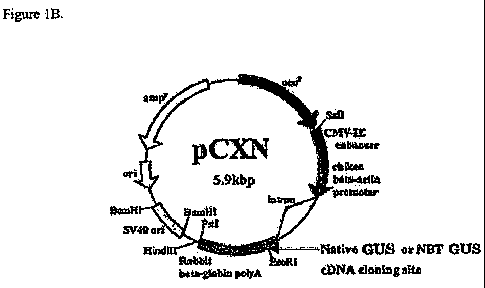
Fig. 1, A and B, is the Gus insert (A) and the mammalian expression vector
pCXN (B)
into which it was cloned (29).
Fig. 2 is a graphical representation of the data obtained in Example 2 showing
stability
data of GUS and PB-GUS at 65 C.
Fig. 3 is a graphical representation of the data obtained in Example 2 showing
stability
data of GUS and PB-GUS at 37 C in the lysosomes of human fibroblasts.
Fig. 4 is a graphical representation of data obtained in Example 3 showing the
clearance of
GUS and PB-GUS from plasma of ERT treated mice as a function of time.
Fig. 5 is a collection of photomicrographs of brain tissue of GUS- and PB-GUS-
treated
mice showing neuronal and meningeal storage of lysomal tissue after treatment
in accordance
with the procedure of Example 5.
Fig. 6 is a graphical representation of data obtained in Example 5 showing the
number of
vacuoles of lysosomal storage per 500 cortical neurons in brains of mice
treated with GUS and
PB-GUS.
SUMMARY OF THE INVENTION
Novel modified lysosomal enzymes and methods of their use in the treatment of
mammals
afflicted with LSDs have now been discovered. Such modified enzymes have
increased half-
Page 2 of 26
CA 02680189 2009-09-04
WO 2008/109677 PCT/US2008/055921
life in the circulatory system resulting in improved treatment of LSDs. Such
modification
chemically inactivates the oligosaccharides on the lysosomal enzymes thereby
inactivating
traditional recognition markers on the enzyme that mediates their rapid
clearance from the
circulation system as will be further described below.
In order to slow down the clearance of (3-glucuronidase after infusion into
the circulatory
system of a mammal, the oligosaccharides on the glycoprotein are chemically
inactivated by
treating the (3-glucuronidase sequentially with sodium-meta-periodate and
sodium
borohydride. This treatment inactivates the two traditional recognition
markers on the enzyme
that mediate its rapid clearance from the circulation by means of the mannose
and mannose 6-
phosphate receptors. This in effect increases the half-life in the circulation
from 11 minutes for
the untreated enzyme (GUS) to 18.5 h for the periodate/borohydride treated
enzyme (PB-GUS,
also known in the art as PerT-GUS). The efficacy of these enzymes was
determined in a 12-
week ERT experiment in which MPS VII mice were treated with weekly infusions
of GUS vs.
PB-GUS at doses of 0, 2mg/kg and 4 mg/kg body weight. A slight improvement was
observed
in the amount of storage material in the cortical neurons in the brains of
mice treated with 4
mg/Kg. There was a remarkable clearance of 95% of storage from the cortical
neurons in the
brains of mice treated with both 2 mg/kg and 4 mg/kg of PB-GUS. Also, there
was observed
significant continued clearance of storage material from the visceral organs
from mice treated
with both types of enzyme at both doses of 2 and 4 mg/kg body weight.
These results seem to indicate that slowing the clearance and maintaining high
concentrations of (3-glucuronidase in the circulation after infusion
facilitates delivery of the
enzyme across the BBB by some mechanism. Since the mannose and mannose 6-
phosphate
delivery systems have been inactivated as a result of the periodate treatment,
this delivery must
be mediated by some other method. One possible method would be by increased
fluid-phase
pinocytosis, a mechanism that would be greatly enhanced by maintaining high
levels of
enzyme present for long periods of time in the circulation. Whatever the
mechanism is, use of
the periodate-treated enzyme shows great promise for treating the brain in MPS
VII and any of
the other lysosomal storage diseases where there is brain pathology. This
method may also be
extended for use for other glycoproteins where rapid clearance from the
circulation by the
mannose or mannose 6-phosphate delivery systems hinders their therapeutic
effect.
Accordingly, in one aspect the invention is directed to a composition useful
in enzyme
replacement therapy, the composition comprising a lysosomal storage enzyme
treated with a
chemical to inactivate carbohydrate moieties on the enzyme, such that the
lysosomal enzyme is
Page 3 of 26
CA 02680189 2009-09-04
WO 2008/109677 PCT/US2008/055921
not readily taken up by a target cell by the mannose and mannose 6-phosphate
delivery
systems. A preferred chemical-to-inactivate is a periodate followed by
treatment with a
borohydride. A preferred MPS enzyme is 0-glucuronidase. It is preferred to
employ any
suitable alkali metal periodate and alkali metal borohydride. The preferred
alkali metal is
sodium.
In another embodiment, the invention is directed to a method of treating a
patient having a
lysosomal storage disease comprising administering to the patient a
therapeutically effective
amount of a composition comprising a medically suitable excipient and a
lysosomal enzyme
treated with a chemical to inactivate carbohydrate moieties on the enzyme,
such that the
enzyme is not readily taken-up by a target cell by the mannose and mannose 6-
phosphate
delivery systems. A preferred treatment is with a periodate followed by
treatment with sodium
borohydride. A preferred MPS enzyme is 0-glucuronidase which is effective to
treat
lysosomal storage disease preferably MPS VII (Sly syndrome).
DETAILED DESCRIPTION OF THE INVENTION
In summary, there has been discovered a means to successfully treat GUS with
periodate
and borohydride without significantly reducing the enzymatic activity or
stability. The treated
protein has been shown to have modified carbohydrate that no longer has
functional
recognition signals for mannose and mannose 6-phosphate receptors. Because of
this, the
enzyme exhibits a vastly increased half-life in the circulation after
intravenous infusion. This
increased availability results in the improved delivery of the enzyme across
the BBB by some
unknown mechanism. Whether it is increased opportunity for fluid phase
pinocytosis or some
other "leakiness", the enzyme, once it has crossed the BBB, has increased
access to cells in the
brain. It is then able to use its enzymatic activity to clear accumulated
storage material in the
cells and hopefully reverse the progression of the disease MPS VII .
While not wishing to be bound by any particular theory, the use of periodate
treated
enzyme shows great promise for treating the brain in MPS VII and any of the
other lysosomal
diseases where there is brain pathology. This method can reasonably be
extended for use with
other glycoproteins where rapid clearance from the circulation hinders their
therapeutic effect.
Any number of lysosomal enzymes are included within the scope of this
invention. Examples
of such enzymes are heparin N-sulfatase for treatment of MPS III (Sanfillipo
A),
hexosaminidase A for treatment of Tay-Sachs disease, a-L-iduronidase for
treatment of MPS I
Hurler Syndrome), palmitoyl thiotransferase (PPTI) for Batten's disease
(CLN1), a-
Page 4 of 26
CA 02680189 2009-09-04
WO 2008/109677 PCT/US2008/055921
glucosidase for Pompe disease, N-acetyl-galactosamine-6-sulfatase for MPS IVA
and 0-
galactosidase for MPS IVB (Morquio disease A and B), and N-acetylgalactosamine
4-sulfatase
for MPS VI (Maroteaux-Lamy syndrome). Other enzymes can be easily envisioned
by those
of ordinary skill in view of this disclosure and are included within the scope
of this invention.
The enzymes disclosed herein when modified in accordance with this invention
are
therapeutically effective to treat various diseases. The effective amount of
such modified
enzymes can be easily determined by simple testing. However the term
"effective amount" as
used herein is intended to mean that amount which will be therapeutically
effective to treat the
disease. Such amount is generally that which is known in the art for the use
of such enzymes
to therapeutically treat known diseases.
GENERATION OF STABLE CELL LINES SECRETING GUS
Using DNA cloning techniques, the cDNA sequence encoding the full length cDNA
for
human (3-glucuronidase was subcloned (Genbank Accession # NM_000181) (Figure
1) into the
mammalian expression vector pCXN (29). This expression vector contains an
expression
cassette consisting of the chicken beta-actin promoter coupled to the CMV
Intermediate-early
(CMV-IE) enhancer. pCXN also contains a selectable marker for G418 allowing
selection of
stably expressing mammalian cells SEQ ID NO. 1.
This plasmid was introduced into the Chinese hamster ovary cell line, CHO-
Kl(34) by
electroporation (30). After selection in growth medium consisting of Minimal
Essential
Medium + 35 g/ml proline + 15 % fetal bovine serum (FBS) + 400 g/ml G418,
colonies
were picked and grown to confluency in 48-well plates. High level expressing
clones were
identified by measuring GUS activity secreted into the conditioned medium from
these clones.
The highest-producing clone was scaled up and secreted enzyme was collected in
protein-free
collection medium PF-CHO. Conditioned medium collected in this way was pooled,
centrifuged at 5000 x g for 20 min and the supematant was collected and frozen
at 20 F until
sufficient quantities were accumulated for purification.
MEASUREMENT OF GUS ACTIVITY
GUS activity was measured using the 10 mM 4-methyl-umbelliferyl (3-D-
glucuronide as
substrate in 0.1M sodium acetate buffer pH 4.8, 1 mg/ml crystalline BSA as
previously
described(31).
PURIFICATION OF GUS
Page 5 of 26
CA 02680189 2009-09-04
WO 2008/109677 PCT/US2008/055921
(3-glucuronidase was purified by two different methods. The first method was
by a multi-
step procedure using conventional column chromatography. The second method
utilized an
anti-human 0-glucuronidase monoclonal antibody affinity resin followed by a
desalting step.
The complete procedures for both methods are outlined below.
CONVENTIONAL PURIFICATION
A: Ultrafiltration: YM-100 membrane; Diafiltrate with 20 mM NaPO4 + 150mM NaCl
+
0.025% NaN3 @ pH 5.5; (2x 2.25L).
B: Blue Sepharose FF(GE Healthcare): Equilibrate lOx column volume column with
20
mM NaPO4 @ pH 5.5; Load concentrate from ultrafiltration (don't adjust pH,
range: 5.5--5.7);
Wash lOx column volume with 20mM NaPO4 + 150mM NaCI @ pH 5.5; Elute column
with
10mM NaPO4 + 800mM NaCl @pH 7.5; Regeneration: Wash with l Ox column 20mM
NaPO4
@ pH 5.5 + 2M NaCl.
C: Phenyl Sepharose (High Sub FF): Equilibrate 30x column volume with 10mM
NaPO4+
1000mM NaCl @ pH 8.0; Load pooled blue elute as is (don't adjust pH, range:
7.2--7.4); Wash
lOx colunm volume with 10mM NaPO4+ 1000mM NaCI @ pH 8.0; Elute colunm with
10mM
Tris + 1mM Na-(3-Glycerophosphate @ pH 8.0; Dialyze elution with 3 changes of
10mM Tris
+ 1mM Na-(3-glycerophosphate @ pH 8.0; Regeneration: Wash with 0.5 M NaOH, 30
min
contact time; Wash with 30 column volumes of ddH2O.
D: DEAE Sephacel: Equilibrate lOx colurnn volume with 10mM Tris + 1mM Na-(3-
glycerophosphate @ pH 8.0; Load pooled dialyzed Phenyl elute. Wash lOx colurnn
volume
with 10mM Tris + 1mM Na-(3-glycerophosphate @ pH 8.0; Elute with 0---0.4M NaCI
gradient; Dialyze DEAE pooled eluate in 25mM Na Acetate + 1mM Na-(3-
glycerophosphate;
+ 0.025% NaN3 @ pH 5.5; Regeneration: Wash with 20x colunm volume 10mM Tris +
1mM
Na-(i-glycerophosphate @ pH 8.0 + 2 M NaCI.
E: CM Sepharose: Equilibrate 10x column volume with 25mM Na Acetate + ImM Na-
(3-
Glycerophosphate + 0.025% NaN3 @ pH 5.5; Load dialyzed DEAE pooled eluate;
Elute with
0---0.3M NaCl gradient. Regeneration: Wash with 20x colunm volume 25mM Na
Acetate +
1mM Na-(3-Glycerophosphate + 0.025% NaN3 @ pH 5.5 + 2M NaCI.
MONOCLONAL PURIFICATION
Page 6 of 26
CA 02680189 2009-09-04
WO 2008/109677 PCT/US2008/055921
Affinity chromatography procedure was performed essentially as follows:
Conditioned
medium from CHO cells overexpressing the GUS protein was filtered through a
0.22 filter.
Sodium chloride (crystalline) was added to a final concentration of 0.5M, and
sodium azide
was added to a final concentration of 0.025% by adding 1/400 volume of a 10%
stock solution.
The medium was applied to a 5 nil column of anti-human (3-glucuronidase-
Affigel 10 (pre-
equilibrated with Antibody Sepharose Wash Buffer: 10 mM Tris pH 7.5, 10 mM
potassium
phosphate, 0.5 M NaCl, 0.025% sodium azide) at a rate of 25 ml/h at 4 C. The
column was
washed at 36 ml/h with 10-20 column volumes of Antibody Sepharose Wash Buffer.
The
colunm was eluted at 36 ml/hour with 50 ml of 10 mM sodium phosphate pH 5.0 +
3.5 M
MgC12. Fractions of 4 ml each were collected and assayed for GUS activity.
Fractions
containing the purifed protein were pooled, diluted with an equal volume of P6
buffer (25 mM
Tris pH 7.5, 1 nilvl (3-glycerophosphate, 0.15 mM NaC1, 0.025% sodium azide)
and desalted
over a BioGel P-6 colunm (pre-equilibrated with P6 buffer) to remove the MgC12
and to
change the buffer to P6 buffer for storage. GUS protein was eluted with P6
buffer, fractions
containing GUS activity were pooled and the final pool assayed for GUS
activity and protein.
Purified GUS was stored frozen at -80 C in P6 buffer for long-tenn stability.
For mouse
infusions, the enzymes were highly concentrated in Centricon YM-30
concentrators and the
buffer was changed to P6 Buffer without azide. These concentrates were frozen
in small
aliquots at -80 C until use.
CHARACTERIZATION OF PURIFIED GUS.
GUS is a 300 kDa protein that exists as a homotetramer consisting of four
identical
monomers of apparent molecular weight of 75 kDa. The purified recombinant GUS
used in
these experiments was similar to that described (11, 19). The apparent
molecular mass of the
enzyme monomer was 75 kDa on reducing SDS-PAGE. The tetrameric enzyme had a
molecular mass of z300 kDa when analyzed by sizing gel filtration
chromatography (data not
shown). The specific activity of the purified enzyme was 5.0 x 10⁶ units/mg.
The KõPtAe was
1.25-2.50 nM, calculated from uptake saturation curves by using human MPS VII
fibroblasts in
which the uptake is almost entirely M6PR-dependent. To confirm molecular
weight, 2 and 4
g of purified GUS were analyzed by SDS-PAGE under reducing conditions (35).
The
apparent molecular weight was 75 kDa as expected.
The following examples are presented to illustrate the instant invention and
are not meant
to limit the scope of the invention to these particular examples. The skilled
artisan, in the
Page 7 of 26
CA 02680189 2009-09-04
WO 2008/109677 PCT/US2008/055921
practice of this invention, will readily and reasonably understand that the
methods and
compositions are applicable to any and all enzymes and proteins that gain
entry into a cell via
the mannose and mannose 6-phosphate pathways.
EXAMPLE 1
TREATMENT OF PURIFIED GUS WITH PERIODATE AND BOROHYDRIDE.
The mannose and manose 6-phosphate recognition sites on GUS are both located
in the
carbohydrate portion of GUS enzyme. In order to inactivate this carbohydrate
moiety, the
enzyme was treated by a well established procedure utilizing reaction with
sodium meta-
periodate followed by sodium borohydride(17, 18). Approximately 10 mg of
purified GUS was
treated with a final concentration of 20 mM sodium meta-periodate in 20 mM
sodium
phosphate, 100 mM NaC1 pH 6.0 for 6.5 h on ice in the dark. The reaction was
quenched by
the addition of 200 mM final concentration ethylene glycol and incubated for
an additional 15
min on ice in the dark. Afterwards, this mixture was dialyzed against 2
changes of 20 mM
sodium phosphate, 100 mM NaCI pH 6.0 at 4 C. The periodate treated, dialyzed
enzyme was
then treated with the addition of 100 mM final concentration sodium
borohydride ovemight on
ice in the dark to reduce reactive aldehyde groups. After this treatment, the
enzyme was
dialyzed against two changes of 20 mM sodium phosphate, 100 mM NaC1, pH 7.5 at
4 C. The
final dialyzed enzyme was stored in this buffer at 4 C where it was stable
indefinitely.
CHARACTERIZATION OF THE PERIODATE AND BOROHYDRIDE TREATED GUS.
Treatment of GUS with periodate and borohydride resulted in only a slight
inactivation of
the enzymatic activity. The specific activity prior to treatment was 5.0 x 10⁶
units/mg and
following treatment was 4.5 x 10⁶ units/mg.
To assess the effectiveness of the periodate and borohydride treatment in
inactivating the
carbohydrate on the enzyme, the ability of the enzyme to be taken up by human
P-
glucuronidase deficient fibroblasts or by the permanent J774E mouse macrophage
line was
analyzed. M6PR-mediated uptake was determined by adding 4,000 units of GUS or
PB-GUS
2 mM M6P in 1 ml of growth medium to 35-mm dishes of confluent GM-2784 GUS-
deficient
fibroblasts. After incubation at 37 C and 5% CO2 for 2 h, the cells were
cooled on ice, washed
five times with cold PBS, then solubilized in 0.5 ml of 1% sodium
deoxycholate. Extracts were
Page 8 of 26
CA 02680189 2009-09-04
WO 2008/109677 PCT/US2008/055921
assayed for GUS activity and protein. Values were expressed as units of enzyme
taken up per
mg of cell protein per hour of uptake.
MR-mediated uptake was measured by adding 10,000 units of GUS or PB-GUS 1.7
mg/ml yeast mannan (Sigma-Aldrich) in 1 ml of growth medium to 35-mm dishes of
confluent
J774E mouse macrophages (33). After incubation at 37 C and 5% COz for 4 h, the
cells were
washed as above and then solubilized in 1 ml of 1% sodium desoxycholate and
assayed for
GUS activity.
Table 1 below shows the M6P-receptor mediated uptake of untreated or mock-
treated
GUS by the human fibroblast cell line. GUS is taken up by this line at the
rate of 377 units/mg
cell protein/1 h of uptake. Two mM M6P completely inhibits this uptake. In
contrast, the
uptake of the periodate and borohydride treated GUS(PBGUS) has been completely
destroyed.
Table 2 below shows that untreated GUS is taken up by the mouse macrophage
line at a rate of
316 u/mg cell protein/1 h of uptake and the uptake is inhibited by the
presence of 1.69 mg/ml
yeast mannan. In contrast, three separate batches of periodate and borohydride
treated
GUS(PBGUS) have essentially no uptake by this cell line.
Page 9 of 26
CA 02680189 2009-09-04
WO 2008/109677 PCT/US2008/055921
TABLE 1
FIBROBLAST UPTAKE ON HBG 5-6 +/- PERIODATE AND BOROHYDRIDE
TREATMENT
Uptake M6P-Specific Uptake
Condition u/mg/lh u/mg/lh
GUS 380 377
GUS + 2mM M6P 3 ---
GUS Mock Treated 363 359
GUS Mock Treated+2mM M6P 3.5 ---
PB-GUS Periodate&Borohydride Treated 1 0
PB-GUS Periodate&Borohydride Treated + 2mM M6P 1 ---
TABLE 2
J774E MACROPHAGE UPTAKE ON HBG 5-6 +/- PERIODATE AND
BOROHYDRIDE TREATMENT
Uptake Man-Specific Uptake
Condition u/mg/lh u/mg/lh
GUS 366 316
GUS + 1.69 mgJml Yeast Mannan 50 ---
PB-GUS 8 3
PB-GUS + Yeast Mannan 5 ---
Page 10 of 26
CA 02680189 2009-09-04
WO 2008/109677 PCT/US2008/055921
PB-GUS 11 2
PB-GUS B34E + Yeast Mannan 9 ---
PB-GUS 12 0
PB-GUS + Yeast Mannan 21 ---
Since both mannose 6-phosphate and mannose receptor mediated uptake are
dependent on
functional mannose 6-phosphate or mannose residues, respectively, these
results indicate that
the periodate and borohydride treatment of GUS (PB-GUS) has inactivated the
carbohydrate
structures on the enzyme.
EXAMPLE 2
STABILITY OF NATIVE GUS OR PB-GUS
The carbohydrates on glycoproteins often confer enhanced thermal stability,
and removal
of oligosaccharide chains often destabilizes glycoproteins (21). Human GUS has
been shown
to be relatively stable to thermal inactivation at 65 C (22-26). Purified GUS
or PB-GUS was
diluted in equal volumes of heat inactivation buffer [40 mM Tris-HCI (pH 7.5),
150 mM NaCI,
10 mg/ml BSA], and aliquots were incubated for 0, 0.5,1, 2, or 3 h at 65 C.
After treatment,
aliquots were cooled on ice and then assayed for GUS activity. Results were
expressed as the
percentage of original units of GUS activity remaining at the indicated times.
As shown in Fig.
2, recombinant GUS retained 90% of initial activity after 3 h at 65 C, whereas
PB-GUS
retained 40% of its activity under these conditions (Fig. 2).
To compare the stability of GUS and PB-GUS in lysosomes of living cells at 37
C, a
study was conducted to determine their half-life after uptake by MPS VII
fibroblasts. The low
rate of endocytosis of PB-GUS by fibroblasts required exposure to 100,000
units/ml PB-GUS
per plate for 48 h to accumulate sufficient enzyme by fluid phase pinocytosis
(28 units per
plate) to allow measurement of its half-life. By contrast, fibroblasts exposed
to 500 units/ml
M6P containing native GUS for 48 h contained 228 units per plate. Tissue
culture dishes (35
Page 11 of 26
CA 02680189 2009-09-04
WO 2008/109677 PCT/US2008/055921
mm) of confluent GM-2784 GUS-deficient fibroblasts were incubated with 500
units of GUS
or 100,000 units of PB-GUS in 1 ml of growth medium at 37 C and 5% CO2 for 48
h under
sterile conditions. The plates were washed twice with sterile growth medium
and then fed with
2 ml of the same. Duplicate plates were taken off at 0, 2, 5, 7,14, and 21
days, washed five
times with PBS and frozen at -20 C. Remaining plates were fed twice weekly
with 2 ml of
growth medium. After all plates had been collected, the cells were solubilized
in 0.5 ml of 1%
desoxycholate and assayed for GUS activity. Values were expressed as
percentage of zero time
cell-associated GUS activity remaining at the indicated time points. Fig. 3
shows the half-life
for the two enzymes in fibroblasts upon subsequent incubation at 37 C. The tõz
of GUS was
18.9 days. The tõZ of PB-GUS was shorter (12.9 days), but nearly one-third of
the initial
activity was still present at 21 days.
EXAMPLE 3
CLEARANCE OF THE PERIODATE AND BOROHYDRIDE TREATED GUS
FROM THE CIRCULATION AFTER IV INFUSION.
As stated previously, the purpose of treating GUS with periodate and
borohydride, was to
drastically slow its clearance time from the circulation after infusion. To
test this, the tail veins
of MPS VII niice were infused with GUS or PB-GUS at a dose of 4 mg/kg body
weight in a
total volume of 125 l11 of PBS. After infusion, blood samples were taken by
supraorbital
puncture at 2, 5, 10, 20, 60, 90, and 120 min for GUS and 4, 240, 1,440, and
2,880 min for PB-
GUS into heparinized capillary tubes. Plasma was collected after
centrifugation and assayed
for GUS activity. Values were expressed as a percentage of GUS activity
remaining compared
with the first time point. Fig. 4 and Table 3 below show the results of that
clearance study. As
can be seen, the clearance of untreated GUS is very rapid with a tõZ of 11.7
min. In contrast,
the clearance of PB-GUS in four separate mice was drastically slower with a
tõz of 18.5f 1.0 h.
This would indicate that the rapid clearance of this enzyme due to the mannose
and mannose 6-
phosphate receptor (15) has been abrogated.
Page 12 of 26
CA 02680189 2009-09-04
WO 2008/109677 PCT/US2008/055921
TABLE 3
CLEARANCE OF GUS AND PB-GUS FROM THE CIRCULATION OF EAM MICE
AFTER INFUSION WITH 4 MG/KG ENZYME
GUS PB-GUS #1 PB-GUS #2 PB-GUS #3 PB-GUS #4
Min. u/ml % u/ml % u/ml % u/ml % u/ml %
2 261,440 100
4 --- --- 318,960 100 228,240 100 285,120 100 369,120 100
174,720 67
73,920 28
11,200 4.3
60 640 0.2
90 0 0
120 0 0
240 177,840 56 147,960 65 176,640 62 225,120 61
1440 75,240 24 64,440 28 68,640 24 94,080 25
2880 21,660 6.8 29,520 12.9 33,120 11.6 41,280 11.1
t,n 11.7 min 1022 min 1195 min 1119 min 1114 min
0.2h 17.Oh 19.9h 18.6h 18.6h
Mean=1113f61min
18.5 t 1.0 h
EXAMPLE 4
5 TISSUE DISTRIBUTION OF GUS vs. PB-GUS
Previously, the plasma clearance of the enzyme was observed to be slowed when
treating
MPS VII mice with high-dose GUS and facilitated enzyme delivery to the brain
(11). In these
experiments, it was not clear whether it was the higher dose of enzyme itself
or the delayed
plasma clearance of the enzyme that accounted for improved delivery to brain.
To address this
10 question, comparative measurements were made of the distribution of GUS and
PB-GUS in
brain and other tissues 48 h after infusion into MPS VII mice. Mice were
perfused with Tris-
buffered saline before collection of tissues to ensure that tissue was not
contaminated with
residual plasma enzyme. MPS VII mice were infused via tail vein with GUS or PB-
GUS at a
Page 13 of 26
CA 02680189 2009-09-04
WO 2008/109677 PCT/US2008/055921
dose of 4 mg/kg in a total volume of 125 l of PBS. At 48 h after infusion,
the mice were
perfused with 30 ml of 25 mM Tris (pH 7.2), 140 mM NaC1. Perfused tissues were
collected
and flash frozen in liquid nitrogen until further processing. Tissues were
thawed, weighed, and
homogenized for 30 s with a Polytron homogenizer in 10-20 volumes of 25 mM
Tris (pH 7.2),
140 mM NaCl, 1 mM phenylmethylsulfonyl fluoride. Total homogenates were frozen
at -80 C,
thawed, and then sonicated for 20 s to produce a homogeneous extract. Extracts
were assayed
for GUS activity and protein, and the results were expressed as
units/milligrams of tissue
protein. The results of these measurements appear in Table 4 below.
TABLE 4
DISTRIBUTION IN BRAIN AND TISSUE OF GUS AND PB-GUS
Wild-type GUS PB-GUS
levels* (4 mg/kg)t (4 mg/kg)
issue (n = 4) (n = 2) (n = 3)
rain 16.7 2 0.23 0.005 1.3010.28
iver 185 f 11.9 892 f 45.5 230 f 63
Spleen 301 f 26.6 558 f 54 122 f 51
eart 20.8 12.5 13.0 1.8 44.1f16.3
'dney 108 7.5 11.9 0.19 21.7 3.6
ung ND 5.1 f0.4 19.9 6.1
4uscle 4.95 1.80 1.2 0.07 6.3 3.5
3one+marrow 161f35 75.6 17 59.5124.8
ye 4.88 0.68 0.90 0.52 4.9 1.5
As is evident from the data in Table 4, delivery of native GUS to brain at 48
h was
minimal. However, native GUS was delivered to other tissues at levels similar
to those
previously reported. PB-GUS was delivered to heart, kidney, muscle, lung, and
eye at levels
higher than those seen with native GUS. The levels in liver and spleen were
nearly 4-fold
lower after PB-GUS infusion than after GUS infusion. This result undoubtedly
reflects the
curtailment of receptor-mediated uptake by the MPR and M6PR that are highly
expressed in
these two tissues. By contrast, brain levels were greatly increased (7.8% of
wild-type) in PB-
GUS-infused animals. This result suggests that the long circulating PB-GUS has
an advantage
in crossing the BBB. Thus, it was of great interest to study its effectiveness
in clearing storage
from cells in the CNS.
Page 14 of 26
CA 02680189 2009-09-04
WO 2008/109677 PCT/US2008/055921
EXAMPLE 5
COMPARISON OF THE EFFICACY OF PERIODATE/ BOROHYDRIDE TREATED GUS FOR ERT IN
CLEARING NEURONAL STORAGE.
As stated previously, it was believed that slowing the clearance of GUS from
the
circulation might facilitate the delivery to the brain. It has been shown
above that the periodate
and borohydride treatment accomplished this producing an enzyme with a much
reduced rate
of clearance from the circulation after IV infusion. The effectiveness of the
treated enzyme in
clearing the storage material from the lysosomes of the MPS VII mouse after a
typical ERT
regimen was tested. MPS VII mice were treated with 12 weekly infusions, one
group with
untreated GUS at doses of 2 or 4 mg/kg body weight and a second group with PB-
GUS at
doses of 2 or 4 mg/kg body weight. Two other groups of MPS VII mice were
infused two
times daily for I week with a total of 48mg/kg, one group with GUS and one
group with PB-
GUS. One week after the last infusion, tissues from the group receiving
untreated GUS (n =
3), 2 mg/kg (n = 3) or 4 mg/kg GUS (n = 2), and PB-GUS, 2 mg/kg (n = 2) or 4
mg/kg (n = 3)
were obtained at necropsy after Tris-buffered saline perfusion, fixed in 2%
paraformaldehyde
and 4% glutaraldehyde, post fixed in osmium tetroxide, and embedded in Spurr's
resin. For
evaluation of lysosomal storage by light microscopy, toluidine blue-stained
0.5- m-thick
sections of liver, spleen, kidney, brain, heart, rib, and bone marrow were
assessed blind. To
evaluate storage in cortical neurons, 500 contiguous parietal neocortical
neurons were scored
for the number of lucent cytoplasmic vacuoles, indicating lysosomal storage. A
maximum of
seven vacuoles were counted per cell, and results were evaluated by ANOVA or
Student's t
test. Also evaluated were the hippocampal neurons by counting the number of
vacuoles in 100
neurons in CA2 sector. Other tissues were examined by using a semiquantitative
scale, as
described in ref. 11.
As can be seen in Figure 5, GUS results in a slight reduction of the storage
material in the
brain whereas PB-GUS results in almost complete reversal of the storage. This
would indicate
that the periodate and borohydride treated GUS was vastly more effective in
treating the brain
storage in this disease.
In Fig. 5, reduction in neuronal and meningeal storage with ERT with GUS and
PB-GUS
is shown as follows: (A) Neocortical neurons from an untreated MPS VII mouse
have
abundant lysosomal storage in the cytoplasm (arrow). (B) After treatment with
4 mg/kg GUS,
Page 15 of 26
CA 02680189 2009-09-04
WO 2008/109677 PCT/US2008/055921
there is still a moderate amount of cytoplasmic storage (arrow) despite the
therapy. (C) After 4
mg/kg PB-GUS, there is a marked reduction in the amount of storage in the
neocortical
neurons (arrow). (D) The CA2 sector hippocampal neurons have abundant storage
(arrow) in
untreated MPS VII mice. (E) After treatment with GUS, the amount of storage in
neurons
(arrow) the same area of the hippocampus is similar to that of the untreated
mouse. (F) After
treatment with PB-GUS, there is a remarkable reduction in the amount of
storage in neurons
(arrow) in the CA2 sector of the hippocampus. (G) The meninges of an untreated
MPS VII
mouse has abundant storage in fibroblasts around vessels (arrow). (H) Storage
(arrow) is
moderately decreased after treatment with GUS. (I) Treatment with PB-GUS also
produces
moderate reduction in storage (arrow) in the meninges. [Scale bars: 10 m (A-
C, uranyl
acetate-lead citrate) and 30 m (D-I, toluidine blue).]
Two of the problems associated in the analysis of micrographs for the
clearance of storage
material in these types of experiments are: 1) that there is some
inconsistency from field to
field i.e. the clearance varies from one microscopic field to another; and 2)
the procedure is
somewhat subjective from person to person as to the amount of storage present.
To address
these problems, a new method was developed to quantify the storage material by
counting the
number of vacuoles (distended lysosomes filled with storage material) present
in a total of 500
cells counted. Fig. 6 shows the results of such an analysis of the mice
treated with GUS or PB-
GUS.
GUS at 2 mg/kg is not very effective at reducing the number of vacuoles,
though
somewhat better at the higher dose of 4 mg/kg. However, PB-GUS appears to be
almost
completely effective at both 2 and 4 mg/kg. This analysis agrees with the
conclusion drawn
from the visual analysis of the images in Fig. 5.
Table 5 below summarizes the results of assessment of storage in neocortical
and
hippocampal neurons of untreated GUS and PB-GUS in MPS VII mice. ERT with GUS
over
12 weeks with both 2 mg/kg and 4 mg/kg GUS reduced storage in neocortical
neurons
compared with untreated MPS VII mice (P = 0.002 and P= 0.003, respectively),
although
hippocampal neurons failed to show a morphological response to this therapy.
PB-GUS at 2
mg/kg also reduced neocortical neuronal storage (P = 0.001). At 4 mg/kg, the
therapeutic effect
of PB-GUS was even more striking (P = 0.003 for 2 vs. 4 mg of PB-GUS and P<
0.001
compared with untreated). In addition, there was virtually no storage in the
hippocampal
neurons in the three PB-GUS-treated mice available for quantitation (the CA2
region was not
present in the section and was therefore not available for quantitation in two
of the five PB-
Page 16 of 26
CA 02680189 2009-09-04
WO 2008/109677 PCT/US2008/055921
GUS-treated mice). These results indicate that ERT with PB-GUS is remarkably
more
effective than traditional GUS at clearing storage in the neocortical and
especially hippocampal
neurons in the MPS VII mouse. As a group, the PB-GUS-treated mice also had
slightly less
storage in glial and perivascular cells than the GUS-treated mice. However,
the dose-dependent
reduction in storage in meninges, which was moderate at 4 mg/kg, was
equivalent in the PB-
GUS- and the GUS-treated animals.
From the above results it is reasonable to expect that treatment of mammalian
species in
accordance with this invention will provide relief of lysosomal storage
disease, particularly in
humans particularly in the brain of humans.
TABLE 5
QUANTITATION OF LYSOSOMAL STORAGE IN NEURONS IN
CONTROL AND TREATED MPS VII MICE
Vacuoles per 500 cells
Neocortical Hippocampal
Treatment neurons neurons
Control MPS VII 1,956 692
1,685 694
1,927
GUS 2 mg/kg 728 641
744 674
1,088
GUS 4 mg/kg 1,274 642
1,213
B-GUS 2 mg 403 2
439
B-GUS 4 mg 73 0
148 5
Page 17 of 26
CA 02680189 2009-09-04
WO 2008/109677 PCT/US2008/055921
72
The following references are cited throughout this disclosure and are herein
incorporated
by reference. They are meant to illustrate and support the invention.
Applicants reserve the
right to challenge the veracity of any statements made therein.
1. Neufeld EF, Muenzer J (2001) in The Metabolic and Molecular Bases of
Inherited
Disease, eds Scriver CR, Beaudet AL, Sly WS, Valle D(McGraw-Hill, New York),
pp 3421-
3451.
2. Desnick RJ (2004) Enzyme replacement and enhancement therapies for
lysosomal
diseases. Jlnherit Metab Dis 27:385-410.
3. Brady RO, Barton NW (1996) Enzyme replacement and gene therapy for
Gaucher's
disease. Lipids 31:S137-S139.
4. Kakkis ED, et al. (2001) Enzyme-replacement therapy in
mucopolysaccharidosis I. N
Engl JMed 344:182-188.
5. Harmatz P, et al. (2006) Enzyme replacement therapy for
mucopolysaccharidosis VI: A
phase 3, randomized, double-blind, placebo-controlled, multinational study of
recombinant
human N-acetylgalactosamine 4-sulfatase (recombinant human arylsulfatase B or
rhASB) and
follow-on, open-label extension study. JPediatr 148:533-539.
6. Brady RO (2006) Enzyme replacement for lysosomal diseases. Annu Rev Med
57:283-
296.
7. Vogler C, et al. (2001) Murine mucopolysaccharidosis VII: Impact of
therapies on the
phenotype, clinical course, and pathology in a model of a lysosomal storage
disease. Pediatr
Dev Pathol 4:421-433.
8. Vogler C, et al. (1993) Enzyme replacement with recombinant p-glucuronidase
in the
newborn mucopolysaccharidosis type VII mouse. Pediatr Res 34:837-840.
9. Vogler C, et al. (1996) Enzyme replacement with recombinant p-glucuronidase
in
murine mucopolysaccharidosis type VII: Impact of therapy during the first six
weeks of life on
subsequent lysosomal storage, growth, and survival. Pediatr Res 39:1050-1054.
Page 18 of 26
CA 02680189 2009-09-04
WO 2008/109677 PCT/US2008/055921
10. Urayama A, Grubb JH, Sly WS, Banks WA (2004) Developmentally regulated
mannose 6-phosphate receptor-mediated transport of a lysosomal enzyme across
the blood-
brain barrier. Proc Natl Acad Sci USA 101:1 26 58-1 2663.
11. Vogler C, et al. (2005) Overcoming the blood-brain barrier with high-dose
enzyme replacement therapy in murine mucopolysaccharidosis VII. Proc Natl Acad
Sci USA
102:14777-14782.
12. Dunder U, et al. (2000) Enzyme replacement therapy in a mouse model of
aspartylg-lycosaminuria. FASEB J 14:361-367.
13. Roces DP, et al. (2004) Efficacy of enzyme replacement therapyin a-
mannosidosis mice: A preclinical animal study. Hum Mol Genet 13:1979-1988.
14. Matzner U, et al. (2005) Enzyme replacement improves nervous system
pathology and function in a mouse model for metachromatic leukodystrophy. Hum
Mol Genet
14:1139-1152.
15. Sly WS, et al. (2006) Enzyme therapy in mannose receptor-null
mucopolysaccharidosis VII mice defines roles for the mannose 6-phosphate and
mannose
receptors. Proc Natl Acad Sci USA 103:1 5172-15177.
16. Ashwell G, Harford J(1982) Carbohydrate-specific receptors of the liver.
Annu
Rev Biochem 51:531-554.
17. Hickman S, Shapiro LJ, Neufeld EF (1974) A recognition marker required for
uptake of a lysosomal enzyme by cultured fibroblasts. Biochem Biophys Res
Commun 57:55-
61.
18. Achord DT, Brot FE, Bell CE, Sly WS (1978) Human p-glucuronidase: In vivo
clearance and in vitro uptake by a glycoprotein recognition system on
reticuloendothelial cells.
Cell 15:269-278.
19. LeBowitz JH, et al. (2004) Glycosylation-independent targeting enhances
enzyme delivery to lysosomes and decreases storage in mucopolysaccharidosis
type VII mice.
Proc Natl Acad Sci USA 101:3083-3088.
20. Houba PH, Boven E, Haisma HJ (1996) Improved characteristics of a human p-
glucuronidase-antibody conjugate after deglycosylation for use in antibody-
directed enzyme
prodrug therapy. Bioconjug Chem 7:606-611.
Page 19 of 26
CA 02680189 2009-09-04
WO 2008/109677 PCT/US2008/055921
21. Wang C, Eufemi M, Turano C, Giartosio A(1996) Influence of the
carbohydrate
moiety on the stability of glycoproteins. Biochemistry 35:7299-7307.
22. Frankel HA, Glaser JH, Sly WS (1977) Human p-glucuronidase. 1. Recognition
and uptake by animal fibroblasts suggests animal models for enzyme replacement
studies.
Pediatr Res 11:811-816.
23. Achord D, Brot F, Gonzalez-Noriega A, Sly W, Stahl P(1977) Human p-
glucuronidase. II. Fate of infused human placental p-glucuronidase in the rat.
Pediatr Res
11:816-822.
24. Brot FE, Bell CE, Jr, Sly WS (1978) Purification and properties of p-
glucuronidase from human placenta. Biochemistry 17:385-391.
25. Wu BM, et al. (1994) Over expression rescues the mutant phenotype of L176F
mutation causing p-glucuronidase deficiency mucopolysaccharidosis in two
Mennonite
siblings. JBiol Chem 269:23681-23688.
26. Natowicz MR, Chi MM, Lowry OH, Sly WS (1979) Enzymatic identification of
mannose 6-phosphate on the recognition marker for receptor-mediated
pinocytosis of p-
glucuronidase by human fibroblasts. Proc Natl Acad Sci USA 76:4322-4326.
27. Schlesinger PH, et al. (1980) The role of extrahepatictissues in the
receptor-
mediated plasma clearance of glycoproteins terminated by mannose or N-
acetylglucosamine.
Biochem J 192:597-606.
28. Banks WA (2004) Are the extracellular [correction of extracelluar]
pathways a
conduit for the delivery of therapeutics to the brain? Curr Pharm Des 10:1365-
1370.
29. Niwa H, Yamamura K, Miyazaki J(1991) Efficient selection for high-
expression transfectants with a novel eukaryotic vector. Gene 108:193-199.
30. Ulmasov B, et al. (2000) Purification and kinetic analysis of recombinant
CA
XII, a membrane carbonic anhydrase over expressed in certain cancers. Proc
Natl Acad Sci
USA 97:14212-14217.
31. Glaser JH, Sly WS (1973) p-Glucuronidase deficiency mucopolysaccharidosis:
Methods for enzymatic diagnosis. JLab Clin Med 82:969-977.
32. Lowry OH, Rosebrough NJ, Farr AL, Randall RJ (1951) Protein measurement
with the folin phenol reagent. JBiol Chem 193:265-275.
Page 20 of 26
CA 02680189 2009-09-04
WO 2008/109677 PCT/US2008/055921
33. Diment S, Leech MS, Stahl PD (1987) Generation of macrophage variants with
5-aza-cytidine: Selection for mannose receptor expression. JLeukocyte Biol
42:485-490.
34. Chinese Hamster Ovary Cell Line American Type Culture Collection, ATCC
CRL 9618.
35. Laemmli, U.K.,(1970) Nature(London) 2'27, 680-685.
Page 21 of 26