Note: Descriptions are shown in the official language in which they were submitted.
CA 02680765 2009-09-14
WO 2008/114065 PCT/GE2008/000003
A DEVICE TO DETECT MALIGNANT PROCESSES IN LIVING ORGANISMS
The invention relates to electronics and biomedicine and it can be used to
evaluate some charac-
teristics of dynamic systems, e.g. to detect (or exclude) malignant processes
in organism via gas-
discharge imaging of a body parts under certain fitted conditions and further
computerized proc-
essing of the images so obtained.
A known device [RU 2110824, prior date 26.11.97. Gas discharge visualization
device. MKI
G03B41/00; International conventional application PCT/RU97/00376, application
analog RU
2110824 and patents AU, BR. Gas discharge visualization device; WO/1999/030612
Method for
determining the energy-information characteristics of a biological object;
WO/2003/053240
Method for diagnosis of human organism; WO/2004/075752) Method for determining
the anxi-
ety level of a human being] comprises a transparent electrode for generating
an electrical field,
dielectric for insulating an object under examination, and a television camera
both the dielectric
and the electrode being made of an optically transparent material the
electrode being positioned
between the dielectric and the television camera.
The shortcoming of the said device is that some spurious light interferes with
gas-discharge im-
age of an object under examination, the light being reflected from the surface
of the latter. Spuri-
ous light is superimposed on emission of the object resulting in poorer
contrast of image that
complicates further computer processing and decreases the reproducibility rate
of results.
Another shortcoming is application of a television camera that fixes frames in
an interlaced
mode. In fact, the frames so obtained contain two halves of an interlaced
field deteriorating qual-
ity of a rapidly altering gas-discharge image of the object under examination.
The gas-discharge glow, which takes place around objects in case they contact
directly with the
surface of rigid dielectric layer, is changing under the influence of numerous
sometimes uncon-
trollable factors like ambient temperature or humidity, or object's variable
pressing force on the
electrode, etc. Also, some functional processes (e.g., psycho-emotional state,
neuro-hormonal
dysfunctions, perspiration and the like) influence the gas-discharge image in
case human finger-
tips are pressed to the free surface of rigid dielectric layer. It is not
likely to be possible to extract
most stable and diagnostically comprehensive components out of a "mixed"
picture acquired in
the above-described conditions.
CA 02680765 2009-09-14
WO 2008/114065 PCT/GE2008/000003
Engineering Result of the Invention
The engineering result of the invention is in detecting an emission in the
range of relatively high
frequencies to evaluate the synchronicity of system processes in the said
range via recording its
superficial emission that might be also used for the detection of malignant
processes in living
systems. This result is achieved by way of clearing and contrasting gas-
discharge images under
conditions of discretely altered external electrical field as well as in
improving image quality and
its reproducibility rate while using some additional (transparent and
nontransparent) layers of
flexible dielectric membranes.
Nature of the Invention
A device for gas-discharge imaging to detect those asynchronous processes that
belong to the
range of high frequencies and are characteristic for malignant processes in
living organisms
comprises an optically transparent electrode to generate an electric field, a
dielectric and an im-
age-receiving television camera wherein a dielectric being capable to be
mounted on an optically
transparent electrode comprises three successive layers, each one in contact
with the adjacent
one: a rigid transparent electrically-insulating layer, a flexible transparent
membrane positioned
on the free surface of the rigid one to improve the quality and stability of
imaging results and a
dark opaque flexible membrane positioned on the free surface of the flexible
transparent mem-
brane to absorb spurious light reflected from the surface of a body part.
An image-receiving progressive-scan monochrome camera is mounted beneath the
optically
transparent electrode to form a continuous analog video signal to be sent to a
computer for fur-
ther processing. This camera can be optionally replaced with a digital
photographic camera.
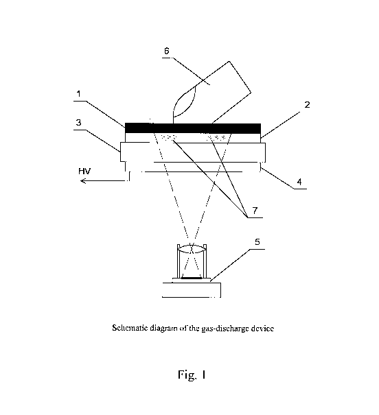
The gas-discharge device comprises a dielectric being capable to be mounted on
an optically
transparent electrode (4), which in turn is a part of a detecting device the
function of which being
to detect malignant processes of a living organism; the dielectric comprises
three successive lay-
ers, each one in contact with the adjacent one: a rigid transparent
electrically-insulating layer (3)
capable to be arranged in surfacing contact with the optically transparent
electrode (4), a flexible
transparent membrane (2) positioned on the free surface of the rigid
transparent electrically-
insulating layer (3) and a dark opaque flexible membrane (1) positioned on the
free surface of
the flexible transparent membrane (2), the free surface of such dark opaque
membrane (1) being
capable to allow contact with a part of a living organism.
An image-receiving camera (5) is mounted under the optically transparent
electrode (4) [Fig.1].
2
CA 02680765 2009-09-14
WO 2008/114065 PCT/GE2008/000003
Device operation is as follows: pulses of high voltage (HV) are applied onto
electrode (4) to
cause gas-discharge glow around the object under examination (6) in planes of
both flexible
transparent membrane (2) and an opaque one (1). The light passes through both
transparent di-
electric (3) and electrode (4) and is then projected onto light-sensitive
matrix of camera (5). Flux
of light directed towards image-receiving camera passes through transparent
insulating dielectric
(3) to reach camera without a hindrance while the light flux directed opposite
is absorbed by the
dark surface of opaque membrane (1). Thus, camera (5) only records a "pure"
discharge picture
free of reflected light beams.
A human finger or any other object is placed onto the surface of opaque
flexible membrane (1).
Pulses from high voltage (HV) generator are sent to transparent electrode (4)
causing both ioni-
zation and discharge of air that surrounds the exposed object, the latter
being pressed to the sur-
face of opaque membrane, while image-receiving camera captures the image and
passes it to
computer using image-capturing software. Gas-discharge imaging is carried out
in the absence of
bright ambient light (an object to be imaged is covered with a dense cloth or
a special cover). A
camera-activating signal also switches on high-voltage generator by means of
computer pro-
gram. The same software controls the entire process of imaging and converts
the image into digi-
tal format whenever necessary. Subsequent processing and analysis of recorded
images is done
by another software upon completion of imaging session.
The device described above makes it possible to obtain information on the mode
of dynamic sys-
tem functioning and to evaluate the degree of synchronicity in the range of
relatively high fre-
quencies via assessment superficial emission of the system. In case malignant
tumor is present in
a body, some processes of high frequency tend to dominate over relatively slow
ones thus alter-
ing the spectrum of superficial emission and affecting, correspondingly, gas-
discharge radiation
of body parts.
In particular, gas-discharge imaging of 10 human fingertips under conditions
of discrete altera-
tion of electrical field (within 1-5 kilohertz) enables to determine an
approximate projec-
tion/location of those areas, where high-frequency components of superficial
emission differ
from background radiation of entire body. Both gradual alteration of
electrical field and the pro-
cedure of video recording are controlled by computer means in such a way that
time spans are
maintained precisely both between switches of electric field and also between
captures of
frames. Precise time-control is crucial while comparing the character of a
system's emission in
various ranges of frequencies.
3
CA 02680765 2009-09-14
WO 2008/114065 PCT/GE2008/000003
External view of the device that is nowadays used for clinical work is
presented on figure 2. The
proper position of an examinee's hand is also shown.
The probability of malignant (dangerous) pathology and its approximate
location in the body are
presently calculated using specified software that enables to display
parametric data and results
of their evaluation in graphic or/and text-forms. Captured images of gas-
discharge glow of a per-
son's ten fingertips are shown on figure 3 along with results of their
evaluation.
As numerous authors have demonstrated, the intensity and character of natural
or induced emis-
sion differ in malignant cells comparing to non-malignant ones [United States
Patent 5131398 -
Method and apparatus for distinguishing cancerous tissue from benign tumor
tissue, benign tis-
sue or normal tissue using native fluorescence; Fritz-Albert Popp and Yu Yan.
Delayed Fluores-
cence of Biological Systems in Terms of Coherent States. Physics Letters A,
293 (1-2) (2002)
pp, 93-97].
Back in 2000 a new physical phenomenon (Holo-diffraction) had been discovered.
The said dis-
covery opened new possibilities of natural systems' study, since the
holographic information on
disordered areas present in self-organizing systems can be obtained using only
minor parts of the
latter (Marina Shaduri. Secondary holodiffractional radiation of biological
systems. Kybernetes:
The International Journal of Systems & Cybernetics. 2005 Volume: 34 Number: 5
Page: 666 -
680).
Subsequent experimental study of the phenomenon (conducted on humans and
various animals)
resulted in the determination of previously unknown functional system. Authors
of the discovery
= Marina Shaduri and George Chichinadze - named it Holo-Informational System
(HIS). They
further studied the matrix of correlation between particular disorders in
human body and spectral
characteristics of his/her fingertips' superficial emission. Clinical and
experimental results of the
study revealed significant influence of malignant pathology upon spectra of
body parts' emis-
sion.
Holographic and parametric information on system disorders is obtained using 3
modes of the
device operation thus enabling to evaluate gas-discharge glow in three various
ranges of fre-
quencies. Therefore, the said technique of system study was named Bioholo-
tomography.
4
CA 02680765 2009-09-14
WO 2008/114065 PCT/GE2008/000003
Obviously, a self-organizing system when it emerges and develops within
another (natural) host-
system, would not function with the latter in synchronicity, since the
synchronization of two
individual dynamic structures implies the subordination of the less powerful
organization to
more powerful one. New system incorporated in another system will become a
subsystem
loosing its independence in case two joined organizations start to function
synchronously and no
competition occurs between them (System Laws and Regulations in
Electrodynamics, Nature
and Society. F. F. Pashchenko and I. V. Prangishvili. The Institute of Control
Problems, Nauka,
ISBN 5020130885).
Two asynchronously developing systems that share one and the same space, would
preserve their
individual features only temporarily and only in case they function with
different rates. All
events in newly formed and fastly developing micro-system would proceed with
higher rates and
frequencies thus altering integral spectrum of the macro-system where new
formation is
incorporated. These theoretical considerations were used as working hypothesis
at the starting
point of research. It became necessary to choose proper tools and technical
means to obtain
information on systems without any intrusion in them and also excluding any
perturbation of
their functioning. Living systems were chosen as objects for the said study.
Natural radiation from living systems' surfaces is too weak to be detected
readily, though it
might be enhanced if external electromagnetic fields of relatively high
frequencies are applied to
the surface of a body. To minimize the perturbation of systems' functioning
only their distal and
minor parts should be exposed to altered ambience. The short-term and harmless
exposure of
human fingertips to external electric fields (within 1-5 kilohertz), enables
researchers to enhance
their emission, transform it to visual glow and thus evaluate the input of
body superficial
emission into the discharge of ionized ambient air. The transformation of weak
emission into the
optic range of radiation provides readily recordable information in the form
of visible glow
around exposed objects, i.e., fingertips.
Known gas-discharge devices and principles of their operation do not provide
reliable results
while recording the emission of living objects. Correspondingly, the
interpretation of non-
reproducible data could not be reliable as well. The principle of living
system evaluation based
on gas-discharge imaging had been considered as non-valuable after numerous
attempts of its
usage demonstrated poor reproducibility of results.
5
CA 02680765 2009-09-14
WO 2008/114065 PCT/GE2008/000003
In order to obtain stable results while detecting various, among them
malignant abnormalities in
living systems, the following was to be done:
- It had been necessary to enhance both reproducibility and stability of gas-
discharge im-
ages of biological objects, i.e., minimize influence of factors beyond control
in both the
surrounding media and objects under examination to final results of imaging.
This result
was partially attained via using additional layer of transparent [P No. 2225]
membrane;
- It was also crucial to improve image quality making it clearer and more
contrasted
through the absorption of spurious light reflected from the surface of objects
under ex-
amination. This result is attained by placing an opaque membrane above a
transparent
layer of elastic dielectric. The usage of additional membrane while conducting
gas-
discharge imaging in discretely changing electric field enables to improve the
reproduci-
bility rate of results significantly;
- It had been important to determine proper frequencies and intensity rates of
electrical
field to acquire those components of body emission, which are characteristic
for malig-
nant processes. Three different modes of electric field application were
selected empiri-
cally and used for gas-discharge imaging thus making possible to evaluate high-
frequency components of the emission;
It was necessary to minimize influence of the human factor to final results of
the examination. In
present invention all modes of operation of gas-discharge device are fully
controlled and the
processing/analysis of captured images are also conducted using dedicated
software. While de-
veloping related software certain body of clinical and experimental work done
with patients da-
tabase was taken into account, since asynchronous processes of relatively high
frequency are
characteristic for malignant growth of tumors.
The device described above had been constructed in compliance with main
requirements and
standards established for medical instruments. Evaluation of its safety and
measurements of
some technical parameters has been conducted at Metrology Institute of Georgia
by National
Agency for Standards, Technical Regulations and Metrology (official
documentation is en-
closed).
Tablel. Some parameters of gas-discharge device evaluated by experts of
Georgian Metrology
Institute by National Agency for Standards, Technical Regulations and
Metrology
PARAMETER VALUE NOTES
maximal instant value of high voltage impulse amplitude, 5
6
CA 02680765 2009-09-14
WO 2008/114065 PCT/GE2008/000003
1 kv
2 error of installation of instant value amplitude, % 20
3 repetition rate of the bundle of damped impulses, hertz 1000, 2000, 4000
4 error of installation of impulses' bundles frequency, % 2.5
duration of one impulse in a set, micro second 11
6 error of installation of one impulse duration, % 10
7 duration of high voltage impulse generation, second 3 T Three-
frequency
impulse charge
8 error of installation of high voltage impulses' duration, % 10
9 image resolution of CCD 640 X 480
0 number of images captured per second > 5
1 alternating current of power supply, v 100 - 240
2 frequency of power supply, hertz 50 - 60
3 maximal current used, a < 1.0
4 duration of continuous work, h > 8
5 Functioning mode short-term, 10 sessions in
repetitive 90 seconds
6 time of functional mode set, seconds < 60 the software
uploading time
is not included
17 maximal temperature of machine external surface after 2h < 40 temperature
of work, OC of
environment,
250C
18 mass of the device, seconds < 2.5
19 overall size, mm < 210x210x235
New principle of living systems study using aforementioned gas-discharge
device in humans en-
ables to detect malignant processes via recording fingertips' emission. To
prove the efficacy of
novel modality for express-detection of cancer, a "blind randomized" trial had
been organized in
5 P. A. Hertzen Research Institute of Oncology" (Moscow, Russian Federation).
Results of this
7
CA 02680765 2009-09-14
WO 2008/114065 PCT/GE2008/000003
small-scale pilot trial (enclosed as supplementary information) demonstrated
high precision of
new principle-based detection of malignant processes. Concise description of
the said approba-
tion is presented below.
The trial has been arranged at the Moscow Institute of Oncology, where 35
examinees underwent
the procedure of their fingertips' bioholo-tomography using aforementioned
device. The group
of patients comprised 19 persons with previously determined cancer of thyroid
or mammary
glands, while as 16 persons were included into the control group as having no
evident pathology.
Cancer had been detected using such methods of standard medical examination as
ultrasound, X-
ray, Computer Tomography and microscopy of biopsy samples.
In the group of "Cancer" there were 2 cases of thyroid gland papillocarcinoma
(stages 2 and 3),
16 cases of mammary gland cancer (stages 1-3) and a case of skin melanoma with
metastases in
auxiliary lymph-nodes. The said group comprised 1 man and 18 women. Age of
examinees var-
ied within the range 32-59 (mean value - 48,5 y). Control group of "healthy"
volunteers com-
prised 16 persons, among them 3 men and 13 women (mean age - 47,5 y).
Bioholo-tomography examination of all 35 persons has been conducted using
aforementioned
gas-discharge device. In the group of persons with known diagnosis of cancer
(the group of
"Cancer") high probability of malignancy presence had been stated in 16 cases
out of 19. Thus, 3
"false-negative" results were obtained while studying emission of patients
included in this group
(one case of thyroid gland carcinoma and 2 cases of mammary gland cancer were
not diagnosed
correctly).
Cancer had been ruled out in 12 cases out of those 16 volunteers, who were
initially considered
as "healthy" examinees (control group), whereas in 4 "healthy" persons the
bioholo-tomography
examination revealed increased probability of malignancy presence. Additional
examination of
examinees whose bioholo-tomography revealed increased risk, did confirm the
presence of ma-
lignant pathology in 2 persons (A case of thyroid gland cancer, stagel and a
case of mammary
gland carcinoma in situ with small areas of invasion into ducts). Only 2 cases
were considered as
"false-positive" so far.
Thus, the very first clinical approbation of the device detecting malignant
pathology in a living
system via analysis of its minor parts' superficial emission, demonstrated
high percentage of its
8
CA 02680765 2009-09-14
WO 2008/114065 PCT/GE2008/000003
results' coincidence (88%) with data obtained while using standard medical
methods. Further
sophistication of present software might increase this percentage by 7-10%.
New principle of systems study that is based on the evaluation of organism's
gas-discharge
emission might be used as a tool for the detection of malignant pathology of
any location and
even at early stages of tumor development, since according to presented
results of clinical trial, it
enables to reveal malignant processes even in clinically "mute" cases of
dangerous pathology.
One may conclude that clinical and experimental results obtained while using
aforementioned
device for the gas-discharge detection of asynchronous processes (in the range
of relatively high
frequencies) proves the correctness of theoretical considerations put in the
base of present inno-
vation. The competition of two self-organizing systems, one of them being
developing within
the space already occupied by another (host) system, alters the emission of
entire body due to the
asynchronous functioning of two individual HI-systems. Since rapidly
developing micro-system
emits in the range of relatively high frequencies, whereas superficial
emission of parental macro-
system occupies the range of relatively low frequencies, the character of gas-
discharge radiation
in such a conglomerate differs from the glow of a solitary system. These
theoretical considera-
tions were substantiated by experimental and clinical results thus confirming
the usefulness of
proposed innovation for the detection of malignant processes via assessment of
body parts' gas-
discharge glow.
9