Note: Descriptions are shown in the official language in which they were submitted.
DEMANDE OU BREVET VOLUMINEUX
LA PRESENTE PARTIE DE CETTE DEMANDE OU CE BREVET COMPREND
PLUS D'UN TOME.
CECI EST LE TOME 1 DE 2
CONTENANT LES PAGES 1 A 69
NOTE : Pour les tomes additionels, veuillez contacter le Bureau canadien des
brevets
JUMBO APPLICATIONS/PATENTS
THIS SECTION OF THE APPLICATION/PATENT CONTAINS MORE THAN ONE
VOLUME
THIS IS VOLUME 1 OF 2
CONTAINING PAGES 1 TO 69
NOTE: For additional volumes, please contact the Canadian Patent Office
NOM DU FICHIER / FILE NAME:
NOTE POUR LE TOME / VOLUME NOTE:
CA 02681404 2009-09-21
WO 2008/118093 PCT/SE2008/050346
Fusion protein capable of degrading amyloid beta peptide
The present invention relates fusion proteins and their use in enzymatic
treatment of
Alzheimer's disease patients. Said fusion protein comprises a component that
cleaves the
s amyloid beta (A(3) peptide, another component that modulates the half-life
in plasma; and
optionally, a third component that connects the first two components.
BACKGROUND OF THE INVENTION
The present invention relates to methods of preventing amyloid plaque
formation and/or
growth by reacting amyloid peptides with an enzyme that specifically
recognizes amyloid
peptides, and inactivates them through degradation or modification. The
present invention
in further relates to a method of treating Alzheimer's disease by
administering an optimized
amyloid peptide-degrading enzyme with improved catalytic activity and/or
selectivity and
also prolonged activity in blood plasma. The present invention also relates to
the field of
medical therapy, in particular to the field of neurodegenerative disease and
provides
methods of eliciting clearance mechanisms for brain amyloid in patients
suffering from
neurodegenerative diseases, in particular Alzheimer's disease. Furthermore,
this invention
relates to the use of proteins and peptides effective in eliciting such
mechanisms.
The present invention describes how an A(3-peptide degrading molecule can
become a
therapeutic relevant agent by attaching a molecule that modulates the
stability and half-life
in blood plasma. The A(3-peptide degrading molecules described in this
invention overall
posseses too short plasma half-life to be useful as an effective therapeutic
agent. However,
by combining these degrading molecules with the described and exemplified
modulator
molecules in this invention, functional agents is produced that can be used
effectively in
treating Alzheimer's disease by administering these optimized amyloid peptide-
degrading
enzyme fusion proteins.
CA 02681404 2009-09-21
WO 2008/118093 PCT/SE2008/050346
2
Neurodegenerative diseases, in particular Alzheimer's disease (AD), have a
strong
debilitating impact on a patient's life. Furthermore, these diseases
constitute an enormous
health, social and economic burden. AD is the most common age-related
neurodegenerative condition affecting about 10% of the population over 65
years of age
s and up to 45% over age 85 (Vickers et al., Progress in Neurobiology 2000,
60:139-165).
Presently, this amounts to an estimated 12 million cases in the US, Europe,
and Japan. This
situation will inevitably, worsen with the demographic increase in the number
of old
people in developed countries. The neuropathological hallmarks that occur in
the brain of
individuals suffering from AD are senile plaques and profound cytoskeletal
changes
coinciding with the appearance of abnormal filamentous structures and the
formation of
neurofibrillary tangles. Both familial and sporadic cases share the deposition
in brain of
extracellular, fibrillary 0-amyloid as a common pathological hallmark that is
believed to be
associated with impairment of neuronal functions and neuronal loss (Younkin S.
G., Ann.
Neurol. 37, 287- 288, 1995; Selkoe, D. J., Nature 399, A23-A31, 1999; Borchelt
D. R. et
is al., Neuron 17, 1005-1013, 1996). B-amyloid deposits are composed of
several species of
amyloid-(3 peptides (A(3); especially A(3 42 is deposited progressively in
amyloid plaques.
AD is a progressive disease that is associated with early deficits in memory
formation and
ultimately leads to the complete erosion of higher cognitive function. A
characteristic
feature of the pathogenesis of AD is the selective vulnerability of particular
brain regions
and subpopulations of nerve cells to the degenerative process. Specifically,
the temporal
lobe region and the hippocampus are affected early and more severely during
the
progression of the disease. On the other hand, neurons within the frontal
cortex, occipital
cortex, and the cerebellum remain largely intact and are protected from
neurodegeneration
(Terry et al., Annals of Neurology 1981, 10:184-192).
Genetic evidence suggests that increased amounts of A(342 are produced in
many, if not all,
genetic conditions that cause familial AD (Borchelt D. R. et al., Neuron 17,
1005- 1013,
1996; Duff K. et al., Nature 383, 710-713, 1996; Scheuner D. et al. , Nat.
Med. 2, 864-870,
1996; Citron M. et al., Neurobiol. Dis. 5, 107-116, 1998), pointing to the
possibility that
amyloid formation may be caused either by increased generation of A(342, or
decreased
CA 02681404 2009-09-21
WO 2008/118093 PCT/SE2008/050346
3
degradation, or both (Glabe, C., Nat. Med. 6, 133-134, 2000). Although these
are rare
examples of early-onset AD, which have been attributed to genetic defects in
the genes for
APP, presenilin-l, and presenilin-2, the prevalent form of late-onset sporadic
AD is of
hitherto unknown etiologic origin. However, several risk factors have been
identified that
s predispose an individual to develop AD, among them most prominently the
epsilon4 allele
of apolipoprotein E (ApoE) and the B-allele of cystatin C. The late onset and
complex
pathogenesis of neurodegenerative disorders pose a formidable challenge to the
development of therapeutic agents.
Currently, there is no cure for AD, nor even a method to diagnose AD ante-
mortem with
high probability. However, 0-amyloid has become a major target for the
development of
drugs designed to reduce its formation (Vassar, R. et al., Science 286, 735-
41, 1999), or to
activate mechanisms that accelerate its clearance from brain.
is However, first experimental results by Schenk et al. (Nature, vol. 400, 173-
177, 1999;
Arch. Neurol., vol. 57, 934-936, 2000) suggest possible new treatment
strategies for AD.
The PDAPP transgenic mouse, which overexpresses mutant human APP (in which the
amino acid at position 717 is phenylalanine instead of the normal valine),
progressively
develops many of the neuropathological hallmarks of AD in an age- and brain
region-
dependent manner. Transgenic animals were immunised with A(34z either before
the onset
of AD-type neuropathologies (at 6 weeks of age) or at an older age (11
months), when
amyloid-(3 deposition and several of the subsequent neuropathological changes
were well
established. Immunisation of the young animals essentially prevented the
development of
(3-amyloid-plaque formation, neuritic dystrophy and astrogliosis. Treatment of
the older
animals also markedly reduced the extent and progression of these AD-like
neuropathologies. It was shown that A(34z immunisation results in the
generation of anti-
A(3 antibodies and that A(3- immunoreactive monocytic/microglial cells appear
in the
region of remaining plaques. However, an active immunisation approach can
entail serious
side effects and hitherto unknown complications in human subjects.
CA 02681404 2009-09-21
WO 2008/118093 PCT/SE2008/050346
4
Bard et al. (Nature Medicine, Vol. 6, Number 8, 916-919, 2000) reports that
peripheral
administration of antibodies against amyloid 0-peptide is sufficient to reduce
amyloid
burden. Despite their relatively modest serum levels, the passively
administered antibodies
were able to cross the blood-brain barrier and enter the central nervous
system, decorate
s plaques and induce clearance of pre-existing amyloid. However, even a
passive
immunisation against (3-peptide may cause undesirable side effects in human
patients.
The present invention is directed to using recombinant protein to treat
Alzheimer's patients.
The balance between the anabolic and catabolic pathways in the metabolism of
the A(3
io peptides is delicate. Although considerable effort has focused on the
generation of the A(3
peptides, until recently considerably less emphasis has been placed on the
clearance of
these peptides. Removal of extracellular A(3 peptide appears to proceed
through two
general mechanisms; cellular internalization and extracellular degradation.
The present
invention describes a novel approach which will complement the natural
catabolic process
is of amyloid (3 peptide.
DeMattos (PNAS 98: 8850-8855. 2001) have described the sink hypothesis that
state that
A(3-peptide can be removed from CNS indirectly by lowering the concentration
of the
peptide in the plasma. They used an antibody that binds the A(3-peptide in the
plasma and
20 thereby sequester A(3 from the CNS. This is accomplished because the
antibody prevent
influx of A(3 from the plasma to CNS and/or change the equilibrium between the
plasma
and CNS due to a lowering of the free A(3 concentration in plasma. Amyloid
binding
agents unrelated to antibodies have also been shown to be effective in
removing amyloid (3-
peptide from CNS through the binding in plasma. Matsuoka et al. (J.
Neuroscience, Vol.
25 23: 29-33, 2003) have presented data using two amyloid (3-peptide binding
agents, gelsolin
and GMl, which sequester plasma A(3 and thereby reduce or prevent brain
amyloidosis.
Another approach to remove or eliminate A(3-peptide is the use of a
degradation enzyme
that degrades the amyloid P peptide into smaller fragments with no or lower
toxicological
30 effects which are more prone for clearence. This enzymatic digestion of the
A(3-peptide
CA 02681404 2009-09-21
WO 2008/118093 PCT/SE2008/050346
will also work through the sink hypothesis mechanism by lowering the free
concentration
of amyloid (3 peptide in plasma. However, there is also a possibility for
direct clearance of
amyloid (3 peptide in the CNS and/or CSF. This approach will not only lower
the free
concentration of A(3 but also directly clear the environment from the full-
length peptide.
s This approach is advantageous because it will not increase the total (free
and bound)
concentration of A(3 in the plasma as been seen in cases when using amyloid (3
peptide
binding agents such as antibodies. There are enzymes described in the
literature that
degrade the A(3-peptide at multiple sites, for example NEP (Leissring et al.,
JBC. 278:
37314-37320, 2003). Degradation of the A(3-peptide at multiple site will
generate small
fragment that are cleared from the blood stream easily.
BRIEF DESCRIPTION OF THE DRA WINGS
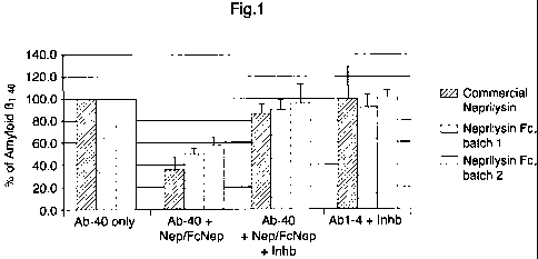
is FIG.1
Degradation of amyloid (31-40 peptide (final concentration 300nM) by
commercial
Neprilysin (2.4 g/ml) or Fc-Neprilysin fusion protein (2.4 g/ml) in buffer.
FIG. 2
A(340 degradation by His-Fc-Nep (SPL061128) and Neprilysin (R&D systems) in
guinea
pig plasma. Two concentrations of His-Fc-Nep are used, and A(3401evels are
measured
after 4 hours. Commercial Neprilysin is used as positive control, and
phosphoramidon is
used as Neprilysin-specific inhibitor.
FIG. 3
A(342 degradation by His-Fc-Nep (SPL061128) and Neprilysin (R&D systems) in
guinea
pig plasma. Two concentrations of His-Fc-Nep are used, and A(3421evels are
measured
after 4 hours. Commercial Neprilysin is used as positive control, and
phosphoramidon is
used as Neprilysin-specific inhibitor.
CA 02681404 2009-09-21
WO 2008/118093 PCT/SE2008/050346
6
FIG. 4
A040 degradation by His-Fc-Nep (SPL061128) and Neprilysin (R&D systems) in
human
plasma. Two concentrations of His-Fc-Nep are used, and A(3401evels are
measured after 4
hours. Commercial Neprilysin is used as positive control, and phosphoramidon
is used as
s Neprilysin-specific inhibitor.
FIG. 5
The PK profile (plasma concentration over time) for Fc-Nep fusion protein
compared to
commercial Neprilysin. Mice were administered with 1 mg/kg commercial
Neprilysin or 1
io alternatively 5 mg/kg in-house produced Fc-Nep.
FIG. 6
Enzymatic activity in cell media from expression of Fc-Neprilysin (N-terminal
fusion of
Fc) compared to Neprilysin-Fc (C-terminal fusion of Fc). Description: PCEP4GW-
Nep-Fc:
is Neprilysin-Fc expressed from pCEP4 plasmid; PEAKlOGW-Nep-Fc: Neprilysin-Fc
expressed from pEAK10 plasmid; com.Nep: Positive control, commercially
available
Neprilysin; PCEP4GW-Fc-Nep: Fc-Neprilysin expressed from pCEP4 plasmid;
PEAKl OGW-Fc-Nep: Fc-Neprilysin expressed from pEAK10 plasmid.
20 FIG. 7
Soluble A(3401evels in plasma of female APPSWE-tg mice after an acute
treatment with Fc-
Nep as well as treatment with the positive control, y-secretase inhibitor
M550426.
25 FIG. 8
Soluble A(3421evels in plasma of female APPSWE-tg mice after an acute
treatment with Fc-
Nep as well as treatment with the positive control, y-secretase inhibitor
M550426.
FIG. 9
CA 02681404 2009-09-21
WO 2008/118093 PCT/SE2008/050346
7
Enzymatic activity of purified protein Fc-Neprilysin (N-terminal fusion of Fc)
compared to
Neprilysin-Fc (C-terminal fusion of Fc).
Description: Nep-Fc: Neprilysin fused to Fc in C-terminal part of Neprilysin;
Fc-Nep:
Neprilysin fused to Fc in N-terminal part of Neprilysin.
FIG. 10
Mouse A(340 levels in plasma of female C57BL/6 mice after an acute treatment
with hFc-
Nep as well as treatment with the positive control, y-secretase inhibitor
M550426.
io FIG. 11
A(3401evels in plasma at different time points after a single injection of hFc-
Nep to female
C57BL6 mice. The percentage shows the reduction compared to vehicle. The
exposure of
hFc-Nep is shown over each treatment bar in the diagram. The effect of
treatment with the
positive control, y secretase inhibitor M550426 is shown in red. The LOQ line
shows the
is limit of quantification in the assay.
FIG. 12
Mean A(340 (A) and A(3421evels (B) in plasma at different time points (from
1.5 up to 336
hours) after a single injection of mFc-Nep to female APPSWE-transgenic mice.
The
20 percentage shows the reduction compared to vehicle. The table (C) shows the
plasma
exposure for respective groups. The effect of treatment with the positive
control, y
secretase inhibitor M550426 is shown in red. The LOQ bar shows the limit of
quantification in the assay. Data was analysed using two-sided t-tests in an
ANOVA model
with time and dose as fixed factors (* p<0.05; ** p<0.01 and *** p<0.001 and
n.s. non-
25 significant).
FIG. 13
Pharmacokinetic and pharmacodynamic diagrams showing the plasma efficacy
effects of
A(340 and A(342, respectively, as percentage of vehicle for all time point
(1.5-336 hours),
CA 02681404 2009-09-21
WO 2008/118093 PCT/SE2008/050346
8
as well as corresponding plasma exposure of mFc-Nep. The line in respective
diagram
shows the predicted exposure and effect.
s In C57BL/6 mice, mFc-Nep significantly reduce mouse A040 in plasma in at
both 5 and 25
mg/kg at all time points (1.5, 168 and 336 hours) (Figure 14). At 168 and 336
hours, both 5
and 25 mg/kg was analysed and the A040 effects are shown to be dose-dependent.
After 2
weeks, a single injection (336 hours) of 25 mg/kg mFc-Nep, significantly
reduce the
mouse A0401evels in plasma by 73% compared to vehicle. The plasma exposure at
this
time point was 48 g/ml and mFc-Nep thereby show to have a long plasma half-
life.
FIG. 14
Mean A(3401evels in plasma at different time points (1.5, 168 and 336 hours)
after a single
intravenous injection of mFc-Nep to female C57BL6 mice. The percentage shows
the
reduction compared to vehicle. The table on the right shows the plasma
exposure for
respective groups. The effect of treatment with the positive control, y
secretase inhibitor
M550426 is shown in red. The LOQ bar shows the limit of quantification in the
assay.
Data was analysed using two-sided t-tests in an ANOVA model with time and dose
as
fixed factors (* p<0.05; ** p<0.01, *** p<0.001 and n.s. non-significant).
FIG. 15
The PK profile (plasma concentration over time) for Fc-Nep fusion protein
compared to in-
house produced Neprilysin. Mice were administered with a single i.v. dose of
10 or 50
nmol enzyme/kg body weight neprilysin (Nep) or Fc-Nep (1 and 5 mg/kg) to mice.
FIG. 16
Table describing degradation of amyloid (3 peptide 1-40 or 1-42 in human
plasma or
APPSWe tg mouse plasma by human or mouse Fc-Neprilysin. EC50 ( M) of
degradation and
% degradation at highest (100 g/mL) concentration of human or mouse Fc-
Neprilysin.
The results are based on 2-3 independent experiments.
CA 02681404 2009-09-21
WO 2008/118093 PCT/SE2008/050346
9
DISCLOSURE OF THE INVENTION
The object of the present invention is to provide fusion proteins capable of
degrading A(3
s peptide. Accordingly, the present invention provides a fusion protein having
the formula
M-A, capable of degrading amyloid beta peptide at one or more cleavage sites
in said
amyloid beta peptide amino acid sequence, wherein M is a protein component
that
prolongs the half-life of the fusion protein, and A is a protein component
that cleaves the
amyloid beta peptide, wherein said M protein component is covalently connected
to the N-
terminus part of the A protein component.
In one aspect of the present invention, there is provided a fusion protein,
wherein A is a
protease.
is In another aspect of the present invention, there is provided a fusion
protein, wherein A is
human Neprilysin.
In another aspect of the present invention, there is provided a fusion
protein, wherein A is
human Neprilysin, wherein said Neprilysin is extracellular Neprilysin.
In another aspect of the present invention, there is provided a fusion
protein, wherein A is
extracellular Neprilysin, comprising an amino acid sequence according to any
one of SEQ
ID NO. 1, 2, 3 or 4.
In another aspect of the present invention, there is provided a fusion
protein, wherein A is
insulin-degrading enzyme.
In another aspect of the present invention, there is provided a fusion
protein, wherein A is
endothelin-converting enzyme 1.
CA 02681404 2009-09-21
WO 2008/118093 PCT/SE2008/050346
In another aspect of the present invention, there is provided a fusion
protein, wherein A is a
scaffold protein.
In another aspect of the present invention, there is provided a fusion
protein, wherein M is
s an Fc part of an antibody. In one embodiment of this aspect, said antibody
is an IgG
antibody. In another embodiment of this aspect, said antibody is an IgG2
antibody.
In another aspect of the present invention, there is provided a fusion
protein, wherein M is
an Fc part from an IgG2 antibody and A is extracellular Neprilysin.
In another aspect of the present invention, there is provided a fusion
protein, comprising an
amino acid sequence according to SEQ ID NO. 11.
In another aspect of the present invention, there is provided a fusion
protein, wherein M is
is an Fc part from an IgG2 antibody and A is insulin-degrading enzyme.
In another aspect of the present invention, there is provided a fusion
protein, comprising an
amino acid sequence according to SEQ ID NO. 12.
In another aspect of the present invention, there is provided a fusion
protein, wherein M is
an Fc part from an IgG2 antibody and A is endothelin-converting enzyme 1.
In another aspect of the present invention, there is provided a fusion
protein, comprising an
amino acid sequence according to SEQ ID NO. 13.
In another aspect of the present invention, there is provided a fusion
protein, wherein M is
selected from pegylation and glycosylation.
In another aspect of the present invention, there is provided a fusion
protein, wherein M is
a HSA.
CA 02681404 2009-09-21
WO 2008/118093 PCT/SE2008/050346
11
In another aspect of the present invention, there is provided a fusion
protein, wherein M is
a HSA binding domain.
s In another aspect of the present invention, there is provided a fusion
protein,, wherein M is
a antibody binding domain.
In another aspect of the present invention, there is provided a fusion
protein,, wherein M
and A is linked together with a linker, L.
In another aspect of the present invention, there is provided a fusion
protein, wherein L is
selected from a peptide and a chemical linker.
In another aspect of the present invention, there is provided a method for
reducing amyloid
0 peptide concentration, said method comprising administration of a fusion
protein,
according to the invention. In one embodiment of this aspect, said reduction
of amyloid (3
peptide is accomplished in plasma. In another embodiment of this aspect, said
reduction of
amyloid 0 peptide is accomplished in CSF. In yet another embodiment of this
aspect, said
reduction of amyloid P peptide is accomplished in CNS.
In another aspect of the present invention, there is provided a pharmaceutical
composition
capable of degrading amyloid 0 peptide, comprising a pharmaceutically
acceptable amount
of fusion protein according to the invention together with a pharmaceutically
acceptable
carrier or excipient.
In another aspect of the present invention, there is provided a method of
prevention and/or
treatment of a condition wherein of degradation of amyloid 0 peptide is
beneficial,
comprising administrering to a mammal, including man in need of such
prevention and/or
treatment, a therapeutically effective amount of a fusion protein according to
the invention.
CA 02681404 2009-09-21
WO 2008/118093 PCT/SE2008/050346
12
In another aspect of the present invention, there is provided a method of
prevention and/or
treatment of Alzheimer's disease, systemic amyloidosis or cerebral amyloid
angiopathy,
comprising administrering to a mammal, including man in need of such
prevention and/or
treatment, a therapeutically effective amount of a fusion protein according to
the invention.
In another aspect of the present invention, there is provided a fusion protein
according to
the invention for use in medical therapy.
In another aspect of the present invention, there is provided use of a fusion
protein of the
invention, in the manufacture of a medicament for prevention and/or treatment
of
conditions wherein of degradation of amyloid 0 peptide is beneficial.
In another aspect of the present invention, there is provided use of a fusion
protein of the
invention, in the manufacture of a medicament for prevention and/or treatment
of
is Alzheimer's disease, systemic amyloidosis or cerebral amyloid angiopathy.
In one
embodiment of this aspect, said medicament reduces amyloid 0 peptide
concentration. Said
reduction of amyloid P peptide is accomplished in plasma, CSF and/or CNS.
The terms used throughout this specification are defined as follows, unless
otherwise
limited in specific instances.
The term "modulator" refers to a molecule that prevents degradation and/or
increases
plasma half-life, reduces toxicity, reduces immunogenicity, or increases
biological activity
of a therapeutic protein. Exemplary modulators include an Fc domain as well as
a linear
polymer (e.g., polyethylene glycol (PEG), polylysine, dextran, etc.); a
branched-chain
polymer (see, for example, U.S. Pat. No. 4,289,872, U.S. Pat. No. 5, 229,490;
WO
93/21259); a lipid; a cholesterol group (such as a steroid); a carbohydrate or
oligosaccharide; or any natural or synthetic protein, polypeptide or peptide
that binds to a
salvage receptor. Glycosylation is also an example of modulator that through
the increase
in size of the fusion protein can prolong the plasma half-life, mainly due to
a change in the
CA 02681404 2009-09-21
WO 2008/118093 PCT/SE2008/050346
13
clearance mechanism. A modulator can also include human serum albumin (HSA)
binding
components which thereby prolong the plasma half-life of the fusion protein.
The term "protein" or "protein component" refers to a molecule that possesses
a catalytic
s activity, which degrades the amyloid 0 peptide by protolytic cleavage at any
possible site
in the amino acid sequence. Examples of proteins include the neprilysin enzyme
as well as
other catalytic active enzymes that degrade the amyloid 0 peptide. Catalytic
antibodies
could also be used as the protein part. The protein can be a natural occurring
variant from
any species (e.g. human, monkey, mice) or a designed variant using rational
design or
io molecular evolution technologies. The protein molecule can also be
different polymorphic
or splice variants. The protein molecule can also be an improved variant of a
natural
occurring variant from any species. Especially a protein can be an improved
variant of
neprilysin that has been modified in the structure by amino acid replacement
to attain
improved properties such as increased activity, improved selectivity towards
the amyloid
is beta peptide and prolonged activity in blood plasma due to increased
stability and/or
reduced inhibition.
The term "fusion" refers to a molecule that is composed of a modulator
molecule and a
protein molecule. The modulator may be covalently linked to the protein part
to create the
20 fusion protein. A non-covalent approach can also be used to connect the
protein to the
modulator part.
The term "degrade", "degrading" or "degradation" refers to a process where one
starting
molecule is divided in two or more molecule(s). More specifically, the amyloid
0 peptide
25 (in any size from amino acid 1-43 and smaller) is cleaved to generate
smaller fragments
compared to the starting molecule. The cleavage can be accomplished through
hydrolysis
of peptide bonds or other type of reaction, which split the molecule in
smaller parts.
The term "native Fc" refers to molecule or sequence comprising the sequence of
a non-
30 antigen-binding fragment resulting from digestion of whole antibody,
whether in
CA 02681404 2009-09-21
WO 2008/118093 PCT/SE2008/050346
14
monomeric or multimeric form. The original immunoglobulin source of the native
Fc may
be of human origin and may be any of the immunoglobulins, although IgGl and
IgG2 are
preferred. Native Fc's are made up of monomeric polypeptides that may be
linked into
dimeric or multimeric forms by covalent (i.e., disulfide bonds) and non-
covalent
s association. The number of intermolecular disulfide bonds between monomeric
subunits of
native Fc molecules ranges from 1 to 4 depending on class (e.g., IgG, IgA,
IgE) or subclass
(e.g., IgGl, IgG2, IgG3, IgAl, IgGA2). One example of a native Fc is a
disulfide- bonded
dimer resulting from papain digestion of an IgG (see Ellison et al. (1982),
Nucleic Acids
Res. 10: 4071-9). The term "native Fc" as used herein is generic to the
monomeric,
dimeric, and multimeric forms.
The term "Fc variant" refers to a molecule or sequence that is modified from a
native Fc
but still comprises a binding site for the salvage receptor, FcRn.
Publications WO
97/34631 and WO 96/32478 describe exemplary Fc variants, as well as
interaction with the
salvage receptor, and are hereby incorporated by reference. Thus, the term "Fc
variant"
comprises a molecule or sequence that is humanized from a non-human native Fc.
Furthermore, a native Fc comprises sites that may be removed because they
provide
structural features or biological activity that are not required for the
fusion molecules of the
present invention. Thus, the term "Fc variant" comprises a molecule or
sequence that lacks
one or more native Fc sites or residues that affect or are involved in (1)
disulfide bond
formation, (2) incompatibility with a selected host cell (3) N-terminal
heterogeneity upon
expression in a selected host cell, (4) glycosylation, (5) interaction with
complement, (6)
binding to an Fc receptor other than a salvage receptor, or (7) antibody-
dependent cellular
cytotoxicity (ADCC). Fc variants are described in further detail hereinafter.
The term "Fc domain" encompasses native Fc and Fc variant molecules and
sequences as
defined above. As with Fc variants and native Fc's, the term "Fc domain"
includes
molecules in monomeric or multimeric form, whether digested from whole
antibody or
produced by other means.
CA 02681404 2009-09-21
WO 2008/118093 PCT/SE2008/050346
The term "pharmacologically active" means that a substance so described is
determined to
have activity that affects a medical parameter (e.g., blood pressure, blood
cell count,
cholesterol level) or disease state (e.g., cancer, autoimmune disorders,
dementia).
s The term" amyloid beta peptide", "A(3 peptide" or "amyloid 0 peptide" means
any form of
the peptide that correlate to amino acid sequence (one letter code) DAEFRHDSG
YEVHHQKLVF FAEDVGSNKG AIIGLMVGGV VIAT in the human A(3 A4 protein
[Precursor], corresponding to amino acid 672 to 714 in the sequence (amino
acid 1-43). It
also includes any shorter forms of this peptide, such as 1-38, 1-40 and 1-42
but not
10 restricted to these forms. Moreover, Amyloid 0 peptide has several natural
occurring
forms. The human forms of Amyloid 0 peptide are referred to as A039, A040,
A041, A(342
and A(343. The sequences of these peptides and their relationship to the APP
precursor are
illustrated by FIG. 1 of Hardy et al., TINS 20, 155-158 (1997). For example,
A(342 has the
sequence:
is H2N-Asp-Ala-Glu-Phe-Arg-His-Asp-Ser-Gly-Tyr-Glu-Val- His- His-Gln-Lys-Leu-
Val-
Phe-Phe-Ala-Glu-Asp-Val-Gly-Ser-Asn-Lys-Gly-Ala- Ile- Ile-Gly-Leu-Met-Val-Gly-
Gly-
Val-Val-Ile-Ala-OH. A(341, A(340 and A(339 differ from A(342 by the omission
of Ala,
Ala-Ile, and Ala-Ile-Val respectively from the C- terminal end. A(343 differs
from A(342 by
the presence of a threonine residue at the C-terminus. Overall, amyloid beta
peptide means
the peptide form that is involved in plaque formation that causes Alzheimer
disease.
The term "half-life" is defined by the time taken for the removal of half the
initial
concentration of the fusion protein from the plasma. This invention describes
ways of
modulating the half-life in plasma. Such modification can produce fusion
proteins with
improved pharmacokinetic properties (e.g., increased in vivo serum half-life).
Prolong the
half-life means that it takes longer time to remove or get a clearance of half
of the initial
concentration of the fusion protein from the plasma. Half-life of a
pharmaceutical or
chemical compound is well defined and known in the art.
CA 02681404 2009-09-21
WO 2008/118093 PCT/SE2008/050346
16
The term"connect" means a covalent or a reversible linkage between two or more
parts. A
covalent linkage can for example be a peptide bond, disulfide bond, carbon-
carbon
coupling or any type of linkage that is based of a covalent linkage between to
atoms.
Reversible linkage can for example be biotin-streptavidin, antibody-antigen or
a linkage,
s which is classified as a reversible linkage known in the art. For example, a
covalent
linkage is directly obtained when the protein part and the modulator part of
the fusion
protein is produced in a recombinant form from the same plasmid, thus the
connection is
designed on DNA level.
The term"covalently connected" means a chemical link between two atoms in
which
electrons are shared between them. Examples of bonds covalently connected are
a peptide
bond, disulfide bond, carbon-carbon coupling. A fusion protein can be linked
together by a
polypeptide bond where the linkage can be accomplished during the
translational process
on the ribosome when the fusion protein are produced. Other type of covalently
connected
component could be modification with a pegylation reagent that is covalently
linked to an
amino residue (for example lysine) on the protein. The chemical coupling
reaction can, for
example, be acylation or other suatible coupling reaction which link the two
components
togheter into a fusion protein. Covalently connected can also mean a linkage
of a linker at
two sites in which the modulator is linked together with the protein part.
The term "cleavage sites" means a specific location/site in a peptide sequence
that can be
cleaved by a protein or an enzyme. Cleavage is normally produced by hydrolysis
of the
peptide bond connecting two amino acids. Cleavage can also take place at
multiple sites in
the same peptide using a single or a combination of proteins or enzymes. A
cleavage site
can also be other site than the peptide bond. This invention describes the
cleavage of the
amyloid 0 peptide in detail.
The term "binding domain" means a molecule that binds the amyloid 0 peptide
with an
affinity of that is therapeutically relevant. These molecules bind to amyloid
P peptide with
a binding affinity greater than or equal to about 106, 107, 10g, 109, or 1010
M-1. Typical
CA 02681404 2009-09-21
WO 2008/118093 PCT/SE2008/050346
17
binding domains are, but not restricted to, antibodies (e.g. Fab, scFv, single
domains all
including the CDR regions), scaffold proteins as described in this invention
and in the
literature or synthetically produced molecules with affinity for the amyloid
(3 peptide.
s The term "protease" means any protein molecule acting in the hydrolysis of
peptide bonds.
It includes naturally occurring proteolytic enzymes, as well as variants
thereof obtained by
site-directed or random mutagenesis or any other protein engineering method,
any
fragment of an proteolytic enzyme, or any molecular complex or fusion protein
comprising
one of the aforementioned proteins. The protease can be a serine, cysteine,
aspartic or a
metalloprotease.
The term "substrate" or "peptide substrate" means any peptide, oligopeptide,
or protein
molecule of any amino acid composition, sequence or length, that contains a
peptide bond
that can be hydrolyzed catalytically by a protease. The peptide bond that is
hydrolyzed is
is referred to as the "cleavage site". Numbering of positions in the substrate
is done according
to the system Introduced by Schlechter & Berger (Biochem. Biophys. Res.
Commun. 27
(1967) 157-162). Amino acid residues adjacent N-terminal to the cleavage site
are
numbered Pl, P2, P3, etc., whereas residues adjacent C-terminal to the
cleavage site are
numbered Pl', P2', P3', etc. The substrate or peptide substrate of this
invention is the
amyloid 0 peptide.
The term "specificity" means the ability of a protein or a protease to
recognize and
hydrolyze selectively certain peptide substrates while others remain
uncleaved. Specificity
can be expressed qualitatively and quantitatively. "Qualitative specificity"
refers to the
kind of amino acid residues that are accepted by a protease at certain
positions of the
peptide substrate. Proteases that accept only a small portion of all possible
peptide
substrates have a "high specificity". Proteases that accept almost any peptide
substrate have
a "low specificity". Proteases with very low specificity are also referred to
as "unspecific
proteases".
CA 02681404 2009-09-21
WO 2008/118093 PCT/SE2008/050346
18
The term "evolved protease" describes any protease that have been obtained
using random
PCR, DNA shuffling or other type of methods that generate diversity on the
DNA/RNA
level. Literature describing these approaches is for example; D.A. Drummond,
B.L.
Iverson, G. Georgiou and F.H. Arnold, Journal of Molecular Biology 350: 806-
816 (2005)
s and S. McQ and D.S. Tawfik, Biochemistry 44: 5444-5452 (2005). Various
approached to
conduct screening and selection among the diversity created are also described
in the
literature (e.g Directed Enzyme Evolution: Screening and Selection Methods
(Methods in
Molecular Biology) Editors: Frances H Arnold and George Georgiou. Volume 230,
2003
and references therein). Various strategies can be used to select for
properties like
increased stability, increased activity, improved selectivity and decreased
inhibition by
known and unknown inhibitors.
The term "improved protease" describes any protease variants that possess
higher catalytic
activity if that is needed. However, in some instances a lower catalytic
activity might be
preferable. Improved protease might also mean a variant that cleaves a certain
substrate
compared to another substrate more efficient that the original protese.
Improved means a
more preferred property, such as catalytic activity and/or selectivity to
obtain a more
optimized pharmaceutical compound. Improved protease can also mean variants
with
increased stability in for example plasma blood (both or either in vitro and
in vivo).
Improved protease can also mean variants with decreased inactivation in for
example
plasma blood (both or either in vitro or in vivo). Decreased inactivation can
be
accomplished by decreasing the protolytic degradation of the protease due to
changed
amino acid sequence, less prone to be cleaved. Decreased proteolytic
degradation can also
be accomplished by modifying the protein surface with for example pegylation
and/or
glycosylation to protect the protein from becoming cleaved. Decreased
inactivation can
also be accomplished by reducing inhibition of the protease by a known or
unknown
inhibitor. Reduced inhibition of an unknown blood plasma inhibitor can be
accomplished
by screening variants for reduced inhibition of protease activity directly in
the blood
plasma.
CA 02681404 2009-09-21
WO 2008/118093 PCT/SE2008/050346
19
The term "human Neprilysin" refers to any natural form of human neprilysin.
This includes
all splice and polymorphic variants that naturally occur in the human
population. A number
of forms of human neprilysin are described in this invention (SEQ ID Nos 1 to
4). The
term also include fragments or extended variants of human Neprilysin, as well
as improved
s variants of human Neprilysin, as described under "improved protease".
The term "scaffold protein" describes any protein that binds amyloid 0
peptide. Examples
of scaffold proteins are tendamistat, affibody, anticalin and ankyrin. These
scaffold
proteins are typically designed and is based on a rigid core structure and a
part, loops,
io surfaces or cavities that can be randomized for the identification of
binders. These scaffold
proteins are well described in the literature.
This invention suggests the possibility that the administration of an
optimized recombinant
A(3 degradation enzyme inhibits amyloid plaque formation by decreasing brain
levels of
is A(3. As a consequence, amyloid plaque-related astrogliosis will also be
reduced.
In one aspect of this invention the therapeutic compound is of fully human
origin. The
fusion protein is composed of fully human proteins that are linked together
using a linker
with lowest possible immunogenic activity.
Advantages using a degrading enzyme compared to a binding molecule such an
antibody
are:
= Degradation with an enzyme of the amyloid 0 peptide will directly remove the
toxic effect compare to a binding approach where the concentration of the
amyloid
0 peptide could potentially increase if the binding molecule in complex with
amyloid 0 peptide is not cleared fast enough. This could he harmful especially
if
the amyloid 0 peptide concentration increases peripherally.
= Catalytic degradation of amyloid P peptide will remove the peptide more
efficiently
that binding. Only a catalytic amount of the degrading enzyme will be
necessary to
CA 02681404 2009-09-21
WO 2008/118093 PCT/SE2008/050346
remove sufficient amyloid (3 peptide whereas a binding molecule such as an
antibody, a stoichiometric amount will be needed for a therapeutic effect.
This will
have a great impact on the amount needed for therapeutic treatment.
s = If the binding molecule is an antibody and cross the BBB allowing binding
to the
amyloid (3 peptide in the plaques, a potential immunological respons that are
harmful is possible. On the other hand, a catalytic fusion protein will not
bind to
the plaques and use the Fc reactivity but only reduce the free concentration
of
amyloid (3 peptide. Thus, A catalytic enzyme will only degrade the free pool
of
10 amyloid (3 peptide. A binding agent like an antibody could potentially
enter the
CNS and dissolve the plaques through Fc activity. This might be unfavorable if
large amount of amyloid (3 peptide is released in the vicinity of the plaque
and they
are toxic to the cells.
is One important enzyme in A(3 catabolism is Neprilysin, also known as neutral
endopeptidase-24.11 or NEP. Iwata et al. (Nature Medicine, 6: 143-149, 2000)
showed that
the A(3 1_42 peptide underwent full degradation through limited proteolysis
conducted by
NEP similar or identical to neprilysin as biochemically analysed.
Consistently, NEP
inhibitor infusion resulted in both biochemical and pathological deposition of
endogeneous
20 A(342 in brain. It was found that this NEP-catalysed proteolysis therefore
limits the rate of
A(342 catabolism.
NEP is a 94 kD, type two membrane-bound Zn-metallopeptidase implicated in the
inactivation of several biologically active peptides including enkephalins,
tachykinins,
bradykinin, endothelins and atrial natriuretic peptide. NEP is present in
peptidergic
neurons in the CNS, and its expression in brain is regulated in a cell-
specific manner
(Roques B. P. et al., Pharmacol. Rev. 45, 87-146, 1993; Lu B. et al., J. Exp.
Med. 181,
2271-2275, 1995; Lu B. et al., Ann. N.Y. Acad. Sci. 780, 156-163, 1996). While
type 2
NEP-transcripts are absent from the CNS, type 1 and type 3 transcripts are
localized in
neurons and in oligodendrocytes of the corpus callosum, respectively (Li C. et
al., J. Biol.
CA 02681404 2009-09-21
WO 2008/118093 PCT/SE2008/050346
21
Chem. 270, 5723-5728, 1995). The Neprilysin family of proteases and
endopeptidases
comprises structurally or functionally homologous members of NEP such as the
recently
described NEP II gene and its isoforms (Ouimet T. et al., Biochem. Biophys.
Res. Commun.
271:565-570, 2000), which are expressed in the CNS in a complementary pattern
to NEP.
s A further member of this family is NL-1 (neprilysin like 1), a soluble
protein efficiently
inhibited by the NEP inhibitor phosphoramidon (Ghaddar G. et al., Biochem. J.
347: 419-
429, 2000).
Other enzymes that are known to catabolise A(3 have also been described. The
zinc
metallopeptidase insulin-degrading enzyme (IDE, EC. 3.4.22.11) cleaves A(31_40
and A(31_42
into what appears to be innocuous products. IDE is a true peptidase; it does
not hydrolyze
proteins. The enzyme cleaves a limited number of peptides in vitro including
insulin and
insulin related peptides, P endorphin, and A(3 peptides. IDE has been
suggested to be one
of the physiological A(3 metabolizing enzymes (W. Q. Qui et al. (1998) J.
Biol. Chem. 273,
is 32730-32738). Kurichkin and Goto (I. V. Kurochkin and S. Gato (1994) FEBS
Lett. 345,
33-37) first reported that insulin degrading enzyme can hydrolyze A(31_40.
This finding was
confirmed in two separate studies (W. Q. Qui et al. (1998) J. Biol. Chem. 273,
32730-
32738; and J. R. McDermott and A. M. Gibson (1997) Neurochem. Res. 22, 49-56).
Moreover, metalloprotease 24.15, a recently identified as a A(3- degrading
enzyme (Yamin
R. et al., J. Biol. Chem. 274, 18777-18784, 1999), was also unchanged in
response to A(3
injections. Angiotensin converting enzyme (ACE), an unrelated neuronal Zn-
metalloendo
peptidase have been also mention as a possible A(3-peptide degrading enzyme
(Barnes N.
M. et al., Eur. J. Pharmacol. 200, 289-292,1991; Alvarez R. et al., J. Neurol.
Neurosurg.
Psychiatzy 67, 733-736, 1999; Amouyel P. et al., Ann. N.Y. Acad. Sci. 903, 437-
441, 2000)
with no known affinity to A(3 (McDermott J. R. and Gibson A. M., Neurochem.
Res. 22,
49-56, 1997). Cathepsin B (CatB) have also been shown to degrade A(3 peptides
(Neuron.
2006 Sep 21;51(6):703-14).
The sequence used from the neprilysin may be the extracellular part of the
protein. The
extracellular part is defined as the part of neprilysin that is defined as
outside the
CA 02681404 2009-09-21
WO 2008/118093 PCT/SE2008/050346
22
membrane region. This invention also includes the use of the whole sequence of
neprilysin
as the amyloid 0 peptide-degrading component. The invention also comprises
smaller
fragments of neprilysin as long as the catalytic activity is preserved against
the amyloid (3
peptide. The invention also comprises any polymorphism variants and splice
variants of
s neprilysin. The invention also comprises any improved variants of
neprilysin.
This invention describes a novel and alternative strategy to hydrolyze A(3
peptides before
they form amyloid plaques or at least prevent the further development of
existing plaques.
It may also be possible to remove existing plaques by hydrolyzing any plaque-
derived A(3
io peptide in equilibrium with free A(3 peptide.
Another embodiment of the present invention refers to a molecule that is
composed of one
part that binds amyloid (3 peptide with high affinity. This affinity is below
micromolar in
binding affinity. The binding affinity for amyloid (3 peptide is preferably at
nanomolar in
is binding affinity. The other part that is involved in the interaction with
amyloid (3 peptide is
an active component that cleaves the amyloid (3 peptide at one or more site in
the structure
of the amyloid (3 peptide. The reason to combine a binding part linked
together with a
catalytic active part that both recognize the amyloid (3 peptide is that the
binding part binds
the amyloid (3 peptide and thereby increase the local concentration (the
binding part and
20 the catalytic part) is binding to the dissociated form of amyloid (3
peptide. Some bind
specifically to the dissociated form without binding to the aggregated form.
Some bind to
both aggregated and dissociated forms. Some such antibodies bind to a
naturally occurring
short form of A(3 (i.e covalently or in another way linked together) of
amyloid (3 peptide to
become cleaved by the active part that is locally around due to the linkage
engineered in
25 the bifunctional molecule. The linkage between the amyloid 0 peptide
binding component
and the amyloid 0 peptide-degrading component is preferably mediated by the
plasma half-
life modulator component with or without a linker component.
In some embodiments of this invention the therapeutic agents include fusion
proteins that
30 specifically bind to amyloid 0 peptide or other component of amyloid
plaques. Such
CA 02681404 2009-09-21
WO 2008/118093 PCT/SE2008/050346
23
compound can be a part of a monoclonal or polyclonal or any other amyloid 0
peptide
binding agent. These compounds bind to amyloid 0 peptide with a binding
affinity greater
than or equal to about 106, 107, 10g, 109, or 1010 M-1. These binding
components are
preferably connected with an amyloid 0 peptide-degrading component.
One aspect of the invention refers to the combination with the "Fc" domain of
an antibody
with a amyloid P peptide degrading component in the fusion protein. Antibodies
comprise
two functionally independent parts, a variable domain known as "Fab", which
binds
antigen, and a constant domain known as "Fc", which links to such effector
functions as
complement activation and attack by phagocytic cells. An Fc has a long serum
half-life,
whereas a Fab is short-lived (Capon et al. (1989), Nature 337: 525-3 1). When
constructed
together with a therapeutic protein, an Fc domain can provide longer half-life
or
incorporate such functions as Fc receptor binding, protein A binding,
complement fixation
and perhaps even placental transfer.
Preferred molecules in accordance with this invention are Fc-linked amyloid (3
peptide
degrading protein such as NEP-related proteins.
Useful modifications of protein therapeutic agents by fusion with the Fc
domain of an
antibody are discussed in detail in a publication entitled, "Modified Peptides
as
Therapeutic Agents (WO 99/25044). That publication discusses linkage to a
"vehicle" such
as PEG, dextran, or an Fc region. Linking to the C-terminal part of an Fc
domain has been
described in the literature as a possible approach (Protein Eng. 1998 11:495-
500). This
allows a N-terminal linkage on the protein part of the fusion protein. This
invention
describes this approach and the beneficial effect of using this strategy
obtaining a fusion
protein with optimized properties for in vivo efficacy.
IgG molecules interact with three classes of Fc receptors (FcR) specific for
the IgG class of
antibody, namely FcyRI, FcyRII and FcyRIII. In preferred embodiments, the
immunoglobulin (Ig) component of the fusion protein has at least a portion of
the constant
region of an IgG that has a low binding affinity for at least one of FcyRI,
FcyRII or
CA 02681404 2009-09-21
WO 2008/118093 PCT/SE2008/050346
24
FcyRIII. In one aspect of the invention, the binding affinity of fusion
proteins for Fc
receptors is reduced by using heavy chain isotypes as fusion partners that
have reduced
binding affinity for Fc receptors on cells. For example, both human IgGl and
IgG3 have
been reported to bind to FcRyI with high affinity, while IgG4 binds 10-fold
less well, and
s IgG2 does not bind at all. The important sequences for the binding of IgG to
the Fc
receptors have been reported to be located in the CH2 domain. Thus, in a
preferred
embodiment, an antibody-based fusion protein with enhanced in vivo circulating
half-life
is obtained by linking at least the CH2 domain of IgG2 or IgG4 to a second non-
immunoglobulin protein. For example, of the four known IgG isotypes, IgGl
(Cyl) and
io IgG3 (Cy3) are known to bind FcRyI with high affinity, whereas IgG4 (Cy4)
has a 10-fold
lower binding affinity, and IgG2 (Cy2) does not bind to FcRyI.
In one embodiment, the A(3-peptide degrading component of the fusion protein
is an
enzyme. The term "enzyme" is used herein to describe proteins, analogs
thereof, and
is fragments thereof, which are active as proteases or petidases. Preferably,
enzymes include
serine, aspartic, metallo and cysteine proteases. Preferably, the fusion
protein of the present
invention displays enzymatic biological activity.
In another embodiment, the immunoglobulin domain is selected from the group
consisting
20 of the Fc domain of IgG, the heavy chain of IgG, and the light chain of
IgG.
In another embodiment, the constant region of the antibody in the fusion
protein will be of
human origin, and belong to the immunoglobulin family derived from the IgG
class of
immunoglobulins, in particular from classes IgGl, IgG2, IgG3 or IgG4,
preferably from
the class IgG2 or IgG4. It is also alternatively possible to use constant
regions of
25 immunoglobulins belonging to the IgG class from other mammals, in
particular from
rodents or primates; however, it is also possible, according to the invention,
to use constant
regions of the immunoglobulin classes IgD, IgM, IgA or IgE. Typically, the
antibody
fragments that are present in the construct according to the invention will
comprise the Fc
domain CH3, or parts thereof, and at least one part segment of the Fc domain
CHz.
3o Alternatively, it is also possible to conceive of fusion constructs
according to the invention
CA 02681404 2009-09-21
WO 2008/118093 PCT/SE2008/050346
which contain, as component (A), the CH3 domain and the hinge region, for the
dimerization.
However, it is also possible to use derivatives of the immunoglobulin
sequences that are
s found in the native state, in particular those variants that contain at
least one replacement,
deletion and/or insertion (combined here under the term "variant"). Typically,
such
variants possess at least 90%, preferably at least 95%, and more preferably at
least 98%,
sequence identity with the native sequence. Variants, which are particularly
preferred in
this context, are replacement variants that typically contain less than 10,
preferably less
10 than 5, and very particularly preferably less than 3, replacements as
compared with the
respective native sequence. Attention is drawn to the following replacement
possibilities as
being preferred: Trp with Met, Val, Leu, Ile, Phe, His or Tyr, or vice versa;
Ala with Ser,
Thr, Gly, Val, Ile or Leu, or vice versa; Glu with Gln, Asp or Asn, or vice
versa; Asp with
Glu, Gln or Asn, or vice versa; Arg with Lys, or vice versa; Ser with Thr,
Ala, Val or Cys,
is or vice versa; Tyr with His, Phe or Trp, or vice versa; Gly or Pro with one
of the other 19
native amino acids, or vice versa.
Soluble receptor-IgG fusion proteins are common immunological reagents and
methods for
their construction are known in the art (see e.g., U.S. Pat. No. 5,225,538). A
functional
20 amyloid 0 peptide-degrading domain may be fused to an immunoglobulin Fc
domain
derived from an immunoglobulin class or subclass. The Fc domains of antibodies
belonging to different Ig classes or subclasses can activate diverse secondary
effector
functions. Activation occurs when the Fc domain is bound by a cognate Fc
receptor.
Secondary effector functions include the ability to activate the complement
system, to
25 cross the placenta, and to bind various microbial proteins. The properties
of the different
classes and subclasses of immunoglobulins are described in Roitt et al.,
Immunology, p.
4.8 (Mosby -Year Book Europe Ltd., 3d ed. 1993). The Fc domains of antigen-
bound
IgGl, IgG3 and IgM antibodies can activate the complement enzyme cascade. The
Fc
domain of IgG2 appears to be less effective, and the Fc domains of IgG4, IgA,
IgD and IgE
are ineffective at activating complement. Thus one can select an Fc domain
based on
CA 02681404 2009-09-21
WO 2008/118093 PCT/SE2008/050346
26
whether its associated secondary effector functions are desirable for the
particular immune
response or disease being treated with the amyloid 0 peptide degrading-Fc
fusion protein.
If it would be advantageous to harm or kill target cells, one could select an
especially
active Fc domain (IgGl) to make the amyloid 0 peptide degrading-Fc-fusion
protein.
s Alternatively, if it would be desirable to produce the amyloid 0 peptide
degrading-Fc-
Fusion without triggering the complement system, an inactive IgG4 Fc domain
could be
selected. This invention describes a fusion protein with a catalytic component
linked to a
Fc part and not a direct binding component. This means that the effect and
activity from
the Fc will be limited because many Fc effects are mediated through the
binding. For
io example complement activation is dependent on binding and the formation of
a network.
C-terminally of the immunoglobulin fragment, a fusion construct according to
the
invention typically, but not necessarily, contains a transition region between
catalytic and
modulator part, which transition region can in turn contain a linker sequence,
with this
is linker sequence preferably being a peptide sequence. This peptide sequence
can have a
length from between 1 and up to 70 amino acids, where appropriate even more
amino
acids, preferably from 10 to 50 amino acids, and particularly preferably
between 12 and 30
amino acids. The linker region of the transition sequence can be flanked by
further short
peptide sequences which can, for example, correspond to DNA restriction
cleavage sites.
20 Any restriction cleavage sites with which the skilled person is familiar
from molecular
biology can be used in this connection. Suitable linker sequences are
preferably artificial
sequences which contain a high number of proline residues (for example at
every second
position in the linker region) and, in addition to that, preferably have an
overall hydrophilic
character. A linker sequence, which consists of at least 30% of proline
residues, is
25 preferred. The hydrophilic character can preferably be achieved by means of
at least one
amino acid having a positive charge, for example lysine or arginine, or
negative charge, for
example aspartate or glutamate. Overall, the linker region therefore also
preferably
contains a high number of glycine and/or proline residues in order to confer
on the linker
region the requisite flexibility and/or rigidity.
CA 02681404 2009-09-21
WO 2008/118093 PCT/SE2008/050346
27
However, native sequences, for example those fragments of ligands belonging to
the NEP
family which are disposed extracellularly, but immediately act, i.e. in front
of, the cell
membrane, are also suitable for use as linkers, where appropriate after
replacement,
deletion or insertion of the native segments as well. These fragments are
preferably the 50
s AA which follow extracellularly after the transmembrane region or else
subfragments of
these first 50 AA. However, preference is given to these segments having at
least 85%
sequence identity with the corresponding natural human sequences, with very
particular
preference being given to at least 95% sequence identity and particular
preference being
given to at least 99% sequence identity in order to limit the immunogenicity
of these linker
regions in the fusion protein according to the invention and not elicit any
intrinsic humoral
defense reaction. Within the context of the present invention, the linker
region should
preferably not possess any immunogenicity.
However, as an alternative to peptide sequences which are linked to the
amyloid 0 peptide
degrading component and the plasma half-life modulator component, by way of
amide-like
bonds, it is also possible to use compounds which are of a nonpeptide or
pseudopeptide
nature or are based on noncovalent bonds. Examples which may be mentioned in
this
connection are, in particular, N-hydroxysuccinimide esters and
heterobifunctional linkers,
such as N-succinimidyl-3-(2-pyridyldi-thio) propionate (SPDP) or similar
crosslinkers.
Other ways of regulating the plasma half-life is to use pegylation or other
type of
modifications that increasing the molecular weight such as glycosylation.
As noted above, polymer modulators may also be used. Various means for
attaching
chemical moieties useful as modulator are currently available, see, e.g.,
patent application
WO 96/11953, entitled "N-Terminally Chemically Modified Protein Compositions
and
Methods, " herein incorporated by reference in its entirety. This PCT
publication discloses,
among other things, the selective attachment of water-soluble polymers to the
N-terminus
of proteins.
CA 02681404 2009-09-21
WO 2008/118093 PCT/SE2008/050346
28
A preferred polymer modulator is polyethylene glycol (PEG). The PEG group may
be of
any convenient molecular weight and may be linear or branched. The average
molecular
weight of the PEG will preferably range from about 2 kiloDalton ("kD") to
about 100 kDa,
more preferably from about 5 kDa to about 50 kDa, most preferably from about 5
kDa to
s about 10 kDa. The PEG groups will generally be attached to the compounds of
the
invention via acylation or reductive alkylation through a reactive group on
the PEG moiety
(e.g., an aldehyde, amino, thiol, or ester group) to a reactive group on the
compound (e.g.
an aldehyde, amino, or ester group).
io A useful strategy for the PEGylation of protein consists of combining,
through forming a
conjugate linkage in solution, a protein and a PEG moiety, each bearing a
special
functionality that is mutually reactive toward the other. The protein can be
prepared with
conventional recombinant expression techniques. The proteins are
"preactivated" with an
appropriate functional group at a specific site. The precursors are purified
and fully
is characterized prior to reacting with the PEG moiety. Ligation of the
protein with PEG
usually takes place in aqueous phase and can be easily monitored by reverse
phase
analytical HPLC. The PEGylated protein can be easily purified by preparative
HPLC and
characterized by analytical HPLC, amino acid analysis and laser desorption
mass
spectrometry.
Polysaccharide polymers are another type of water-soluble polymer which may be
used for
protein modification. Dextrans are polysaccharide polymers comprised of
individual
subunits of glucose predominantly linked by al-6linkages. The dextran itself
is available
in many molecular weight ranges, and is readily available in molecular weights
from about
1 kD to about 70 kD. Dextran is a suitable water-soluble polymer for use in
the present
invention as a modulator by itself or in combination with another modulator
(e.g., Fc), see
e.g. WO 96/11953 and WO 96/05309. The use of dextran conjugated to therapeutic
or
diagnostic immunoglobulins has been reported; see, for example, European
Patent
Publication EP 0 315 456, which is hereby incorporated by reference. Dextran
of about 1
CA 02681404 2009-09-21
WO 2008/118093 PCT/SE2008/050346
29
kD to about 20 kD is preferred when dextran is used as a vehicle in accordance
with the
present invention.
Carbohydrate (oligosaccharide) groups may conveniently be attached to sites
that are
s known to be glycosylation sites in proteins. Generally, 0-linked
oligosaccharides are
attached to serine (Ser) or threonine (Thr) residues while N-linked
oligosaccharides are
attached to asparagine (Asn) residues when they are part of the sequence Asn-X-
Ser/Thr,
where X can be any amino acid except proline. X is preferably one of the 19
naturally
occurring amino acids other than proline. The structures of N-linked and 0-
linked
oligosaccharides and the sugar residues found in each type are different. One
type of sugar
that is commonly found on both is N-acetylneuraminic acid (referred to as
sialic acid).
Sialic acid is usually the terminal residue of both N- linked and 0- linked
oligosaccharides
and, by virtue of its negative charge, may confer acidic properties to the
glycosylated
compound. Such site(s) may be incorporated in the linker of the compounds of
this
invention and are preferably glycosylated by a cell during recombinant
production of the
polypeptide compounds (e.g., in mammalian cells such as CHO, BHK, COS).
However,
such sites may further be glycosylated by synthetic or semi- synthetic
procedures known in
the art. Amino acids that are suitable for glycosylation can be incorporated
at specific sites
both in the modulator and the protein part. Preferable techniques to use for
engineering
these specific amino acids are site-directed mutagenesis or comparable method.
Other possible modifications include hydroxylation of proline and lysine,
phosphorylation
of hydroxyl groups of seryl or threonyl residues, oxidation of the sulfur atom
in Cys,
methylation of the alpha-amino groups of lysine, arginine, and histidine side
chains.
Creighton, Proteins: Structure and Molecule Properties (W. H. Freeman & Co.,
San
Francisco), pp. 79-86 (1983). Thus, glycosylation sites in the amyloid 0
peptide degrading
component can be engineered. For example, residues preferably on the surface
of
neprilysin structure are modified to allow the glycosylation. The 3D structure
of neprilysin
is know an can be used to select suitable amino acid replacement for the
introduction of
both glycosylation and pegylation sites. Glycosylation sites are introduced
using for
example the Asn-X- Ser/Thr sequence. For pegylation, suitable surface exposed
amino
CA 02681404 2009-09-21
WO 2008/118093 PCT/SE2008/050346
acids are for example replaced to cystine residues for specific and efficient
coupling of the
pegylation component.
Compounds of the present invention may be changed at the DNA level, as well.
The DNA
s sequence of any portion of the compound may be changed to codons more
compatile with
the chosen host cell. For E. coli, which is the preferred host cell, optimized
codons are
known in the art. Codons may be substituted to eliminate restriction sites or
to include
silent restriction sites, which may aid in processing of the DNA in the
selected host cell.
The vehicle, linker and peptide DNA sequences may be modified to include any
of the
10 foregoing sequence changes.
Linkers: Any "linker" group is optional. When present, its chemical structure
is not critical,
since it serves primarily as a spacer. The linker is preferably made up of
amino acids
linked together by peptide bonds. Thus, in preferred embodiments, the linker
is made up of
is from 1 to 20 amino acids linked by peptide bonds, wherein the amino acids
are selected
from the 20 naturally occurring amino acids. Some of these amino acids may be
glycosylated, as is well understood by those in the art. In a more preferred
embodiment, the
1 to 20 amino acids are selected from glycine, alanine, proline, asparagine,
glutamine, and
lysine. Even more preferably, a linker is made up of a majority of amino acids
that are
20 sterically unhindered, such as glycine and alanine. Thus, preferred linkers
are polyglycines
(particularly (Gly) 4, (Gly)s), poly(Gly-Ala), and polyalanines.
The quantitative specificity of proteases varies over a wide range. There are
very
unspecific proteases known, such as papain which cleaves all polypeptides that
contain a
25 phenylalanine, a valine or an leucine residue, or trypsin which cleaves all
polypeptides that
contain an arginine or a lysine residue. On the other hand, there are highly
specific
proteases known, such as the tissue-type plasminogen activator (t-PA) which
cleaves
plasminogen only at a single specific sequence. Proteases with high substrate
specificity
play an important role in the regulation of protein functions in living
organisms. The
30 specific cleavage of polypeptide substrates, for example, activates
precursor proteins or
CA 02681404 2009-09-21
WO 2008/118093 PCT/SE2008/050346
31
deactivates active proteins or enzymes, thereby regulating their functions.
Several
proteases with high substrate specificities are used in medical applications.
Pharmaceutical
examples for activation or deactivation by cleavage of specific polypeptide
substrates are
the application of t-PA in acute cardiac infarction, which activates
plasminogen to resolve
s fibrin clots, or the application of Ancrod in stroke which deactivates
fibrinogen, thereby
decreasing blood viscosity and enhancing its transport capacity. While t-PA is
a human
protease with an activity necessary in human blood regulation, Ancrod is a non-
human
protease. It was isolated from the viper Agkistrodon rhodostoma, and comprises
the main
ingredient of the snake's poison. Therefore, there exist a few non-human
proteases with
io therapeutic applicability. Their identification, however, is usually highly
incidental.
The treatment of diseases by administering drugs is typically based on a
molecular
mechanism initiated by the drug that activates or inactivates a specific
protein function in
the patient's body, be it an endogenous protein or a protein of an infecting
microbe or virus.
While the action of chemical drugs on these targets is still difficult to
understand or to
is predict, protein drugs are able to specifically recognize these target
proteins among
millions of other proteins. Prominent examples of proteins that have the
intrinsic
possibility to recognize other proteins are antibodies, receptors, and
proteases. Although
there are a huge number of potential target proteins, only very few proteases
are available
today to address these target proteins. Due to their proteolytic activity,
proteases are
20 particularly suited for the inactivation of protein or peptide targets.
When considering
human proteins only, the number of potential target proteins is yet enormous.
It is
estimated that the human genome comprises between 30,000 and 100,000 genes,
each of
which encodes a different protein. Many of these proteins or peptides are
involved in
human diseases and are therefore potential pharmaceutical targets. It might be
unlikely to
25 find such a protease with a particular qualitative specificity by screening
natural isolates.
Therefore there is a need to optimize the catalytic selectivity of a known
protease or other
scaffold proteins including catalytic antibodies.
Selection systems for proteases of known specificity are known in the art, for
instance,
30 from Smith et al., Proc. Natl. Acad. Sci USA, Vol. 88 (1991). As
exemplified, the system
CA 02681404 2009-09-21
WO 2008/118093 PCT/SE2008/050346
32
comprises the yeast transcription factor GAL4 as the selectable marker, a
defined and
cleavable target sequence inserted into GAL4 in conjunction with the TEV
protease. The
cleavage separates the DNA binding domain from the transcription activation
domain and
therewith renders the transcription factor inactive. The phenotypical
inability of the
s resulting cells to metabolize galactose can be detected by a calorimetric
assay or by the
selection on the suicide substrate 2-deoxygalactose.
Further, selection may be performed by the use of peptide substrates with
modifications as,
for example, fluorogenic moieties based on groups as ACC, previously described
by Harris
io et al. (US 2002/022243).
Identical or similar approaches could be used in order to identify or produce
an effective
amyloid 0 peptide-degrading component as described in this invention. That
starting point
for the engineering of this amyloid P peptide-degrading component could be an
enzyme
is that possesses some activity against amyloid (3 peptide or that have no
activity at all. Other
components could be a scaffold protein where specific regions are randomized
to possess
activity against the amyloid P peptide. There are described various scaffold
proteins in the
literature where one part of the scaffold structure is the core structure
holding the
randomized part in a relative fixed positions to generate a binding or active
site. Enzymes
20 that possess some activity against amyloid (3 peptide could be natural
proteases that are
described to degrade amyloid (3 peptide. For example, neprilysin could be
engineered either
by rationale design or a more random approach to become more efficient as a
amyloid (3
peptide-degrading component.
25 Laboratory techniques to generate proteolytic enzymes with altered sequence
specificities
are in principle known. They can be classified by their expression and
selection systems.
Genetic selection means to produce a protease or any other protein within an
organism
which protease or any other protein is able to cleave a precursor protein
which in turn
results in an alteration of the growth behavior of the producing organism.
From a
30 population of organisms with different proteases those having an altered
growth behavior
CA 02681404 2009-09-21
WO 2008/118093 PCT/SE2008/050346
33
can be selected. This principle was reported by Davis et al. (U.S. Pat. No.
5258289). The
production of a phage system is dependent on the cleavage of a phage protein,
which is
activated in the presence of a proteolytic enzyme, or antibody which is able
to cleave the
phage protein. Selected proteolytic enzymes, scaffolds or antibodies would
have the ability
s to cleave an amino acid sequence for activation of phage production.
A system to generate proteolytic enzymes with altered sequence specificities
with
membrane-bound proteases is reported. Iverson et al. (WO 98/49286) describe an
expression system for a membrane-bound protease which is displayed on the
surface of
cells. An essential element of the experimental design is that the catalytic
reaction has to be
performed at the cell surface, i.e., the substrates and products must remain
associated with
the bacterium expressing the enzyme at the surface. Another example of a
selection system
is the use of FACS sorting (Varadarajan et al., Proc. Natl. Acad. Sci USA,
Vol. 102, 6855
(2005)) that express the active protein on a cell surface and sort cells that
contains variants
is with improved properties. They showed a three million-fold change in
specificity for a
protease cleavage site.
A system to generate proteolytic enzymes with altered sequence specificities
with self-
secreting proteases is also known. Duff et al. (WO 98/11237) describe an
expression
system for a self-secreting protease. An essential element of the experimental
design is that
the catalytic reaction acts on the protease itself by an autoproteolytic
processing of the
membrane-bound precursor molecule to release the matured protease from the
cellular
membrane into the extracellular environment.
Broad et al. (WO 99/11801) disclose a heterologous cell system suitable for
the alteration
of the specificity of proteases. The system comprises a transcription factor
precursor
wherein the transcription factor is linked to a membrane-anchoring domain via
a protease
cleavage site. The cleavage at the protease cleavage site by a protease
releases the
transcription factor, which in turn initiates the expression of a target gene
being under the
control of the respective promotor. The experimental design of alteration of
the specificity
CA 02681404 2009-09-21
WO 2008/118093 PCT/SE2008/050346
34
consists in the insertion of protease cleavage sites with modified sequences
and the
subjection of the protease to mutagenesis.
According to the invention, any protein or peptide can be used directly or as
a starting
s point to generate a suitable amyloid 0 peptide-degrading component. For
example,
according to the invention, any protease can be used as first protease.
Preferably, any
protein or peptide that are of human origin is used. If a natural protein or
peptide, normally
existing in the human body, is used, the smallest possible changes are
preferred.
In some methods, two or more fusion proteins with different binding
specificities and/or
degradation activity are administered simultaneously, in which case the dosage
of each
fusion protein administered falls within the ranges indicated. Fusion protein
is usually
administered on multiple occasions. Intervals between single dosages can be,
for example,
weekly, monthly, every three monthgs or yearly. Intervals can also be
irregular as indicated
by measuring blood levels of fusion protein in the plasma of the patient. In
some methods,
is dosage is adjusted to achieve a plasma fusion protein concentration of 1-
1000 ug/ml and in
some methods 25-300 ug/ml. Also in some methods, dosage is adjusted to achieve
a
plasma fusion protein concentration of 1-1000 ng/ml and in some methods 25-300
ng/ml.
Alternatively, fusion protein can be administered as a sustained release
formulation, in
which case less frequent administration is required. Dosage and frequency vary
depending
on the half-life of the fusion protein in the patient. In general, fusion
protein with an Fc
part shows a long half-life. The dosage and frequency of administration can
vary
depending on whether the treatment is prophylactic or therapeutic. In
prophylactic
applications, a relatively low dosage is administered at relatively infrequent
intervals over
a long period of time. Some patients continue to receive treatment for the
rest of their lives.
In therapeutic applications, a relatively high dosage at relatively short
intervals is
sometimes required until progression of the disease is reduced or terminated,
and
preferably until the patient shows partial or complete amelioration of
symptoms of disease.
Thereafter, the patent can be administered a prophylactic regime. It is
predicted that a
catalytic active amyloid 0 peptide degrading fusion protein can be
administrated at a lower
dose compare to a binding agent such as for example an antibody.
CA 02681404 2009-09-21
WO 2008/118093 PCT/SE2008/050346
Actual dosage levels of the active ingredients in the pharmaceutical
compositions of the
present invention may be varied so as to obtain an amount of the active
ingredient which is
effective to achieve the desired therapeutic response for a particular
patient, composition,
s and mode of administration, without being toxic to the patient. The selected
dosage level
will depend upon a variety of pharmacokinetic factors including the activity
of the
particular compositions of the present invention employed, or the ester, salt
or amide
thereof, the route of administration, the time of administration, the rate of
excretion of the
particular compound being employed, the duration of the treatment, other
drugs,
io compounds and/or materials used in combination with the particular
compositions
employed, the age, sex, weight, condition, general health and prior medical
history of the
patient being treated, and like factors well known in the medical arts.
All publications or patents cited herein are entirely incorporated herein by
reference as
is they show the state of the art at the time of the present invention and/or
to provide
description and enablement of the present invention. Publications refer to any
scientific or
patent publications, or any other information available in any media format,
including all
recorded, electronic or printed formats. The following references are entirely
incorporated
herein by reference: Ausubel, et al., ed., Current Protocols in Molecular
Biology, John
20 Wiley & Sons, Inc., NY, N.Y. (1987-2001); Sambrook, et al., Molecular
Cloning: A
Laboratory Manual, 2nd Edition, Cold Spring Harbor, N. Y. (1989); Harlow and
Lane,
antibodies, a Laboratory Manual, Cold Spring Harbor, N.Y. (1989); Colligan, et
al., eds.,
Current Protocols in Immunology, John Wiley & Sons, Inc., NY (1994-2001);
Colligan et
al., Current Protocols in Protein Science, John Wiley & Sons, NY, N.Y., (1997-
2001).
One aspect of the present invention is the possibility to modify natural wild
type proteins
to become even more selective in the degradation of amyloid 0 peptide. Site-
directed
mutagenesis can be used to introduce/replace amino acids in the wild type
sequence. On
approach is to use rational design by investigating the active site of the
degrading enzymes.
Amino acids that potentially will alter the selectivity profile (degradation
of amyloid (3
CA 02681404 2009-09-21
WO 2008/118093 PCT/SE2008/050346
36
peptide compare to other peptides/proteins) can be replace with other amino
acids a the
new variants can be tested in cleavage assays known in the art. Preferably,
variants that
have a higher catalytic degradation activity towards amyloid 0 peptide compare
to other
related peptides are useful. Other related peptides include but are not
limited to
s Enkephalin, Neuropeptide Y, Substance P, somastatin and cholecystokinin.
The three-dimensional structure of neprilysin is known (Oefner et al (2000) J.
Mol. Biol.
296:341-9; Sahli et al. (2005) Helv.Chim.Acta. 88:73 1). This structure can
guide the way
changes are introduced in the structure and also which part that are most
efficient to
change in order to make libraries for screening or selection. The active site
of neprilysin is
very deeply buried in the structure explaining the enzymes preference for
small substrate
such a peptide frgaments with a molecular weight below about 5000 Da. The
active site
residues include N542, H583, H587, E646 and R717. Amino acid residues close to
the
active site also include V580, F563, F564, M579, F716, I718, F106, I558, F563,
F579,
is V580, H583, V692, W693 and A543 (Voisin et al (2004) JBC 279:46172-8 1).
These and
other residues can be changed by rationale design investigating the three-
dimensional
structure, or be randomly changed in a various libraries to obtained improved
variants of
neprilysin.
It is an object of the present invention to provide methods and materials,
which are suited
for the development of a treatment for neurodegenerative diseases and for the
identification
of compounds useful for therapeutic intervention in such diseases. Based on
the finding
that 0-amyloid can be clearance through an optimized enzymatic-mediated
mechanism the
present invention sets out for providing such methods and materials as laid
out in the
claims section and described hereinafter.
The invention provides a method for preventing and treating neurodegenerative
disorders
comprising administering to the peripheral system of a mammalian an effective
amount of
an optimized enzymatic active compound. In particular, the enzymatic active
compound is
a fusion protein where one part has enzymatic activity and the other part
regulate the half-
CA 02681404 2009-09-21
WO 2008/118093 PCT/SE2008/050346
37
life in plasma. The method is suited for preventing and treating brain
amyloidosis such as
Alzheimer's disease. The invention also provides different assay principles -
biochemical
and in particular cellular assays for testing an optimized enzymatic compound,
preferably
screening a plurality of compounds, for modulating activity and plasma half-
life.
s In a further embodiment, the assay comprises the addition of a known
inhibitor of the
member of the neprilysin family before detecting said enzymatic activity.
Suitable
inhibitors are e.g. phosphoramidon, thiorphan, spinorphin, or a functional
derivative of the
foregoing substances.
In a general sense, assays according to the invention measure the enzymatic
activity and
half-life in plasma, both in vitro and in vivo.
In another aspect, the present invention provides a method for producing a
medicament
comprising the steps of (i) identifying a compound which degrades A(3-
peptides, preferably
is a compound that is highly specific and with high A(3-peptides degrading
activity (ii)
linking this A(3-peptides degrading compound to a modulator compound that
determine the
half-time in plasma.
The compounds of this invention may be made in transformed host cells using
recombinant
DNA techniques. To do so, a recombinant DNA molecule coding for the fusion
protein is
prepared. Methods of preparing such DNA molecules are well known in the art.
For
instance, sequences coding for the modulator and protein could be excised from
DNA
using suitable restriction enzymes. Alternatively, the DNA molecule could be
synthesized
using chemical synthesis techniques, such as the phosphoramidate method. Also,
a
combination of these techniques could be used.
The invention also includes a vector capable of expressing the modulator,
protein or fusion
in an appropriate host. The vector comprises the DNA molecule that codes for
the
modulator, protein and/or fusion operatively linked to appropriate expression
control
sequences. Methods of effecting this operative linking, either before or after
the DNA
CA 02681404 2009-09-21
WO 2008/118093 PCT/SE2008/050346
38
molecule is inserted into the vector, are well known. Expression control
sequences include
promoters, activators, enhancers, operators, ribosomal binding sites, start
signals, stop
signals, cap signals, polyadenylation signals, and other signals involved with
the control of
transcription or translation.
The resulting vector having the DNA molecule thereon is used to transform an
appropriate
host. This transformation may be performed using methods well known in the
art.
Any of a large number of available and well-known host cells may be used in
the practice
of this invention. The selection of a particular host is dependent upon a
number of factors
io recognized by the art. These include, for example, compatibility with the
chosen
expression vector, toxicity of the fusion encoded by the DNA molecule, rate of
transformation, ease of recovery of the fusion, expression characteristics,
bio-safety and
costs. A balance of these factors must be struck with the understanding that
not all hosts
may be equally effective for the expression of a particular DNA sequence.
Within these
is general guidelines, useful microbial hosts include bacteria (such as E.
coli sp.), yeast (such
as Saccharomyces sp.) and other fungi, insects, plants, mammalian (including
human) cells
in culture, or other hosts known in the art.
Next, the transformed host is cultured and purified. Host cells may be
cultured under
20 conventional fermentation conditions so that the desired compounds are
expressed. Such
fermentation conditions are well known in the art. Finally, the fusion is
purified from
culture by methods well known in the art. One preferably approach is to use
Protein A or
similar technique to purify the fusion protein when using a Fc part as a
modulator.
The modulator, protein and fusion may also be made by synthetic methods. For
example,
25 solid phase synthesis techniques may be used. Suitable techniques are well
known in the
art, and include those described in Merrifield (1973), Chem. Polypeptides, pp.
335-61
(Katsoyannis and Panayotis eds.); Merrifield (1963), J. Am. Chem. Soc. 85:
2149; Davis et
al. (1985), Biochem. Intl. 10: 394-414; Stewart and Young (1969), Solid Phase
Peptide
Synthesis; U.S. Pat. No. 3,941,763; Finn et al. (1976), The Proteins (3rd ed.)
2: 105-253;
30 and Erickson et al. (1976), The Proteins (3rd ed.) 2: 257-527. Solid phase
synthesis is the
CA 02681404 2009-09-21
WO 2008/118093 PCT/SE2008/050346
39
preferred technique of making individual peptides or proteins since it is the
most cost-
effective method of making small peptides or proteins.
In general, the compounds of this invention have pharmacologic activity
resulting from
s their ability to degrade the amyloid 0 peptide in vivo. The activity of
these compounds can
be measured by assays known in the art. For the Fc-NEP compounds, in vivo
assays are
further described in the Examples section herein.
In general, the present invention also provides the possibility of using
pharmaceutical
compositions of the inventive compounds. Such pharmaceutical compositions may
be for
administration for injection, or for oral, pulmonary, nasal, transdermal or
other forms of
administration. In general, the invention encompasses pharmaceutical
compositions
comprising effective amounts of a compound of the invention together with
pharmaceutically acceptable diluents, preservatives, solubilizers,
emulsifiers, adjuvants
is and/or carriers. Such compositions include diluents of various buffer
content (e.g., Tris-
HC1, acetate, phosphate), pH and ionic strength; additives such as detergents
and
solubilizing agents (e.g., Tween 80, Polysorbate 80), anti-oxidants (e.g.,
ascorbic acid,
sodium metabisulfite), preservatives (e.g., Thimersol, benzyl alcohol) and
bulking
substances (e.g., lactose, mannitol); incorporation of the material into
particulate
preparations of polymeric compounds such as polylactic acid, polyglycolic
acid, etc. or
into liposomes. Hyaluronic acid may also be used, and this may have the effect
of
promoting sustained duration in the circulation. Such compositions may
influence the
physical state, stability, rate of in vivo release, and rate of in vivo
clearance of the present
proteins and derivatives. See, e.g. Remington's Pharmaceutical Sciences, 18th
Ed. (1990,
Mack Publishing Co., Easton, Pa. 18042) pages 1435-1712 which are herein
incorporated
by reference. The compositions may be prepared in liquid form, or may be in
dried
powder, such as lyophilized form. Implantable sustained release formulations
are also
contemplated, as are transdermal formulations. These administration
alternatives are well
known in the art.
CA 02681404 2009-09-21
WO 2008/118093 PCT/SE2008/050346
The dosage regimen involved in a method for treating the above-described
conditions will
be determined by the attending physician, considering various factors which
modify the
action of drugs, e.g. the age, condition, body weight, sex and diet of the
patient, the
severity of any infection, time of administration and other clinical factors.
Generally, the
s daily regimen should be in the range of 0.1-1000 micrograms of the inventive
compound
per kilogram of body weight, preferably 0.1-150 micrograms per kilogram.
This invention describe clearly that an amyloid 0 degrading protein can be
modified in a
specific way to maintain significant degrading activity and become suitable
for in vivo
10 usage. Experimental evidence is disclosed supporting this invention.
In some embodiments, the present invention provides a method for the treatment
of A(3-
related pathologies such as Downs syndrome and (3-amyloid angiopathy, such as
but not
limited to cerebral amyloid angiopathy, systemic amyloidosis, hereditary
cerebral
15 hemorrhage, disorders associated with cognitive impairment, such as but not
limited to
MCI ("mild cognitive impairment"), Alzheimer Disease, memory loss, attention
deficit
symptoms associated with Alzheimer disease, neurodegeneration associated with
diseases
such as Alzheimer disease or dementia including dementia of mixed vascular and
degenerative origin, pre-senile dementia, senile dementia and dementia
associated with
20 Parkinson's disease, progressive supranuclear palsy or cortical basal
degeneration,
comprising administering to a mammal (including human) a therapeutically
effective
amount of a fusion protein according to the present invention.
EXAMPLES
The invention is herein described by the following, non-limiting examples:
Example 1
Description of the protein domains
The extracellular domain of Neprilysin is defined as amino acid 51-749
(excluding the first
Methionine) (SEQ ID NOS 1-4). There are two polymorphisms that lead to amino
acid
CA 02681404 2009-09-21
WO 2008/118093 PCT/SE2008/050346
41
difference identified in this domain, and the amino acid sequence for the
different variants
are described in SEQ ID NO 1-4.
IDE (insulin degrading enzyme) is a 1018 amino acid long protein (SEQ ID NO
5). There
s are splice variants and polymorphism variants described of IDE. In one
splice variant, one
exon is replaced with another exon of the same size, encoding a peptide
sequence similar
to the "wt" exon (described in SEQ ID NO 6). This variant has been described
to be less
efficient in degrading both insulin and A(3. There are also several
polymorphisms in the
IDE gene described, that lead to amino acid difference identified in this
domain: D947N,
io E612K, L298F and E408G (numbering according to SEQ ID NO 5). All
combinations of
these polymorphisms are also possible.
The extra-cellular domain of ECEl (endothelin-converting enzyme 1) (SEQ ID NO
7) is a
681 amino acids long protein, defined as amino acid 90-770 of the full-length,
membrane-
is bound ECEl protein. The ECEl gene contains several possible polymorphisms
that lead to
amino acid difference: R665C, W541R, L494Q and T2521. All combinations of
these
polymorphisms are also possible.
The extracellular domain of Neprilysin, IDE and ECEl are fused to the human
IgG2 Fc
20 domain (including the hinge region). A signal sequence (SEQ ID NO 8) is
introduced to
enable secretion of the protein into the culture media during expression. The
sequence of
the hinge region is shown in SEQ ID NO 9 and the IgG2 Fc domain is shown in
SEQ ID
NO 10. The complete fusion proteins (excluding the signal sequence) with a
human
Neprilysin variant corresponding to SEQ ID NO 1, IDE corresponding to SEQ ID
NO 5
25 and ECEl corresponding to SEQ ID NO 7, are described in SEQ ID NOS 11-13.
The final
fusion proteins (excluding the signal sequence) have predicted molecular
weights of 211
kDa (Fc-Nep as a dimer), 294 kDa (Fc-IDE as a dimer) and 206 kDa (Fc-ECEl as a
dimer).
30 Example 2
Description of the construction of the gene encoding the fusion protein Fc-
Neprilysin
CA 02681404 2009-09-21
WO 2008/118093 PCT/SE2008/050346
42
The gene encoding the extra-cellular domain of Neprilysin as fusion to the
gene encoding
Fc domain of IgG2, was synthetically made (GeneArt). The complete gene
(encoding the
Fc-Neprilysin) including the signal sequence was transferred from the GeneArt
vector
(pCR-Script, pGA4 or pUC-Kana) to a Gateway donor vector. The Gateway donor
vectors
s are used to introduce the complete gene into several expression vectors. By
using the
Gateway system, the transfer from donor vectors to the expression vectors
csould be done
by using recombination instead of restriction enzymes. The mammalian
expression vectors
investigated were primarily pCEP4, pEAK10, pEF5/FRT/V5-DEST and pcDNA5/FRT/TO
(Gateway adapted). All these are standard mammalian expression vectors based
on a CMV
io promoter (pCEP4, pEAK10 and pcDNA5/FRT/TO) or EF-la promotor (pEF5/FRT/V5-
DEST). The genes were sequenced after all cloning steps to verify the DNA
sequence.
Example 3
Description of the construction of the genes encoding the fusion proteins Fc-
IDE and
is Fc-ECE1
The gene encoding the enzymes IDE and ECEl as fusions to the gene encoding Fc
domain
of IgG2, are synthetically made. The complete genes (encoding the Fc-IDE and
Fc-ECEl
fusion protein including the signal sequence) are transferred from the initial
cloning
vectors (pCR-Script, pGA4 or pUC-Kana) to a Gateway donor vector. The Gateway
donor
20 vectors are used to introduce the complete gene into several expression
vectors. The
mammalian expression vectors investigated are primarily pCEP4, pEAK10,
pEF5/FRT/V5-DEST and pcDNA5/FRT/TO (Gateway adapted). All these are standard
mammalian expression vectors based on a CMV promoter (pCEP4, pEAK10 and
pcDNA5/FRT/TO) or EF-la promotor (pEF5/FRT/V5-DEST). The genes are sequenced
25 after all cloning steps to verify the DNA sequence.
Example 4
Expression of extra-cellular domain of Neprilysin and fusion protein Fc-
Neprilysin in
HEK293 cells
30 The protein Neprilysin (extra-cellular domain only) and Fc-Neprilysin (Fc-
Nep) were
transiently expressed in suspension-adapted mammalian cells. The cell lines
used in the
production experiments were cell lines derived from HEK293, including HEK293S,
CA 02681404 2009-09-21
WO 2008/118093 PCT/SE2008/050346
43
HEK293S-T and HEK293S-EBNA cells. Expression from plasmids pCEP4 and pEAK10
encoding the protein of interest was tested. Transfection was performed at
cell density of
approximately 0.5-1x10⁶ and with plasmid DNA at concentrations ranging from
0.3-0.8
g/ml cell suspension (final concentration). Tested transfection reagents are
s Polyethylenimine (Polyscience) at 2 g/ml cell suspension (final
concentration).
Expression was performed in cell culture volumes of 30 ml to 1000 ml (shaker
flasks), and
5L to lOL Wave Bioreactor. Expression was followed by taking samples from the
culture
supematants at different days and analyzing cell density, cell viability,
protein expression
and enzyme activity. Cell cultures were harvested after 4 to 14 days by
centrifugation. The
cell culture media was used in protein purification experiments. All plasmid
concentrations
and vectors were successful, giving different levels of production, typically
in the range of
1-3 mg/L.
Example 5
is Expression of fusion proteins Fc-IDE and FcECEl in HEK293 cells
The proteins Fc-IDE and Fc-ECEl are transiently expressed in suspension-
adapted
mammalian cells. The cell lines used in the production experiments are cell
lines derived
from HEK293, including HEK293S, HEK293S-T and HEK293S-EBNA cells. Expression
from plasmids pCEP4 and pEAKlO encoding the protein of interest is tested.
Transfection
is performed at cell density of approximately 0.5-1x10⁶ and with plasmid DNA
at
concentrations ranging from 0.3-0.8 g/ml cell suspension (final
concentration). Tested
transfection reagents are Polyethylenimine (Polyscience) at 2 g/ml cell
suspension (final
concentration). Expression is performed in cell culture volumes of 30 ml to
1000 ml
(shaker flasks), and 5L to l OL in Wave Bioreactor. Expression is followed by
taking
samples from the culture supematants at different days and analyzing cell
density, cell
viability, protein expression and enzyme activity. Cell cultures are harvested
after 4 to 14
days by centrifugation. The cell culture media is used in protein purification
experiments.
Example 6
Expression of extra cellular domain of Neprilysin and fusion protein Fc-
Neprilysin in
CHO-S cells
CA 02681404 2009-09-21
WO 2008/118093 PCT/SE2008/050346
44
The proteins Neprilysin (extra cellular domain only) and Fc-Nep were stably
expressed in
suspension-adapted mammalian cells. The host cells used in the production
experiments
were the F1pIn CHO-cells (Invitrogen), which have been adapted to suspension
growth.
Expression from plasmids pcDNA5/FRT/TO-DEST30 and pEF5/FRT/V5-DEST encoding
s the protein of interest was tested. The expression was driven by either the
CMV promoter
or the EF 1 alpha promoter. Transfection was performed at a cell density of
approximately
1x10⁶ cells / ml in F12 media using plasmid DNA at concentrations of about 0.1
g/ml
(final concentration). A helper plasmid pOG44 coding for a recombinase was
cotransfected
at a final concentration of 0.8 g/ml. Polyethylenimine (Polyscience) at 2
g/ml cell
io suspension (final concentration) was used as transfection reagent.
Expression was
performed in cell culture volumes of 30 ml to 1000 ml in shaker flasks.
Samples from the
culture supematants were taken at different days and cell density, cell
viability, protein
expression and enzyme activity were analyzed. Cell cultures were harvested
after 4 to 11
days by centrifugation. Finally, the cell culture media was used in protein
purification
is experiments. Both expression vectors used were successful in producing the
desired
proteins. The production levels were typically in the range of 10-50 mg/L.
Example 7
Expression of fusion protein Fc-IDE and Fc-ECE1 in CHO-S cells
20 The proteins Fc-IDE and Fc-ECEl are stably expressed in suspension-adapted
mammalian
cells. The host cells used in the production experiments are the F1pIn CHO-
cells
(Invitrogen), which have been adapted to suspension growth. Expression from
plasmids
pcDNA5/FRT/TO-DEST30 and pEF5/FRT/V5-DEST encoding the protein of interest is
tested. The expression is driven by either the CMV promoter or the EF l alpha
promoter.
25 Transfection is performed at a cell density of approximately 1x10⁶ cells /
ml in F 12 media
using plasmid DNA at concentrations of about 0.1 g/ml (final concentration).
A helper
plasmid pOG44 coding for a recombinase is cotransfected at a final
concentration of 0.8
g/ml. Polyethylenimine (Polyscience) at 2 g/ml cell suspension (final
concentration) is
used as transfection reagent. Expression is performed in cell culture volumes
of 30 ml to
30 1000 ml in shaker flasks. Samples from the culture supematants are taken at
different days
and cell density, cell viability, protein expression and enzyme activity are
analyzed. Cell
cultures are harvested after 4 to 11 days by centrifugation.
CA 02681404 2009-09-21
WO 2008/118093 PCT/SE2008/050346
Example 8
Purification of expressed Fc-Neprilysin protein by affinity chromatography
s Purification of the fusion protein was performed using cell media from
expression in
mammalian cells. The purification was performed by Affinity chromatography
(Protein A)
followed by low pH elution, and was performed on AKTA Chromatography systems
(Explorer or Purifier, GE Healthcare). rProtein A Sepharose FF (GE Healthcare)
in an
XK26 column (GE Healthcare) was equilibrated with 10 column volumes (CV) of
PBS
io (2.7 mM KC1, 138 mM NaC1, 1.5 mM KH2PO4, 8 mM NazHPO4-7Hz0, pH 6.7-7.0,
Invitrogen). Cell culture media with expressed fusion protein (Fc-Neprilysin)
was applied
on the column. The column was washed with 20 CV PBS before bound protein was
eluted
with Elution buffer (0.1 M Glycine, pH 3.0). Purified fractions were
immediately
neutralized by adding 50 l of 1M Tris Base to 1 ml of eluted protein.
Purified fractions
is were pooled and buffer was exchanged to 50 mM Tris-HC1, pH 7.5, 150 mM NaC1
using
PD10 Columns (GE Healthcare). Purified protein was analyzed on SDS-PAGE, and
was
found to be approximately 90% pure.
20 Example 9
Purification of expressed Fc-IDE and Fc-ECE1 by affinity chromatography
Purification of the fusion protein is performed using cell media from
expression in
mammalian cells. rProtein A Sepharose FF (GE Healthcare) in an XK26 column (GE
Healthcare) is equilibrated with 10 column volumes (CV) of PBS (2.7 mM KC1,
138 mM
25 NaC1, 1.5 mM KH2PO4, 8 mM NazHPO4-7Hz0, pH 6.7-7.0, Invitrogen). Cell
culture
media with expressed fusion protein (Fc-IDE or Fc-ECEl) is applied on the
column. The
column is washed with 20 CV PBS before bound protein is eluted with Elution
buffer (0.1
M Glycine, pH 3.0). Purified fractions are immediately neutralized by adding
50 l of 1M
Tris Base to 1 ml of eluted protein. Purified fractions are pooled and buffer
is exchanged to
30 50 mM Tris-HC1, pH 7.5, 150 mM NaC1 using PD10 Columns (GE Healthcare).
Example 10
SDS-PAGE and Western Blot analysis of expression of Fc-Neprilysin
CA 02681404 2009-09-21
WO 2008/118093 PCT/SE2008/050346
46
Cell culture media from expression in mammalian cells was analyzed using
western blot.
20 l of cell culture media was diluted in 4x LDS Sample Buffer (Invitrogen)
including
Sample Reducing Agent (Invitrogen). The samples were heated to 95 C for 5
minutes and
loaded on an SDS-PAGE gel (4-12% Gradient gel, 10 wells (1 mm), Invitrogen).
MES
s Buffer was used as running buffer. The gels were run at 200 V for 35
minutes.
Electro blotting was performed at 30 V for 1 hour, to transfer the proteins to
PVDF
membranes. The membranes were blocked in TBST (TBS (20 mM Tris, 500 mM NaC1,
pH 7.5 (BioRad) plus 0.05% Tween-20) including 5% BSA overnight, before they
were
incubated with 30 l of primary antibody (Biotinylated Goat Anti-human
Neprilysin
io Antibody, 50 g/ml (R&D Systems)) in 15 ml TBST. The membranes were
incubated in
room temperature for two hours, washed three times with TBST, and incubated
for one
hour with Streptavidin-horseradish peroxidase conjugate (GE Healthcare,
diluted 1:10 000
(1.5 l in 15 ml TBST)). The membranes were washed three times with TBST and
three
times with water before the bands were visualized using ECL plus reagent (GE
Healthcare)
is and ECL films (GE Healthcare). SDS-PAGE showed that the purified protein
was of the
correct size and approximately 90 % pure. Western blot verified the identity
of the
Neprilysin domain.
Example 11
20 Neprilysin Enzyme Activity FRET-assay
The Neprilysin enzymatic activity was determined in a fluorescence resonance
energy
transfer (FRET) assay. Recombinant Human Neprilysin (R&D Systems), culture
medium
from Neprilysin or Fc-Neprilysin producing cells (AZ S6dertalje) or purified
Neprilysin or
Fc-Neprilysin was added into 96-well plate containing 10 M of fluorogenic
peptide
25 substrate V - Mca-Arg-Pro-Gly-Phe-Ser-Ala-Phe-Lys(Dnp)-OH (R&D Systems).
The final
concentration of the control recombinant human Neprilysin was 0.1 or 0.25
g/ml. 10 M of
Neprilysin inhibitor phosphoramidone (BIOMOL) was added into some wells in
order to
control the specificity of the signal in the assay and verify the specific
Neprilysin activity.
Following addition of all components to wells, plate was immediately placed
into a
30 fluorescent plate reader (Ascent) and signal was recorded for every minute
for 20 minutes
at the excitation 340 nm and emission 405 nm. The activity of enzyme was
evaluated by
calculating the velocity of reaction - Slope coefficient = E ARFU /At. In
order to compare
CA 02681404 2009-09-21
WO 2008/118093 PCT/SE2008/050346
47
the specific activity of the commercial recombinant Neprilysin and Fc-
Neprilysin fusion
protein, we introduced the specific activity coefficient, which is calculated
according to
this formula: Specific activity = slope coefficient/pmol of Neprilysin or
monomer of Fc-
Neprilysin in assay.
Example 12
IDE and ECE1 Enzyme Activity FRET-assay
The enzymatic activity is determined in a fluorescence resonance energy
transfer (FRET)
assay. Recombinant enzyme without Fc domain (commercial or in-house produced),
io culture medium (from Fc-IDE or Fc-ECEl-producing cells) or purified protein
(Fc-IDE or
Fc-ECEl) is added into 96-well plate containing 10 M of fluorogenic peptide
substrate V -
Mca-Arg-Pro-Gly-Phe-Ser-Ala-Phe-Lys(Dnp)-OH (R&D Systems). Following addition
of
all components to wells, plate is immediately placed into a fluorescent plate
reader
(Ascent) and signal is recorded for every minute for 20 minutes at the
excitation 340 nm
is and emission 405 nm. The activity of enzyme is evaluated by calculating the
velocity of
reaction - Slope coefficient = E ARFU /At. In order to compare the specific
activity of the
control (commercial recombinant enzyme) the specific activity coefficient is
calculated
according to this formula: Specific activity = slope coefficient/pmol of
enzymatic domain
in assay.
Example 13
Measurement of Neprilysin and Fc-Neprilysin concentration in cell culture
supernatant
Neprilysin concentration in cell culture supematant was measured using GyrosTM
BioaffyTM CD microlaboratory method and Gyrolab Workstation LIF equipment
(Gyros
AB, Sweden). Samples from different cell cultures were diluted in Standard
Diluent (Gyros
AB) and placed into Thermo-Fast 96-well PCR plate (Abgene, UK). Monoclonal
mouse
biotinylated anti-human Neprilysin antibody (Serotec) was used as a capturing
reagent
(final concentration 0.05 mg/ml) and polyclonal goat anti-human Neprilysin
antibody
(R&D Systems) labeled with Alexa Fluor 647 dye (Molecular Probes) served as a
detection antibody (final concentration 100nM) for measurement of Neprilysin
concentrations. Commercial recombinant Neprilysin (R&D Systems) was used as a
CA 02681404 2009-09-21
WO 2008/118093 PCT/SE2008/050346
48
standard in a concentration range from l Ong/ml to 10000ng/ml in order to
construct a
standard curve. Polyclonal biotinylated anti-human Neprilysin antibody (R&D
Systems)
was used as a capturing antibody, while polyclonal goat anti-human IgG
antibody
(Molecular Probes) labeled with Alexa Fluor 647 dye (Molecular probes) was
used as a
s detection antibody for Fc-Neprilysin construct detection. In-house produced
and purified
Fc-Neprilysin fusion protein served as a standard in a concentration range
from 10 ng/ml to
10000ng/ml. Standards, capturing and detection antibodies were placed to
Thermo-Fast
96-well PCR plate (Abgene). Both plates as well as Gyrolab Bioaffy TM CD were
placed
into Gyrolab Workstation LIF instrument and concentration measurement
performed
according to the manufacturers protocol using Gyrolab BioaffyTM Software
Package
Version 1.8 (Gyros AB).
Example 14
Measurement of IDE, ECE1, Fc-IDE and Fc-ECE1 concentration in cell culture
is supernatant
Protein concentration in cell culture supematant is measured using GyrosTM
BioaffyTM CD
microlaboratory method and Gyrolab Workstation LIF equipment (Gyros AB,
Sweden).
Samples from different cell culture conditions are diluted in Standard Diluent
(Gyros AB)
and placed into Thermo-Fast 96-well PCR plate (Abgene, UK). Biotinylated IDE
or
ECEl-specific antibodies are used as a capturing reagent and Alexa Fluor 647
dye
(Molecular Probes) labelled IDE or ECEl-specific antibodies are used as
detection
antibodies. Commercial or in-house produced recombinant IDE and ECEl is used
to
construct a standard curve. When measuring Fc-IDE or Fc-ECEl concentration,
the
difference is that a polyclonal goat anti-human IgG antibody (Molecular
Probes) labeled
with Alexa Fluor 647 dye (Molecular probes) is used as a detection antibody.
Standards,
capturing and detection antibodies are placed to Thermo-Fast 96-well PCR
plate
(Abgene). Both plates as well as Gyrolab Bioaffy TM CD are placed into Gyrolab
Workstation LIF instrument and concentration measurement performed according
to the
manufacturers protocol using Gyrolab BioaffyTM Software Package Version 1.8
(Gyros
AB).
Example 15
CA 02681404 2009-09-21
WO 2008/118093 PCT/SE2008/050346
49
Degradation of amyloid 0 peptide by Fc-Neprilysin and Neprilysin in buffer
The goal of this experiment was to demonstrate that Fc-Neprilysin is capable
to degrade
amyloid 0 1-40 peptide. The assay is measuring the remaining amyloid 0 1-40
peptide
(Bachem) concentration following its incubation in the presence of Neprilysin
(R&D
s Systems) or Fc-Neprilysin with or without Neprilysin inhibitor. 100 1 of
reaction mixture
containing amyloid 0 1-40 peptide (final concentrations, 300, 30 or 3nM)
and/or Neprilysin
(2.4 g/ml), and/or Fc-Neprilysin construct (2.4 g/ml), and/or Phosphoramidone
(10 M)
was incubated in a round bottom 96-well polypropylene plate at 37 C for 2.5
hours.
Following incubation, 10 l of reaction mixture was transferred into Thermo-
Fast 96-
io well PCR plate (Abgene, UK) containing 10 1 of Standard Diluent (Gyros AB).
Amyloid (3
1-40 concentration was determined using Gyrolab Workstation LIF system.
Biotinylated
anti-amyloid (3 antibodies (6E10; final concentration 50 g/ml; Signet) were
used as
capturing antibodies and polyclonal anti-human amyloid (3 antibodies (44-348;
Biosource)
labeled with Alexa Fluor 647 dye (Molecular probes) were used as detection
antibodies.
15 Amyloid 0 1-40 peptide concentration measurement performed according to the
manufacturers protocol using Gyrolab BioaffyTM Software Package Version 1.8
(Gyros
AB). Amyloid (3 1-40 peptide degradation by Neprilysin was calculated as a
percentage of
Amyloid (3 1-40 peptide left after incubation in the presence of Neprilysin
compared to the
amyloid (3 1-40 peptide concentration in the absence of Neprilysin.
20 Recombinant human Neprilysin at the concentration of 2.4 g/ml after 2.5
hours incubation
at 37 C degraded 64% of Amyloid 0 1-40 peptide (300nM). In-house produced Fc-
Neprilysin construct at approximately the same concentration (2.4 g/ml)
degraded 50%
(batch 1) and 42% (batch 2) of amyloid (31-40 peptide (300 nM). The specific
Neprilysin
activity was almost completely abolished in the presence of 10 M
Phosphoramidone
25 (Figure 1). This example shows that Fc-Neprilysin effectively degrades the
amyloid 0 1-40
peptide.
Example 16
Degradation of amyloid 0 peptide by IDE, ECE1, Fc-IDE and Fc-ECE1 in buffer
30 The goal of this experiment is to demonstrate that Fc-IDE and Fc-ECEl is
capable to
degrade amyloid 0 1-40 peptide. The assay is measuring the remaining amyloid
(3 1-40
peptide (Bachem) concentration following its incubation in the presence of
enzyme (Fc-
CA 02681404 2009-09-21
WO 2008/118093 PCT/SE2008/050346
IDE or Fc-ECE 1). 100 1 of reaction mixture containing amyloid 0 1-40 peptide
(final
concentrations, 300, 30 or 3nM), Fc-IDE or Fc-ECEl is incubated at 37 C for
2.5 hours.
Following incubation, 10 l of reaction mixture is transferred into Thermo-
Fast 96-well
PCR plate (Abgene, UK) containing 10 1 of Standard Diluent (Gyros AB). Amyloid
(3 1-
s 40 concentration is determined using Gyrolab Workstation LIF system.
Biotinylated anti-
amyloid (3 antibodies (6E10; final concentration 50 g/ml; Signet) are used as
capturing
antibodies and polyclonal anti-human amyloid (3 antibodies (44-348; Biosource)
labeled
with Alexa Fluor 647 dye (Molecular probes) are used as detection antibodies.
Amyloid
1-40 peptide degradation by Neprilysin is calculated as a percentage of
Amyloid (3 1-40
io peptide left after incubation in the presence of enzymes compared to the
amyloid (3 1-40
peptide concentration in the absence of enzymes.
Example 17
Degradation of amyloid 0 peptide 1-40 and amyloid 0 peptidel-42 in guinea pig
is plasma by Fc-Neprilysin
Degradation of amyloid 0 peptidel-40 (A(340) and amyloid (3 peptidel-42
(A(342) by
neprilysin was investigated using heparinized plasma from male Dunkin Hartley
guinea
pigs, weighing 250-300g (HBLidkoping ka). Blood was withdrawn from
anaesthetized
guinea pigs by heart puncture. The blood were collected into prechilled
heparin-plasma
20 tubes and centrifuged for 10 min at 4 C at 3000 x g within 20 minutes of
sampling. Plasma
samples were transferred to pre-chilled polypropylene tubes and immediately
frozen on dry
ice and stored at -70 C prior to use. The experiments were performed on a pool
of plasma
from seven guinea pigs. His-Fc-Nep (6 g/ml or 208 g/ml) or 5 g/ml
recombinant
human Neprilysin (R&D systems) with corresponding vehicles (50 mM Tris-HC1,
150 mM
25 NaC1 pH 7.5 or 25 mM Tris-HC1, 0.1 M NaC1 pH 8.0) were incubated with a
pool of
plasma in presence or absence of 10 M phosphoramidon (BIOMOL) at 37 C for 0
and 4h.
A final concentration of 4.7 mM EDTA was added into the tubes before the
amount of
A040 and A042 was analysed using a commercial ELISA kit obtained from
Biosource
(A(31-40) or Innogenetics (A(31-42).
30 Ex-vivo incubation of 4 hours in 37 C with guinea pig plasma and 6 g/ml or
208 g/ml
His-Fc-Nep resulted in reduction of A(340 with 26% and 51 %, respectively,
compared to
vehicle. Commercial human recombinant neprilysin (5 g/ml) degraded A(340 with
49%
CA 02681404 2009-09-21
WO 2008/118093 PCT/SE2008/050346
51
compared to vehicle. The A(3401evels were unaffected after addition of 10 M
phosphoramidon (Figure 2).
A(3421evels in guinea pig plasma were reduced more than 57%, compared to
vehicle when
s incubated either with 208 g/ml His-Fc-Nep or 5 g/ml Neprilysin (R&D
Systems). The
reduction of A(342 was not inhibited by phosphoramidon when combined with 208
g/ml
of the His-Fc-Nep. There was no degradation in A(342 with the low
concentration of His-
Fc-Nep (Figure 3).
io Example 18
Degradation of amyloid 0 peptidel-40 in human plasma by Fc-Neprilysin
Blood from eight individuals (5 females and 3 males) were collected into pre-
chilled
heparin-plasma tubes at the healthcare centre (AstraZeneca) at two different
time points.
Plasma was prepared by centrifugation for 20 min at 4 C at 2500 x g within 30
minutes of
is sampling. Plasma samples were transferred to pre-chilled polypropylene
tubes and
immediately frozen and stored at -70 C prior to use. His-Fc-Nep (6 g/ml) or 5
g/ml
recombinant human Neprilysin (R&D systems) with corresponding vehicles (50 mM
Tris-
HC1, 150 mM NaC1 pH 7.5 or 25 mM Tris-HC1, 0.1 M NaC1 pH 8.0) in presence or
absence of 10 M phosphoramidon was incubated with a pool of plasma at 37 C
for 0 and
20 4h. A final concentration of 4.7 mM EDTA was added into the tubes before
the amount of
A040 was analysed using a commercial ELISA kit obtained from Biosource. His-Fc-
Nep
(6 g/ml) and commercial human recombinant neprilysin (5 g/ml) degraded A040
with
33% and 70%, respectively, compared to vehicle after 4 hours incubation at 37
C The
A0401evels were unaffected after addition of 10 M phosphoramidon (Figure 4).
Example 19
Degradation of amyloid 0 peptidel-40 and amyloid 0 peptidel-42 in guinea pig
plasma by in-house produced Fc-Nep (in vivo studies)
In vivo studies in guinea pigs are performed in order to test the in vivo
efficacy of in-house
produced Fc-Nep. The read-out is plasma A(3 levels and plasma drug
concentration. The y-
secretase inhibitor, AZ10420130 (M550426) is used as reference (positive
control for
reduction of plasma A(3levels).
CA 02681404 2009-09-21
WO 2008/118093 PCT/SE2008/050346
52
The guinea-pigs (Male Dunkin Hartley Guinea pigs, 250-300 g) are weighed and
i.v.
administrated with a single dose. The The aim is that the dose should give the
plasma
exposures 0, 5, and 20 g/ml at termination. Observations of the animal health
are made
during the whole experiment. 8 animals are included in each time point and
each time
s points has its own vehicle group. The animals are anaesthetized with
Isoflurane and blood
is sampled by heart puncture. For information about blood sample handling and
analysis of
A(31-40 or A(31-42 (See Example 23). All plasma samples will be sent for PK
studies to
determine drug exposure (For method description, see Example 20).
io Example 20
Pharmacokinetics of Fc-Nep and Neprilysin only
The Fc-Nep fusion protein was developed to improve the pharmacokinetic
entities of
neprilysin with the specific aims to reduce clearance and improve half-life.
To test this we
have administrated a single i.v. dose of either 1 mg/kg commercial neprilysin
or 1
is alternatively 5 mg/kg in-house produced Fc-Nep to mice. At set times after
dosing blood
samples were drawn from the tail vein or by heart puncture at termination.
Upon sampling
into tubes containing EDTA the aliquots were put on ice. Plasma was prepared
by
centrifugation within 15 minutes of sampling (typically 1500g at 4 C for 10
min) and
immediately frozen. Plasma concentrations of Fc-Nep and neprilysin were
determined via
20 immunoassays using either anti-Nep for commercial neprilysin or anti-human
IgG for Fc-
Nep as capture antibodies while both substances were detected via an anti-Nep
antibody.
Pharmacokinetic parameters are calculated using a software package (WinNonlin,
Pharsight Corporation, USA) and in this example experiment the calculated half-
life had
increased from about 5 minutes for Nep to about 20 hours for Fc-Nep. The
results are
25 shown in Figure 5.
Example 21
Comparison of the enzymatic activity of Neprilysin-Fc and Fc-Neprilysin in
cell
media using Enzyme Activity FRET-assay
30 In order to compare C-terminal fusion of Fc to Neprilysin and N-terminal
fusion of Fc to
Neprilysin, both proteins (Fc-Neprilysin and Neprilysin-Fc) was produced
according to
Example 4 and purified as described in Example 8. The enzymatic activity of
the protein in
CA 02681404 2009-09-21
WO 2008/118093 PCT/SE2008/050346
53
cell media was determined in a fluorescence resonance energy transfer (FRET)
assay.
Recombinant Human Neprilysin (R&D Systems), culture medium from Fc-Neprilysin
producing cells and from Neprilysin-Fc producing cells was added into 96-well
plate
containing 10 M of fluorogenic peptide substrate V - Mca-Arg-Pro-Gly-Phe-Ser-
Ala-Phe-
s Lys(Dnp)-OH (R&D Systems). The final concentration of the control
recombinant human
Neprilysin was 0.1 or 0.25 g/ml. Following addition of all components to
wells, plate was
immediately placed into a fluorescent plate reader (Ascent) and signal was
recorded for
every minute for 20 minutes at the excitation 340 nm and emission 405 nm. The
activity of
enzyme was evaluated by calculating the velocity of reaction - Slope
coefficient = E
ARFU /At. In order to compare the specific activity of the commercial
recombinant
Neprilysin, Neprilysin-Fc fusion protein and Fc-Neprilysin fusion protein, we
introduced
the specific activity coefficient, which is calculated according to this
formula: Specific
activity = slope coefficient/pmol of Neprilysin or monomer of fusion protein
in assay. The
results (shown in Figure 6) show that the expression of Nep-Fc resulted in a
very low
is specific activity (0.1 for expression with pCEP4 vector and 0.55 for
expression with
pEAK 10 vector) but the expression of Fc-Nep resulted in a much higher
specific activity
(13.4 for expression with pCEP4 vector and 15.2 for expression with pEAK10
vector).
Example 22
Comparison of the enzymatic activity of purified Neprilysin-Fc and Fc-
Neprilysin
using Enzyme Activity FRET-assay
In order to compare C-terminal fusion of Fc to Neprilysin and N-terminal
fusion of Fc to
Neprilysin, both proteins (Fc-Neprilysin and Neprilysin-Fc) was produced
according to
Example 4 and purified as described in Example 8. The Neprilysin enzymatic
activity was
determined in a fluorescence resonance energy transfer (FRET) assay.
Recombinant
Human Neprilysin (R&D Systems), purified Neprilysin-Fc protein and purified Fc-
Neprilysin was added into 96-well plate containing 10 M of fluorogenic peptide
substrate
V - Mca-Arg-Pro-Gly-Phe-Ser-Ala-Phe-Lys(Dnp)-OH (R&D Systems). Following
addition of all components to wells, plate was immediately placed into a
fluorescent plate
reader (Ascent) and signal was recorded for every minute for 20 minutes at the
excitation
340 nm and emission 405 nm. The activity of enzyme was evaluated by
calculating the
velocity of reaction - Slope coefficient = E ARFU /At. In order to compare the
specific
CA 02681404 2009-09-21
WO 2008/118093 PCT/SE2008/050346
54
activity of the commercial recombinant Neprilysin, Neprilysin-Fc fusion
protein and Fc-
Neprilysin fusion protein, we introduced the specific activity coefficient,
which was
calculated according to this formula: Specific activity = slope
coefficient/pmol of
Neprilysin or monomer of Neprilysin-Fc in assay. The results (shown in Figure
9) show
s that the specific activity of the purified fusion protein Nep-Fc was very
low (0.001) but the
specific activity of the purified fusion protein Fc-Nep was much higher
(14.1).
Example 23
Treatment with Fc-Neprilysin on soluble A(3levels in plasma in APPSWE-
transgenic
mice
The objective with this study was to evaluate the time and dose-response
effect of Fc-Nep
in plasma of female APPSWE-tg mice after acute intraveneous treatment. The
specific
purpose is to find an effect on plasma A04o and A042. The y-secretase
inhibitor M-550426
is included as a reference compound.
25-31 weeks old female APPSWE-transgenic mice (10 mice/group) received vehicle
or the
Fc-Nep at 1 or 5 mg/kg as a single intravenous injections. As a reference
compound,
300 moUkg of the y-secretase inhibitor M-550426 was used and these animals
was treated
in 3 hours (4 mice). A blank group (4 untreated mice) was also incluced in the
study.
Blood was sampled from vehicle- and compound-treated animals at 1,5 and 3
hours after
dose. Blood was withdrawn from anaesthetized mice by heart puncture into pre-
chilled
microtainer tubes containing EDTA. Blood samples were immediately put on ice
prior to
centrifugation. Plasma was prepared by centrifugation for 10 minutes at
approximately
3000 x g at +4 C within 20 minutes from sampling. After blood sampling, mice
were
terminated. A(340 and A(3421evels in plasma were analyzed by commercial ELISA
kit
obtained from Biosource and Innogenetics, respectively.
The concentrations of Fc-Nep in plasma and in the formulations were assayed
according to
the procedures described in Example 20. The exposure in plasma was analysed in
samples
from non-treated animals (blank) and in samples from animals treated with
M550426.
CA 02681404 2009-09-21
WO 2008/118093 PCT/SE2008/050346
Results
The Fc-Nep significantly reduced the level of soluble A040 with approximately
20%
compared to vehicle (P<0.05) in plasma at 1.5 hours after 1 or 5 mg/kg i.v.
injection dose,
s but not at 3 hours after dose in APPSWe transgenic mice. The mean plasma
exposure of Fc-
Nep at 1 and 5 mg/kg at 1.5 hours was 9.8 and 33.6 g/ml, respectively. No
significant
changes in A040 was seen after 3 hours although the Fc-Nep plasma exposure at
1 and 5
mg/kg was 7.6 and 27.3 g/ml, respectively. As expected, decreased levels of
A040 was
observed in plasma after treatment with the positive control, y-secretase
inhibitor
io M550426. The mean plasma exposure of M550426 at 3 hours after dose was 33.5
M in
mice receiving 300 moUkg (Figure 7).
The level of A042 in plasma after 1.5 hours treatment with 5 mg/kg Fc-Nep was
reduced
is by approximatelty 20% compared to vehicle (P<0.05) No significant change
was seen
after 1,5 hours of 1 mg/kg administration. No significant changes in A042 was
seen in any
of the doses after 3 hours although the Fc-Nep plasma exposure at 1 and 5
mg/kg was 7.6
and 27.3 g/ml, respectively. Decreased levels of A042 was observed in plasma
after
treatment with the positive control, y-secretase inhibitor M550426. The mean
plasma
20 exposure of M550426 at 3 hours after dose was 33.5 M in mice receiving 300
moUkg
(Figure 8).
Example 24
Treatment with hFc-Nep and the effect on soluble A(3 levels in plasma in
C57BL/6
25 mice (time- and dose response study: 1.5 & 3 hours)
The objective with this study was to evaluate the time and dose-response
effect of hFc-Nep
in plasma of female C57BL/6 mice after an acute treatment. The specific
purpose is to find
an effect on plasma A040 and to correlate effect to exposure level of hFc-Nep
in plasma.
The y-secretase inhibitor M-550426 is included as a positive control.
30 13 weeks old female C57BL/6 mice (10 mice/ group) received vehicle or hFc-
Nep at 1 or 5
mg/kg as a single intravenous injection. M-550426 was administraded per orally
at
300 moUkg 3 hours before termination. A blank group was also included in the
study.
CA 02681404 2009-09-21
WO 2008/118093 PCT/SE2008/050346
56
Blood was sampled from vehicle- and compound-treated animals at 1,5 and 3
hours after
dose. Blood was withdrawn from anaesthetized mice by heart puncture into pre-
chilled
microtainer tubes containing EDTA. Blood samples were immediately put on ice
prior to
centrifugation. Plasma was prepared by centrifugation for 10 minutes at
approximately
s 3000 x g at +4 C within 20 minutes from sampling. After blood sampling,
mice were
terminated. Observations of the animal health were made during the whole
experiment
revealing no overt adverse effects. Mouse A(3401evels in plasma were analysed
by
commercial ELISA kit obtained from Biosource. The concentrations of Fc-Nep in
plasma
and in the formulations were assayed according to the procedures described in
Example 27.
Results
The results showed that mouse A(340 is significantly reduced by treatment with
hFc-Nep in
a dose-dependent manner both after 1.5 and 3 hours in C57BL/6 mice. After 1.5
hours, a
reduction of A(340 of 17% was seen at 1 mg/kg dose (p=0.1638) and 76%
reduction at 5
is mg/kg dose (p<0.0001) compared to vehicle. The mean plasma exposure of hFc-
Nep at 1
and 5 mg/kg at 1.5 hours was 14 and 89 g/ml, respectively. After 3 hours,
A(340 was
significantly reduced with 36% at 1 mg/kg dose (p<0.005) and with 72% at 5
mg/kg dose
(p<0.0001) compared to vehicle. The mean plasma exposure of hFc-Nep at 1 and 5
mg/kg
at 3 hours was 17 and 78 g/ml, respectively. As expected, decreased levels of
A(340 were
also observed in plasma after treatment with the positive control, y-secretase
inhibitor M-
550426. The mean plasma exposure of M-550426 at 3 hours after dose was 42 M
in mice
receiving 300 moUkg (Figure 10).
Example 25
Time-response relationship using hFc-Nep given as a single dose via
intravenous
injection to C57BL/6 mice.
The objective of this study was to evaluate the time-response relationship of
the hFc-Nep
in plasma of female C57BL/6 mice after a single dose. The specific purpose is
to find how
long the reducing effect of hFc-Nep stays in the plasma, and to correlate the
effect to the
CA 02681404 2009-09-21
WO 2008/118093 PCT/SE2008/050346
57
level of exposure of test compound in plasma. The y-secretase inhibitor M-
550426 is
included as a positive compound.
20-21 weeks old female C57BL/6 mice (8 mice/ group) received vehicle or hFc-
Nep at 5
mg/kg as a single intravenous injection and A040 was analysed at different
time points
s after injection (between 1.5-168 hours, i.e., up to 1 week). The y-secretase
inhibitor M-
550426 was given per orally and the animals were treated for 3 hours. A blank
group was
also included in the study. Observations of the animal health were made during
the whole
experiment revealing no overt adverse effects. Blood collection, plasma
processing and
measurement of mouse A0401evels in plasma were basically as described in
Example 27.
Results
The results (Figure 11) showed that plasma A040 is significantly reduced after
1.5-168
hours' treatment of hFc-Nep when given as a single intravenous injection to
C57BL/6
mice. The A(340 reduction was persistent (between 67-80% compared to vehicle)
at all
is time points (1.5, 6, 12, 24, 36, 72 and 168 hours). The mean plasma
exposure of hFc-Nep
at 5 mg/kg was 87 g/ml at 1.5 hours and was slowly reduced to a level of 38
g/ml after 1
week (168 hours). These data show that the half-life of Fc-Nep in mice is
considerably
long. As expected, decreased levels of A(340 was observed in plasma after
treatment with
the positive control, y secretase inhibitor M550426. The mean plasma exposure
of M-
550426 at 3 hours after dose was 34 M in mice receiving 300 mol/kg (Figure
11).
Example 26
Time-response relationship using mouse Fc-Nep given as a single dose via
intravenous
injection to APPSWE-tg mice and C57BL/6
The objective of this study was to evaluate the time-response relationship of
the mouse
version of the Fc-Nep (mFc-Nep, SEQ ID NO 14) in plasma of female APPSWE-tg
mice
and C57BL/6 mice after a single dose. The specific purpose is to find out how
long the
reducing effect of mFc-Nep on A(3 stays in the plasma, and to correlate the
effect to the
level of exposure of test compound in plasma. The y-secretase inhibitor M-
550426 is
included as a positive compound.
CA 02681404 2009-09-21
WO 2008/118093 PCT/SE2008/050346
58
21-23 weeks old female APPSWE-tg mice and 24 weeks old female C57BL/6 mice (6
mice/
group) received vehicle or mFc-Nep at 5 or 25 mg/kg as a single intravenous
injection and
A040 was analysed at different time points after injection (between 1.5-336
hours, i.e., up
to 2 weeks). M-550426 was administrated per orally at 300 moUkg 3 hours
before
s termination. For both mouse models, APPSWE-tg and C57BL/6, a positive
control and
blank groups were included. The following groups were included for the APPSWE-
tg mice:
25 mg/kg: 1.5, 72, 168 and 336 hours; 5 mg/kg): 336 hous (2 weeks). For
C57BL/6 mice:
25 m k: 168 and 336 hours; 5 mg/kg): 1.5, 168 and 336 hours. Observations of
the
animal health were made during the whole experiment revealing no overt adverse
effects.
Blood collection and plasma processing were basically as described in Example
25 The
analysis of mouse A0401evels in plasma of C57BL6 mice was as described in
Example 25
The analysis of human A040 and A042 levels in plasma of APPSWE-tg mice was as
described in Example 25 (as described in the last APP-tg study).
is Results
In APPSWE-transgenic mice, mFc-Nep significantly reduced human A040 and A042
in
plasma at all time points after a single administration of 25 mg/kg (Figure
12, a and b).
After 1.5 hours, the A(3levels are 91 % and 87% for A040 and A042,
respectively, when
compared to vehicle and the A(3levels gradually increased when the exposure is
decreased.
After two weeks (336 hours), the A(3levels are 58% and 44% for A040 and A(342,
respectively, when compared to vehicle. After two weeks, the exposure after a
single
intravenous injection of 25 mg/ml mFc-Nep has reduced from 299 g/ml (1.5
hours) down
to 60 g/ml (336 hours) (Figure 12, c). For 168 and 336 hours, an additional
group of
animals were used that was given 5 mg/kg. As shown in Figure 12, a and b, A(3
is degraded
in a dose-dependent manner at those time points for both A(340 and A(342. The
plasma
efficacy effects of both A(340 and A(342 are inversely correlated to the
plasma exposure of
mFc-Nep (Figure 13). These results indicate that mFc-Nep's A(3 degrading
effect is greater
for A(340 than for A(342.
In C57BL/6 mice, mFc-Nep significantly reduce mouse A(340 in plasma in at both
5 and 25
mg/kg at all time points (1.5, 168 and 336 hours) (Figure 14). At 168 and 336
hours, both 5
CA 02681404 2009-09-21
WO 2008/118093 PCT/SE2008/050346
59
and 25 mg/kg was analysed and the A040 effects are shown to be dose-dependent.
After 2
weeks, a single injection (336 hours) of 25 mg/kg mFc-Nep, significantly
reduce the
mouse A0401evels in plasma by 73% compared to vehicle. The plasma exposure at
this
time point was 48 g/ml and mFc-Nep thereby show to have a long plasma half-
life.
Example 27
Pharmacokinetics of Fc-Nep and in-house produced Neprilysin
Pharmacokinetic studies were repeated using different bathces of Fc-Nepa and
Nep and
different PK profile was obtained. Most important is the significant
prologantion of plasma
half-life of the compound including the Fc-part for an IgG.
The Fc-Nep fusion protein was developed to improve the pharmacokinetic
entities of
neprilysin with the specific aims to reduce clearance and improve half-life.
To test this we
have administrated a single i.v. dose of 10 or 50 nmol enzyme/kg body weight
neprilysin
(Nep) or Fc-Nep (1 and 5 mg/kg) to mice. At set times the dose blood samples
were drawn
from the tail vein or by heart puncture at termination. Upon sampling into
tubes containing
EDTA the aliquots were put on ice. Plasma was prepared by centrifugation
within 15
minutes of sampling (typically 1500g at 4 C for 10 min) and immediately
frozen. Plasma
concentrations of Nep and Fc-Nep were determined via immunoassays using either
anti-
Nep for Nep or anti-human IgG for Fc-Nep as capture antibodies while both
substances
were detected via an anti-Nep antibody. Pharmacokinetic parameters are
calculated using a
software package (WinNonlin, Pharsight Corporation, USA) and in this example
experiment the calculated half-life had increased from about 1 day for Nep to
about 2.5
weeks for Fc-Nep. The results are shown in Figure 15.
Example 28
Degradation of amyloid 0 peptide 1-40, 1-42 in human and APPSWe-tg mouse
plasma
by human or mouse Fc-Neprilysin
Blood from twelve individuals (6 females and 6 males) were collected into pre-
chilled
heparin-plasma tubes at the healthcare centre (AstraZeneca) at three different
time points.
Plasma was prepared by centrifugation at 2500 x g for 20 min at 4 C. Plasma
samples were
CA 02681404 2009-09-21
WO 2008/118093 PCT/SE2008/050346
collected and transferred to pre-chilled polypropylene tubes and immediately
frozen and
stored at -70 C prior to use. Plasma was thawed and pooled from 12 individuals
just before
the experiment. A(3 1-40 and 1-42 in plasma pool was degraded by human Fc-Nep
or
mouse Fc-Nep with corresponding vehicles (50 mM Tris-HC1, 150 mM NaC1 pH 7.5).
The
s following final concentrations of the Fc-Nep constructs were used, 100, 32,
10, 3.2, 1, 0.3,
0.1 and 0 pg/ml and the degradation occurred at room temperature for 1 hour
while shaking
on an orbital shaker. The enzymatic reaction was stopped by adding
Phosphoramidone (10
M final concentration). Concentration of amyloid 0 1-40 in human plasma pool
was
measured using ELISA kit (Biosource; KHB348 1) according to the manufacturers
10 instructions.
A final concentration of 4.7 mM EDTA was added to the tubes before the
concentration of
A042 was analyzed using ELISA kit Innotest (3-Amyloidi_42 (Innogenetics, lot#
177462,
ref# 80177) according to the manufacturers instructions.
The highest concentration (100 g/ml) of human Fc-Nep and mouse Fc-Nep degraded
is human plasma amyloid (3 1-40 by 66% and 71%, respectively and A(3 1-42 by
28% and
19%, respectively, as compared to plasma without Fc-Nep treatment. ECso values
of
degradation by human Fc-Nep and mouse Fc-Nep was for human A(3 1-40 0.58 M
and
0.40 M, respectively and for A(3 1-42 0.25 M and 0.18 M respectively.
Results are
summarized in Figure 16.
20 Mouse plasma collected from 9 animals was stored at -70 C. Plasma was
thawed and
pooled just before the experiment. A(3 1-40 and 1-42 in plasma pool was
degraded by
human Fc-Nep or mouse Fc-Nep with corresponding vehicles (50 mM Tris-HC1, 150
mM
NaC1 pH 7.5). The following final concentrations of the Fc-Nep constructs were
used, 100,
32, 10, 3.2, 1, 0.3, 0.1 and 0 pg/ml and the degradation occurred at room
temperature for 1
25 hour while shaking on an orbital shaker. The enzymatic reaction was stopped
by adding
Phosphoramidone (10 M final concentration).
After degradation, before concentration of amyloid (3 1-40 in tg-mouse plasma
pool was
measured using ELISA kit (Biosource; KHB3481), the plasma samples were diluted
20
times in standard diluent buffer, according to manufacturers instructions.
CA 02681404 2009-09-21
WO 2008/118093 PCT/SE2008/050346
61
After degradation, a final concentration of 4.7 mM EDTA was added to the tg-
mouse
plasma tubes and the plasma samples were diluted 3 times in sample diluent,
before the
concentration of A042 was analyzed using ELISA kit Innotest (3-Amyloidi_42
(Innogenetics, lot# 177462, ref# 80177) according to the manufacturers
instructions.
s The highest concentration (100 g/ml) of human Fc-Nep and mouse Fc-Nep
degraded
human plasma amyloid (3 1-40 by 71% and 77%, respectively and A(3 1-42 by 34%
and
53%, respectively, as compared to plasma without Fc-Nep treatment. ECso values
of
degradation by human Fc-Nep and mouse Fc-Nep was for human A(3 1-40 0.47 M
and
0.34 M, respectively and for A(3 1-42 1.3 M and 0.82 M respectively.
Results are
io summarized in Figure 16.
CA 02681404 2009-09-21
WO 2008/118093 PCT/SE2008/050346
63
WTQQSASNFKEQSQCMVYQYGNFSWDLAGGQHLNGINTLGENIADNGGLGQAY
RAYQNYIKKNGEEKLLPGLDLNHKQLFFLNFAQVWCGTYRPEYAVNSIKTDVHSP
GNFRIIGTLQNSAEFSEAFHCRKNSYMNPEKKCRVW
s SEQ ID NO 3
Amino acid sequence of extracellular domain of Neprilysin, version 3
YDDGICKSSDCIKSAARLIQNMDATTEPCTDFFKYACGGWLKRNVIPETSSRYGNF
DILRDELEVVLKDVLQEPKTEDIVAVQKAKALYRSCINESAIDSRGGEPLLKLLPDI
YGWPVATENWEQKYGASWTAEKAIAQLNSKYGKKVLINLFVGTDDKNSVNHVI
io HIDQPRLGLPSRDYYECTGIYKEACTAYVDFMISVARLIRQEERLPIDENQLALEMN
KVMELEKEIANATAKPEDRNDPMLLYNKMRLAQIQNNFSLEINGKPFSWLNFTNEI
MSTVNISITNEEDVVVYAPEYLTKLKPILTKYSARDLQNLMSWRFIMDLVSSLSRT
YKESRNAFRKALYGTTSETATWRRCANYVNGNMENAVGRLYVEAAFAGESKHV
VEDLIAQIREVFIQTLDDLTWMDAETKKRAEEKALAIKERIGYPDDIV SNDNKLNN
is EYLELNYKEDEYFENIIQNLKFSQSKQLKKLREKVDKDEWISGAAVVNAFYSSGR
NQIVFPAGILQPPFFSAQQSNSLNYGGIGMVIGHEITHGFDDNGRNFNKDGDLVDW
WTQQSASNFKEQSQCMVYQYGNFSWDLAGGQHLNGINTLGENIADNGGLGQAY
RAYQNYIKKNGEEKLLPGLDLNHKQLFFLNFAQVWCGTYRPEYAVNSIKTDVHSP
GNFRIIGTLQNSAEFSEAFHCRKNSYMNPEKKCRVW
SEQ ID NO 4
Amino acid sequence of extracellular domain of Neprilysin, version 4
YDDGICKSSDCIKSAARLIQNMDATTEPCRDFFKYACGGWLKRNVIPETSSRYGNF
DILRDELEVVLKDVLQEPKTEDIVAVQKAKALYRSCINESAIDSRGGEPLLKLLPDI
YGWPVATENWEQKYGASWTAEKAIAQLNSKYGKKVLINLFVGTDDKNSVNHVI
HIDQPRLGLPSRDYYECTGIYKEACTAYVDFMISVARLIRQEERLPIDENQLALEMN
KVMELEKEIANATAKPEDRNDPMLLYNKMRLAQIQNNFSLEINGKPFSWLNFTNEI
MSTVNISITNEEDVVVYAPEYLTKLKPILTKYSARDLQNLMSWRFIMDLVSSLSRT
YKESRNAFRKALYGTTSETATWRRCANYVNGNMENAVGRLYVEAAFAGESKHV
VEDLIAQIREVFIQTLDDLTWMDAETKKRAEEKALAIKERIGYPDDIVSNDNKLNN
CA 02681404 2009-09-21
WO 2008/118093 PCT/SE2008/050346
64
EYLELNYKEDEYFENIIQNLKFSQSKQLKKLREKVDKDEWISGAAVVNAFYSSGR
NQIVFPAGILQPPFFSAQQSNSLNYGGIGMVIGHEITHGFDDNGRNFNKDGDLVDW
WTQQSASNFKEQSQCMVYQYGNFSWDLAGGQHLNGINTLGENIADNGGLGQAY
RAYQNYIKKNGEEKLLPGLDLNHKQLFFLNFAQVWCGTYRPEYAVNSIKTDVHSP
s GNFRIIGTLQNSAEFSEAFHCRKNSYMNPEKKCRVW
SEQ ID NO 5
Amino acid sequence of IDE (insulin degrading enzyme)
MRYRLAWLLHPALPSTFRSVLGARLPPPERLCGFQKKTYSKIVINNPAIKRIGNHITK
io SPEDKREYRGLELANGIKVLLMSDPTTDKSSAALDVHIGSLSDPPNIAGLSHFCEH
MLFLGTKKYPKENEYSQFLSEHAGSSNAFTSGEHTNYYFDVSHEHLEGALDRFAQ
FFLCPLFDESCKDREVNAVDSEHEKNVMNDAWRLFQLEKATGNPKHPFSKFGTG
NKYTLETRPNQEGIDVRQELLKFHSAYYSSNLMAVCVLGRESLDDLTNLVVKLFS
EVENKNVPLPEFPEHPFQEEHLKQLYKIVPIKDIRNLYVTFPIPDLQKYYKSNPGHY
is LGHLIGHEGPGSLLSELKSKGWVNTLVGGQKEGARGFMFFIINVDLTEEGLLHVED
IILHMFQYIQKLRAEGPQEWVFQECKDLNAVAFRFKDKERPRGYTSKIAGILHYYP
LEEVLTAEYLLEEFRPDLIEMVLDKLRPENVRVAIVSKSFEGKTDRTEEWYGTQYK
QEAIPDEVIKKWQNADLNGKFKLPTKNEFIPTNFEILPLEKEATPYPALIKDTVMSK
LWFKQDDKKKKPKACLNFEFFSPFAYVDPLHCNMAYLYLELLKDSLNEYAYAAE
20 LAGLSYDLQNTIYGMYLSVKGYNDKQPILLKKIIEKMATFEIDEKRFEIIKEAYMRS
LNNFRAEQPHQHAMYYLRLLMTEVAWTKDELKEALDDVTLPRLKAFIPQLLSRL
HIEALLHGNITKQAALGIMQMVEDTLIEHAHTKPLLPSQLVRYREVQLPDRGWFV
YQQRNEVHNNCGIEIYYQTDMQSTSENMFLELFCQIISEPCFNTLRTKEQLGYIVFS
GPRRANGIQSLRFIIQSEKPPHYLESRVEAFLITMEKSIEDMTEEAFQKHIQALAIRR
25 LDKPKKLSAECAKYWGEIISQQYNFDRDNTEVAYLKTLTKEDIIKFYKEMLAVDA
PRRHKVSVHVLAREMDSCPVVGEFPCQNDINLSQAPALPQPEVIQNMTEFKRGLPL
FPLVKPHINFMAAKL
SEQ ID NO 6
30 Amino acid sequence of IDE (insulin degrading enzyme) (splice variant)
CA 02681404 2009-09-21
WO 2008/118093 PCT/SE2008/050346
MRYRLAWLLHPALPSTFRSVLGARLPPPERLCGFQKKTYSKIVINNPAIKRIGNHITK
SPEDKREYRGLELANGIKVLLISDPTTDKSSAALDVHIGSLSDPPNIAGLSHFCEHM
LFLGTKKYPKENEYSQFLSEHAGSSNAFTSGEHTNYYFDVSHEHLEGALDRFAQFF
LCPLFDESCKDREVNAVDSEHEKNVMNDAWRLFQLEKATGNPKHPFSKFGTGNK
s YTLETRPNQEGIDVRQELLKFHSAYYSSNLMAVCVLGRESLDDLTNLVVKLFSEV
ENKNVPLPEFPEHPFQEEHLKQLYKIVPIKDIRNLYVTFPIPDLQKYYKSNPGHYLG
HLIGHEGPGSLLSELKSKGWVNTLVGGQKEGARGFMFFIINVDLTEEGLLHVEDIIL
HMFQYIQKLRAEGPQGWVFQECKDLNAVAFRFKDKERPRGYTSKIAGILHYYPLE
EVLTAEYLLEEFRPDLIEMVLDKLRPENVRVAIVSKSFEGKTDRTEEWYGTQYKQE
io AIPDEVIKKWQNADLNGKFKLPTKNEFIPTNFEILPLEKEATPYPALIKDTAMSKLW
FKQDDKFFLPKACLNFEFFSRYIYADPLHCNMTYLFIRLLKDDLKEYTYAARLSGL
SYGIASGMNAILLSVKGYNDKQPILLKKIIEKMATFEIDEKRFEIIKEAYMRSLNNFR
AEQPHQHAMYYLRLLMTEVAWTKDELKEALDDVTLPRLKAFIPQLLSRLHIEALL
HGNITKQAALGIMQMVEDTLIEHAHTKPLLPSQLVRYREVQLPDRGWFVYQQRNE
is VHNNCGIEIYYQTDMQSTSENMFLELFCQIISEPCFNTLRTKEQLGYIVFSGPRRAN
GIQGLRFIIQSEKPPHYLESRVEAFLITMEKSIEDMTEEAFQKHIQALAIRRLDKPKK
L SAECAKYW GEIIS QQYNFDRDNTEVAYLKTLTKEDIIKFYKEMLAVDAPRRHKV
SVHVLAREMDSCPVVGEFPCQNDINLSQAPALPQPEVIQNMTEFKRGLPLFPLVKP
HINFMAAKL
SEQ ID NO 7
Amino acid sequence of ECEl (endothelin-converting enzyme 1)
QYQTRSPSVCLSEACVSVTSSILSSMDPTVDPCHDFFSYACGGWIKANPVPDGHSR
WGTFSNLWEHNQAIIKHLLENSTASVSEAERKAQVYYRACMNETRIEELRAKPLM
ELIERLGGWNITGPWAKDNFQDTLQVVTAHYRTSPFFSVYVSADSKNSNSNVIQV
DQSGLGLPSRDYYLNKTENEKVLTGYLNYMVQLGKLLGGGDEEAIRPQMQQILD
FETALANITIPQEKRRDEELIYHKVTAAELQTLAPAINWLPFLNTIFYPVEINESEPIV
VYDKEYLEQISTLINTTDRCLLNNYMIWNLVRKTSSFLDQRFQDADEKFMEVMYG
TKKTCLPRWKFCVSDTENNLGFALGPMFVKATFAEDSKSIATEIILEIKKAFEESLS
TLKWMDEETRKSAKEKADAIYNMIGYPNFIMDPKELDKVFNDYTAVPDLYFENA
CA 02681404 2009-09-21
WO 2008/118093 PCT/SE2008/050346
66
MRFFNFSWRVTADQLRKAPNRDQWSMTPPMVNAYYSPTKNEIVFPAGILQAPFYT
RSSPKALNFGGIGVVVGHELTHAFDDQGREYDKDGNLRPWWKNSSVEAFKRQTE
CMVEQYSNYSVNGEPVNGRHTLGENIADNGGLKAAYRAYQNWVKKNGAEHSLP
TLGLTNNQLFFLGFAQVWCSVRTPESSHEGLITDPHSPSRFRVIGSLSNSKEFSEHFR
s CPPGSPMNPPHKCEVW
SEQ ID NO 8
Amino acid sequence of signal peptide used in expression constructs
METDTLLLWVLLLWVPGSTGD
SEQ ID NO 9
Amino acid sequence of hinge region (from IgG2)
ERKCCVECPPCP
is SEQ ID NO 10
Amino acid sequence of Fc domain (from IgG2)
APPVAGPSVFLFPPKPKDTLMISRTPEVTCVVVDVSHEDPEVQFNWYVDGVEVHN
AKTKPREEQFNSTFRVVSVLTVVHQDWLNGKEYKCKVSNKGLPAPIEKTISKTKG
QPREPQVYTLPPSREEMTKNQVSLTCLVKGFYPSDIAVEWESNGQPENNYKTTPP
MLDSDGSFFLYSKLTVDKSRWQQGNVFSCSVMHEALHNHYTQKSLSLSPGK
SEQ ID NO 11
Amino acid sequence of complete fusion protein Fc-Neprilysin
ERKCCVECPPCPAPPVAGPSVFLFPPKPKDTLMISRTPEVTCVVVDVSHEDPEVQF
NWYVDGVEVHNAKTKPREEQFNSTFRVVSVLTVVHQDWLNGKEYKCKVSNKGL
PAPIEKTISKTKGQPREPQVYTLPPSREEMTKNQVSLTCLVKGFYPSDIAVEWESNG
QPENNYKTTPPMLDSDGSFFLYSKLTVDKSRWQQGNVFSCSVMHEALHNHYTQK
SLSLSPGKYDDGICKSSDCIKSAARLIQNMDATTEPCTDFFKYACGGWLKRNVIPE
TSSRYGNFDILRDELEVVLKDVLQEPKTEDIVAVQKAKALYRSCINESAIDSRGGEP
LLKLLPDIYGWPVATENWEQKYGASWTAEKAIAQLNSKYGKKVLINLFVGTDDK
CA 02681404 2009-09-21
WO 2008/118093 PCT/SE2008/050346
67
NSVNHVIHIDQPRLGLPSRDYYECTGIYKEACTAYVDFMISVARLIRQEERLPIDEN
QLALEMNKVMELEKEIANATAKPEDRNDPMLLYNKMTLAQIQNNF SLEINGKPF S
WLNFTNEIMSTVNISITNEEDVVVYAPEYLTKLKPILTKYSARDLQNLMSWRFIMD
LVSSLSRTYKESRNAFRKALYGTTSETATWRRCANYVNGNMENAVGRLYVEAAF
s AGESKHVVEDLIAQIREVFIQTLDDLTWMDAETKKRAEEKALAIKERIGYPDDIVS
NDNKLNNEYLELNYKEDEYFENIIQNLKF SQSKQLKKLREKVDKDEWISGAAVVN
AFYSSGRNQIVFPAGILQPPFFSAQQSNSLNYGGIGMVIGHEITHGFDDNGRNFNKD
GDLVDWWTQQSASNFKEQSQCMVYQYGNFSWDLAGGQHLNGINTLGENIADNG
GLGQAYRAYQNYIKKNGEEKLLPGLDLNHKQLFFLNFAQVWCGTYRPEYAVNSI
io KTDVHSPGNFRIIGTLQNSAEFSEAFHCRKNSYMNPEKKCRVW
SEQ ID NO 12
Amino acid sequence of complete fusion protein Fc-IDE
ERKCCVECPPCPAPPVAGPSVFLFPPKPKDTLMISRTPEVTCVVVDVSHEDPEVQF
is NWYVDGVEVHNAKTKPREEQFNSTFRVVSVLTVVHQDWLNGKEYKCKVSNKGL
PAPIEKTISKTKGQPREPQVYTLPPSREEMTKNQVSLTCLVKGFYPSDIAVEWESNG
QPENNYKTTPPMLDSDGSFFLYSKLTVDKSRWQQGNVFSCSVMHEALHNHYTQK
SLSLSPGKMRYRLAWLLHPALPSTFRSVLGARLPPPERLCGFQKKTYSKIVINNPAIK
RIGNHITKSPEDKREYRGLELANGIKVLLMSDPTTDKSSAALDVHIGSLSDPPNIAG
20 LSHFCEHMLFLGTKKYPKENEYSQFLSEHAGSSNAFTSGEHTNYYFDVSHEHLEG
ALDRFAQFFLCPLFDESCKDREVNAVDSEHEKNVMNDAWRLFQLEKATGNPKHP
FSKFGTGNKYTLETRPNQEGIDVRQELLKFHSAYYSSNLMAVCVLGRESLDDLTN
LVVKLFSEVENKNVPLPEFPEHPFQEEHLKQLYKIVPIKDIRNLYVTFPIPDLQKYY
KSNPGHYLGHLIGHEGPGSLLSELKSKGWVNTLVGGQKEGARGFMFFIINVDLTEE
25 GLLHVEDIILHMFQYIQKLRAEGPQEWVFQECKDLNAVAFRFKDKERPRGYTSKIA
GILHYYPLEEVLTAEYLLEEFRPDLIEMVLDKLRPENVRVAIVSKSFEGKTDRTEE
WYGTQYKQEAIPDEVIKKWQNADLNGKFKLPTKNEFIPTNFEILPLEKEATPYPALI
KDTVMSKLWFKQDDKKKKPKACLNFEFFSPFAYVDPLHCNMAYLYLELLKDSLN
EYAYAAELAGL SYDLQNTIYGMYLS VKGYNDKQPILLKKIIEKMATFEIDEKRFEII
30 KEAYMRSLNNFRAEQPHQHAMYYLRLLMTEVAWTKDELKEALDDVTLPRLKAFI
CA 02681404 2009-09-21
WO 2008/118093 PCT/SE2008/050346
68
PQLLSRLHIEALLHGNITKQAALGIMQMVEDTLIEHAHTKPLLPSQLVRYREVQLP
DRGWFVYQQRNEVHNNCGIEIYYQTDMQSTSENMFLELFCQIISEPCFNTLRTKEQ
LGYIVFSGPRRANGIQSLRFIIQSEKPPHYLESRVEAFLITMEKSIEDMTEEAFQKHIQ
ALAIRRLDKPKKL SAECAKY WGEIIS QQYNFDRDNTEVAYLKTLTKEDIIKFYKEM
s LAVDAPRRHKVSVHVLAREMDSCPVVGEFPCQNDINLSQAPALPQPEVIQNMTEF
KRGLPLFPLVKPHINFMAAKL
SEQ ID NO 13
Amino acid sequence of complete fusion protein Fc-ECEl
io ERKCCVECPPCPAPPVAGPSVFLFPPKPKDTLMISRTPEVTCVVVDVSHEDPEVQF
NWYVDGVEVHNAKTKPREEQFNSTFRVVSVLTVVHQDWLNGKEYKCKVSNKGL
PAPIEKTISKTKGQPREPQVYTLPPSREEMTKNQVSLTCLVKGFYPSDIAVEWESNG
QPENNYKTTPPMLDSDGSFFLYSKLTVDKSRWQQGNVFSCSVMHEALHNHYTQK
SLSLSPGKQYQTRSPSVCLSEACVSVTSSILSSMDPTVDPCHDFFSYACGGWIKANP
is VPDGHSRWGTFSNLWEHNQAIIKHLLENSTASVSEAERKAQVYYRACMNETRIEE
LRAKPLMELIERLGGWNITGPWAKDNFQDTLQVVTAHYRTSPFFSVYVSADSKNS
NSNVIQVDQSGLGLPSRDYYLNKTENEKVLTGYLNYMVQLGKLLGGGDEEAIRPQ
MQQILDFETALANITIPQEKRRDEELIYHKVTAAELQTLAPAINWLPFLNTIFYPVEI
NESEPIVVYDKEYLEQISTLINTTDRCLLNNYMIWNLVRKTSSFLDQRFQDADEKF
20 MEVMYGTKKTCLPRWKFCVSDTENNLGFALGPMFVKATFAEDSKSIATEIILEIKK
AFEESLSTLKWMDEETRKSAKEKADAIYNMIGYPNFIMDPKELDKVFNDYTAVPD
LYFENAMRFFNFSWRVTADQLRKAPNRDQWSMTPPMVNAYYSPTKNEIVFPAGIL
QAPFYTRSSPKALNFGGIGVVVGHELTHAFDDQGREYDKDGNLRPWWKNSSVEA
FKRQTECMVEQYSNYSVNGEPVNGRHTLGENIADNGGLKAAYRAYQNWVKKNG
25 AEHSLPTLGLTNNQLFFLGFAQVWCSVRTPESSHEGLITDPHSPSRFRVIGSLSNSKE
FSEHFRCPPGSPMNPPHKCEVW
SEQ ID NO 14
Amino acid sequence of complete murine fusion protein Fc-Neprilysin
CA 02681404 2009-09-21
WO 2008/118093 PCT/SE2008/050346
69
METDTLLLWVLLLWVPGSTGDVPRDCGCKPCICTVPPVSSVFIFPPKPKDVLTITLT
PKVTCVVVAISKDDPEVQFSWFVDDVEVHTAQTQPREEQFASTFRSVSELPIMHQD
WLNGKEFKCRVNSAAFPAPIEKTISKTKGRPKAPQVYTIPPPKEQMAKDKVSLTCM
ITDFFPEDITVEWQWNGQPAENYKNTQPIMNTNGSYFVYSKLNVQKSNWEAGNTF
s TCSVLHEGLHNHHTEKSLSHSPGKYDDGICKSSDCIKSAARLIQNMDASVEPCTDF
FKYACGGWLKRNVIPETSSRYSNFDILRDELEVILKDVLQEPKTEDIVAVQKAKTL
YRSCINESAIDSRGGQPLLKLLPDIYGWPVASDNWDQTYGTSWTAEKSIAQLNSKY
GKKVLINFFVGTDDKNSTQHIIHFDQPRLGLPSRDYYECTGIYKEACTAYVDFMIS
VARLIRQEQSLPIDENQLSLEMNKVMELEKEIANATTKPEDRNDPMLLYNKMTLA
io KLQNNFSLEVNGKSFSWSNFTNEIMSTVNINIQNEEEVVVYAPEYLTKLKPILTKYS
PRDLQNLMSWRFIMDLVSSLSRNYKESRNAFRKALYGTTSETATWRRCANYVNG
NMENAV GRLYVEAAFAGESKHV VEDLIAQIREVFIQTLDDLT WMDAETKKKAEE
KALAIKERIGYPDDIISNENKLNNEYLELNYREDEYFENIIQNLKFSQ SKQLKKLRE
KVDKDEWISGAAVVNAFYSSGRNQIVFPAGILQPPFFSAQQSNSLNYGGIGMVIGH
is EITHGFDDNGRNFNKDGDLVDWWTQQSANNFKDQSQCMVYQYGNFSWDLAGG
QHLNGINTLGENIADNGGIGQAYRAYQNYVKKNGEEKLLPGLDLNHKQLFFLNFA
QVWCGTYRPEYAVNSIKTDVHSPGNFRIIGTLQNSAEFADAFHCRKNSYMNPERK
CRVW
SEQ ID NO 15
Amino acid sequence of human amyloid (3 peptide 1-38
DAEFRHDSGYEVHHQKLVFFAEDVGSNKGAIIGLMVGG
SEQ ID NO 16
Amino acid sequence of human amyloid (3 peptide 1-39
DAEFRHDSGYEVHHQKLVFFAEDVGSNKGAIIGLMVGGV
CA 02681404 2009-09-21
WO 2008/118093 PCT/SE2008/050346
SEQ ID NO 17
Amino acid sequence of human amyloid (3 peptide 1-40
DAEFRHDSGYEVHHQKLVFFAEDVGSNKGAIIGLMVGGVV
s
SEQ ID NO 18
Amino acid sequence of human amyloid (3 peptide 1-41
DAEFRHDSGYEVHHQKLVFFAEDVGSNKGAIIGLMVGGVVI
io SEQ ID NO 19
Amino acid sequence of human amyloid (3 peptide 1-42
DAEFRHDSGYEVHHQKLVFFAEDVGSNKGAIIGLMVGGVVIA
SEQ ID NO 20
is Amino acid sequence of human amyloid (3 peptide 1-43
DAEFRHDSGYEVHHQKLVFFAEDVGSNKGAIIGLMVGGVVIAT
DEMANDE OU BREVET VOLUMINEUX
LA PRESENTE PARTIE DE CETTE DEMANDE OU CE BREVET COMPREND
PLUS D'UN TOME.
CECI EST LE TOME 1 DE 2
CONTENANT LES PAGES 1 A 69
NOTE : Pour les tomes additionels, veuillez contacter le Bureau canadien des
brevets
JUMBO APPLICATIONS/PATENTS
THIS SECTION OF THE APPLICATION/PATENT CONTAINS MORE THAN ONE
VOLUME
THIS IS VOLUME 1 OF 2
CONTAINING PAGES 1 TO 69
NOTE: For additional volumes, please contact the Canadian Patent Office
NOM DU FICHIER / FILE NAME:
NOTE POUR LE TOME / VOLUME NOTE: