Note: Descriptions are shown in the official language in which they were submitted.
CA 02681944 2009-09-25
WO 2008/118300 PCT/US2008/003511
SPINAL TREATMENT METHOD AND ASSOCIATED APPARATUS
BACKGROUND OF THE INVENTION
The present invention relates to method for treating certain kinds of spinal
disease.
More specifically, the present invention relates to a method of intradiscal
heat therapy. The
present invention also relates to an associated apparatus utilizable in the
method.
Recent research has determined that a spinal disc may become painful as the
disc
annulus cracks and fissures, owing to natural degeneration or injury. These
fissures in the
disc annulus may become infiltrated with abnormal, pain-sensing nerve fibers
and may allow
inflammatory chemicals to leak into the spinal canal. Previously no treatment
existed for
chronically painful, degenerative discs short of major lumbar fusion surgery
with removal of
the painful discs and implantation of spinal hardware or bone.
Intradiscal endoscopic techniques, such as laparoscopic anterior lumbar
interbody
fusion, have been adapted for interior approaches to the lumbar spine.
Although the
endoscopic approach is promising, some limitations exist. Scientists have
found the
laparoscopic approach to involve longer operative times and a much higher rate
of sexual
dysfunction in men, whereas the open approach provides better visualization
and is
technically less demanding.
Intradiscal endoscopic treatment (IDET) is a new minimally invasive treatment
for
patients with low back pain caused by tears in the outer wall of one or more
intervertebral
discs. The therapy entails the application of heat to modify. the collagen
fibers of the
degenerative disc and destroy the,.pain receptors in the area. AYi afflicted
disc is heated by
inserting an electrothermal catheter through which an electrical current
passes.
IDET is performed as an outpatient procedure while the patient is awake and
under a
local anesthesia. The surgeon inserts the catheter through a small incision on
the patient's
back and into an afflicted disc under the guidance of an X-ray camera. Once in
the disc
space, the catheter heats the disc to a temperature of 90 C over the course
of about 20
minutes. The patient is observed for a while and then is allowed to go home.
Pain relief may
be seen within a few days following the procedure, or relief can take up to
eight weeks to be
noticed. Early studies indicate that in some patients the pain relief may
continue for up to six
months or longer. However, some patients do not experience any pain relief.
The long-term
effects of this procedure on the disc are not yet known.
Recovery from IDET takes one to two weeks. An exercise program after the
procedure is often recommended. Early results with IDET show that some
patients who
undergo the procedure report an increased activity level, a reduced use of
pain medications,
CA 02681944 2009-09-25
WO 2008/118300 PCT/US2008/003511
2
and improved sitting tolerance. Later published results have been less
positive. Long-term
outcomes must be examined and compared to other forms of pain relief. More
data into the
effectiveness of IDET are needed especially in the form of placebo-controlled,
randomized
clinical trials.
The IDET's therapeutic functions are based on using heat to modify the disc's
collagen fibers and destroying pain receptors in the target area.
SUMMARY OF THE INVENTION
The present invention aims to provide a noninvasive method and/or associated
apparatus for treating spinal pain originating intradiscally. More
specifically, the present
invention provides a method and/or associated apparatus for generating heat in
a spinal disc
to modify the disc's collagen fibers and destroy pain receptors in the target
area.
A method for treating spinal pain comprises, in accordance with the present
invention,
operating a scanning apparatus to locate a spinal disc afflicted with cracks
or fissures, and
applying waveform energy to the afflicted spinal disc to heat the spinal disc
sufficiently to
modify collagen fibers of the spinal disc and destroy pain receptors in the
spinal disc. The
waveform energy is generated outside the patient and travels through the
patient's tissues to a
focal point or other locus.
While the waveform energy may take any effective fonm, such as microwave or
radio-
frequency radiation, the waveform energy is preferably ultrasonic waveform
energy. In that
case, the applying of the waveform energy includes generating ultrasonic
pressure waves in
the spinal disc.
Pursuant to another feature of the present invention, the ultrasonic pressure
waves are
focused in the spinal disc, for instance, by operating a high-intensity
focused ultrasound
(HIFU) device. The HIFU transducer or wave generator module may comprise
multiple
transducer elements disposed in a fixed configuration of parabolic transverse
cross-section
that permits an optimization of the transducer's length/width ratio.
Pursuant to a further feature of the present invention, the scanning apparatus
is an
ultrasound apparatus. Accordingly, the operating of the scanning apparatus
includes
generating ultrasonic pressure waves in the spinal disc. Where both the
scanning apparatus
and the heat-inducing waveform generator are ultrasound devices, the devices
may be
separate dedicated devices. Alternatively, at least some transducer elements
may be used to
carry out both the imaging function and the therapeutic function. For
instance, an ultrasound
apparatus may include a multiplicity of transducer elements that are operated
in a non-
focused phased-array mode to extract image information that is processed to
produce images
CA 02681944 2009-09-25
WO 2008/118300 PCT/US2008/003511
3
that are displayed on a video monitor. Once an operating physician detects an
afflicted spinal
disc from the displayed images, the physician may operate the ultrasound
apparatus to
energize the phased transducer array so as to focus ultrasonic waves within
the afflicted disc.
The waveform generating apparatus may include circuitry or programming for
ensuring that a proper amount of ultrasonic waveform energy is applied to an
afflicted disc.
The control circuitry or programming ensures that enough energy is applied to
heat the spinal
disc sufficiently to modify collagen fibers of the spinal disc and destroy
pain receptors in the
spinal disc. The control circuitry or programming also ensures that the
applied energy is
limited to avoid overheating and consequent damage to the spinal disc
collagen.
The scanning apparatus typically includes one or more electromechanical
transducers,
while the high-intensity focused ultrasound (HIFU) device includes at least
one
electromechanical transducer. Mounting structure may be provided for fixing
the transducers
of the scanning apparatus relative to the transducer of the HIFU device. The
method of the
present invention then further comprises moving the at least one second
electromechanical
transducer in tandem with the at least one first electromechanical transducer
over a skin
surface of the patient. The operating of the scanning apparatus includes
energizing at least
one electromechanical transducer to generate diagnostic ultrasonic pressure
waves in the
spinal disc, while the applying of the waveform energy includes energizing at
least one other
electromechanical transducer to generate therapeutic ultrasonic pressure waves
in the spinal
disc.
The transducers of the HIFU device may be dedicated elements, separate from
the
transducers of the scanning apparatus. This is likely to be the case where the
treatment
apparatus includes a probe having transducer elements fixed in a form
conducive for wave
concentration at a focal point or other locus. The treatment probe head may
have its
transducers disposed along a parabolic cylinder.
Alternatively, the HIFU device and the scanning apparatus may share transducer
elements. This is possible, for instance, if the transducers are operated as a
phased array first
for imaging purposes to locate an afflicted spinal disc and subsequently for
treatment
purposes to heat the collagen material of the target disc.
Concomitantly, the treatment probe may include a dedicated set of transducers
operated as a phased array, while the scanning apparatus includes another set
of transducers
operated separately as a phased array. Using such hardware, one may merely
position the
treatment probe and the scanning transducer array in juxtaposition to a
patient's spinal cord at
an approximate location of an afflicted or degenerative disc. Once the probe
and the a
CA 02681944 2009-09-25
WO 2008/118300 PCT/US2008/003511
4
scanning array are in place, the scanning and treatment may be effectuated
without moving
the transducers.
Alternatively, the scanning transducers as well as the treatment transducers
may be
located on a movable probe head. The probe is moved over a skin surface of the
patient
during a scanning procedure to locate an afflicted or degenerative disc. Once
the disc is
located, the probe head may be held in a fixed position during the application
of focused
waveform energy.
Accordingly, apparatus for treating spinal pain comprises, in accordance with
the
present invention, a waveform scanner adapted for locating a spinal disc
afflicted with cracks
or fissures, the waveform scanner including at least one sensor element
disposable proximate
to a patient. The apparatus further comprises a source of waveform energy for
application to
the afflicted spinal disc. The source includes a control circuit controlling
the amount of
applied waveform energy to heat the spinal disc sufficiently to modify
collagen fibers of the
spinal disc and destroy pain receptors in the spinal disc.
Where the waveform energy is ultrasonic waveform energy, the source includes
at
least one electromechanical transducer. The source includes means for focusing
the
ultrasonic waveform energy in the spinal disc. This means for focusing may
take the form of
a software program for energizing a plurality of spaced transducer elements in
a phased array
process. Alternatively, the means for focusing may include additional
hardware, such as a
multiplicity of piezoelectric transducers disposed in a parabolic array to
generate high-
intensity focused ultrasound.
In a particular embodiment of the present invention, at least a portion of the
source of
treatment waveform energy is fixed relative to the sensor and movable at in
tandem with the
sensor element relative to a skin surface of the patient.
The present invention provides a noninvasive method and associated apparatus
for
treating spinal pain originating intradiscally. The method and associated
apparatus generate
heat in a spinal disc to modify the disc's collagen fibers and destroy pain
receptors in
the target area
BRIEF DESCRIPTION OF THE DRAWINGS
FIG. 1 is a block diagram of a system for treating spinal cord discs, in a
method
according to the present invention.
FIG. 2 is a block diagram of selected components of a control unit shown in
FIG. 1.
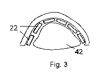
FIG. 3 is a schematic cross-sectional view of an ultrasound treatment probe
utilizable
in a method in accordance with the present invention.
CA 02681944 2009-09-25
WO 2008/118300 PCT/US2008/003511
DETAILED DESCRIPTION
In a method for treating spinal pain, one operates a scanning apparatus 12
(FIG. 1) to
locate, in a patient PA, a spinal disc SD afflicted with cracks or fissures.
One then operates a
treatment device 14 to apply waveform energy 16 to the afflicted spinal disc
SD to heat the
5 spinal disc sufficiently to modify collagen fibers of the spinal disc and
destroy pain receptors
in the spinal disc. The waveform energy 16 is generated outside the patient PA
and travels
through the patient's tissues PT to a focal point 18 or other locus.
Waveform energy 16 may take any effective form, such as microwave or radio-
frequency radiation. Preferably, the waveform energy 16 is ultrasonic waveform
energy. In
that case, treatment device 14 comprises an array 20 of scanning transducers
22 that are
placed into wave-transmitting contact with the patient's skin PS. Appropriate
activation of
transducers 22 generates ultrasonic pressure waves in the patient PA that are
focused at point
18 in spinal disc SD.
Transducers 22 of array 20 are connected to a treatment waveform generator 24
that is
in turn activated by a control unit 26 in response to instructions entered by
a user via an input
terminal or periphera128. During a scanning of a spinal column SC of the
patient PA via
scanning apparatus 12, the user views an image produced on a video monitor 30.
Scanning
apparatus 12 may take any convenient form (MRI, CAT) but preferably comprises
an
ultrasound scanner having an array 32 of transducer elements 34 that are
selectively
energized by a waveform generator 36 under the control of control unit 26.
Transducer
elements 34 may be piezoelectric crystals and are placed in wave-transmitting
contact (e.g.,
using a gel) with the patient's skin surface PS generally over spinal column
SC. Transducer
elements 34 generate unfocused ultrasonic pressure waves in the patient's
tissues PT that are
partially reflected back to array 32. Transducers 34 are selectively polled by
a signal
processor 37 that conducts a preliminary processing of the incoming reflected
waves and
provides an analyzed or partially analyzed image-data-containing signal to
control unit 26.
Control unit 26 modulates the operation of waveform generator 24 so that
transducers
22 focus waveform energy 16 at point 16 in the afflicted or degenerative
spinal disc SD to
heat the spinal disc sufficiently to modify collagen fibers of the spinal disc
and destroy pain
receptors in the spinal disc. To that end, control unit 26 includes an
intensity control module
38 and a duration control module 40 (FIG. 2) that regulate the amplitude and
timing of the
focused ultrasound. The therapeutic ultrasound radiation may be applied in
pulses for better
distribution and control. Intensity control module 38 and duration control
module 40
cooperate to ensure that a proper amount of ultrasonic waveform energy is
applied to an
CA 02681944 2009-09-25
WO 2008/118300 PCT/US2008/003511
6
afflicted disc. The control circuitry or programming ensures that enough
energy is applied to
heat the spinal disc sufficiently to modify collagen fibers of the spinal disc
and destroy pain
receptors in the spinal disc. The control circuitry or programming also
ensures that the
applied energy is limited to avoid overheating and consequent damage to the
spinal disc
collagen.
Treatment device 14 may be a high-intensity focused ultrasound (HIFU) device.
In
that case, transducer elements 22 of treatment transducer array 20 are
disposed in a fixed
configuration of parabolic transverse cross-section (see FIG. 3) that permits
an optimization
of the transducer's length/width ratio. Reference numeral 42 represents a
fluid-filled flexible
pouch that facilitates the creation of an effective patient-probe interface
over which ultrasonic
pressure waves are conducted into the patient's tissues PT.
The operating of scanning apparatus 12 includes generating ultrasonic pressure
waves
in the target spinal disc SD. Where both scanning apparatus 12 and heat-
inducing waveform-
generating treatment device 14 are ultrasound devices, the devices may be
separate dedicated
devices. In that case the scanning arrays 20 and 32 may be mounted to
respective substrates
or carriers (not illustrated). Alternatively, at least some transducer
elements 22, 34 may be
used to carry out both the imaging function and the therapeutic function. In
that case,
scanning apparatus 12 and treatment device 14 are implemented via a single
hardware
arrangement. A common set of transducers, e.g., transducers 22 or array 20,
perform the
functions of transducers 22 and 34, while waveform generator 36 carries out
the functions of
treatment waveform generator 24, all in response to signals from control unit
26. In this
combined functioning, transducers 22 may be operated in a non-focused phased-
array mode
to extract image information that is processed to produce images that are
displayed on video
monitor 30. Once an operating physician detects an afflicted spinal disc SD
from the
displayed images, the physician may instruct control unit 26 to energize the
phased
transducer array 20 so as to focus ultrasonic waves within the afflicted disc
SD.
The common set of transducers may take the parabolic configuration illustrated
in
FIG. 3. Transducers 22 are energized according to different algorithms for
imaging and
therapy, respectively. In the case of therapy, the transducers are energized
simultaneously to
focus ultrasound simultaneously at the focal point 16 or other locus of the
parabolic array 20.
(For focusing at a point, the transducers are disposed along a parabola of
revolution, while
focusing along a line is implemented by a prismatic parabola configuration.)
During
scanning, the transducers 22 of FIG. 3 are energized one at a time and may
also be polled in
sequence.
CA 02681944 2009-09-25
WO 2008/118300 PCT/US2008/003511
7
Even where scanning apparatus 12 and heat-inducing waveform-generating
treatment
device 14 have respective dedicated transducer arrays 20 and 32, the arrays
may be disposed
on the same substrate, for instance, the same probe head 44, as
diagrammatically illustrated in
FIG. 1. Probe head 44 comprises mounting structure that fixes transducers 34
of scanning
apparatus 12 relative to the transducers 22 of the HIFU device 14. Where the
probe head 44
is movable by the operator over the patient's skin surface PS, the operator
naturally moves
treatment transducers 22 in tandem with the scanning transducers 34.
Transducers 22 and 34
are typically electromechanical elements such as piezoelectric crystals.
Although the invention has been described in terms of particular embodiments
and
applications, one of ordinary skill in the art, in light of this teaching, can
generate additional
embodiments and modifications without departing from the spirit of or
exceeding the scope
of the claimed invention. Accordingly, it is to be understood that the
drawings and
descriptions herein are proffered by way of example to facilitate
comprehension of the
invention and should not be construed to limit the scope thereof.
