Note: Descriptions are shown in the official language in which they were submitted.
CA 02682005 2009-09-25
WO 2008/148547 PCT/EP2008/004457
- 1-
Detection of an analyte in a sample of hemolyzed whole blood
The present invention relates to a method of detecting an analyte in a
hemolyzed
whole blood sample the method comprising the steps of applying a sample of
hemolyzed whole blood known or suspected to contain an analyte of interest to
a
column comprising a restricted access chromatography material (RAM) thereby
binding the analyte, eluting the analyte from the RAM and detecting the
analyte,
wherein at least in the first step a buffer with a pH above 8.0 is used. The
novel
method ensures that hemoglobin does not interfere with the detection of the
analyte of interest. The method can be easily used in the online detection of
many
analytes, e.g. from a hemolyzed whole blood sample, like in the detection of
an
antibiotic, of folate or of immunosuppressive drugs, like tacrolimus or
sirolimus.
Background of the Invention
The more constituents are present in a sample the more difficult is the
analysis of a
target analyte comprised therein. Red blood cells contain a dramatic amount of
proteins and small molecular weight constituents that potentially interfere
with the
detection of an analyte of interest from a biological fluid like whole blood.
This is
one of the major reasons why in clinical routine preferably blood plasma or
simply
referred to as plasma (i.e. an anticoagulated whole blood sample; deprived of
cells
and erythrocytes) or blood serum or referred to simply as serum (i.e.
coagulated
whole blood; deprived of cells, erythrocytes and most proteins of the
coagulation
system, especially fibrin/fibrinogen), respectively, are used. Whole blood
samples
also tend to be more difficult to handle, e.g., as compared to serum or
plasma.
Whole blood tends to be less stable and slow rupture of erythrocytes impairs a
reliable measurement of quite a few analytes of interest.
As indicated above, serum or plasma may be obtained from whole blood and used
in the detection of an analyte. Cells and erythrocytes in theory may also be
removed
by filtration or centrifugation from whole blood. However, these methods are
neither appropriate for use in a routine diagnostic setting, nor would they
allow for
a correct meH ~.II\.111\.lll oJc~.ror.~o.,t ;F1~. tl.1..... a;~1al_.~.. .
tL~la~ ~. are at 1leasi
~~~ y~ pariiaiiy present inside red
blood cells.
The vast majority of procedures known in the art for the detection of an
analyte
from a whole blood sample require a further processing of the sample before
the
analyte can be quantified. In many procedures the analyte of interest is first
CA 02682005 2009-09-25
WO 2008/148547 PCT/EP2008/004457
-2-
separated from the majority of potentially interfering substances by selective
precipitation or extraction methods. Extraction can be performed in liquid
phase or
on a solid phase. This shall be exemplified by illustrating some of the
procedures
used in the detection of some immunosuppressive drugs or folate, respectively.
Well-known immunosuppressive drugs are e.g. mycophenolate mofetil (MMF),
rapamycin (RAPA also known as sirolimus) and tacrolimus (FK-506). Therapeutic
drug monitoring for immunosuppressive drugs is especially important for
transplant patients as well as for patients suffering from AIDS (cf. e.g.:
Drug. Ther.
Perspect. 17(22) (2001) 8-12). Most patients who undergo solid organ
transplantation require lifelong immunosuppressive therapy to prevent
allograft
rejection. But, because many immunosuppressive agents have narrow therapeutic
ranges, and are associated with various toxicities and the potential for drug
interactions, the use of therapeutic drug monitoring (TDM) in conjunction with
clinical assessment of patients may be particularly important.
Tacrolimus is a macrolide antibiotic that was first approved by the US Food
and
Drug Administration (FDA) in 1994 for the prevention of liver allograft
rejection. It
is up to 100 times more potent than cyclosporin in vitro, and clinically, it
is
associated with a greater reduction in the incidence of tissue rejection.
Tacrolimus
has demonstrated efficacy both as primary immunosuppressive therapy in
patients
undergoing various transplantation procedures, and as rescue therapy for
patients
with refractory acute allograft rejection after liver or kidney
transplantation. The
therapeutic trough concentration is in the range of 5-20 g/L.
Since at least part of the tacrolimus present in the circulation is
compartmented
within erythrocytes, a whole blood sample is used in the clinical routine
measurement of this drug. Tacrolimus can e.g. be detected by high performance
liquid chromatography (HPLC), HPLC interfaced to mass spectrometry (MS),
radio receptor assay (RRA), or by an immunoassay (IA). The latter two
methodologies do not detect tacrolimus and certain of its various metabolites
with
the same sensitivity. This may lead to an interference in the procedure used
(Murthy, J. N., et al., Clin. Biochem. 31 (1998) 613-617). At least in the
detection of
the various tacrolimus metabolites the HPLC-MS-procedure may be considered the
gold standard. All the procedures mentioned above, however, require the
extraction
of tacrolimus from whole blood. Usually acetonitrile is used in clinical
routine for
the extraction of tacrolimus from whole blood and no method appears to exist
that
would allow for an online measurement of tacrolimus from a whole blood sample.
CA 02682005 2009-09-25
WO 2008/148547 PCT/EP2008/004457
-3-
Sirolimus is, like tacrolimus, a macrolide antibiotic. It was first approved
in 1999 by
the US FDA for the prevention of allograft rejection after kidney
transplantation,
and indeed has shown promising results in this respect when used acutely in
combination with cyclosporin and corticosteroids. In vitro, sirolimus is up to
100
times more potent than cyclosporin, and clinically, it may exhibit synergism
with
cyclosporin, perhaps permitting a reduction in cyclosporin dosage. The
therapeutic
trough concentration is in the range of 5-15 g/L.
As for tacrolimus, a significant amount of sirolimus is present within
erythrocytes.
Therefore extraction of a whole blood sample is required no matter which
detection
method is used. In clinical routine a sample suspected to comprise sirolimus
is
subjected to HPLC and sirolimus is detected by ultraviolet light (UV) or by
tandem
mass spectrometry (MS/MS). Recently also a microparticle enzyme immunoassay
has been described (Jones, K., et al., Clinical Therapeutics 22, Suppl. B
(2000) B49-
B61).
Folate is the collective name of a group of related molecules differing in
oxidation
state. Folates are part of the water-soluble vitamin B group and are important
as
coenzymes for homocysteine metabolism and in the transfer of one-carbon groups
required for DNA replication. Inadequate folate status is related to increased
risk of
neural tube defects, is associated with cardiovascular disease, anemia, with
certain
cancers and with Alzheimer's disease. Serum or plasma folate concentrations
reflect
recent dietary intake, whereas erythrocyte folate concentrations are more
indicative
of body stores (Gunter, E.W., et al., Clin. Chem. 42 (1996) 1689-1694; Fazili,
Z., et
al., Clin. Chem. 51 (2005) 2318-2325; Pfeiffer, C.M., et al., Clin. Chem. 50
(2004)
423-432). Erythrocyte total folate (red blood cell folate = RBC-folate) is the
best
measure of whole body folate status. Recent studies have shown that 5-methyl
tetrahydrofolate is the dominant folate vitamer in circulating erythrocytes.
For the
diagnosis of folate deficiency it is recommended that determinations are
performed
not only from serum or from plasma but also from erythrocytes, since folate is
localized to more than 95% in the latter. The concentration in the
erythrocytes
more truly reflects the actual folate status.
A number of methods are available to measure folate in different matrices. The
major analytical methods are microbiological assay, radio immuno assay,
chemiluminescence, chromatographic methods and mass spectrometric methods.
Most methods are based on competitive binding of folate to folate binding
protein.
CA 02682005 2009-09-25
WO 2008/148547 PCT/EP2008/004457
-4-
For the measurement of RBC-folate the use of a hemolyzing reagent is obviously
mandatory. For example the Elecsys' assay (Elecsys is a trademark of a member
of
the Roche Group) for determination of RBC folate uses ascorbic acid as lysis
reagent. Elecsys RBC-folate hemolyzing reagent is used together with the
Elecsys
folate assay for the quantitative determination of folate in erythrocytes (RBC-
folate). Whole blood treated with anticoagulants (heparin or EDTA) is diluted
with
ascorbic acid solution (0.2%) and incubated for approximately 90 minutes at 20-
25 C. Lysis of the erythrocytes takes place, with liberation of the
intracellular folate.
The hemolysate is then used as a "prediluted" sample (in analogy to serum) for
subsequent measurement in the Elecsys folate assay. The hematocrit value
determined in whole blood and the dilution effect brought about by
pretreatment
of the sample is compensated for in the calculation of the erythrocyte folate
concentration (Greiling, H., Gressner, A.M., Lehrbuch der Klinischen Chemie
und
Pathobiochemie, 3rd ed., Stuttgart-New York, Schattauer (1995), pp. 460-462;
Gunter, E.W., et al., Clin. Chem. 42(1996) 1689-1694).
The hemolysate generated by treatment with ascorbic acid can not be used for
routine chromatographic procedures, because of interfering substances
contained
therein. For use of such hemolysate in a chromatographic procedure or mass
spectrometric determination it would necessary to remove cell debris and
precipitated protein prior to analysis.
Debris and precipitated proteins usually are removed from a sample by
centrifugation, offline filtration or solid phase extraction.
Solid phase extraction (SPE) is a chromatographic technique which is widely
used,
e.g., for preconcentration and cleanup of analytical samples, for purification
of
various chemicals, and for removal of toxic or valuable substances from
aqueous
solutions. SPE is usually performed using a column or cartridge containing an
appropriate resin. SPE procedures have been developed using sorbents which can
interact with analytes by hydrophobic, ion exchange, chelation, sorption, and
other
mechanisms, to bind and remove the analytes from fluids. Since different SPE
applications for different classes of analytes can require different sorbents,
there is a
concomitant need for sorbents with specific properties which have unique
selectivities for the analyte or class of analytes of interest. Representative
examples
of SPE materials and SPE columns, respectively, can be found in US 6,322,695
and
US 6,723,236.
CA 02682005 2009-09-25
WO 2008/148547 PCT/EP2008/004457
-5-
As obvious from the above discussion of the state-of-the-art procedures, the
direct,
especially the online measurement of these analytes from whole blood is not
possible at all or at least suffers from complicated and/or time-consuming
handling
steps. Without wanting to be bound to the following theory, it may well be
that the
lack of appropriate procedures in the art is due to at least two reasons: a) a
sample
of hemolyzed whole blood often contains aggregates or precipitates and b) the
extremely high concentration of hemoglobin interferes with the detection
method.
The inventors of the present invention have addressed and solved both
problems.
First a method has been developed called differential hemolysis that allows
e.g. for
the complete lysis of the erythrocytes as contained in a sample of whole blood
without formation of interfering aggregates or precipitates. The procedure for
differential hemolysis is described in some detail below.
The inventors of the present invention, however, also discovered that the high
concentration of hemoglobin as obviously present in a hemolyzed blood sample
tends to interfere with the sensitive detection of an analyte of interest.
This has even
been observed when using the most advanced chromatography materials, the so-
called restricted access chromatography materials (RAMs). If a sample of
hemolyzed blood is applied to a column comprising a RAM according to standard
procedures hemoglobin is not completely removed, rather hemoglobin tends to
bind to the RAM column and thus tends to interfere in the analyte detection.
The inventors of the present invention thus were facing the problem that
hemoglobin as e.g. present in a hemolyzed blood sample - even after
differential
hemolysis - may interfere with the detection of an analyte of interest. As
obvious to
the skilled artisan this problem becomes the more pronounced the more
sensitive
and the more precise the measurement has to be.
It would, however, be highly desirable if whole blood could be used directly
as a
sample, especially in the detection of low molecular weight analytes. This
would in
addition be especially advantageous in an online detection procedure making
use of
a liquid chromatography (LC) separation step. It is also obvious that e.g. the
direct
online detertinn of an immun:.suppri.ssive drug 1ioi11 whole blood would be an
important progress for a clinical routine laboratory.
It has now surprisingly been found and could be established that it is
possible to
efficiently avoid interference by hemoglobin, e.g. by hemoglobin as comprised
in a
CA 02682005 2009-09-25
WO 2008/148547 PCT/EP2008/004457
-6-
sample of hemolyzed whole blood. It has been found and is described in detail
below that under special buffer conditions hemoglobin does not bind to RAMs,
or
at least does not interfere with an online detection of the analyte of
interest. Under
these conditions RAMs can be used in an elegant way in the detection of an
analyte
of interest from a hemolyzed whole blood sample.
Summarv of the Invention
In a first embodiment the present invention relates to a method of detecting
an
analyte in a hemolyzed whole blood sample the method comprising the steps of
applying the sample of hemolyzed whole blood known or suspected to contain the
analyte of interest to a column comprising a restricted access chromatography
material (RAM) thereby binding the analyte, eluting the analyte from the RAM
and,
detecting the analyte, wherein at least in the step of applying the sample to
the RAM
a buffer with a pH above 8.0 is used.
In preferred embodiments it is e.g. described that the RAM is selected from
porous
silica or porous polymer-based particles and may have certain advantageous
modifications of the inner and/or outer surface. The method described herein
can
be used in the detection of certain clinically important analytes.
Detailed Description of the Invention
The method according to the present invention is performed in vitro, i.e. not
on the
human or animal body.
In a preferred embodiment the present invention relates to a method of
detecting
an analyte in a hemolyzed whole blood sample the method comprising the steps
of
a) applying the sample of hemolyzed whole blood known or suspected to contain
the analyte of interest to a column comprising a restricted access
chromatography
material (RAM) thereby binding the analyte, b) eluting the analyte from the
RAM
and, c) detecting the analyte, wherein at least in step (a) a buffer with a pH
above
8.0 is used.
, ~.. nerein to
Unless the context dictates otl:er :vi,,-,.-+~., ~ articles ~'~~ "a" and ~ '
"a~~-i" are used ' '
refer to one or to more than one (i.e. to at least one) of the grammatical
object of
the article. By way of example, "an analyte of interest" means one analyte of
interest
or more than one analyte of interest.
CA 02682005 2009-09-25
WO 2008/148547 PCT/EP2008/004457
-7-
A "restricted access material" or restricted access chromatography material
(RAM)
according to the present invention is a chromatography material having pores
with
an inner surface capable of binding the analyte of interest and a pore size
appropriate to prevent the protein fraction of the hemolyzed blood from
entering
into said pores.
The restricted access materials as originally described (Boos, K.S. and
Rudolphi, A.,
LC-GC 15 (1997) 602-611, and Rudolphi, A. and Boos, K.S., LC-GC 15 (1997)
814-823) have a hydrophobic inner surface that works alike a reversed phase
chromatographic material. The chromatography based on such RAM can be
considered to be a combination of a reversed-phase chromatography and size
exclusion chromatography.
The various types of RAM available are known to the skilled artisan.
Preferably the RAM used in a method according to the present invention is
selected
from porous silica or polymer-based particles. The RAM particles used in a
method
according to the present invention are in addition characterized by a
hydrophilic
coating.
The hydrophilic coating of the RAM, i.e. of the RAM outside of the pores is
preferably based on a coating material providing hydroxyl groups like
alkyldiol,
carboxylic groups like carboxymethyl, amino groups like aminopropyl or
bis(hydroxyethyl)aminoethyl, respectively. Also preferred is the use of
hydrophilic
polymers like polyethyleneglycol, polyvinylalcohol, oligo- and
polysaccharides,
dextran, peptides or proteins, respectively, to render the RAM outside the
pores
hydrophilic. Commercially available and preferred examples of RAM materials
that
can be used in a method as disclosed herein are Biotrap 500 MS (Chromtech,
Cogleton, United Kingdom) or LiChrosphere RP-18 ADS (Merck, Darmstadt,
Germany).
The inner surface of the RAM is capable of binding the analyte of interest.
The
inner surface of the RAM used in a method according the present invention will
be
chosen by the skilled artisan to match the chemical properties of the analyte
of
interest. The binding can be achieved by any appropriate interaction, like
molecular
imprinting, hydrophobic interaction, ionic interaction, or polar interaction.
The
skilled artisan is fully familiar with these types of interaction.
CA 02682005 2009-09-25
WO 2008/148547 PCT/EP2008/004457
-8-
In a preferred embodiment the RAM used in a method according to the present
invention is binding the analyte of interest by hydrophobic interaction or by
polar
interaction.
Preferably the inner pores of silica or polymer-based particles used in a
method
according to the present invention are coated with groups selected from anion
or
cation exchange materials or from hydrophobic groups, respectively.
Preferably the inner pores of silica or polymer-based particles used in a
method as
disclosed herein have a hydrophobically coated inner surface of the pores
wherein
the binding material is selected from the group consisting of aliphatic
groups,
phenyl groups, hydrophobic peptides or other reversed phase materials.
Preferably
the aliphatic group has between 4 and 20 C-atoms. Also preferred, the inner
surface
is a C4-, C8- C12-, C16- or C18-matrix. Preferred materials have a C4, C8 or a
C18
coating of the inner surface.
The pore size is selected in such a way that the analyte(s) can enter the
pores and
therefore are bound to the inner surface of the pores, e.g. to the reversed
phase.
Ideally, polymeric matrix constituents, typically blood proteins, especially
hemoglobin, are a) excluded from the pores by appropriate selection of the
pore
size and b) do not stick to the hydrophilic outer surface. If these
requirements are
met the polymers are not retained by the RAM and pass through the column in
the
void volume. In addition, since these polymers cannot enter the pores, almost
no
surface fouling which could alter the retention characteristics of the
material,
occurs. Generally, the analyte separation is performed on a second column,
which is
filled with a standard RP material. In a preferred embodiment the pore size of
the
RAM used in a method according to the present invention is between 60 and 120
A,
also preferred between 60 and 100 A.
RAMs are supposed for use in on-line extraction procedures wherein the analyte
of
interest is present in a very difficult matrix. However, it is desired and has
proven
necessary to remove as many proteins from a hemolyzed blood sample as possible
in order to minimize any interference of such molecules with the measurement
of
an analyte. Dependent on the pore siz? selected it is pos;ible to exciiide
liroieins
above a certain molecular weight. Preferably the protein fraction excluded
comprises the polypeptides of 20 kDa and larger. Also preferred the protein
fraction
excluded comprises polypeptides of 19 kDa and larger, 18kD and larger, 17kD
and
larger, 16kD and larger, and 15 kDa and larger.
CA 02682005 2009-09-25
WO 2008/148547 PCT/EP2008/004457
-9-
The lower limit for size exclusion of commercially available RAM materials is
given
by the respective manufacturers as being about 15 to 20 kDa. Whereas RAMs work
quite well with polypeptides having an apparent molecular weight of 20 kDa or
more, hemoglobin - which has a molecular weight of only 16 kDa - represents a
significant problem with the commercially available RAMs investigated. In fact
this
problem may be one of the reasons, or even the most important reason, why the
online chromatographic detection of an analyte from a sample of hemolyzed
blood
still is not established in clinical diagnostic routine.
As can be seen from the experiments presented further below, hemoglobin
unspecifically binds for example to a commercially available RAM ADS column,
if
this column is used according to a standard protocol. Upon elution of the
bound
materials, including the analyte of interest, hemoglobin is also set free from
the
RAM and interferes with the detection of the analyte. Surprisingly it has been
found
that the non-specific binding of hemoglobin, e.g. as contained in a sample of
hemolyzed whole blood can be drastically reduced if the sample comprising
hemoglobin is applied in a buffer having a pH of above 8Ø
"Applying" the sample at a certain pH to a RAM does not necessarily mean that
the
sample has this pH or has been adjusted to this pH, but means that the sample
application buffer into which the sample is injected has this pH. The skilled
artisan
often refers to such buffer also as the first eluent or as buffer (A). In a
routine and
preferred mode of liquid chromatography the RAM column usually is equilibrated
with the application buffer and the system is running, i.e. the application
buffer is
flowing through the chromatography setup and the sample is injected into the
buffer stream. This is why the application buffer can also be referred to as
an eluent,
e.g. as eluent (A). As the skilled artisan will appreciate the buffering
capacity of the
application buffer will be chosen to match the buffer strength of the sample.
In case
of a hemolysate the molarity of the buffer can be as low as 5mM. Preferably
the
buffer will be used at a concentration of about 10 or of about 20mM. As
indicated,
the buffer strength can be easily selected by the skilled artisan to match the
requirements.
At a pH above 8."v ihe non-specific binding of hemoglobin to a RAM is
abolished or
reduced to a level that does not interfere with the detection of an analyte of
interest,
respectively. Preferably the pH of the buffer used to apply the sample to the
RAM is
at least pH 8.5 or higher or also preferred at least pH 9.0 or higher, or pH
9.5 or
higher. The high end of the pH-range can be selected as appropriate. The
skilled
CA 02682005 2009-09-25
WO 2008/148547 PCT/EP2008/004457
-10-
artisan will choose a pH that does neither destroy the analyte of interest nor
the
RAM used. Preferably the pH at the high end will be pH 12.5 or less. Also
preferred
it will be pH 12.0 or less, or pH 11.5 or less, or pH 11.0 or less,
respectively.
In an elegant and preferred online setup, the sample is applied to a RAM under
appropriate buffer conditions and the RAM is washed with an appropriate
washing
buffer. By adjusting the valves and flow direction of the system, the analyte -
if
present - is eluted, passed over an analytical column and detected. In a
further
preferred embodiment the method of the present invention is practiced by using
the same pH for application and elution buffer. As the skilled artisan knows
the
elution of an analyte of interest can be adjusted to meet the conditions most
appropriate for the analyte under investigation. In many cases a gradient
elution
will serve the needs.
An analyte according to the present invention may be any inorganic or organic
molecule, including a biomolecule, excluding nucleic acids. The analyte will
not be
a nucleic acid, especially it will not be a DNA. Preferably the analyte is
selected from
the group consisting of a polypeptide, a carbohydrate, and an inorganic or
organic
drug molecule. Preferably the analyte of interest has a molecular weight of 10
kDa
or below, also preferred of 9 kDa or below, of 8 or below, of 7 kDa or below,
of 6
kDa or below, or of 5 kDa or below, respectively.
A polypeptide or protein is a molecule that is essentially composed of amino
acids
and that has at least two amino acids linked by peptidic linkage. In case the
analyte
of interest to be investigated in a method disclosed here, the polypeptide
preferably
will consist of at least 3, 4, 5, 6, 7, 8, 9, 10, 12, 15, 20, 25, and 30 to up
to about 100
amino acids. Preferably the polypeptide will contain from 5 to 100, also
preferred
from 10 to 40 amino acids. Suitable peptidic analytes of interest are e.g.
peptide
hormones, and other polypeptides present in the circulation and especially
polypeptides released from red blood cells due to the treatment with a
membrane
solubilizing agent according to the present invention.
Preferably the method according to the present invention is used in the online
u '
detection of an analyte from a;y1,~.lvi~, 0 1v.1tU..V...U7 ~-C wherein ~----'--
sai' ana
,~., ia salilpliyte is at least
partially located inside a red blood cell, like sirolimus, tacrolimus or
folate.
A preferred target analyte according to the present invention is selected from
the
group consisting of the drugs of abuse and the immunosuppressive drugs.
CA 02682005 2009-09-25
WO 2008/148547 PCT/EP2008/004457
-11-
Preferred target analytes are the drugs of abuse. The drug of abuse is
preferably
selected from the group consisting of amphetamine, cocaine and cocaine
metabolites like benzoylecgnonine, methamphetamine, opiate and opiate
derivatives, cannabinoids like tetrahydrocannabinol, and phencyclidine.
Another preferred target analyte is an immunosuppressive drug. The
immunosuppressive drug is preferably selected from the group consisting of
cyclosporine (CsA), mycophenolate mofetil (MMF), rapamycin (RAPA also known
as sirolimus), tacrolimus (FK-506) azathioprine (AZA), and methylprednisolone
(MP).
A further preferred target analyte is folic acid or a folic acid vitamer,
respectively.
One preferred analyte is the total folate as comprised in both the blood
plasma and
in the red blood cells.
In the method according to the present invention the analyte of interest is
first
bound to a RAM under appropriate conditions. For detection and/or quantitation
the analyte is eluted from the RAM and thereafter detected and or quantified
by an
appropriate method. Preferably the analyte of interest is eluted from the RAM
and
thereafter separated by aid of liquid chromatography. Preferably the material
eluted
from the RAM is further separated by reversed phase HPLC
Liquid chromatography (LC) is an extremely important analytical technique
which
is used for the separation, identification and quantitation of an analyte of
interest
even if present in a complex mixture of different sample constituents. During
LC
the chemical components in a mixture are carried through a column bed packed
with a suitable stationary phase by the flow of a liquid mobile phase. The
stationary
phase, usually irregularly or spherical particles, have a surface that is
suitable for
reversible interactions with the analytes. The particles may be porous in
order to
increase the surface area available for interactions. Separation in liquid
chromatography is achieved by means of differences in the interactions of the
analytes with both the mobile and stationary phases. As the skilled artisan
appreciates both a stationary phase and a mobile phase appropriate to the
analytes
under investigation }lavP to be chosen. In addiiion, tlie user will identify
chromatographic conditions appropriate to maintain the sharpness of analyte
bands as a sample moves through the stationary phase column to the detector.
CA 02682005 2009-09-25
WO 2008/148547 PCT/EP2008/004457
- 12-
High Performance Liquid Chromatography, also known as High Pressure Liquid
Chromatography, abbreviated as HPLC, is a special form of liquid
chromatography
and nowadays used frequently in biochemistry and analytical chemistry.
Compared
to LC, the particle size of the stationary phase is smaller, which, according
to well
known theory, results in less band dispersion and narrower peaks in the
resulting
chromatogram and thence to better resolution and sensitivity. Because of the
smaller particle diameter, the mobile phase in an HPLC application has to be
forced
through a column at high pressure.
The mobile phase is chosen to ensure solubility of the sample solutes. For the
stationary phase, preferably microparticulate silica (bare or chemically
modified) is
used, because its high surface area accentuates the differences in solute-
stationary
phase interactions. The use of a stationary phase that interacts strongly with
solutes
relative to solute mobile-phase interactions will result in very long
retention times,
a situation which is not analytically useful. Hence the stationary phase must
be
selected so as to provide weak to moderate solute interactions relative to
those in
the mobile phase. As a consequence, the nature of the solute governs the type
of LC
selected. The stronger interactions should occur in the mobile phase to ensure
sample solubility and ready elution, while the stationary phase should be
responsive
to more subtle differences among the solutes. For example, polar neutral
compounds are usually better analyzed using a polar mobile phase together with
a
nonpolar stationary phase that distinguishes subtle differences in the
dispersive
character of the solutes. One of the powerful aspects of HPLC is that the
mobile
phase can be varied to alter the retention mechanism. Modifiers can be added
to the
mobile phase to control retention. For example, pH is an important variable in
aqueous mobile phases.
Five general classes of LC can be distinguished:
1. Normal-phase chromatography calls for the use of a polar stationary phase
in
conjunction with a non-polar (dispersive) mobile phase.
2. Reversed-phase chromatography, the opposite possibility, calls for the use
of a
non-polar stationarv phase and a polar mcbile phase (co~~posed of one or
more of the polar solvents, e.g. water, methanol, acetonitrile, and
tetrahydrofuran).
CA 02682005 2009-09-25
WO 2008/148547 PCT/EP2008/004457
- 13 -
3. Ion-exchange chromatography involves ionic interactions. In this case the
mobile phase must support ionization to ensure solubility of ionic solutes.
The
stationary phase must also be partially ionic to promote some retention.
4. Size-Exclusion chromatography involves separations based on molecular size
alone and ideally requires that there be no adsorptive interaction of the
solutes
with the stationary phase.
5. Affinity chromatography is based on a specific interaction, e.g. between
the
members of a specific binding pair, like antigen and corresponding antibody or
receptor and corresponding ligand. For example a first partner of a binding
pair is bound to an appropriate stationary phase and used to capture the
second partner of the binding pair. The second partner can be released and
isolated by appropriate means.
The general classification of separation principles given above must not be
exhaustive and therefore is non-limiting, there are other separation
principles
which can be used for the separation of liquid samples, e.g. hydrophobic
interaction
chromatography, hydrophilic interaction chromatography, ion-pair reversed-
phase
chromatography, molecular imprinted materials based separation.
The characterization and or quantification of an analyte of interest can be
performed by any appropriate method. Appropriate and preferred detectors sense
the presence of a compound passing through, and provide an electronic signal
to a
recorder or computer data station. The output is usually in the form of a
chromatogram and a substance of interest is usually found in a certain peak.
The
peak area or peak height can be used to quantify the amount of analyte present
in
the sample investigated.
The detector for an HPLC system is the component that emits a response due to
the
eluting sample compound and subsequently signals a peak on the chromatogram.
It
is positioned immediately posterior to the stationary phase in order to detect
the
compounds as they elute from the column. The bandwidth and height of the peaks
may usually be adjusted using the coarse and fine tuning controls, and the
detection
and sensitivity parameters may also be controlled by the skilled artisan.
There are
many types of detectors that can be used with HPLC. Some of the more common
detectors include: Refractive Index (RI), Ultra-Violet (UV), Fluorescence,
CA 02682005 2009-09-25
WO 2008/148547 PCT/EP2008/004457
- 14-
Radiochemical, Electrochemical, Near-Infra Red (Near-IR), Mass Spectrometry
(MS), Nuclear Magnetic Resonance (NMR), and Light Scattering (LS).
Refractive Index (RI) detectors measure the ability of sample molecules to
bend or
refract light. This property for each molecule or compound is called its
refractive
index. For most RI detectors, light proceeds through a bi-modular flow-cell to
a
photodetector. One channel of the flow-cell directs the mobile phase passing
through the column while the other directs only the mobile phase. Detection
occurs
when the light is bent due to samples eluting from the column, and this is
read as a
disparity between the two channels.
Fluorescence detectors measure the ability of a compound to absorb then re-
emit
light at different wavelengths. Each compound has a characteristic
fluorescence.
The excitation light passes through the flow-cell while a monochromator and
photodetector measure the emitted light intensity.
Radiochemical detection involves the use of radiolabeled material, usually
tritium
(3H) or carbon-14 (14C). It operates by detection of fluorescence associated
with
beta-particle ionization, and it is most popular in metabolite research.
Electrochemical detectors measure compounds that undergo oxidation or
reduction reactions. This is usually accomplished by measuring gain or loss of
electrons from migrating samples as they pass between electrodes at a given
difference in electrical potential.
Mass spectrometry is an analytical technique used to measure the mass-to-
charge
ratio (m/z (or m/q)) of ions. It is most generally used to analyze the
composition of
a physical sample by generating a mass spectrum representing the masses of
sample
components. The technique has several applications, including: identifying
unknown compounds by the mass of the compound and/or fragments thereof;
determining the isotopic composition of one or more elements in a compound;
determining the structure of compounds by observing the fragmentation of the
compound; quantitating the amount of a compound in a sample using carefully
designed methods (mass spectrometry is not inherently quantitative); studying
the
fundamentals of gas phase ion chemistry (the chemistry of ions and neutrals in
vacuum); determining other physical, chemical or even biological properties of
compounds with a variety of other approaches.
CA 02682005 2009-09-25
WO 2008/148547 PCT/EP2008/004457
-15-
A mass spectrometer is a device used for mass spectrometry, and produces a
mass
spectrum of a sample to analyze its composition. This is normally achieved by
ionizing the sample components and separating ions of differing masses (m/z
ratios) and recording their relative abundance by measuring intensities of ion
flux.
A typical mass spectrometer comprises three parts: an ion source, a mass
analyzer,
and a detector.
The kind of ion source is a contributing factor that strongly influences what
types of
samples can be analyzed by mass spectrometry. Electron ionization and chemical
ionization are used for gases and vapors. In chemical ionization sources, the
analyte
is ionized by chemical ion-molecule reactions during collisions in the source.
Two
techniques often used with liquid and solid biological samples include
electrospray
ionization (ESI) and matrix-assisted laser desorption/ionization (MALDI).
Other
techniques include fast atom bombardment (FAB), thermospray, atmospheric
pressure chemical ionization (APCI), secondary ion mass spectrometry (SIMS)
and
thermal ionization.
In a preferred embodiment the detecting of an analyte in a method according to
the
present invention is performed by mass spectroscopy.
Nuclear magnetic resonance (NMR) detection is based on the fact that certain
nuclei with odd-numbered masses, including H and 13C, spin about an axis in a
random fashion. However, when placed between poles of a strong magnet, the
spins
are aligned either parallel or anti-parallel to the magnetic field, with the
parallel
orientation favored since it is slightly lower in energy. The nuclei are then
irradiated
with electromagnetic radiation which is absorbed and places the parallel
nuclei into
a higher energy state; consequently, they are now in "resonance" with the
radiation.
Each H or C will produce different spectra depending on their location and
adjacent atoms or atom groups in the compound, because all nuclei in molecules
are surrounded by electron clouds which change the encompassing magnetic field
and thereby alter the absorption frequency.
When a source emits a parallel beam of light which strikes particles in
solution,
some light is reflected, absorbed, transmitted, or scattered. TheSe
pi12nGme;,a l.d,l
be measured by a light-scattering (LS) detector. The most prominent forms of
LS
detection are termed nephelometry and turbidimetry. Nephelometry is defined as
the measurement of light scattered by a particulate solution. This method
enables
the detection of the portion of light scattered at a multitude of angles.
Turbidimetry
CA 02682005 2009-09-25
WO 2008/148547 PCT/EP2008/004457
- 16-
is defined as the measure of the reduction of light transmitted due to
particles in
solution. It measures the light scatter as a decrease in the light that is
transmitted
through the particulate solution. Therefore, it quantifies the residual light
transmitted.
Near-infrared detectors operate by scanning compounds in a spectrum from 700
to
1100 nm. Stretching and bending vibrations of particular chemical bonds in
each
molecule are detected at certain wavelengths.
The method described herein can be used in the detection of an analyte from a
sample of hemolyzed whole blood. As obvious to the skilled artisan the method
according to the present invention will also work with other basic sample
materials
like plasma, serum, urine or CSF. A preferred sample to be used in a method
according to the present invention thus can be selected from the group
consisting
of plasma, serum, urine, CSF and hemolyzed whole blood. A sample of hemolyzed
whole blood is preferred.
As mentioned above, the authors of the present invention have also discovered
a
method for differential hemolysis. This method is not yet available to the
public
and therefore shall be described in some detail below. The term differential
hemolysis relates to method of treating a sample comprising erythrocytes with
a
membrane solubilizing agent under conditions appropriate to lyse cell
membranes
of red blood cells and at the same time not to cause precipitation of sample
constituents. As will be obvious from the below description, the method
according
to the present invention is preferably practiced with a sample of
differentially
hemolyzed whole blood.
"Red blood cells" in the sense of the present invention are red blood cells
not
having a cell nucleus. Such red blood cells not having a cell nucleus are e.g.
the
mature red blood cells as found in the circulation of mammals. This invention
does
not relate to nucleated red blood cells as e.g. known from avian species. The
latter
ones would meet the criteria for nucleated or eukaryotic cell.
"Mammal" for purpose of the present invention refers to any animal classified
as a
mammal, including humans, domestic and farm animals, and zoo, sports, or pet
animals, such as dogs, cats, cattle, horses, sheep, pigs, goats, rabbits, etc.
Preferably,
the mammal is human.
CA 02682005 2009-09-25
WO 2008/148547 PCT/EP2008/004457
- 17-
A "eukaryotic cell" or "nucleated cell" in the sense of the present invention
is a cell
derived from a eukaryotic organism and still having its cell nucleus. Examples
of
eukaryotic cells are cells derived from nucleated tissue, nucleated tissue
culture cells
and nucleated blood cells. In a preferred embodiment the eukaryotic cell is a
nucleated blood cell like a thrombocyte, a monocyte, neutrophils, eosinophils
or a
leukocyte. Cells from lower organisms, like bacteria, though containing
genetic
material, are not eukaryotic cells.
Hemolysis of a whole blood sample can be performed by any hemolysis method
known in the art. Preferably the hemolyzed whole blood sample is obtained by
differential hemolysis. The advantageous properties of differential hemolysis,
i.e. of
processing a liquid sample with a membrane solubilizing agent under conditions
appropriate to lyse cell membranes of red blood cells and at the same time not
to
cause precipitation of sample constituents, as demonstrated in the Examples
section, have been established by using whole blood samples.
Preferably the liquid sample subjected to a differential hemolysis with an
appropriate membrane solubilizing agent comprises red blood cells and may
comprise or comprises, respectively, nucleated cells. Further preferred the
liquid
sample comprises both red blood cells and nucleated cells. Preferably the
liquid
sample used in a method subjected to differential hemolysis and thereafter
used in a
method according to the present invention will be whole blood. As will be
appreciated a whole blood sample contains both red blood cells without nuclei
as
well as nucleated blood cells.
Preferably the whole blood sample is processed directly, i.e. directly after
sampling
it is subjected to the differential hemolysis. Also preferred, the whole blood
will be
collected/treated with an appropriate anti-coagulant to yield an anti-
coagulated
whole blood sample. Well-known anti-coagulants frequently used in clinical
diagnostic routine are heparin, citrate and EDTA. Preferably the sample
according
to the present invention is an anti-coagulated whole blood sample, especially
a
citrated whole blood sample or an EDTA-anti-coagulated whole blood sample that
has been differentially hemolyzed.
In a method for differential hemolysis the liquid sample is treated in such a
manner
that two requirements are met: a) if red blood cells are present, the
membranes of
red blood cells are disrupted and b) at the same time no precipitation of
sample
constituents is caused. As mentioned, this process is termed differential
hemolysis.
CA 02682005 2009-09-25
WO 2008/148547 PCT/EP2008/004457
- 18-
In case the method is practiced on a whole blood sample a processed sample, a
differentially hemolyzed whole blood sample, is obtained containing lyzed red
blood cells but at the same time no precipitate.
By applying a suitable membrane solubilizing agent under appropriate
conditions
the integrity of the cellular membrane that is e.g. essential for shielding
the contents
of a red blood cell from the blood plasma is lost. The content of the
erythrocytes
(e.g. hemoglobin but also some analytes of interest) is released into the
surrounding
liquid. At the same time no precipitation of sample constituents is caused.
As the skilled artisan will appreciate sample constituents that might
interfere with a
later analysis may especially be DNA and proteins. As long as the nuclei of
eukaryotic cells, e.g. like lymphocytes or monocytes are not destroyed, no DNA
is
released from these nuclei. As long as no proteins precipitate, proteins
comprised in
the sample subjected to differential hemolysis will not interfere, at least
not to a
significant extend with a routine chromatography step. However, as illustrated
further above, RBC constituents - especially hemoglobin - may nonetheless
interfere with the detection of an analyte of interest.
The integrity of red blood cells can for example be easily assessed by
appropriate life
stains. In a preferred embodiment according to the present invention trypane
blue
is used in order to assess the integrity of a red blood cell membrane. Intact
red
blood cells do not accumulate trypane blue, whereas a red blood cell with a
disrupted membrane does stain with trypane blue. The membrane integrity of a
red
blood cell is easily assessed under the microscope after staining a sample
with
trypane blue. The percentage of disrupted red blood cells is calculated by
counting
intact red blood cells before and after the treatment, by then dividing the
first
number by the latter number and by then multiplying this value by 100. Red
blood
cells that are solubilized are referred to as lyzed red blood cells or as
lyzed
erythrocytes.
The appropriate treatment will be adequate to lyse a red blood cell, but at
the same
time it will not cause precipitation of sample constituents. It is expected
that the
appropriate hemolysis treatment in a method according l, +.. + i.~
~., ,., prc^Sciii 111Ve1111VI1
will also effects the outer membranes of eukaryotic cells. However, care can
and
must be taken that the DNA contained in the cell nuclei is not released into
the
sample. The hemolysis reagent and conditions used will either and preferably
leave
the nuclear membrane and thus the nuclei macroscopically intact or at least
DNA
CA 02682005 2009-09-25
WO 2008/148547 PCT/EP2008/004457
- 19-
will not be set free from its surrounding and DNA-stabilizing nuclear
proteins. If
DNA would be released to a significant extend such DNA might or even would
interfere with further handling of the sample. Released DNA e.g. tends to make
the
liquid very viscous. It is then no longer possible to pipette or transfer such
sample
or to pass it through certain filters or columns.
Care can and must also be taken that no protein precipitation occurs. As the
skilled
artisan will appreciate, there are many many different proteins present in a
biological sample, e.g. in a whole blood sample. All these proteins have
individual
properties influencing their tendency to precipitate or aggregate.
It has now been found that it is possible to describe and define whether
sample
processing with a membrane solubilizing agent is performed under appropriate
conditions i.e. in a mode to lyse cell membranes of red blood cells on the one
hand
and at the same time not to cause precipitation of sample constituents. Both,
red
blood cells not lysed as well as precipitated sample constituents would have a
negative impact on the properties of such sample.
Whether the conditions for differential hemolysis are appropriate can be
easily and
preferably determined by using the following standardized procedure. A whole
blood sample with a hematocrit of 40 is diluted 1:10 with the candidate
hemolysis
reagent. The efficacy of a reagent for bringing about differential hemolysis
is seen
visually. Upon lysis of the erythrocytes the mixture becomes clear. If
precipitation
of sample constituents occurs the sample becomes turbid or viscous or both.
As indicated above, the conditions used in a method of differential hemolysis
according to the present invention can easily be assessed visually. If a whole
blood
sample is incubated with an appropriate candidate reagent for differential
hemolysis the minimal concentration required to hemolyse red blood cells can
be
recognized as the lowest concentration rendering the turbid blood sample
transparent or clear. The highest possible concentration is the one still
leading to a
transparent and non-viscous sample.
It has turned out rather easy to determine the appropriate minimal final
concentration of the candidate hemolysis reagent as the concentration leading
to
the change in transparency of a treated whole blood sample. This change in
transparency correlates well with the suitability of such processed sample for
direct
analysis by HPLC. However, for the sake of an unambiguous definition it is
CA 02682005 2009-09-25
WO 2008/148547 PCT/EP2008/004457
-20-
preferred that minimal concentration of a hemolysis reagent is confirmed by
the
HPLC method as described below.
The maximal concentration of hemolysis reagent possible is the concentration
still
not causing release of DNA and/or precipitation of a protein. The sample
thereby
would turn viscous or turbid or both and is not suitable for a direct HPLC
application anymore. Whereas viscosity and turbidity can be followed visually
it is
preferred that maximal concentration of a hemolysis reagent is confirmed by an
HPLC method as described below.
Both, a whole blood sample still comprising too many non-lysed erythrocytes as
well as a treated whole blood sample comprising precipitated sample
constituents
will not be suitable for any chromatographic procedure. This is why the
conditions
appropriate to bring about differential hemolysis preferably are determined by
applying in a standardized manner a sample of whole blood treated with a
candidate reagent for differential hemolysis to an HPLC column.
Incomplete hemolysis and/or precipitation of sample constituents is assessed
by
applying 50 times 10 l of the processed whole blood sample to an HPLC column.
To assess whether a candidate hemolysis reagent for differential hemolysis is
appropriate, said hemolysis reagent is mixed with a sample of whole blood.
Preferably EDTA-blood with a hematocrit of 40 that has been prediluted 1:10 in
physiological saline is used. It is mixed in a 1:1 ratio with the candidate
hemolysis
reagent and the mixture is incubated for 30 min at 20 C. 50 aliquots of 10 L
of the
this mixture, i.e. a processed whole blood sample are applied to a filter with
a
diameter of 2 mm and 0.5 m pore size that is part of an HPLC system. In case
the
frit is part of an HPLC column the stationary phase must be selected not to
cause
any interference or blocking. The back-pressure is monitored. A candidate
reagent
for differential hemolysis that would cause an increase in back-pressure of 20
bar or
more - if the back-pressure for injection 50 and the back-pressure for the
first
injection are compared to each other - would be deemed not to be appropriate.
This way both the minimal as well as the maximal final concentration of an
appropriate reagent for differential hemolysis can easily be identified.
Preferably the filter used in the above assessment of a candidate reagent for
differential hemolysis is an HPLC frit. Also preferred the frit is part of an
HPLC
column of 20 mm in length filled with 3.5 m Symmetry C18 particles with a
pore
size of 100A as bed material, and having an inner column diameter of 2 mm.
CA 02682005 2009-09-25
WO 2008/148547 PCT/EP2008/004457
-21-
As the skilled artisan will readily appreciate the whole blood sample used for
such
assessment is obtained from a healthy individual, i.e. an individual having no
known disease and biochemical values in the normal range.
It has been found and established that appropriate conditions can be
established for
quite many chemicals in order to meet both the requirements for differential
hemolysis. The reagent for differential hemolysis comprises a membranolytic (=
membrane solubilizing) chemical in the appropriate concentration to after
mixing
with the sample result in differential hemolysis.
The membrane solubilizing agent according to the present invention preferably
is
based on water as a solvent comprises a chemical or reagent bringing about the
differential hemolysis as described above, and also preferred it may comprise
a
buffer and/or a preservative. The agents used for differential hemolysis
preferably
are based on chemicals or reagents with membrane solubilizing activity that
have a
molecular weight of less than 1000 Dalton.
The membrane solubilizing agent preferably is based on the membranolytic
action
of one or more of the following chemicals: KBr; KJ; and KSCN or on a salt
consisting of one or more of the following cations and anions:
The cation preferably is selected from
mH(2m+l)
j
N+ N
i
CnH(2n+1) C8H17
0
NH2
N,C4H9 N
CH20C8H17
wherein m is 0 or 1 and n is 4 or 6.
The anion is preferably selected from chloride, tetrafluoroborate,
octylsulfate,
iodide und thiocyanate. It is also possible to use mixtures of the above
mentioned
CA 02682005 2009-09-25
WO 2008/148547 PCT/EP2008/004457
-22-
chemicals. As the skilled artisan appreciates it is these chemicals that
facilitate the
differential hemolysis whereas other ingredients of a hemolysis reagent may
serve
different purposes and may e.g. function as a buffer or as a preservative.
Preferably the chemical comprised in a reagent for differential hemolysis is a
salt
wherein the cation preferably is selected from
mH(2m+1)
+
N+ N
i
CnH(2n+,) C8H17
0
NH2
NN-C4H9 N
CH20C8H17
wherein m is 0 or 1 and n is 4 or 6, and wherein the anion is preferably
selected
from chloride, tetrafluoroborate, octylsulfate, iodide und thiocyanate.
Appropriate membranolytic chemicals comprised in a membrane solubilizing agent
are preferably selected from the group consisting of 1-Butyl-4-
methylpyridinium
tetrafluoroborate; 1-Butyl-3-methyl-imidazolium tetrafluoroborate; 1-Butyl-3-
methyl-imidazoliumoctylsulfate; 1-Butyl-3-methyl pyridiniumchloride; 1-
Hexylpyridiniumchloride; 1-Methyl-l-octyl pyrrolidiniumchloride; N-
Octylpyridiniumchloride; 3-Carbamoyl-l-octyloxymethyl pyridiniumchloride;
KBr; KJ; and KSCN, and of combinations thereof.
Preferably the membranolytic chemicals comprised in a membrane solubilizing
agent are selected from the group consisting of 1-Butyl-4-methylpyridinium
tetrafluoroborate; 1-Butyl-3-methyl-imidazolium tetrafluoroborate; 1-Butyl-3-
methyl-imidazoliumocryls,ulfate; 1-R,wtyl-3_n:ethyl Yyridiniun-1chloride; 1-
Hexylpyridiniumchloride; 1-Methyl-l-octyl pyrrolidiniumchloride; N-
Octylpyridiniumchloride; and 3-Carbamoyl-l-octyloxymethyl pyridiniumchloride.
It is further preferred to use a mixture of one these reagents and of KSCN.
CA 02682005 2009-09-25
WO 2008/148547 PCT/EP2008/004457
-23-
As obvious to the skilled artisan, once an appropriate concentration of a
candidate
reagent for differential hemolysis has been identified in the above defined
method
that is based on a 1 in 20 dilution of a whole blood sample in a candidate
hemolysis
reagent, another ratio of whole blood sample to an adjusted hemolysis reagent
can
be used as required.
In case the analyte of interest is expected to be highly concentrated in the
blood
sample under investigation, the final concentration of the hemolysis reagent
can
stay the same as identified in the above setting and lower ratios of whole
blood to
hemolysis reagent, e.g., 1:30, 1:40 or 1:50 can be used. Preferably in a
membrane
solubilizing agent according to the present invention the reagent for
differential
hemolysis is used in at least the minimal concentration sufficient to achieve
differential hemolysis as determined above.
In case the analyte of interest is present in rather a low concentration it
may be
necessary not to dilute the whole blood sample 1:20 but less. This is feasible
by
adjusting the concentration of the hemolysis reagent accordingly, such that
the final
relative concentration of hemolysis reagent to whole blood in the mixture of
the
hemolysis reagent and the whole blood sample stays within the ratio identified
for
the required minimal and maximal concentration, respectively, of hemolysis
reagent as determined in the above described assessment.
By way of example: It has been found that 1-Methyl-1-octyl
pyrrolidiniumchloride/KSCN if used in a final concentration of 1% and 0.4% in
a
membrane solubilizing agent according to the present invention, respectively,
are
appropriate to achieve the desired result, i.e. the differential hemolysis of
a whole
blood sample at a final dilution of 1:20. Dilution of an analyte in the
processed
blood sample can be reduced if for example the concentration of this hemolysis
reagent is adjusted to 2% for 1-Methyl-l-octyl pyrrolidiniumchloride and 0.8%
for
KSCN, respectively. The membrane solubilizing agent comprising this adjusted
concentration of hemolysis reagent, if later mixed 1:1 with a 1:5 diluted
whole
blood sample, also leads to differential hemolysis of the whole blood sample.
Since
the ratio of whole blood to hemolysis reagent is kept constant, this processed
blood
sar,iple is oiiiy diluteci 1:10. if 1 mi of a membrane solubilizing agent
comprising
10% of 1-Methyl-l-octyl pyrrolidiniumchloride and 4% of KSCN, respectively, is
mixed with 1 ml of whole blood diluted 1:1 in PBS differential hemolysis is
also
observed. Alternatively 1 ml of whole blood could be added to 2 ml of a
membrane
CA 02682005 2009-09-25
WO 2008/148547 PCT/EP2008/004457
-24-
solubilizing agent comprising 10% of 1-Methyl-l-octyl pyrrolidiniumchloride
and
4% of KSCN, respectively.
For many routine applications it is expected that the ideal ratio of whole
blood
sample to a membrane solubilizing agent will be between 10:1 and 1:20.
Preferably
in a method according to the present invention the sample of whole blood is
mixed
with the hemolysis reagent at a ratio from 5 to 1 to 1 to 15. More preferred
the ratio
is between 2 to 1 and 1 to 10, also preferred between 1 to 1 and 1 to 5. The
final, i.e.
highest possible concentration of an adjusted hemolysis reagent used in the
clinical
routine will depend on the solubility and also the price of such reagent.
Preferably an appropriate reagent for different hemolysis is further
characterized in
that the (minimal) concentration of the chemical required for disrupting the
membrane of a red blood cell and the (maximal) concentration tolerated for
said
chemical at which at the same time no precipitation of sample constituents is
caused are at least two-fold apart. The broader the window between minimal and
maximal concentration for a the reagent responsibly for differential hemolysis
the
more easily such reagent can be used in clinical diagnostic routine.
It is further preferred that the membranolytic chemical comprised in a reagent
for
differential hemolysis is used at a concentration that after mixing said
reagent with
a sample the final concentration of this membranolytic chemical corresponds to
the
mean value plus 30% of the minimal concentration minus 30% the maximal
concentration, respectively. Further preferred the concentration of the
membranolytic chemical comprised in the reagent for differential hemolysis
will be
adjusted that after mixing it with a sample it is within plus or minus 25%,
20% or
15% of the mean value of the minimal and maximal concentration, respectively.
It is also preferred that the membranolytic chemical comprised in a reagent
for
differential hemolysis is used at a concentration that after mixing with a
sample a
final concentration of this hemolytic chemical corresponding to a
concentration
between one and four times the minimal concentration, and also preferred
between
1.5 times and 3 times the minimal concentration determined as described above
is
3(1 nhtainPd
Preferably the reagent for differential hemolysis in a membrane solubilizing
agent
of the present invention is used at a concentration of no more than 75%
weight/volume, also preferred at no more than 50% weight/volume.
CA 02682005 2009-09-25
WO 2008/148547 PCT/EP2008/004457
-25-
Preferably the method of differentially hemolyzing a sample is combined with a
method according to the present invention. Preferably this method also uses a
further liquid chromatography (LC) step.
In a further preferred embodiment the present invention relates to a method of
detecting an analyte in a liquid sample the method comprising the steps of
obtaining said liquid sample, subjecting said liquid sample to a method of
sample
processing by a membrane solubilizing agent, wherein the solubilizing agent is
appropriate to disrupt the membrane of red blood cells, and not to destroy the
nuclei of eukaryotic cells, if required, adjusting the pH to a value above pH
8.0,
applying the sample to a RAM, and thereafter eluting the bound material,
subjecting it to liquid chromatography and analyzing the analyte under
investigation by appropriate means. Preferably the steps of applying the
sample to
RAM, eluting and chromatographing the sample as well as the method of analysis
are all performed directly (online).
In routine applications the stationary phase, the so-called bed material, e.g.
derivatized silica particles in an RP-HPLC-application, is packed into an
appropriate column, and is protected by a frit. The frit material usually is
selected
to have e.g. a smaller pore size as compared to the pore size of the bed
material.
In HPLC methods the diameter of the stationary phase particles usually is in
the
range of 1 to 10 m. These small particles necessitate the high pressure used
in
HPLC. The bed material usually is protected by a frit. Typical frits have a
pore size
of 1 m, 0.45 m or 0.2 m. The smaller the particles the smaller is usually
the pore
size of the frit. If a sample comprises a constituent capable of blocking an
HPLC frit
this is detrimental for any routine analysis. A whole blood sample, as well as
an
"over-treated" whole blood sample comprising precipitates of sample
constituents
causes a rapid blocking of any routine HPLC frit or column. As the skilled
artisan
will appreciate blocking of the frit used in an HPLC column will occur the
more
rapidly the lower the pore size of the frit, the smaller the diameter of the
stationary
phase particles and the smaller the column diameter. In case the frit would
not be
selected appropriately, i.e. a too large pore size, the particle size of the
column
material would also matter and the column itself would block more rapidly the
smaller the particles are.
By subjecting a sample to differential hemolysis, e.g. to a sample of whole
blood, it
is possible to directly apply such treated sample to an HPLC column, without
CA 02682005 2009-09-25
WO 2008/148547 PCT/EP2008/004457
-26-
running the risk of blocking the column. Preferably, the stationary phase
particles
used in such HPLC step are in the range of 1 to 10 m, also preferred in the
range
of 2 to 7 m in diameter. Preferably the frit used in such HPLC step has a
pore size
of 0.5 m or also preferred of 0.2 m.
As illustrated in the Examples, it is now possible to subject a sample, e.g.
of whole
blood to differential hemolysis, to apply such hemolysate to a RAM under
conditions wherein specific pH requirements are met, to elute the RAM-bound
material and to detect the absence, presence or quantity of an analyte of
interest like
an antibiotic or an immunosuppressive drug, like tacrolimus or rapamycin.
The following examples and figures are provided to aid the understanding of
the
present invention, the true scope of which is set forth in the appended
claims. It is
understood that modifications can be made in the procedures set forth without
departing from the spirit of the invention.
Description of the Figures
Figure 1 pH 6.6: Chromatogram of hemoglobin at 396 nm. Peak #1
represents hemoglobin in the flow through, peak #2 represents
hemoglobin as eluted at 15% buffer B and peak #3 represents
hemoglobin found once the column is washed with 100% buffer
B, respectively.
Figure 2 pH 10.7: Chromatogram of hemoglobin at 396 nm. Peak #1
represents hemoglobin in the flow through; peak #2 represents
hemoglobin as eluted at 20% buffer (B), respectively.
Figure 3 Investigation of hemoglobin carry-over at pH 10.7. Hemoglobin
carry-over was investigated by performing a blank injection after
the analysis of a haemoglobin-containing sample. As can be seen
from the chromatogram, no haemoglobin can be identified at
20% buffer (B).
Figure 4 Experimental setup used to extract analytes from hemolysates.
Hemoglobin was monitored after Biotrap 500 MS with UV-Vis
d?tec tinn. A na1 ~.teS '~ + ~+^a ~`"' `L _ '_ , ,
., ue~,., ~,.u a. ~ u~c aiia~yiicai coiumn by
mass spectrometry.
Figure 5 Blank injection of whole blood hemolysate. The chromatogram
shows that at the position where tetracycline should elute, no
peak is found.
CA 02682005 2009-09-25
WO 2008/148547 PCT/EP2008/004457
-27-
Figure 6 Injection of whole blood hemolysate spiked with tetracycline. The
chromatogram shows that at the position where tetracycline
should elute, a clear peak for this analyte is found.
Figure 7 Blank chromatographic run on Biotrap 500 MS after an injection
of hemolysate as a sample. As obvious from this chromatogram
no hemoglobin is detected.
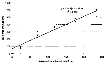
Figure 8 Calibration curve for tetracycline spiked into a human whole
blood hemolysate. A linear correlation is observed as indicated by
the extrapolation line shown in this Figure and by the high
coefficient of correlation calculated (r2 = 0.945).
Figure 9 Analytical HPLC setup used to extract and detect immuno
suppressive drugs from a whole blood hemolysate.
Figure 10 Extraction of tacrolimus from a whole blood hemolysate with on-
line SPE-HPLC-UV. The chromatogram taken at 291 nm (the
maximal adsorption of tacrolimus) is depicted. The arrow
indicates the peak corresponding to tacrolimus.
Figure 11 Extraction of rapamycin from a whole blood hemolysate with on-
line SPE-HPLC-UV. The chromatogram taken at 278 nm (the
maximal adsorption of rapamycin) is depicted. The arrow
indicates the peak corresponding to rapamycin.
Example 1
Evaluation of various RAMs at different pH values
The RAM columns were directly installed into an analytical HPLC system. Ten L
of hemoglobin solution were injected. First tests were performed with sample
application buffers (Eluent buffer A) of different pH of 6.6 and at pH 10.7,
respectively. For each pH value, amounts of hemoglobin flowing through and
retained on the column were computed. Carry-overs were also evaluated by
performing blank injections consecutive to injections of hemoglobin.
The column effluent was monitored at 396 nm (the latter being the absorption
maximum of hemoglobin). The columns tested included LiChrospher RP-18 ADS
from Merck (Merck, Darmstadt, Germany, ordering number 1.50947.0001 ), and
Biotrap 500 MS from ChromTech Ltd., Cogleton, United Kingdom, ordering
number BMS134K
CA 02682005 2009-09-25
WO 2008/148547 PCT/EP2008/004457
-28-
a) Evaluation of hemoglobin interference at pH 6.6 (5mM ammonium acetate)
A LiChrospher RP-18 ADS column from Merck was used. 10 L of sample (150
mg/mL of hemoglobin dissolved in 5 mM ammonium acetate at pH 6.6) was
applied to the column. For elution of hemoglobin a mobile phase using the two
buffers (A) 5 mM ammonium acetate in H20 (pH 6.6) and (B) 5 mM ammonium
acetate in acetonitrile (ACN) (pH 6.6), respectively was used. Elution was
performed for 15.0 min isocratic at 100 % (A), then using a linear gradient
from
100% (A)-0% (B) to 0% (A)-100% (B) in 30.0 min, and thereafter by a wash step
for 2.0 min at 100 % (B) to detect any hemoglobin reversibly bound to the RAM.
The flow rate was 1.5 mL/min and all steps were carried out at room
temperature.
At pH 6.6, hemoglobin is predominantly present in the flow-through (set to 100
%), partially elutes at around 15 % B (24 %), and is also present in the wash
step at
100 % (B) (4 %). However, this separation is not always reproducible and the
correct quantitation of both the unretained as well as the retained
hemoglobin,
respectively, is rather difficult because of detector saturation with the flow-
through
peak and the large width and the flatness of the retained peaks (cf. Figurel).
Hemoglobin in the wash peak is critical, since this would most likely imply
interference by hemoglobin in an online set-up for an analyte measurement from
a
hemolysate. Since hemoglobin was already present in the wash step, the risk of
hemoglobin carry-over, i.e. the risk of interference in any further analyte
detection
by hemoglobin contained in a sample previously applied to the online system is
very high and would for example result in contamination of electrospray ion
sources used for mass spectrometric detection.
b) Evaluation of hemoglobin interference at pH 10.7 (10 mM ethanolamine)
A LiChrospher RP-18 ADS column from Merck was used. 10 L of sample (150
mg/mL of hemoglobin dissolved in 10 mM ethanolamine at pH 10.7) was applied
to the column. For elution of RAM-bound hemoglobin a mobile phase using the
two buffers (A) 10 mM ethanolamine in H20 (pH 10.7) and (B) 10 mM
ethanolamine in ACN (pH 10.7), respectively was used. Elution was performed
for
15.0 min isocratic at 100 % (A), then using a linear gradient from 100% (A)- 0
io
(B) to 0% (A)-100% (B) in 30.0 min, and thereafter by a wash step for 2.0 min
at
100 % B. The flow rate was 1.5 mL/min and all steps were carried out at room
temperature. At pH 10.7, hemoglobin is predominantly present in the flow-
through
(set to 100 %), partially elutes at around 20 % (B) (20 %), and is not present
to any
CA 02682005 2009-09-25
WO 2008/148547 PCT/EP2008/004457
-29-
significant level in the cleaning peak (cf. Figure 2). The absence of
hemoglobin from
the cleaning peak as observed after application of 100 % (B) is very
important, since
this would indicate that under these buffer conditions hemoglobin would not
cause
any memory effect, i.e. it would not be present in the next measurement using
the
same column. Practically no carry-over was observed at pH 10.7. This pH value
appears to be appropriate to avoid carry-over of hemoglobin between different
injections onto RAM and to thus to avoid memory effects.
In order for an online method to be fully compatible with routine
requirements,
such method also necessitates not be hampered by carry-over problems. When
measuring an analyte from a sample of hemolyzed whole blood, carry-over
problems would most likely result from hemoglobin carry-over, since hemoglobin
represents the by far most abundant polypeptide in a hemolysate. Hemoglobin
carry-over was investigated by performing a blank injection after the
hemoglobin
sample. Instead of a sample containing hemoglobin 10 L of 10 mM ethanolamine
(pH 10.7) were applied to the same column as above. All other buffer/elution
conditions remained unchanged. As can be seen from Figure 3, no hemoglobin can
be identified in this blank injection. This means under this buffer conditions
hemoglobin is neither found in the flow-through nor in the next run. Under
these
buffer conditions no hemoglobin would interfere with measurement of an analyte
of interest.
It should be mentioned that these very positive observations were made under
buffer/run conditions that are far above the pH-range as specified by the
manufacturer for this RAM column.
Example 2
Quantitation of tetracycline hydrochloride in human whole blood hemolysates
In this series of experiments a Biotrap 500 MS column has been used. Whole
blood has been differentially hemolyzed and used as a sample or as a matrix
for
spiking with an analyte of potential interest. Due to the positive experiences
described in Example 1,. application and elution buffers with a pH of 10.7
were
used. For specific analyte detection a linear q adrupole mass spectrometer was
used.
CA 02682005 2009-09-25
WO 2008/148547 PCT/EP2008/004457
-30-
a) Preparation of blood hemolysates
Anti-coagulated whole blood was stored at +4 C until analysis. The reagent for
differential hemolysis was prepared by mixing 0.7 g C13H2SC1N (1-methyl-octyl-
pyrrolidinium chloride) and 0.3 g KSCN in 25 mL H20.
Blood hemolysates were obtained by mixing under gentle agitation a sample of
1:10
diluted blood (diluted in 150mM NaCI) and lysis reagent in a 1 to 1 ratio.
After a
few minutes the blood sample became clear and ready for injection in the HPLC-
MS system. Unfortunately the striking effect of differential hemolysis can not
be
easily visualized on black and white reproductions. Therefore no picture is
shown.
However, this procedure, resulting in a clear sample of differentially
hemolyzed
whole blood, can easily be reproduced by the skilled artisan. It should be
mentioned
that preparation of appropriate (differential) hemolysates was not possible
after
dilution of blood in 10 mM ethanolamine. Consequently, blood hemolysates were
first prepared at neutral conditions by mixing 300 L whole blood to 2.7 mL of
150
mM NaCl and then wit 3 mL lysis reagent. The sample of differentially
hemolyzed
blood was then injected under basic conditions (10 mM ethanolamine, pH 10.7)
in
the SPE-HPLC-MS setup as described below.
b) Experimental setup
A scheme showing the analytical setup is depicted in Figure 4. The sample was
injected over Biotrap 500 MS at pH 10.7 and after the hemoglobin comprised in
the
sample had flown through the column, the analyte of interest was transferred
to
HPLC and mass spectrometric detection by switching a 6-way valve. The loading
pump was a P680 HPLC pump from Dionex, Germany, and the injection system
consisted of a Rheodyne 6-port valve (7725) mounted with a 2.5 mL external
loop.
Hemoglobin monitoring was performed with a diode array detector UVD340U
(Dionex) implemented after the Biotrap 500 MS trap column. The switching unit
consisted of a Rheodyne 6-port valve (7000). The elution was performed over a
Prontosil 300-5-C18-H 5 m (125 x 2.0 mm) analytical column from Bischoff
Analysentechnik und Gerate Leonberg, Germany. Eluent delivery was accomplished
with a Rheos 2000 pump from Flux Instruments, Switzerland. I11:S detectiot-i
of the
analytes was performed with a linear quadrupole Surveyor MSQ from Thermo
Finnigan, CA, USA.
CA 02682005 2009-09-25
WO 2008/148547 PCT/EP2008/004457
-31 -
c) Detection of tetracycline
Two different samples were used and corresponding chromatograms compared to
one another. The first sample was pure hemolysate to which no tetracycline had
been added. The second one was hemolysate spiked with tetracycline. For the
second sample 30 ng of tetracycline hydrochloride were spiked in 3 mL whole
blood
hemolysate (150 L blood + 1,350 L 150 mM NaCI + 1,500 L of lysis reagent).
The resulting concentration of tetracycline hydrochloride should correspond to
200
pg/ L in whole blood.
2.5 mL of each sample were injected over Biotrap 500 MS and washed with 10 mM
ethanolamine at 3.2 mL/min. After 18 min the 6-port valve was switched and the
elution occurred over the analytical column at 300 L/min. (buffer (A): H20 +
0.05
% TFA, buffer (B): ACN + 0.05 % TFA; linear gradient 10-30 % (B) in 7.5 min
followed by 30-100 % (B) in 2.5 min and isocratic conditions of 100% (B) for 2
min). Mass spectrometric detection was performed by monitoring m/z 445.2
1Ø
Ion monitoring chromatograms corresponding to a blank run and to an injection
of 200 pg tetracycline hydrochloride per L of blood are depicted in Figures 5
and
6, respectively. As shown in these Figures, it was possible to detect
tetracycline at
200 pg/ L in whole blood hemolysates. However, the neighboring elution of a
compound with the same m/z does not permit to detect tetracycline at much
lower
levels or in less concentrated samples. This problem will be addressed in
future
experiments by using selected reaction monitoring on an ion-trap- or triple
quadrupole mass spectrometer.
d) Carry-over experiments
To check the carry-over of hemoglobin on Biotrap 500 MS, 2.5 mL of a whole
blood hemolysate were injected. The loading step on Biotrap 500 MS was
performed with 10 mM ethanolamine at 3.2 mL/min for 18 min. The 6-port valve
was then switched and back flush elution was performed with a gradient of ACN
+
0.05 % TFA (10-30 % (B) in 7.5 min, 30-100 % (B) in 2.5 min, 100 % (B) for 2.0
min, and 0 % (B) for 7.9 min).
During the back flush elution, the sample loop was washed by applying a
gradient
of 10 mM ethanolamine in H20 (A) to 10 mM ethanolamine in ACN (B), (i.e., 0 %
(B) for 2 min, 0-100 % (B) in 4 min, 100 % (B) for 6 min, and 0 % (B) for 7.9
min).
CA 02682005 2009-09-25
WO 2008/148547 PCT/EP2008/004457
-32-
The carry-over of hemoglobin on Biotrap 500 MS was checked by performing a
blank chromatographic run after the injection of hemolysate. Detection was
performed at 396 nm.
As obvious from Figure 7, no carry-over of hemoglobin was observed even when
the hemolysate obtained by lyzing a sample of whole blood was applied as a
sample
on Biotrap 500 MS at pH 10.7.
e) Calibration curve
Five samples consisting of tetracycline hydrochloride spiked into blood
hemolysate
were prepared. The concentrations of tetracycline hydrochloride in whole blood
were 200 pg/ L, 600 pg/ L, 1.000 pg/ L, 1.500 pg/ L, and 2.000 pg/ L. The
corresponding concentrations in the hemolysate solutions to be injected were:
9.5
pg/ L, 28.5 pg/ L, 47.5 pg/ L, 70.9 pg/ L, and 94.3 pg/ L. For each sample,
2.5 mL
were injected and analyzed as described in (c) above. Samples were analyzed
twice
or three times.
A calibration curve was computed and is plotted in Figure 8. A linear
correlation is
observed (R 2 = 0.945), proving the ability of Biotrap 500 MS to
quantitatively
extract tetracycline and thus most likely also other antibiotics from whole
blood
hemolysates at clinically meaningful concentrations (200 pg/ L in whole
blood).
MRM detections on triple quadrupole analyzers should permit to avoid
integration
errors due to coelution of the analyte with other compounds and thus lead to
even
further improved measurement of antibiotics. MRM detections are expected to
permit that even lower limits of detection can be achieved.
Example 3
Extraction of immunosuppressive drugs from a whole blood hemolysate
In Example 2, the ability of the Biotrap 500 MS RAM to extract antibiotics
from a
whole blood hemolysate has been shown. Encouraged by these positive results
further investigations were initiated whether the method might be applicable
to
other analytes of interest as well. Since immunosuppressive drugs are very
lmt~nrtant ..rli........a~~y .-..ll.. 1 relC~___; _a,_i. ~ ana1
r.,. ~ .... .iytes, it was investigated whether this class of
analytes might now be also amenable to an elegant online detection procedure
based on the method described and used in Example 2.
CA 02682005 2009-09-25
WO 2008/148547 PCT/EP2008/004457
-33-
The analytes investigated were tacrolimus and rapamycin. Tacrolimus is
commercially available under FK-506 monohydrate and was purchased from Sigma
and (CAS # 109581-93-3). Rapamycin (also called sirolimus) is also available
from
Sigma (CAS # 53123-88-9).
A whole blood hemolysate was spiked with different amounts of an
immunosuppressive drug. The sample was injected and extracted over Biotrap 500
MS at pH 10.7. After 10 min, the immunosuppressive drug (i.e., the analyte of
interest) was transferred to the analytical column by switching a 6-way valve.
A
scheme of the analytical setup is depicted in Figure 9. The loading pump was a
P680
HPLC pump from Dionex and the injection system consisted of a Rheodyne 6-port
valve (7725) mounted with a 2.5 mL external loop. The switching unit consisted
of
a Rheodyne 6-port valve (7000). The elution was performed over a Prontosil 300-
5-
C18-H 5 m (125 x 2.0 mm) analytical column from Bischoff. Eluent delivery was
accomplished with a Rheos 2000 pump from Flux Instruments. Analyte monitoring
was performed with a diode array detector UVD340U (Dionex) implemented after
the analytical column. Thus unlike to the setting of Example 2 e) here a less
sensitive and selective means of detection was chosen.
A hemolysate without spiking of any immunosuppressive drug was first injected
into the HPLC system and monitored at 291 nm and 278 nm (i.e. the maximal
absorption of tacrolimus and rapamycin, respectively).
Then, solutions of tacrolimus and rapamycin were injected into the HPLC-UV in
order to determine their retention time. Large amounts of immunosuppressive
were injected into the HPLC-UV system in order to get unambiguous
identifications.
Finally, tacrolimus and rapamycin were spiked into whole blood hemolysates and
analyzed with on-line SPE-HPLC-UV. Chromatograms are depicted in Figure 10
and in Figure 11, obtained after spiking tacrolimus and rapamycin into a
hemolysate corresponding to a concentration of 291.1 ng/ L and was 20.0 ng/ L,
respectively. As demonstrated (indicated by the arrow in these Figures), it
was
possible to unambiguously identify the tacrolimus and rapamycin spiked into a
whole blood hemolysate. As the skilled artisan will note, the amounts of
immunosuppressive drugs injected into the SPE-HPLC-UV system were rather
high. The limits of detection using diode-array UV-detection are at about 291
ng/pL and at about 20 ng/ L for tacrolimus and rapamycin, respectively.
However,
CA 02682005 2009-09-25
WO 2008/148547 PCT/EP2008/004457
-34-
as the skilled artisan will readily appreciate clinically relevant lower
detection limits
should be achieved when using for example selected reaction monitoring tandem
mass spectrometry as described above for determination of tetracycline.
Therefore in conclusion it appears that by use of appropriate buffers in
combination with commercially available RAM the online detection of many
analytes of interest from a whole blood sample is now possible and may have
significant clinical diagnostic as well as commercial utility.