Note: Descriptions are shown in the official language in which they were submitted.
CA 02682940 2009-09-30
WO 2008/125870 PCT/GB2008/001352
- 1 -
A SUPPORTING STRUCTURE AND A WORKSTATION INCORPORATING THE
SUPPORTING STRUCTURE FOR IMPROVING, OBJECTIFYING AND DOCUMENTING
IN VIVO EXAMINATIONS OF THE UTERUS
FIELD OF THE INVENTION
The invention relates to a supporting structure. In particular, the supporting
structure supports a workstation. Further, the workstation is for improving,
objectifying
and/or documenting examination of the uterus.
The invention also relates to a workstation comprising at least a supporting
structure of
the present invention. In particular, the workstation is for improving,
objectifying and/or
documenting examination of the uterus.
The invention also relates to a workstation programmed to operate for
improving,
objectifying and/or documenting examination of the uterus, and which allows
image
comparison of various captured and stored images.
BACKGROUND
Women with an abnormal Pap-test are referred for colposcopic examination.
Colposcopy is an established procedure involving the examination of the
woman's lower
genital track and in particular the area in the vicinity of the transformation
zone, with the
aid of either a low magnification microscope or a camera lens arrangement with
or
without zoom optics.
The purpose of the examination is to locate abnormal areas for biopsy
sampling.
Localization of abnormal areas is assisted with the aid of diagnostic chemical
markers,
such as acetic acid solutions, which when administered topically provoke a
transient
alteration of the optical properties of the tissue. These alterations become
evident to the
examiner as color alterations (acetowhitening (AW) effect), thus enhancing the
perceived
contrast and consequently assisting the localization and identification of
suspicious areas
for diagnosis, biopsy sampling and treatment.
Colposcopic examination procedures performed with the aid of conventional
colposcopes are not standardized and the associated ergonomics are poor.
Colposcopic
examination involves the insertion of a speculum to open the vagina for
allowing the
observation of the cervix of the uterus.
SUBSTITUTE SHEET (RULE 26)
CA 02682940 2009-09-30
WO 2008/125870 PCT/GB2008/001352
2 -
The examiner holds the speculum in a proper position, with one hand, providing
the optimum field-of-view and with the other hand manipulates the colposcope
for
microscopic examination, while observing through binoculars. Colposcopes
equipped
with a camera and display monitor improve the comfort of the examiner, but the
associated ergonomics are very poor, due to the space restrictions of the
examination
field. As a result, the monitor is normally located outside the examiner's
viewing angle
and in many case the monitor may be located behind the examiner, which forces
the
examiner to turn around to view the monitor.
Another main drawback of existing digital and video colposcopes is that they
do
not provide stereo imaging, which is essential for performing treatment and
biopsy and =
for observing surface elevation effects associated with the AW phenomenon. A
yet
another drawback of both optical and digital colposcopes is associated with
the fact that
they may not enable inspection of the endocervical canal. This is important
because a
vast majority of neoplasias are developed in the vicinity of the
transformation zone of the
endocervical canal. Microscopic examination is combined with the topical
application of
acetic acid solution and the induced alterations are observed in various
magnifications
performed during the evolution of the acetowhitening effect which lasts 3-8
minutes
depending on the neoplasia grade.
One other major drawback of existing colposcopes is associated with their
optical
zooming facility, which is used to magnify suspicious sub areas of the
examined tissue.
Optical zooming may cause a loss of examined area overview. That is, the
viewable
area may be reduced when optical zooming is used. As a result, the AW
responsive
area may be located outside the zooming window, and therefore may remain
undetected. Zooming in and out cannot address this inherent limitation since
the AW
evolution is relatively fast. This limitation of existing colposcopes.is
directly associated
with the high risk for other abnormal areas to remain undetected and to
progress to
invasiveness and metastases. In order to maintain the AW effect for longer
times, the
examiner repeats the- application of the marker without any control on the
quantity and
application uniformity; although it is well known that the lack of this
control affects
substantially the AW effect, which may result in over diagnosis and
unnecessary
biopsies. In addition, multiple applications of the marker results in the
excess
accumulation of the marker, which may obstruct the area under examination.
CA 02682940 2009-09-30
WO 2008/125870 PCT/GB2008/001352
- 3 -
Another important drawback of conventional colposcopes is that they do not
provide quantitative diagnostic information. Rather, the diagnostic
performance relies
totally on the experience and visual acuity of the examiner. A high inter-and
intra-
observer disagreement has been reported in various studies, while the average
diagnostic performance is very low. Due to this, colposcopy does not provide a
definitive
diagnosis and its role is restricted to locate abnormal areas for biopsy
sampling. The
obtained biopsy samples are then submitted for histological examination, which
provides.
the definitive diagnosis. Due to the dynamic nature of the AW effect and to
the visual
limitations of the human optical system in memorizing dynamic phenomena,
colposcopy
is subjected to a high biopsy sampling error rate. Conventional colposcopes
neither
provide guidance for biopsy sampling, nor recording and documentation of the
biopsy
sampling procedure. The latter is essential in order to elucidate whether a
negative
histological assessment refers to a healthy tissue sample or to a sampling
error.
These diagnostic deficiencies are attributed largely to the lack of knowledge
of
the correlation degree between observable macroscopic tissue features and the
actual
tissue pathology and to the lack of quantitative methods for assessing these
features in
vivo. Recent clinical trials have shown that the measurement and mapping of
dynamic
optical phenomena provoked by the topical application of diagnostic markers,
such as
acetic acid solution, could provide a means for improving, objectifying and
for
documenting colposcopy. In_ particular, it is shown that the measured in vivo
dynamic
optical phenomena and parameters are highly statistically correlated with the
cervical
neoplasia grade.
BRIEF SUMMARY
Exemplary embodiments provide an integrated imaging workstation and a
method for improving, objectifying and documenting in vivo examinations of the
uterus.
The integrated imaging workstation may be portable.
It is one purpose of current invention to provide an imaging workstation for
digital
imaging of the uterus, with improved ergonomics. The imaging workstation may
have
electronic display means for digital image inspection, along with an imaging
sensor and
CA 02682940 2009-09-30
WO 2008/125870 PCT/GB2008/001352
- 4 -
optics. The electronic display means and the examination area are positioned
so that
both the electronic display means and the examination area are simultaneously
located
within the examiner's viewing angle. This is achieved with the aid of properly
designed
mechanical supporting structures of the imaging workstation.
It is yet another purpose of current invention to integrate in one workstation
both
stereo digital and endoscopy for the imaging of the cervix and of the
endocervical canal
of the uterus through a dual sensor stereo display means integrated with
endoscope.
It is yet another purpose of current invention to provide mechanical
stabilization
of the speculum in relation with the imaging unit for substantially
maintaining the same
field-of-view during monitoring of dynamic phenomena of diagnostic importance.
This
may be achieved using lockable supporting structures of both an imaging head
unit and
a speculum.
It is yet another purpose of current invention to provide an imaging unit
providing
a shadow free, overview high quality image, image enhancing optics and
software, while
simultaneously allowing for local magnification. This is achieved with a
properly designed
imaging unit image, display size and resolution.
It is yet another purpose of current invention to provide standardization of
the
marker application uniformity and quantity and to provide embodiments for
synchronizing
marker application with the image capturing procedure. Such standardizations
and
synchronizations may be achieved with arrangements including proper marker
applicators, sensors and control electronics mounted properly on lockable
supporting
structures.
It is yet another purpose of current invention to objectify the diagnostic
performance of colposcopy through the reliable quantitative assessment of the
dynamic
optical characteristics of the tissue, which may be provoked from the topical
application
of diagnostic markers, such as acetic acid solution. Reliable measurements are
achieved
with proper mechanical stabilization and marker application standardization,
as
described above, combined with digital image and signal processing, which
enables the
CA 02682940 2009-09-30
WO 2008/125870 PCT/GB2008/001352
- 5 -
elimination of artifacts and the calculation and mapping of dynamic optical
parameters
with high diagnostic value.
It is yet another purpose of current invention to provide automatic detection
of
abnormal areas and lesion quantitative information for the lesion's size
distribution as a
function of the grade, which is achieved through the automatic segmentation of
the
dynamic map.
It is yet another purpose of current invention to provide guidance for biopsy
sampling and treatment through the automatic detection of abnormal areas and
super
positioning of digital markings onto the real time displayed image, thus
enabling dynamic
map guided surgical treatment, laser treatment and biopsy sampling.
It is yet another purpose of current invention to provide a complete
documentation of biopsy sampling and treatment procedures, together with
dynamic
imaging data, patient's personal data, past examiriations and diagnostic
tests. This may
enable a complete review of the examination and post processing, may also
facilitate off-
site digital window-based microscopy, telemedicine and comparison with
subsequent
examinations for objective follow-up
In a first aspect, the present invention provides a. supporting structure, for
an integrated
portable imaging workstation operable by an examiner for improving,
objectifying and
documenting in vivo examination of the uterus, the workstation comprising at
least an
imaging head module operably-connected to the supporting structure, for
imaging an
examination area of a patient situated on an examination plafform, wherein the
supporting structure controls movement and positioning of at least the imaging
head
module in to an imaging position in close proximity to said examination area
and away
from said examination area allowing for the patient's access to the
examination area and
comprises control means for locking the imaging head module in position in the
examination area and unlocking to allow translation away from the examination
area.
According to an aspect of the present invention, there is provided a
supporting structure,
for an integrated portable imaging workstation operable by an examiner for
improving,
objectifying and documenting in vivo examination of the uterus, the
workstation
CA 02682940 2009-09-30
WO 2008/125870 PCT/GB2008/001352
- 6 -
comprising at least an imaging head module operably-connected to the
supporting
structure, for imaging an examination area of a patient situated on an
examination
platform,
wherein the supporting structure comprises
(a) a base member
(b) a planar positioning structure mounted onto the said base member in a
manner such
that said planar positioning structure can move, relative to the base member,
from a
position away from the examination area, allowing for the patient's access to
the
examination platform, to an imaging position, translating at least said
imaging head
module in close proximity with the examination area
(c) a space micro-positioning structure disposed directly onto the said planar
positioning
structure
(d) a weight counterbalancing mechanism integrated in said space micro-
positioning
structure
(e) a pivoting structure disposed directly onto said space micro-positioning
structure,
wherein the imaging head module is disposed directly on the pivoting structure
(f) wherein motion of the space micro-positioning structure and the pivoting
structure
may be locked to fix the imaging head module in position in the examination
area and
unlocked to allow translation away from the examination area
(g) a handle for the control of the position.of said space micro-positioning
and pivoting
structures.
The present invention also provides an integrated portable imaging workstation
for
improving, objectifying and documenting in vivo examination of the uterus,
comprising a
supporting structure of the present invention.
Preferably, the workstation, further comprises one or more of:
an imaging head module, for imaging an examination area, operably-connected
to the supporting structure;
display means, for displaying images and/or data of said examination area
received from the imaging head module, operably-connected to the supporting
structure;
computer means connected to the imaging head module and the display means;
and/or
CA 02682940 2009-09-30
WO 2008/125870 PCT/GB2008/001352
_ 7 _
software means installed in the computer means which causes the computer
means to process images obtained.by the imaging head module to permit display
of an image of said examination area by the display means.
The present invention also provides an integrated portable imaging workstation
for
improving, objectifying and documenting in vivo examinations of the uterus
comprising:
an imaging head module for imaging an examination area, comprising one or more
of
an imaging sensor, imaging optics and/or a light source ;
computer means connected to the imaging head module;
display means connected to the computer means for displaying an image of said
examination area;
user interface means, and;
software means installed in the computer means, which causes the computer
means
to capture, store and process images. obtained by the imaging head module to
permit
display of an image of the examination area by the display means,
wherein the imaging sensor has a first spatial resolution, the imaging optics
is a lens
providing a constant first magnification, the display means has a given size
and a
second spatial resolution and wherein the entire image captured by the sensor
is
displayed at lesser or equal than the first resolution on the display means
providing a
first magnification, and wherein a second magnification is achieved by
displaying and
overlaying selected image sub-areas at a resolution at least equal with the
first
resolution, for allowing magnification of multiple sub-areas, without moving
the
imaging head and without changing magnification optics, and for post
examination
magnification and analysis of the captured images, while maintaining the image
overview.
In a further aspect, the invention is provided by an integrated portable
imaging
workstation for improving, objectifying and documenting in vivo examinations
of the
uterus comprising:
a supporting structure;
an imaging head module;
computer means;
CA 02682940 2009-09-30
WO 2008/125870 PCT/GB2008/001352
_ 8 _
display means; and
software means installed in the computer means,
wherein the supporting structure allows for both mechanical support and for
positioning of at least the imaging head module in close proximity to an
examination area
and for moving the imaging head module away from the examination area, the
imaging
head module, display means are substantially located within an examiner's
viewing
angle when the supporting structure positions the imagining head module in
close
proximity to the examination area and
wherein at least one of component of the supporting structure has at least two
translation modes: one free moving mode, allowing for the free and
counterbalanced
spatial movement of the imaging module in and out of the examination area
before the
connection and after the disconnection of the imaging head module with a
speculum
shaft and one substantially locked mode for locking at least one degree of
freedom of the
supporting structure duration connection,
wherein when the connection is established, the imaging axis, illumination ray
symmetry axis, and the agent disperising pattern longitudinal axis become
substantially
collinear with the speculum's longitudinal axis.
The supporting structure may comprise:
a basic member;
a planar positioning structure;
a space micro-positioning structure;
a pivoting structure;
a weight counter bal.ance mechanism integrated in the space micro-positioning
structure.
The imaging head module may comprise:
imaging sensor means coupled with imaging optics means;
light source means for the illumination of the imaging optics field-of-view;
light beam manipulation optics;
diagnostic marker dispensing means;
a speculum with an extension shaft for opening the vagina walls;
CA 02682940 2009-09-30
WO 2008/125870 PCT/GB2008/001352
_ 9 _
a first mechanical support, disposed on the pivoting structure, with locking
mechanisms for its detachable connection with the agent dispenser and the
speculum's shaft; and
a second mechanical support disposed on the first supporting structure for
permanent mounting at least the imaging sensor and the light source.
The diagnostic marker dispenser is an application mechanism for dispensing a
diagnostic marker onto the surface of the examined tissue, the dispensing
means
comprising:
an application probe;
a diagnostic marker container; and
means for enabling the application of the marker,
wherein the application probe is disposed and fixed on a fixture disposed
directly
or indirectly, by way of an extension bracket, at a certain position on the
first mechanical
support and wherein the orientation of its longitudinal axis is prefixed so
that when the
imaging head module is connected with the speculum shaft, the marker is
applied
substantially homogeneously onto a tissue area of at least equal size with the
light
source spot and the imaging sensor field-of-view.
In a further aspect, the present invention provides an integrated portable
imaging
workstation for improving, objectifying and documenting in vivo examinations
of the
uterus comprising:
a supporting structure, comprising one or more of:
o a base member comprising an eccentric ellipsoid shape, further
comprising rotational members with an allowable range of motion of about
90 ;
o a planar positioning structure comprising an articulating extension
mounted onto the rotating members of the base member and wherein the
planar positioning structure is a relatively longish member with a vertically
supporting foot, fixed near to its other end, with a lockable, integrated
wheel, and wherein following the range of motion allowed by the rotating
members, the planar positioning structure rotates from its extended (rest)
position, allowing for the patient's access to the examination platform, to
CA 02682940 2009-09-30
WO 2008/125870 PCT/GB2008/001352
- 10 -
its closed (imaging) position, translating at least the imaging head module
in close proximity with the examination area;
o a space micro-positioning structure comprising an XYZ translator
disposed directly onto the said planar positioning structure;
o a weight counterbalancing mechanism is integrated in the space micro-
positioning structure and wherein the suspended weight is balanced using
constant force springs mounted fixedly to the Z-axis motion element;
o a pivoting structure is disposed directly onto the space micro-positioning
structure and wherein the pivoting structure is a limited ball-joint;
o XY motion of said XYZ translator is locked/unlocked using
electromagnetic means, Z motion of the XYZ translator is
locked/unlocked using a motor coupled with a timing belt and pulley, the
pivoting structure motion is locked/unlocked using counteracting
compression springs and a cam-follower mechanism; and/or
o a handle for the control of the position of said space micro-positioning
and pivoting structures is disposed onto the pivoting structure, further
incorporating a microswitch to trigger substantially the locking/unlocking of
said XY, Z and ball-joint motions;
an imaging head module disposed directly onto the pivoting structure,
comprising one or more of:
o a imaging sensor comprising at least one CCD sensor, coupled with a
polarizer with a first orientation of its polarization plane;
o a imaging lens comprising lens with at least 20 mm focal length;
o a light source means comprising a white-LED light source equipped with
optical elements for light beam focusing on the examination area and
wherein the light source is coupled with a polarizer with a second
orientation of its polarization plane and wherein the second orientation is
adjusted to become substantially perpendicular with the first polarization
plane;
o at least one of the imaging sensor and the illumination means are
affixed on the second mechanical support and wherein the second
mechanical support is affixed on the pivoting structure through a linear
slider for fine focusing;.
CA 02682940 2009-09-30
WO 2008/125870 PCT/GB2008/001352
- 11 -
o beam manipulation optics comprising at least one light deflector for
deflecting the light rays of at least one of the imaging and illumination
means to become substantially co-axial and wherein the light deflector is
placed distantly enough from the one of the imaging and illumination
means, that is subjected light ray deflection, forming a clear aperture from
which the light rays of the other of the imaging and illumination means are
passing substantially unobstructed;
o a diagnostic marker dispenser comprises a bottle containing a volume
of the diagnostic marker and is connected via a 2-way valve and tubing to
a syringe-like mechanism of fixed volume, a narrow angle, full-cone, axial
spray nozzle and wherein the nozzle is detachably connected with the
extension bracket and aligned properly so that the marker is uniformly
applied onto the examination area covering at least the imaging sensor's
field-of-view and wherein the nozzle is connected with the syringe-like
mechanism via tubes and the valves for transferring to and dispensing
from the nozzle the marker , and wherein the syringe-like mechanism is
housed in an appropriately designed casing comprising of photosensors
for detecting the complete depressing of the syringe-like mechanism and
wherein the output signal of the photosensor is used to synchronize the
image capturing with the application of the diagnostic marker;
o a speculum shaft is detachably connected with the first mechanical
support via mechanical locking means disposed onto the first mechanical
support via an extension bracket and wherein the locking means is a
bayonet type mechanism and wherein the bayonet type mechanism
comprises of a pre-loaded sleeve with an incorporated angled groove, a
pre-load mechanism for the sleeve, by means of which an extension shaft
at the back side of the vaginal speculum is locked into the sleeve and
wherein the pre-loaded sleeve. is comprised of a receptacle for the
extension shaft attached to the speculum shaft and wherein the speculum
shaft has a dowel pin pressed through it close to its distal end and
perpendicular to the axis of the speculum shaft and wherein the dowel
pin mates with the receptacle, and wherein the speculum extension shaft
comprises shape features to spatially position the speculum longitudinal
axis substantially coaxially with the central imaging and illumination axes
CA 02682940 2009-09-30
WO 2008/125870 PCT/GB2008/001352
- 12 -
inside the speculum, when the speculum shaft is locked on said first
mechanical support;
computer means disposed directly onto the XY member of the space micro-
positioning structure, wherein the computer means is based on multiple core
microprocessor which different cores handling different tasks in parallel, and
wherein the computer means further include control means for controlling at
least
the locking mechanisms and for synchronization and triggering image capturing
with agent application, computer memory means, hardware interface means for
connecting computer peripherals including but not limited to:. a display, a
user
interface means, a local network, a hospital data bases, the internet, a
printer;
user interface means, wherein the user interface means are selected among
a touch screen, a keyboard, a wireless keyboard, a voice interface, a foot
switch or
combinations thereof;
display means, wherein the display means are selected among, a monitors, a
touch-screen monitor, head-mounted display, video goggles and combinations
thereof and wherein the monitor is placed on one side of the of the
examination
platform and is disposed directly onto the base member and wherein the monitor
is
positioned spatially so as to be within the viewing angle of the user and
wherein
the viewing angle also including the examined area and the imaging head
module;
and/or
software means wherein the software is used for programming the computer
to perform at least in part the following functions: image calibration, image
capturing initialization, image registration, dynamic curve calculation,
processing
and analysis, dynamic pseudo-color map calculation and segmentation, biopsy
sampling/treatment guiding documentation, image magnification, and/or data
base
operations for storing, retrieval and post-processing images and data.
DETAILED DESCRIPTION
In order that the invention may be full disclosed, embodiment will now be
described, by
way of example only, with reference to the accompanying drawings, in which:
Figure 1 is a perspective view of a workstation according to the present
invention,
showing a supporting structure according to the present invention;
CA 02682940 2009-09-30
WO 2008/125870 PCT/GB2008/001352
13 -
Figure 2 is a perspective view of an imaging head module, including a
speculum,
according to the invention of Figure 1;
Figures 3 (a) and 3 (b) are simplified views of an imaging head module and
speculum of Figure 2;
Figure 4 is a perspective view of an imaging head module and speculum,
according to the invention of Figure 1;
Figure 5 is a perspective view of an alternative embodiment of workstation
according to the present invention;
Figure 6 is an internal view of parts of a space micro-positioning structure
according to the present invention;
Figure 7 is an exploded-view of further parts of the space micro-positioning
structure of Figure 6;
Figure 8 is an exploded-view of a ball-joint according to the present
invention;
Figure 9 is a perspective view of an imaging head module according to the
present invention, including both a speculum and a diagnostic marker
dispensing
container according to the present invention;
Figure 10 is an exploded view of a speculum and its attachment apparatus,
according to the present invention;
Figure 11 is a flow chart showing various stages of examination and analysis
carried out by the workstation of the present invention;
Figure 12 is a flow chart showing a number of stages carried out during in
vivo
examination of the uterus, according to the present invention;
Figure 13 is a display means according to the present invention showing a
uterus
under examination in which an area of the uterus has been highlighted and the
view expanded in order to facilitate analysis;
Figure 14 is a flow chart showing the process of capturing images and
analysing
a number of the captured images;
Figures 15 to 29 show various sets of data in graphic form, covering various
aspects of data analysis and results provided from analysis of captured
images;
and
Figure 30 is a flow chart showing various operations of the workstation
according
to the present invention, in particular, triggering image acquisition with
biomarker
application.
Exemplary embodiments provide an imaging workstation for digital imaging of
the
uterus, with improved ergonomics. Exemplary embodiments allow for digital
image
inspection on electronic display means. The electronic display means,
examination
area, imaging sensor and optics can be simultaneously located within the
examiner's
viewing angle. This can be achieved with the aid of properly designed
mechanical
supporting structures.
CA 02682940 2009-09-30
WO 2008/125870 PCT/GB2008/001352
- 14 -
Exemplary embodiments also provide an imaging workstation with mechanical
stabilization of the speculum in relation with the imaging unit for achieving
diagnostic
marker application uniformity and for substantially maintaining the same field-
of-view
during monitoring of dynamic optical phenomena of diagnostic importance.
Exemplary embodiments of the imaging workstation can include mechanical
structures, such as a base member, a planar positioning structure, a space
micro-
positioning structure, and a pivoting structure. The base member can provide a
stable
platform for the planar positioning structure, space micro-positioning and
pivoting
structures. The planar positioning structure allows for the manual translation
of critical
components in close proximity with the examination area. The space micro-
positioning
and pivoting structures allow for micromanipulations necessary for the
mechanical
connection of an optical imaging module with a speculum. After establishing
the
connection, motion-locking mechanisms can be activated to ensure stable
imaging
conditions for the duration of the examination.
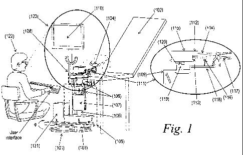
Figure 1 depicts an exemplary imaging workstation for colposcopic examination.
The imaging workstation can include.a base member (101), a planar positioning
structure (103), a space micro-positioning structure (105), a pivoting
structure (108), a
display (110), an imaging head module (111), a computing means (121) as well
as other
various components as discussed herein.
A supporting structure can include. a base member (101) with the principle
purpose of providing a stable platform for the workstation and acts as a
chassis for the
mounting and coupling of the rest of the components of the workstation. The
base
member (101) can be a means of mounting the rest of the components of the
workstation on a solid datum such as a floor, a permanent fixture in the
environment
such as the examination platform (102) (gynecological bed), or can be an
independent
base member (101) capable of being temporarily or permanently affixed to the
abovementioned fixtures.
Said supporting structure can include a planar positioning structure (103)
which
may be an articulating arm with one or more articulation joints capable of
positioning the
CA 02682940 2009-09-30
WO 2008/125870 PCT/GB2008/001352
- 15, -
arm in a two-dimensional space. The planar positioning structure (103) may be
moved
linearly (X), using slides or rotationally (0) using articulation joints which
may disposed
on said base member (101). The range of motion of the planar positioning
structure
(103) may be limited to a pre-specified range of motion. The planar
positioning structure
(103) serves to bring the additional components mounted on it close to the
examination
area (104). The planar positioning structure (103) can provide coarse
positioning of the
some of the components of the workstation with respect to the target area to
be
examined to bring the components in proximity of the examination area (104).
Said supporting structure can include a space micro-positioning structure
(105),
which may be affixed to the previously described planar positioning structure
(103). The
function of the space micro-positioning structure (105) can be used to
accurately position
the rest of the components of the claimed workstation with respect to the
target area to
be examined. The space micro-positioning structure (105) may work in the
Cartesian
(x,y,z), Polar or Spherical space or combinations thereof to achieve the
desired position
of the rest of the components of the claimed workstation, such as sensors,
light sources
etc, which are mounted on to said space micro-positioning structure (105).
Additionally,
the space micro-positioning structure (105) may include a mechanism to balance
the
weight and the torque exerted on it by the components mounted to it. Weight
counterbalance (107) assists the user to perform said micromanipulations for
connecting/disconnecting of said imaging head module (111) with said speculum.
extension shaft (118). The weight counterbalance may be achieved with the aid
of
counteracting compression springs, rotational springs, self compensating gas
dampers,
hydraulic suspension elements or pneumatic means, or a combination thereof.
Additionally, all or some of the degrees of freedom of both planar and space
micro-positioning structures may be temporarily locked, with the aid of
suitable
elements for locking/unlocking (106), once the desired position has been
achieved. The
locking may be affected by mechanical, electro-mechanical, pneumatic,
hydraulic means
or combination thereof. Additionally, all temporary locks may be
activated/released by a
single user action.
Said supporting structure can also include a pivoting structure (108) with the
capability of providing some or all of tilting, pitching and yawing motions
(6, w)to the
CA 02682940 2009-09-30
WO 2008/125870 PCT/GB2008/001352
- 16 -
components attached to it: Additionally, the pivoting structure (108) may
comprise a
temporary locking mechanism _ to allow the user to lock the motion of the
pivoting
structure (108) in one or more of the pivoting structure's (108) degrees of
freedom with a
single user action allowing the user to fix the position of the components
attached to the
pivoting structure (108) when the desired position has been achieved. The user
action
described may be the same user action required for the activation/release of
the locks on
the space micro-positioning structure (105) thereby having the effect of
activating/releasing the locks on both the space micro-positioning structure
(105) and the
pivoting structure (108) with a single user action. The locks incorporated
into the
pivoting structure (108) may be mechanical, electro-mechanical, hydraulic,
pneumatic or
a combination thereof. Additionally, the user action may be, performed through
a handle
(109) used for the manual manipulation of said positioning structures.
Additionally, said supporting structure can also include a means of attaching
a
display (110) for the displaying images and data captured by the imaging head
module
(111), described hereinafter. Preferably said display (110) supporting
structures are
disposed either on said base member (101) or on the other positioning
structures, so that
said display (110) is encompassed by the viewing angle (123) of the user,
where the
viewing angle (123) also includes at least said examination area (104) and
said imaging
head module (111).
The workstation can also include an imaging head module (111). Said imaging
head module (111) has the principle function of capturing images from the
examination
area (104), and may also provide illumination of the examination area. The
imaging
head module can also house suitable imaging and illumination optics and
optomechanical elements for allowing light beam manipulation. The image
capturing can
be accomplished with the use of imaging sensor (115) means which may be one or
more
of a CCD, CMOS imager or a combination thereof. The imaging sensor (115) means
can be configured to capture images in color or black and white. The imaging
sensor
(115) means can operate in conjunction with suitable imaging optics (112)
means.
Additionally, said imaging optics (112) provides an imaging field of view
substantially
equal to the size of the examination area (104). Additionally, the mentioned
illumination
can be derived from a light source (113) which may be mounted substantially at
right
angles, substantially parallel to the imaging sensor (115) and imaging optics
(112), or at
CA 02682940 2009-09-30
WO 2008/125870 PCT/GB2008/001352
- 17 -
any angle in between. The illumination source comprises of suitable optical
elements to
focus the beam to provide an illumination spot (206), (see Figure 2),
substantially equal
to the imaging field of view and the size of the target area.
Said imaging head module (111) comprises of beam manipulation optical
elements used to provide substantial overlapping of both imaging and
illumination spots
irrespective of the angle formed between said imaging sensor (115)/optics and
said light
source (113). Said beam manipulation optical elements may be a partly or fully
reflective
mirror element, a prism a polarizing beam splitter or a combination thereof.
The light
beam may be manipulated to illuminate the target examination area from, for
example, a
location above the imaging optics means. Manipulating the light beam in this
manner
may provide a shadow free examination area so that the target area to be
examined can
be substantially illuminated.
Said imaging head module (111) can include a means of dispensing a diagnostic
marker. The means of dispensing a diagnostic marker may include a spray
nozzle, full
cone or hollow cone, a means of pressurizing said agent before delivery to the
spray
nozzle. The pressurizing means may include. a manual, pneumatic or electrical
mechanism such that sufficient back pressure can be built up at the inlet to
the spray
nozzle so that a proper spray pattern can be fully developed. The diagnostic
marker
may be stored in a container as shown in figure 4, (402) pre-filled with the
marker, which
may attached on said supporting and pivoting structures, or the marker may be
introduced to the dispensing system at the moment of examination.
Said imaging head module (111) may be connected to a speculum (117) via an
extension shaft (temporarily attached to said imaging head module (111)) for
the
duration of the examination in a releasable way. Said extension shaft can be
designed
so as when attached to said imaging head module (111) the imaging,
illumination ray
symmetry axes and said agent dispensing pattern longitudinal axis become
substantially
collinear with said speculum's longitudinal axis (204), see Figure 2, so that
said imaging
field-of-view, said light source (113) spot and the tissue area covered by
said agent are
substantially overlapping.
CA 02682940 2009-09-30
WO 2008/125870 PCT/GB2008/001352
- 18 -
Additionally, the imaging module can include a first mechanical support (119)
for
the attachment of the speculum (117) and its extension shaft in a releasable
way. The
mechanical support (119) may also include means of attaching the previously
described
diagnostic marker system. Additionally, said imaging module can include a
second
mechanical support (120) for permanently fixing the imaging head module (111)
on to
the previously described supporting structure.
The workstation additionally can include a computer (121) means interfaced
with
at least one said imaging sensor (115) described previously, and with some or
all of the
positioning structures locking means. Said computer (121) means can have a
hardware
interface to interface the computing (121) means with the imaging sensor
(115). The
computer (121) means and imaging sensor (115) may be interfaced using one or
more of
a selection including, but not limited to video, USB, IEEE1394 (A, or B),
camera link
Ethernet, etc., or any combinations thereof. Additionally, the hardware
interface
interfaces said computer (121) means with said display (110) means mounted on
the
previously described supporting structure to display the images and data.
The workstation also comprises a software means installed in said computer
(121) means comprising modules for hardware control, image and data capturing,
image
processing, analysis and display and image and data storage and retrieval for
review.
The supporting structure and/or workstation can be characterized in that said
planar
positioning structure (103) allows for both mechanical support and for
positioning at least
said imaging head module (111) in close proximity to the examination area
(104) and to
move away from said examination area (104) and whereas at least at the
proximity
position said examined area, said imaging head module (111) and said display
(110) are
substantially located within the user's field-of-view, and in that at least
one of said planar
positioning structure (103), said space micro-positioning structure (105) and
pivoting
structure (108) has at least two translation modes: one free moving mode,
allowing for
the manual free and counterbalanced spatial movement of said imaging head
module
(111) in and out of the examination area (104) before the connection and after
the
disconnection of said imaging module with said speculum extension shaft (118)
and one
substantially locked mode for the duration of said connection, and in that
when said
connection can be established, the imaging, illumination ray symmetry axes and
said
CA 02682940 2009-09-30
WO 2008/125870 PCT/GB2008/001352
- 19 -
agent dispensing pattern longitudinal axis become substantially collinear with
said
speculum's longitudinal axis (204). This is achieved through proper focusing
and
mounting of the corresponding components at proper positions on said first and
second
mechanical supports, so that said imaging field-of-view, said light source
(113) spot and
the tissue area covered by said agent are substantially overlapping.
In some embodiments, said base member (101) of the supporting structure as
described previously can be a mobile base. The base member (101) can use of
one or
more individually lockable castors for enabling mobility. Additionally, at
least one of the
planar positioning structure (103), space micro-positioning structure (105) or
the imaging
head module (111) can be mounted directly on to the base member (101).
Therefore,
the claimed workstation may be configured to be comprised of a mobile base
member
(101), a space micro-positioning structure (105) that comprises at least a
vertically
telescoping columnar member at one end of which is attached a pivoting
structure (108)
onto which said imaging head module (111) can be affixed. As a result, the
workstation
itself may be mobile.
In other embodiments of the supporting structure and/or workstation, the
previously described planar positioning structure (103) can be affixed to a
mobile base
and the previously described space micro-positioning structure (105) can be
affixed to
the planar positioning structure (103). In yet other embodiments, the base
member (101)
comprises of an immobile datum such as the floor or ceiling of the environment
or
examination bed, and the planar positioning structure (103) can be mounted
fixedly to
the datum.
In yet other embodiments of the claimed workstation, the previously described
space micro-positioning structure (105) can be affixed directly on to the base
member
(101) and the planar positioning structure (103) can be affixed to the space
micro-
positioning structure (105).
In yet other embodiments, the space micro-positioning structure (105) and the
planar positioning structure (103) comprise a multi-jointed articulating arm.
The arm may
work in the spherical space to achieve the desired positioning accuracy of the
imaging
head module (111) with the use of horizontal and vertical rotational elements.
These
CA 02682940 2009-09-30
WO 2008/125870 PCT/GB2008/001352
- 20 -
said elements may be roller bearings of the axial thrust or rotational type,
or self
lubricating bushings, or a combination thereof. Additionally, the arm may be
lockable at
some or all of its articulating joints using some or all of pneumatic,
electrical, mechanical,
electro-magnetic or hydraulic means.
In other embodiments, the space micro-positioning structure (105) may be a
linear translator working in the Cartesian space (x,y,z) comprising of linear
guide
elements that may be of the type linear slideways or pillow blocks mounted on
suitable
guide rails and either of which may move on incorporated roller balls, cross-
rollers or
self-lubricating bushings.
In other embodiments, the planar positioning structure (103) may be a movable
structure rotating (0) around appropriately fixed and stable vertical members
on the base
member (101). The planar positioning structure (103) may consist of a rotating
part
rotating around the fixed members of the base around one or more of roller
bearings, a
set of axial thrust bearings, and/or self lubricating bushings. Additionally,
the planar
positioning structure (103) may possess a longish extension (i.e: may be an
elongate
member).
In other embodiments of the claimed workstation, the planar positioning
structure
(103) can be a mechanical slider (X) which may be composed of a stable.
platform and a
movable carriage which may be brought in close proximity to the target area to
be
examined. The motion may be accomplished by using a movabie carriage mounted
on a
closed circuit of rolling balls, rotating rollers moving on guide rails or
bushing elements
sliding on corresponding guide elements.
In other embodiments of the claimed workstation, said planar positioning
structure (103) can be a wheeled trolley upon which all other components are
mounted.
The trolley may include two platforms supported on columns where the first
platform
serves as the mounting platform for all other structures of the workstation
and the
second platform serves as the location surface of the wheels in the trolley.
Additionally,
the trolley wheels may be individually lockable facilitating its positioning
and
locking/unlocking in close proximity to the examination area (104).
CA 02682940 2009-09-30
WO 2008/125870 PCT/GB2008/001352
- 21 -
In other embodiments of the claimed workstation, said trolley can be
collapsible
by virtue of possessing collapsible or telescoping columns. Additionally, the
trolley can
be composed of two platforms where the first of the two platforms serves as a
mounting
platform for all other structures on the workstation and the second platform
serves as the
location surface for the wheels in the trolley.
In other embodiments of the claimed workstation, said pivoting structure (108)
is
at least one degree of freedom axial joint and may be mounted directly on to
one of
either the planar positioning structure (103) or the base member (101). This
degree of
freedom may provide the pivoting structure (108) with the capability of pitch,
yaw or tilt
and may be comprised of a solid rod like member to accomplish this motion.
In other embodiments of said workstation said pivoting structure (108) may be
a
ball-joint structure attached to either of the planar positioning structure
(103), the space
micro-positioning structure (105) or to the base member (101). Said ball-joint
may
comprise of a ball, see figure 8, (810) and a suitable casing to encase the
ball (810),
suitable means of attaching the ball-joint to either of the planar positioning
structure
(103), the space micro-positioning structure (105) or to the base member.
(101).
In other embodiments of said workstation, one or both of the space micro-
positioning structure (105) and the planar positioning structure (103)
consists of the
weight counterbalancing means. These means may include constant force. springs
(603), see Figure 6 constant torque spring sets, counteracting compression
springs, self
compensating gas dampers, multi-chamber hydraulic dampers or active pneumatic
circuits and circulating and suspended pulley weights in the configuration of
an Atwood's
machine.
In other embodiments of the claimed workstation, the motion of the various
movable members can be locked/unlocked using one or more of mechanical,
electrical,
pneumatic, electromagnetic, electrical drive means of activating and
deactivating friction
inducing elements. The mechanical means may include mechanical stops, high
tension
steel cable actuated lever, cam (807), see Figure 8, follower and multi-
pivoting
mechanisms whereas the electrical means may comprise servomotors supplied with
holding torque inducing current, current to induce or change polarities in
ferro-magnetic
CA 02682940 2009-09-30
WO 2008/125870 PCT/GB2008/001352
- 22 -
elements while pneumatic means may include pneumatically actuated clutches to
engage and disengage relatively mobile members or pneumatically actuated
friction
elements.
Furthermore, the claimed workstation can include means of controlling the
friction
level of one or more of moving parts of one or more from amongst the planar
positioning
structure (103), the space micro-positioning structure (105) or the pivoting
structure
(108). By using variable friction levels on one of the structures, and
suitably designing
the remaining, the claimed workstation can achieve the desired functionality.
These
means may include the use of manually actuated screws or knobs, or these means
may.
be actuated by using a remotely activated mechanism. Furthermore, the remote
activation of the means may be affected by an actuation signal located on the
handle
(109), as described previously. The triggering may be affected by means
analogous to
the mechanism used for activating and deactivating the friction elements and
may
include the use of a high leverage ratio pivoted lever, a microswitch (812),
see Figure 8,
to trigger electrical elements, or a pneumatic pilot line to activate and
deactivate
respective pneumatic components. This handle (109) may be located directly on
the
pivoting structure (108), or any position in space allowing the use of the
handle (109) for
the desired positioning of the various elements.
In other embodiments; said triggering means can be a high leverage ratio,
pivoted hand lever (811), see figure 8, that serves to compress and decompress
suitable
springs to activate and deactivate a direct manual brake for the pivoting
structure (108).
Simultaneously, said hand lever (811) acts as a means of triggering remotely
located
brakes for the braking of relatively mobile members. Said hand lever (811) may
use one
or more of remote activation and deactivation means from amongst, but not
limited to,
mechanical, electrical, hydraulic or pneumatic means.
In other embodiments, said triggering handle (109) can be supplied with manual
force and the force can be transmitted from the triggering handle (109) to
remotely
located brakes using a high tension steel cable which can be housed in an
appropriately
sized external sheath which can be substantially flexible but incompressible.
Said sheath
may be comprised of an outer covering made of hardened polymeric compounds
CA 02682940 2009-09-30
WO 2008/125870 PCT/GB2008/001352
- 23 -
whereas the inner portion of the sheath may be comprised of a continuous
compression
spring.
In other embodiments, said imaging head module (111) can be affixed directly
on
to said pivoting structure (108)
The imaging head module (111) can be configured so that focused, shadow and
glare-free tissue overview images can be obtained, once said imaging head
module
(111) is connected with said speculum such as by an extension shaft (118). To
achieve
imaging through the relatively small rear aperture of said speculum (117),
small imaging
and illumination elements are employed, which are mounted in close proximity
on said
second mechanical support (120) so that their respective light spots
substantially overlap
onto the examined area, without the corresponding light ray being obstructed
by said
speculum (117). Said second mechanical support (120) may be affixed onto said
first
mechanical support (119), which may be detachably connected with said speculum
extension shaft (118) through a shaft locking mechanism (205), see Figure 2
and 10.
Fine focusing is allowed either through auto or manual focusing optics or
through a linear
translator (801) allowing for the relative translation of said first
mechanical support (119)
in relation to said second mechanical support (120), through a fine focusing
knob.
In addition and for the purpose of a more .realistic and complete
documentation
and for facilitating treatment operations said workstation may be configured
with two
imaging sensors and image focusing optics and appropriate display means to
provide
stereo digital imaging. Furthermore it may be configured with two imaging
sensors, one
coupled with magnifying optics for imaging of the cervix and the other with an
endoscope
probe for the imaging of the endocervix. A more detailed description of the
abovementioned configurations is provided below with reference to figures 2,3,
and 8.
In some embodiments, said imaging sensor (115) means in the imaging head
module (111) can be comprised of one or more of, but not limited to, a CCD
camera,
CMOS camera or a combination thereof. The cameras can provide color images
and/or
black and white images. Additionally, the imaging sensor (115) can have a
spatial
resolution of at least 640x420 pixels and the imaged data from the sensor can
be
CA 02682940 2009-09-30
WO 2008/125870 PCT/GB2008/001352
- 24 -
transmitted using a protocol selected from, but not limited to, video, USB,
IEEE1394a,
IEEE1394b, camera link, Ethernet, etc.
In other embodiments, said imaging head module (111) can include imaging
optics (112) which are comprised from a group including, but not limited to
constant
magnification optics, zoom optics, scalable magnification optics and endoscope
optics.
In other embodiments, said imaging optics (112) used in conjunction with the
imaging sensor (115) means may be a 25-35mm lens or a zoom lens and may be of
the
type C-mount, CS mount or of any other mount type.
In other embodiments, the imaging head module (111) of the claimed workstation
can include the illumination source which may be selected from a group
including, but
not limited to Xenon, Light Emitting Diodes (LED), Halogen and any other light
source
(113) that can emit light at least in the spectral range 400nm-700nm.
Additionally, the imaging head module (111) can include first and second
polarizers (207). The first polarizer (207) can be placed in the imaging
sensor's imaging
path and the second polarizer (207) can be placed in the light path of the
illumination
source, with their polarization planes being substantially at right angles to
each other.
The polarizers may be placed in the paths by temporary or permanent means and
are
adjusted to achieve the desired angle between their polarizations planes.
Furthermore, the imaging head module (111) described previously may comprise
of a first camera used for the imaging of the vagina and the cervix of the
uterus while a
second imaging sensor (115) may be coupled with an endoscope for the imaging
of the
endocervical canal and the endocervix.
Furthermore, and with reference to Figure 3 (a), the imaging head module (111)
as described previously and in particular the imaging lens means is a
microlens with a
diameter less than 1 cm and is positioned parallel to the illumination source
allowing the
imaging field of view and the illumination field to be substantially coaxial
at the target
area. This is achieved by the use of members in the illumination source that
possess a
CA 02682940 2009-09-30
WO 2008/125870 PCT/GB2008/001352
- 25 -
similar size envelope as said microlens so as to be in close proximity with
the imaging
means.
In other embodiments of the workstation, as depicted in figure. 3 (b) said
imaging
sensors may be two in number and are placed in close proximity to each other
and at
each others' side and are coupled with the previously described microlens
allowing for
stereo vision of the vagina and that of the cervix of the uterus, provided
that the images
are displayed on display means providing stereo perception.
In other embodiments at least said camera and said light source (113) can be
mounted on said second mechanical support (120) and whereas said second
mechanical support (120) can be mounted on said first mechanical support (119)
which
in turn can be mounted on said pivoting structure (108) through a linear
translator (801),
said linear translator (801) allowing for fine focusing (see fig 2) In this
figure the cooling
fan (211) module with the threaded shafts (212), spacers (210) and heat sink
flange
(209) for the heat sink (208) is indicated which in turn absorbs/dissipates
heat from the
light source (113).
In some embodiments said beam manipulation optics (114) can be a light
deflector (201) selected from a group including but not limited to a prism,
polarization
beam splitters, dichroic mirrors, dichroic reflectors, fully or partially
reflective mirrors of
combinations thereof. In some cases the sizes of said imaging sensor (115) and
said
light source (113) do not permit side-by-side placement so that the spot
overlapping
requirement, as described above, can be fulfilled. In these cases light
deflection of the
rays of at least one of said imaging sensor (115) and said light source (113)
to become
substantially coaxial with each other and with the speculum longitudinal axis
(204) (when
connected) provide an optimum configuration for the fulfilment of this
requirement. As
depicted in figure 2, light deflector (201) may deflect the light of either
said imaging
sensor (115) or of the light source (113) or of both. In other embodiments,
the beam
manipulation optics (114) include at least one planar mirror which is oriented
in a fashion
so as to achieve coaxial illumination with the imaging field of view. The
planar mirror
may be supported along an off-center axis along its surface with the
capability of being
fixed in the desired position by fastener means or by permanent means once the
desired
position has been achieved. In other embodiments, the beam manipulation optics
(114)
CA 02682940 2009-09-30
WO 2008/125870 PCT/GB2008/001352
- 26 -
may be comprised of a non - planar mirror which is encased and held in a
position
appropriate to achieving a coaxial illumination beam with the imaging field of
view.
In yet other embodiments, said light manipulation optics (114) further
comprise
laser beam manipulation optics (114) to manipulate a laser beam for image
guided laser
treatment. Beam manipulation may be carried out by altering the relative
orientation of
these elements with respect to the illumination source and the orientation may
be altered
by mechanical or electrical means. The orientation may be achieved by using
pre-
determined coordinates or by using electrical feedback for the imaging data
from sources
external to the claimed workstation. In other embodiments, the beam
manipulation
optics (114) may be a set of galvanic mirrors to manipulate a laser beam for
tissue
treatment that may be added in a retro-fit fashion to the workstation. In
other
embodiments, the beam manipulation means includes at least one mirror
controlled with
a joystick to manipulate a laser beam. In such case, the beam manipulation
means may
be driven by electrical drive means such as micro-motors, servomotors or
stepper
motors that interface directly with the joystick to achieve the desired
orientation of the
beam manipulation means and the laser beam.
In other embodiments of the claimed workstation (as it is depicted in fig. 4),
said
imaging means and the illumination means may be placed at substantially right
angles to
each other within the imaging head module (111). Additionally, said beam
manipulation
optics (114) are held at approximately 450 with one of the axes of either the
imaging
means or of the iliumination means. This has the effect of reflecting the rays
incident
onto the beam manipulation optics (114) approximately 90 and thereby making
it
substantially parallel with the other axis.
In other embodiments of the imaging head module (111), the light deflector
(201)
and the light source (113) are located on the same side of the central ray
axis of the
imaging means (as shown in Figure 2). Both the light deflector (201) and the
light source
(113) are positioned so as to not obstruct the field of view of the imaging
means but, at
the same time, provide illumination that, after interacting with the light
deflector (201), is
substantially coincident with the field of view of the imaging means at the
surface of the
tissue to be examined, or being examined. This is accomplished by
maintaining,the light
deflector (201) on one side of the central ray axis of the imaging means, but
as close as
CA 02682940 2009-09-30
WO 2008/125870 PCT/GB2008/001352
- 27 -
possible to it, and by positioning it at 450 to the central ray axis.
Additionally, the light
deflector (201) is also positioned at 45 to the central axis on the same
relative side - as
the light source (113) of the of the illumination module. Light from the
illumination source
(113) interacts with the light deflector (201), the central axis of the
emanating light is at
90 . to the central axis of the illumination means.
In an alternative embodiment of the imaging head module (111), the light
deflector (201) and the light source (113) are located on opposite sides of
the central ray
axis of the imaging means (as shown in Figure 4). This is a preferred
embodiment in
cases where the upper half of the rear aperture of the speculum (117) is
wider, so that
the entering light bean is not obscured. Both the light deflector (201) and
the light source
(113) are positioned so as to not obstruct the field of view of the imaging
means but, at
the same time, provide illumination that, after interacting with the light
deflector (201), is
substantially coincident with the filed of view of the imaging means at the
surface of the
tissue to be examined or being examined. This is accomplished by maintaining
the light
deflector (201) on one side of the central ray axis of the imaging means, but
as close as
possible to it, and positioning it at 450 to the central ray axis.
Additionally, the light
deflector (201) is positioned on the opposite side of the central ray axis of
the illumination
means with respect to the light source (113) and at 45 to the central axis of
the
illumination module. Before light from the illumination source (113) interacts
with the
light deflector (201), the central axis of the emanating light is at 90 to
the central axis of
the illumination means.
The disclosed workstation may also incorporate a mechanism allowing for the
uniform and standardized applicatiori of a diagnostic marker, such as acetic
acid
solution, onto a surface of the tissue to be examined. In a case where
recording of
dynamic optical phenomena, provoked by the marker, is required, means for
synchronization of initiation of the image capturing procedure with the
completion of the
marker application are also integrated in to the disclosed workstation.
In some embodiments of the workstation, the agent dispenser (116) (diagnostic
marker dispensing means) may be an application mechanism for dispensing the
diagnostic marker onto the surface of the examined tissue. The proposed
mechanism
consists of an application probe which may be a narrow angle full-cone or
hollow-cone,
CA 02682940 2009-09-30
WO 2008/125870 PCT/GB2008/001352
- 28 -
axial spray nozzle, a container (402), See Figure 4, for the diagnostic marker
and a
means for delivering the diagnostic marker from the container (402) to the
application
probe. Furthermore, the application probe is disposed and fixed on a mount
disposed
directly or indirectly by way of an extension bracket (202), at a certain
position on the first
mechanical support (119) and wherein the orientation of its longitudinal axis
is prefixed
so that, when the imaging head module (111) is connected with the speculum
extension
shaft (118), the marker is applied substantially homogeneously onto a tissue
area of at
least equal size with the light source (113) spot and the imaging sensor's
field-of-view.
In other embodiments, the described probe may be mounted on a mechanical
mount which includes a pre-aligned fixture for alignment of the probe. The
alignment
fixture is designed such that when the probe is locked into the fixture, its
orientation
ensures a substantially homogeneous application of the diagnostic marker onto
the
examined tissue.
In yet other embodiments, the described diagnostic marker container (402) is a
single compartment container (402), fillable with a standardized volume of the
diagnostic
marker and delivered to the application probe with means appropriate for
creating the
necessary pressure and flow conditions required to affect the desired
homogeneous
application onto the examined tissue.
In an alternative embodiment the agent dispenser (116) has a protective
injector
cap (1006), fixed on a nozzle cylinder (1012) and fastened to ensure proper
alignment in
line with the central optical axis of the speculum, with a fastening nut
(1011) mounted on
the speculum locking mechanism (205) with bracket (1013), see Figures 2, 4, 9
and 10,
the diagnostic marker container (402) is a dual compartment arrangement where
the first
compartment is a reservoir, volume of the diagnostic marker and the second
compartment contains a standardized fraction of the volume of the diagnostic
marker,
and the two compartments are connected via appropriate means, including,
valves, and
pressure and vacuum creation means. Additionally, the agent dispenser (116)
includes
means for delivering the diagnostic marker from the second compartment to the
application probe.
CA 02682940 2009-09-30
WO 2008/125870 PCT/GB2008/001352
- 29 -
In other embodiments of the agent dispenser (116), the means for enabling
application are manual and manually delivered force is used for the creation
of the
requisite back pressure at the inlet to the application probe, in order to
create the desired
spray pattern to achieve the desired homogeneous application of the diagnostic
marker
onto the examined tissue.
In other embodiments of the agent dispenser (116), the means for enabling the
application of the diagnostic marker are electro-mechanical in nature and
comprise drive
components chosen from a group including, but not limited to, one or more
stepper
motors and servomotors, which are connected directly or indirectly to a
pumping
mechanism chosen from a group including, but not limited to, reciprocating
positive
displacement pumps, peristaltic pumps, centrifugal pumps or diaphragm pumps.
The
motors are controlled and the pumps are appropriately calibrated so as to
deliver a
standardized volume of the diagnostic marker to inlet of the application probe
at
appropriate flow conditions required to develop the spray pattern required to
achieve the
desired homogeneous application of the diagnostic marker onto the examined
tissue
surface. Additionally, the motors are operated by an electrical signal which
may be
generated by the previously described computer means (12 1).
In other embodiments of the agent dispenser (116) as described, the manual
means for delivering the diagnostic marker to the application probe comprise
manually
depressing a syringe-type mechanism (501), see Figure 9. An end of the syringe-
type
mechanism (501) is connected detachably to the application probe and manual
force is
used to depress the syringe plunger and create the requisite back pressure at
the inlet to
the application probe, in order to provide the desired homogeneous application
of the
diagnostic marker onto the examined tissue surface.
In other embodiments of the agent dispenser (116), the electrical signal is
used to
trigger initiation of image capturing by the previously described imaging
means and to
synchronize image capture with the end of application of the diagnostic
marker. The
computer (121) means may be programed to record completion of application of
the
diagnostic marker, or may be pre-programed to initiate image capturing at a
pre-
determined time interval after commencement of application of the diagnostic
marker.
CA 02682940 2009-09-30
WO 2008/125870 PCT/GB2008/001352
- 30 -
In other embodiments of the agent dispenser (116), the elements for enabling
the
manual delivery of the diagnostic marker to the inlet of the application probe
comprise a
syringe-type mechanism (501) with an integrated piston.
In other embodiments of the agent dispenser (116) as described, sensors are
incorporated to detect completion of manual delivering of the diagnostic
marker onto the
examined tissue surface. The sensors are electrical in nature and may be
chosen from a
group including, but not limited to, one or more optical sensors, capacitive
sensors,
proximity sensors, motion sensors, pressure sensors, flow sensors,
displacement
sensors or a mechanical toggle switch: Activation of the sensors is further
used to
initiate image capturing using the previously described imaging means and,
thereby,
synchronizing image capture with completion of application of the diagnostic
marker onto
the examined tissue surface.
In other embodiments of the agent dispenser (116), the means for enabling
manual delivery of the diagnostic marker to the inlet of theapplication probe
comprise a
syringe-type (501) mechanism with an integrated piston having an opaque and
air-tight
end. Furthermore, the syringe-type mechanism (501) is supported on a structure
that
fully - or partially - covers the container (402) of the syringe-type
mechanism (501) along
its length. Furthermore, the structure comprises the sensor to detect motion
of the
moving parts in the syringe-type mechanism (501). Additionally, the sensor is
a
combination of a light source (113) and a photo-sensor (903), see Figure 9,
which is of
the normally on (NO) type. Furthermore, the manually depressing the plunger of
the
syringe-type mechanism (501) causes interruption of the photo contact between
the light
source (113) and the photo-sensor (903) by the opaque and air-tight end,
causing
generation of a triggering signal for initiation of the image capturing
process.
Furthermore, the syringe-type mechanism (501) is supported on a structure that
fully - or partially - covers the container of the syringe-type mechanism
(501) along its
length. Furthermore, the sensor comprises a pair of electrical. contacts that
are brought
into contact when the depression of the plunger of the syringe-type mechanism
(501) is
completed. The electrical contacts may be brought into contact using a
mechanical
toggle switch or any other means, and contact of the electrical contacts- has
the effect of
CA 02682940 2009-09-30
WO 2008/125870 PCT/GB2008/001352
- 31 -
generating a triggering signal to initiate image capture so as to synchronize
image
capture with the end of the diagnostic marker application.
In other embodiments of the agent dispenser (116), the previously described
sensors are located directly on the diagnostic marker container or are
appropriately
placed so as to detect the motion of the moving parts of the described manual
means of
application of the diagnostic marker.
In other embodiments of the agent dispenser (116), the sensors may be located
on mechanical supports or structures that hold all or part of the diagnostic
marker
container. This may include mechanical brackets, plastic housings or other
such
encapsulations and supports as required for the support of the diagnostic
marker
container.
As stated above, imaging dynamic phenomena requires substantially maintaining
stability of the imaging sensor's field-of-view for required periods during
prolonged
examination. The . disclosed workstation integrates means for such mechanical
stabilization. In addition, the disclosed workstation corrects image motion
artifacts
occurring within said field-of-view by integrating image registration (1103),
see Figure 11,
algorithms, which are described below. In some embodiments of imaging the
stabilization is achieved by detachably connecting the imaging head module
(111) with
the speculum (117), equipped with an extension shaft. Once the connection is
established, the supporting and pivoting structures may be locked to further
secure
stabilization and to support the weight of the speculum (117).
As stated above, and given that the marker is application probe is properly
positioned and aligned on the imaging head module (111), this connection
provides for
reproducible and uniform application of the marker. Mechanical stabilization
means may
include a bayonet mechanism, spring loaded, wedge-shaped pins or positive
engagement spring-loaded couplings. The bayonet mechanism may include a spring
preloaded probe, while the speculum extension shaft (118) may be a female
shaft
designed to accept the probe. The wedge-shaped pin mechanism may include an
eccentric wedge which pivots around a fixed pivot and which is preloaded with
a leaf-
spring. The extension shaft is designed to accept the wedge feature in it when
properly
CA 02682940 2009-09-30
WO 2008/125870 PCT/GB2008/001352
- 32 -
aligned. Alternatively, a spring-loaded coupling may be used that is preloaded
both
axially and radially, so as to securely lock the speculum extension shaft
(118) in the
coupling whilst facilitating release of the shaft when the radial spring is
released.
In some embodiments of the workstation, the speculum (117) is detachably
attached to the imaging head module (11,1) with an extension shaft. The shaft
is so
designed as to be coaxial with the central axis of the imaging means
incorporated in the
imaging module head. Additionally, the shaft is attached to the imaging module
head
with semi-permanent means, the manner of which may be chosen from a group
comprising, but not limited to, mechanical locking means, magnetic means,
electromagnetic means and/or pneumatic means.
In other embodiments of the workstation, the computer (121) means further
comprises components and modules for interfacing with at least one of the
imaging
sensor (115) means, the user interface means, the display means and/or the
agent
dispenser (116) means. Additionally, the computer (121) means comprises
connection
means for printers, local networks and/or the internet.
In other embodiments of the computer (121) means, one of the interface means
is wireless and may comprise Bluetooth 1.2, Bluetooth 2.0, Infrared or any
other protocol
for wireless data transfer.
In other embodiments of the computer (121) means, the computer (121) means is
mounted directly on the supporting structures.
In other embodiments of the workstation, the previously described interfaces
are
selected from a group including but not limited to a keyboard, a mouse, a
track ball, a
voice interface, touchscreen (502), see Figure 5, and/or a foot-switch.
In other embodiments of the computer (121) means, the previously described
interfaces are located on the previously described supporting structures.
In other embodiments of the computer (121) means, the interface means are
located directly on the computer (121) means.
CA 02682940 2009-09-30
WO 2008/125870 PCT/GB2008/001352
- 33 -
In other embodiments, the display (110) is a monitor that is mounted on a
stand.
Furthermore, the stand is located on the previously described supporting
structures in a
spaced-location but within the viewing angle (123) of the user, where the
viewing angle
(123) also includes the examined area. This allows the user to visualize both
the
examined area and the displayed image without moving his/her head. This is, of
course,
an advantage over the prior art.
In other embodiments of the display (110) means, the stand is located on the
previously described base member (101) and is placed on one side of the
examination
bed outside the angle subtended by a patient's legs. Additionally, the monitor
is
provided at a spaced-location but within the viewing angle (123) of the user,
where the
viewing angle (123) includes the examined area. Such that the user may
visualize both
the display (110) means and the examined area without turning his/her head.
Again, this
is an advantage over the prior art.
In other embodiments of the display (110) means, the stand is located on the
previously described planar positioning structure (103). Additionally, the
display (110) is
located at a spaced-location but within the angle subtended by a patient's
legs and is
within the viewing angle (123) of the user, which also includes the examined
area such
that the user may visualize both the display (110) and the examined are
without turning.
his/her head. This is an advantage over the prior art.
In other embodiments of the display (110) means, the display means may be
chosen from a group including, but not limited to, a head-mounted display,
video
goggles, touchscreen (502) and/or a projection display.
A further embodiment of workstation is described with reference to Figures 4
to
10 in particular. In its preferred embodiment, the base member (101) is an
eccentric,
ellipsoid-shaped base-plate mounted on individually lockable wheels,
additional braking
and stabilization members being integrated into the base-plate. The
stabilizing members
are used to provide temporary fixation of the base to the datum with respect
to the
examination piatform (102), in use. The base member (101) has 2 tubular
elements, one
of which is fixed on to the base plate while the second rotates around the
fixed tubular
CA 02682940 2009-09-30
WO 2008/125870 PCT/GB2008/001352
- 34 -
member with the help of a self-lubricating bushing or a set of axial thrust
bearings.
Rotation of the tubular assembly is limited to a maximum of 90 by the
presence of a
press-fit dowel pin moving in a machined groove. Also mounted to the fixed
tubular
member is a vertical columnar member which supports a large format image,
display
(110) unit.
A planar positioning structure (103) is fixedly-mounted at one of its ends to
the
rotating tubular member. In its preferred embodiment, the planar positioning
structure
(103) is a relatively long member which has.a vertically-supporting foot fixed
near to its
other end. The foot is a lockable, integrated wheel capable of swiveling
through 360 .
The foot supports at least the planar positioning structure (103) and the
imaging head
module (111). Following from the range of motion allowed by the 2 tubular
sections
mentioned, the planar positioning structure (103) rotates from its extended
(rest)
position, allowing for a patient's access to the examination platform (102),
to its closed
(imaging) position, translating at least the imaging head module (111) in
close proximity
with the examination area (104).
In its preferred embodiment, the space micro-positioning structure (105) works
in
Cartesian coordinates. Motion is provided in the XY-plane by 2 sets of guide
elements
in each direction, working on a set of three, parallel, equally-sized plates .
The guide
elements may be linear coller-ball type guide elements, linear cross-roller
guide
elements, linear self-lubricating bushing elements or a combination thereof,
such that
unrestricted motion is substantially frictionless. Motion along the Z-axis is
provided by a
linear guide element (602) which comprises a splined, non-rotational shaft
moving along
a closed circuit of roller balls retained appropr'iately. The top end of the
splined shaft
(601) terminates in a ball (810) fixedly-attached to the shaft. The Z linear
guide element
(602) is supported on a support member affixed to columnar structures, mounted
on the
top-plate (606) of the 3 plates used for affecting the XY motion.
In its preferred embodiment, the space micro-positioning structure further
comprises suitably sized constant-force springs (603) mounted on the support
member
and affixed permanently to the splined shaft (601). The constant-force springs
(603)
rotate on a substantially frictionless drum and shaft, which are of the needle-
bearing-
type with hardened steel shafts.
CA 02682940 2009-09-30
WO 2008/125870 PCT/GB2008/001352
- 35
Additionally, the space micro-positioning structure (105) can be temporarily
fixed
along all its axes of motion, XY and Z. The X motion is achieved with X motion
sliders
.(613) along with X mounting slider holders (612) on middle plate (607) and
the Y motion
is achieved using Y motion sliders (611) along with Y mounting slider holders
(610) on
the bottom plate (608). Y motions are temporarily fixed by stopping the
relative motion of
the top (606) and bottom plate (608) with respect to each other. The XY motion
is
affected by using a brake mechanism housing module (705) with a suitably sized
helical
counteracting spring (702) inserted on an electromagnet pivot (704) holding an
electromagnet (701), see Figure 7, pressing on a friction element (703)
through the
brake pad housing (706). As a result, this mechanism brakes on brake pad
(609). The
brake is of the normally open
(NO)-type and is engaged at all times and can be released by the action of the
user,
described herein. The action of the user serves to activate the electromagnet
(701),
which retracts the friction element (703) mounted at the distal end of a
suitable, ferro-
magnetic mount.
The motion along the Z-axis, see Figure 6, is temporarily fixed by using a
motion
drive apparatus having a stepper motor (605) and a timing belt (604) fixedly-
attached to
the splined shaft (601). The motion drive apparatus is of the normally closed
(NC)-type
and provides a holding torque to the stepper motor (605) thus preventing the
motion of
the splined shaft (601). The circuit is opened and the motion released using
the same
user action; described herein, for releasing the XY brake.
In its preferred embodiment, the workstation is a pivoting structure (108),
where
the pivoting structure (108) is a limited ball-joint providing unlimited
rotational motion,
limited pitching motion and zero tilting motion. The ball-joint uses as its
central member
the previously described ball (810) affixed permanently to the top-end of the
previously
described splined shaft (601) of the space micro-positioning structure (105).
The ball-
joint has an upper, middle and lower disc-shaped member. The middle and the
lower-
disc-shaped members are complimentary concave-shaped and are interconnected by
a
pair of parallel rod members. The rod members pass through the disc-shaped
members,
through respective openings, trapping and thus restricting the ball (810).of
the ball-joint
CA 02682940 2009-09-30
WO 2008/125870 PCT/GB2008/001352
_ 36 _
within the middle disc-shaped member (805), the lower disc-shaped. member
(806), see
Figure 8, and the pair of parallel rod members.
The lower disc-shaped member (806) acts as a motion limiter as it limits
motion
of the ball-joint when approaching the middle disc-shaped member (805) and
traps and
immobilizes the ball (810) of the ball-joint between the two approaching disc-
shaped
concave members. Additionally, the lower disc-shaped member (806) restricts
motion of
the ball-joint with respect to the splined shaft (601), which is achieved by
providing a
linear slit in the lower disc-shaped member (806) that acts as the entry point
of the
splined shaft (601) into the ball-joint. By virtue of this slit, limited
pitching is allowed and
no tilting is allowed to the ball-joint.
Affixed on top of the middle disc-shaped member (805), is the upper disc-
shaped
member (804). The parallel rod members (808), passing through respective
openings in
both the middle and lower disc-shaped members, terminate in the upper disc-
shaped
member (804) . Mounted, coaxially with the parallel rod members, is a pair of
suitably-
sized helical springs (809), encapsulated between the upper and middle disc-
shaped
members. The other ends of the parallel rod members are secured by using
threaded
fasteners (814) housed in suitable cavities in the lower disc-shaped members
(806).
The parallel rod members are joined together by using a suitable shaft, so as
to
maintain the rod members relatively congruent to each other and for depressing
the
helical springs upon the action of a follower - cam (807) mechanism, described
herein.
An eccentric cam (807) is housed and permanently affixed at one of its ends to
the upper disc-shaped member (804) with mounting screws (819) is with a
suitable
surface created in it for depressing the shaft (821) connecting the parallel
rod members
and connecting to top round part (804) through shaft member (818). A suitably
shaped
lever (811) is in contact with the free end of the cam (807), with a
corresponding follower
path created at the end in contact with the cam (807),. and is housed in a
suitably
designed casing (813) with handle mounting pins (822). Also mounted along the
lever
(811) is a mechanism for transmitting a signal for the motive release of the
previously
mentioned micro-positioning structure (816) affixed into handle (109) with
lower plug
(815), which is activated when the lever (811) is depressed. In its preferred
embodiment, this mechanism is a microswitch (812) that transmits an electrical
signal to
the respective motion locking members in the micro-positioning structure.
Depressing
CA 02682940 2009-09-30
WO 2008/125870 PCT/GB2008/001352
- 37 -
the lever (811) and activating the incorporated follower - cam (807) has the
effect of
depressing the incorporated helical springs in the ball-joint and thereby
creates a
separation between the lower and the middle disc-shaped bodies - including the
ball-joint
- which has the effect of releasing motion on the allowed degrees of freedom
in the ball-
joint. The lever (811) and its casing (813) further act as a handle (109)
which is held
together with screws (830) to allow for manual positioning of the positioning
structures
upon releasing the motion of the ball-joint.
Additionally, mounted on top of the upper disc-shaped member (804) of the ball-
joint, is an asymmetric bracket (401), with an opening (803) created in a
protrusion for
receiving a container (402), for suitable marking agents. Additionally,
mounted on the
asymmetric bracket (401) is a linear translator (801) incorporating an
internal rack and
pinion mechanism, used for fine focusing, or fine manouvering of the imaging
head
module (111), described elsewhere. The linear translator (801) is activated by
using a
thumb screw (802) present on either side of the translator (801) and provides
symmetric
positive and negative motion around nominal.
In its preferred embodiment, the workstation additionally has an imaging head
module
(111) comprising of an imaging sensor (115) and associated imaging optics
(112). In its
preferred embodiment, the imaging sensor (115) is at least one color CCD
sensor of at
least 1024X768 resolution coupled with an appropriate imaging lens of at least
20mm
focal length imaging lens with a 20 to 35 cm working distance. The imaging
lens has the
desired characteristic of providing the correct-sized field of view at the
desired axial
distance, and has variable but lockable aperture settings.
Additionally, the imaging head module (111) consists of an LED light source
(113) of
suitable intensity and spectral range that may cover, at least, the range of
about 400nm-
700nm to work in conjunction with said color CCD. The light source (113) also
includes
suitable focusing optics, so as to achieve illumination of the imaging field
of view.
Additionally, the light source (113) comprises a mechanism to allow beam
manipulation
to achieve coaxial illumination with the imaging field of view. In its
preferred
embodiment, the imaging head module (111) has the light source (113)
positioned at
substantially right-angles to the CCD and said imaging lens. The beam output
from the
light source (113) is reflected towards the target area with the use of a
suitable reflective
CA 02682940 2009-09-30
WO 2008/125870 PCT/GB2008/001352
- 38 -
mirror. Coaxial illumination with the imaging field of view is achieved by
manipulating the
relative angle of the mirror, the relative angle of the light. source (113) or
both.
Additionally, coaxial field of view is achieved by means of vertical
adjustments provided
for the position of the CCD and imaging lens . The net result of the provided
adjustments is that the illumination cone and the imaging cone are
substantially
coincident.
In the preferred embodiment, at least one of the imaging sensor (115) and the
illumination means are affixed on the second mechanical support (120) and
wherein the
second mechanical support (120) is affixed on the pivoting structure (108)
through a
linear slider for allowing fine focusing.
In the preferred embodiment, the light deflector (201) is placed distantly
enough
from one of the imaging and illumination means, that is subjected to light ray
deflection
and, thus, forming a clear aperture, from which the light rays of the other of
the imaging
and illumination means may pass substantially unobstructed.
In the preferred embodiment the CCD imaging sensor (115) is coupled with a
polarizer (203) with a first orientation of its polarization plane. The light
source (113)
means is a white LED light source (113) equipped with optical elements for
focussing the
light beam on the examination area (104). In addition, the light source (113)
is coupled
with a polarizer (203) with a second orientation of its polarization plane.
The second
orientation is adjusted to become substantially perpendicular with the first
polarization
plan.
In the preferred embodiment, the imaging head module (111) has a diagnostic
marker dispenser system. The system is comprises a diagnostic marker container
(402)
fixedly-mounted on to the asymmetric bracket (401) (previously described) with
a.
suitable opening (803) for supporting the container (402), located on top of
the limited
ball-joint (previously described). The diagnostic marker dispenser system
further
consists of a medical syringe of fixed capacity which is temporarily mounted
in its
dedicated holder, the houlder being mounted on the imaging head module (111).
Furthermore, the syringe is connected to the diagnostic marker container (402)
via a two-
way valve (904), see Figure 9, affixed directly to the syringe. Additionally,
the second
CA 02682940 2009-09-30
WO 2008/125870 PCT/GB2008/001352
- 39 -
port of the two-way valve (904) is connected to a flexible tube terminating in
a
permanently-bonded, narrow-angle, full-cone, axial spray nozzle . The nozzle
possesses the characteristic of spraying uniform-sized droplets of the
diagnostic marker
onto the target tissue area. Additionally, it is aligned such that the spray
cone of the
nozzle is substantially coincident with the previously described illumination
and imaging
cones. The nozzle is fixed in a detachable way to a speculum attachment block,
described herein, to allow changing of the nozzle while maintaining its
position and angle
of spray.
Additionally, the imaging head module (111) comprises a mechanism for
detachably attaching a vaginal speculum (117) to the imaging head module. The
speculum (117) is attached to a multi-member block (attachment block), via
means of an
extension bracket (202), fixedly attached to the asymmetric bracket (401)
previously
described. The block is supported at a distal end of the extension bracket,
(202) and the
block comprises a base member (101) fixed to the bracket, and means for
supporting a
vaginal speculum (117) in a releasable way.
In its preferred embodiment, the base member (101) has a bayonet-type
mechanism, including a sleeve (1004), see Figure 10, with an incorporated
angled-
groove (1003), a pre-load mechanism for the sleeve (1004), which in the
preferred
embodiment consists of screw-type, spring-loaded balls, by means of which an
extension
shaft at the back side of the vaginal speculum (117) is locked into the sleeve
(1004).
The extension shaft attached to the speculum (117) is substantially hollow and
has a
dowel pin (1002) pressed through it close to its distal end, and in a
direction
perpendicular to the axis of the shaft. Inside the pre-loaded sleeve (1004),
is placed a
receptacle (1005) for the dowel pin (1002) that forms part of the guide for
motion of the
extension shaft and the speculum (117) but without allowing any rotation as it
opens and
closes in the Z direction moving on the groove (1001) of member (118). During
engagement, the pin is aligned with the opening in the angled groove (1003) in
the
sleeve (1004) and with the inner receptacle (1005). The provided lever may
then be
turned counterclockwise to force the dowel pin (1002) to move back along the
receptacle
(1005) by a distance governed by the angled groove (1003). Since the entire
sleeve
(1004) is pre-loaded using spring loaded balls, the effect is to provide a
positive pressure
between the dowel pin (1002) and the angled groove (1003) to prevent
accidental
CA 02682940 2009-09-30
WO 2008/125870 PCT/GB2008/001352
- 40 -
release of the speculum (117) from the system. Additionally, both the
extension bracket
(202) and the speculum extension shaft (118) are designed so that the central
axis of the
speculum (117) is coincident with the axis of the described CCD and also that
of the
described imaging cone. Additionally, the speculum extension shaft (118)
comprises a
groove (1001) at around its midway point that is shaped to follow the motion
of the
speculum (117) thereby maintaining the axis of the speculum (117) in space and
always
ensuring alignment with the CCD axis and the illumination cone.
In the preferred embodiment said computer (121) means is based on a multiple-
core microprocessor in which different cores handle different tasks in
parallel. The
computer (121) means further includes control means, for controlling at least
the locking
mechanisms, and for synchronization and triggering image capture with agent
application; computer memory means, and hardware interface means, for
connecting
computer peripherals including, but not limited to one or more displays, user
interface
means, a local network, hospital data bases, the internet, printers.
Additionally user
interface means, are selected from among a touch-screen (502), a keyboard, a
wireless
keyboard, a voice interface, a foot switch or combinations thereof.
The computer (121) also controls activation and deactivation of the space
micro-
positioning locks. Additionally, the computer (121) means is designed to
receive the
captured images from the optical head module, process those using specially
developed
algorithms, and display the results on the display (110) monitor. The computer
(121)
means also includes a touch-screen (502) user interface that is also used for
displaying
of images, while its principle purpose is to act as the data entry/user
interface point. The
computer (121) means further includes, a mother board and graphics cards to
support
and carry out the various processes required to conduct the examination.
In the preferred embodiment the display (110) means is selected from among,
monitors, touchscreen (502) monitors, head-mounted displays, video goggles and
combinations thereof. In addition, the monitor is placed on one side of the
examination
platform (102) and is disposed directly onto the base member (101), through a
stand.
Furthermore, the monitor is positioned so as to be within the viewing angle
(123), where
the viewing angle (123) also includes both the examination area (104) and the
imaging
head module (111)
CA 02682940 2009-09-30
WO 2008/125870 PCT/GB2008/001352
- 41 -
In the preferred embodiment, software means are used for programming the
computer (121) to perform at least in part the following functions: image
calibration,
image capture initialization, image registration (1103), dynamic curve
calculation,
processing and analysis, dynamic pseudo-color map calculation and
segmentation,
biopsy sampling/treatment guiding documentation, image magnification, and/or
database
operations for storing, retrieval and post-processing images and data.
In another preferred embodiment of said workstation the base member (101) and
planar positioning structure (103) is a collapsible trolley onto which the
space micro-
positioning, pivoting structures and the imaging head module (111) are
disposed. In
addition, the display is selected from among a monitor, provided on the
trolley, head-
mounted displays, video goggles, and the computer (121) means is disposed on
locations selected from among the trolley and the space micro-positioning
structure.
It is another aspect of this invention to provide high-quality, user
independent
performance through the quantitative assessment of the dynamic optical
phenomena
generated after application of diagnostic markers, such as acetic acid
solution, onto the
tissue surface. These markers alter the optical properties of the tissue in a
transient
fashion and, in the case of an effective marker, providing reliable and
reproducible
assessment and mapping of the dynamic optical characteristics provides a means
to
improve diagnostic performance up to a standardized base-line. Clinical trials
using
acetic acid as diagnostic marker have shown that calculation of Diffuse
reflectance
(1101) versus. time curves and derivative dynamic optical characteristics
provide a
means for improving diagnostic performance and for standardizing colposcopic
procedures. For example, it has been found that the time integral of the
Diffuse
Reflectance (DR) versus time curves taken over four minutes can provide a
reliable cut-
off value for determining low-grade from high-grade cervical neoplasia. It is
therefore
very desirable to, and comprises an embodiment of current invention, provide a
means
for reliable calculation of both dynamic optical characteristics and
parameters in order to
eliminate artifacts due to tissue motion and to noise factors, than can be
introduced
during measurement of the dynamic optical characteristics.
CA 02682940 2009-09-30
WO 2008/125870 PCT/GB2008/001352
- 42 -
The disclosed workstation includes software means for enabling unit control,
for
performing acquisition of cervical images, processing and analysis in a
standardized,
user independent fashion. One main feature of the current invention is
quantitative
monitoring, analysis and mapping of the acetowhitening effect a dynamic
optical effect
taking place after application of acetic acid solution, which has proven
diagnostic value.
In addition, the current invention provides means for digital image
magnification and
enhancement, further improving the provided diagnostic information: Both
hardware and
software of the workstation enable implementation of a method for standardized
examination of the cervix, the method comprising a series of steps determined
by
execution sequence of the workstation functions, both described below with
reference to
Figures 11 to 13:
The workstation functions and operations are:
= image calibration;
= image capture initialization;
= image registration (1103);
= dynamic curve calculation, processing and analysis;
= dynamic pseudo-color map calculation and segmentation;
= biopsy sampling/treatment guiding and documentation;
= image magnification module; and/or
= data storage and retrieval in a data base.
Image calibration ensures reproducible device independent image acquisition
and compensates for the.variability of light intensity remitted by the tissue
surface. The
former is achieved by the interactive procedure for color balancing and the
latter with
image brightness control.
The image acquisition system, comprises the imaging sensor (115) and optics,
the imaging data transfer interface, the computer (121) and the display (110),
which can
be calibrated using a graphical user interface following the steps below:
= place a calibration plate with known reflectance characteristics in the
filed-of-view
of the imaging sensor (115)
= illuminate the calibration plate with the light source (113);
= record images and data with the imaging sensor (115), the imaging data
corresponding to at least sub-areas of the calibration plate;
CA 02682940 2009-09-30
WO 2008/125870 PCT/GB2008/001352
- 43 -
= regulate imaging parameters, selected from among a list including, but not
limited
to: grey values, Red, Green, Blue channels, brightness, and/or shutter, until
the
output readings of the imaging sensor (115) reaches the desirable levels
corresponding to the reflectance characteristics of the calibration plate.
= store of the regulated values of the imaging parameters in the computer
(121)
memory means; and/or
= set the regulated values as default for subsequent examinations
In some embodiments, the image calibration is performed manually using scroll
bars for regulating the imaging parameters using the output readings of the
imaging
sensor (115), displayed on the display means, as feed-back.
In other embodiments, the regulation is performed automatically by the
computer
(121) means, using the output readings of the imaging sensor (115) as feed-
back.
In yet other embodiments, said regulation is performed automatically by the
computer (121) means, using the output readings of at least one optical sensor
placed in
the light path of the light source (113) as feedback.
As soon as the desired resuits are achieved, the settings can be saved to
become the default imaging parameter values for subsequent examinations.
For reliable quantitative monitoring of the acetowhitening effect, it is
desirable to
capture a reference image just before the application of the diagnostic marker
(i.e. acetic
acid solution) and to initiate snap-shot imaging just after application of the
diagnostic
marker. The current invention addresses this issue with the following steps:
capture and store a reference image in the computer memory means of the
computer (121);
apply marker; and
capture and display images in time sequence, and at predetermined time
intervals and duration.
Some additional steps may include as follows:
set the workstation in stand-by mode;
CA 02682940 2009-09-30
WO 2008/125870 PCT/GB2008/001352
- 44 -
capture and store a reference image in the computer memory means of the
computer (121);
capture and store new reference image replacing the previously stored
reference image in the computer memory means and repeat this procedure for
the duration of the stand-by mode;
use the electrical signal for triggering and synchronization of the initiation
of the
image capture procedure, generated with the completion of injection of the
diagnostic marker, to end the stand-by mode and to store the most recently
captured image, just before the arrival of the electrical signal, to be used
as
reference image; and/or
capture and display images in time sequence and at predetermined time
intervals and duration.
In some embodiments, the predetermined time intervals are 1.5-10 minutes.
In other embodiments, the predetermined time intervals are variable with time
intervals being shorter at the earlier phase and longer at the later phase of
the
acquisition process.
For reliable quantitative monitoring of the acetowhitening effect, it is also
desirable to ensure alignment of the images acquired in time sequence, which
is a basic
prerequisite for the per pixel calculation of the dynamic optical
characteristics and
parameters. The stability of the relative position of the imaging sensor (115)
and
examination area (104) is a basic requirement for achieving substantially
aligned image
acquisition. This is ensured with the opto-mechanical arrangements described
above,
such as the supporting structures with locking mechanisms, connection of said
imaging
head module (111) with the speculum's shaft, etc. Nevertheless, there are
additional
micro-movements caused by breathing, tissue contractions, etc. that could
result in
erroneous results. This problem is addressed in the current invention with the
aid of
image registration (1103) algorithms. The latter are necessary to compensate
for
misalignments caused by micro-movements occurring during a prolonged image
acquisition procedure required for the quantitative monitoring of the
acetowhitening
effect
CA 02682940 2009-09-30
WO 2008/125870 PCT/GB2008/001352
- 45 -
The reflectance images of the cervix captured in time sequence are registered
using
an automatic image-based nonlinear (deformable) registration (1103) method.
Image
registration (1103) is the process of determining the point-by-point
correspondence
between two images. During acquisition, and as soon as the second image is
available,
it is registered to the previous one and so on. This way all images are
registered relative
to the reference image. Some or all of, the following steps may be implemented
for
registration (1103) of the images:
= Preprocess acquired images to remove noise;
= Compare images captured in the time intervals;
= Determine translational relative movements of sequential images using rigid
registration (1103) algorithms;
= Reject images with excessive relative movements;
= Perform image registration (1103) using rigid registration algorithms;
= Determine relative movements due to tissue deformation in rigid-based
registered
images using deformable registration algorithms;
= Reject images with excessive deformations;
= Perform image registration (1103) using deformable registration algorithms;
= Store registered images to the computer memory means.
In some embodiments, image registration (1103) is performed in parallel with
image acquisition in order to reduce the time required to process the imaging
data and,
consequently, the examination time is reduced.
In other embodiments, image registration (1103) is performed with reference to
the reference image for documentation purposes.
In yet other embodiments, image registration (1103) is performed with
reference
to the last acquired image.
A more detailed description of the algorithms involved in image registration
(1103) of cervical images acquired by the workstation is now provided.
A'reference
image' is defined as the first image in a set of two images, which is the
image that is kept
unchanged. A second image in the set of two images is defined as a 'target
image' and
is the image that is re-sampled in order to be registered to the reference
image.
CA 02682940 2009-09-30
WO 2008/125870 PCT/GB2008/001352
- 46 -
Preprocessing images involves image improvementusing methods such as noise
removal and feature enhancement. Noise removal is achieved using the Median
filtering
method. The intensity of each pixel of the image is replaced by the median
intensity in a
circular window of radius of 3 pixels. Image enhancement is achieved by
subtracting
from each image a background. The background image corresponds to the zero
scale
wavelet transform computed with the atrous algorithm. These methods typically
apply
only to those images that will be used for registration and not the original
images or the
ones displayed on the screen for diagnostic purposes.
In some embodiments, image registration is performed using a rigid-body
registration. For registering the target image to the reference image, the
transformation
function that determines the correspondence between all points of the two
images is
estimated. The problem to be solved is: given the coordinates of N
corresponding points
in the reference and target images
{(xi,Y;),(X,,Yi) : i
to determine a transformation function f(x,y) with components fX(x,y) and
fy(x,y) that
satisfies
X; =.fX(x;, Yr)Yi =fy(xr, Y;), l =1,...,N
Once f(x,y) is determined, then given the coordinates of a point in the
reference
image, the coordinates of the corresponding point in the target image can be
computed.
In the frame of the rigid-body registration procedure it is assumed that the
transformation function is linear and represents global translational and
rotational
differences between the two images. In that case the transformation function
can be
defined by:
x
" X cosO sinO tx t~ = =
X=x0cos0+y=sin9+t
Y=-x=sin0+y=cos0+ty Y -sinB cosO ty y
1 0 0 1 1
CA 02682940 2009-09-30
WO 2008/125870 PCT/GB2008/001352
- 47 -
Where 9 and tx, ty represent rotational and translational differences between
the
images respectively. These parameters can be determined if the coordinates of
two
corresponding points in the images are known. However considering that
determination
of the correspondence of two points will be noisy or inaccurate, more points
are used. In
5. order to refine the transformation parameters so as to better align the
features present in
the images, all pixels whose value is not below a threshold value are
selected. Thus, the
problem to be solved is an optimization problem with 3 paramenters: two
translations
and one rotation. The simplex optimization method (Numerical recipes) is used
in order
to maximize a similarity metric that truthfully represents image alignment.
Simplex is
selected because it offers good convergence behavior and good behavior for
local
minima.
As a similarity metric for the optimization, two different measures can be
utilized
namely: the spatial-frequency characteristics computed using the Fast Fourier
Transform
and the Normalized Mutual Information.
The spatial-frequency characteristics of two images can be. used as a
similarity
metric. In order to compute the spatial-frequency characteristics of the
images the Fast
Fourier Transform (FFT) can be adopted. Low-order transform coefficients
measure low-
frequency contents in an image and high-order coefficients reflect high-
spatial
frequencies present in an image. The method can have best results for
determining
translational differences so it can be used as a first step of the rigid-body
registration
algorithm for determining a first approximation for the simplex method.
An alternative similarity metric between two images is the Normalized Mutual
Information (NMI) that explores the statistical dependence of images. NMI is
appropriate
for handling noise and occlusions. Determination of the similarity between
template ftQ
and window f,,[], P,(a) is based upon the probability that the intensity at a
pixel in ft[] is a
and P,(b) is the probability that the intensity at a pixel in Q is b. Then by
overlaying the
template and the window, the probability that the intensity a in the template
lies on top of
the intensity b in the window will be equal to their joint probability
Ptw(a,b). If the template
and the window truly correspond to each other, their intensities will be
highly dependent
and they will produce high-joint probabilities. However, if the template and
the window do
CA 02682940 2009-09-30
WO 2008/125870 PCT/GB2008/001352
- 48 -
not correspond to each other, they will produce small-joint probabilities.
Given the above
the Normalized mutual information is computed as follows:
Y(t, w) - H(t) + H(w)
H(t, w)
Where H(t), H(w) represent the entropies of images t,w to be registered, and
H(t,w) the joint entropy of t, w.
Another feature of rigid-body registration is the adoption of a multi-
resolution
approach in order to reduce the computation time and avoid local minima. That
means to
compute similarity and optimization in various image scales. Cole-Rhodes et al
found
that mutual information produces a sharper peak at the best-match position,
thus, being
more suitable for sub-pixel registration of images than the correlation
coefficient.
The algorithm for determining the Transformation Function can be pseudo-coded
as follows:
Initial Estimate Ro based on acquisition and FFT.
For scale 0 to n do begin
Initial Estimate Ro computed from previous scale
Until "THE RESULTS ARE SATISFACTORY"
Compute NMI(R)
Compute 3 new rigid parameters according to optimizer
END UNTIL
As long as the Transformation function is determined and given the (x,y)
coordinates of a point in the reference image, the (X,Y) coordinates of the
corresponding
point in the target image can be determined. By reading the intensity at (X,Y)
in the
target image and saving it at (x,y) in a new image, the target image is point-
by-point
resampled to the geometry of the reference image. Although (x,y) are integers,
(X,Y) are
floating point numbers. Thus the intensity at point (X,Y) has to be estimated
from the
intensities of a small number of'surrounding pixels. An appropriate method for
estimating
the intensity at a point (X,Y) based on its 4x4 neighborhood points is the
Cubic.splines
method (Numerical Recipes).
CA 02682940 2009-09-30
WO 2008/125870 PCT/GB2008/001352
- 49 -
After performing rigid-body registration of the images, deformable
registration
follows. Given the fact that the Cervix is live tissue, the images to be
registered often
have nonlinear geometric differences that cannot be corrected using the rigid-
body
registration. Thus, it is more appropriate to use a nonlinear transformation
function that
will register accurately different parts of the images. In this case the Thin
Plate Spline
Transformation (TPS) function is adopted. TPS can be combined with robust
similarity
measures and local motion tracking algorithms. It does not require regular
distribution of
control points and allows for space-variant control-point density based on
local image
characteristics. TPS transformation function can be determined by searching
for local
image characteristics and establishing point correspondences. In order to
achieve this,
the image is divided into a number of blocks. The upper left corner of each
block defines
one control point. Initially the homologous points are determined based on the
results
from the rigid transformation. A template matching algorithm is further used
to refine the
pairs of homologous points and establish the final correspondence. Once
homologous
-15 points are established, a closed-form solution of the TPS can be found. A
linear system
with a large number of parameters is solved for each dimension. As in the case
of the
rigid body, singular value decomposition (simplex) is used for solving the
linear system in
order to obtain robust.and numerically stable solutions.
Another feature of the current invention is rejection of images with excessive
displacements and deformations based on the results of the rigid and
deformable
registration. The rejection decision can be made if the translational and
rotational
differences are of more than a predefined number blocks, exceed certain
limits. If it is
decided that an image should be rejected, then it is exempted from the time
sequence
25. and from further processing.
It is another aspect of the current invention to provide a reliable, artifact-
free
quantitative assessment of the DR vs time curves and associated parameters.
Besides
the motion artifacts which are eliminated with image registration algorithms,
a series of
events may be responsible for distorting the line shape of the DR versus time
curves.
Line shape distortion may result in an erroneous calculation of derivative
parameters,
which may in turn result to false positive or false negative diagnosis. These
events may
be, for example, the generation of foam after application of the diagnostic
marker, the
presence of blood, mucus, etc. The steps followed for providing a reliable,
artifact-free
CA 02682940 2009-09-30
WO 2008/125870 PCT/GB2008/001352
- 50 -
quantitative assessment of the DR vs time curves and associated parameters are
listed
below:
= Calculate the defuse reflectance versus time curves for every spatial
location
form images captured and stored in time sequence before.and after the
application of the diagnostic marker;
= Display the defuse reflectance versus time curves during and after
acquisition;
= Smooth the defuse reflectance versus time curves using algorithms selected
from
among a group including, but not limited to: Butterworth, Fast Fourier
Transformation, single and multiple exponential fitting based filters,
difference
based filters or combinations thereof;
= Fit at least in part the defuse reflectance versus time curves using the
functions
selected among a group including, but not limited to: single and multiple
exponential fitting, polynomial or combinations thereof;
=. Calculate from the defuse reflectance versus time curves a group of
parameters
including but not limited to: time integral calculated for at least in part of
the
predetermined time duration of the acquisition process, maximum, time-to-max,
defuse reflectance curve slopes; and/or
= Compare the parameters with predetermined cut-off values. discriminating
between various pathologic conditions
Once image acquisition and registration is completed, a Butterworth Smoothing
algorithm is applied to the kinetic curves to smooth out their line shape and
to eliminate
their noise. The algorithm is based on a Fast Fourier Transformation (FFT)
that produces
faster results when applied on 2" points. If the acquired data points are not
exactly 2",
additional points are added at the beginning and the end of the curve having
the same
value as the first point and being an average of the last 4 points
respectively. A
Butterworth filter is applied on the spectrum of this data set of 2" points,
which cuts off
high-frequencies. An inverse FFT and the rejection of the extra points results
in the
smoothed curve of the raw data set. In an alternative embodiment, a cubic
spline
interpolation is employed in order to smooth the DR versus time curves. Given
the
intensities {I;: i=-1,0,1,2} of the time points {u;: i=-1,0,1,2} of the
sequence, the intensity at
point Osu<1 can be estimated using a B-spline curve of order four (degree
three).
CA 02682940 2009-09-30
WO 2008/125870 PCT/GB2008/001352
- 51 -
An alternative embodiment is uses a bi-exponential fitting in order to smooth
the DR vs
time curves and determine said dynamic optical parameters. The data is tttted
with a
function of the form:
DR = a exp(bt) + c exp(dt)
The four parameters of the fitting function can be determined by using the
Levenberg-Marquardt algorithm. The Levenberg-Marquardt (LM) algorithm is an
iterative
technique that locates the minimum of a multivariate function that is
expressed as the
sum of squares of non-linear real-valued functions. LM can be thought of as a
combination of steepest descent and the Gauss-Newton method. When the current
solution is far from the correct one, the algorithm behaves like a steepest
descent
method: slow, but guaranteed to converge. When the current solution is close
to the
correct solution, it becomes a Gauss-Newton method, rapidly converging to the
solution.
In other emb~odiments a difference based filter is employed to reject noisy
curves.
This filter is indented to reject curves that were corrupted due to glare from
the cervical
tissue or due to movement that was not corrected by registration. The
difference
between the raw and the smoothed data is calculated as follows:
(DR; mooth _ DRiraw )2
r_5
If this difference exceeds an empirically determined threshold then this curve
is
also rejected.
Another feature of the system is Curve Tendency Prediction. In most cases,
dynamic optical parameters can be computed reliably even though the time
duration of
the examination procedure is shorter than the optimum one determined
experimentally.
This is possible in cases where the line shape of the DR versus time curve is
substantially known and predictable after a first set of measurements. For
example, the
shape of DR versus time curves is substantially predictable and linear after
they reach
their maximum value in the time range 1 to 2 minutes. This experimental
evidence can
be used to extrapolate the curves of longer time periods although the actual
raw data
within these periods are missing (interruption of the examination due to
patient's
discomfort) or rejected due to excessive noise. As soon as the minimum
required images
CA 02682940 2009-09-30
WO 2008/125870 PCT/GB2008/001352
- 52 -
(related to the shape of the curve) are captured, an extrapolation of the DR
vs. time
curves is computed for each pixel of the image. In case the examination is
ended after
enough images has been captured but earlier than the predefined duration, the
user is
able to observe an extrapolation of the DR vs time curves up to the predefined
end point,
which extrapolation may be displayed with a different color. The Curve
Tendency
Prediction algorithm produces a straight line based on the average slope of
the points
measured after the curve has passed its maximum point (descending phase). The
line is
plotted until it reaches either the last point on the time axis or the
reference level. This
way, even if the total number of images have not been acquired or rejected, it
is possible
extrapolate the existing ones and continue with the diagnostic calculations.
In some embodiments, calculation.and display of the curve is performed during
evolution of the image acquisition procedure for at least one image point
selected
automatically as the point whose parameter values are above the cut-off value,
indicating
the presence of a disease for attracting attention of the user to potentially
abnormal
tissue areas.
In other embodiments, captured and stored images are selected from a group
including but not limited to: colour images, colour image RGB channels,
spectral, black
and white images or combinations thereof.
In yet other embodiments, captured and stored images are the green channel (G)
images of the corresponding colour images:
It is another purpose to provide quantitative parameters for expressing and
mapping the dynamic optical characteristics derived from registered images and
processed DR vs. time curves as described above. The parameters calculated as
the
slope, time integral, DR maximum value, and/or time-to-max from the fitted or
unfitted
curves DR vs. time curves. In the case that data fitting is employed using,
for example,
single or multiple exponential fitting polynomial fitting, fitting parameters
may be included
in the list of the above referred parameters. It is another purpose of this
invention to
provide high-quality, user-independent diagnostic performance through the use
of the
parameter cut-off values discriminating normal from various pathologic
conditions as well
as low-grade from high-grade lesions. The parameter cut-off values may be
determined
CA 02682940 2009-09-30
WO 2008/125870 PCT/GB2008/001352
- 53 -
experimentally by comparing the parameter values obtained from a certain
tissue area
with the results obtained from a standard method and reefing to a tissue
sample
obtained from the same tissue area. For example, in the case of cervical
tissue, and
using acetic acid solution as a diagnostic marker, it has been found (by
comparing the
DR time integral taken over four minutes with histology) that an optimum cut-
off value for
discriminating high-grade from non-high-grade cervical neoplasia may lie in
the range of
about 500-600.
It is another purpose of this invention to provide mapping of the lesion for
facilitating diagnosis, biopsy sampling and treatment based on the display of
the spatial
distribution of said dynamic optical parameters the values of which are
represented as
pseudo-colors taken from a pseudo-color scale. The spatial distribution of
said
pseoudocolors comprises a dynamic pseoudocolor map image. The steps followed
for
the calculation and segmentation of said dynamic pseudo-color map are listed
below:
= Assign pseudo-colors to said parameter value ranges;
= Generate said dynamic pseudo-color map representing the spatial distribution
of
said parameter ranges;
= Overlay and display said dynamic pseudo-color map, aligned with reference to
the last captured image, onto the real time displayed image of the tissues
after
the end of the image acquisition procedure;
= Display said dynamic curve calculation for image points of said dynamic
pseudo-
color map selected though said interfaces;
= Segment said dynamic pseudo-color map and display size distribution of at
least
one pseudo-colored area; and/or
= Store said dynamic pseudo-color map, aligned with reference to said
reference
image
In some embodiments, the pseudocolours are assigned to areas with the
parameter values being above and below the cut-off values.
In other embodiments, the dynamic pseoudocolor map is used for guiding and
documenting biopsy sampling and treatment. This is performed with the steps
listed
below:
CA 02682940 2009-09-30
WO 2008/125870 PCT/GB2008/001352
- 54 -
Select clusters of the dynamic pseoudocolor map overlaid onto the real time
displayed image of the tissue and overlay a closed-line markings through the
interfaces;
Calculate and display a representative the dynamic curve and the
parameters corresponding to each marking;
Remove the dynamic pseoudocolor map through the interfaces and perform
biopsy sampling and/or treatment by simultaneously inspecting both tools for
biopsy sampling/treatment and the markings on the display means, using the
markings as guidance for aiming the tools towards the selected tissue areas;
and
Activate image recording to record in the computer memory means the
biopsy sampling and treatment procedure.
The pseudo-colors are attributed to each pixel according to the parameter
values
indicating the presence of a disease, compared to certain cut-off values. If
there are
pixels that their dynamic parameter value indicates possible pathologic
conditions, then
the map is segmented in various grades, and clusters of pixels of a certain
lesion grade
are determined.
In some embodiments, the cluster with the higher-grade and with a size being
greater than a certain limit may be automatically located and a circle
centered on the
pixel corresponding to the gravity center of the lesion is displayed and
overlaid on the
map.
In other embodiments, the image for recording the biopsy sampling and
treatment
procedure is selected from a group including but not limited to: still images,
sequence of
images, and/or video.
In yet other embodiments, activation is performed through the interfaces.
In yet other embodiments, activation is performed automatically using motion
tracking algorithms of the biopsy sampling/treatment tool.
CA 02682940 2009-09-30
WO 2008/125870 PCT/GB2008/001352
- 55 -
It is another purpose of current invention to provide local magnification of
the
acquired images and, thus, enabling detailed examination without loosing the
overview
of the examined area. To achieve this it, it may be preferred to configure the
workstation
to include:
= The imaging sensor (115) means coupled with imaging optics (112) means;
= The light source (113) with focusing optics for the illumination of the
imaging
optics (112) field-of-view;
= The display means with a given size and a second spatial resolution;
= The computer (121) means;
= The software (control and processing means) means; and/or
= The interface means.
The current invention provides local magnification by displaying on the
display
(110), and within a window of pre-defined dimensions and resolution, a part of
the image
magnified, while the rest of 'the display still contains the full image
recorded by the
imaging sensor (115). This provides for simultaneous viewing of a specific
area
magnified and the entire field of view. The sub area of the image to be
magnified is
selected via the user interface.
In some embodiments, the image magnification step also allows enhancement of
image characteristics by the application of different kinds of spectral
filtering or color
filtering or contrast or color channel dynamic range control. The selection of
these is
done via the user interface.
Local magnification is achieved by configuring the imaging sensor (115) to
have a
first spatial resolution, the imaging optics (112) is a lens providing a first
magnification,
the display means has a given size and a second spatial resolution, and the
overview
image captured by the sensor is displayed at lesser or equal than the first
resolution on
the display means, providing a first magnification, then a second
magnification may be
achieved by displaying and overlaying selected image sub-areas at a resolution
at least
equal with the first resolution.
CA 02682940 2009-09-30
WO 2008/125870 PCT/GB2008/001352
- 56 -
One indicative configuration, presented here as an example, may include a
first
resolution of at least 1024X768 pixels, the display (110) of at least 14
inches diagonal
size, the second resolution of at least 640X420 pixels, and with the first
magnification
being in the range of times 6 to 15 and said second magnification being in the
range of
times 1.5 to 2.5.
In yet other embodiments, local magnification applies to a colour image,
colour
image channels, spectral image, enhanced image or combinations thereof.
It is another purpose of current invention to provide means for user-friendly
dynamic image data parameters and curves storage and retrieval for
facilitating
documentation of the examination and follow-up through a dedicated data base.
Storage,
retrieval and post processing and analysis operatives may be performed through
the
user interfaces. In one preferred embodiment, database entries are performed
through a
touchscreen (502). The data storage and retrieval steps comprise storage in
the
computer memory means, and retrieval and play-back through the interface means
of a
group of data including, but not limited to:
= Patient personal data;
= Patient referral reason and history;
= In vitro and in vivo test results;
= Patient management plan;
= At least a subset of the acquired images;
= The pseudo-color map;
= The markings with the corresponding parameter values and dynamic curves;
and/or
= Images recording and documenting biopsy sampling/treatment.
Data storage and retrieval in the database updates patient records with all
the
data recorded during an examination performed with the workstation, which
includes the
sequence of acquired images, the pseudocolor map (1102), the markings of the
sites
selected as biopsy points with their parameter values and dynamic curves, the
biopsy
sampling imaging record, etc.
CA 02682940 2009-09-30
WO 2008/125870 PCT/GB2008/001352
57 -
Optical biomarkers are chemical substances that induce impermanent alterations
of the optical response of the abnormal tissue. In the case of efficient
biomarkers, the
structural, morphological and functional alterations of the abnormal tissue
are manifested
in the optical signal generated during the biomarker tissue interaction
facilitating lesion
identification and localization.
A typical diagnostic procedure involving biomarker application includes:
Administrating topically or systematically one or more biomarkers.
Inspection of the biomarker induced alterations in the optical properties of
the
tissue.
Locating abnormal areas for diagnosis and treatment.
Traditional diagnostic methods involving biomarkers suffer from several
drawbacks
mainly related with the fact that the visual assessment of dynamic optical
phenomena
cannot be effective, due to physiological limitations of the human optical
system in .
detecting and recording fast changing phenomena with different kinetics in
different
tissue sites.
A solution to this problem is provided by a method and device disclosed by
Balas
C. (2001) IEEE Trans. on Biomedical Engineering, 48:96-104; Balas CJ, et al.
(1999)
SPIE 3568: 31-37; and PCT Publication No. WO 01/72214 Al, wherein quantitative
assessment and mapping of the dynamic optical phenomena generated from the
biomarker-tissue interaction is provided.
As indicated above, the present invention provides improved methods as
compared to the foregoing methods. For example, the present invention provides
a
systematic parametric analysis of DOC and comparative evaluation of the
derived DOPs
in terms of both predictive value and efficiency in discriminating various
normal, and
pathologic conditions.
The invention described herein pertains to methods for automated diagnosis for
screening purposes, or for semi-automated clinical diagnosis in colposcopy,
based on
selecting appropriate DOPs, along with their corresponding cut-off values,
that best
discriminate various pathologic conditions. This is achieved via correlation
of the DOPs,
CA 02682940 2009-09-30
WO 2008/125870 PCT/GB2008/001352
- 58 -
extracted from the DOC, with both qualitative and quantitative pathology.
Another
objective of the invention disclosed herein is to present a method for
assessing both
structural and functional features in a living tissue via modelling of
epithelial transport
phenomena, and their correlation with in vivo measured dynamic optical
characteristics.
As used interchangeably herein, the terms "dynamic optical curve" or "DOC" are
intended to include a curve representing an optical characteristic of tissue
under
observation, such as intensity of backscattered light from a tissue or portion
thereof,
reflectance of light, diffusive reflectance of light from a tissue or a
portion thereof, or
fluorescence from a tissue or a portion thereof that has been exposed to a
biomarker
over time.
As used herein, the term "biomarker" is intended to include any chemical agent
capable of altering an optical signal from the tissue sample being tested. Non-
limiting
examples of such agents include, but are not limited to acetic acid, formic
acid, propionic
acid, butyric acid, Lugol's iodine, Shiller's iodine, methylene blue,
toluidine blue, osmotic
agents, ionic agents, and indigo carmine. Any solutions of the foregoing
agents may be
used. In a preferred embodiment, the biomarker is an acetic acid solution,
e.g., a 3-5%
acetic acid solution.
As used herein, the term "dynamic optical parameter" is intended to include
the
one or more parameters based on which one of skill in the art may
characterize, e.g.,
grade, a tissue. As described herein such parameters may be derived via a
mathematical analysis of one or more of the dynamic optical curves plotted
based on the
intensity of backscattered light from a cancer tissue, or portion thereof,
that has been
exposed to a biomarker over time. Such parameters may also be derived by an .
empirical, manual, or visual analysis of one or more of said dynamic optical
cUrves.
Non-limiting examples of the dynamic optical parameters contemplated by the
present
invention are 'Integral', 'Max', 'Time to Max', 'Area to Max', 'SlopeA', and
'SlopeB'.
The articles "a" and "an" are used herein to refer to one or to more than one
(i.e.
to at least one) of the grammatical object of the article. By way of example,
"a dynamic
optical parameter" means one or more dynamic optical parameters.
CA 02682940 2009-09-30
WO 2008/125870 PCT/GB2008/001352
- 59 -
As used herein, the term "tissue" is intended to include any tissue, or
portions
thereof, including cancerous and pre-cancerous tissues. For example, the
tissue may be
an epithelial tissue, a connective tissue, a muscular tissue or a nervous
tissue. In a
preferred embodiment of the invention, the tissue is an epithelial tissue, or
a portion
thereof, e.g., covering and lining epithelium or glandular epithelium. For
example, the
tissue may be cervical tissue; skin tissue; gastrointestinal tract tissue,
e.g., oral cavity
tissue, stomach tissue, esophageal tissue, duodenal tissue, small intestine
tissue, large
intestine tissue, pancreatic tissue, liver tissue, gallbladder tissue or colon
tissue; or nasal
cavity tissue. In a preferred embodiment, the tissue is a pre-cancer or cancer
tissue,
such as, for example, a dysplasia, a neoplasia or a cancerous lesion.
As used herein, the phrase "characterizing" a cancer tissue is intended to
include
the characterization of a cancer tissue using the methods described herein
such that the
screening, clinical diagnosis, guided biopsy sampling and/or treatment of a
cancer tissue
is facilitated. For example, a cancer tissue may be graded, e.g.,
characterized as a low
grade (LG) lesion (i.e., an HPV infection, an inflammation or a CINGrade I
lesion, or a
subcombination thereof) or a high grade (HG) lesion (i.e., a CINGrade II
lesion, a
CINGrade III lesion, or Invasive Carcinoma (CA) or a subcombination thereof).
There are various degrees of cervical intraepithelial neoplasia (CIN),
formerly
called dysplasia. Histologically evaluated lesions are typically characterized
using the
CIN nomenclature; cytologic smears are typically classified according to the
Bethesda
system; and cervical cancer is typically staged based on the International
Federation of
Gynecology and Obstetrics (FIGO) system. CIN Grade I (mild dysplasia) is
defined as
the disordered growth of the lower third of the epithelial lining; CIN Grade
II (moderate
dysplasia) is defined as the abnormal maturation of two-thirds of the lining;
CIN Grade III
(severe dysplasia): encompasses more than two thirds of the epithelial
thickness with
carcinoma in situ (CIS) representing full-thickness dysmaturity. There are
well known
classification systems for the characterization of cervical dysplasia, i.e.,
the disordered
growth and development of the epithelial lining of the cervix (see, for
example,
DeCherney, A. et a/., Current Obstetric & Gynecologic Diagnosis & Treatment,
9th ed.,
CA 02682940 2009-09-30
WO 2008/125870 PCT/GB2008/001352
- 60 -
The McGraw-Hill Companies, New York, NY (2003), the contents of which are
incorporated herein by reference).
FIG. 14 illustrates the basic. steps of the disclosed method
Acquisition of a reference image of the tissue before biomarker application,
1402.
This step is required in order to record the original optical properties of
the examined
tissue.
Application of a biomarker, e.g., by means of an applicator, 1404. The
biomarker
applicator may also provide a triggering signal to initiate image acquisition,
right after
(i.e., less than 1 second) the biomarker application, thus ensuring the
synchronization and the standardization of the acquisition process.
Acquisition of a series of images in time succession at a sampling or
acquisition
rate of between about five and seven seconds, at predetermined spectral bands,
and
for a predetermined time period of about four minutes, 1406. The time period
is
determined taking into account the duration of the_optical phenomena induced
by the
biomarker. Those skilled in the art will recognize that the time period can
extend.
beyond four minutes to one or two hours or any time interval therebetween, but
factors such as patient comfort, patient convience, effectiveness of optical
phenomena induced by the biomarker beyond a certain period, system
capabilities
such as storage capacity and processing capacity, and other like factors can
be used
to determine a desired time period. Alternatively, the time period can be
measured in
terms of the number of images acquired, for example, thirty images, thirty-
five
images, forty images and the like. Spectral bands are selected such that
maximum
contrast between biomarker responsive and non responsive areas is achieved.
Align captured images, 1408. This step is desirable for obtaining the temporal
variation of light intensity emitted by every tissue point. Image pixels
corresponding
to a specific image location need to correspond to.the same tissue point. In
several
cases of in vivo measurements, the optical sensor-tissue relative movements
are
present due to breathing, etc, during successive acquisition of tissue images.
Constant relative position between the optical sensor and the examined tissue
area
may be ensured, for example, through either mechanical stabilization means,
and/or
image registration algorithms. Proper alignment of the captured images with
the
reference image (1402) ensures also valid extraction of the DOC from every
image
pixel or group of pixels corresponding to a specific location of the examined
tissue.
CA 02682940 2009-09-30
WO 2008/125870 PCT/GB2008/001352
- 61 -
Calculation from some or all of said acquired series of images of the DOC at
every image location (i.e., every pixel location or a location defined by a
group of
pixels) for selected images, expressing the diffuse reflectance [DR], or
fluorescence
intensity (FI), as a function of time at predetermined spectral bands, 1410.
The
selection of the optical property (DR, FI) is determined by the property of
the
employed biomarker to alter either the diffuse reflectance, or fluorescence
characteristics, respectively. As indicated above, proper spectral bands are
selected
providing the maximum contrast between biomarker responsive and non-responsive
tissues and tissue areas. In an illustrative embodiment, FIG. 15-18 to be
described
below, show DOC curves obtained from cervical tissue sites interacting with
acetic
acid solution (biomarker) corresponding to various pathologies, as classified
by
histology.
Calculation of DOPs from DOC obtained from each image location (i.e., every
pixel location or a location defined by a group of pixels) for selected
images, 1412. A
number of parameters expressing the dynamic characteristics of the phenomenon
are derived. Depending on the efficiency of the biomarker in selectively
staining
tissue abnormalities, DOPs could potentially provide a quantitative means for
assessing in vivo various tissue pathologies. These parameters can then be
displayed in the form of a pseudocolor map, with different colors representing
different parameter values. Such a pseudocolor map can be used for determining
the lesion's grade and margins, thus, facilitating biopsy sampling, treatment,
and in
general lesion management. In one embodiment of the current invention, a
variety of
DOPs are calculated from DOC (e.g., DOC integral over selected time ranges,
maxima, slopes as indicated in, for example, Table 1 below) expressing the
dynamic
characteristics of the optical phenomena generated by biomarker-tissue
interaction.
Detailed analysis of indicative DOPs is provided below for the case where the
tissue
is cervical epithelium and the biomarker is an acetic acid solution with
reference to
FIG. 19.
In another embodiment the predictive value of the DOPs and DOC is determined
experimentally in a statistically sufficient tissue population by comparing
DOP and
DOC vales with standard methods providing definite diagnosis, such as
histology
(gold standards). For those DOPs displaying adequate ptedictive values, cut-
off
values that best discriminate various pathological conditions are determined,
1416.
For a specific biomarker and epithelial tissue this step could be performed
separately
CA 02682940 2009-09-30
WO 2008/125870 PCT/GB2008/001352
- 62 -
and not as a part of the routine implementation of the method. This step is
desirable
for correlating DOPs and DOC with specific pathological conditions. After
establishing this correlation discrimination of pathological conditions based
on
predetermined cut-off values of DOPs is enabled 1420. Detailed analysis of the
assessment of the predictive values of various DOPs in the case where the
tissue is
cervical epithelium and the biomarker is acetic acid solution is provided
below with
reference to FIGS. 20-22.
DOP and DOC values representing different pathological conditions and grades
can be displayed in a form of a pseudocolor map, wherein different colors
represent
different grades, 1424. The pseudocolor map expresses a pathology map which
can
be used for the in vivo grading of the lesion, and the determination of the
lesion
margins, facilitating biopsy sampling, treatment and in general the management
of
the lesion.
In another embodiment of the current invention, biophysical models of both
transport phenomena and structural features of an epithelial tissue are
developed
based on the understanding and the analysis of biomarker-tissue interaction
through
in vivo and in vitro experiments, 1414. In cases where epithelial transport
phenomena are determined by the functional characteristics of the tissue, and
in
cases where the functional characteristics are expressed in DOPs and DOC, the
model parameters are correlated with the later, thus providing a means for the
in vivo
assessment of functional and structural characteristics of the tissue. In
particular,
DOP values may be converted to express functional and/or structural features
of the
tissue in various normal and pathological conditions, 1418. It is worth
noticing that
functional properties can be determined only in living tissues, whereas
structural
features can be determined in-vitro by analyzing tissue samples (biopsies).
The
methods of the present invention provide a means for assessing both features
in
vivo, thus, enabling more complete epithelial system characterization or
identification.
Complete epithelial system characterization/identification is expected to
improve the
diagnostic performance since various pathological conditions are affecting
both
functional and structural properties of an epithelial tissue. As an example,
and
referring to structural phenomena for the case of cervical cancer where acetic-
acid
solution is used as a biomarker, DOP values are correlated with quantitative
data
expressing nuclear density obtained through quantitative pathology methods.
The
correlation is illustrated in FIG. 27-28, which enables the conversion of DOP
to
CA 02682940 2009-09-30
WO 2008/125870 PCT/GB2008/001352
- 63 -
nuclear-to-cytoplasmic-ratio. In both cases of either functional or structural
features,
a pseudocolor map may be generated with different colors representing
different
functional and structural features, 1422. The pseudocolor map expresses either
a
tissue functionality and/or structural map, which can be used for the in vivo
grading of
the lesion, and the determination of the lesion margins, facilitating biopsy
sampling,
treatment and in general management of the lesion. The pseudocolor map may be
also used for in vivo monitoring of the effects of the biomarker in both
structural and
functional features of the tissue and, consequently, for assessing the
efficiency of the
biomarker in highlighting abnormal tissue areas.
As an illustrative embodiment of the present invention in the case of cervical
tissue, the appropriate DOPs, and corresponding cut-off values were determined
that
best discriminate among conditions including normal, HPV (Human
Papillomavirus)
infection, Inflammation, and Cervical Intraepithelial Neoplasia (CIN) of
different grades.
Acetic acid solution 3-5% was used as the biomarker and the above mentioned
measuring procedure for obtaining the DOC was followed. In order to determine
the
predictive value of DOC and DOPs, experimental data were obtained from a multi-
site
clinical trial, where 310 women with abnormal Pap-test were enrolled and
examined.
DOCs were obtained though image capturing in time sequence of the cervical
tissue in
the blue-green spectral range. The acetic acid responsive tissue areas, as
depicted by a
DOC and DOPs pseudocolor map, were biopsied and submitted for histological
evaluation and grading. The histology classification was then compared with a
set of
DOPs in order to determine those that best correlate with histology grading
through ROC
analysis. From the ROC curve, the optimum cut-off values for each parameter,
or for a
set of parameters, were derived providing the desirable SS and SP values.
In an illustrative embodiment, FIG. 15 to FIG. 18 show typical DOC obtained
from
cervical tissue sites classified by the histologists as: HPV infection,
Inflammation, CIN1,
and high-grade (HG) lesions, respectively. As a further categorisation used
commonly in
clinical practice, HPV, Inflammation, CIN1, or combination thereof, are
referred to as low-
grade (LG) lesions. HG lesions correspond to either, or combination of, CIN2,
CIN3, or
Invasive Carcinoma (CA). Histological grades CIN1, CIN2, and CIN3 are
precursors of
CA (CIN1-lowest, CIN3-highest). The vertical axis corresponds to the IBSL
(expressed in
arbitrary units), and the horizontal axis represents the elapsed time.(in
seconds) after the
CA 02682940 2009-09-30
WO 2008/125870 PCT/GB2008/001352
- 64 -
application of acetic acid to the tissue. It is clearly seen that the DOC
corresponding to
the various pathologic conditions differ in various ways in terms of intensity-
temporal
alterations.
In particular, it can be seen that the HPV-classified curves increase almost
exponentially and then reach a saturation level, whereas the curves
corresponding to
inflammation reach a higher peak value earlier, and then decay abruptly. CIN1-
classified
curves reach their maximum later than the curves corresponding to HPV or
inflammation,
and then decay with a slow rate, but notably slower than that observed in the
inflammation cases. For the HG lesions, the maximum of the curves is reached
later and
with a higher value than that observed in the HPV and CIN1 cases, whereas the
decay
rate is very small; much smaller than that seen in the inflammation-classified
curves. In
contrast to these findings, the DOC obtained from a normal tissue site are
almost
constant across the entire measurement period (see FIG 29).
Although helpful, the previous description of the DOC in relation to a
specific
pathological condition is rather qualitative Hence, the following sections
describe the
quantitative parameters extracted from the dynamic curves which are able to
discriminate robustly LG from HG lesions, and HPV infections from HG lesions.
In a preferred embodiment of the invention, the DOC obtained from the tissue
can be further processed using mathematical formulations, including, but not
limited to,
polynomial, single-, bi-, and multi-exponential fitting, linear and non-linear
decomposition,
or combinations thereof, in order to derive a single, or combination of, DOPs
depicting
various characteristics of the recorded DOC in relation to a pathological
condition.
In another embodiment, the derived DOPs can be also weighted based on
features particular to tiie examined tissue sample, such as, for example,
patient age,
menopausal period (for women), or on features characterizing the regional,
global,
population of the subject whose tissue is examined, or both.
In another preferred embodiment of the method, the DOPs with a high diagnostic
value in discriminating LG from HG lesions are the foilowing:
CA 02682940 2009-09-30
WO 2008/125870 PCT/GB2008/001352
- 65 -
1. Max
This parameter is defined as the diference between maximum value of the
recorded
DOC, after the application of a biomarker and DOC value at t=O.
2. Integral
This parameter is defined as the area sorounded by the recorded DOC, and the
parallel
to the time axis line intersecting the first DOC experimental point. The
integral is
calculated for a predetermined time period, which depends on the time duration
of optical
effects generated by the biomarker-tissue interaction. In the case of cervical
tissue and
acetic acid solution (biomarker) the intergral is taken for t=O to t=4min.This
parameter
can be also calculated analytically through the integral of a mathematical
formula, after
approximation of the measured curve with a closed mathematical form.
3. Tmax
This parameter is defined as the time required for reaching the maximum of the
DOC,
where said maximum is the Max parameter.
4. Area to Max
This parameter is defined as the area of the curve corresponding to the DOC
from t = 0
sec (i.e., initialization time of the acetowhitening phenomenon), until t =
Tmax. Again, this
parameter can also be calculated analytically through the.integral of a
mathematical
formula, after approximation of the measured curve with a closed mathematical
form.
5. SlopeA
This is a parameter expressing the rate of intensity increase until the 'Max'
value.
Indicatively, it can be calculated as the first derivative of the curve, or as
the average of
the intermediate slopes until the 'Max' value.
6. SlopeB
This is a parameter expressing the rate of intensity decrease starting from
the 'Max'
value of the curve. Indicatively, it can be calculated as the last derivative
of the curve, or
as the average of the intermediate slopes, starting from the 'Max' value.
CA 02682940 2009-09-30
WO 2008/125870 PCT/GB2008/001352
- 66 -
FIG. 19 illustrates four of the previously defined parameters on the curve of
a
DOC: 'Max', 'Tmax', 'SlopeA', and 'SlopeB'. The other two parameters
('integral', and
'Area to Max'), represent essentially the area enclosed by the indicated
points: KLNP,
and KLM, respectively.
FIG. 20 iilustrates the LG/HG ROC analysis of the cumulative results for the
'Integral' parameter described previously. The area under the ROC curve is
0.83,
implying high discrimination.
FIG. 21 illustrates the sensitivity (grey) and specificity (black) plots
derived from
the ROC analysis for various values of the'lntegral' parameter used for the
quantification
of the acetowhitening characteristics. It is clearly seen that for a certain
value both
sensitivity and specificity are maximized reaching 78%.
FIG. 22-26 illustrates the mean values, with corresponding error-bars
representing 95% confidence intervals, for some of the parameters described
previously,
for the LG and HG diagnostic conditions, as concluded through biopsy
examination
performed by the histologists.
The optimum value ranges in discriminating LG from HG lesions were calculated
with ROC analysis, as shown previously for the 'Integral' parameter. In
particular, for
each parameter type the percentage of true positives (TP) and false positives
(FP) was
calculated for various threshold values spanning the entire range: [Pmin,
Pmax], where P
denotes the value of a specific parameter. The threshold value where the
sensitivity (SS
= TP), and specificity (SP = 1 00-FP), approximately coincide with one another
was used
as the optimum (cut-off) value for discriminating LG from HG.
TABLE I illustrates the optimum value ranges for discriminating LG from HG
lesions for some of the previously defined parameters, leading to a
performance dictated
by specificity and sensitivity greater than 60%.
CA 02682940 2009-09-30
WO 2008/125870 PCT/GB2008/001352
- 67 -
TABLE I
Parameter Optimum parameter cut-off values for
LG/HG discrimination
Max 70 to 90 (a.u.)
Integral" 480 to 650 (a.u.) 5
Area To Max 120 to 170 (a.u.)
SlopeA 1.1 to 1.3 (rad)
SlopeB -0.012 to -0.090 (rad)
*The presented integral cut-off values have been calculated from a DOC
corresponding to a 4 minute integration time. Diferent acquisition and
integration time
periods will result in different cut-off values. The 4 minute time perod is
selected as an
optimum time period and it is presented here as an example and not as a
restriction.
Based on the previous analysis, in one preferred embodiment the 'lntegral'
parameter of the DOC with the about 480-650 cut-off value range is used for
discriminating LG from HG lesions.
In another preferred embodiment the 'Max' parameter of the DOC with the about
70-90 cut-off value range is used for discriminating LG from HG lesions.
In yet another embodiment, the 'Area to Max' parameter with the about 120-170
cut-off value range is used for discriminating LG from HG lesions.
In another preferred embodiment, the 'SlopeA' parameter with the about 1.1-1.3
value range is used for discriminating LG from HG lesions.
In a still further embodiment, the 'SlopeB' parameter with the about -0.012 to
0.090 cut-off value range is used for discriminating LG from HG lesions.
A similar analysis was also performed for deriving the appropriate cut-off
values
of the previous parameters for discriminating HPV infections from HG lesions.
CA 02682940 2009-09-30
WO 2008/125870 PCT/GB2008/001352
- 68 -
TABLE 2 illustrates the optimum value ranges generating specificity and
sensitivity greater than 60% for HPV/HG discrimination, for the 'Max' and
'Integral'
parameters.
TABLE 2 5
Parameter Optimum parameter cut-off values for
HPV/HG discrimination
Max 65 to 90 (a.u.)
Integral 380 to 490 (a.u.)
In a preferred embodiment, the'lntegral' parameter of the DOC with the about
380-490 cut-off value range is used for discriminating HPV infections from HG
lesions.
In another embodiment the 'Max' parameter of the DOC with the about 65-90 cut-
off value range is used for discriminating HPV infections from HG lesions.
As shown in Figure 21, the range of cut-off values provided herein represents
the
values obtained at different SS and SPs. For example, if the DOP selected were
the
'integral', a value of at least 480 would indicate a high-grade cervical
neoplasia with a
sensitivity of 90% and a specificity of 60% and a value of less than 480 would
indicate a
low-grade cervical neoplasia with a sensitivity of 90% and a specificity of
60%. Similarly,
if the 'integral' value selected were a value of 650, then a value of at least
650 would
indicate a high-grade cervical neoplasia with a sensitivity of 60% and a
specificity of 90%
and a value of less than 650 would indicate a low-grade cervical neoplasia
with a
sensitivity of 60% and a specificity of 90%. Moreover, if the 'integral' value
selected
were a value of 580, then a value of at least 580 would indicate a high-grade
cervical
neoplasia with a sensitivity of 80% and a specificity of 80% and a value of
less than 580
would indicate a low-grade cervical neoplasia with a sensitivity of 80% and a
specificity
of 80%.
In view of the foregoing; one of skill in the art will appreciate that
depending on
the SP and SS desired, any cut-off value within the claimed range may be
selected. For
example, in the case of the DOP being the 'integral', a value of at least
about 480, 490,
500, 510, 520, 530, 540, 550, 560, 570, 580, 590, 600, 610, 620, 630, 640 or
650
CA 02682940 2009-09-30
WO 2008/125870 PCT/GB2008/001352
- 69 -
indicates that the cervical tissue being tested is a high grade cervical
neoplasia. A value
of less than about 480, 490, 500, 510, 520, 530, 540, 550, 560, 570, 580, 590,
600, 610,
620, 630, 640 or 650 in each corresponding case would indicate that the
cervical tissue
being tested is a low grade cervical neoplasia or a normal tissue.
Similarly, in the case of the DOP being the 'Max', a value of at least about
70, 75,
80, 85, 86, 87, 88, 89 or 90 would indicate that the cervical tissue being
tested is a high
grade cervical neoplasia. A value of less than about 70, 75, 80, 85, 86, 87,
88, 89 or 90
in each corresponding case'would indicate that the cervical tissue being
tested is a low
grade cervical neoplasia or a normal tissue.
In the case of the DOP being the'Area to Max', a value of at least about 120,
130, 140, 150, 160 or 170 would indicate that the cervical tissue being tested
is a high
grade cervical neoplasia. A value of less than about 120, 130, 140, 150, 160
or 170 in
each corresponding case would indicate that the cervical tissue being tested
is low grade
cervical neoplasia or a normal tissue.
In the case of the DOP being the'SlopeA', a value of at least about 1.1, 1.2
or
1.3 rad would indicate that the cervical tissue being tested is a high grade
cervical
neoplasia. A value of less than about 1.1, 1.2 or 1.3 rad in each
corresponding case
would indicate that the cervical tissue being tested is low grade cervical
neoplasia.
In the case of the DOP being the'SlopeB', a value of at least about -0.012, -
0.020, -0.025, -0.030, -0.040, -0.050, -0.050, -0.060, -0.070, -0.080, or -
0.090 would
indicate that the cervical tissue being tested is a high grade cervical
neoplasia. A value
of less than about -0.012, -0.020, -0.025, -0.030, -0.040, -0.050, -0.050, -
0.060, -0.070, -
0.080, or -0.090 in each corresponding case would indicate that the cervical
tissue being
tested is low grade cervical neoplasia.
Beyond the 'hard-clustering' approach using a cut-off parameter value for
discriminating LG from HG lesions, or HPV from HG lesions, more advanced
statistical
and pattern recognition analysis techniques (such as Bayesian classification,
Artificial
Neural Networks (ANNs), classification trees), may be employed to extract
other linear,
or non-linear, of single or combinations of multiple, parameters for achieving
high
CA 02682940 2009-09-30
WO 2008/125870 PCT/GB2008/001352
- 70 -
discrimination. In yet another embodiment, a parametric approach, using
Bayesian
modelling (as described in, for example, Fukunaga K. (1990) New York:
Academic, 2"d
Ed.),..and a non-parametric approach, using ANNs (Learning Vector Quantization-
LVQ,
see as described in, for example, Kohonen T., (1986) Int. J. Quant. Chem.,
Suppl. 13,
209-21), were employed for differentiating the DOPs obtained from
corresponding DOC
of tissue sites with LG and HG neoplasia. For both Bayes and NN
classification, the
overall discrimination performance of LG and HG lesions was greater than 75%,
for
various combinations of the optical parameters described previously, and for a
variable
number of training sets selected from the overall sample.
In another embodiment, the invention comprises a means for automated cervical
screening through the mapping of the dynamic parameter values, and the
corresponding
cut-off values, showing presence of the disease.
In yet another embodiment, the invention comprises a means for semi-automated
colposcopy through the mapping of the dynamic parameter values and
corresponding
cut-off values showing presence of the disease. Such a methodology ensures a
base-
line colposcopy performance independently of the practitioner's skills,
facilitating the
overall diagnostic procedure, follow-up, and guidance during biopsy sampling
and
treatment.
Another aspect of the present invention comprises the interpretation of the
acetowhitening phenomenon dictated by the dynamic parameters in relation to
the
functional and structural alterations in the epithelium. In one embodiment,
distinctive
parameters related to the cervical tissue structural properties are computed
and
correlated with a number of functional features derived from the DOC recorded
from the
same tissue sites. Specifically, there is a common agreement in terms of the
direct
correlation between the nuclear volume and grading of neoplasia (HPV, CIN 1,
CIN 2
and CIN3), or cervical cancer [Walker DC, et al. (2003) Physiological
Measurement,
24:1-15]. The nuclear-to-cytoplasmic-ratio (NCR), which expresses the nuclear
density in
the epithelial tissue, is the most common parameter used to describe this
correlation with
certain diagnostic conditions. In a preferred embodiment, the cellular
structure of the
tissue could be assessed by finding the correlation formula between either, or
combination, of the aforementioned dynamic parameters with the NCR computed
from
CA 02682940 2009-09-30
WO 2008/125870 PCT/GB2008/001352
- 71 -
the biopsy material extracted from corresponding cervical locations. To this
end, the
NCR was correlated with the DOC parameters reflecting the abnormal functioning
of the
epithelium, after acetic acid induction into the tissue area.
In yet another embodiment, this correlation could lead to the extraction of a
pseudocolor map representing the structural properties of the examined
cervical tissue at
every location, in addition to the map representing the acetowhitening kinetic
characteristics, along with highlighted sites of high nuclear density. Such an
implementation has an exceptional value if one thinks that by quantifying the
in vivo
optical curve obtained from the tissue, which represents an in vivo assessment
of the
macro-structural tissue state; one can also derive direct conclusions about
the cellular
properties of the tissue, which constitutes a representative view of its
structure at a
microscopic level.
In order to calculate the NCR for a corresponding number of epithelial tissue
sites
from which the dynamic parameters were obtained by the method disclosed
herein, an
equal number of cervical biopsy samples were obtained during colposcopy. The
biopsied
tissue was processed through standard procedures, immunohistochemically
stained, and
placed . on slides for further evaluation through microscopic image analysis.
After
acquiring an equivalent number of microscopic histological images, a
multistage image-
analysis algorithm was employed for segmenting the cell-nuclei displayed in
the images
[Loukas CG, et al. (2003) Cytometry, 55A(1): 30-42]. The NCR quantity was
calculated
as the sum of the area occupied by the nuclei enclosed in the epithelium,
divided by the
overall area of the epithelial tissue. NCR is also known as the 'cell-packing'
property of
the epithelial tissue, expressing essentially the cross-sectional structure of
the tissue's
cellular population.
In an illustrative embodiment, FIG. 27 and FIG. 28 show scatter plots of two
different DOPs exhibiting the strongest correlation coefficient (R), against
NCR. These
parameters are the 'Integral', and the maximum value (Max), of the dynamic
optical
curve, as defined previously. The lines in the graphs represent linear
regression curves,
whereas the DOP to NCR conversion equation and correlation results obtained
from
least-squares fitting on the experimental data are shown in TABLE 3.
CA 02682940 2009-09-30
WO 2008/125870 PCT/GB2008/001352
- 72 -
TABLE 3
NCR vs DOP Correlation Coefficient Conversion Equation
NCR vs 'Integral' 0.71
NCR vs 'Max' 0.64
From this table it can be seen that both parameters present a significant
correlation with the cell-packing property of the tissue. In one embodiment of
the
method, the linear equations allow conversion of a DOP corresponding to a DOC
obtained from a specific tissue site, to the underlying NCR property of the
tissue site.
In another embodiment of the method, either of the quantitative pseudocolor
maps of 'Integral', or 'Max', can be converted to the NCR map of the
epithelial tissue,
using the previously shown conversion formulas.
In addition to the structural alterations of the epithelial tissue in relation
to the
neoplasia progress, there are also several functional changes in the
extracellular and
intracellular space of the epithelium after applying the acetic acid solution.
In particular,
solid tumours are known to live in an acidic microenviroment [Webb SD, at al.
(1999) J.
Theor. Biol., 196: 237-250; Lee AH, et al. (1998) Cancer Research, 58: 1901-
1908;
Yamagata M et al. (1996) Br. J. Cancer, 73: 1328-1334; and Marion S, et al.
(2000)
Molecular Medicine Today, 6: 15-19]. Besides, experimental measurements have
shown
that extracellular pH in tumors is on average 0.5 units lower than that of
normal tissues,
with tumor extracellular pH lying typically in the range [6.6, 7.0] (see
[Yamagata M et al.
(1996) Br. J. Cancer, 73: 1328-1334]). Tumor cells also have a neutral or
slightly
alkaline intracellular pH [Marion S, et al. (2000) Molecular Medicine Today,
6: 15-19].
Similar to the normal cells, tumor cells regulate their cytoplasmic pH within
a narrow
range to provide a favorable environment for various intracellular activities.
Although the issue regarding the presence of acidic extracellular pH in tumors
is
still controversial, there is a common belief that the acidic environment of
tumors arises
from the high rate of metabolic acid production, such as lactic acid, and from
its
CA 02682940 2009-09-30
WO 2008/125870 PCT/GB2008/001352
- 73 -
inefficient removal from the extracellular space [Webb SD, at al. (1999) J.
Theor. Biol.,
196: 237-250; Lee AH, et a!. (1998) Cancer Research, 58: 1901-1908; Marion S,
et al.
(2000) Molecular Medicine Today, 6: 15-19; and Prescott DM, et a!. (2000)
Clinical
Cancer Research, 6;(6): 2501-2505]. Besides, tumor cells have a high rate of
glycolysis,
regardless their oxygen supply level. As a consequence, large quantities of
lactic acid
(and subsequently H+) are produced outwards from the cellular environment. Due
to a
number of factors such as a disorganized vasculature, or poor lymphatic
drainage, and
elevated interstitial pressure, the acid clearance (H clearance) to the blood
is very slow,
and thus a reversed pH gradient between the extracellular and the
intracellular space of
tumors cells is observed, [Webb SD, at al. (1999) J. Theor. Biol., 196: 237-
250; Lee AH,
et al. (1998) Cancer Research, 58: 1901-1908; Yamagata M et al. (1996) Br. J.
Cancer,
73: 1328-1334; and Marion S, et a!. (2000) Molecular Medicine Today, 6: 15-
19]. It is
also reasonable to assume that the CIN extracellular environment is also
acidic (perhaps
less acidic), provided that cancer is a transitional process and CIN is a
precursor of
cancer. Moreover, tumor as well as dysplastic cells are known to employ the
same short-
term, [Marion S, et al. (2000) Molecular Medicine Today, 6: 15-19], and long-
term [Lee
AH, et al. (1998) Cancer Research, 58: 1901-1908; Yamagata M et al. (1996) Br.
J.
Cancer, 73: 1328-1334 and Prescott DM, et a!. (2000) Clinica/ Cancer Research,
6;(6):
2501-2505], pH regulation mechanisms as those of normal cells. The excess of
protons
produced by tumor cell metabolism is excreted from the cell via specific
hydrogen pumps
[Prescott DM, et a!. (2000) Clinical Cancer Research, 6;(6): 2501-2505].
The observation of the acetowhitening effect in the cervix is used in
colposcopy
to characterize abnormal tissue (i.e. HPV, CIN, or cancer). The acetowhitening
effect
refers to the phenomenon induced by the application of acetic acid solution to
the
cervical transformation zone. The acetic acid application selectively induces
a transient
whitening of abnormal cervical areas. Although it has been used for more than
70 years
in clinical practice to locate abnormal areas, the exact physicochemical
mechanisms
involved in tissue whitening remain still unknown. Similar phenomena are
observed
when Formic, Propionic, and Butyric, acids are employed as biomarkers.
Two major explanations for the interpretation of the acetowhitening effect
prevail
in the relative literature. In vitro studies have shown that the acetic acid
effect is related
to the amount of certain cytokeratines (proteins present in epithelial cells)
[Maddox P, et
CA 02682940 2009-09-30
WO 2008/125870 PCT/GB2008/001352
- 74 -
a/. (1999) Journal of Clinical Pathology, 52: 41-46 and Carrilho C, et al.
(2004) Human
Pathology, 35: 546 - 551]. Since in cervical neoplasias the extra-cellular
environment is
acidic, the topically administrered acidic acid molecule is not disassociated
to its
composing ions and as such can penetrate passively the cell membrane. Entering
into
the neutral pH cytoplasm the acetic acid molecules are disassociated giving
hydrogen
and carboxylic ions which interact with nuclear proteins resulting in the
alteration of the
scattering properties of the abnormal cells selectively.
Cytosolic pH value is crucial for the conformational stability of these
proteins. At
neutral pH values, proteins are stable in solution. As pH drops, they become
unstable
and insoluble depending on their pl (isoelectrical point). The process of
protein
destabilization is called denaturation and this partial denaturation is a
reversible process
which lasts only for some milliseconds. Denatured or unfolded proteins have a
different
refractive index, and this may be the reason for the whitening effect. The
decrease of pH
in normal cells may not be enough to cause the proteins to unfold and perhaps
this is the
reason that in normal tissue no variation in the IBSL is detected. Thus, the
back-
scattered light is strongly related to the pH dynamics influenced by the
acetic acid
penetration in the cervical epithelium. Nevertheless, the proteins that
contribute.to the
effect are not well established. Moreover, each of these proteins may denature
at a
different pH value.
According to the other interpretation, the action of acetic acid on the
epithelium of
the transformation zone is related to its concentration [MacLean AB. (2004)
Gynecologic
Oncology, 95: 691-694]. Acetic acid enters in the cellular environment of the
dysplastic
layers altering the structure of different nucleoproteins and hence causing
the cells to
appear opaque. Thus, the dynamics of the back-scattered light follows the
dynamics of
the acetic acid concentration. In normal tissue, no whitening occurs because
the quantity
of nucleoprotein is very small.
Based on the above mentioned analysis of the functional and structural
features
of the epithelium undergoing changes during neoplasia development it is
possible to
correlate dynamic optical data with epithelial features of diagnostic
importance. In
particular, the measured dynamic characteristics can be used to decouple
various
epithelial structural and transport phenomena occurring in time sequence after
the
CA 02682940 2009-09-30
WO 2008/125870 PCT/GB2008/001352
- 75 -
application of the biomarker, and to correlate them with in vivo measurable
optical
parameters thus providing a solution to the inverse problem. In other words,
it is possible
to obtain information for various epithelial features by measuring in vivo
dynamic
characteristics and parameters.
In one embodiment of the method, 'SlopeA' is used to obtain information for
the
extracellular acidity, and in turn for the passive diffusion constant, and for
the number of
cell layers of the stratified epithelium. In another embodiment of the method,
'Max' is
used to determine the NCR of the epithelium since the intensity of the back-
scattered
light is proportional to the density of signal sources (cell nuclei). In
another embodiment
of the method, 'SlopeB' is used to obtain information in regard to the cell
malfunction in
regulating the intracellular pH, and to the existence of disorganized
vasculature, or to the
poor lymphatic drainage associated with neoplasia development. In another
embodiment, the 'Integral' parameter is used to obtain combined information
for both
functional and structural features as described above.
Clinical validation of this biophysical model has been performed by
correlating
NCR with the 'Max' and 'Integral' parameters described previously. However,.
clinical
validation of the functional features is clinically impracticable due to the
lack of reference
methods capable of measuring these features in vivo. In contrast, the method
disclosed
herein is capable of modelling and predicting in vivo functional features of
the tissue,
based on its inherent capability of recording, analysing, and displaying
dynamic optical
characteristics obtained in vivo from a tissue interacting with a biomarker.
FIG. 30 depcits another illustrative embodiment of the present invention.
Computing device 1070 executes instructions embodied on a computer readable
medium defining at least the steps illustrated in image processing engine 1085
and in
conjunction with a hardware set-up utilized to obtain the tissue image data.
In particular,
the tissue 1020, is constantly illuminated with a light source 1010. After
application of a
suitable biomarker by means of an applicator 1030, a trigger signal is
provided to initiate
image acquisition using an image acquisition device 1040 such as a video CCD
or other
suitable image acquisition device. Between the tissue 1020 and the image
acquisition
device 1040 are optical filter 1050 and lenses 1060, for example, one or more
zoomable
lenses can be interposed. The optical filter 1050 can be tuned to a preferred
spectral
CA 02682940 2009-09-30
WO 2008/125870 PCT/GB2008/001352
- 76 -
band, at which maximum contrast is obtained between areas that are subjected
to
different grade of alterations in their optical reflectance or fluorescence
characteristics
after administering an appropriate agent.
Before agent administration a tissue image is obtained as a reference. After
agent administration, a series of images 1080, in time succession, at
predetermined
spectral bands, and for a predetermined time period, is obtained and stored in
memory
or a storage device internal to or external to the computing device 1070, for
further
processing by the image processing engine 1085. After proper alignment of some
or all
of the acquired images, a DOC 1090is generated for a specific image location
corresponding to the same tissue point. In step 1100, a number of dynamic
optical
parameters expressing the dynamic characteristics of the phenomenon are
derived from
the DOCs, 1100.
After extracting the DOPs, in step 1110 their values can be compared with
predetermined cut-off values to, in turn, in step 1120, classify various
pathological
conditions of the tissue. As one result, a pseudolor map 1130, can then be
displayed on
a display device 1140, with different colors, or grey-shades, representing
different
pathologies. Alternatively, the classification of the various pathological
conditions of the
tissue can be stored for display at another time or sent to another computing
device by,
for example, a packet or other unit suitable for use in transporting data in a
network
environment.
Alternatively, in step 1150, the DOP values can be converted using
predetermined mathematical formulas, to express functional and structural
features of
the tissue. In this case, a pseudolor map 1130, can be displayed on the
display device
1140 with different colors, or grey-shades, representing different functional
and structural
features.
Colposcopy is the technique used to evaluate women with an abnormal smear.
However its sensitivity is reported to range from 56-67% and its specificity
from 54-80%.
It is a subjective process, dependent on the skill and experience of the
operator.
CA 02682940 2009-09-30
WO 2008/125870 PCT/GB2008/001352
- 77 -
Dynamic Spectral Imaging measures objectively the changes induced by acetic
acid and produces a pseudo-colour map of the cervix charting the changes
induced by
acetic acid. The DySIS instrument can include components depicted in Fig. 12AA
including components 1010, 1020, 1030, 1040, 1050, 1060, 1070 and/or may
include
components of the imaging head module (111) and the computer (121) means. The
DySiS instrument may be incorporated into the workstation described herein.
The DysSIS records these changes using a superior optical and digital camera
system. We have studied prospectively 447 women referred to colposcopy in two
London clinics and a clinic in Athens using the first clinical prototype. All
the women were
examined with the DySIS machine and with colposcopy by an operator blinded to
the
DySIS results. 72 women had high grade disease or pre-clinical invasive
disease. The
analysis was based on the ability of the system to identify these women.
The receiver operator characteristic curve of the per patient DySIS data had
an
area under the curve of 0.844, indicating- good performance. The sensitivity,
specificity
and diagnostic odds ratio of the referral smear, colposcopy and DySIS are
shown in
Table 4.
Referral Smear Colposcopy DySIS
Sensitivity 53% 49% 79%
Specificity 86% 89% 76%
Diagnostic Odds 6.88 7.91 11.81
Ratio
TABLE 4
DySIS was much more sensitive than colposcopy or the referral smear at the
cost of a small reduction in specificity. The improvement in overall
performance is
illustrated by the diagnostic odds ratio. These results were obtained with the
first
prototype and further improvements can be anticipated with future models based
on the
experience of this trial. These results are obtained by an objective process
rather than
being dependent on the subjective impression of an experienced colposcopist.
This
instrument would be equally suitable for use by colposcopists, trained nurse
practitioners
or paramedical staff. It may also have a primary screening role in the
Developing World.
CA 02682940 2009-09-30
WO 2008/125870 PCT/GB2008/001352
- 78 -
The present invention is not to be limited in scope by the specific
embodiments
described herein. Indeed, various modifications of the invention in addition
to those
described herein will become apparent to those skilled in the art from the
foregoing
description and accompanying figures. Such modifications are intended to fall
within the
scope of the appended claims. Moreover, all embodiments described herein are
considered to be broadly applicable and combinable with any and all other
consistent
embodiments, as appropriate.
The contents of all references, figures, patents and published patent
applications
cited throughout this application are hereby incorporated by reference.