Note: Descriptions are shown in the official language in which they were submitted.
CA 02684647 2009-10-16
WO 2008/131004 PCT/US2008/060477
Telencephalic Glial-Restricted Cell Populations and Related
ComPositions and Methods
CROSS-REFERENCE TO RELATED APPLICATIONS
This application claims priority to U.S. Provisional Application No.
60/912,387, filed April 17, 2007, which is incorporated herein by reference in
its
entirety.
STATEMENT REGARDING FEDERALLY FUNDED RESEARCH
This invention was made with government support under Grant Nos.
NS042800251 and 1T32NS051152-01 awarded by the National Institutes of Health.
The government has certain rights in the invention.
BACKGROUND
Injury to the central nervous system (CNS) is associated with multiple types
of
damage, all of which pose substantial challenges to tissue repair.
SUMMARY
Provided herein are telencephalic glial-restricted precursor cell populations
and related compositions. Further provided are methods of using and producing
telencephalic glial-restricted precursor cell populations and related
compounds. For
example, the disclosed methods include methods of treating a CNS lesion in a
subject
comprising administering telencephalic glial-restricted precursor cells, or
cells
derived from a telencephalic glial-restricted precursor cell, to the subject.
DESCRIPTION OF DRAWINGS
The accompanying drawings, which are incorporated in and constitute a part
of this specification, illustrate several of the disclosed methods and
compositions and
together with the description, serve to explain the principles of the
disclosed methods
and compositions.
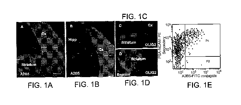
Figures 1 A, 1 B, 1 C andl D are micrographs showing A2B5+ cells in the
telencephalon. FIG. 1A shows A2B5+ cells in coronal sections of the developing
striatum and dorsolateral neocortex of the E15 telencephalon. FIG. 1B shows
that
1
CA 02684647 2009-10-16
WO 2008/131004 PCT/US2008/060477
A2B5+ cells are absent in the developing hippocampal region. FIG. 1 C and 1 D
show
that the dorsal A2B5+ region is not Olig2+ (FIG. 1C) while the ventral A2B5+
region
partially overlaps with the Olig2+ domain in the developing striatum (FIG.
1D). FIG
1E shows FACS data of A2B5+/PSA-NCAM" stained cells shows three cell
populations, including PSA-NCAM+, A2B5+/PSA-NCAM+, and A2B5+. Scale bar,
100 m.
Figures 2A, 2B, and 2C are micrographs showing a subset of A2B5+ cells are
also beta III tubulin+ in the E15 dorsal telencephalon. FIGs. 2A-C show the
isolated
A2B5+/PSA-NCAM- cell population from the dorsal telencephalon included a beta
III
tubulin+ population, seen at 1 hour (FIG. 2A), 12 hours (FIG. 2B), and 4 days
(FIG.
2C) post isolation. FIG. 2D is a histogram showing isolated A2B5+/PSA-NCAM"
cells stained and analyzed for beta III tubulin presence between E13 and E20.
E15
was determined to be the peak time to isolate A2B5+/PSA-NCAM-/beta III
tubuliri
cells as 21% of the E15 A2B5+/PSA-NCAM- population was beta III tubulin . DAPI
nuclear stain. Scale bars, 100 m.
Figures 3A, 3B and 3C show an outline of the isolation procedure used to
characterize the putative glial restricted precursor population. A2B5+/PSA-
NCAM"
cells were selected by MACS resulting in a heterogeneous mixture of cells. For
mass
culture studies (FIG. 3A) and clonal analysis (FIG. 3B), cells were maintained
in
culture for two cell passages to select for proliferative cells and to remove
the A2B5+
neuronal population. The resultant putative glial restricted precursor
population was
then plated at mass culture or clonal density and exposed to differentiating
conditions
including a pro-oligodendrocytic condition, a pro-astrocytic condition, or a
pro-
neuronal condition. Alternatively, the heterogeneous mixture of cells obtained
from
the MACS selection was plated at clonal density, and resultant clones were
selectively
passaged and split into the differentiation conditions (FIG. 3C).
Figures 4A, 4B, 4C, 4D, 4E and 4F are micrographs showing that the putative
dorsal glial restricted precursor population can generate macroglial subtypes
in mass
culture. Putative glial restricted precursor cells generate Ga1C+ cells (FIG.
4A) and
GFAP+ cells (FIG. 4C) but do not generate neurons (FIG. 4D) after 6 days of
exposure to the appropriate differentiation conditions. FIG. 4B shows that
after 4
days of growth in the pro-oligodendrocyte condition, O4+ cells were readily
2
CA 02684647 2009-10-16
WO 2008/131004 PCT/US2008/060477
identifiable. FIGs. 4E and 4F show exposure of the putative glial restricted
precursor
population to BMP-4 is insufficient to result in detection of the known
astrocyte
marker GFAP until 10 days (FIG. 4E), but does induce the astrocyte precursor
cell
marker, CD44, after 6 days (FIG 4F). DAPI nuclear stain (FIGs. 4D and 4F).
Scale
bars, 100 m.
Figures 5A and 5B show photomicrographs of neuron generation from E15
unsorted dorsal and ventral telencephalic cells. In order to validate the pro-
neuronal
condition used, cells present in the E15 dorsal (FIG. 5A) and ventral (FIG 5B)
telencephalon before MACS selection were exposed to the pro-neuronal condition
used for glial restricted precursor characterization and were found to
generate beta III
tubulin+ cells after 6 days in culture. Scale bars, 100 m.
Figures 6A, 6B and 6C are micrographs showing clonal analysis of the
putative dorsal glial restricted precursor further indicates glial
restriction. To
distinguish between the potential presence of an APC/OPC cell mixture and the
presence of a glial restricted precursor population, the putative glial
restricted
precursor population was grown at clonal density and exposed to the
differentiating
conditions, resulting in the detection of clones containing Ga1C+ cells (FIG.
6A)
clones containing GFAP+ cells (FIG 6B) but no neuron containing clones (FIG
6C).
DAPI nuclear stain. Scale bars, 100 m.
Figures 7A, 7B and 7C are micrographs showing clone splitting confinning
the ability of the putative glial restricted precursor cell to generate both
oligodendrocytes and astrocytes. Split clones of A2B5+/PSA-NCAM- founder cells
can generate GalC+ cells (FIG. 7A) GFAP+ cells (FIG. 7B) but not neurons (FIG.
7C)
and allows for the classification of the A2B5+/PSA-NCAM-/beta III tubuliri
cell as a
glial restricted precursor cell. DAPI nuclear stain. Scale bars, 100 m.
Figures 8A, 8B, 8C, 8D, 8E, 8F, 8G, 8H, and 81 are micrographs showing the
dorsal telencephalon has the potential to generate glial restricted precursor
cells
independent of ventral cell infiltration. FIGs. 8A, 8B and 8C show that cells
with the
similar antigenic profile described for the dorsal glial restricted precursor
population
were isolated from two day in vitro grown dorsal explants, and can generate
GaIC+
cells (FIG. 8A) GFAP+ cells (FIG. 8B) but not neurons (FIG. 8C) in mass
culture.
FIGs. 8D, 8E and 8F show explant derived putative glial restricted precursors
can
3
CA 02684647 2009-10-16
WO 2008/131004 PCT/US2008/060477
generate clones containing Ga1C+ cells (FIG. 8D) clones containing GFAP+ cells
(FIG. 8E) but no clones containing neurons (FIG. 8F) when exposed to the
differentiation conditions. FIGs. 8G, 8H and 81 show split clones of explant
derived
putative glial restricted precursor founder cells can generate GalC+ cells
(FIG. 8G)
GFAP+ cells (FIG. 8H) but not neurons (FIG. 81). DAPI nuclear stain. Scale
bars, 100
m.
Figures 9A, 9B, 9C, 9D, 9E, 9F, 9G, 9H, 91 and 9J are micrographs showing a
glial restricted precursor population cell can be isolated from the E15
ventral
telencephalon. FIGs. 9A, 9B and 9D show putative glial restricted precursor
cells
sharing the similar antigenic profile of the dorsal glial restricted precursor
population
were isolated from the E15 ventral telencephalon, consisting of the AEP and
MGE.
This cell population generated Ga1C+ cells (FIG. 9A) and GFAP+ cells (FIG. 9B)
but
not neurons (FIG. 9D) in mass culture. FIG. 9C shows putative glial restricted
precursor cells do not make A2B5+/GFAP+ Type-II astrocytes in response to
CNTF.
To distinguish between APC/OPC presence and glial restricted precursor
presence,
ventral putative glial restricted precursor cells were grown at clonal density
and
generated Ga1C+ cells (FIG. 9E) and GFAP+ cells (FIG. 9F) but not neurons
(FIG. 9G)
when examined at the clonal level. Split clones of ventral putative glial
restricted
precursor founder cells generated Ga1C+ cells (FIG. 9H) and GFAP+ cells (FIG.
91)
but not neurons (FIG. 9J). DAPI nuclear stain, (FIGs. 9A, 9C-9J). Scale bars,
100
m.
Figure 10 is a histogram showing a summary of the generated clones from
dorsal, ventral, and explant derived glial restricted precursor, with no
significant
difference (p>0.05; Student's t-test) between astrocyte and oligodendrocyte
containing clone numbers.
Figures 11 A, 11 A', 11 B, 11 B', 11 C, 11 C' are electronmicrographs and 11
D,
11 E, 11 F, 11 G, 11 H and 11 I are fluorescent micrographs, showing dorsal
glial
restricted precursors and explant derived dorsal glial restricted precursors
produce
compact myelin, in addition to the ability of both ventral and dorsal glial
restricted
precursors to make astrocytes in vivo. FIGs. 11A-C' show EM images from the
contralateral hemisphere of the transplanted shiverer forebrains showed a lack
of
dense, compacted myelin, consistent with the shiverer mutant phenotype, on
4
CA 02684647 2009-10-16
WO 2008/131004 PCT/US2008/060477
longitudinally sectioned (FIG. 1 lA) and cross-sectioned (FIG. 11A') neuronal
fibers.
The dorsal glial restricted precursor isolated from the E15 dorsal
telencephalon and
transplanted into the postnatal day 18 (P 18) shiverer forebrain is capable of
myelin
formation as seen in longitudinally sectioned (FIG. 11B) and cross-sectioned
(FIG.
11B') neuronal fibers. Transplantation of the dorsal glial restricted
precursor cell
derived from two day in vitro grown E13 dorsal telencephalic explants into the
P18
shiverer mutant forebrain produces compacted myelin as seen in longitudinally
sectioned (FIG. 11C) and cross-sectioned (FIG. 11C') neuronal fibers. FIGs.
11D-F
show hPAP+ dorsal glial restricted precursors transplanted into the forebrains
of P0 rat
pups generate hPAP+/GFAP+ cells after 10 days, as well as Olig2+
oligodendroglial
cells (FIGs. 11G-I). DAPI nuclear stain (FIG. 11F). Scale bars for 11A-C' as
indicated, scale bars for 11D-I, 100 m.
Figure 12 shows a model for the generation of glial subtypes through
telencephalic Glial Restricted Precursor (tGRP) populations. The dorsal
telencephalon and ventral telencephalon give rise to glial restricted
precursor
populations with a primary developmental fate towards astrocyte and OPC
generation,
respectively. The classification of these two populations as true tGRP
populations
uses their isolation and in vitro characterization in order to remove the
normal
developmental cues promoting dorsal astrocyte generation and ventral OPC
formation. As the ventral and dorsal telencephalon continues through
development,
each tGRP population has the potential to participate in a secondary
developmental
fate towards astrocytes ventrally, or OPCs dorsally. The developmental
plasticity of
each population is revealed in vitro and demonstrates the potential for
oligodendrocyte and astrocyte development from a common precursor cell type.
Figure 13A is a micrograph showing spinal cord GDAgP130 (CNTF induced)
astrocytes express both GFAP and Olig2. Cells were grown for 4 days in the
presence
of growth factors.
Figure 13B is a micrograph showing CNTF induced GFAP+ astrocytes derived
from tGRPs do not resemble scGDAgbp13o based on a lack of Olig2/GFAP
colocalization.
5
CA 02684647 2009-10-16
WO 2008/131004 PCT/US2008/060477
Figure 14 shows intracellular redox status of ventral and dorsal tGRPs. As
measured by the geometric mean of oxidized dye fluorescence, dorsal tGRPs have
a
higher intracellular redox level when compared to ventral tGRPs.
Figure 15 A, B and C, are micrographs showing an indication that tGRPs
generate Ga1C+ oligodendrocytes via a PSA-NCAM/PDGFRalpha/Olig2+
intermediate. The passage of a tGRP through a classically described OPC (PSA-
NCAM/PDGFRalpha/Olig2+) intermediate provides evidence that tGRps are
responsible for the generation of OPCs in vivo and adds to the number of
possible
intermediate cell fates that are achievable with the use of tGRPs as a
starting
population.
DETAILED DESCRIPTION
This disclosure is related to lineage restricted glial precursor cells from
the
telencephalon. For example, provided herein are telencephalic glial-restricted
precursor (tGRP) cell populations. Related compositions are also provided and
include, but are not limited to, any cell or cell population derived from a
population of
telencephalic glial-restricted precursor cells. An example of a related
composition is
a type-1 astrocyte, or population thereof, derived from a telencephalic glial-
restricted
precursor cell. Related compositions can also include other compounds, agents
or
molecules in combination with a tGRP cell or population, or a cell or cell
population
derived from a tGRP cell or cell population. Also provided are 01ig2" glial
restricted
precursor (GRP) cells and cell populations. Optionally, the Olig2- GRPs are
isolated
from the dorsal telencephalon.
Further provided are methods of using and producing telencephalic glial-
restricted precursor cell populations and related compositions. These methods
include,
but are not limited to, treating a CNS lesion in a subject comprising
administering
telencephalic glial-restricted precursor cells, or cells derived from a
telencephalic
glial-restricted precursor cell, to the subject. The cells can be administered
in
combination with other compounds, agents or molecules as described herein.
Telencephalic glial-restricted precursor cell populations include precursor
populations in the ventral and dorsal telencephalon that generate astrocytes
and
oligodendrocytes. The dorsal glial precursor cells can be generated de novo
from the
6
CA 02684647 2009-10-16
WO 2008/131004 PCT/US2008/060477
dorsal telencephalon and they can be used for in vivo production of both
myelin-
forming oligodendrocytes and astrocytes upon transplantation into a subject.
Within the central nervous system (CNS), the greatest progress in identifying
the
specific cell populations involved in development has been achieved in the
spinal cord.
In the rat spinal cord, embryonic day 10.5 (E10.5 ) cells have been shown to
represent a
homogenous population of multipotent neuroepithelial stem cells (NEPs) capable
of
generating cells of both the neuronal and glial lineage.
Differentiated cell types arise from these NEP cells by way of lineage
restricted
intermediate precursor populations capable of extended proliferation and the
generation
of neurons or glia. The cells comprising the earliest intermediate precursor
population
restricted to oligodendrocyte and astrocyte formation, called glial restricted
precursor
cells (GRPs), can be isolated from the embryonic spinal cord as early as E12.
Their
ability to generate two antigenically distinct populations of astrocytes and
oligodendrocytes has been established both in vitro and in vivo.
GRP cells are identified with the A2B5 antibody and do not express the
Polysialylated form of Neural Cell Adhesion Molecule (PSA-NCAM). Freshly
isolated
GRP cells depend on basic fibroblast growth factor (bFGF) for survival and
proliferation
but, unlike oligodendrocyte progenitor cells (OPCs), are not defined by the
expression of
platelet-derived growth factor receptor-alpha (PDGFR-alpha) or Olig2. The OPC
has
been shown in vivo to arise at a later time point than the GRP, and the
generation of
oligodendrocytes from a GRP population has been demonstrated in vitro to occur
through an OPC intermediate stage.
Additional characteristics distinguishing GRP cells from OPCs are the ability
of
the GRP cells to generate two types of astrocytes (that have been designated
type-1 and
type-2) in vitro and to generate both oligodendrocytes and astrocytes in vivo.
Both type-
1 and type-2 astrocytes are GFAP+, but only type-2 astrocytes co-label with
the A2B5
antibody. Type-1 astrocytes are thought to arise from GRP cells through
intermediate
astrocyte progenitor cells (APC), while Type-2 astrocytes can require prior
generation of
OPCs as an intermediate step. Unlike OPCs, GRP cells readily generate
astrocytes
following transplantation into the adult CNS, while primary OPCs only generate
oligodendrocytes in such transplantations.
7
CA 02684647 2009-10-16
WO 2008/131004 PCT/US2008/060477
The identification of GRP cells in the spinal cord gave rise to a generalized
model of gliogenesis. This model of gliogenesis involves the progression from
a
multipotential NEP cell to a lineage restricted multipotent precursor cell
population
(e.g. GRPs) that in turn give rise to more restricted glial precursor cell
types (e.g.
OPCs and possibly APCs) and the eventual mature glial cells of the CNS (e.g.
oligodendrocytes and astrocytes).
It has been ascertained through genetic and clonal in vitro experiments that a
subset of cells from ventral regions of the telencephalon differentiate into
PDGFR-
alpha+ and/or Olig2+ oligodendrocyte progenitors, migrate away from their
ventral
origin, and give rise to mature oligodendrocytes throughout the brain. It
appears that
these cells express Oligl/2 to be fated towards oligodendrocytes as compound
disruption of Oligl and Olig2 results in a complete loss of oligodendrocytes.
Provided herein are telencephalic precursor cell populations capable of
generating oligodendrocytes and astrocytes but that are unable to generate
neurons
under conditions that generally promote neuronal lineage. Examples of
conditions
that generally promote neuronal lineage in vitro include exposure to
Neurotrophin-3
(NT-3) (e.g., at lOng/ml) plus All-trans Retinoic Acid (RA) (e.g., at IOOnM),
to Glial
Growth Factor (GGF) (e.g., at lOng/ml), or to Brain Derived Neurotrophic
Factor
(BDNF) (e.g., at l Ong/ml). The provided tGRP cells do not produce neurons
under
these example conditions.
Cell populations were isolated from the dorsal telencephalon based on the
antigenic phenotype of restricted precursor cells previously identified in the
spinal
cord. These telencephalic cells were characterized in mass culture and at the
clonal
level and were found to generate all macroglial subtypes but were unable to
generate
neurons under under conditions that generally promote neuronal lineage.
The dorsal telencephalon was determined to be capable of generating this glial
restricted population de novo by separating the dorsal telencephalon at a time
point
where the cell populations present are exclusively of a dorsal origin. A
ventral glial
restricted cell population was detected in parallel.
The ability of the dorsal cell population to differentiate into myelin
producing
oligodendrocytes upon transplantation in a myelin deficient background was
confirmed, as well as GFAP+ astrocytes when transplanted into the perinatal
8
CA 02684647 2009-10-16
WO 2008/131004 PCT/US2008/060477
forebrain. Thus, described are populations of precursor cells isolated from
the
embryonic telencephalon that are able to generate both oligodendrocytes and
astrocytes but are unable to generate neuronal progeny under under conditions
that
generally promote neuronal lineage.
Also provided is a defined cell population that is generated de novo in the
dorsal aspect of the telencephalon and is a source for dorsally derived glial
cells.
Further provided is a cell population in the telencephalon that can act as a
source of
astrocytic cells both ventrally as well as dorsally. Thus, disclosed is a
model of
gliogenesis by which glial cells originate in a timely and organized manner in
the
developing telencephalon.
Provided herein are compositions and methods for the treatment of CNS
injury, including traumatic or degenerative conditions of the CNS, promotion
of axon
regeneration, suppression of astrogliosis, re-alignment of host tissues, and
the delay of
axon growth inhibitory proteoglycan expression. Thus, provided are methods of
treating a CNS lesion in a subject, comprising administering to the subject a
composition comprising telencephalic glial-restricted cell populations and/or
cells
derived from a telencephalic glial-restricted cell, including tGRP progeny or
combinations thereof tGRP progeny include any GFAP+ cell derived or produced
from a tGRP. For example, tGRP progeny include tGRP derived astrocytes, GDAs,
and APCs. Optionally, the GDA is a type-1 GDA. Optionally, the astrocyte is a
type-
1 astrocyte. tGRP progeny also include any GaIC+ cell derived or produced from
a
tGRP. For example, tGRP progeny include oligodendrocytes. Methods of treating
a
CNS lesion in a subject, comprising administering to the subject an Olig2-
cell or cells
are also provided. Described cells or combinations thereof can be administered
in
combination with other compositions as described herein.
The methods can be used for the treatment of spinal cord injury or other CNS
injuries. The methods can also be used in CNS lesions in which it is desirable
to
promote regeneration and/or re-alignment of host tissues, modulate the CNS
scarring
response, and rescue neurons from atrophy and death, or any combination
thereof.
9
CA 02684647 2009-10-16
WO 2008/131004 PCT/US2008/060477
As used herein, the term GDAs (glial restricted precursor derived astrocyte)
refers to glial fibrillary acidic protein (GFAP)+/A2B5- cells, also referred
to herein as
type-1 GDAs, unless type-2 GDAs (GFAP+/A2B5+ cells) are specifically
referenced.
The limited success of stem cell and neural precursor cell transplantation is
likely due to the inflammatory environment of adult CNS injuries, which direct
undifferentiated neural stem cells or glial precursors to a scar astrocyte
like
phenotype. Scar astrocytes are poorly supportive of axon growth.
Methods and compositions described herein can provide an alternative to
allowing the lesion environment to direct differentiation of stem or precursor
cells
while still retaining the benefit of starting with an undifferentiated cell.
Provided
herein are methods of treating a CNS lesion in a subject, comprising
administering to
the subject a composition comprising telencephalic glial restricted precursor
cells or
cells derived from a tGRP cell. The term lesion is used herein to refer to a
site of
injury to the CNS, a site of a CNS disease process, degenerative damage, or
scarring,
wherein promotion of regeneration would provide benefit.
Telencephalic glial-restricted precursor (tGRP) populations can generate
oligodendrocytes, APCs, and can preferentially generate type-1 GDAs and type-1
astrocytes versus type 2 astrocytes. tGRP cells are restricted to the glial
lineage in
vivo as they are unable to generate neuronal phenotypes in an in vivo
neurogenic
environment. tGRP cells survive and migrate in the neonatal and adult brain.
Transplanted tGRP cells can differentiate into myelin-forming oligodendrocytes
in a
myelin-deficient background and can also generate immature oligodendrocytes in
the
normal neonatal brain. Transplanted tGRP cells can also differentiate into
type-1
GDAs and type-1 astrocytes when administered to a CNS lesion. In some aspects,
such transplanted tGRP cells do not produce type-2 astrocytes.
Cell culture technologies can be used for the preparation of tGRPs, APCs,
GDAs, astrocytes and oligodendrocytes. As an example, A2B5+ tGRPs can be
isolated from dissociated cell suspensions of telencephalon of embryos using
standard
methods such as, for example, flow cytometry or immunopanning.
tGRPs or tGRP derived APCs, GDAs, astrocytes, or oligodendrocytes can be
immortalized by procedures known in the art, so as to preserve a continuing
source of
tGRPs, or tGRP derived APCs, GDAs, astrocytes, or oligodendrocytes.
Immortalized
CA 02684647 2009-10-16
WO 2008/131004 PCT/US2008/060477
tGRPs or tGRP derived APCs, GDAs, astrocytes, or oligodendrocytes can be
maintained in vitro indefinitely. Various methods of immortalization are known
in
the art including, but not limited to, viral transformation (e.g., with SV40,
polyoma,
RNA or DNA tumor viruses, Epstein Barr Virus, bovine papilloma virus, or a
gene
product thereof) and chemical mutagenesis. The cell line can be immortalized
by a
virus defective in replication, or is immortalized solely by expression of a
transforming virus gene product. For example, tGRPs or tGRP derived APCs,
GDAs,
astrocytes, or oligodendrocytes can be transformed by recombinant expression
vectors
which provide for the expression of a replication-defective transforming virus
or gene
product thereof. Such procedures are known in the art.
tGRPs can be maintained in culture in a suitable medium. For example,
tGRPs can be maintained in culture with approximately 0.1-100ng/ml bFGF and
SATO supplements on a mixed laminin/fibronectin substrate. In order to
differentiate
tGRPs to GDAs, the tGRPs can be exposed to, for example, approximately 1-100
ng/ml of recombinant BMP-4 (for approximately 7 days in culture) to
differentiate
them into GDAs. Also disclosed is the use of other members of the BMP family,
or
other signaling molecules that induce differentiation along the astrocyte
pathway
within the antigenic range of type-1 astrocytes.
tGRPs or tGRP derived APCs, GDAs, astrocytes, or oligodendrocytes can be
cryopreserved. Various methods for cryopreservation of viable cells are known
and
can be used (see, e.g., Mazur, 1977, Cyrobiology 14:251-272; Livesey and
Linner,
1987, Nature 327:255; Linner, et al., 1986, J. Histochem. Cytochem. 34(9):1123-
1135; U.S. Pat. No. 4,199,022 to Senkan et al.; U.S. Pat. No. 3,753,357 to
Schwartz;
U.S. Pat. No. 4,559,298 to Fahy, which are incorporated by reference at least
for the
methods described therein).
GDAs for use in the methods described herein can be generated by the method
comprising isolating telencephalic cells from the subject, purifying A2B5
positive
tGRPs, and culturing said cells with a BMP.
To ensure GDA suspensions for transplantation do not contain
undifferentiated tGRPs or cells with the phenotype of type-2 astrocytes,
contaminating cell types can be removed from the suspension by, for example,
immuno-panning with the A2B5 antibody. A small volume of the resulting
11
CA 02684647 2009-10-16
WO 2008/131004 PCT/US2008/060477
suspension can be plated onto glass coverslips and labeled with antibodies to
A2B5
and GFAP to verify a uniform type-1 astrocyte phenotype. For transplantation,
GFAP
positive/A2B5 negative GDAs can be suspended in a suitable medium such as, for
example, Hanks Balanced Salt Solution, at a density of 103-106 cells/gL.
tGRP-derived GDAs can be generated by BMP exposure and fall within the
population of cells defined by their antigenic phenotype as type-1 astrocytes.
In vitro
studies on cells purified from the postnatal CNS have shown that type-1
astrocytes of
postnatal origin promote extensive neurite growth from a variety of neurons in
vitro,
express high levels of axon growth supportive molecules such as
laminin/fibronectin
and NGF / NT-3 and also exhibit minimal chondroitin sulfate proteoglycan
immunoreactivity in vitro. However, while transplantation of immature cortical
astrocytes into adult brain injuries or acute adult spinal cord injuries have
been shown
to suppress astrogliosis, only limited sprouting of endogenous axons have been
observed, with axons failing to penetrate the center of grafts or re-enter
white matter
beyond the sites of injury.
Thus, although GDAs show antigenic phenotypes like type-1 astrocytes,
GDAs are a unique cell type that, when transplanted into CNS lesion sites,
promote an
unprecedented level of tissue reorganization, axon regeneration and locomotor
recovery.
GDAs promote robust axon regeneration and functional recovery after
transplantation into CNS lesion sites. The ability of GDAs to fill an injury
site,
suppress astrogliosis, re-align host tissues and delay expression of axon
growth
inhibitory proteoglycans indicate that these cells possess an effective
ability to
provide an axon regenerative environment. These attributes, in combination
with
their striking ability to significantly reduce atrophy of axotomized CNS
neurons and
support a robust behavioral recovery, make GDAs a highly effective cell type
with
which to repair a damaged or diseased CNS. Thus, the GDAs can promote axon
regeneration, suppress astrogliosis, re-align host tissues, delay expression
of axon
growth inhibitory proteoglycans, or any combination thereof.
Provided herein is an isolated tGRP cell or a population of isolated tGRP
cells. As used herein, the term isolated refers to a cell or population of
cells which
has been separated from its natural environment, e.g., removal from a donor
animal,
12
CA 02684647 2009-10-16
WO 2008/131004 PCT/US2008/060477
e.g., human or embryo. The isolated cell or population of cells can be in the
form of a
tissue sample, e.g., an intact sheet of cells, e.g., a monolayer of cells, or
it can be in a
cell suspension. The term isolated does not preclude the presence of other
cells. The
term population is intended to include two or more cells. Cells in a
population can be
obtained from the same or different source(s).
The telencephalic glial restricted precursor cells can be isolated from a
mammal, including an embryo, selected from the group consisting of human and
non-
human primates, equines, canines, felines, bovines, porcines, ovines, rats and
lagomorphs.
Provided herein are isolated cell populations comprising at least about a 10%,
20%, 30%, 40%, 50%. 60%, 70%, 80%, 85%, 90%, 95%, 96%, 97%, 98%, 99%, or
100% pure population of tGRPs or any percent between 10 to 100%. Thus, for
example, the isolated cell population can comprise at least 90% tGRPs. The
isolated
population can also comprise at least 95% tGRPs or at least 99% tGRPs. Cell
populations comprising the same percentages of Olig2" GRP cells are also
provided.
The Olig2" GRP cells are optionally isolated from the dorsal telencephalon.
Optionally, the isolated cell population does not comprise type-2 astrocytes.
Optionally, the isolated cell population does not comprise pluripotential or
multipotential stem cells, such as ES cells or neuroepithelial stem cells.
However, the
isolated cell population can also comprise about 0.01 %, 0.05%, 0.1%, 0.5%,
1%, 2%,
3%, 4%, 5%, 6%, 7%, 8%, 9%, or 10% type-2 GDAs, type-2 astrocytes, APCs,
pluripotential stem cells, multipotential cells, undifferentiated glial
precursors, or any
combination thereof. Thus, for example, the isolated cell population can
comprise
less than 10% type-2 GDAs. The isolated cell population can also comprise less
than
5% type-2 GDAs. The purity of a cell population can be determined by, for
example,
detecting markers specific for various cell types in culture and determining
by visual
observation the percentage of cell types in the population. Also provided are
compositions comprising the isolated cell populations in combination with
other
compositions including compounds, agents or molecules.
A purified population of cells can be grown in feeder-cell-independent culture
on a substratum and in a medium configured for supporting adherent growth of
the
telencephalic glial restricted precursor cells or derivatives thereof and at a
temperature
13
CA 02684647 2009-10-16
WO 2008/131004 PCT/US2008/060477
and in an atmosphere conducive to growth of the precursor cells and
derivatives
thereof. The telencephalic glial restricted precursor cells and derivatives
can be
purified using procedures such as specific antibody capture, fluorescence
activated
cell sorting, magnetic bead capture, and the like.
Provided herein is an isolated tGRP derivative or progeny cell, or a
population
of isolated tGRP derivative or progeny cells. Optionally, the tGRP derivative
or
progeny cell or cells are GFAP+. For example, the derivative or progeny cell
or cells
can be an APC, type-1 GDA or type-1 astrocyte. In another aspect, the tGRP
derivative or progeny cell or cells are Ga1C+. For example, the tGRP
derivative or
progeny cell can be an oligodendrocyte.
Thus, provided herein is an isolated APC, GDA, astrocyte or oligodendrocyte
cell, or a population of isolated APC, GDA, astrocyte or oligodendrocyte
cells,
derived from a tGRP, or isolated tGRP population. tGRP derived isolated APC,
GDA, astrocyte or oligodendrocyte populations can comprise at least about an
10%,
20%, 30%. 40%, 50%, 60%, 70%, 80%, 85%, 90%, 95%, 96%, 97%, 98%, 99%, or
100% pure population of each respective cell type or any percent between 10%
to
100%. Thus, for example, the isolated cell population can comprise at least
90%
APCs, GDAs, astrocytes, or oligodendrocytes. The isolated population can also
comprise at least 95% APCs, GDAs, astrocytes, or oligodendrocytes or at least
99%
APCs, GDAs, astrocytes, or oligodendrocytes. In certain aspects, the isolated
cell
population does not comprise type-2 astrocytes or type-2 GDAs. Optionally, the
isolated cell population does not comprise pluripotential or multipotential
stem cells,
such as ES cells or neuroepithelial stem cells. However, the isolated cell
population
of the method can comprise at most about 0.01%, 0.05%, 0.1%, 0.5%, 1%, 2%, 3%,
4%, 5%, 6%, 7%, 8%, 9%, or 10% type-2 GDAs, type-2 astrocytes, pluripotential
stem cells, multipotential cells, undifferentiated glial precursors (e.g.,
GRPs), or any
combination thereof. Thus, for example, the isolated cell population can
comprise
less than 10% type-2 GDAs. The isolated cell population can also comprise less
than
5% type-2 GDAs.
The purity of a cell population can be determined by, for example, detecting
markers specific for various cell types in culture and determining by visual
observation the percentage of cell types in the population. Also provided
herein are
14
CA 02684647 2009-10-16
WO 2008/131004 PCT/US2008/060477
compositions comprising the isolated cell populations in combination with
other
compositions including compounds, agents or molecules.
The tGRPs or tGRP derived APCs, GDAs, astrocytes, oligodendrocytes or
combinations thereof can be administered using standard methods known in the
art for
use in the promotion of CNS nerve regeneration and/or scar reduction. The
tGRPs or
tGRP derived APCs, GDAs, astrocytes, oligodendrocytes or combinations thereof
can
be administered to treat subjects in which it is desired to promote CNS
regeneration
and/or reduce scar formation. Thus, tGRPs or tGRP derived APCs, GDAs,
astrocytes,
oligodendrocytes, or combinations thereof can be applied in any conventional
formulation to areas of a lesion.
There is no restriction to the location of a lesion. Thus, any part of the
brain
or spinal cord can be treated. For example, the cerebral cortex, the mid-
brain, the
thalamus, the hypothalamus, the striatum, the substantia nigra, the pons, the
cerebellum, the medulla, or any cervical, thoracic, lumbar, or sacral spinal
segment.
The methods are applicable for any nervous system lesion including, for
example,
those caused by spinal cord injury (resulting, for example, in respiratory
paralysis,
quadriplegia, and paraplegia).
The tGRPs or tGRP derived APCs, GDAs, astrocytes, oligodendrocytes, or
combinations thereof can also be administered to patients in whom the nervous
system has been damaged or injured by trauma, surgery, ischemia, infection,
metabolic disease, nutritional deficiency, malignancy, toxic agents,
paraneoplastic
syndromes and degenerative disorders of the nervous system. Examples of such
disorders include, but are not limited to, Alzheimer's Disease, Parkinson's
Disease,
Huntington's chorea, amyotrophic lateral sclerosis, progressive supranuclear
palsy,
and neuropathies. tGRPs or tGRP derived APCs, GDAs, astrocytes,
oligodendrocytes
or combinations thereof, can be administered to a wound to reduce scar
formation.
Thus, after an operation, tGRPs or tGRP derived APCs, GDAs, astrocytes,
oligodendrocytes, or combinations thereof, can be administered in order to
reduce
scar formation from lesions due to, for example, arterio-venous malformation,
necrosis, bleeding, and craniotomy, which can secondarily give rise to
epilepsy.
tGRPs or tGRP derived APCs, GDAs, astrocytes, oligodendrocytes, or
combinations
CA 02684647 2009-10-16
WO 2008/131004 PCT/US2008/060477
thereof, can also be used for treatment of epilepsy, by stabilizing the
epileptic focus
and reducing scar formation.
Treatment can be performed, for example, within 24 hours, or alternatively,
for example, one week, 5 years, or even more than 10 years after onset of the
lesion.
In cases where a lesion can be predicted, for example, during surgery, the
tGRPs or
tGRP derived APCs, GDAs, astrocytes, oligodendrocytes, or combinations
thereof,
can be delivered prior to or during the occurrence.
tGRPs or tGRP derived APCs, GDAs, astrocytes, oligodendrocytes, or
combinations thereof, can be delivered by direct application, for example, by
direct
injection of a sample of tGRPs or tGRP derived APCs, GDAs, astrocytes,
oligodendrocytes, or combinations thereof, into the site of neural tissue
damage. For
example, the spinal cord can be exposed by laminectomy, and a cellular
suspension
injected using a microsyringe under a surgical microscope. When high
resolution
MRI images are obtained, the cell suspension can be injected without
laminectomy as
in intervertebrally (e.g., by the technique of lumbar puncture).
Methods for treating a neurological or neurodegenerative injury comprises
administering to a mammal in need of such treatment an effective amount of
telencephalic glial restricted precursor cells or derivatives thereof. The
tGRP cells or
derivatives thereof can be caused to (1) proliferate and differentiate in
vitro prior to
being administered, or (2) proliferate in vitro prior to being administered
and to
further proliferate and differentiate in vivo after being administered, or (3)
proliferate
in vitro prior to being administered and then to differentiate in vivo without
further
proliferation after being administered, or (4) proliferate and differentiate
in vivo after
being injected directly after being freshly isolated. The tGRP cells or
derivatives
thereof can be from a heterologous donor or an autologous donor. The donor can
be a
fetus, a juvenile, or an adult. The injury to be treated can be multiple
sclerosis, spinal
cord injury, CNS trauma, conditions in which axonal regeneration is desired,
conditions in which control or reduction in glial scarring is desired, any
dysmyelinating disorder, or an enzymatic disorder. The tGRP cells,
derivatives, or
combinations thereof, can be administered locally or widely in the CNS.
16
CA 02684647 2009-10-16
WO 2008/131004 PCT/US2008/060477
Optionally, tGRPs or tGRP derived APCs, GDAs, astrocytes,
oligodendrocytes, or combinations thereof, are delivered in a media which
partially
impedes their mobility so as to localize the tGRPs or tGRP derived APCs, GDAs,
astrocytes, oligodendrocytes, or combinations thereof, to a site of lesion. By
way of
example, tGRPs or tGRP derived APCs, GDAs, astrocytes, oligodendrocytes, or
combinations thereof, can be delivered in a paste or gel comprising, for
example, a
biodegradable gel-like polymer such as fibrin or a hydrogel. Such a semi-solid
medium can impede the migration of (scar-producing) undesirable mesenchymal
components such as fibroblasts into the site.
Optionally, tGRPs or tGRP derived APCs, GDAs, astrocytes,
oligodendrocytes, or combinations thereof, can be administered with the use of
polymer implants and surgical bypass techniques. Uses of polymer implants and
surgical techniques are known to those of skill in the art. For example, tGRPs
or
tGRP derived APCs, GDAs, astrocytes, oligodendrocytes, or combinations
thereof,
can be applied to a site of a lesion in a form in which the tGRPs or tGRP
derived
APCs, GDAs, astrocytes, oligodendrocytes, or combinations thereof, are seeded
or
coated onto a polymer implant. Various types of polymer implants can be used
herein, with various compositions, pore sizes, and geometries. Such polymers
include, but are not limited to, those made of nitrocellulose, polyanhydrides,
and
acrylic polymers (see e.g., those described in European Patent Publication No.
286284; Aebischer, et al., 1988, Brain Res. 454:179-187; Aebischar, et al.,
1988,
Prog. Brain Res. 78:599-603; Winn, et al., 1989, Exp. Neurol. 105:244-250,
which
are incorporated by reference at least for the polymers described therein).
Polymers can be used as synthetic bridges, over which nerve regeneration can
be promoted and scar formation can be reduced by application of tGRPs or tGRP
derived APCs, GDAs, astrocytes, oligodendrocytes, or combinations thereof, to
the
end(s), or in the vicinity of, the bridge. For example, an acrylic polymer
tube with
tGRPs or tGRP derived APCs, GDAs, astrocytes, oligodendrocytes, or
combinations
thereof, at one or more ends, or throughout the tube, can be used to bridge
lesions
rostrally or bypass lesions, e.g., of the spinal cord, over which regeneration
can be
induced. Semi-penneable tubes can be used, e.g., in the dorsal columns or
dorsal
afferents, which tubes can contain and provide for the release of trophic
factors or
17
CA 02684647 2009-10-16
WO 2008/131004 PCT/US2008/060477
anti-inflammatory agents. The types of tubes which can be used are well known
to
those of skill in the art.
Axon fibers that demonstrate regenerative growth or collateral sprouting
encounter an inhibitory environment as well as a physical gap that requires a
permissive bridging substance. Thus synthetic bridges can be used in the
methods
described herein. Advances in the field of biomatrix material have provided
opportunities to bridge the gap with artificial material, such as
biodegradable
hydrogels, or combinations of hydrogels and cells, that may promote
regeneration.
Desired properties of a synthetic bridge are to provide simultaneously a
physical
substrate for axonal attachment and growth without triggering antigenic host
reactions.
Optionally, tGRPs or tGRP derived APCs, GDAs, astrocytes,
oligodendrocytes, or combinations thereof, can be administered in combination
with
other compositions including therapeutic or pharmacological compounds, agents
and
molecules. For example, several agents have been applied to acute spinal cord
injury
(SCI) management and CNS lesions that can be used in combination with the
compositions and methods. Such agents include agents that reduce edema and/or
the
inflammatory response. Exemplary agents include, but are not limited to,
steroids,
such as methylprednisolone; inhibitors of lipid peroxidation, such as
tirilazad mesylate
(lazaroid); and antioxidants, such as cyclosporin A, EPC-K1, melatonin and
high-dose
naloxone. Thus, the compositions including tGRPs or tGRP derived APCs, GDAs,
astrocytes, oligodendrocytes, or combinations thereof, can further comprise
methylprednisolone, tirilazad mesylate, cyclosporin A, EPC-Kl, melatonin, or
high-
dose naloxone or any combination thereof.
The compositions including tGRPs or tGRP derived APCs, GDAs, astrocytes,
oligodendrocytes, or combinations thereof, can also comprise, glutamate
receptor
antagonists including, but not limited to, the noncompetitive N-methyl-D-
aspartate
(NMDA) ion channel blocker MK-801 (dizocilpine, Merck & Co., Inc., Whitehouse
Station, NJ), 1,2,3,4-tetrahydro-6-nitro-2,3-dioxobenzo[f]quinoxaline-7-
sulfonamide
(NBQX), gacyclidine (GK-11, Beaufour-Ipsen, Paris, France), and agmatine.
Anti-inflammatory agents, such as, for example, CM101, cytokine IL- 10, and
selective cyclooxygenase (COX)-2 inhibitors can be used in conjunction with
the
tGRPs or tGRP derived APCs, GDAs, astrocytes, oligodendrocytes, or
combinations
18
CA 02684647 2009-10-16
WO 2008/131004 PCT/US2008/060477
thereof. Thus, the compositions can further comprise CM101, IL-10, or a
selective
COX-2 inhibitor or any combination thereof.
The tGRPs or tGRP derived APCs, GDAs, astrocytes, oligodendrocytes, or
combinations thereof, can also be used in conjunction with inhibitors of
apoptosis,
such as caspase inhibitors, for example, Bcl-2, and calpain inhibitors.
Compositions including tGRPs or tGRP derived APCs, GDAs, astrocytes,
oligodendrocytes, or combinations thereof, can also comprise exogenous
neurotrophins, including, but not limited to, nerve growth factor (NGF), glial-
derived
neurotrophic factor (GDNF), cilliary neurotrophic factor (CNTF), neurotrophic
factor-
3 and 4/5 (NT-3, NT-4/5), fibroblastic growth factor (FGF), and brain-derived
neurotrophic factor (BDNF) or any combination thereof.
Inhibitors of netrins, semaphorins, ephrins, tenascins, integrins, and
chondroitin sulfate proteoglycans (CSPG) can be used in combination with tGRPs
or
tGRP derived APCs, GDAs, astrocytes, oligodendrocytes, or combinations
thereof.
For example, chondroitinase can be used to remove CSPG. Thus, the compositions
can further comprise an inhibitor of netrins, semaphorins, ephrins, tenascins,
integrins, or CSPG. Thus, the compositions can further comprise a
chondroitinase.
The compositions including tGRPs or tGRP derived APCs, GDAs, astrocytes,
oligodendrocytes, or combinations thereof, can also comprise, the IN-1
antibody,
which neutralizes the inhibitory protein activity of NoGo, the myelin-derived
growth-
inhibitory protein, myelin-associated glycoprotein (MAG) or any combination
thereof.
Agents that act through direct intracellular mechanisms in the nerve cell body
to promote neurite growth can be used in combination with tGRPs or tGRP
derived
APCs, GDAs, astrocytes, oligodendrocytes, or combinations thereof. Thus,
inosine, a
purine nucleoside, and cAMP and the compound AIT-082, a synthetic hypoxanthine
derivative containing a para-aminobenzoic acid moiety (e.g., Neotrofin;
NeoTherapeutics, Newport Beach, CA) can be used in the compositions and
methods.
Thus, the compositions can further comprise AIT-082.
Gene therapy allows the engineering of cells, which combines the therapeutic
advantage of the cells in combination with a gene delivery system. For
example, if
delivery of neurotrophins is desired, cells that form myelin and secrete
neurotrophins
can be engineered to both promote neurite growth and restore nerve function.
19
CA 02684647 2009-10-16
WO 2008/131004 PCT/US2008/060477
Macrophages from the patient's own blood (autologous macrophages) can be
activated and implanted at the site of the injury in combination with tGRPs or
tGRP
derived APCs, GDAs, astrocytes, oligodendrocytes, or combinations thereof. The
patient's own activated macrophages can scavenge degenerating myelin debris,
rich in
non-permissive factors, and thus encourage regenerative growth without
eliciting an
immune response.
The compositions including tGRPs or tGRP derived APCs, GDAs, astrocytes,
oligodendrocytes, or combinations thereof, can further comprise immuno-
suppressive
drugs such as cyclosporins, tacrolimus (FK505), cyclophosamid, azathioprines,
methotrexate, mizoribin alone or in any combination or the use thereof. Thus,
the
compositions can further comprise cyclosporins, tacrolimus (FK505),
cyclophosamid,
azathioprines, methotrexate, or mizoribin.
Administration of any composition in combination with the administration of
tGRPs or tGRP derived APCs, GDAs, astrocytes, oligodendrocytes, or
combinations
thereof, can be performed prior to, concurrent with, or after the
administration of a
tGRPs or tGRP derived APCs, GDAs, astrocytes, oligodendrocytes or a
combination
thereof. Thus, the methods described herein can further comprise,
administration of a
composition including agents, compounds or molecules, prior to, during, or
after
administration of the tGRPs or tGRP derived APCs, GDAs, astrocytes,
oligodendrocytes, or combinations thereof. The compositions and methods
described
herein may comprise a composition including agents, compounds or molecules in
any
combination. By way of example, the compositions containing tGRPs or tGRP
derived APCs, GDAs, astrocytes, oligodendrocytes, or combinations thereof,
described herein may also comprise a glutamate receptor antagonist and a
neurotrophin. One or more of the compositions including agents, compounds or
molecules can be formulated with the tGRPs or tGRP derived APCs, GDAs,
astrocytes, oligodendrocytes, or combinations thereof, containing composition
or can
be administered separately from the tGRPs or tGRP derived APCs, GDAs,
astrocytes,
oligodendrocytes, or combinations thereof, containing compositions described
herein.
If administered separately, the one or more additional composition including
agents,
compounds or molecules can be administered before, after or simultaneously
with the
CA 02684647 2009-10-16
WO 2008/131004 PCT/US2008/060477
tGRPs or tGRP derived APCs, GDAs, astrocytes, oligodendrocytes, or
combinations
thereof, containing compositions as appropriate.
Any combination of composition including agents, compounds or molecules,
or therapies can be combined with the tGRPs or tGRP derived APCs, GDAs,
astrocytes, oligodendrocytes, or combinations thereof, described herein even
if not
explicitly mentioned as a combination. For example, combinations of
immunosuppressive drugs and tGRPs or tGRP derived APCs, GDAs, astrocytes,
oligodendrocytes, or combinations thereof, can further include any other agent
mentioned herein (e.g., bridges, neurotrophic factors and/or anti-inflammatory
agents).
The number of tGRPs or tGRP derived APCs, GDAs, astrocytes,
oligodendrocytes, or combinations thereof, to be administered can depend on
the
species, age, weight and the extent of the lesion(s). Optionally, administered
doses
range from about 103-108, including 103-105, 105-108, 104-107, cells or any
amount in
between in total for an adult patient.
An effective amount of tGRP cells or derivatives thereof or mixtures thereof
for administration refers to an amount or number of cells sufficient to obtain
the
selected effect. For example, an effective amount of tGRP cells for treating
scarring
can be an amount of cells sufficient to obtain a measurable decrease in the
amount of
scarring. tGRP cells can generally be administered at concentrations of about
5-
50,000 cells/microliter. Optionally, administration can occur in volumes up to
about
15 microliters per injection site. However, administration to the central
nervous
system can involve volumes many times this size.
As used herein treating or treatment does not have to mean a complete cure. It
can also mean that one or more symptoms of the underlying disease are reduced,
and/or that one or more of the underlying cellular, physiological, or
biochemical
causes or mechanisms causing the symptoms are reduced. It is understood that
reduced, as used in this context, means relative to the state of the disease,
including
the molecular state of the disease, not just the physiological state of the
disease.
When the tenns prevent, preventing, and prevention are used herein in
connection with a given treatment for a given condition (e.g., prevention of a
CNS
lesion), they mean that the treated subject either does not develop an
observable level
21
CA 02684647 2009-10-16
WO 2008/131004 PCT/US2008/060477
of the condition at all, or develops it more slowly and/or to a lesser degree
than he/she
would have absent the treatment. These terms are not limited solely to a
situation in
which the subject experiences no aspect of the condition whatsoever. For
example, a
treatment can be said to have prevented the condition if it is given during
exposure of
a subject to a stimulus that would have been expected to produce a given
manifestation of the condition, and results in the subject's experiencing
fewer and/or
milder symptoms of the condition than otherwise expected. A treatment can
prevent
lesions of the CNS, for example, by resulting in the subject's displaying only
mild
overt symptoms of the lesion.
The compositions including agents, compounds or molecules can be delivered
at effective amounts or concentrations. An effective concentration or amount
of a
substance is one that results in treatment or prevention of lesions of the
CNS,
promotion of axon regeneration, suppression of astrogliosis, re-alignment of
host
tissues, and the delay of axon growth inhibitory proteoglycans expression. The
term
therapeutically effective means that the amount of the composition used is of
sufficient quantity to ameliorate one or more causes or symptoms of a disease
or
disorder. Such amelioration only requires a reduction or alteration, not
necessarily
elimination.
Effective dosages and schedules for administering the compositions can be
determined empirically. The dosage ranges for the administration of the
compositions
are those large enough to produce the desired effect in which the symptoms
disorder
are affected. The dosage should not be so large as to cause adverse side
effects, such
as unwanted cross-reactions, anaphylactic reactions, and the like. The exact
amount
of the compositions required can vary from subject to subject. Generally, the
dosage
can vary with the age, condition, sex and extent of the disease in the
patient, route of
administration, or whether other drugs are included in the regimen, and can be
determined by one of skill in the art. The dosage can be adjusted by the
individual
physician in the event of any counter indications. Dosage can vary, and can be
administered in one or more dose administrations daily, for one or several
days.
Guidance can be found in the literature for appropriate dosages for given
classes of
pharmaceutical products.
22
CA 02684647 2009-10-16
WO 2008/131004 PCT/US2008/060477
The provided tGRPs or tGRP derived APCs, GDAs, astrocytes,
oligodendrocytes, or combinations thereof, can be prepared by making cell
suspensions of the cultured tGRPs or tGRP derived APCs, GDAs, astrocytes, or
oligodendrocytes in a culture medium or a pharmaceutically acceptable carrier.
Cell
density for application can be from about 103-106 cells/ L. Thus, provided
herein is a
pharmaceutical composition comprising an effective amount of the disclosed
tGRPs
or tGRP derived APCs, GDAs, astrocytes, oligodendrocytes, or combinations
thereof,
in a pharmaceutically acceptable carrier.
The term carrier means a compound, composition, substance, or structure that,
when in combination with a compound or composition, aids or facilitates
preparation,
storage, administration, delivery, effectiveness, selectivity, or any other
feature of the
compound or composition for its intended use or purpose. For example, a
carrier can
be selected to minimize any degradation of the active ingredient and to
minimize any
adverse side effects in the subject. Such pharmaceutically acceptable carriers
include
sterile biocompatible pharmaceutical carriers, including, but not limited to,
saline,
buffered saline, dextrose, and water.
The compositions for use with the tGRPs or tGRP derived APCs, GDAs,
astrocytes, or oligodendrocytes or combinations thereof, including agents,
compounds
or molecules can be incorporated into microparticles, liposomes, or cells. Any
of the
microparticles, liposomes or cells, including the tGRPs or tGRP derived APCs,
GDAs, astrocytes, oligodendrocytes, or combinations thereof, can be targeted
to a
particular cell type via antibodies, receptors, or receptor ligands. Targeting
can be
accomplished by various means known to those of skill in the art, including,
for
example, by way of genetic engineering.
Suitable carriers and their formulations are described in Remington: The
Science and Practice of Pharmacy (21 th ed.) Lippincott Williams & Wilkins
(2005).
Examples of the pharmaceutically-acceptable carrier include, but are not
limited to,
saline, Ringer's solution and dextrose solution. The pH of the solution can be
from
about 5 to about 8 or from about 7 to about 7.5. Further carriers include
sustained
release preparations such as semi-permeable matrices of solid hydrophobic
polymers,
which matrices are in the form of shaped articles, e.g., films, liposomes or
microparticles. Preparations for parenteral administration include sterile
aqueous or
23
CA 02684647 2009-10-16
WO 2008/131004 PCT/US2008/060477
non-aqueous solutions, suspensions, and emulsions. Aqueous carriers include
water,
alcoholic/aqueous solutions, emulsions or suspensions, including saline and
buffered
media. Parenteral vehicles include sodium chloride solution, Ringer's
dextrose,
dextrose and sodium chloride, lactated Ringer's, or fixed oils. Intravenous
vehicles
include fluid and nutrient replenishers and electrolyte replenishers (such as
those
based on Ringer's dextrose). Preservatives and other additives can also be
present
such as, for example, antimicrobials, anti-oxidants, chelating agents, and
inert gases.
Delivery systems for other optional compositions, such as neurotrophic
factors, include administration by direct injections through catheters
attached to
indwelling osmotic pumps, through genetically engineered biological delivery
systems
such as transduced fibroblasts or immortalized cell lines, and by direct
injection of
genes or proteins into the spinal parenchyma at or near the lesion site.
Parenteral administration of the compositions can be accomplished by
injection. Injectables can be prepared in conventional forms, either as liquid
solutions
or suspensions, solid forms suitable for solution of suspension in liquid
prior to
injection, or as emulsions. A more recently revised approach for parenteral
administration involves use of a slow release or sustained release system such
that a
constant dosage is maintained. See, e.g., U.S. Patent No. 3,610,795, which is
incorporated by reference herein.
Disclosed herein are kits that are drawn to reagents that can be used in
practicing the methods disclosed herein. The kits can include any reagent or
combination of reagents that would be understood to be required or beneficial
in the
practice of the disclosed methods. For example, the kits could include tGRPs
or tGRP
derived APCs, GDAs, astrocytes, oligodendrocytes, or combinations thereof, as
well
as, buffers and compositions for using them. Other examples of kits, include
tGRPs
or tGRP derived APCs, GDAs, astrocytes, oligodendrocytes, or combinations
thereof,
described herein, as well as neurotrophic factors, such as NGF, as well as the
buffers
and compositions for using them. Optionally, kits include tGRPs or tGRP
derived
APCs, GDAs, astrocytes, oligodendrocytes, or combinations thereof, and
instructions
to use the same in the methods described herein.
The disclosed methods and compositions are applicable to numerous areas
including, but not limited to, the treatment of CNS lesions. The disclosed
24
CA 02684647 2009-10-16
WO 2008/131004 PCT/US2008/060477
compositions and methods can also be used in a variety of ways as research
tools.
Other uses are disclosed, apparent from the disclosure, and/or will be
understood by
those in the art.
Disclosed are materials, compositions, and components that can be used for,
can be used in conjunction with, can be used in preparation for, or are
products of the
disclosed methods and compositions. These and other materials are disclosed
herein,
and it is understood that when combinations, subsets, interactions and groups
of these
materials are disclosed that while specific reference of each various
individual and
collective combinations and permutation of these compounds may not be
explicitly
disclosed, each is specifically contemplated and described herein. For
example, if a
cell is disclosed and discussed and a number of modifications that can be made
including the cell are discussed, each and every combination and permutation
of the
cell and the modifications that are possible are specifically contemplated
unless
specifically indicated to the contrary. Thus, if a cell type A, B, and C are
disclosed as
well as a cell type D, E, and F and an example of a combination of cells, A-D
is
disclosed, then even if each is not individually recited, each is individually
and
collectively contemplated. Thus, this example, each of the combinations A-E, A-
F,
B-D, B-E, B-F, C-D, C-E, and C-F are specifically contemplated and should be
considered disclosed from disclosure of A, B, and C; D, E, and F; and the
example
combination A-D. Likewise, any subset or combination of these is also
specifically
contemplated and disclosed. Thus, for example, the sub-group of A-E, B-F, and
C-E
are specifically contemplated and should be considered disclosed from
disclosure of
A, B, and C; D, E, and F; and the example combination A-D. This concept
applies to
all aspects of this application including, but not limited to, steps in
methods of making
and using the disclosed compositions. Thus, if there are a variety of
additional steps
that can be performed it is understood that each of these additional steps can
be
performed with any specific element or combination of elements of the
disclosed
methods, and that each such combination is specifically contemplated and
should be
considered disclosed.
Ranges can be expressed herein as from about one particular value, and/or to
about another particular value. When such a range is expressed, this includes
a range
from the one particular value and/or to the other particular value. It will be
further
CA 02684647 2009-10-16
WO 2008/131004 PCT/US2008/060477
understood that the endpoints of each of the ranges are significant both in
relation to
the other endpoint, and independently of the other endpoint. It is also
understood that
there are a number of values disclosed herein, and that each value is also
herein
disclosed as about that particular value in addition to the value itself. For
example, if
the value 10 is disclosed, then about 10 is also disclosed. It is also
understood that
each unit between two particular units are also disclosed. For example, if 10
and 15
are disclosed, then 11, 12, 13, and 14 are also disclosed.
As used throughout by a subject is meant an individual. Thus, the subject can
include, for example, domesticated animals, such as cats and dogs, livestock
(e.g.,
cattle, horses, pigs, sheep, and goats), laboratory animals (e.g., mice,
rabbits, rats, and
guinea pigs) mammals, non-human mammals, primates, non-human primates,
rodents, birds, reptiles, amphibians, fish, and any other animal. The subject
can be a
mammal such as a primate or a human.
Optional or optionally means that the subsequently described event or
circumstance may or may not occur, and that the description includes instances
where
said event or circumstance occurs and instances where it does not.
Throughout this application, various publications are referenced. The
disclosures of these publications in their entireties are hereby incorporated
by
reference into this application in order to more fully describe the state of
the art to
which this pertains. The references disclosed are also individually and
specifically
incorporated by reference herein for the material contained in them that is
discussed
in the sentence in which the reference is relied upon.
Unless defmed otherwise, all technical and scientific terms used herein have
the same meanings as commonly understood by one of skill in the art to which
the
disclosed method and compositions belong. No admission is made that any
reference
constitutes prior art. The discussion of references states what their authors
assert, and
applicants reserve the right to challenge the accuracy and pertinency of the
cited
documents. It will be clearly understood that, although a number of
publications are
referred to herein, such reference does not constitute an admission that any
of these
documents forms part of the common general knowledge in the art.
26
CA 02684647 2009-10-16
WO 2008/131004 PCT/US2008/060477
Examples
Example 1
Materials and Methods
Cell culture. A2B5+/PSA-NCAM- cells were isolated from embryonic day 15
(E15) Sprague Dawley rat telencephala using A2B5 and an antibody recognizing
the
polysialylated form of neural cell adhesion molecule (PSA-NCAM) (Rao et al.,
PNAS
95:3996-4001 (1998); Rao and Mayer-Proschel, Dev. Biol. 188:48-63 (1997); and
Mayer-Proschel et al., Neuron 19:773-785 (1997)) in combination with magnetic
separation using Miltenyi MACS Cell Separation Columns (Miltenyi Biotech,
Auburn, CA). For explant studies, the dorsal telencephala was removed from E13
Sprague Dawley rats and placed on Millicell culture plate inserts for two days
of in
vitro growth in GIBCO Neural Basal Media (Invitrogen, Carlsbad, CA) with the
addition of 2 mM GIBCO Glutamax (Invitrogen, Carlsbad, CA) and GIBCO B27
Supplement minus AO (Invitrogen, Carlsbad, CA), before being immunopurified as
above. Cells were grown on fibronectin/laminin-coated glass coverslips at 1000
cells
per well of a 24 well plate for mass culture experiments or at 500 cells per
T25 flask
and/or 40 cells per well of a 24 well plate for clonal analysis. For
propagation,
cultures were grown in DMEM-F12 supplemented with additives as described
(Bottenstein and Sato, PNAS 76:514-7 (1979)) and basic fibroblast growth
factor
(bFGF: l Ong/ml). At the specified time, cells were stained with A2B5 antibody
(Schnitzer and Schachner, Cell Tissue Res. 224:625-36 (1982)) to detect
precursor
cells, anti-galactocerebroside (Ga1C) (Bansal et al., J. Neurosci. Res. 24:548-
57
(1989)) to identify oligodendrocytes, anti-GFAP antiserum to identify
astrocytes
(Bignami and Dahl, Brain Res. 49:393-402 (1973) and Norton and Farooq, Brain
Res.
Dev. Brain Res. 72:193-202 (1993)) and anti-beta III tubulin (Caccamo et al.,
Lab
Invest. 60:390-8 (1989)) to detect neurons, followed by the appropriate
fluorochrome
conjugated secondary antibodies (Molecular Probes, Inc., Eugene, OR).
Mass culture and clonal analysis of telencephalon populations. Mass culture
and clonal differentiation analyses were used to confirm the differentiation
potential
of cell populations and individual precursor cells, respectively, as used
previously in
GRP cell characterization from the spinal cord (Rao et al., PNAS 95:3996-4001
(1998); Herrera et al., Exp. Neurol. 171:11-21 (2001); and Mayer-Proschel et
al.,
27
CA 02684647 2009-10-16
WO 2008/131004 PCT/US2008/060477
Neuron 19:773-785 (1997)), as well as in characterization of OPCs (Ibarrola
and
Rodriguez-Pena, Bran Res. 752:285-293 (1997) and Smith et al., PNAS 97:10032-7
(2000)). Cells were isolated as described above and grown in bFGF for 1 week
prior
to replating for mass culture or clonal density. Cells were propagated in bFGF
for 2
days prior to exposure to one of the following conditions: lOng/ml bFGF
(control:
proliferative), lOng/ml Bone Morphogenic Protein 4 (BMP-4: astrocyte
induction),
1% Fetal Bovine Serum (FBS: astrocyte induction), ing/ml Platelet Derived
Growth
Factor (PDGF-AA) plus a mixture of 49 nM Triiodothyronine and 45nM Thyroxine
(PDGF-AA + T3/T4: oligodendrocyte induction), or l Ong/ml Neurotrophin-3 plus
100nM Retinoic Acid (NT3 + RA: neuron induction).
Section preparation. Embryos from various developmental ages were
immersed in cold isopentane (Sigma-Aldrich, St. Louis, MO) and stored at -80 C
until sectioned. 10 m sections were cut using a Shandon Cryotome Cryostat and
collected on Superfrost Plus slides (VWR, West Chester, PA). Slides were air
dried at
room temperature overnight and processed for primary antibody staining or
stored at -
80 C. Sections were fixed by immersion in 4% paraformaldehyde for 10 minutes
at
room temperature followed by a 2 minute acetone exposure at -20 C. All washing
steps were carried out in Tris buffered saline. Blocking buffer consisted of
0.5M TBS
with 5% Goat Serum and 4% Bovine Seruxn Albumin.
Fluorescence Activated Cell Sorting Analysis. Freshly dissociated cells were
stained with primary antibodies that included anti-PSA-NCAM with a secondary
anti-
IgM-PE conjugate, and A2B5 conjugated directly to fluorescein. FACS staining
was
conducted at 4 C in the following sequence: Primary PSA-NCAM, secondary IgM-
PE, primary A2B5-FITC. Flow cytometry was performed on a Becton Dickinson
FACSCaliburTM (Becton Dickinson, Franklin Lakes, NJ) and analysis was done
using
CELLQuestTM software (Becton Dickinson, Franklin Lakes, NJ).
Immunostaining of cells and sections. All primary antibody stains were done
at 4 C overnight, followed by a 30 minute stain with the appropriate
secondary.
A2B5, PSA-NCAM, 04, Ran2 and Ga1C hybridoma supernatants (American Type
Culture Collection, Manassas, VA) were used at 1:10 dilutions. 3CB2 and RC2
hybridoma supematants (Developmental Studies Hybridoma Bank, Iowa City, IA)
were used at 1:50. GFAP rabbit polyclonal antibody (Dako, Denmark) and beta
III
28
CA 02684647 2009-10-16
WO 2008/131004 PCT/US2008/060477
tubulin (BioGenex, San Ramon, CA) were used at 1:400. Sox2 (Millipore,
Temecula,
CA), Sox 10 (Sigma-Aldrich, St. Louis, MO), Nestin (Rat 401; Millipore,
Temecula,
CA), NG2 (Millipore, Temecula, CA) and PDGFR alpha (Santa Cruz Biotechnology,
Santa Cruz, CA) antibodies were used at 1:500. CD44 antibody (Accurate
Chemical,
Westbury, NY) and human Placental Alkaline Phosphatase antibody (Sigma-
Aldrich,
St. Louis, MO) were used at 1:1000. Olig2 antibody (Takebayashi et al.,
Mechanisms
of Development 99:143-8 (2000)) was used at 1:40,000. All secondary antibodies
were purchased from Molecular Probes and included goat anti-mouse IgG3, IgM,
IgG2a, and goat anti-rabbit Ig (heavy and light chain) conjugated to Alexa-
488,
Alexa-350, Alexa-546 or Alexa-568.
Clonal splitting experiments. Immunopurified cells were plated at clonal
density and grown in lOng/ml bFGF until clones were detected containing
approximately 200 cells. These clones were then selectively passaged and split
into
four separate wells containing one of the following: lOng/ml bFGF, 1% FBS,
ing/ml
PDGF-AA plus a mix of 45nM T3 and 49nM T4, or 10ng/ml NT-3 plus 100nM RA.
Media was changed every other day for six days and cells were processed for
immunostaining as indicated above.
Transplantation. Postnatal day 18 homozygous shiverer mice were
anesthetized with 25 1 of a 100 g/ 1 solution of ketamine prior to
transplantation. A
0.34 mm needle was used to inject 1.5 1 of PBS containing 1x10⁵ A2B5+/PSA-
NCAM- cells at four injection sites lateral to the cortical hem of the left
hemisphere.
The needle was inserted to a depth of 3 mm and remained in the injection site
for 1
minute prior to removal. Shiverer mice undergoing the transplantation
procedure
were sacrificed three weeks post-transplantation for analysis. Postnatal day 0
Sprague
Dawley rat pups were anesthetized by hypothermia for hPAP expressing,
telencephalic cell transplantation. 8-9 sites were injected with 27.6 nl per
injection
site at a depth of 1mm into the left hemisphere. Rat pups receiving cell
transplantations were sacrificed at postnatal day 10 and processed for
immunofluorescence as described above.
Electron Microscopy. Animals that underwent cell transplantation were
perfused with a mixture of paraformaldehyde and gluteraldehyde warmed to 38 C.
Brains were removed and sectioned into 1 mm coronal sections using a Braintree
29
CA 02684647 2009-10-16
WO 2008/131004 PCT/US2008/060477
Scientific (Braintree, MA) 1 mm mouse acrylic matrix. Each section was fixed
overnight in paraformaldehyde/gluteraldehyde mix, rinsed with phosphate
buffer, pH
7.4, and post-fixed in phosphate buffered 1.0% osmium tetroxide for 1.5 hours.
The
1mm sections were dehydrated in a graded series of ethanol (ETOH) to 100%,
transitioned into 100% propylene oxide and infiltrated in Epon/Araldite
(Electron
Microscopy Sciences, Fort Washington, PA) epoxy resin overnight. Sections were
embedded into molds with fresh resin and polymerized for two days at 70 C.
Semi-
thin two micron sections were cut and stained with 0.5% toluidine blue in 1%
sodium
borate and examined under a light microscope to determine the area to be thin
sectioned. Thin sections were cut with a diamond knife and placed on 200 mesh
copper grids and stained with uranyl acetate and lead citrate. The grids were
examined with a Hitachi 7100 Transmission Electron Microscope (Tokyo, Japan)
and
digital images were captured using a MegaView III digital camera (AnalySIS,
Lakewood, CO).
Results
A2B5+ cells can be detected in the dorsal telencephalon outside of the
ventral Olig2 domain.
The dorsal telencephalon was used to pursue initial identification of a glial
restricted precursor in the telencephalon as this region provides two major
advantages
over the ventral telencephalon for cell identification: First, OPCs are not
detected in
the dorsal telencephalon until after E15 (based on PDGFR-alpha expression),
while
the ventral telencephalon has been reported to contain OPCs (defined as PDGFR-
alpha+ cells) as early as E12.5. As both GRPs and OPCs are A2B5+, an initial
distinction between these two cell types necessitated cell isolation from a
specific
developmental window in a region such as the E15 dorsal telencephalon, known
to
possess gliogenic potential but being devoid of the OPC. Second, the dorsal
telencephalon consists entirely of dorsal born cells until the time of ventral
cell
infiltration, at approximately E13.5 in the rat, providing the opportunity to
explore the
origin of an identified precursor population.
First characterized was the distribution of A2B5+ cells in the embryonic
telencephalon, and as shown in Figure 1A and B, A2B5 labeled cells are present
in
both the E15 dorsal and ventral telencephalon, whereas Olig2, a marker for
OPCs,
CA 02684647 2009-10-16
WO 2008/131004 PCT/US2008/060477
was found only in the ventral telencephalon (Fig.1C and D). To detennine the
presence of a glial restricted precursor population among the widely A2B5
positive
telencephalon, cell isolation and sorting was conducted using the antigenic
phenotype
that defines spinal cord GRP cells: A2B5+/PSA-NCAM-. As A2B5 and anti-PSA-
NCAM are both IgM antibodies, the A2B5 primary antibody directly conjugated to
fluorescein was used allowing for simultaneous labeling of A2B5 and anti-PSA-
NCAM immunoreactive cells. FACS analysis revealed three distinct cell
populations:
PSA-NCAM+ only cells, A2B5+ only cells, and cells that co-label with anti-PSA-
NCAM and A2B5 (Fig. lE). These results confirm the presence of an A2B5+/PSA-
NCAM" cell population in the dorsal telencephalon located outside of the Olig2
domain. The A2B5+ only population was the focus of further analysis as this
antigenic
phenotype is shared by the previously identified spinal cord GRP cell. It is
important
to note, however, that both the A2B5+/PSA-NCAM+ and the PSA-NCAM+ only
populations contained at least a subset of cells capable of glial cell
generation, as seen
in preliminary mass culture experiments.
A2B51abels a subset of neurons in the dorsal telencephalon
The purification of A2B5+/PSA-NCAM" cells from the E15 dorsal
telencephalon yielded a heterogeneous population of putative glial precursors
and
neurons. A2B5+/PSA-NCAM- populations isolated as early as E13 to as late as
E20
from the dorsal telencephalon contained A2B5+ cells expressing the neuronal
marker
beta III tubulin, detected by immunofluorescence at 4 hours, 12 hours and 4
days
post-dissection (Fig. 2A-C). The lack of glial precursor-restricted labeling
with A2B5
prompted examination of the A2B5+/PSA-NCAM- cell populations in combination
with beta III tubulin to determine the appropriate developmental time point
that would
yield specifically A2B5+/PSA-NCAM"/beta III tubulin cells. Acute staining of
cells
directly after dissection indicated that the peak time for isolating an
optimal number
of A2B5+/PSA-NCAM"/beta III tubulin" cells was E15, when A2B5+/beta III
tubulin
cells represented approximately 21% of the subpopulation of A2B5+/PSA-NCAM-
E15 dorsal telencephalic cells (Fig. 2D). This time point as the peak time to
isolate a
putative glial restricted precursor population identified as A2B5+/PSA-
NCAM"/beta
III tubulin .
31
CA 02684647 2009-10-16
WO 2008/131004 PCT/US2008/060477
Defining the A2B5+/PSA-NCAM7beta III tubulin- population
To further characterize the antigenic profile of the A2B5+/PSA-NCAM-/beta
III tubulin putative glial restricted precursor population, freshly isolated
and MACS
sorted cells were allowed to adhere to a FN/LN coated surface over a maximum
of 8
hours. Cells were then stained with antibodies directed against spatially
relevant and
cell-type specific antigens. Table 1 provides a summary of the antibodies used
and
the determined presence or absence of their respective antigens in the
putative glial
restricted precursor population.
Table 1
Antigenic profile of the A2B5+/PSA-NCAM- population, pre- and post-in vitro
growth
Freshly isolated In vitro expanded
Antigen A2B5+/PSA-NCAM- cells A2B5+/PSA-NCAM- cells
A2B5 + +
CD44 - -
GFAP - -
Nestin + +
NG2 - -
04 - -
Olig2 - -
PDGFR alpha - -
PSA-NCAM - -
Ran2 - -
5100 - -
3CB2 - -
Sox2 + +
Sox10 - -
Beta III Tubulin + -
32
CA 02684647 2009-10-16
WO 2008/131004 PCT/US2008/060477
More mature glial markers were absent as expected, including Olig2, PDGFR
alpha,
NG2, GFAP, CD44 and SOX1O, Ran2 and 04. Antigens associated with neurons and
their precursors including NeuN and Doublecortin were not detected. Cells were
also
negative for the radial glial markers RCB2 and RC2.
In contrast to the absence of neuronal markers and more mature glial lineage
markers, putative glial restricted precursor population were immunoreactive
for both
Nestin and Sox2, antigens that have been shown to be present in various
populations
of stem cells and in GRP cells. While the antigenic profile of the A2B5+/PSA-
NCAM"
/beta III tubuliri cell population was not consistent with OPCs, the
expression of
Nestin and Sox2 did not allow for distinguishing between stem cells and GRP
cells.
As stem cells differ from GRP cells in their differentiation potential in
vitro and in
vivo, a number of experiments were conducted that were geared towards the
identification of the differentiation potential of the A2B5+/PSA-NCAM-/beta
III
tubuliri cell pool. To determine a possible lineage restriction of the
A2B5+/PSA-
NCAM- cell population, the defined cell pool was calculated over a minimum of
7
days in a defined condition that allowed the expansion of the cells without
changing
their phenotype.
To establish such a condition, freshly isolated, MACS sorted A2B5+/PSA-
NCAM- cells (comprised of a heterogeneous population of A2B5+/PSA-NCAM"/beta
III tubulin+ and of A2B5+/PSA-NCAM-/beta III tubulin) were plated in defined
medium supplement with bFGF and cultured for 7 days. During this culture
period,
the cells were passaged twice, which resulted in a loss of the A2B5+/PSA-NCAM"
/beta III tubulin+ neuronal population. The loss of this neuronal population
was
attributable to two factors: (i) the medium condition was not permissive for
the
survival of the neuronal A2B5+/PSA-NCAM"/beta III tubulin+ cells, but was
sufficient
to allow survival and proliferation of the non-neuronal A2B5+/PSA-NCAM"/beta
III
tubuliri population, and (ii) a difference in substrate binding between the
neuronal
and putative glial precursor populations. To confirm that the loss of the
neuronal
population was due to cell death, the neuronal A2B5+/PSA-NCAM"/beta III
tubulin+
cells were cultured in the presence of PDGF-AA, a factor that has been shown
to
support neuronal survival. This condition was supportive of the survival of
A2B5+/PSA-NCAM"/beta III tubulin+ neurons (as determine by
33
CA 02684647 2009-10-16
WO 2008/131004 PCT/US2008/060477
immunofluorescence) but did not support the survival of the non-neuronal
A2B5+/PSA-NCAM"/beta III tubulin cell pool. The observed difference in
substrate
binding of the neuronal A2B5+/PSA-NCAM-/beta III tubulin+ cells compared to
the
non-neuronal A2B5+/PSA-NCAM"/beta III tubulin cells resulted in the neuronal
cells
tightly binding to the growth substrate while the non-neuronal cells could be
removed
with ease, allowing for selective passaging. As a direct application of the
above
findings, growth of the freshly isolated A2B5+/PSA-NCAM- population
(containing
both putative glial restricted precursors and neurons) in lOng/ml bFGF alone
resulted
in preferential survival of the non-neuronal A2B5+/PSA-NCAM"/beta III tubuliri
population.
To detennine whether the cells remained unchanged during in vitro growth,
the resultant population that was grown for 7 days as describe above and
passaged
twice were stained with the antibodies listed in Table 1 and compared to
freshly
isolated cells. The antigenic profile of the cell population that underwent
growth and
expansion in bFGF in vitro was identical to the antigenic profile of freshly
isolated
and MACS sorted cells (see Table 1). Importantly, the A2B5+/PSA-NCAM"/beta III
tubulin cell population remained Olig2 negative (even after 3 weeks of in
vitro
growth in basal media supplemented with lOng/ml bFGF). This observation is
important as it has been suggested by Gabay et al that bFGF might have a
"ventralizing" effect on Olig2 negative dorsal derived spinal cord cells.
Results did
not suggest such a role of bFGF in the dorsal-derived telencephalic A2B5+/PSA-
NCAM-/beta III tubuliri cells. In addition, no spontaneously appearing beta
III
tubulin+ cells or any obvious differences in cell morphology, growth rate, or
survival
during this in vitro growth, further arguing against the "ventralizing"
effects in
response to bFGF as described by Gabay et al., 2003.
The A2B5+/PSA-NCAM population generates astrocytes and
oligodendrocytes in mass culture but does not generate neurons
The culture conditions identified allowed for the expansion of cells while
maintaining their antigenic phenotype. This in vitro culture system was used
to
determine whether the A2B5+/PSA-NCAM"/beta III tubuliri population represented
neural stem cells or lineage restricted precursor cells. While both cells
population
34
CA 02684647 2009-10-16
WO 2008/131004 PCT/US2008/060477
share a similar antigenic profile, their in vitro and in vivo differentiation
potential
were fundamentally different. Neural stem cells are considered to be
multipotent and
are able to give rise to glial as well as neuronal populations. In contrast,
lineage
restricted cells have lost their multipotency and are restricted in their
differentiation
potential to either glial or neuronal lineages or to a specific subset of
cells of either
lineage. To determine the differentiation potential of the A2B5+/PSA-NCAM-
/beta III
tubulin" cell population from the E15 dorsal telencephalon, mass culture
analyses (as
shown in Figures 3A), clonal analyses (3B), and clonal splitting analyses (3C)
were
conducted. Each experiment was designed to determine the ability of the
isolated cell
populations to generate astrocytes, oligodendrocytes and neurons.
Differentiation
conditions used for these analyses were based on our previous data on spinal
cord
derived GRPs and on many reposts in the literature. As a pro-astrocyte
condition,
cells were exposed to 1% FBS. To determine whether cells are capable of
generating
oligodendrocytes, cultures were exposed to PDGF-AA plus T3/T4 (pro-
oligodendrocye). To facilitate neuronal differentiation cells, were exposed to
NT3
plus RA (pro-neuron), a condition that has been shown to be effective in
directing
beta III tubulin+ neuron formation from spinal cord NEP cells. Control
cultures were
kept in bFGF and represented the proliferate condition.
Cells were isolated from the E15 dorsal telencephalon, MACS sorted for
A2B5+/PSA-NCAM" cells and expanded for 7 days in bFGF. Cultures were then
switched to differentiation conditions and labeled after 6-9 days (depending
on
condition) with markers that identified differentiated progeny. As show in
Figure
4A,C and D, cells were capable of generating GaIC+ oligodendrocytes in PDGF-AA
plus T3/T4 and GFAP+ astrocytes in 1% FBS, but were unable to generate neurons
in
NT3 and RA. To exclude the possibility that the failure of neuronal generation
from
the A2B5+/PSA-NCAM"/beta III tubuliri was due to an inadequate pro-neuronal
environment, freshly isolated, non-selected cells from E15 dorsal telencephala
were
cultured at clonal density in the presence of NT3 and RA for 6 days and
labeled
clones with anti-beta III tubulin. As shown in Figure 5A, clones possessing
the ability
to generate neurons in the pro-neuron condition were readily identifiable,
indicating
the pro-neuronal condition used was adequate to elicit neuron formation from a
competent cell.
CA 02684647 2009-10-16
WO 2008/131004 PCT/US2008/060477
In accordance with the generation of oligodendrocytes from spinal cord
derived GRP cells, an O4+ intermediate cell type was seen upon exposure to
PDGF-
AA plus T3/T4 for 4 days (Fig. 4B). BMP-4, shown previously to increase
astroglial
cell commitment and implicated in the switch from neuron to astrocyte
formation in
the telencephalon was unable to generate GFAP+ cells until 10 days after the
onset of
BMP exposure (Fig. 4E), but did induce expression of the known GRP derived
astrocyte precursor cell marker, CD44, after 6 days in vitro (Fig. 4F). Taken
together, the results presented confirmed that the A2B5+/PSA-NCAM- dorsal
telencephalic cell population is capable of generating oligodendrocytes and
astrocytes
but not neurons.
The A2B5+/PSA-NCAM population generates similar numbers of clones
containing oligodendrocytes or astrocytes, but no clones containing neurons.
While the initial in vitro differentiation experiments indicated the
restriction of
the A2B5+/PSA-NCAM" population to the glial lineage, a distinction between the
presence of a bipotential cell that can generate oligodendrocytes and
astrocytes and
the presence of a heterogeneous population of APCs and OPCs was necessary. To
distinguish between these two possibilities, A2B5+/PSA-NCAM" cells grown in
culture for one week were passaged and re-plated at clonal density. Clones
were then
exposed to bFGF (proliferative), PDGF-AA plus T3T4 (pro-oligodendrocyte), 1%
FBS (pro-astrocyte), or NT3 plus RA (pro-neuron) in order to determine the
differentiation potential of individual clones. A clone was considered to be
capable of
generating the specified cell types by the presence of at least one
oligodendrocyte per
clone, at least one astrocyte per clone, or at least one neuron per clone, in
the
respective condition.
A2B5+/PSA-NCAM" cells from the dorsal telencephalon gave rise to clones
capable of generating oligodendrocytes (Fig. 6A), astrocytes (Fig. 6B) but not
neurons
(Fig. 6C) after six days of exposure to the differentiation conditions. In
four
independent experiments, a total of 223 clones exposed to PDGF-AA plus T3/T4,
a
total of 164 clones exposed to 1% FBS, and more than 200 clones exposed to NT3
plus RA were analyzed. 79% of the clones exposed to PDGF-AA plus T3/T4
contained at least one Ga1C+ oligodendrocyte, 87% of all clones exposed to 1%
serum
36
CA 02684647 2009-10-16
WO 2008/131004 PCT/US2008/060477
(115 clones) contained at least one GFAP+ astrocyte, while none of the clones
exposed to NT3 plus RA contained a neuron. A summary of the GFAP+ and Ga1C+
clones is presented in Figure 10, and indicates a similar percentage of
astrocyte-
containing clones and oligodendrocyte-containing clones in the respective
conditions,
a result consistent with a cell capable of generating both oligodendrocytes
and
astrocytes.
The splitting of A2B5+/PSA-NCAM" clones reveals the potential to
generate oligodendrocytes and astrocytes from a single founder cell
The analysis of the clonal data demonstrate that the A2B5+/PSA-NCAM-
population comprised a cell capable of generating both oligodendrocytes and
astrocytes when exposed to appropriate conditions in parallel wells. As the
presently
known conditions that are required to induce cell differentiation along a
specific
lineage do not allow the generation of oligodendrocytes and astrocytes in a
single
clone at the same time, an alternative method was needed to determine whether
the
progeny arising from a single A2B5+/PSA-NCAM- cell was able to generate
oligodendrocytes and astrocytes. "Clone-splitting" analysis was initiated, as
outlined
in Figure 3C. The cells were plated at clonal density in 100mm dishes and
allowed to
propagate in bFGF (10ng/ml) until a clone size of approximately 200 cells was
achieved. Clones were selected based on the presence of cells consistent with
the
bipolar morphology of precursor cells. Each selected clone was passaged and re-
plated amongst four wells of a 24 well plate and exposed to the previously
used
differentiating conditions. Clones passaged in this manner gave rise to
oligodendrocytes in PDGF-AA plus T3T4 (Fig. 7A), astrocytes in 1% FBS (Fig.
7B)
but did not generate neurons in NT3 and RA (Fig. 7C) after 6 days of exposure
to the
indicated conditions. Each split clone was capable of generating
oligodendrocytes
and astrocytes but not neurons in the respective conditions, confirming the
potential
of the initial A2B5+/PSA-NCAM" founder cell to generate both oligodendrocytes
and
astrocytes, and allowing for its classification as a glial restricted
precursor cell.
37
CA 02684647 2009-10-16
WO 2008/131004 PCT/US2008/060477
Dorsal glial restricted precursor cells are generated de novo from the
dorsal telencephalon
In order to determine if the dorsal telencephalon is competent to generate
glial
restricted precursor cells de novo, or is a result of ventral cell
infiltration, E12.5 dorsal
telencephalon was mechanically separated from the ventral telencephalon and
the
dorsal explant was grown for 2 days in vitro. The physical separation of the
dorsal
telencephalon from the ventral telencephalon allowed for the simulated
development
of the dorsal telencephalon in the absence of ventral cell types until a time
period
comparable to an E15 dorsal telencephalon. As E12.5 is prior to the known
entrance
of ventral cells into the dorsal telencephalon, any cells present or generated
in the two
day culture period were decisively of dorsal origin.
Explants were harvested after two days of in vitro growth in Neural Basal
Media in the absence of bFGF. This was important to minimize the possibility
that the
culture conditions would lead to a "ventralization" of the explants, although
no such
an effect was observed in vitro when dissociated cells were cultured in the
presence
of bFGF.
Explant tissue was cultured for 2 days, after which A2B5+/PSA-NCAM" cells
were selected by MACS separation from the dissociated explants and cultured
for an
additional 7 days before being subjected to mass culture differentiation and
clonal
analyses. Mass culture studies indicated that the explant-derived A2B5+/PSA-
NCAM-
cell population possessed similar in vitro differentiation abilities as the
glial restricted
precursor population from the dorsal telencephalon. Explant cells were induced
to
generate Ga1C+ oligodendrocytes with PDGF-AA plus T3/T4 (Fig. 8A), GFAP+
astrocytes with 1% FBS (Fig. 8B), and did not generate neurons in NT3 plus RA
(Fig.
8C). The explant derived A2B5+/PSA-NCAM- cells grown at clonal density gave
rise
to 145 out of 190 (76%) clones containing at least one Ga1C+ oligodendrocyte
when
exposed to PDGF-AA plus T3/T4 (Fig. 8D). 144 out of 173 (84%) clones contained
at least one astrocyte when exposed to 1% FBS (Fig. 8E), and clones containing
at
least one neuron when exposed to NT3 and RA could not be detected (Fig. 8F). A
summary of the clones generated by the dorsal explant A2B5+/PSA-NCAM- cell
population is provided (Figure 10).
38
CA 02684647 2009-10-16
WO 2008/131004 PCT/US2008/060477
To further the characterization of the explant derived putative glial
restricted
precursor population, A2B5+/PSA-NCAM" cells isolated from 2 day in vitro grown
explants were plated at clonal density and the differentiation potential of
the clonal
progeny was characterized as outlined in Figure 3C. Six clones were
selectively
passaged and the cells from each clone were divided among four wells of a 24
well
plate for exposure to the differentiation conditions. Cells from the split
clones were
able to generate Ga1C+ oligodendrocytes in PDGF-AA plus T3/T4 (Fig. 8G), GFAP+
astrocytes in 1% FBS (Fig. 8H), but were unable to generate neurons in NT3 and
RA
(Fig. 81). These data confirm the ability of the dorsal telencephalon to give
rise to an
A2B5+/PSA-NCAM- glial restricted precursor population independent of cellular
migration from ventral regions and indicates a potential dorsal origin for the
telencephalic glial restricted precursor population in vivo.
A ventral glial restricted precursor cell can be isolated from the E15 rat
telencephalon
As no ventral telencephalic cell from the developing telencephalon has been
reported to be able to give rise to astrocytes and oligodendrocytes but not
neurons, the
analysis was expended to determine whether a glial restricted precursor cell
also
exists in the ventral aspect of the early telencephalon.
Due to the multiple origins of OPC generation, analysis of a putative ventral
glial restricted precursor population was begun by dissecting the medial
ganglionic
eminence (MGE) and the anterior entopeduncular area (AEP) of E15 ventral
telencephala. Pdgf-alpha expression studies indicated OPC presence in these
areas.
The potential problem of isolating a heterogeneous population of glial
restricted
precursor cells and OPCs was addressed by growing freshly isolated A2B5+/PSA-
NCAM- cells in the presence of 1 Ong/ml PDGF. This condition has been
previously
shown to maintain OPCs but unable to support GRP cell survival. Surviving
cells
grown in this manner were beta III tubulin+ and few if any A2B5+ cells were
detected.
Taken together, the absence of a PDGF responsive A2B5+ population and the
known
inability of OPCs to generate type-I astrocytes (A2B5"/GFAP+) allowed for the
selective determination of a ventral glial restricted population.
39
CA 02684647 2009-10-16
WO 2008/131004 PCT/US2008/060477
A2B5+/PSA-NCAM" cells were isolated and characterized in vitro using the
same experimental approaches described before and summarized in Figure 3. Mass
culture studies confirmed the ability of this ventral A2B5+/PSA-NCAM" cell
population to generate Ga1C+ oligodendrocytes in PDGF-AA plus T3/T4 (Fig. 9A),
GFAP+ astrocytes in 1% FBS (Fig. 9B) and the inability to generate neurons in
NT3
and RA (Fig. 9D). Clonal analysis established the capacity of individual
A2B5+/PSA-
NCAM- cells to generate 174 out of 223 (78%) total clones counted containing
at least
one GalC+ oligodendrocytes in PDGF plus T3T4 (Fig. 9E), 115 clones out of 164
(70%) total clones counted containing at least one GFAP+ astrocytes (Fig. 9F),
but an
inability to generate clones containing at least one neuron in NT3 and RA
(Fig. 9G).
A summary of the clones counted is provided in Figure 10. In order to confirm
the
effectiveness of NT3 and RA to induce a neuronal cell fate, freshly isolated
unselected ventral telencephalic cells were plated at clonal density.
Unselected cells
from the ventral telencephalon possessing the necessary differentiation
potential
generated beta III tubulin+ cell clones identifiable after 6 days of exposure
to NT3
plus RA (Fig. 5B).
A2B5+/GFAP+ cells were not detected in 1% FBS or with exposure to ciliary
neurotrophic factor (CNTF; Fig. 9C), a condition known to induce A2B5+/GFAP+
Type-I1 astrocytes from spinal cord derived GRPs. Type-II astrocyte generation
and
oligodendrocyte generation is presently thought to be the differentiation
profile of the
OPC, while the ability to generate both Type-I (A2B5"/GFAP+) and Type-I1
(A2B5+/GFAP+) astrocytes and Ga1C+ oligodendrocytes from a restricted glial
precursor is characteristic only of the GRP cell.
For further in vitro characterization, freshly isolated ventral A2B5+/PSA-
NCAM" cells were plated at clonal density and selectively passaged and split
as
outlined in Figure 3C. The cells from a single divided clone generated Ga1C+
oligodendrocytes in PDGF-AA plus T3/T4 (Fig. 9H), GFAP+ astrocytes in 1% FBS
(Fig. 91) but did not generate neurons in NT3 plus RA (Fig. 9J). These results
confirm glial restricted precursor cells are present in the E15 ventral
telencephalon.
CA 02684647 2009-10-16
WO 2008/131004 PCT/US2008/060477
In vivo production of myelinating oligodendrocytes and astrocytes by
telencephalic glial restricted precursor cells
The in vitro analyses identified the existence of dorsal and ventral
A2B5+/PSA-NCAM- glial restricted precursor populations in the E15
telencephalon
capable of generating oligodendrocytes and/or astrocytes but unable to
generate
neurons under under conditions that generally promote neuronal lineage. Data
also
indicated that the dorsal telencephalon possesses the potential to generate
the
A2B5+/PSA-NCAM" glial restricted precursor population without the presence of
ventral cell components.
A2B5+/PSA-NCAM- glial restricted precursor cells were isolated from 1) the
E15 dorsal telencephalon and 2) E12.5 dorsal telencephalic explants grown in
vitro
for two days for transplantation into the forebrain of postnatal shiverer
mice. The
shiverer mouse contains a deletion in the MBP gene resulting in little to no
compacted
myelin formation. This animal provided an avenue for examining the ability of
the
dorsal glial restricted precursor population to generate functional
oligodendrocytes
that, importantly, can contribute to the myelin composition of the forebrain.
The
dorsal and explant derived glial restricted precursor populations were
transplanted
into the subcortical region of the left hemisphere of postnatal day 18
homozygous
shiverer mice. The contralateral hemisphere of each mouse was not injected and
served as the control for basal myelin presence and appearance. At three weeks
post-
transplantation, animals were perfused and 1.5 mm coronal sections were
prepared for
electron microscopy. EM images taken of the non-injected hemispheres showed
thin,
non-compacted myelin sheets, typical of shiverer forebrains, in longitudinally
sectioned (Fig. 11A) and cross-sectioned (Fig. 11A') axonal fibers present in
the
coronal sections. EM images of the hemisphere containing the transplanted E15
dorsal glial restricted precursor population showed numerous dense, compacted
myelinated fibers in the subcortical white matter, seen in longitudinally
sectioned
fibers (Fig. 11B) and cross-sectioned fibers (Fig. 11B'), extending from the
site of
injection to more lateral aspects of the dorsal forebrain. Longitudinal and
cross-
sections of dense, compacted myelinated fibers were readily identifiable in EM
images acquired from coronal sections of the hemisphere containing the
transplanted
explant derived glial restricted precursor population as well (Figs. 11C and
C').
41
CA 02684647 2009-10-16
WO 2008/131004 PCT/US2008/060477
One hallmark of the spinal cord derived GRP cell that distinguishes this cell
from an OPC is its ability to produce astrocytes upon transplantation. In
order to
determine the in vivo astrocytic potential of the dorsal and ventral
telencephalic glial
restricted precursor cells, isolated glial restricted precursor populations
from E15
telencephala of transgenic rat embryos expressing human placental alkaline
phosphatase (hPAP) were transplanted into the forebrains of P0 Sprague Dawley
rat
pups, a time point coinciding with peak astrocyte formation and the beginning
of
dorsal born oligodendrocyte precursors. At postnatal day 10, pups were
sacrificed
and sections were analyzed for co-localization of hPAP and GFAP. Double
positive
cells could be found throughout the transplanted regions of host brains
receiving
dorsal (Fig. 11 D-F) glial restricted precursors, although regions showing
hPAP+ cells
not co-localizing with GFAP were also seen. Olig2+/hPAP+ cells could also be
visualized in the transplanted regions, indicating the presence of
oligodendrocyte
precursors (O2As) and/or oligodendrocytes (Fig. 11G-I). These transplantation
studies confirmed the ability of the dorsal glial restricted precursor
population to
generate myelinating oligodendrocytes, as well as the ability of the dorsal
glial
restricted precursor population to generate astrocytes and cells of the
oligodendrocyte
lineage upon transplantation.
Two A2B5+/PSA-NCAM- cell populations were identified: one isolated from
the E15 dorsal telencephalon and the other isolated from the E15 ventral
telencephalon. The designation of cells as GRP, OPC or NSC can include the
analysis
of the cell type-specific differentiation potential (for review, see Noble et
a12006).
l'Vhile it can be expected that NSC can generate oligodendrocytes, astrocytes
and
neurons, lineage restricted cells do not display the full array of cell types
upon
differentiation.
The mass culture analyses, clonal analyses, clone splitting analyses, and in
vivo transplantation experiments of the A2B5+/PSA-NCAM"/beta III tubulin
telencephalic cell population demonstrated their ability to generate cells of
the glial
lineage but an inability to differentiate into neurons. This differentiation
profile
strongly resembles that of the previously described GRP population of the
E13.5
spinal cord. In addition to the similar differentiation profile, the
telencephalic glial
restricted precursor populations are, like the spinal cord GRP population,
responsive
42
CA 02684647 2009-10-16
WO 2008/131004 PCT/US2008/060477
to bFGF as a mitogen and survival factor and can also be isolated from both
dorsal
and ventral aspects of the respective tissues. The data also establishes the
capability
of the dorsal telencephalon to generate a telencephalic glial restricted
precursor
population in the absence of ventral cell tissue.
There were, however, detectable differences between the telencephalic cells
and the previously studied spinal cord cells, including the astrocyte
generation upon
exposure to BMP-4, as well as a lack of Type-II astrocyte generation in
response to
CNTF. The last characteristic, in particular, makes a distinction between this
telencephalic precursor cell population and the extensively studied OPCs
isolated
from postnatal rat brains.
The identification of tGRPs also offers a defmed source for astrocytes. It has
been shown in the spinal cord that astrocytes occur in both dorsal and ventral
regions,
and a subset of astrocytes and oligodendrocytes arises from cells of ventral
origin
migrating to and residing in the dorsolateral subventricular zone. Astrocytic
populations have also been identified in other regions of the developing
telencephalon, but the source of these cells has remained elusive. tGRPs that
arise
both ventrally and dorsally can account for the generation of at least a
subset of
astrocytes in the developing telencephalon.
The identification of tGRPs allows for the unification of the various existing
models of glial origin, and to this end the following model for gliogenesis in
the
telencephalon if shown (Figure 12). The data show that at least two tGRP
populations
are generated independently in the ventral and dorsal aspect of the embryonic
telencephalon. The dorsal tGRP population is developmentally fated towards APC
and astrocyte generation early in development, while the ventral tGRP
population
shows an initial developmental fate towards OPC generation due to
environmental
signals. Removal of environmental cues (e.g. BMP dorsally and Shh ventrally)
by
isolation and in vitro culture allows for the emergence of the developmental
plasticity
of each population, as seen with the generation of astrocytes and
oligodendrocytes
from ventral and dorsal tGRPs, respectively.
Later in development, as signals change or are modified to provide a
pennissive environment for glial cell maturation, this model affords the
potential of
each tGRP population to contribute to the generation of an alternate glial
cell type,
43
CA 02684647 2009-10-16
WO 2008/131004 PCT/US2008/060477
revealing the secondary developmental fate of each tGRP population.
Importa.ntly,
the isolation of a prototypical tGRP population from either the ventral or
dorsal
regions, regardless of the time point, provides a cell population capable of
generating
both oligodendrocytes and astrocytes, but not neurons.
Example 2
A. Astrocytes derived from tGRP using CNTF are distinct from
astrocytes derived from scGRPs.
The transplantation of spinal cord derived GDAsgp130 (glial restricted
precursor cells induced to differentiate into astrocytes using signaling
molecules that
act through the gp130) or undifferentiated GRP cells resulted in robust
neuropathic
pain. Forepaw withdrawal thresholds to a mechanical stimulus and the
withdrawal
response latency of any paw from a heat source were measured before and after
dorsolateral funiculus transection. GDA9p130 transplanted animals showed a
significant increase in sensitivity to both mechanical and heat stimuli by 2
weeks post
injury, an effect that intensified between the second and third weeks and
persisted
through 5 weeks post injury, the last time point tested. Animals that received
intra-
injury transplants of undifferentiated GRP cells also developed increased
sensitivity to
both mechanical and heat stimuli, although with a delayed time course to that
shown
by GDAP130 transplanted animals. GRP transplanted animals began to show
increased sensitivity in both tests by 3 weeks post injury/transplantation, a
sensitivity
that also persisted through 5 weeks post injury. In contrast, transplantation
of
astrocyte generated from GRP cells via induction using BMP (GDABMP) did not
show
any increased sensitivity to mechanical or heat stimuli at any time point up
to 5 weeks
post injury compared to pre-injury responses (2 Way Repeated Measures ANOVA p>
0.05) a result in striking contrast with the effects of transplantations of
GDAs$P130 or
GRP cells.
Independent studies showed that one of the major differences of GDABMP and
GDA9P130 is their expression of the transcription factor Olig2. GDABMP express
GFAP but are not Olig2+. In contrast, GDA1130 co-express GFAP and Olig2. In
light of these data, the expression of Olig2 was characterized in astrocytes
derived
from tGRPs.
44
CA 02684647 2009-10-16
WO 2008/131004 PCT/US2008/060477
As shown in Figure 13, tGRP cells induced with CNTF do not express Olig2
and are hence distinct from scGRP derived GDAgp130
B. tGRPs derived from the dorsal versus the ventral telencephalon have
distinct redox status.
Intracellular redox status of dorsal and ventral tGRPs was assayed using
Dihydrocalcein (DHC), a cell permeable fluorescent measure of intracellular
oxidases. dtGRPs were found to be more oxidized than vtGRPs (Figure 14). As a
comparison, the intracellular redox status of OPCs from corpus callosum (CC)
and
cortex (Cx) were included as a comparison.
C. Intermediate generation of oligodendrocytes from tGRPs
Previously, tGRPs were shown to generate Ga1C+ oligodendrocytes. Further
investigation has expanded this characterization and indicates tGRPs generate
Ga1C+
oligodendrocytes via a PSA-NCAM/PDGFRalpha/Olig2+ intermediate (Figure 15).
This intermediate cell, generated from tGRP cultures by removing bFGF and
adding
PDGF, is distinguishable from the tGRP, shown previously to be negative for
PSA-
NCAM, PDGFRalpha and Olig2.