Note: Descriptions are shown in the official language in which they were submitted.
CA 02687583 2009-11-18
WO 2008/144890
PCT/CA2008/000978
Chimeric And Humanized Anti-CD44 Antibodies That Mediate Cancer Cell
Cytotoxicity
FIELD OF THE INVENTION
This invention relates to the diagnosis and treatment of cancerous diseases,
particularly to the mediation of cytotoxicity of tumor cells; and most
particularly to the use of
cancerous disease modifying antibodies (CDMAB), optionally in combination with
one or
more CDMAB/chemotherapeutic agents, as a means for initiating the cytotoxic
response. The
invention further relates to binding assays, which utilize the CDMAB of the
instant invention.
BACKGROUND OF THE INVENTION
CD44 in Cancer: Raising monoclonal antibodies against human white blood
cells led to the discovery of the CD44 antigen; a single chain hyaluronic acid
(HA) binding
glycoprotein expressed on a wide variety of normal tissue and on all types of
hematopoietic
cells. It was originally associated with lymphocyte activation and homing.
Currently, its
putative physiological role also includes activation of inflammatory genes,
modulation of cell
cycle, induction of cell proliferation, induction of differentiation and
development, induction
of cytoskeletal reorganization and cell migration and cell survival/resistance
to apoptosis.
In humans, the single gene copy of CD44 is located on the short arm of
chromosome 11, 11p13. The gene contains 19 exons; the first 5 are constant,
the next 9 are
variant, the following 3 are constant and the final 2 are variant.
Differential splicing can lead
to over 1000 different isoforms. However, currently only several dozen
naturally occurring
variants have been identified.
The CD44 standard glycoprotein consists of a N-terminal extracellular
(including a 20 a.a. leader sequence, and a membrane proximal region (85
a.a.)) domain (270
a.a.), a transmembrane region (21 a.a.) and a cytoplasmic tail (72 a.a.). The
extracellular
region also contains a link module at the N-terminus. This region is 92 a.a.
in length and
shows homology to other HA binding link proteins. There is high homology
between the
mouse and human forms of CD44. The variant forms of the protein are inserted
to the
carboxy terminus of exon 5 and are located extracellularly when expressed.
A serum soluble form of CD44 also occurs naturally and can arise from either
a stop codon (within the variable region) or from proteolytic activity.
Activation of cells from
1
CA 02687583 2009-11-18
WO 2008/144890
PCT/CA2008/000978
a variety of stimuli including 1NF-a results in shedding of the CD44 receptor.
Shedding of
the receptor has also been seen with tumor cells and can result in an increase
in the human
serum concentration of CD44 by up to 10-fold. High CD44 serum concentration
suggests
malignancy (ovarian cancer being the exception).
The standard form of CD44 exists with a molecular weight of approximately
37 kD. Post-translational modifications increase the molecular weight to 80-90
kD. These
modifications include amino terminus extracellular domain N-linked
glycosylations at
asparagine residues, 0-linked glycosylations at serine/threonine residues at
the carboxy
terminus of the extracellular domain and glycosaminoglycan additions. Splice
variants can
range in size from 80-250 kD.
HA, a polysaccharide located on the extracellular matrix (ECM) in mammals,
is thought to be the primary CD44 ligand. However, CD44 has also been found to
bind such
proteins as collagen, fibronectin, laminin etc. There appears to be a
correlation between HA
binding and glycosylation. Inactive CD44 (does not bind HA) has the highest
levels of
glycosylation, active CD44 (binding HA) the lowest while inducible CD44 (does
not or
weakly binds HA unless activated by cytokines, monoclonal antibodies, growth
factors, etc.)
has glycoslyation levels somewhere in between the active and inactive forms.
CD44 can mediate some of its functions through signal transduction pathways
that depend on the interaction of the cell, stimulus and the environment. Some
of these
pathways include the NFic13 signaling cascade (involved in the inflammatory
response), the
Ras-MAPK signal transduction pathway (involved with activating cell cycling
and
proliferation), the Rho family of proteins (involved with cytoskeleton
reorganization and cell
migration) and the P13-K-related signaling pathway (related to cell survival).
All of the
above-mentioned functions are closely associated with tumor disease initiation
and
progression. CD44 has also been implicated in playing a role in cancer through
a variety of
additional mechanisms. These include the presentation of growth factors,
chemokines and
cytokines by cell surface proteoglycans present on the cell surface of CD44 to
receptors
involved in malignancy. Also, the intracellular degradation of HA by lysosomal
hyaluronidases after internalization of the CD44-HA complex can potentially
increase the
likelihood of tumor invasiveness and induction of angiogenesis through the
ECM. In addition,
the transmission of survival or apoptotic signals has been shown to occur
through either the
2
CA 02687583 2009-11-18
WO 2008/144890
PCT/CA2008/000978
standard or variable CD44 receptor. CD44 has also been suggested to be
involved in cell
differentiation and migration. Many, if not all, of these mechanisms are
environment and cell
dependent and several give rise to variable findings. Therefore, more research
is required
before any conclusions can be drawn.
In order to validate a potential functional role of CD44 in cancer, expression
studies of CD44 were undertaken to determine if differential expression of the
receptor
correlates with disease progression. However, inconsistent findings were
observed in a
majority of tumor types and this is probably due to a combination of reagents,
technique,
pathological scoring and cell type differences between researchers. Renal cell
carcinoma and
non-Hodgkin's lymphoma appear to be the exception in that patients with high
CD44
expressing tumors consistently had shorter survival times than their low or
non-CD44
expressing counterparts.
Due to its association with cancer, CD44 has been the target of the
development of anti-cancer therapeutics. There is still controversy as to
whether the standard
or the variant forms of CD44 are required for tumor progression. There is in
vivo animal data
to support both views and again it may be tumor type and even cell type
dependent. Different
therapeutic approaches have included injection of soluble CD44 proteins,
hyaluronan synthase
cDNA, hyaluronidase, the use of CD44 antisense and CD44 specific antibodies.
Each
approach has led to some degree of success thereby providing support for anti-
CD44 cancer
therapeutics.
Both variant and standard CD44 specific monoclonal antibodies have been
generated experimentally but for the most part these antibodies have no
intrinsic biological
activity, rather they bind specifically to the type of CD44 they recognize.
However, there are
some that are either active in vitro or in vivo but generally not both.
Several anti-CD44
antibodies have been shown to mediate cellular events. For example the murine
antibody
A3D8, directed against human erythrocyte Lutheran antigen CD44 standard form,
was shown
to enhance CD2 (9-1 antibody) and CD3 (OKT3 antibody) mediated T cell
activation; another
anti-CD44 antibody had similar effects. A3D8 also induced IL-1 release from
monocytes and
IL-2 release from T lymphocytes. Interestingly, the use of A3D8 in conjunction
with drugs
such as daunorubicin, mitoxantrone and etoposide inhibited apoptosis induction
in HL60 and
NB4 AML cells by abrogating the generation of the second messenger ceramide.
The J173
3
CA 02687583 2009-11-18
WO 2008/144890 PCT/CA2008/000978
antibody, which does not have intrinsic activity and is directed against a
similar epitope of
CD44s, did not inhibit drug-induced apoptosis. Also, A3D8 and another anti-
CD44
monoclonal antibody, H90, as well as hyaluronan, induced differentiation in
leukemic blasts
from acute myeloid leukemia (AML) patients. In the same study however, the
J173 antibody
that also binds the standard form of CD44, did not induce differentiation of
the same cells.
Interestingly, 1190 did not bind to the AML cells from a subgroup of patients
whose AML
cells were bound by J173, indicating that these antibodies recognize distinct
epitopes. In a
separate study both A3D8 and H90 induced terminal differentiation of several
AML-derived
cell lines. The NIH44-1 antibody, directed against an 85-110 kD and 200 kD
form of CD44,
augmented T-cell proliferation through a pathway the authors speculated as
either cross-
linking or aggregation of CD44. Taken together, there is no evidence that
antibodies such as
these are suitable for use as cancer therapeutics since they either are not
directed against
cancer (e.g. activate lymphocytes), induce cell proliferation, or when used
with cytotoxic
agents inhibited drug-induced death of cancer cells.
Several anti-CD44 antibodies have been described which demonstrate anti-
tumor effects in vivo. The antibody 1190 is a mouse monoclonal antibody
generated by
immunization of mice with human red blood cells (RBCs), and that reportedly
binds all
isoforms of CD44. Administration of this antibody, three times per week for
four weeks, to
irradiated NOD-SCID mice that had been innoculated with human AML cells,
blocked
repopulation by these cells. In addition, serial passage of AML cells from
these animals failed
to repopulate the recipient mice when the cells were obtained from animals
that had
undergone treatment with the H90 antibody. The effect of this antibody
appeared to be
mediated by interference with the differentiation of leukemic stem cells and
with the
interaction of the AML cells with the appropriate niche. In addition,
repopulation of the
irradiated NOD-SCID animals with human cord blood stem cells was not impaired
by the
treatment, indicating a selective effect of the antibody and also important
phenotypic
differences between AML and normal human hemopoietic stem cells.
The antibody 1.1ASML, a mouse IgG1 directed to the v6 variant of CD44, has
been shown to decrease the lymph node and lung metastases of the rat
pancreatic
adenocarcinoma BSp73ASML. Survival of the treated animals was concomitantly
increased.
The antibody was only effective if administered before lymph node
colonization, and was
4
CA 02687583 2009-11-18
WO 2008/144890 PCT/CA2008/000978
postulated to interfere with cell proliferation in the lymph node. There was
no direct
cytototoxicity of the antibody on the tumor cells in vitro, and the antibody
did not enhance
complement-mediated cytotoxicity, or immune effector cell function. Utility of
the antibody
against human cells was not described.
Breyer et al. described the use of a commercially-available antibody to CD44s
to disrupt the progression of an orthotopically-implanted rat glioblastoma.
The rat
glioblastoma cell line C6 was implanted in the frontal lobe, and after 1 week,
the rats were
given 3 treatments with antibody by intracerebral injection. Treated rats
demonstrated
decreased tumor growth, and higher body weight than buffer or isotype control
treated rats.
The antibody was able to inhibit adhesion of cells in vitro to coverslips
coated with
extracellular matrix components, but did not have any direct cytotoxic effects
on cells. This
antibody was not tested against human cells.
A study was carried out which compared the efficacy of an antibody to CD44s
(IM-7.8.1) to an antibody to CD44v10 (K926). The highly metastatic murine
melanoma line
B16F10, which expresses both CD44 isoforms, was implanted intravenously into
mice. After
2 days, antibodies were given every third day for the duration of the study.
Both antibodies
caused a significant reduction of greater than 50 percent in the number of
lung metastases;
there was no significant difference in efficacy between/the two antibodies.
The antibody did
not affect proliferation in vitro, and the authors, Zawadzki et al.,
speculated that the inhibition
of tumor growth was due to the antibody blocking the interaction of CD44 with
its ligand. In
another study using 1M-7.8.1, Zahalka et al. demonstrated that the antibody
and its F(ab')2
fragment were able to block the lymph node infiltration by the murine T-cell
lymphoma LB.
This conferred a significant survival benefit to the mice. Wallach-Dayan et
al. showed that
transfection of LB-TRs murine lymphoma, which does not spontaneously form
tumors, with
CD44v4-v10 conferred the ability to form tumors. IM-7.8.1 administration
decreased tumor
size of the implanted transfected cells in comparison to the isotype control
antibody. None of
these studies demonstrated human utility for this antibody.
GKW.A3, a mouse IgG2a, is specific for human CD44 and prevents the
formation and metastases of a human melanoma xenograft in SCID mice. The
antibody was
mixed with the metastastic human cell line SMMU-2, and then injected
subcutaneously.
Treatments were continued for the following 3 weeks. After 4 weeks, only 1 of
10 mice
5
CA 02687583 2009-11-18
WO 2008/144890 PCT/CA2008/000978
developed a tumor at the injection site, compared to 100 percent of untreated
animals. F(ab')2
fragments of the antibody demonstrated the same inhibition of tumor formation,
suggesting
that the mechanism of action was not dependent on complement or antibody-
dependent
cellular cytotoxicity. If the tumor cells were injected one week prior to the
first antibody
injection, 80 percent of the animals developed tumors at the primary site.
However, it was
noted that the survival time was still significantly increased. Although the
delayed antibody
administration had no effect on the primary tumor formation, it completely
prevented the
metastases to the lung, kidney, adrenal gland, liver and peritoneum that were
present in the
untreated animals. This antibody does not have any direct cytotoxicity on the
cell line in vitro
nor does it interfere with proliferation of SMMU-2 cells, and appears to have
its major effect
on tumor formation by affecting metastasis or growth. One notable feature of
this antibody
was that it recognized all isoforms of CD44, which suggests limited
possibilities for
therapeutic use.
Strobel et al. describe the use of an anti-CD44 antibody (clone 515) to
inhibit
the peritoneal implantation of human ovarian cancer cells in a mouse xenograft
model. The
human ovarian cell line 36M2 was implanted intraperitoneally into mice in the
presence of the
anti-CD44 antibody or control antibody, and then treatments were administered
over the next
days. After 5 weeks, there were significantly fewer nodules in the peritoneal
cavity in the
antibody treated group. The nodules from both the anti-CD44 and control
treated groups were
20 the same size, suggesting that once the cells had implanted, the
antibody had no effect on
tumor growth. When cells were implanted subcutaneously, there was also no
effect on tumor
growth, indicating that the antibody itself did not have an anti-proliferative
or cytotoxic effect.
In addition, there was no effect of the antibody on cell growth in vitro.
VFF-18, also designated as BIWA 1, is a high-affinity antibody to the v6
variant of CD44 specific for the 360-370 region of the polypeptide. This
antibody has been
used as a 99mTechnetium-1abelled conjugate in a Phase 1 clinical trial in 12
patients. The
antibody was tested for safety and targeting potential in patients with
squamous cell
carcinoma of the head and neck. Forty hours after injection, 14 percent of the
injected dose
was taken up by the tumor, with minimal accumulation in other organs including
the kidney,
spleen and bone marrow. The highly selective tumor binding suggests a role for
this antibody
in radioimmunotherapy, although the exceptionally high affinity of this
antibody prevented
6
CA 02687583 2009-11-18
WO 2008/144890
PCT/CA2008/000978
penetration into the deeper layers of the tumor. Further limiting the
application of BIWA 1 is
the irnmunogenicity of the murine antibody (11 of 12 patients developed human
anti-mouse
antibodies (HAMA)), heterogenous accumulation throughout the tumor and
formation of
antibody-soluble CD44 complexes. WO 02/094879 discloses a humanized version of
VFF-18
designed to overcome the HAMA response, designated BIWA 4. BIWA 4 was found to
have
a significantly lower antigen binding affinity than the parent VFF 18
antibody. Surprisingly,
the lower affinity BIWA 4 antibody had superior tumor uptake characteristics
than the higher
affinity BIWA 8 humanized VFF-18 antibody. Both "'Technetium-labelled and
186Rhenium-
labelled BIWA 4 antibodies were assessed in a 33 patient Phase 1 clinical
trial to determine
safety, tolerability, tumor accumulation and maximum tolerated dose, in the
case of 186Re-
labelled BIWA 4. There appeared to be tumor related uptake of 99mTc4abelled
BIWA 4.
There were no tumor responses seen with all doses of 186Re-labelled BIWA 4,
although a
number had stable disease; the dose limiting toxicity occurred at 60 mCi/m2.
There was a 50-
65 percent rate of adverse events with 12 of 33 patients deemed to have
serious adverse
events (thrombocytopenia, leucopenia and fever) and of those 6, all treated
with 186Re-
labelled BIWA 4, died in the course of treatment or follow-up due to disease
progression.
Two patients developed human anti-human antibodies (HAHA). A Phase 1 dose
escalation
trial of 186Re-labelled BIWA 4 was carried out in 20 patients. Oral mucositis
and dose-
limiting thrombocytopenia and leucocytopenia were observed; one patient
developed a
HAHA response. Stable disease was seen in 5 patients treated at the highest
dose of 60
mCi/m2. Although deemed to be acceptable in both safety and tolerablility for
the efficacy
achieved, these studies have higher rates of adverse events compared to other
non-
radioisotope conjugated biological therapies in clinical studies. U.S. Patent
Application US
2003/0103985 discloses a humanized version of VFF-18 conjugated to a
maytansinoid,
designated BIWI 1, for use in tumor therapy. A humanized VFF 18 antibody, BIWA
4, when
conjugated to a toxin, i.e. BIWI 1, was found to have significant anti-tumor
effects in mouse
models of human epidermoid carcinoma of the vulva, squamous cell carcinoma of
the
pharynx or breast carcinoma. The unconjugated version, BIWA 4, did not have
anti-tumor
effects. In one Phase 1 trial of BIWI 1, with patients affected by incurable
head and neck
cancer, the maximum tolerated dose could not be determined because of
premature
interruption of the trial due to death of one of the patients as a result of
massive skin toxicity.
7
CA 02687583 2009-11-18
WO 2008/144890 PCT/CA2008/000978
In a parallel second trial of BIWIl, also with patients with head and neck
cancer, MTD was
determined and it was a result of skin toxicity. In a third trial with BIWIl,
with metastatic
breast cancer patients that previously had undergone chemotherapy, the most
common
toxicities were mild and transient skin disorders. In this study, even though
there was no
objective measure of efficacy, 50 percent of the treated patients showed dose-
independent
stable disease. An overall negative risk vs. efficacy assessment of all
trials, due to the lack of
predictability of fatal events, resulted in discontinuation of further
development of this drug.
Mab U36 is a murine monoclonal IgG1 antibody generated by UM-SCC-22B
human hypopharyngeal carcinoma cell immunization and selection for cancer and
tissue
specificity. Antigen characterization through cDNA cloning and sequence
analysis identified
the v6 domain of keratinocyte-specific CD44 splice variant epican as the
target of Mab U36.
Immunohistochemistry studies show the epitope to be restricted to the cell
membrane.
Furthermore, Mab U36 labeled 94 percent of the head and neck squamous cell
carcinomas
(HNSCC) strongly, and within these tumors there was uniformity in cell
staining. A 10 patient
99mTc-labelled Mab U36 study showed selective accumulation of the antibody to
HNSCC
cancers (20.4 +/- 12.4 percent injected dose/kg at 2 days); no adverse effects
were reported
but two patients developed HAMA. In a study of radio-iodinated murine Mab U36
there were
3 cases of HAMA in 18 patients and selective homogenous uptake in HNSCC. In
order to
decrease the antigenicity of Mab U36 and decrease the rate of HAMA a chimeric
antibody
was constructed. Neither the chimeric nor the original murine Mab U36 has ADCC
activity.
There is no evidence of native functional activity of Mab U36. 186Re-labelled
chimeric Mab
U36 was used to determine the utility of Mab U36 as a therapeutic agent. In
this Phase 1
escalating dose trial 13 patients received a scouting dose of 99mTc-labelled
chimeric Mab U36
followed by 186Re-labelled chimeric Mab U36. There were no acute adverse
events reported
but following treatment dose limiting myelotoxcity (1.5 GBq/m2) in 2 of 3
patients, and
thrombocytopenia in one patient treated with the maximum tolerated dose (1.0
GBq/m2) were
observed. Although there were some effects on tumor size these effects did not
fulfill the
criteria for objective responses to treatment. A further study of 186Re-
labelled chimeric Mab
U36 employed a strategy of using granulocyte colony-stimulating factor
stimulated whole
blood reinfusion to double the maximum-tolerated activity to 2.8 Gy. In this
study of nine
patients with various tumors of the head and neck, 3 required transfusions for
drug related
8
CA 02687583 2009-11-18
WO 2008/144890 PCT/CA2008/000978
anemia. Other toxicity includes grade 3 myelotoxicity, and grade 2 mucositis.
No objective
tumor responses were reported although stable disease was achieved for 3-5
months in 5
patients. Thus, it can be seen that although Mab U36 is a highly specific
antibody the
disadvantage of requiring a radioimmunoconjugate to achieve anti-cancer
effects limits its
usefulness because of the toxicity associated with the therapy in relation to
the clinical effects
achieved.
To summarize, a CD44v6 (1.1ASML) and CD44v10 (K926) monoclonal
antibody have been shown to reduce metastatic activity in rats injected with a
metastatic
pancreatic adenocarcinoma or mice injected with a malignant melanoma
respectively.
Another anti-CD44v6 antibody (VFF-18 and its derivatives), only when
conjugated to a
maytansinoid or a radioisotope, has been shown to have anti-tumor effects.
Anti-standard
CD44 monoclonal antibodies have also been shown to suppress intracerebral
progression by
rat glioblastoma (anti-CD44s), lymph node invasion by mouse T cell lymphoma
(1M-7.8.1) as
well as inhibit implantation of a human ovarian cancer cell line in nude mice
(clone 515), lung
metastasis of a mouse melanoma cell line (IM-7.8.1) and metastasis of a human
melanoma
cell line in SCID mice (GKW.A3). The radioisotope conjugated Mab U36 anti-
CD44v6
antibody and its derivatives had anti-tumor activity in clinical trials that
were accompanied by
significant toxicity. These results, though they are encouraging and support
the development
of anti-CD44 monoclonal antibodies as potential cancer therapeutics,
demonstrate limited
effectiveness, safety, or applicability to human cancers.
Thus, if an antibody composition were isolated which mediated cancerous cell
cytotoxicity, as a function of its attraction to cell surface expression of
CD44 on said cells, a
valuable diagnostic and therapeutic procedure would be realized.
Monoclonal Antibodies as Cancer Therapy: Each individual who presents with
cancer is unique and has a cancer that is as different from other cancers as
that person's
identity. Despite this, current therapy treats all patients with the same type
of cancer, at the
same stage, in the same way. At least 30 percent of these patients will fail
the first line
therapy, thus leading to further rounds of treatment and the increased
probability of treatment
failure, metastases, and ultimately, death. A superior approach to treatment
would be the
customization of therapy for the particular individual. The only current
therapy which lends
9
CA 02687583 2009-11-18
WO 2008/144890 PCT/CA2008/000978
itself to customization is surgery. Chemotherapy and radiation treatment
cannot be tailored to
the patient, and surgery by itself, in most cases is inadequate for producing
cures.
With the advent of monoclonal antibodies, the possibility of developing
methods for customized therapy became more realistic since each antibody can
be directed to
At the present time, the cancer patient usually has few options of treatment.
Thus, if a methodology was put forth which enabled the practitioner to treat
each tumor independently of other patients in the same cohort, this would
permit the unique
success in the treatment of human cancers. Lymphomas and leukemias have been
treated
with human plasma, but there were few prolonged remission or responses.
Furthermore, there
Solid tumors such as breast cancers, melanomas and renal cell carcinomas have
also been
CA 02687583 2009-11-18
WO 2008/144890 PCT/CA2008/000978
treated with human blood, chimpanzee serum, human plasma and horse serum with
correspondingly unpredictable and ineffective results.
There have been many clinical trials of monoclonal antibodies for solid
tumors. In the 1980s there were at least four clinical trials for human breast
cancer which
produced only one responder from at least 47 patients using antibodies against
specific
antigens or based on tissue selectivity. It was not until 1998 that there was
a successful
clinical trial using a humanized anti-Her2/neu antibody (Herceptin ) in
combination with
CISPLATIN. In this trial 37 patients were assessed for responses of which
about a quarter
had a partial response rate and an additional quarter had minor or stable
disease progression.
The median time to progression among the responders was 8.4 months with median
response
duration of 5.3 months.
Herceptin was approved in 1998 for first line use in combination with
Taxol . Clinical study results showed an increase in the median time to
disease progression
for those who received antibody therapy plus Taxol (6.9 months) in comparison
to the group
that received Taxol alone (3.0 months). There was also a slight increase in
median survival;
22 versus 18 months for the Herceptin plus Taxol treatment arm versus the
Taxol
treatment alone arm. In addition, there was an increase in the number of both
complete (8
versus 2 percent) and partial responders (34 versus 15 percent) in the
antibody plus Taxol
combination group in comparison to Taxol alone. However, treatment with
Herceptin and
Taxol led to a higher incidence of cardiotoxicity in comparison to Taxol
treatment alone
(13 versus 1 percent respectively). Also, Herceptin G4_ therapy was only
effective for patients
who over express (as determined through immunohistochemistry (IHC) analysis)
the human
epidermal growth factor receptor 2 (Her2/neu), a receptor, which currently has
no known
function or biologically important ligand; approximately 25 percent of
patients who have
metastatic breast cancer. Therefore, there is still a large unmet need for
patients with breast
cancer. Even those who can benefit from Herceptin treatment would still
require
chemotherapy and consequently would still have to deal with, at least to some
degree, the side
effects of this kind of treatment.
The clinical trials investigating colorectal cancer involve antibodies against
both glycoprotein and glycolipid targets. Antibodies such as 17-1A, which has
some
specificity for adenocarcinomas, has undergone Phase 2 clinical trials in over
60 patients with
11
CA 02687583 2009-11-18
WO 2008/144890 PCT/CA2008/000978
only 1 patient having a partial response. In other trials, use of 17-1A
produced only 1
complete response and 2 minor responses among 52 patients in protocols using
additional
cyclophosphamide. To date, Phase III clinical trials of 17-1A have not
demonstrated
improved efficacy as adjuvant therapy for stage III colon cancer. The use of a
humanized
murine monoclonal antibody initially approved for imaging also did not produce
tumor
regression.
Only recently have there been any positive results from colorectal cancer
clinical studies with the use of monoclonal antibodies. In 2004, ERBITUX was
approved
for the second line treatment of patients with EGFR-expressing metastatic
colorectal cancer
who are refractory to irinotecan-based chemotherapy. Results from both a two-
arm Phase II
clinical study and a single arm study showed that ERBITUX in combination with
irinotecan
had a response rate of 23 and 15 percent respectively with a median time to
disease
progression of 4.1 and 6.5 months respectively. Results from the same two-arm
Phase II
clinical study and another single arm study showed that treatment with ERBITUX
alone
resulted in an 11 and 9 percent response rate respectively with a median time
to disease
progression of 1.5 and 4.2 months respectively.
Consequently in both Switzerland and the United States, ERBITUX
treatment in combination with irinotecan, and in the United States, ERBITUX
treatment
alone, has been approved as a second line treatment of colon cancer patients
who have failed
first line irinotecan therapy. Therefore, like Herceptine, treatment in
Switzerland is only
approved as a combination of monoclonal antibody and chemotherapy. In
addition, treatment
in both Switzerland and the US is only approved for patients as a second line
therapy. Also,
in 2004, AVASTINO was approved for use in combination with intravenous 5-
fluorouracil-
based chemotherapy as a first line treatment of metastatic colorectal cancer.
Phase III clinical
study results demonstrated a prolongation in the median survival of patients
treated with
AVASTINO plus 5-fluorouracil compared to patients treated with 5-fluourouracil
alone (20
months versus 16 months respectively). However, again like Herceptin and
ERBITUX ,
treatment is only approved as a combination of monoclonal antibody and
chemotherapy.
There also continues to be poor results for lung, brain, ovarian, pancreatic,
prostate, and stomach cancer. The most promising recent results for non-small
cell lung
cancer came from a Phase II clinical trial where treatment involved a
monoclonal antibody
12
CA 02687583 2009-11-18
WO 2008/144890
PCT/CA2008/000978
(SGN-15; dox-BR96, anti-Sialyl-LeX) conjugated to the cell-killing drug
doxorubicin in
combination with the chemotherapeutic agent TAXOTERE . TAXOTERE is the only
FDA
approved chemotherapy for the second line treatment of lung cancer. Initial
data indicate an
improved overall survival compared to TAXOTERE alone. Out of the 62 patients
who
were recruited for the study, two-thirds received SGN-15 in combination with
TAXOTERE
while the remaining one-third received TAXOTERE alone. For the patients
receiving SGN-
in combination with TAXOTERE , median overall survival was 7.3 months in
comparison to 5.9 months for patients receiving TAXOTERE alone. Overall
survival at 1
year and 18 months was 29 and 18 percent respectively for patients receiving
SNG-15 plus
10 TAXOTERE compared to 24 and 8 percent respectively for patients
receiving
TAXOTERE alone. Further clinical trials are planned.
Preclinically, there has been some limited success in the use of monoclonal
antibodies for melanoma. Very few of these antibodies have reached clinical
trials and to date
none have been approved or demonstrated favorable results in Phase III
clinical trials.
15 The discovery of new drugs to treat disease is hindered by the
lack of
identification of relevant targets among the products of 30,000 known genes
that could
contribute to disease pathogenesis. In oncology research, potential drug
targets are often
selected simply due to the fact that they are over-expressed in tumor cells.
Targets thus
identified are then screened for interaction with a multitude of compounds. In
the case of
potential antibody therapies, these candidate compounds are usually derived
from traditional
methods of monoclonal antibody generation according to the fundamental
principles laid
down by Kohler and Milstein (1975, Nature, 256, 495-497, Kohler and Milstein).
Spleen cells
are collected from mice immunized with antigen (e.g. whole cells, cell
fractions, purified
antigen) and fused with immortalized hybridoma partners. The resulting
hybridomas are
screened and selected for secretion of antibodies which bind most avidly to
the target. Many
therapeutic and diagnostic antibodies directed against cancer cells, including
Herceptin0 and
RITUXIMAB, have been produced using these methods and selected on the basis of
their
affinity. The flaws in this strategy are two-fold. Firstly, the choice of
appropriate targets for
therapeutic or diagnostic antibody binding is limited by the paucity of
knowledge surrounding
tissue specific carcinogenic processes and the resulting simplistic methods,
such as selection
by overexpression, by which these targets are identified. Secondly, the
assumption that the
13
CA 02687583 2009-11-18
WO 2008/144890 PCT/CA2008/000978
drug molecule that binds to the receptor with the greatest affinity usually
has the highest
probability for initiating or inhibiting a signal may not always be the case.
Despite some progress with the treatment of breast and colon cancer, the
identification and development of efficacious antibody therapies, either as
single agents or co-
treatments, have been inadequate for all types of cancer.
Prior Patents:
U.S. Patent No. 5,750,102 discloses a process wherein cells from a patient's
tumor are transfected with MHC genes, which may be cloned from cells or tissue
from the
patient. These transfected cells are then used to vaccinate the patient.
U.S. Patent No. 4,861,581 discloses a process comprising the steps of
obtaining monoclonal antibodies that are specific to an internal cellular
component of
neoplastic and normal cells of the mammal but not to external components,
labeling the
monoclonal antibody, contacting the labeled antibody with tissue of a mammal
that has
received therapy to kill neoplastic cells, and determining the effectiveness
of therapy by
measuring the binding of the labeled antibody to the internal cellular
component of the
degenerating neoplastic cells. In preparing antibodies directed to human
intracellular
antigens, the patentee recognizes that malignant cells represent a convenient
source of such
antigens.
U.S. Patent No. 5,171,665 provides a novel antibody and method for its
production. Specifically, the patent teaches formation of a monoclonal
antibody which has
the property of binding strongly to a protein antigen associated with human
tumors, e.g. those
of the colon and lung, while binding to normal cells to a much lesser degree.
U.S. Patent No. 5,484,596 provides a method of cancer therapy comprising
surgically removing tumor tissue from a human cancer patient, treating the
tumor tissue to
obtain tumor cells, irradiating the tumor cells to be viable but non-
tumorigenic, and using
these cells to prepare a vaccine for the patient capable of inhibiting
recurrence of the primary
tumor while simultaneously inhibiting metastases. The patent teaches the
development of
monoclonal antibodies, which are reactive with surface antigens of tumor
cells. As set forth at
col. 4, lines 45 et seq., the patentees utilize autochthonous tumor cells in
the development of
monoclonal antibodies expressing active specific immunotherapy in human
neoplasia.
14
CA 02687583 2009-11-18
WO 2008/144890 PCT/CA2008/000978
U.S. Patent No. 5,693,763 teaches a glycoprotein antigen characteristic of
human carcinomas and not dependent upon the epithelial tissue of origin.
U.S. Patent No. 5,783,186 is drawn to anti-Her2 antibodies, which induce
apoptosis in Her2 expressing cells, hybridoma cell lines producing the
antibodies, methods of
treating cancer using the antibodies and pharmaceutical compositions including
said
antibodies.
U.S. Patent No. 5,849,876 describes new hybridoma cell lines for the
production of monoclonal antibodies to mucin antigens purified from tumor and
non-tumor
tissue sources.
U.S. Patent No. 5,869,268 is drawn to a method for generating a human
lymphocyte producing an antibody specific to a desired antigen, a method for
producing a
monoclonal antibody, as well as monoclonal antibodies produced by the method.
The patent
is particularly drawn to the production of an anti-HD human monoclonal
antibody useful for
the diagnosis and treatment of cancers.
U.S. Patent No. 5,869,045 relates to antibodies, antibody fragments, antibody
conjugates and single chain immunotoxins reactive with human carcinoma cells.
The
mechanism by which these antibodies function is 2-fold, in that the molecules
are reactive
with cell membrane antigens present on the surface of human carcinomas, and
further in that
the antibodies have the ability to internalize within the carcinoma cells,
subsequent to binding,
making them especially useful for forming antibody-drug and antibody-toxin
conjugates. In
their unmodified form the antibodies also manifest cytotoxic properties at
specific
concentrations.
U.S. Patent No. 5,780,033 discloses the use of autoantibodies for tumor
therapy and prophylaxis. However, this antibody is an anti-nuclear
autoantibody from an aged
mammal. In this case, the autoantibody is said to be one type of natural
antibody found in the
immune system. Because the autoantibody comes from "an aged mammal", there is
no
requirement that the autoantibody actually comes from the patient being
treated. In addition
the patent discloses natural and monoclonal antinuclear autoantibody from an
aged mammal,
and a hybridoma cell line producing a monoclonal antinuclear autoantibody.
U.S. Patent No. 5,916,561 discloses a specific antibody, VFF-18, and its
variants directed against the variant exon v6 of the CD44 gene. This antibody
is an
CA 02687583 2009-11-18
WO 2008/144890 PCT/CA2008/000978
improvement over the comparator antibody in that it recognizes a human CD44 v6
variant
rather than a rat CD44 v6 variant. In addition this antibody discloses
diagnostic assays for
CD44 v6 expression. There was no in vitro or in vivo function disclosed for
this antibody.
U.S. Patent No. 5,616,468 discloses a monoclonal antibody, Var3.1, raised
against a synthetic peptide containing a sequence encoded by the human exon 6A
of the CD44
gene. Specifically this antibody does not bind to the 90 lcD form of human
CD44 and is
distinguished from the Hermes-3 antibody. A method for detection of the v6
variant of CD44
is provided, as well as a method for screening and assaying for malignant
transformation
based on this antigen. A method for screening for inflammatory disease based
on detecting the
antigen in serum is also provided.
U.S. Patent No. 5,879,898 discloses a specific antibody that binds to a 129 bp
exon of a human CD44 variant 6 that produces a 43 amino acid peptide. The
monoclonal
antibody is produced by a number of hybridoma cell lines: MAK<CD44>M-1.1.12,
MAK<CD44>M-2.42.3, MAK<CD44>M-4.3.16. The antibody is generated from a fusion
protein that contains at least a hexapeptide of the novel CD44 v6 amino acid
sequence.
Further, there is a disclosure of an immunoassay for the detection of exon 6
variant that can
be used as a cancer diagnostic. Significantly, there is no in vitro or in vivo
function of this
antibody disclosed.
U.S. Patent No. 5,942,417 discloses a polynucleotide that encodes a CD44 like
polypeptide, and the method of making a recombinant protein using the
polynucleotide and its
variants. Antibodies are claimed to these polypeptides however there are no
specific examples
and there are no deposited clones secreting such antibodies. Northern blots
demonstrate the
appearance of the polynucleotide in several types of tissues, but there is no
accompanying
evidence that there is translation and expression of this polynucleotide.
Therefore, there is no
evidence that there were antibodies to be made to the gene product of this
polynucleotide, that
these antibodies would have either in vitro or in vivo function, and whether
they would be
relevant to human cancerous disease.
U.S. Patent No. 5,885,575 discloses an antibody that reacts with a variant
epitope of CD44 and methods of identifying the variant through the use of the
antibody. The
isolated polynucleotide encoding this variant was isolated from rat cells, and
the antibody,
mAb1.1ASML, directed against this variant recognizes proteins of molecular
weight 120 kD,
16
CA 02687583 2009-11-18
WO 2008/144890 PCT/CA2008/000978
150 kD, 180 kD, and 200 kD. The administration of monoclonal antibody 1.1ASML
delayed
the growth and metastases of rat BSp73ASML in isogenic rats. Significantly
1.1ASML does
not recognize human tumors as demonstrated by its lack of reactivity to LCLC97
human
large-cell lung carcinoma. A human homolog was isolated from LCLC97 but no
equivalent
antibody recognizing this homolog was produced. Thus, although an antibody
specific to a
variant of rat CD44 was produced and shown to affect the growth and metastasis
of rat tumors
there is no evidence for the effect the this antibody against human tumors.
More specifically
the inventors point out that this antibody does not recognize human cancers.
SUMMARY OF THE INVENTION
This application utilizes methodology for producing patient specific anti-
cancer antibodies taught in the U.S. 6,180,357 patent for isolating hybridoma
cell lines which
encode for cancerous disease modifying monoclonal antibodies. These antibodies
can be
made specifically for one tumor and thus make possible the customization of
cancer therapy.
Within the context of this application, anti-cancer antibodies having either
cell-killing
(cytotoxic) or cell-growth inhibiting (cytostatic) properties will hereafter
be referred to as
cytotoxic. These antibodies can be used in aid of staging and diagnosis of a
cancer, and can
be used to treat tumor metastases. These antibodies can also be used for the
prevention of
cancer by way of prophylactic treatment. Unlike antibodies generated according
to traditional
drug discovery paradigms, antibodies generated in this way may target
molecules and
pathways not previously shown to be integral to the growth and/or survival of
malignant
tissue. Furthermore, the binding affinities of these antibodies are suited to
requirements for
initiation of the cytotoxic events that may not be amenable to stronger
affinity interactions.
Also, it is within the purview of this invention to conjugate standard
chemotherapeutic
modalities, e.g. radionuclides, with the CDMAB of the instant invention,
thereby focusing the
use of said chemotherapeutics. The CDMAB can also be conjugated to toxins,
cytotoxic
moieties, enzymes e.g. biotin conjugated enzymes, cytokines, interferons,
target or reporter
moieties or hematogenous cells, thereby forming an antibody conjugate. The
CDMAB can be
used alone or in combination with one or more CDMAB/chemotherapeutic agents.
The prospect of individualized anti-cancer treatment will bring about a change
in the way a patient is managed. A likely clinical scenario is that a tumor
sample is obtained
at the time of presentation, and banked. From this sample, the tumor can be
typed from a
17
CA 02687583 2009-11-18
WO 2008/144890 PCT/CA2008/000978
panel of pre-existing cancerous disease modifying antibodies. The patient will
be
conventionally staged but the available antibodies can be of use in further
staging the patient.
The patient can be treated immediately with the existing antibodies, and a
panel of antibodies
specific to the tumor can be produced either using the methods outlined herein
or through the
use of phage display libraries in conjunction with the screening methods
herein disclosed. All
the antibodies generated will be added to the library of anti-cancer
antibodies since there is a
possibility that other tumors can bear some of the same epitopes as the one
that is being
treated. The antibodies produced according to this method may be useful to
treat cancerous
disease in any number of patients who have cancers that bind to these
antibodies.
In addition to anti-cancer antibodies, the patient can elect to receive the
currently recommended therapies as part of a multi-modal regimen of treatment.
The fact that
the antibodies isolated via the present methodology are relatively non-toxic
to non-cancerous
cells allows for combinations of antibodies at high doses to be used, either
alone, or in
conjunction with conventional therapy. The high therapeutic index will also
permit re-
treatment on a short time scale that should decrease the likelihood of
emergence of treatment
resistant cells.
If the patient is refractory to the initial course of therapy or metastases
develop,
the process of generating specific antibodies to the tumor can be repeated for
re-treatment.
Furthermore, the anti-cancer antibodies can be conjugated to red blood cells
obtained from
that patient and re-infused for treatment of metastases. There have been few
effective
treatments for metastatic cancer and metastases usually portend a poor outcome
resulting in
death. However, metastatic cancers are usually well vascularized and the
delivery of anti-
cancer antibodies by red blood cells can have the effect of concentrating the
antibodies at the
site of the tumor. Even prior to metastases, most cancer cells are dependent
on the host's
blood supply for their survival and an anti-cancer antibody conjugated to red
blood cells can
be effective against in situ tumors as well. Alternatively, the antibodies may
be conjugated to
other hematogenous cells, e.g. lymphocytes, macrophages, monocytes, natural
killer cells, etc.
There are five classes of antibodies and each is associated with a function
that
is conferred by its heavy chain. It is generally thought that cancer cell
killing by naked
antibodies are mediated either through antibody dependent cellular
cytotoxicity (ADCC) or
complement dependent cytotoxicity (CDC). For example murine IgM and IgG2a
antibodies
18
CA 02687583 2009-11-18
WO 2008/144890 PCT/CA2008/000978
can activate human complement by binding the C-1 component of the complement
system
thereby activating the classical pathway of complement activation which can
lead to tumor
lysis. For human antibodies the most effective complement activating
antibodies are
generally IgM and IgGl. Murine antibodies of the IgG2a and IgG3 isotype are
effective at
recruiting cytotoxic cells that have Fc receptors which will lead to cell
killing by monocytes,
macrophages, granulocytes and certain lymphocytes. Human antibodies of both
the IgG1 and
IgG3 isotype mediate ADCC.
The cytotoxicity mediated through the Fc region requires the presence of
effector cells, their corresponding receptors, or proteins e.g. NK cells, T-
cells and
complement. In the absence of these effector mechanisms, the Fc portion of an
antibody is
inert. The Fc portion of an antibody may confer properties that affect the
pharmacokinetics of
an antibody in vivo, but in vitro this is not operative.
Another possible mechanism of antibody mediated cancer killing may be
through the use of antibodies that function to catalyze the hydrolysis of
various chemical
bonds in the cell membrane and its associated glycoproteins or glycolipids, so-
called catalytic
antibodies.
There are three additional mechanisms of antibody-mediated cancer cell
killing. The first is the use of antibodies as a vaccine to induce the body to
produce an
immune response against the putative antigen that resides on the cancer cell.
The second is
the use of antibodies to target growth receptors and interfere with their
function or to down
regulate that receptor so that its function is effectively lost. The third is
the effect of such
antibodies on direct ligation of cell surface moieties that may lead to direct
cell death, such as
ligation of death receptors such as TRAIL R1 or TRAIL R2, or integrin
molecules such as
alpha V beta 3 and the like.
The clinical utility of a cancer drug is based on the benefit of the drug
under an
acceptable risk profile to the patient. In cancer therapy survival has
generally been the most
sought after benefit, however there are a number of other well-recognized
benefits in addition
to prolonging life. These other benefits, where treatment does not adversely
affect survival,
include symptom palliation, protection against adverse events, prolongation in
time to
recurrence or disease-free survival, and prolongation in time to progression.
These criteria are
generally accepted and regulatory bodies such as the U.S. Food and Drug
Administration
19
CA 02687583 2009-11-18
WO 2008/144890 PCT/CA2008/000978
(F.D.A.) approve drugs that produce these benefits (Hirschfeld et al. Critical
Reviews in
Oncology/Hematolgy 42:137-143 2002). In addition to these criteria it is well
recognized that
there are other endpoints that may presage these types of benefits. In part,
the accelerated
approval process granted by the U.S. F.D.A. acknowledges that there are
surrogates that will
likely predict patient benefit. As of year-end 2003, there have been sixteen
drugs approved
under this process, and of these, four have gone on to full approval, i.e.,
follow-up studies
have demonstrated direct patient benefit as predicted by surrogate endpoints.
One important
endpoint for determining drug effects in solid tumors is the assessment of
tumor burden by
measuring response to treatment (Therasse et al. Journal of the National
Cancer Institute
92(3):205-216 2000). The clinical criteria (RECIST criteria) for such
evaluation have been
promulgated by Response Evaluation Criteria in Solid Tumors Working Group, a
group of
international experts in cancer. Drugs with a demonstrated effect on tumor
burden, as shown
by objective responses according to RECIST criteria, in comparison to the
appropriate control
group tend to, ultimately, produce direct patient benefit. In the pre-clinical
setting tumor
burden is generally more straightforward to assess and document. In that pre-
clinical studies
can be translated to the clinical setting, drugs that produce prolonged
survival in pre-clinical
models have the greatest anticipated clinical utility. Analogous to producing
positive
responses to clinical treatment, drugs that reduce tumor burden in the pre-
clinical setting may
also have significant direct impact on the disease. Although prolongation of
survival is the
most sought after clinical outcome from cancer drug treatment, there are other
benefits that
have clinical utility and it is clear that tumor burden reduction, which may
correlate to a delay
in disease progression, extended survival or both, can also lead to direct
benefits and have
clinical impact (Eckhardt et al. Developmental Therapeutics: Successes and
Failures of
Clinical Trial Designs of Targeted Compounds; ASCO Educational Book, 39th
Annual
Meeting, 2003, pages 209-219).
Using substantially the process of U.S. 6,180,357, the mouse monoclonal
antibody 11460-16-2 was obtained following immunization of mice with cells
from both a
patient's lung tumor biopsy and the NCI-H460 lung cancer cell line. The H460-
16-2 antigen
was expressed on the cell surface of a broad range of human cell lines from
different tissue
origins. The breast cancer cell line MDA-MB-231 and skin cancer cell line
A2058 were
susceptible to the cytotoxic effects of 11460-16-2 in vitro.
CA 02687583 2013-11-15
WO 2008/144890 PCT/CA2008/000978
The result of H460-16-2 cytotoxicity against MDA-MB-231 cells in culture
was further extended by its anti-tumor activity towards these cancer cells
when transplanted
into mice. Pre-clinical xenograft tumor models are
considered valid predictors of therapeutic efficacy.
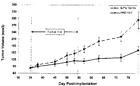
In the preventative in vivo model of human breast cancer, H460-16-2 treatment
was significantly (p<0.0001) more effective in suppressing tumor growth during
the treatment
period than an isotype control antibody. At the end of the treatment phase,
mice given 11460-
16-2 had tumors that grew to only 1.3 percent of the control group. During the
post treatment
follow-up period, the treatment effects of H460-16-2 were sustained and the
mean tumor
volume in the treated groups continued to be significantly smaller than
controls until the end
of the measurement phase. Using survival as a measure of antibody efficacy, it
was estimated
that the risk of dying in the 11460-16-2 treatment group was about 71 percent
of the antibody
buffer control group (p=0.028) at 70 days post-treatment. These data
demonstrated that H40-
16-2 treatment conferred a survival benefit compared to the control-treated
groups. H460-16-
2 treatment appeared safe, as it did not induce any signs of toxicity,
including reduced body
weight and clinical distress. Thus, H460-16-2 treatment was efficacious as it
both delayed
tumor growth and enhanced survival compared to the control-treated groups in a
well-
established model of human breast cancer.
In addition, 11460-16-2 demonstrated anti-tumor activity against MDA-MB-
231 cells in an established in vivo tumor model (as disclosed in S.N.
10/603,000). Treatment
with 11460-16-2 was compared to the standard chemotherapeutic drug, Cisplatin,
and it was
shown that the Cisplatin and H460-16-2 treatment groups had significantly
(p<0.001) smaller
mean tumor volumes compared with groups treated with either antibody dilution
buffer or the
isotype control antibody. H460-16-2 treatment mediated tumor suppression that
was
approximately two-thirds that of Cisplatin chemotherapy but without the
significant (19.2
percent) weight loss (p<0.003) and clinical distress, including 2 treatment-
associated deaths,
that was observed with Cisplatin treatment The anti-tumor activity of H460-16-
2 and its
minimal toxicity make it an attractive anti-cancer therapeutic agent.
In addition, in the post-treatment period, 11460-16-2 showed a significant
survival benefit (p<0.02) as the risk of dying in the H460-16-2 group was
about half of that in
the isotype control antibody group at >70 days after treatment The observed
survival benefit
21
CA 02687583 2013-11-15
=
WO 2008/144890
PCT/CA2008/000978
continued past 120 days post-treatment where 100 percent of the isotype
control and Cisplatin
treated mice had died compared to 67 percent of the H460-16-2 treatment group.
H460-16-2
maintained tumor suppression by delaying tumor growth by 26 percent compared
to the
isotype control antibody group. At 31 days post treatment, H460-16-2 limited
tumor size by
reducing tumor growth by 48 percent compared to the isotype control group,
which is
comparable to the 49 percent reduction observed at the end of the treatment.
In the
established tumor model of breast cancer, these results indicated the
potential of H460-16-2 to
maintain tumor suppression beyond the treatment phase and demonstrated the
ability of the
antibody to reduce the tumor burden and enhance survival in a mammal.
In addition to the beneficial effects in the established in vivo tumor model
of
breast cancer, H460-16-2 treatment in combination with a chemotherapeutic drug
(Cisplatin)
had anti-tumor activity against PC-3 cells in an established in vivo prostate
cancer model.
Using a paired t-test, 11460-16-2 plus Cisplatin treatment was
significantly more effective in suppressing tumor growth shortly after the
treatment period
than buffer control (p<0.0001), Cisplatin treatment alone (p.004) or H460-16-2
treatment
alone (p<0.0001). At the end of the treatment phase, mice given 11460-16-2
plus Cisplatin had
tumors that grew to only 28.5 percent of the buffer control group. For PC-3
SC1D xenograft
models, body weight can be used as a surrogate indicator of disease
progression. Mice in all
the groups experienced severe weight loss. In this study, mice in all groups
showed a weight
loss of approximately 23 to 35 percent by the end of the treatment period. The
group treated
with H460-16-2 showed the smallest degree of weight loss (21.7 percent). After
treatment,
day 48, there was no significant increase in weight loss associated with the
treatment of
H460-16-2 and Cisplatin in comparison to buffer control (1)=-0.5042). Thus,
11460-16-2 plus
Cisplatin treatment was efficacious as it delayed tumor growth compared to the
isotype
control treated group in a well-established model of human prostate cancer.
In order to validate the H460-16-2 epitope as a drug target, the expression of
11460-16-2 antigen in normal human tissues was previously determined.
This work was extended by comparison with the anti-CD44 antibodies; clone L178
(disclosed
in S.N. 10/647,818, now U.S. patent 7,189,397) and clone BU75.
By IHC staining with H460-16-2, the majority of the tissues failed to express
the H460-16-2 antigen, including the cells of the vital organs, such as the
liver, kidney (except
22
CA 02687583 2013-11-15
WO 2008/144890 PCT/CA2008/000978
for marginal staining of tubular epithelial cells), heart, and lung. Results
from tissue staining
indicated that H460-16-2 showed restricted binding to various cell types but
had binding to
infiltrating macrophages, lymphocytes, and fibroblasts. The BU75 antibody
showed a similar
staining pattern. However, there was at least one difference of note; staining
of lymphocytes
was more intense with BU75 in comparison to 11460-16-2.
Localization of the H460-16-2 antigen and determining its prevalence within
the population, such as among breast cancer patients, is important in
assessing the therapeutic
use of H460-16-2 and designing effective clinical trials. To address H460-16-2
antigen
expression in breast tumors from cancer patients, tumor tissue samples from 50
individual
breast cancer patients were previously screened for expression of the 11460-16-
2 antigen
and was compared to L178 (S.N. 10,647,818, now U.S. patent 7,189,397),
BU75 and the anti-Her2 antibody c-erbB-2, The results of
these studies were similar and showed that 62 percent of tissue samples
stained positive for
the H460-16-2 antigen while 73 percent of breast tumor tissues were positive
for the BU75
epitope. Expression of 11460-16-2 within patient samples appeared specific for
cancer cells as
staining was restricted to malignant cells. H460-16-2 stained 4 of 10 samples
of normal tissue
from breast cancer patients while BU75 stained 8. Breast tumor expression of
both the 11460-
16-2 and BU75 antigen appeared to be mainly localized to the cell membrane of
malignant
cells, making CD44 an attractive target for therapy. 11460-16-2 expression was
further
evaluated based on breast tumor expression of the receptors for the hormones
estrogen and
progesterone, which play an important role in the development, treatment, and
prognosis of
breast tumors. No correlation was apparent between expression of the H460-16-2
antigen and
expression of the receptors for either estrogen or progesterone. When tumors
were analyzed
based on their stage, or degree to which the cancer advanced, again there was
no clear
correlation between 11460-16-2 antigen expression and tumor stage. Similar
results were
obtained with BU75. In comparison to c-erbB-2, 11460-16-2 showed a completely
different
staining profile where 52 percent of the breast tumor tissue samples that were
positive for the
11460-16-2 antigen were negative for Her2 expression indicating a yet unmet
targeted
therapeutic need for breast cancer patients. There were also differences in
the intensity of
staining between the breast tumor tissue sections that were positive for both
11460-16-2 and
Her2. The c-erbB-2 antibody also positively stained one of the normal breast
tissue sections.
23
CA 02687583 2013-11-15
WO 2008/144890 PCT/CA2008/000978
Further localization of the H460-16-2 antigen and determination of its
prevalence within the population, such as among prostate cancer patients.
Binding of antibodies to 53 human prostate tumor and 3 normal prostate
tissues was performed using a human, prostate normal and tumor tissue
microarray (Imgenex,
San Diego, CA). 19/53 (36 percent) of the tested tumors
were positive for H460-16-2. H460-16-2 was specific for tumor cells and stroma
fibroblasts.
Cellular localization was mostly membranous and cytoplasmic membranous with or
without
luminal localization. The percentage of positive cells ranged from <10 percent-
>50 percent
indicating heterogenous binding of the antibody to tumor cells. The relation
of the antibody
binding to tumor stage could not be assessed properly due to a discrepancy in
the number of
tumors among different tumor stages, being 1/1 (100 percent), 4/12 (33
percent), 0/2 (0
percent) and 11/33 (33 percent) to tumor stage I, II, III and IV,
respectively. There was higher
binding to Gleason score G3-G4 (36 percent) than to G1-G2 (25 percent). All 3
normal
prostate tissue sections were positive for the antibody. However, the tissue
specificity was for
myoepithelium and stromal fibroblasts and spared the glandular epithelium..
There was
heterogeneity of the binding of H460-16-2 to tested prostate tumors: 10/53,
6/53, 3/53
positive tumors were in the categories of <10-10 percent, <50-50 percent and
>50 percent,
respectively. As a result of its binding to prostate cancer cells, the
therapeutic benefit of
11460-16-2 can potentially be extended to the treatment of prostate cancer.
Further localization of the 11460-16-2 antigen and determination of its
prevalence within the population, such as among liver cancer patients.
The H460-16-2 antibody showed binding to 21/49 (43 percent) of tested liver
cancers, including 11/37 (30 percent) of primary, 7/8 (88 percent) of
metastatic hepatocellular
carcinoma, 1/2 (50 percent) of primary and 2/2 (100 percent) of metastatic
cholangiocarcinomas. The antibody showed significant higher binding to
advanced tumors'
stages LII and IV in comparison with early stages I and 11(j) = 0.03) [stage
I, 0/2 (0 percent);
stage II, 2/17 (12 percent); stage III, 8/16 (50 percent) and stage IV, 6/8
(75 percent)). 11460-
16-2 was specific for tumor cells and infiltrating inflammatory cells.
Cellular localization was
mainly membranous. Some tumors also displayed a diffuse cytoplasmic staining
pattern. The
antibody bound to 9/9 of non-neoplastic liver tissues. However, the binding
was restricted to
the sinusoidal cells and infiltrating lymphocytes. The 11460-16-2 antigen
appears to be
24
CA 02687583 2013-11-15
WO 2008/144890 PCT/CA2008/000978
specifically expressed on advanced liver tumor tissue. H460-16-2 therefore has
potential as a
therapeutic drug in the treatment of liver cancer.
To further extend the potential therapeutic benefit of H460-16-2, the
frequency
and localization of the antigen within various human cancer tissues was also
previously
determined and was compared to clone L178 (S.N. 10,647,818, now U.S.
patent 7,189,397). The majority of these tumor types were also positive for
the L178 antigen.
As with human breast tumor tissue, H460-16-2 and L178 localization occurred on
the
membrane of tumor cells. However, there was substantially more membrane
localization with
the L178 compared to the H460-16-2 antibody. Also, of the tumor types that
were stained by
both H460-16-2 and Li 78, 43 percent of the tissues showed higher intensity
staining with the
L178 antibody.
There appears to be no form of CD44 that exactly matches the IHC data
presented herein based on comparisons with the IHC data from the literature.
The standard
form of CD44 is normally expressed in the human brain; the 11460-16-2 antigen
is not.
Antibodies directed against pan-CD44 isoforms do not stain the liver
(including Kuppfer
cells) and positively stain the endometrial glands in all phases of the
reproductive cycle. The
H460-16-2 antigen is clearly present on Kuppfer cells and is only present on
the secretory
endometrial glands of the reproductive cycle. 11460-16-2 antigen is clearly
present on tissue
macrophages and only the variant forms V4/5 and V8/9 show occasional
macrophage
staining. The similar yet distinct binding pattern seen with H460-16-2 in
comparison to anti-
CD44 L178 and now BU75 indicates that the H460-16-2 antigen is an unique
epitope of
CD44.
As disclosed previously (S.N. 10/647,818, now U.S. patent 7,189,397),
additional biochemical data also indicate that the antigen recognized by H460-
16-2 is one of
the forms of CD44. This is supported by studies that showed a monoclonal
antibody (L178)
reactive against CD44 identifies proteins that were bound to 11460-16-2 by
immunoprecipitation. Western blotting studies also suggest that the epitope of
CD44
recognized by 11460-16-2 is not present on v6 or v10. The 11460-16-2 epitope
is also
distinguished by being carbohydrate and conformation dependent, whereas many
anti-CD44
antibodies are directed against peptide portions of CD44. These IHC and
biochemical results
demonstrate that H460-16-2 binds to a variant of the CD44 antigen. Thus, the
preponderance
CA 02687583 2013-11-15
WO 2008/144890 PCT/CA2008/000978
of evidence shows that 11460-16-2 mediates anti-cancer effects through
ligation of an unique
carbohydrate dependent conformational epitope present on a variant of CD44.
For the
purpose of this invention, said epitope is defined as a "CD44 antigenic
moiety" characterized
by its ability to bind with a monoclonal antibody encoded by the hybridoma
cell line H460-
16-2, antigenic binding flagments thereof, antigenic binding ligands thereof
or antibody
conjugates thereof.
In order to further elucidate the mechanism behind H460-16-2's anti-cancer
effects, hyaluronic acid (HA) binding assays were performed.
It was determined that an average concentration of 1.87 (+/- 1.01)
micrograms/It-IL of H460-16-2 was required to inhibit adhesion of MDA-MB-231
cells to HA
by 50 percent. These results indicate that 11460-16-2 interacts with, at least
in part, the
region(s) on CD44 that are responsible for binding to HA and consequently
could be
mediating its anti-cancer effects through down regulation of angiogenesis or
tumor
invasiveness through the ECM.
In addition to the HA binding assays, a cell cycling experiment was performed
in order to determine if the H460-16-2 in vitro and in vivo anti-cancer
effects were due to
regulation of the cell cycle. After 24 hours and with 20
micrograms/mL of 11460-16-2, there was an increase in the number of MDA-MB-231
apoptotic cells in comparison to the isotype control. This effect also
appeared to be dose
dependent. Therefore, the efficacy of 11460-16-2 might also be due, in whole
or in part, to its
apoptotic inducing capabilities.
To further elucidate the mechanism of action for H460-16-2, the effect of
11460-16-2 treatment upon apoptosis in MDA-MB-231 tumors grown in vivo in a
xenograft
model of breast cancer was performed. Serial sections of the
ApoTag stained tumors were subsequently H & E stained and these were examined
for
apoptotic cells using morphological criteria such as deletion of single cells,
cell shrinkage and
compaction of chromatin into a dense mass. Counts for cells meeting these
criteria were done
as described in the section above to give average counts for the treatment
groups. The buffer
control group yielded an average total score of 17 cells (+ 5.29) while the
11460-16-2 treated
group yielded an average total score of 22.5 cells ( 4.20). Therefore, there
is a trend towards
increased apoptosis with 11460-16-2 treatment as determined using cellular
morphology.
26
CA 02687583 2013-11-15
WO 2008/144890 PCT/CA2008/000978
To facilitate production of antibody chimera, the genes encoding the variable
regions of both heavy and light chains were separately cloned and sequenced.
11460-16-2 chimeric light and heavy chains of a human IgG1 and IgG2
isotype were then constructed and expressed.
To determine the relative efficacy of the chimeric versus the murine antibody,
an in vivo model of human breast cancer was performed.
Both murine 11460-16-2 and (ch)ARH460-16-2-IgG1 reduced tumor growth in an
established
MDA-MB-231 in vivo model of human breast cancer. At day 62, 5 days after the
last dose
was administered, treatment with H460-16-2 resulted in a tumor growth
inhibition of 39
percent (Mean TIC = 57 percent). This reduction in tumor growth was
significantly different
from the control (p=0.0037). The chimeric antibody (ch)ARH460-16-2-IgG1
resulted in an
enhanced tumor growth inhibition of 64 percent (Mean TIC = 26.9 percent;
p<0.0001). By
contrast, the IgG2 version of the chimeric antibody, (ch)ARH460-16-2-IgG2
showed no
inhibition in tumor growth when compared with the buffer control (tumor growth
inhibition =
0 percent; Mean TIC = 122 percent; p=0.7264). There were no clinical signs of
toxicity
throughout the study. In summary, (ch)ARH460-16-2-IgG1 demonstrates the same
or greater
efficacy compared to the murine antibody in the 1ViDA-MB-231 breast cancer
model.
Annexin-V staining was previously performed
to determine whether the chimeric versions of1-1460-16-2 were able to induce
apoptosis in the same manner as the murine counterpart on the MDA-MB-231 human
breast
cancer cell line. All 3 antibodies showed a dose-dependent increase in the
percentage necrotic
and necrotic/apoptotic populations over their prospective isotype controls.
The largest
increase in the percentage necrotic and necrotic/apoptotic populations was
seen with
(ch)ARH460-16-2-IgG2, then (ch)ARH460-16-2-IgG 1 and then H460-16-2.
In toto, this data demonstrates that the 11460-16-2 antigen is a cancer
associated antigen and is expressed in humans, and is a pathologically
relevant cancer target.
Further, this data also demonstrates the binding of the 11460-16-2 antibody to
human cancer
tissues, and can be used appropriately for assays that can be diagnostic,
predictive of therapy,
or prognostic. In addition, the cell membrane localization of this antigen is
indicative of the
cancer status of the cell due to the lack of expression of the antigen in most
non-malignant
27
CA 02687583 2009-11-18
WO 2008/144890
PCT/CA2008/000978
cells, and this observation permits the use of this antigen, its gene or
derivatives, its protein or
its variants to be used for assays that can be diagnostic, predictive of
therapy, or prognostic.
Other studies, involving the use of anti-CD44 antibodies, have limitations of
therapeutic potential that are not exhibited by H460-16-2. H460-16-2
demonstrates both in
vitro and in vivo anti-tumor activity. Previously described antibodies such as
MAK<CD44>M-1.1.12, MAK<CD44>M-2.42.3 and MAK<CD44>M-4.3.16 have no in vitro
or in vivo cytotoxicity ascribed to them and VFF-18 and Mab U36 show no
intrinsic tumor
cytotoxicity. In addition other anti-CD44 antibodies that have shown in vivo
tumor effects
also have certain limitations that are not evident with H460-16-2. For
example, ASML1.1,
K926, anti-CD44s and IM-78.1 show in vivo anti-tumor activity against rat,
murine, rat and
murine tumors grown in xenograft models respectively. H460-16-2 demonstrates
anti-tumor
activity in a model of human cancer. H460-16-2 is also directed against human
CD44 while
antibodies such as ASML1.1 recognize only rat CD44. The clone 515 anti-CD44
antibody
does inhibit peritoneal tumor implantation of a human ovarian cell line but
does not prevent or
inhibit tumor growth. H460-16-2 is capable of inhibiting human breast tumor
growth in a
SCID mouse xenograft model. GKW.A3 is an anti-human CD44 monoclonal antibody
capable of inhibiting tumor growth of a human metastasizing melanoma grown in
mice in a
preventative but not an established model. H460-16-2 has demonstrated
significant anti-
tumor activity in both preventative and established murine xenograft models of
human breast
cancer. Consequently, it is quite apparent that H460-16-2 has superior anti-
tumor properties
in comparison to previously described anti-CD44 antibodies. It has
demonstrated both in
vitro and in vivo anti-tumor activity on a human breast tumor in SCID mice and
is directed
against human CD44. It also exhibits activity in a preventative and
established (more
clinically relevant) model of human breast cancer and it exhibits activity
with Cisplatin in an
established model of human prostate cancer.
The present invention describes the development and use of H460-16-2, it's
corresponding chimeric antibodies, (ch)ARH460-16-2-IgG1 and (ch)ARH460-16-2
(VKOVHO), and it's corresponding humanized antibody variants, (hu)ARH460-16-2.
H460-
16-2 was identified by, its effect, in cytotoxic assays, in tumor growth
models and in
prolonging survival time in mammals suffering from cancerous disease. This
invention
represents an advance in the field of cancer treatment in that it describes,
for the first time,
28
SUBSTITUTE SHEET (RULE 26)
CA 02687583 2009-11-18
WO 2008/144890
PCT/CA2008/000978
reagents that bind specifically to an epitope or epitopes present on the
target molecule, CD44,
and that also have in vitro cytotoxic properties, as a naked antibody, against
malignant tumor
cells but not normal cells, and which also directly mediate, as a naked
antibody, inhibition of
tumor growth and extension of survival in in vivo models of human cancer. This
is an advance
in relation to any other previously described anti-CD44 antibody, since none
have been shown
to have similar properties. It also provides an advance in the field since it
clearly
demonstrates, and for the first time, the direct involvement of CD44 in events
associated with
growth and development of certain types of tumors. It also represents an
advance in cancer
therapy since it has the potential to display similar anti-cancer properties
in human patients. A
further advance is that inclusion of these antibodies in a library of anti-
cancer antibodies will
enhance the possibility of targeting tumors expressing different antigen
markers by
determination of the appropriate combination of different anti-cancer
antibodies, to find the
most effective in targeting and inhibiting growth and development of the
tumors.
In all, this invention teaches the use of the H460-16-2 antigen as a target
for a
therapeutic agent, that when administered can reduce the tumor burden of a
cancer expressing
the antigen in a mammal, and can also lead to a prolonged survival of the
treated mammal.
This invention also teaches the use of CDMABs (H460-16-2, (ch)ARH460-16-2-IgG
1,
(ch)ARH460-16-2 (VKOVHO) and variants of (hu)ARH460-16-2), and their
derivatives, and
antigen binding fragments thereof, and cellular cytotoxicity inducing ligands
thereof to target
their antigen to reduce the tumor burden of a cancer expressing the antigen in
a mammal, and
lead to prolonged survival of the treated mammal. Furthermore, this invention
also teaches the
use of detecting the H460-16-2 antigen in cancerous cells that can be useful
for the diagnosis,
prediction of therapy, and prognosis of mammals bearing tumors that express
this antigen.
Accordingly, it is an objective of the invention to utilize a method for
producing cancerous disease modifying antibodies (CDMAB) raised against
cancerous cells
derived from a particular individual, or one or more particular cancer cell
lines, which
CDMAB are cytotoxic with respect to cancer cells while simultaneously being
relatively non-
toxic to non-cancerous cells, in order to isolate hybridoma cell lines and the
corresponding
isolated monoclonal antibodies and antigen binding fragments thereof for which
said
hybridoma cell lines are encoded.
29
SUBSTITUTE SHEET (RULE 26)
CA 02687583 2009-11-18
WO 2008/144890
PCT/CA2008/000978
It is an additional objective of the invention to teach cancerous disease
modifying antibodies, ligands and antigen binding fragments thereof.
It is a further objective of the instant invention to produce cancerous
disease
modifying antibodies whose cytotoxicity is mediated through antibody dependent
cellular
toxicity.
It is yet an additional objective of the instant invention to produce
cancerous
disease modifying antibodies whose cytotoxicity is mediated through complement
dependent
cellular toxicity.
It is still a further objective of the instant invention to produce cancerous
disease modifying antibodies whose cytotoxicity is a function of their ability
to catalyze
hydrolysis of cellular chemical bonds.
A still further objective of the instant invention is to produce cancerous
disease
modifying antibodies which are useful for in a binding assay for diagnosis,
prognosis, and
monitoring of cancer.
Other objects and advantages of this invention will become apparent from the
following description wherein are set forth, by way of illustration and
example, certain
embodiments of this invention.
BRIEF DESCRIPTION OF THE FIGURES
The patent or application file contains at least one drawing executed in
color.
Copies of this patent or patent application publication with color drawing(s)
will be provided
by the Office upon request and payment of the necessary fee.
Figure 1 demonstrates the effect of H460-16-2 on tumor growth in an
established human breast MDA-MB-468 cancer model. The vertical dashed lines
indicate the
period during which the antibody was intraperitoneally administered. Data
points represent
the mean +/- SEM.
Figure 2 demonstrates the effect of H460-16-2 on mouse body weight in an
established MDA-MB-468 breast cancer model. Data points represent the mean +/-
SEM.
Figure 3 demonstrates the effect of H460-16-2 on tumor growth in an
established human PC-3 prostate cancer model. The vertical dashed lines
indicate the period
during which the antibody was intraperitoneally administered. Data points
represent the mean
+/- SEM.
SUBSTITUTE SHEET (RULE 26)
CA 02687583 2009-11-18
WO 2008/144890 PCT/CA2008/000978
Figure 4 demonstrates the effect of H460-16-2 on mouse body weight in an
established PC-3 prostate cancer model. Data points represent the mean +/-
SEM.
Figure 5 demonstrates the effect of (ch)ARH460-16-2-IgG1 in a dose-
dependent manner on the tumor growth in an established human breast (MDA-MB-
231)
cancer model. The vertical dashed lines indicate the period during which the
antibody was
intraperitoneally administered. Data points represent the mean +/- SEM.
Figure 6 demonstrates the effect of (ch)ARH460-16-2-IgG1 on mouse body
weight in an established MDA-MB-231 breast cancer model. Data points represent
the mean
+/- SEM.
Figure 7 is a summary of H460-16-2 binding on a human colon tumor tissue
microarray.
Figure 8. Representative micrographs showing the binding pattern on colon
tumor tissue obtained with H460-16-2 (A) or the isotype control antibody (B).
Magnification
is 200X.
Figure 9 is a summary of (ch)ARH460-16-2-IgG1 binding on a human and
cynomolgus monkey tissue microarray.
Figure 10. Representative micrographs showing the binding pattern with
(ch)ARH460-16-2-IgG1 on human spleen (white pulp) (A) and cynomolgus monkey
spleen
(white pulp) (B). Staining was observed with both species. Magnification is
40X.
Figure 11. Western blot of 500 micrograms of MDA-MB-231 membrane
proteins probed with different primary antibody solutions. Lanes 1-5 were
probed with
biotinylated H460-16-2 mixed with 2 micrograms/mL, 10 micrograms/mL, 100
micrograms/mL, 500 micrograms/mL or 1000 micrograms/mL of non-biotinylated
H460-16-2
respectively. Lanes 6-10 were probed with biotinylated H460-16-2 mixed with 2
micrograms/mL, 10 micrograms/mL, 100 micrograms/mL, 500 micrograms/mL or 1000
micrograms/mL of non-biotinylated AR37A335.8 respectively. Lanes 11-15 were
probed
with biotinylated 11460-16-2 mixed with 2 micrograms/mL, 10 micrograms/mL, 100
micrograms/mL, 500 micrograms/mL or 1000 micrograms/mL of non-biotinylated
1B7.11
respectively.
Figure 12. Western blot of 500 micrograms of MDA-MB-231 membrane
proteins probed with different primary antibody solutions. Lanes 1-5 were
probed with
31
CA 02687583 2009-11-18
WO 2008/144890 PCT/CA2008/000978
biotinylated AR37A335.8 mixed with 2 micrograms/mL, 10 micrograms/mL, 100
micrograms/mL, 500 micrograms/mL or 1000 micrograms/mL of non-biotinylated
11460-16-2
respectively. Lanes 6-10 were probed with biotinylated AR37A335.8 mixed with 2
micrograms/mL, 10 micrograms/mL, 100 micrograms/mL, 500 micrograms/mL or 1000
micrograms/mL of non-biotinylated AR37A335.8 respectively. Lanes 11-15 were
probed
with biotinylated AR37A335.8 mixed with 2 micrograms/mL, 10 micrograms/mL, 100
micrograms/mL, 500 micrograms/mL or 1000 micrograms/mL of non-biotinylated
8B1B.1
respectively.
Figure 13. Binding affinity of AR37A335.8. Dissociation constants for the
binding of the antibodies to the purified recombinant human CD44 was assessed
by surface
plasmon resonance.
Figure 14. List of RTKs whose phosphorylation is affected by treatment of
MDA-MB-231 cells with (ch)ARH460-16-2-IgG1 followed by serum and supplement
stimulation.
Figure 15. Primers used in the PCR amplification of light chain (SEQ ID NOS:
10-28, respectively, in order of appearance).
Figure 16. Primers used in the PCR amplification of heavy chain (SEQ ID
NOS: 29-44, respectively, in order of appearance).
Figure 17. Mouse H460-16-2 VII Sequence (SEQ ID NO: 45).
Figure 18. Mouse H460-16-2 VL Sequence (SEQ ID NO: 46).
Figure 19. Oligonucleotides (SEQ ID NOS: 47-58, respectively, in order of
appearance) used for the generation of chimeric and variant humanized H460-16-
2 VII
sequences.
Figure 20. Oligonucleotides (SEQ ID NOS: 59-68, respectively, in order of
appearance) used for the generation of chimeric and variant humanized 11460-16-
2 VL
sequences.
Figure 21. pANT18 expression vector.
Figure 22. Humanized H460-16-2 VII variants (SEQ ID NOS: 7 and 9,
respectively, in order of appearance). CDRs are underlined.
Figure 23. Humanized H460-16-2 VL variants (SEQ ID NO: 8). CDRs are
underlined.
32
CA 02687583 2009-11-18
WO 2008/144890 PCT/CA2008/000978
Figure 24. Binding data for chimeric and humanized variants of H460-16-2.
Figure 25. Binding affinity of murine H460-16-2, and of the humanized
variants, (hu)ARH460-16-2 variant HV1/KV1 and (hu)ARH460-16-2 variant HV2/KV1.
Dissociation constants for the binding of the antibodies to the purified
recombinant human
CD44 was assessed by surface plasmon resonance.
DETAILED DESCRIPTION OF THE INVENTION
In general, the following words or phrases have the indicated definition when
used in the summary, description, examples, and claims.
The term "antibody" is used in the broadest sense and specifically covers, for
example, single monoclonal antibodies (including agonist, antagonist, and
neutralizing
antibodies, de-immunized, murine, chimeric or humanized antibodies), antibody
compositions
with polyepitopic specificity, single-chain antibodies, diabodies, triabodies,
immunoconjugates and antibody fragments (see below).
The term "monoclonal antibody" as used herein refers to an antibody obtained
from a population of substantially homogeneous antibodies, i.e., the
individual antibodies
comprising the population are identical except for possible naturally
occurring mutations that
may be present in minor amounts. Monoclonal antibodies are highly specific,
being directed
against a single antigenic site. Furthermore, in contrast to polyclonal
antibody preparations
which include different antibodies directed against different determinants
(epitopes), each
monoclonal antibody is directed against a single determinant on the antigen.
In addition to
their specificity, the monoclonal antibodies are advantageous in that they may
be synthesized
uncontaminated by other antibodies. The modifier "monoclonal" indicates the
character of the
antibody as being obtained from a substantially homogeneous population of
antibodies, and is
not to be construed as requiring production of the antibody by any particular
method. For
example, the monoclonal antibodies to be used in accordance with the present
invention may
be made by the hybridoma (murine or human) method first described by Kohler et
al., Nature,
256:495 (1975), or may be made by recombinant DNA methods (see, e.g., U.S.
Pat.
No.4,816,567). The "monoclonal antibodies" may also be isolated from phage
antibody
libraries using the techniques described in Clackson etal., Nature, 352:624-
628 (1991) and
Marks et al., J Mol. Biol., 222:581-597 (1991), for example.
33
CA 02687583 2009-11-18
WO 2008/144890 PCT/CA2008/000978
"Antibody fragments" comprise a portion of an intact antibody, preferably
comprising the antigen-binding or variable region thereof. Examples of
antibody fragments
include less than full length antibodies, Fab, Fab', F(ab1)2, and Fv
fragments; diabodies; linear
antibodies; single-chain antibody molecules; single-chain antibodies, single
domain antibody
molecules, fusion proteins, recombinant proteins and multispecific antibodies
formed from
antibody fragment(s).
An "intact" antibody is one which comprises an antigen-binding variable
region as well as a light chain constant domain (CO and heavy chain constant
domains, CHI,
CH2 and C13. The constant domains may be native sequence constant domains
(e.g. human
native sequence constant domains) or amino acid sequence variant thereof.
Preferably, the
intact antibody has one or more effector functions.
Depending on the amino acid sequence of the constant domain of their heavy
chains, intact antibodies can be assigned to different "classes". There are
five-major classes of
intact antibodies: IgA, IgD, IgE, IgG, and IgM, and several of these may be
further divided
into "subclasses" (isotypes), e.g., IgGl, IgG2, IgG3, IgG4, IgA, and IgA2. The
heavy-chain
constant domains that correspond to the different classes of antibodies are
called a, 8, z, 7, and
II, respectively. The subunit structures and three-dimensional configurations
of different
classes of immunoglobulins are well known.
Antibody "effector functions" refer to those biological activities
attributable to
the Fc region (a native sequence Fe region or amino acid sequence variant Fe
region) of an
antibody. Examples of antibody effector functions include Clq binding;
complement
dependent cytotoxicity; Fe receptor binding; antibody-dependent cell-mediated
cytotoxicity
(ADCC); phagocytosis; down regulation of cell surface receptors (e.g. B cell
receptor; BCR),
etc.
"Antibody-dependent cell-mediated cytotoxicity" and "ADCC" refer to a cell-
mediated reaction in which nonspecific cytotoxic cells that express Fe
receptors (FcRs) (e.g.
Natural Killer (NK) cells, neutrophils, and macrophages) recognize bound
antibody on a
target cell and subsequently cause lysis of the target cell. The primary cells
for mediating
ADCC, NK cells, express Fc7RIII only, whereas monocytes express Fc7RI, Fc7RII
and
Fc7RIII. FcR expression on hematopoietic cells is summarized in Table 3 on
page 464 of
Ravetch and Kinet, Annu. Rev. Immunol 9:457-92 (1991). To assess ADCC activity
of a
34
CA 02687583 2009-11-18
WO 2008/144890 PCT/CA2008/000978
molecule of interest, an in vitro ADCC assay, such as that described in U.S.
Pat. No.
5,500,362 or 5,821,337 may be performed. Useful effector cells for such assays
include
peripheral blood mononuclear cells (PBMC) and Natural Killer (NK) cells.
Alternatively, or
additionally, ADCC activity of the molecule of interest may be assessed in
vivo, e.g., in a
animal model such as that disclosed in Clynes et al. PNAS (USA) 95:652-656
(1998).
"Effector cells" are leukocytes which express one or more FcRs and perform
effector functions. Preferably, the cells express at least FcyRIII and perform
ADCC effector
function. Examples of human leukocytes which mediate ADCC include peripheral
blood
mononuclear cells (PBMC), natural killer (NK) cells, monocytes, cytotoxic T
cells and
neutrophils; with PBMCs and NK cells being preferred. The effector cells may
be isolated
from a native source thereof, e.g. from blood or PBMCs as described herein.
The terms "Fe receptor" or "FcR" are used to describe a receptor that binds to
the Fe region of an antibody. The preferred FcR is a native sequence human
FcR. Moreover, a
preferred FcR is one which binds an IgG antibody (a gamma receptor) and
includes receptors
of the FcyRI, FcyRII, and Fey Rill subclasses, including allelic variants and
alternatively
spliced forms of these receptors. FcyRII receptors include FcyRIIA (an
"activating receptor")
and FcyRIIB (an "inhibiting receptor"), which have similar amino acid
sequences that differ
primarily in the cytoplasmic domains thereof. Activating receptor FcyRIIA
contains an
immunoreceptor tyrosine-based activation motif (ITAM) in its cytoplasmic
domain. Inhibiting
receptor FcyRIIB contains an immunoreceptor tyrosine-based inhibition motif
(ITIM) in its
cytoplasmic domain. (see review M. in Daeron, Annu. Rev. Immunol. 15:203-234
(1997)).
FcRs are reviewed in Ravetch and Kinet, Annu. Rev. Immunol 9:457-92 (1991);
Capel et al.,
Immunomethods 4:25-34 (1994); and de Haas et al., J. Lab. Clin. Med. 126:330-
41 (1995).
Other FcRs, including those to be identified in the future, are encompassed by
the term "FcR"
herein. The term also includes the neonatal receptor, FcRn, which is
responsible for the
transfer of maternal IgGs to the fetus (Guyer et al., .1 Immunol. 117:587
(1976) and Kim et
al., Eur. Immunol. 24:2429 (1994)).
"Complement dependent cytotoxicity" or "CDC" refers to the ability of a
molecule to lyse a target in the presence of complement. The complement
activation pathway
is initiated by the binding of the first component of the complement system
(Clq) to a
molecule (e.g. an antibody) complexed with a cognate antigen. To assess
complement
CA 02687583 2009-11-18
WO 2008/144890 PCT/CA2008/000978
activation, a CDC assay, e.g. as described in Gazzano-Santoro et al., J.
ImmunoL Methods
202:163 (1996), may be performed.
The term "variable" refers to the fact that certain portions of the variable
domains differ extensively in sequence among antibodies and are used in the
binding and
specificity of each particular antibody for its particular antigen. However,
the variability is not
evenly distributed throughout the variable domains of antibodies. It is
concentrated in three
segments called hypervariable regions both in the light chain and the heavy
chain variable
domains. The more highly conserved portions of variable domains are called the
framework
regions (FRs). The variable domains of native heavy and light chains each
comprise four FRs,
largely adopting a13-sheet configuration, connected by three hypervariable
regions, which
form loops connecting, and in some cases forming part of, the 13-sheet
structure. The
hypervariable regions in each chain are held together in close proximity by
the FRs and, with
the hypervariable regions from the other chain, contribute to the formation of
the antigen-
binding site of antibodies (see Kabat etal., Sequences of Proteins of
Immunological Interest,
5th Ed. Public Health Service, National Institutes of Health, Bethesda, Md.
(1991)). The
constant domains are not involved directly in binding an antibody to an
antigen, but exhibit
various effector functions, such as participation of the antibody in antibody
dependent cellular
cytotoxicity (ADCC).
The term "hypervariable region" when used herein refers to the amino acid
residues of an antibody which are responsible for antigen-binding. The
hypervariable region
generally comprises amino acid residues from a "complementarity determining
region" or
"CDR" (e.g. residues 24-34 (L1), 50-56 (L2) and 89-97 (L3) in the light chain
variable
domain and 31-35 (H1), 50-65 (H2) and 95-102 (113) in the heavy chain variable
domain;
Kabat et al., Sequences of Proteins of Immunological Interest, 5th Ed. Public
Health Service,
National Institutes of Health, Bethesda, Md. (1991)) and/or those residues
from a
"hypervariable loop" (e.g. residues 2632 (L1), 50-52 (L2) and 91-96 (L3) in
the light chain
variable domain and 26-32 (111), 53-55 (H2) and 96-101 (143) in the heavy
chain variable
domain; Chothia and Lesk I MoL Biol. 196:901-917 (1987)). "Framework Region"
or "FR"
residues are those variable domain residues other than the hypervariable
region residues as
herein defined. Papain digestion of antibodies produces two identical antigen-
binding
fragments, called "Fab" fragments, each with a single antigen-binding site,
and a residual
36
CA 02687583 2009-11-18
WO 2008/144890 PCT/CA2008/000978
"Fe" fragment, whose name reflects its ability to crystallize readily. Pepsin
treatment yields
an F(ab')2fragment that has two antigen-binding sites and is still capable of
cross-linking
antigen.
"Fv" is the minimum antibody fragment which contains a complete antigen-
recognition and antigen-binding site. This region consists of a dimer of one
heavy chain and
one light chain variable domain in tight, non-covalent association. It is in
this configuration
that the three hypervariable regions of each variable domain interact to
define an antigen-
binding site on the surface of the VH-VL dimer. Collectively, the six
hypervariable regions
confer antigen-binding specificity to the antibody. However, even a single
variable domain
(or half of an Fv comprising only three hypervariable regions specific for an
antigen) has the
ability to recognize and bind antigen, although at a lower affinity than the
entire binding site.
The Fab fragment also contains the constant domain of the light chain and the
first constant
domain (CH I) of the heavy chain. Fab' fragments differ from Fab fragments by
the addition
of a few residues at the carboxy terminus of the heavy chain CHI domain
including one or
more cysteines from the antibody hinge region. Fab'-SH is the designation
herein for Fab' in
which the cysteine residue(s) of the constant domains bear at least one free
thiol group.
F(ab1)2 antibody fragments originally were produced as pairs of Fab' fragments
which have
hinge cysteines between them. Other chemical couplings of antibody fragments
are also
known.
The "light chains" of antibodies from any vertebrate species can be assigned
to
one of two clearly distinct types, called kappa (K) and lambda (X), based on
the amino acid
sequences of their constant domains.
"Single-chain Fv" or "scFv" antibody fragments comprise the VH and VL
domains of antibody, wherein these domains are present in a single polypeptide
chain.
Preferably, the Fv polypeptide further comprises a polypeptide linker between
the VH and VL
domains which enables the scFv to form the desired structure for antigen
binding. For a
review of scFv see Pliickthun in The Pharmacology of Monoclonal Antibodies,
vol. 113,
Rosenburg and Moore eds., Springer-Verlag, New York, pp. 269-315 (1994).
The term "diabodies" refers to small antibody fragments with two antigen-
binding sites, which fragments comprise a variable heavy domain (VH) connected
to a
variable light domain (VL) in the same polypeptide chain (VH-VL). By using a
linker that is
37
CA 02687583 2009-11-18
WO 2008/144890 PCT/CA2008/000978
too short to allow pairing between the two domains on the same chain, the
domains are forced
to pair with the complementary domains of another chain and create two antigen-
binding
sites. Diabodies are described more fully in, for example, EP 404,097; WO
93/11161; and
Hollinger etal., Proc. Natl. Acad. Sci. USA, 90:6444-6448 (1993).
The term "triabodies" or "trivalent trimers" refers to the combination of
three
single chain antibodies. Triabodies are constructed with the amino acid
terminus of a VL or
VH domain, i.e., without any linker sequence. A triabody has three Fv heads
with the
polypeptides arranged in a cyclic, head-to-tail fashion. A possible
conformation of the
triabody is planar with the three binding sites located in a plane at an angle
of 120 degrees
from one another. Triabodies can be monospecific, bispecific or trispecific.
An "isolated" antibody is one which has been identified and separated and/or
recovered from a component of its natural environment. Contaminant components
of its
natural environment are materials which would interfere with diagnostic or
therapeutic uses
for the antibody, and may include enzymes, hormones, and other proteinaceous
or
nonproteinaceous solutes. Isolated antibody includes the antibody in situ
within recombinant
cells since at least one component of the antibody's natural environment will
not be present.
Ordinarily, however, isolated antibody will be prepared by at least one
purification step.
An antibody "which binds" an antigen of interest, e.g. CD44 antigen, is one
capable of binding that antigen with sufficient affinity such that the
antibody is useful as a
therapeutic or diagnostic agent in targeting a cell expressing the antigen.
Where the antibody
is one which binds CD44, it will usually preferentially bind CD44 as opposed
to other
receptors, and does not include incidental binding such as non-specific Fc
contact, or binding
to post-translational modifications common to other antigens and may be one
which does not
significantly cross-react with other proteins. Methods, for the detection of
an antibody that
binds an antigen of interest, are well known in the art and can include but
are not limited to
assays such as FACS, cell ELISA and Western blot.
As used herein, the expressions "cell", "cell line", and "cell culture" are
used
interchangeably, and all such designations include progeny. It is also
understood that all
progeny may not be precisely identical in DNA content, due to deliberate or
inadvertent
mutations. Mutant progeny that have the same function or biological activity
as screened for
38
CA 02687583 2009-11-18
WO 2008/144890 PCT/CA2008/000978
in the originally transformed cell are included. It will be clear from the
context where distinct
designations are intended.
"Treatment or treating" refers to both therapeutic treatment and prophylactic
or
preventative measures, wherein the object is to prevent or slow down (lessen)
the targeted
pathologic condition or disorder. Those in need of treatment include those
already with the
disorder as well as those prone to have the disorder or those in whom the
disorder is to be
prevented. Hence, the mammal to be treated herein may have been diagnosed as
having the
disorder or may be predisposed or susceptible to the disorder.
The terms "cancer" and "cancerous" refer to or describe the physiological
condition in mammals that is typically characterized by unregulated cell
growth or death.
Examples of cancer include, but are not limited to, carcinoma, lymphoma,
blastoma, sarcoma,
and leukemia or lymphoid malignancies. More particular examples of such
cancers include
squamous cell cancer (e.g. epithelial squamous cell cancer), lung cancer
including small-cell
lung cancer, non-small cell lung cancer, adenocarcinoma of the lung and
squamous carcinoma
of the lung, cancer of the peritoneum, hepatocellular cancer, gastric or
stomach cancer
including gastrointestinal cancer, pancreatic cancer, glioblastoma, cervical
cancer, ovarian
cancer, liver cancer, bladder cancer, hepatoma, breast cancer, colon cancer,
rectal cancer,
colorectal cancer, endometrial or uterine carcinoma, salivary gland carcinoma,
kidney or renal
cancer, prostate cancer, vulval cancer, thyroid cancer, hepatic carcinoma,
anal carcinoma,
penile carcinoma, as well as head and neck cancer.
A "chemotherapeutic agent" is a chemical compound useful in the treatment of
cancer. Examples of chemotherapeutic agents include alkylating agents such as
thiotepa and
cyclosphosphamide (CYTOXANTm); alkyl sulfonates such as busulfan, improsulfan
and
piposulfan; aziridines such as benzodopa, carboquone, meturedopa, and uredopa;
ethylenimines and methylamelamines including altretamine, triethylenemelamine,
triethylenephosphoramide, triethylenethiophosphoramide and
trimethylolomelamine; nitrogen
mustards such as chlorambucil, chlomaphazine, cholophosphamide, estramustine,
ifosfamide,
mechlorethamine, mechlorethamine oxide hydrochloride, melphalan, novembichin,
phenesterine, prednimustine, trofosfamide, uracil mustard; nitrosureas such as
carmustine,
chlorozotocin, fotemustine, lomustine, nimustine, ranimustine; antibiotics
such as
aclacinomysins, actinomycin, authramycin, azaserine, bleomycins, cactinomycin,
39
CA 02687583 2009-11-18
WO 2008/144890 PCT/CA2008/000978
calicheamicin, carabicin, carnomycin, carzinophilin, chromomycins,
dactinomycin,
daunorubicin, detorubicin, 6-diazo-5-oxo-L-norleucine, doxorubicin,
epirubicin, esorubicin,
idarubicin, marcellomycin, mitomycins, mycophenolic acid, nogalamycin,
olivomycins,
peplomycin, potfiromycin, puromycin, quelamycin, rodorubicin, streptonigrin,
streptozocin,
tubercidin, ubenimex, zinostatin, zorubicin; anti-metabolites such as
methotrexate and 5-
fluorouracil (5-FU); folic acid analogues such as denopterin, methotrexate,
pteropterin,
trimetrexate; purine analogs such as fludarabine, 6-mercaptopurine,
thiamiprine, thioguanine;
pyrimidine analogs such as ancitabine, azacitidine, 6-azauridine, carmofur,
cytarabine,
dideoxyuridine, doxifluridine, enocitabine, floxuridine, 5-FU; androgens such
as calusterone,
dromostanolone propionate, epitiostanol, mepitiostane, testolactone; anti-
adrenals such as
aminoglutethimide, mitotane, trilostane; folic acid replenisher such as
frolinic acid;
aceglatone; aldophosphamide glycoside; aminolevulinic acid; amsacrine;
bestrabucil;
bisantrene; edatraxate; defofamine; demecolcine; diaziquone; elformithine;
elliptinium
acetate; etoglucid; gallium nitrate; hydroxyurea; lentinan; lonidamine;
mitoguazone;
mitoxantrone; mopidamol; nitracrine; pentostatin; phenamet; pirarubicin;
podophyllinic acid;
2-ethylhydrazide; procarbazine; PSKO; razoxane; sizofiran; spirogermanium;
tenuazonic
acid; triaziquone; 2,2',2"-trichlorotriethylamine; urethan; vindesine;
dacarbazine;
mannomustine; mitobronitol; mitolactol; pipobroman; gacytosine; arabinoside
("Ara-C");
cyclophosphamide; thiotepa; taxanes, e.g. paclitaxel (TAXOLO, Bristol-Myers
Squibb
Oncology, Princeton, N.J.) and docetaxel (TAXOTERE , Aventis, Rhone-Poulenc
Rorer,
Antony, France); chlorambucil; gemcitabine; 6-thioguanine, mercaptopurine;
methotrexate;
platinum analogs such as cisplatin and carboplatin; vinblastine; platinum;
etoposide (VP-16);
ifosfamide; mitomycin C; mitoxantrone; vincristine; vinorelbine; navelbine;
novantrone;
teniposide; daunomycin; aminopterin; xeloda; ibandronate; CPT-11;
topoisomerase inhibitor
RFS 2000; difluoromethylomithine (DMF0); retinoic acid; esperamicins;
capecitabine; and
pharmaceutically acceptable salts, acids or derivatives of any of the above.
Also included in
this definition are anti-hormonal agents that act to regulate or inhibit
hormone action on
tumors such as anti-estrogens including for example tamoxifen, raloxifene,
aromatase
inhibiting 4(5)-imidazoles, 4-hydroxytamoxifen, trioxifene, keoxifene,
LY117018,
onapristone, and toremifene (Fareston); and anti-androgens such as flutamide,
nilutamide,
CA 02687583 2009-11-18
WO 2008/144890 PCT/CA2008/000978
bicalutamide, leuprolide, and goserelin; and pharmaceutically acceptable
salts, acids or
derivatives of any of the above.
"Mammal" for purposes of treatment refers to any animal classified as a
mammal, including humans, mice, SCID or nude mice or strains of mice, domestic
and farm
animals, and zoo, sports, or pet animals, such as sheep, dogs, horses, cats,
cows, etc.
Preferably, the mammal herein is human.
"Oligonucleotides" are short-length, single- or double-stranded
polydeoxynucleotides that are chemically synthesized by known methods (such as
phosphotriester, phosphite, or phosphoramidite chemistry, using solid phase
techniques such
as described in EP 266,032, published 4 May 1988, or via deoxynucleoside H-
phosphonate
intermediates as described by Froehler et al.,Nucl. Acids Res., 14:5399-5407,
1986. They are
then purified on polyacrylamide gels.
In accordance with the present invention, "humanized" and/or "chimeric"
forms of non-human (e.g. murine) immunoglobulins refer to antibodies which
contain specific
chimeric immunoglobulins, immunoglobulin chains or fragments thereof (such as
Fv, Fab,
Fab', F(ab1)2 or other antigen-binding subsequences of antibodies) which
results in the
decrease of a human anti-mouse antibody (HAMA), human anti-chimeric antibody
(HACA)
or a human anti-human antibody (HAHA) response, compared to the original
antibody, and
contain the requisite portions (e.g. CDR(s), antigen binding region(s),
variable domain(s) and
so on) derived from said non-human immu,noglobulin, necessary to reproduce the
desired
effect, while simultaneously retaining binding characteristics which are
comparable to said
non-human immunoglobulin. For the most part, humanized antibodies are human
immunoglobulins (recipient antibody) in which residues from the
complementarity
determining regions (CDRs) of the recipient antibody are replaced by residues
from the CDRs
of a non-human species (donor antibody) such as mouse, rat or rabbit having
the desired
specificity, affinity and capacity. In some instances, Fv framework region
(FR) residues of the
human immunoglobulin are replaced by corresponding non-human FR residues.
Furthermore,
the humanized antibody may comprise residues which are found neither in the
recipient
antibody nor in the imported CDR or FR sequences. These modifications are made
to further
refine and optimize antibody performance. In general, the humanized antibody
will comprise
substantially all of at least one, and typically two, variable domains, in
which all or
41
CA 02687583 2009-11-18
WO 2008/144890 PCT/CA2008/000978
substantially all of the CDR regions correspond to those of a non-human
immunoglobulin and
all or substantially all of the FR residues are those of a human
immunoglobulin consensus
sequence. The humanized antibody optimally also will comprise at least a
portion of an
immunoglobulin constant region (Fc), typically that of a human immunoglobulin.
"De-immunized" antibodies are immunoglobulins that are non-immunogenic,
or less immunogenic, to a given species. De- immunization can be achieved
through structural
alterations to the antibody. Any de- immunization technique known to those
skilled in the art
can be employed. One suitable technique for de- immunizing antibodies is
described, for
example, in WO 00/34317 published June 15, 2000.
An antibody which induces "apoptosis" is one which induces programmed cell
death by any means, illustrated by but not limited to binding of annexin V,
caspase activity,
fragmentation of DNA, cell shrinkage, dilation of endoplasmic reticulum, cell
fragmentation,
and/or formation of membrane vesicles (called apoptotic bodies).
As used herein "antibody induced cytotoxicity" is understood to mean the
cytotoxic effect derived from the hybridoma supernatant or antibody produced
by the
hybridoma deposited with the ATCC as accession number PTA-4621, a humanized
antibody
of the isolated monoclonal antibody produced by the hybridoma deposited with
the ATCC as
accession number PTA-4621, a chimeric antibody of the isolated monoclonal
antibody
produced by the hybridoma deposited with the ATCC as accession number PTA-
4621,
antigen binding fragments, or antibody ligands thereof, which effect is not
necessarily related
to the degree of binding.
Throughout the instant specification, hybridoma cell lines, as well as the
isolated monoclonal antibodies which are produced therefrom, are alternatively
referred to by
their internal designation, H460-16-2 (murine), (ch)ARH460-16-2-IgG1,
(ch)ARH460-16-2
(VKOVHO), (hu)ARH460-16-2 or Depository Designation, ATCC PTA-4621.
As used herein "antibody-ligand" includes a moiety which exhibits binding
specificity for at least one epitope of the target antigen, and which may be
an intact antibody
molecule, antibody fragments, and any molecule having at least an antigen-
binding region or
portion thereof (i.e., the variable portion of an antibody molecule), e.g., an
Fv molecule, Fab
molecule, Fab' molecule, F(ab')<sub>2</sub> molecule, a bispecific antibody, a
fusion protein, or any
genetically engineered molecule which specifically recognizes and binds at
least one epitope
42
CA 02687583 2009-11-18
WO 2008/144890
PCT/CA2008/000978
of the antigen bound by the isolated monoclonal antibody produced by the
hybridoma cell line
designated as ATCC PTA-4621 ( the ATCC PTA-4621 antigen), a humanized antibody
of the
isolated monoclonal antibody produced by the hybridoma deposited with the ATCC
as
accession number PTA-4621, a chimeric antibody of the isolated monoclonal
antibody
produced by the hybridoma deposited with the ATCC as accession number PTA-4621
and
antigen binding fragments.
As used herein "cancerous disease modifying antibodies" (CDMAB) refers to
monoclonal antibodies which modify the cancerous disease process in a manner
which is
beneficial to the patient, for example by reducing tumor burden or prolonging
survival of
tumor bearing individuals, and antibody-ligands thereof.
A "CDMAB related binding agent", in its broadest sense, is understood to
include, but is not limited to, any form of human or non-human antibodies,
antibody
fragments, antibody ligands, or the like, which competitively bind to at least
one CDMAB
target epitope.
A "competitive binder" is understood to include any form of human or non-
human antibodies, antibody fragments, antibody ligands, or the like which has
binding affinity
for at least one CDMAB target epitope.
Tumors to be treated include primary tumors and metastatic tumors, as well as
refractory tumors. Refractory tumors include tumors that fail to respond or
are resistant to
treatment with chemotherapeutic agents alone, antibodies alone, radiation
alone or
combinations thereof. Refractory tumors also encompass tumors that appear to
be inhibited
by treatment with such agents but recur up to five years, sometimes up to ten
years or longer
after treatment is discontinued.
Tumors that can be treated include tumors that are not vascularized, or not
yet
substantially vascularized, as well as vascularized tumors. Examples of solid
tumors, which
can be accordingly treated, include breast carcinoma, lung carcinoma,
colorectal carcinoma,
pancreatic carcinoma, glioma and lymphoma. Some examples of such tumors
include
epidermoid tumors, squamous tumors, such as head and neck tumors, colorectal
tumors,
prostate tumors, breast tumors, lung tumors, including small cell and non-
small cell lung
tumors, pancreatic tumors, thyroid tumors, ovarian tumors, and liver tumors.
Other examples
include Kaposi's sarcoma, CNS neoplasms, neuroblastomas, capillary
hemangioblastomas,
43
CA 02687583 2009-11-18
WO 2008/144890 PCT/CA2008/000978
meningiomas and cerebral metastases, melanoma, gastrointestinal and renal
carcinomas and
sarcomas, rhabdomyosarcoma, glioblastoma, preferably glioblastoma multiforme,
and
leiomyosarcoma.
As used herein "antigen-binding region" means a portion of the molecule
which recognizes the target antigen.
As used herein "competitively inhibits" means being able to recognize and
bind a determinant site to which the monoclonal antibody produced by the
hybridoma cell line
designated as ATCC PTA-4621, (the ATCC PTA-4621 antibody), a humanized
antibody of
the isolated monoclonal antibody produced by the hybridoma deposited with the
ATCC as
accession number PTA-4621, a chimeric antibody of the isolated monoclonal
antibody
produced by the hybridoma deposited with the ATCC as accession number PTA-
4621,
antigen binding fragments, or antibody ligands thereof, is directed using
conventional
reciprocal antibody competition assays. (Belanger L., Sylvestre C. and Dufour
D. (1973),
Enzyme linked immunoassay for alpha fetoprotein by competitive and sandwich
procedures.
Clinica Chimica Acta 48, 15).
As used herein "target antigen" is the ATCC PTA-4621 antigen or portions
thereof.
As used herein, an "immunoconjugate" means any molecule or CDMAB such
as an antibody chemically or biologically linked to cytotoxins, radioactive
agents, cytokines,
interferons, target or reporter moieties, enzymes, toxins, anti-tumor drugs or
therapeutic
agents. The antibody or CDMAB may be linked to the cytotoxin, radioactive
agent, cytokine,
interferon, target or reporter moiety, enzyme, toxin, anti-tumor drug or
therapeutic agent at
any location along the molecule so long as it is able to bind its target.
Examples of
immunoconjugates include antibody toxin chemical conjugates and antibody-toxin
fusion
proteins.
Radioactive agents suitable for use as anti-tumor agents are known to those
skilled in the art. For example, 1311 or 211At is used. These isotopes are
attached to the
antibody using conventional techniques (e.g. Pedley et al., Br. J. Cancer 68,
69-73 (1993)).
Alternatively, the anti-tumor agent which is attached to the antibody is an
enzyme which
activates a prodrug. A prodrug may be administered which will remain in its
inactive form
until it reaches the tumor site where it is converted to its cytotoxin form
once the antibody
44
CA 02687583 2009-11-18
WO 2008/144890 PCT/CA2008/000978
complex is administered. In practice, the antibody-enzyme conjugate is
administered to the
patient and allowed to localize in the region of the tissue to be treated. The
prodrug is then
administered to the patient so that conversion to the cytotoxic drug occurs in
the region of the
tissue to be treated. Alternatively, the anti-tumor agent conjugated to the
antibody is a
cytokine such as interleukin-2 (IL-2), interleukin-4 (IL-4) or tumor necrosis
factor alpha
(TNF-a). The antibody targets the cytokine to the tumor so that the cytokine
mediates damage
to or destruction of the tumor without affecting other tissues. The cytokine
is fused to the
antibody at the DNA level using conventional recombinant DNA techniques.
Interferons may
also be used.
As used herein, a "fusion protein" means any chimeric protein wherein an
antigen binding region is connected to a biologically active molecule, e.g.,
toxin, enzyme,
fluorescent proteins, luminescent marker, polypeptide tag, cytokine,
interferon, target or
reporter moiety or protein drug.
The invention further contemplates CDMAB of the present invention to which
target or reporter moieties are linked. Target moieties are first members of
binding pairs.
Anti-tumor agents, for example, are conjugated to second members of such pairs
and are
thereby directed to the site where the antigen-binding protein is bound. A
common example
of such a binding pair is avidin and biotin. In a preferred embodiment, biotin
is conjugated to
the target antigen of the CDMAB of the present invention, and thereby provides
a target for
an anti-tumor agent or other moiety which is conjugated to avidin or
streptavidin.
Alternatively, biotin or another such moiety is linked to the target antigen
of the CDMAB of
the present invention and used as a reporter, for example in a diagnostic
system where a
detectable signal-producing agent is conjugated to avidin or streptavidin.
Detectable signal-producing agents are useful in vivo and in vitro for
diagnostic purposes. The signal producing agent produces a measurable signal
which is
detectable by external means, usually the measurement of electromagnetic
radiation. For the
most part, the signal producing agent is an enzyme or chromophore, or emits
light by
fluorescence, phosphorescence or chemiluminescence. Chromophores include dyes
which
absorb light in the ultraviolet or visible region, and can be substrates or
degradation products
of enzyme catalyzed reactions.
CA 02687583 2009-11-18
WO 2008/144890 PCT/CA2008/000978
Moreover, included within the scope of the present invention is use of the
present CDMAB in vivo and in vitro for investigative or diagnostic methods,
which are well
known in the art. In order to carry out the diagnostic methods as contemplated
herein, the
instant invention may further include kits, which contain CDMAB of the present
invention.
Such kits will be useful for identification of individuals at risk for certain
type of cancers by
detecting over-expression of the CDMAB's target antigen on cells of such
individuals.
Diagnostic Assay Kits
It is contemplated to utilize the CDMAB of the present invention in the form
of a diagnostic assay kit for determining the presence of a tumor. The tumor
will generally be
detected in a patient based on the presence of one or more tumor-specific
antigens, e.g.
proteins and/or polynucleotides which encode such proteins in a biological
sample, such as
blood, sera, urine and/or tumor biopsies, which samples will have been
obtained from the
patient.
The proteins function as markers which indicate the presence or absence of a
particular tumor, for example a colon, breast, lung or prostate tumor. It is
further
contemplated that the antigen will have utility for the detection of other
cancerous tumors.
Inclusion in the diagnostic assay kits of binding agents comprised of CDMABs
of the present
invention, or CDMAB related binding agents, enables detection of the level of
antigen that
binds to the agent in the biological sample. Polynucleotide primers and probes
may be used to
detect the level of mRNA encoding a tumor protein, which is also indicative of
the presence
or absence of a cancer. In order for the binding assay to be diagnostic, data
will have been
generated which correlates statistically significant levels of antigen, in
relation to that present
in normal tissue, so as to render the recognition of binding definitively
diagnostic for the
presence of a cancerous tumor. It is contemplated that a plurality of formats
will be useful for
the diagnostic assay of the present invention, as are known to those of
ordinary skill in the art,
for using a binding agent to detect polypeptide markers in a sample. For
example, as
illustrated in Harlow and Lane, Antibodies: A Laboratory Manual, Cold Spring
Harbor
Laboratory, 1988. Further contemplated are any and all combinations,
permutations or
modifications of the afore-described diagnostic assay formats.
The presence or absence of a cancer in a patient will typically be determined
by (a) contacting a biological sample obtained from a patient with a binding
agent; (b)
46
CA 02687583 2009-11-18
WO 2008/144890 PCT/CA2008/000978
detecting in the sample a level of polypeptide that binds to the binding
agent; and (c)
comparing the level of polypeptide with a predetermined cut-off value.
In an illustrative embodiment, it is contemplated that the assay will involve
the
use of a CDMAB based binding agent immobilized on a solid support to bind to
and remove
the polypeptide from the remainder of the sample. The bound polypeptide may
then be
detected using a detection reagent that contains a reporter group and
specifically binds to the
binding agent/polypeptide complex. Illustrative detection reagents may include
a CDMAB
based binding agent that specifically binds to the polypeptide or an antibody
or other agent
that specifically binds to the binding agent, such as an anti-immunoglobulin,
protein G,
protein A or a lectin. In an alternative embodiment, it is contemplated that a
competitive assay
may be utilized, in which a polypeptide is labeled with a reporter group and
allowed to bind to
the immobilized binding agent after incubation of the binding agent with the
sample.
Indicative of the reactivity of the sample with the immobilized binding agent,
is the extent to
which components of the sample inhibit the binding of the labeled polypeptide
to the binding
agent. Suitable polypeptides for use within such assays include full length
tumor-specific
proteins and/or portions thereof, to which the binding agent has binding
affinity.
The diagnostic kit will be provided with a solid support which may be in the
form of any material known to those of ordinary skill in the art to which the
protein may be
attached. Suitable examples may include a test well in a microtiter plate or a
nitrocellulose or
other suitable membrane. Alternatively, the support may be a bead or disc,
such as glass,
fiberglass, latex or a plastic material such as polystyrene or
polyvinylchloride. The support
may also be a magnetic particle or a fiber optic sensor, such as those
disclosed, for example,
in U.S. Pat. No. 5,359,681.
It is contemplated that the binding agent will be immobilized on the solid
support using a variety of techniques known to those of skill in the art,
which are amply
described in the patent and scientific literature. The term "immobilization"
refers to both
noncovalent association, such as adsorption, and covalent attachment, which,
in the context of
the present invention, may be a direct linkage between the agent and
functional groups on the
support, or may be a linkage by way of a cross-linking agent. In a preferred,
albeit non-
limiting embodiment, immobilization by adsorption to a well in a microtiter
plate or to a
membrane is preferable. Adsorption may be achieved by contacting the binding
agent, in a
47
CA 02687583 2009-11-18
WO 2008/144890 PCT/CA2008/000978
suitable buffer, with the solid support for a suitable amount of time. The
contact time may
vary with temperature, and will generally be within a range of between about 1
hour and
about 1 day.
Covalent attachment of binding agent to a solid support would ordinarily be
accomplished by first reacting the support with a bifunctional reagent that
will react with both
the support and a functional group, such as a hydroxyl or amino group, on the
binding agent.
For example, the binding agent may be covalently attached to supports having
an appropriate
polymer coating using benzoquinone or by condensation of an aldehyde group on
the support
with an amine and an active hydrogen on the binding partner (see, e.g., Pierce
Immunotechnology Catalog and Handbook, 1991, at Al2 A13).
It is further contemplated that the diagnostic assay kit will take the form of
a
two-antibody sandwich assay. This assay may be performed by first contacting
an antibody,
e.g. the instantly disclosed CDMAB that has been immobilized on a solid
support, commonly
the well of a microtiter plate, with the sample, such that polypeptides within
the sample are
allowed to bind to the immobilized antibody. Unbound sample is then removed
from the
immobilized polypeptide-antibody complexes and a detection reagent (preferably
a second
antibody capable of binding to a different site on the polypeptide) containing
a reporter group
is added. The amount of detection reagent that remains bound to the solid
support is then
determined using a method appropriate for the specific reporter group.
In a specific embodiment, it is contemplated that once the antibody is
immobilized on the support as described above, the remaining protein binding
sites on the
support will be blocked, via the use of any suitable blocking agent known to
those of ordinary
skill in the art, such as bovine serum albumin or Tween 20TM (Sigma Chemical
Co., St. Louis,
Mo.). The immobilized antibody would then be incubated with the sample, and
polypeptide
would be allowed to bind to the antibody. The sample could be diluted with a
suitable diluent,
such as phosphate-buffered saline (PBS) prior to incubation. In general, an
appropriate
contact time (i.e., incubation time) would be selected to correspond to a
period of time
sufficient to detect the presence of polypeptide within a sample obtained from
an individual
with the specifically selected tumor. Preferably, the contact time is
sufficient to achieve a
level of binding that is at least about 95 percent of that achieved at
equilibrium between
bound and unbound polypeptide. Those of ordinary skill in the art will
recognize that the time
48
CA 02687583 2009-11-18
WO 2008/144890 PCT/CA2008/000978
necessary to achieve equilibrium may be readily determined by assaying the
level of binding
that occurs over a period of time.
It is further contemplated that unbound sample would then be removed by
washing the solid support with an appropriate buffer. The second antibody,
which contains a
reporter group, would then be added to the solid support. Incubation of the
detection reagent
with the immobilized antibody-polypeptide complex would then be carried out
for an amount
of time sufficient to detect the bound polypeptide. Subsequently, unbound
detection reagent
would then be removed and bound detection reagent would be detected using the
reporter
group. The method employed for detecting the reporter group is necessarily
specific to the
type of reporter group selected, for example for radioactive groups,
scintillation counting or
autoradiographic methods are generally appropriate. Spectroscopic methods may
be used to
detect dyes, luminescent groups and fluorescent groups. Biotin may be detected
using avidin,
coupled to a different reporter group (commonly a radioactive or fluorescent
group or an
enzyme). Enzyme reporter groups may generally be detected by the addition of
substrate
(generally for a specific period of time), followed by spectroscopic or other
analysis of the
reaction products.
In order to utilize the diagnostic assay kit of the present invention to
determine
the presence or absence of a cancer, such as prostate cancer, the signal
detected from the
reporter group that remains bound to the solid support would generally be
compared to a
signal that corresponds to a predetermined cut-off value. For example, an
illustrative cut-off
value for the detection of a cancer may be the average mean signal obtained
when the
immobilized antibody is incubated with samples from patients without the
cancer. In general,
a sample generating a signal that is about three standard deviations above the
predetermined
cut-off value would be considered positive for the cancer. In an alternate
embodiment, the
cut-off value might be determined by using a Receiver Operator Curve,
according to the
method of Sackett et al., Clinical Epidemiology. A Basic Science for Clinical
Medicine, Little
Brown and Co., 1985, p. 106-7. In such an embodiment, the cut-off value could
be
determined from a plot of pairs of true positive rates (i.e., sensitivity) and
false positive rates
(100 percent-specificity) that correspond to each possible cut-off value for
the diagnostic test
result. The cut-off value on the plot that is the closest to the upper left-
hand corner (i.e., the
value that encloses the largest area) is the most accurate cut-off value, and
a sample
49
CA 02687583 2009-11-18
WO 2008/144890 PCT/CA2008/000978
generating a signal that is higher than the cut-off value determined by this
method may be
considered positive. Alternatively, the cut-off value may be shifted to the
left along the plot,
to minimize the false positive rate, or to the right, to minimize the false
negative rate. In
general, a sample generating a signal that is higher than the cut-off value
determined by this
method is considered positive for a cancer.
It is contemplated that the diagnostic assay enabled by the kit will be
performed in either a flow-through or strip test format, wherein the binding
agent is
immobilized on a membrane, such as nitrocellulose. In the flow-through test,
polypeptides
within the sample bind to the immobilized binding agent as the sample passes
through the
membrane. A second, labeled binding agent then binds to the binding agent-
polypeptide
complex as a solution containing the second binding agent flows through the
membrane. The
detection of bound second binding agent may then be performed as described
above. In the
strip test format, one end of the membrane to which binding agent is bound
will be immersed
in a solution containing the sample. The sample migrates along the membrane
through a
region containing second binding agent and to the area of immobilized binding
agent.
Concentration of the second binding agent at the area of immobilized antibody
indicates the
presence of a cancer. Generation of a pattern, such as a line, at the binding
site, which can be
read visually, will be indicative of a positive test. The absence of such a
pattern indicates a
negative result. In general, the amount of binding agent immobilized on the
membrane is
selected to generate a visually discernible pattern when the biological sample
contains a level
of polypeptide that would be sufficient to generate a positive signal in the
two-antibody
sandwich assay, in the format discussed above. Preferred binding agents for
use in the instant
diagnostic assay are the instantly disclosed antibodies, antigen-binding
fragments thereof, and
any CDMAB related binding agents as herein described. The amount of antibody
immobilized on the membrane will be any amount effective to produce a
diagnostic assay,
and may range from about 25 nanograms to about 1 microgram. Typically such
tests may be
performed with a very small amount of biological sample.
Additionally, the CDMAB of the present invention may be used in the
laboratory for research due to its ability to identify its target antigen.
In order that the invention herein described may be more fully understood, the
following description is set forth.
CA 02687583 2009-11-18
WO 2008/144890 PCT/CA2008/000978
The present invention provides CDMAB (i.e., ATCC PTA-4621 CDMAB, a
humanized antibody of the isolated monoclonal antibody produced by the
hybridoma
deposited with the ATCC as accession number PTA-4621, a chimeric antibody of
the isolated
monoclonal antibody produced by the hybridoma deposited with the ATCC as
accession
number PTA-4621, antigen binding fragments, or antibody ligands thereof) which
specifically
recognize and bind the ATCC PTA-4621 antigen.
The CDMAB of the isolated monoclonal antibody produced by the hybridoma
deposited with the ATCC as accession number PTA-4621 may be in any form as
long as it
has an antigen-binding region which competitively inhibits the imrnunospecific
binding of the
isolated monoclonal antibody produced by hybridoma ATCC PTA-4621 to its target
antigen.
Thus, any recombinant proteins (e.g., fusion proteins wherein the antibody is
combined with a
second protein such as a lymphokine or a tumor inhibitory growth factor)
having the same
binding specificity as the ATCC PTA-4621 antibody fall within the scope of
this invention.
In one embodiment of the invention, the CDMAB is the ATCC PTA-4621
antibody.
In other embodiments, the CDMAB is an antigen binding fragment which may
be a Fv molecule (such as a single-chain Fv molecule), a Fab molecule, a Fab'
molecule, a
F(ab')2 molecule, a fusion protein, a bispecific antibody, a heteroantibody or
any recombinant
molecule having the antigen-binding region of the ATCC PTA-4621 antibody. The
CDMAB
of the invention is directed to the epitope to which the ATCC PTA-4621
monoclonal antibody
is directed.
The CDMAB of the invention may be modified, i.e., by amino acid
modifications within the molecule, so as to produce derivative molecules.
Chemical
modification may also be possible. Modification by direct mutation, methods of
affinity
maturation, phage display or chain shuffling may also be possible.
Affinity and specificity can be modified or improved by mutating CDR and/or
phenylalanine tryptophan (FW) residues and screening for antigen binding sites
having the
desired characteristics (e.g., Yang et al.,J. Mol. Biol., (1995) 254: 392-
403). One way is to
randomize individual residues or combinations of residues so that in a
population of otherwise
identical antigen binding sites, subsets of from two to twenty amino acids are
found at
particular positions. Alternatively, mutations can be induced over a range of
residues by error
51
CA 02687583 2009-11-18
WO 2008/144890 PCT/CA2008/000978
prone PCR methods (e.g., Hawkins et al., J. Mol. Biol., (1992) 226: 889-96).
In another
example, phage display vectors containing heavy and light chain variable
region genes can be
propagated in mutator strains of E. coil (e.g., Low et al., J. Mol. Biol.,
(1996) 250: 359-68).
These methods of mutagenesis are illustrative of the many methods known to one
of skill in
the art.
Another manner for increasing affinity of the antibodies of the present
invention is to carry out chain shuffling, where the heavy or light chain are
randomly paired
with other heavy or light chains to prepare an antibody with higher affinity.
The various
CDRs of the antibodies may also be shuffled with the corresponding CDRs in
other
antibodies.
Derivative molecules would retain the functional property of the polypeptide,
namely, the molecule having such substitutions will still permit the binding
of the polypeptide
to the ATCC PTA-4621 antigen or portions thereof
These amino acid substitutions include, but are not necessarily limited to,
amino acid substitutions known in the art as "conservative".
For example, it is a well-established principle of protein chemistry that
certain
amino acid substitutions, entitled "conservative amino acid substitutions,"
can frequently be
made in a protein without altering either the conformation or the function of
the protein.
Such changes include substituting any of isoleucine (I), valine (V), and
leucine
(L) for any other of these hydrophobic amino acids; aspartic acid (D) for
glutamic acid (E)
and vice versa; glutamine (Q) for asparagine (N) and vice versa; and serine
(S) for threonine
(T) and vice versa. Other substitutions can also be considered conservative,
depending on the
environment of the particular amino acid and its role in the three-dimensional
structure of the
protein. For example, glycine (G) and alanine (A) can frequently be
interchangeable, as can
alanine and valine (V). Methionine (M), which is relatively hydrophobic, can
frequently be
interchanged with leucine and isoleucine, and sometimes with valine. Lysine
(K) and arginine
(R) are frequently interchangeable in locations in which the significant
feature of the amino
acid residue is its charge and the differing pK's of these two amino acid
residues are not
significant. Still other changes can be considered "conservative" in
particular environments.
52
CA 02687583 2013-11-15
WO 2008/144890
PCT/CA2008/000978
EXAMPLE 1
In -vivo Tumor Experiment with human MDA-MB-468 Breast Cancer Cells
H460-16-2 has previously demonstrated
efficacy against a MDA-MB-231 human breast cancer xenograft model. To extend
this
finding, H460-16-2 was tested in a MDA-MB-468 human breast cancer xenograft
model.
With reference to Figures 1 and 2, 8 to 10 week old female athymic nude mice
were
implanted with 5 million human breast cancer cells (MDA-MB-468) in 100
microliters PBS
solution injected subcutaneously in the right flank of each mouse. The mice
were randomly
divided into 2 treatment groups of 10. On day 35 after implantation when the
average tumor
volume of the mice reached approximately 83 mm3, 20 mg/kg of H460-16-2 test
antibody or
buffer control was administered intraperitoneally to each cohort in a volume
of 300
microliters after dilution from the stock concentration with a diluent that
contained 2.7 mM
KC1, 1 mM KH2PO4, 137 mM NaC1 and 20 mM Na2HPO4. The antibody and control
samples
were then administered three times per week for around 3 weeks. Tumor growth
was
measured approximately every 3-10 day with calipers. The treatment was
completed after 8
doses of antibody. Body weights of the animals were recorded at the same time
as tumor
measurement. All animals were euthanized according to CCAC guidelines at the
end of the
study once they had reached endpoint.
H460-16-2 significantly inhibited tumor growth in the MDA-MB-468 in vivo
established model of human breast cancer cells. Treatment with ARIUS antibody
H460-16-2
reduced the growth of MDA-MB-468 tumors by 62.8 percent (p=0.005506, t-test),
compared
to the buffer treated group, as determined on day 79, 26 days after last dose
of antibody
(Figure 1). Tumor growth inhibition was calculated by subtracting the initial
tumor volume
for both the control and treatment groups.
There were no obvious clinical signs of toxicity throughout the study. Body
weight measured at weekly intervals was a surrogate for well-being and failure
to thrive. The
mean body weight increased in all groups over the duration of the study
(Figure 2). The mean
weight gain between day 35 and day 79 was 1.82 g (7.2 percent) in the control
group and 2.30
g (9.9 percent) in the H460-16-2-treated group. There was no significant
difference between
the groups during the treatment period.
53
CA 02687583 2013-11-15
WO 2008/144890 PCT/CA2008/000978
In summary, H460-16-2 was well-tolerated and significantly inhibited the
tumor growth in a human breast cancer xenograft model.
EXAMPLE 2
In vivo Tumor Experiment with human PC-3 Prostate Cancer Cells
H460-16-2 has previously demonstrated
efficacy against a PC-3 human prostate cancer xenograft model in conjunction
with the
chemotherapeutic drug Cisplatin. To determine if efficacy could be
demonstrated in the
absence of drug, H460-16-2 was tested alone in a different mouse strain
xenograft
model.With reference to Figures 3 and 4, 8 to 10 week old male athymic nude
mice were
implanted with 5 million human prostate acancer cells (PC-3) in 100
microliters PBS solution
injected subcutaneously in the right flank of each mouse. The mice were
randomly divided
into 2 treatment groups of 10. On day 6 after implantation when the average
mouse tumor
volume reached approximately 95 nun3, 20 mg/kg of H460-16-2 test antibody or
buffer
control was administered intraperitoneally to each cohort in a volume of 300
microliters after
dilution from the stock concentration with a diluent that contained 2.7 mM
KC1, 1 mM
K112PO4, 137 mM NaC1 and 20 mM Na2HPO4. The antibody and control samples were
then
administered three times per week for around 3 weeks. Tumor growth was
measured every 4-
10 days with calipers. The treatment was completed after 10 doses of antibody.
Body
weights of the animals were recorded at the same time as tumor measurement.
All animals
were euthanized according to CCAC guidelines at the end of the study once they
had reached
endpoint.
H460-16-2 significantly inhibited tumor growth in the PC-3 in vivo established
model of human prostate cancer. Treatment with ARIUS antibody H460-16-2
reduced the
growth of PC-3 tumors by 61.9 percent (p----0.002414, t-test), compared to the
buffer treated
group, as determined on day 71, 44 days after last dose of antibody (Figure
3). Tumor growth
inhibition was calculated by subtracting the initial tumor volume for both the
control and
treatment groups.
There were no obvious clinical signs of toxicity throughout the study. Body
weight measured at weekly intervals was a surrogate for well-being and failure
to thrive. The
mean body weight increased in all groups over the duration of the study
(Figure 4). The mean
weight gain between day 6 and day 71 was 3.47 g (14.3 percent) in the control
group and 3.86
54
CA 02687583 2009-11-18
WO 2008/144890 PCT/CA2008/000978
g (15.1 percent) in the 11460-16-2-treated group. There was no significant
difference between
the groups during the treatment period.
In summary, 11460-16-2 was well-tolerated and, as antibody alone,
significantly inhibited the tumor growth in this human prostate cancer
xenograft model.
EXAMPLE 3
In vivo Tumor Experiment with human MDA-MB-231 Breast Cancer Cells
11460-16-2 has previously demonstrated (as disclosed in S.N. 10/603,000)
efficacy against a MDA-MB-231 human breast cancer xenograft model. To
determine
effective dose levels, (ch)ARH460-16-2-IgG1 was tested at various doses in an
established
MDA-MB-231 human breast cancer xenograft model. With reference to Figures 5
and 6, 8 to
10 week old female SCID mice were implanted with 5 million human breast cancer
cells
(MDA-MB-231) in 100 microliters PBS solution injected subcutaneously in the
right flank of
each mouse. The mice were randomly divided into 5 treatment groups of 10 when
the
average mouse tumor volume reached approximately 100 mm3. On day 11 after
implantation,
20, 10, 2 or 0.2 mg/kg of (ch)ARH460-16-2-IgG1 test antibody or buffer control
was
administered intraperitoneally to each cohort in a volume of 300 microliters
after dilution
from the stock concentration with a diluent that contained 2.7 mM KC1, 1 mM
KH2PO4, 137
mM NaC1 and 20 mM Na2HPO4. The antibody and control samples were then
administered
three times per week for around 3 weeks. Tumor growth was measured every 4-7
days with
calipers. The treatment was completed after 10 doses of antibody. Body weights
of the
animals were recorded at the same time as tumor measurement. All animals were
euthanized
according to CCAC guidelines at the end of the study once they had reached
endpoint.
(ch)ARH460-16-2-IgG1 demonstrated dose-dependent inhibition and
regression of tumor growth in the MDA-MB-231 in vivo established model of
human breast
cancer at the lowest dose of 0.2 mg/kg during the treatment period between day
11 and day
32, and still continuously sustained tumor growth inhibition after dosing.
Treatment with
ARRJS antibody (ch)ARH460-16-2-IgG1 at doses of 20, 10, 2 or 0.2 mg/kg reduced
the
growth of MDA-MB-231 tumors by 91.3, 89.0, 86.1 or 63.5 percent (p<0.00001, t-
test),
compared to the buffer-treated group, as determined on day 55, 23 days after
last dose of
antibody (Figure 5).
CA 02687583 2009-11-18
WO 2008/144890 PCT/CA2008/000978
There were no obvious clinical signs of toxicity throughout the study. Body
weight measured at weekly intervals was a surrogate for well being and failure
to thrive. The
mean body weight increased in all groups over the duration of the study
(Figure 6). The mean
weight gain between day 0 and day 55 was 2.33 g (11.9 percent) in the control
group and 2.44
g (12.8 percent), 2.0 g (10.0 percent), 2.0 g (10.5 percent), and 2.0 g (10.5
percent) in the
11460-16-2-treated group at doses of 20, 10, 2 and 0.2 mg/kg, respectively.
There was no
significant difference between the groups during the treatment period.
In summary, (ch)ARH460-16-2-IgG1 was well-tolerated and significantly
inhibited tumor growth and produced regression of tumor size in this human
breast
adenocarcinoma xenograft model in dose-dependent manner at all tested doses.
EXAMPLE 4
Human Colon Tumor Tissue Staining
IHC studies on human colon tumor tissues were conducted to further evaluate
the binding of H460-16-2 to human cancers. IHC optimization studies were
performed in
order to determine the conditions for further experiments.
Binding of 11460-16-2 to 59 human colon tumor tissues was performed using a
human, colon tumor tissue microarray (Imgenex, San Diego, CA). Tissue sections
were
deparaffinized by drying in an oven at 58 C for 1 hour and dewaxed by
immersing in xylene 5
times for 4 minutes each in Coplin jars. Following treatment through a series
of graded
ethanol washes (100 percent to 75 percent) the sections were re-hydrated in
water. The slides
were immersed in 10 mM citrate buffer at pH 6 (Dako, Toronto, Ontario) then
microwaved at
high, medium, and low power settings for 5 minutes each and finally immersed
in cold PBS.
Slides were then immersed in 3 percent hydrogen peroxide solution for 6
minutes, washed
with PBS three times for 5 minutes each, dried and incubated with Universal
blocking
solution (Dako, Toronto, Ontario) for 5 minutes at room temperature. 11460-16-
2, anti-human
muscle actin (Clone HHF35, Dako, Toronto, Ontario) or isotype control antibody
(directed
towards Aspergillus niger glucose oxidase, an enzyme which is neither present
nor inducible
in mammalian tissues; Dako, Toronto, Ontario) was diluted in antibody dilution
buffer (Dako,
Toronto, Ontario) to its working concentration (5 micrograms/mL for each
antibody except
for anti-actin which was diluted to 0.5 microgram/mL) and incubated for 1 hour
at room
temperature in a humidified chamber. The slides were then washed with PBS 3
times for 5
56
CA 02687583 2013-11-15
WO 2008/144890 PCT/CA2008/000978
minutes each. Immunoreactivity of the primary antibodies was
detected/visualized with HRP
conjugated secondary antibodies as supplied (Dako Envision System, Toronto,
Ontario) for 30
minutes at room temperature. Following this step the slides were washed with
PBS 3 times for
minutes each and a color reaction was developed by adding DAB (3,3'-
diaminobenzidine
5 tetrahydrachloride, Dako, Toronto, Ontario) chromogen substrate solution
for
immunopermddase staining for 10 minutes at room temperature. Washing the
slides in tap
water terminated the chromogenic reaction. Following counterstaining with
Meyer's
Hematoxylin (Sigma Diagnostics, Oakville, ON), the slides were dehydrated with
graded
ethanols (75 percent to 100 percent) and cleared with xylene. Using mounting
media (Dako
Faramount, Toronto, Ontario) the slides were coverslipped. Slides were
microscopically
examined using an Axiovert 200 (Ziess Canada, Toronto, ON) and digital images
were
acquired and stored using Northern Eclipse Imaging Software (Mississauga, ON).
Results
were read, scored and interpreted by a histopathologist
Figure 7 presents a summary of the results of H460-16-2 staining of an array
of
human colon tumor tissues. From the table, 36/59 (61 percent) of the tested
tumors were
positive for H460-16-2. H460-16-2 was specific for tumor cells and stroma
fibroblasts (Figure
8). Cellular localization was mostly membranous and cytoplasmic membranous.
The
percentage of positive cells ranged from <10 percent to >50 percent indicating
heterogenous
binding of the antibody to tumor cells. The relation of the antibody binding
to tumors' stages
(American Joint Committee on Cancer, AJCC staging) could not be assessed
properly due to a
discrepancy in the number of tumors among different tumor stages, being 0/0,
19/29 (66
percent), 14/25 (56 percent) and 3/5 (60 percent) to stages I, II, III and IV,
respectively. Anti-
actin, as the positive antibody control, showed the expected positive binding
to muscular
tissues. IgG isotype negative control showed negative binding to the tested
tissues.
As a result of its binding to colon cancer cells, the therapeutic benefit of
H460-
16-2 can be extended to the treatment of colon cancer.
EXAMPLE 5
Normal Human & Cynomolgus Monkey Cross Reactivity
IHC studies were conducted to characterize the H460-16-2 antigen on the
normal tissues of cynomolgus monkey. Antibody titration experiments were
conducted with
57
*Trademark
CA 02687583 2009-11-18
WO 2008/144890 PCT/CA2008/000978
antibody (ch)ARH460-16-2-IgG1 (FITC labeled by LifeSpan, Seattle, WA, USA) and
an
isotype control antibody (Sigma, labeled by LifeSpan, Seattle, WA, USA) to
establish the
concentration that would result in minimal background and maximal detection of
signal. For
optimization, serial dilutions were performed at 20 micrograms/mL, 10
micrograms/mL, 5
micrograms/mL, and 2.5 micrograms/mL on formalin-fixed, paraffin-embedded and
fresh-
frozen tissues. Antibody (ch)AR11460-16-2-IgG1 and the isotype control
antibody were used
as the primary antibodies, the secondary antibody was an anti-FITC antibody
made in rabbit
(DAKO, Mississauga, ON, Canada). The principal detection system consisted of
DAKO
Envision peroxidase labeled polymer (DAKO, Mississauga, ON, Canada) with DAB
as the
chromogen, which was used to produce a brown-colored deposit. The negative
control
consisted of performing the entire immunohistochemistry procedure on adjacent
sections in
the absence of primary antibody. High powered images of slides were captured
with a DVC
1310C digital camera coupled to a Nikon microscope. Images of full sections
were captured
with a LifeSpan proprietary imaging apparatus (ALIAS system) equipped with a
Leica
DMLA microscope. Images were stored as TIFF files with Adobe Photoshop.
Antibody (ch)ARH460-16-2-IgG1 showed strong staining of positive human
control tissues at 2.5 micrograms/mL, with higher background at higher
concentrations of
antibody. Therefore, a concentration of 2.5 micrograms/mL was used for further
immunohistochemistry studies. As well, formalin fixed, paraffin embedded
tissues showed
less background staining than the frozen sections; therefore fixed tissues
were used for further
IHC studies. In summary, from the optimization study on a limited number of
samples, the
signal was present in both human and primate samples of formalin-fixed
tissues, although
human skin was more positively stained than the primate skin tissues.
Expanded IHC on 16 normal human and cynomolgus monkey (blood, bone
marrow, brain, colon, eye, heart, kidney, liver, lung, skeletal muscle, ovary,
pancreas, skin,
spleen, testis and thyroid) formalin fixed paraffin embedded tissues was
conducted (Figures 9
and 10).
Within the human samples, (ch)ARH460-16-2-IgG1 demonstrated faint to
moderate cytoplasmic or membrane staining in the following cell types:
neutrophils, subsets
of macrophages and lymphocytes, the myeloid series in the bone marrow, subsets
of plasma
cells, type II pneumocytes, epidermal keratinocytes and skin appendages. White
matter tracts
58
CA 02687583 2009-11-18
WO 2008/144890 PCT/CA2008/000978
were also faintly positive. Faint nuclear staining was seen in neurons and
glia, enteric
ganglion cells, and one sample each of testis and respiratory epithelium.
Other cell types and
tissues were negative, including the erythroid series and megakaryocytes,
neurons and glia,
colonic epithelium, smooth muscle, endothelium, fibroblasts, the eye except
for macrophages,
heart, liver, ovary, pancreas, skeletal muscle, lymphocytes and endothelium of
the spleen, and
thyroid. The IgG isotype control antibody was negative in all human tissues
tested.
Within the monkey tissues, there was a higher level of nuclear staining across
many tissues compared to the human samples. Faint to moderate cytoplasmic
staining was
also observed in neutrophils, subsets of neurons, subsets of colonic
epithelial cells, subsets of
lymphocytes and macrophages, occasional collecting ducts, multiple cell types
in the ovary
and eye, and spermatocytes. Nuclear staining was seen in most cell types that
were also
positive for cytoplasmic staining. The following cell types showed nuclear
staining in the
absence of cytoplasmic or membrane staining: glia, meningeal cells, cardiac
myocytes,
subsets of cells in renal glomeruli and renal tubules and ducts, bile duct
epithelium,
respiratory epithelium and pneumocytes, and islets of Langerhans. The IgG
isotype control in
cynomolgus monkey tissues showed faint nuclear staining in neurons, and faint
cytoplasmic
staining in neuroendocrine cells of the colon and white matter tracts.
When comparing the staining patterns between the two species, the increased
nuclear staining of the cynomolgus monkey samples was evident across several
cell types,
including neurons, cardiac myocytes, renal tubular epithelium, and bile ducts.
With
cytoplasmic or membrane staining, the following differences were observed:
slightly
increased staining was seen in cynomolgus monkey colonic epithelial cells,
neurons, oocytes
and follicular epithelium of the ovary, and spermatocytes. The human bone
marrow myeloid
precursors were more positive than the early precursors seen in the cynomolgus
monkey
samples. Other tissues, including peripheral blood samples, lung, skeletal
muscle, pancreas,
skin, spleen, and thyroid showed similar staining between the cynomolgus
monkey and
human samples.
To address the nuclear staining observed in some of the sections, especially
those in the cynomolgus monkey tissues, frozen sections were used on human and
cynomolgus monkey lung, skin, and heart tissues at (ch)ARH460-16-2-IgG1
concentrations
of 2.5, 1.25, 0.6, 0.3, 0.15, 0.08, and 0.04 micrograms/mL to determine the
concentration that
59
CA 02687583 2013-11-15
WO 2008/144890
PCT/CA2008/000978
would retain primary signal yet reduce the background in collagen and
connective tissues and
to also evaluate the nuclear staining that was present in some formalin-fixed
tissues. The
slides at that concentration were then compared to previous studies on
formalin-fixed tissues
of the same organs in these two species.
At a concentration of 1.5 micrograms/mL, nuclear staining was substantially
reduced in both the human and primate formalin-fixed tissue samples, and
within frozen
samples, nuclear staining was largely absent, strongly suggesting that the
nuclear staining that
was prevalent in formalin-fixed tissues was artifactual and due to methodology
or a fixation
artifact. In conclusion, chimeric antibody (ch)ARH460-16-2-IgG1 cross reacts
with
cynomolgus monkey normal tissues.
EXAMPLE 6
Cross Competition Studies
In order to further characterize the binding properties of H460-16-2 and
AR37A335.8, antibody competition experiments were
carried out by Western blot to determine if H460-16-2 and AR37A335.8 recognize
similar or
distinct epitopes of CD44. Five hundred micrograms of an MDA-MB-231 total
membrane
preparation was subjected to SDS-PAGE under non-reducing conditions using
preparative
well combs that spanned the entire length of each of two 10 percent
polyacrylamide gels. The
proteins from the gels were transferred to PVDF membranes at 150V for 2 hours
at 4 C. The
membranes were blocked with 5 percent skim milk in TBST for approximately 17
hours at
4 C on a rotating platform. The membranes were washed twice with approximately
20 mL of
TBST and were placed in a Western multiscreen apparatus creating twenty
separate channels
in which different probing solutions were applied. Previously, biotinylated
H460-16-2 and
AR37A335.8 had been prepared using EZ-Link NHS-PEO Solid Phase Biotinylation
Kit
(Pierce, Rockford, IL). Primary antibody solutions were prepared by mixing
biotinylated
H460-16-2 or biotinylated AR37A335.8 with varying concentrations of non-
biotinylated
antibodies. Specifically, solutions were prepared containing 1 microgram/mL of
biotinylated
1-1460-16-2 in 3 percent skim milk in TBST plus 2 micrograms/mL, 10
micrograms/mL, 100
micrograms/mL, 500 micrograms/mL or 1000 micrograms/mL of non-biotinylated
antibody.
The non-biotinylated antibodies that were used were H460-16-2, AR37A335.8 and
control
CA 02687583 2009-11-18
WO 2008/144890 PCT/CA2008/000978
antibody 1B7.11 (isotype control, anti-TNP murine IgG1 , purified in-house).
Solutions
containing 1 microgram/mL of biotinylated AR37A335.8 were prepared with the
same
concentrations listed above of the non-biotinylated antibodies AR37A335.8,
11460-16-2 and
control antibody 8B1B.1 (isotpye control, anti-bluetongue virus murine IgG2b,
purified in-
house).
The primary antibody solutions were incubated in separate channels on the
membranes for 2 hours at room temperature on a rocking platform. Each channel
was washed
3 times with TBST for 10 minutes on a rocking platform. Secondary solution of
0.01
micrograms/mL peroxidase conjugated streptavidin (Jackson Imtnunoresearch,
West Grove,
PA) in 3 percent skim milk in TBST was applied to each channel on the
membrane. The
membranes were incubated in secondary solution for 1 hour at room temperature
on a rocking
platform. Each channel was washed 3 times with TBST for 10 minutes on a
rocking platform.
The membranes were removed from the multiscreen apparatus and incubated with
an
enhanced chemiluminescence detection solution (GE Healthcare, Life Sciences
formerly
Amersham Biosciences, Piscataway, NJ) according to manufacturer's directions.
The
membranes were then exposed to film and developed.
Figures 11 and 12 show the results of the antibody competition experiments.
Binding of biotinylated 11460-16-2 was completely inhibited when mixed with
non-
biotinylated 11460-16-2 at concentrations of 100 micrograms/mL and greater
(100X excess;
Figure 11, Panel A, lanes 1-5) while the binding of biotinylated AR37A335.8
was completely
inhibited when mixed with non-biotinylated AR37A335.8 at concentrations of 10
micrograms/mL and greater (10X excess; Figure 12, Panel B, lanes 6-10). The
binding of
biotinylated H460-16-2 was not inhibited in any of the samples containing IgG1
isotype
control antibody (Figure 11, Panel C, lanes 11-15) and the binding of
biotinylated
AR37A335.8 was not inhibited in any of the samples containing IgG2b isotype
control
antibody (Figure 12, Panel C, lanes 11-15). This indicates that the inhibition
of binding
observed with the biotinylated antibodies mixed with the same non-biotinylated
antibody was
due to the occupation of antigen binding sites by the non-biotinylated
antibody, not by non-
specific interactions of excess antibody alone. The binding of biotinylated
AR37A335.8 was
completely inhibited when mixed with non-biotinylated 11460-16-2 at
concentrations of 500
micrograms/mL and higher (500X excess; Figure 12, Panel A, lanes 1-5), and the
binding of
61
CA 02687583 2013-11-15
WO 2008/144890 PCT/CA2008/000978
biotinylated H460-16-2 was completely inhibited when mixed with non-
biotinylated
AR37A335.8 at all concentrations tested (Figure 11, Panel B, lanes 6-10).
These results
indicate that the binding of 11460-16-2 prevents the binding of AR37A335.8 and
vice versa.
Overall, the results of the competition Western blots suggest that the
epitopes of the CD44
molecule that are recognized by 11460-16-2 and AR37A335.8 are either identical
or spatially
very close to each other, such that binding of one antibody can completely
block the binding
of the other antibody.
EXAMPLE 7
Determination of the binding affinity of AR37A335.8 to rhCD44
The binding affinity of AR37A335.8 to recombinant CD44 (rhCD44) was
determined by surface plasmon resonance (SPR).
Recombinant human CD44/Fc (R&D Systems, Minneapolis, MN, USA) was
immobilized using a standard amine coupling procedure. The surface of a CM5
sensor chip
(GE Healthcare, Piscataway, NJ USA formerly Biacore) was activated by
injection of 104
microliters of a 1:1 mixture of 0.4 M EDC and 0.1 M NHS (flow rate 10
microliters/minute).
The rhCD44 was injected at a concentration of 20 micrograms/mL (diluted in 10
mM sodium
acetate pH 5.5) to reach approximately 500 RU. Finally, 119 microliters of 1.0
M
ethanolamine-Ha pH 8.5 was injected over the surface to block any unoccupied
activated
sites on the sensor chip surface. Varying concentrations of AR37A335.8
antibody were
injected. Regeneration of the sensor chip surface for subsequent injections
was accomplished
by injection of 10 mM Glycine-HCI pH 2.0 for 70 seconds at a flow rate of 50
microliters/minute. Antibodies were diluted in running buffer (HBS-EP+, GE
Healthcare,
Piscataway, NJ USA formerly Biacore) and serially injected at concentrations
ranging from
0.67 to 667 nM, and the surface was regenerated between each cycle. As a
control, each
antibody concentration was also injected over a reference surface which did
not have rhCD44
immobilized on the surface. Using Biacore T100 Evaluation Software Version
1.1, kinetic
analysis was performed on the obtained sensograms using a simple 1:1
interaction model.
The association and dissociation constant were used to calculate the KD of the
antibodies.
The experiments were conducted using a Biacore*T100 system (GE Healthcare,
Piscataway,
NJ USA formerly Biacore). The results of this experiment yielded a KD of
0.09425 nM for
AR37A335.8. (Figure 13). These results indicate AR37A335.8 has a KD in the sub-
62
*Trademark
CA 02687583 2013-11-15
WO 2008/144890 PCT/CA2008/000978
nanomolar range and that the affinity of AR37A335.8 is higher than that of
H460-16-2 (refer
to Example 10 below). The association constants (Ka) and dissociation
constants (Kd) were
also tabulated (Figure 13).
EXAMPLE 8
Phospho-RTK (Receptor Tyrosine Kinase) Proteome Profiler Blots
To identify intracellular signaling pathways affected by chimeric (ch)ARH460-
16-2-IgG1 treatment, lysates from cells treated with (ch)ARH460-16-2-IgG1 were
screened
using a proteome profiler human phospho-RTK antibody array (ARY001, R&D
Systems Inc.,
Minneapolis, MN).
Treatment and preparation of cells
Previous work demonstrated in vivo efficacy
of (ch)ARH460-16-2-IgG1 in a breast cancer xenograft model using MDA-MB-231
breast
cancer cells grown in severe combined immunodeficient (SCID) mice.
Accordingly,
screening for activation of intracellular signaling molecules was performed
using the MDA-
MB-231 cell line. MDA-MB-231 cells were grown to near confluence, washed with
phosphate buffered saline (PBS) and then starved in serum and
supplement¨deficient media
for overnight at 37 C. After this, (ch)ARH460-16-2-IgG1 (20 rnicrograms/mL) or
human
IgG1 (Sigma-Aldrich, St. Louis, MD (20 micrograms/mL) was added to the cells
and allowed
to bind for 20 minutes at 4 C. Cells were then stimulated by adding fetal
bovine serum
(FBS), L-glutamime and sodium pyruvate to the cells to give a final
concentration of 10
percent FBS, 1 percent L-glutamine, and 1 percent sodium pyruvate. The cells
were placed in
an incubator at 37 C and the cell lysate was collected 1 hour after
stimulation. Lysates were
collected by washing the cells twice with PBS and harvesting in NP-40 lysis
buffer (20 mM
Tris-HC1 (pH 8.0), 137 mM sodium chloride, 10 percent Glycerol, 2 mM EDTA, 1
mM
sodium orthovanadate, 10 micrograms/mL Aprotinin, 10 micrograms/mL Leupeptin,
1
percent NP-40 (Igepal CA-630, Sigma-Aldrich, St. Louis, MI)). The cells were
resuspended
by pipetting, transferred to a 1.5 mL microfuge tube and mixed by rotation at
4 C for 30
minutes. Lysates were the centrifuged at 14000xg for five minutes and the
supernatant was
transferred to a clean tube. Protein concentration was determined by the
bicinchoninic acid
(BCA) protein assay (Pierce, Rockford, IL).
63
CA 02687583 2009-11-18
WO 2008/144890 PCT/CA2008/000978
Human phospho-RTK antibody array
The human phospho-RTK antibody array was screened with MDA-MB-231
cell lysates according to the protocol described by the manufacturer (Third
Revision,
November 2005, R&D Systems antibody array ARY001). Briefly, each human phospho-
RTK profiler membrane was prepared by incubating in 1.5 mL of array buffer 1
(Part no.
895477: R&D Systems antibody array ARY001) for 1 hour on a rocking platform
shaker. For
each treatment, 150 micrograms of total protein was diluted with array buffer
1 to a total
volume of 1.5 mL. This mixture was added to the prepared profiler membranes
and incubated
at 4 C overnight on a rocking platform shaker. Each membrane was then washed 3
times in
1X wash buffer (diluted in purified distilled water from a 25X stock, (Part
no. 895003: R&D
Systems antibody array ARY001)) and incubated for 2 hours with 1.5 mL of anti-
phospho-
tyrosine-HRP detection antibody cocktail (Part no. 841403: R&D Systems
antibody array
ARY001) diluted in lx array buffer 2 (5X array buffer 2, Part no. 895478: R&D
Systems
antibody array ARY001). The membranes were washed 3 times in IX wash buffer
and
exposed to ECL plus Western detection reagents (GE Healthcare, Life Sciences,
Piscataway,
NJ) for developing. Membranes were exposed to chemiluminescent film (Kodak,
Cedex,
France) and developed using an X-ray medical processor. Phospho-RTK array data
on
developed X-ray films were quantitated by scanning the film on a transmission-
mode scanner
and analyzing the array image file using Image J analysis software (Image J
1.37v, NIH). For
each RTK, the average pixel density for corresponding duplicate spots was
calculated and
subtracted from background signal using the pixel density of a clear area on
the membrane.
The average normalized pixel density of (ch)ARH460-16-2-IgG1 -treated samples
was
divided by the average normalized pixel density of istoype control, human IgGl-
treated
samples for each corresponding phospho-protein target to obtain a ratio of
relative change.
The percent reduction of phospho-protein signal was determined by subtracting
the ratio of
relative change from 1 and multiplying by 100.
The results from phospho-RTK array incubated with (ch)ARH460-16-2-IgG1
is shown in Figure 14. Compared with isotype control, (ch)ARH460-16-2-IgG1
treatment
induced a reduction in the phosphorylation of the RTK tyrosine kinase with
immunoglobulin-
like and EGF-like domains 1 (Tie-1) (approximately 51 percent). Tie-1 together
with Tie-2
form the receptor for angiopoietins, growth factors that promote angiogenesis.
Binding of
64
CA 02687583 2009-11-18
WO 2008/144890
PCT/CA2008/000978
angiopoietins to their receptor induces phosphorylation of Tie-1 and Tie-2 and
the initiation of
cell signaling that promotes cell growth. That (ch)ARH460-16-2-IgG1 can reduce
the
phosphorylation of Tie-1 upon stimulation by serum and supplements suggest
that
(ch)ARH460-16-2-IgG1 can block growth factor induction of cellular
differentiation and
tumor progression through the activation of angiopoietin/Tie-1/2 receptor
ligand complex.
EXAMPLE 9
Humanization of H460-16-2
Recombinant DNA techniques were performed using methods well known in
the art and, as appropriate, supplier instructions for use of enzymes used in
these methods.
Detailed laboratory methods are also described below.
mRNA was extracted from the hybridoma 11460-16-2 1 cells using a Poly A
Tract System 1000 mRNA extraction kit: (Promega Corp., Madison, WI) according
to
manufacturer's instructions. mRNA was reverse transcribed as follows. For the
kappa light
chain, 5.0 microliters of mRNA was mixed with 1.0 microliter of 20 pmol/
microliter
MuIgGEIVL-3' primer 0L040 (Figure 15) and 5.5 microliters nuclease free water
(Promega
Corp., Madison, WI). For the lambda light chain, 5.0 microliters of mRNA was
mixed with
1.0 microliter of 20 pmol/ microliter MuIgGE VL-3' primer 0L042 (Figure 15)
and 5.5
microliters nuclease free water (Promega Corp., Madison, WI). For the gamma
heavy chain, 5
microliters of mRNA was mixed with 1.0 microliter of 20 pmol/ microliter
MuIgGVH-3'
primer 0L023 (Figure 16) and 5.5 microliters nuclease free water (Promega
Corp., Madison,
WI). All three reaction mixes were placed in the pre-heated block of the
thermal cycler set at
70 C for 5 minutes. These were chilled on ice for 5 minutes before adding to
each 4.0
microliters ImPromII 5x reaction buffer (Promega Corp., Madison, WI), 0.5
microliters
RNasin ribonuclease inhibitor (Promega Corp., Madison, WI), 2.0 microliters 25
mM MgC12
(Promega Corp., Madison, WI), 1.0 microliter 10 mM dNTP mix (Invitrogen,
Paisley, UK)
and 1.0 microliter Improm II reverse transcriptase (Promega Corp., Madison,
WI). The
reaction mixes were incubated at room temperature for 5 minutes before being
transferred to a
pre-heated PCR block set at 42 C for 1 hour. After this time the reverse
transcriptase was heat
inactivated by incubating at 70 C in a PCR block for fifteen minutes.
Heavy and light chain sequences were amplified from cDNA as follows. A
PCR master mix was prepared by adding 37.5 microliters 10x Hi-Fi Expand PCR
buffer
CA 02687583 2009-11-18
WO 2008/144890 PCT/CA2008/000978
(Roche, Mannheim, Germany), 7.5 microliters10 mM dNTP mix (Invitrogen,
Paisley, UK)
and 3.75 microliters Hi-Fi Expand DNA polymerase (Roche, Mannheim, Germany) to
273.75
microliters nuclease free water. This master mix was dispensed in 21.5
microliter aliquots into
15 thin walled PCR reaction tubes on ice. Into six of these tubes was added
2.5 microliters of
MuIgVH-3' reverse transcription reaction mix and 1.0 microliter of heavy chain
5' primer
pools HA to HF (see Figure 16 for primer sequences and primer pool
constituents). To
another seven tubes was added 2.5 microliters of MuIgKVL-3' reverse
transcripton reaction
and 1.0 microliter of light chain 5' primer pools LA to LG (Figure 15). Into
the final tube was
added 2.5 microliters of MuIgKVL-3' reverse transcripton reaction and 1.0
microliter of
lambda light chain primer MuIgkVL5'-LI. Reactions were placed in the block of
the thermal
cycler and heated to 95 C for 2 minutes. The polymerase chain reaction (PCR)
reaction was
performed for 40 cycles of 94 C for 30 seconds, 55 C for 1 minute and 72 C for
30 seconds.
Finally the PCR products were heated at 72 C for 5 minutes, and then held at 4
C.
Amplification products were cloned into pGEM-T easy vector using the
pGEM-T easy Vector System I (Promega Corp., Madison, WI) kit and sequenced.
The
resultant VH and VL sequences are shown in Figures 17 and 18 respectively.
For generation of a chimeric antibody, VH region genes were amplified by
PCR using the primers 0L437 and 0L438 (Figure 19); these were designed to
engineer in a
5' MluI and a 3' HindIII restriction enzyme site using plasmid DNA from one of
the cDNA
clones as a template. Into a 0.5 mL PCR tube was added 5 microliters 10X Hi-Fi
Expand PCR
buffer: (Roche, Mannheim, Germany), 1.0 microliter 10 mM dNTP mix (Invitrogen,
Paisley,
UK), 0.5 microliters of Primer 0L437, 0.5 microliters of primer 0L43 8, 1.0
microliter
template DNA and 0.5 microliters Hi-Fi Expand DNA polymerase (Roche, Mannheim,
Germany) to 41.5 microliters nuclease free water.
VL regions were amplified in a similar method using the oligonucleotides
0L439 and 0L090 (Figure 20) to engineer in BssHII and BamHI restriction enzyme
sites.
Reactions were placed in the block of the thermal cycler and heated to 95 C
for 2 minutes.
The polymerase chain reaction (PCR) reaction was performed for 30 cycles of 94
C for 30
seconds, 55 C for 1 minute and 72 C for 30 seconds. Finally the PCR products
were heated at
72 C for 5 minutes, and then held at 4 C. VII and VL region PCR products were
then cloned
66
CA 02687583 2013-11-15
WO 2008/144890 PCT/CA2008/000978
into into the dual vector pANT18 (Figure 21) at the Mlul/HindIII and
BssHII/BamHI sites
respectively.
pANT18 is a pAT153-based plasmid containing a human Ig heavy and light
chain expression cassette and a dhfr selection gene. The heavy chain cassette
consists of a
human genomic IgG1 constant region gene driven by the hCMVie promoter with a
downstream human IgG polyA region. The light chain cassette is comprised of
the genomic
human kappa constant region driven by the hCMVie promoter with a downstream
light chain
polyA region. Cloning sites between a human Ig leader sequence and the
constant regions
allow the insertion of the variable region genes. pANT18 also contains a
hamster dhfr gene
driven by the SV40 promoter with a downstream SV40 polyA region
Humanized V region genes were constructed using the mouse H460-16-2 VII
and VL templates for PCR using long overlapping oligonucleotides to introduce
amino acids
from homologous human VII and VL sequences. Oligonucleotides used for
generation of
variant humanized VII and VL sequences are shown in Tables 19 and 20
respectively.
Humanized variants were also cloned directly into the expression vector
pANT18. The
sequences for the humanized VH and VL variants are shown in Figures 22 and 23
respectively.
The resulting chimeric (ch)ARH460-16-2 (VKOVHO), and humanized
constructs were transfected into CHO/dhfr- cells (ECACC, 94060607) by
electroporation and
selected in media (high glucose DMEM with L-glutamine and Na pyruvate
(Invitrogen,
Paisley, UK) plus 5 percent dialysed FBS (Cat No. 26400-044 Invitrogen,
Paisley, UK),
Proline (Sigma, Poole, UK) and Penicillin/Streptomycin (Invitrogen, Paisley,
UK) depleted of
Hypoxanthine and Thymidine. Colonies were selected based on levels of human
IgG secreted
in the medium as measured by the human IgGl/kappa capture ELISA described
below.
Selected colonies were expanded and antibodies were purified from cell culture
supernatants
by Protein A affinity chromatography using a 1 mL HiTrap*MabSelect SuRe column
(GE
Healthcare, Amersham, UK) following the manufacturers recommended conditions.
The
purified antibodies were filter sterilised before storing (in PBS pH 7.4) at
+4 C.
The concentrations of the antibodies were calculated by the hIgGl/kappa
capture ELISA using purified human IgGl/Kappa (Sigma, Poole, UK) as standards.
Imrnunosorb 96 well plates (Nalge nunc, Hereford, UK) were coated with mouse
anti-human
67
*Trademark
CA 02687583 2013-11-15
WO 2008/144890 PCT/CA2008/000978
IgG Fc-specific antibody (16260 Sigma, Poole, UK) diluted at 1:1500 in 1X PBS
(pH 7.4) at
37 C for 1 hour. Plates were washed three times in PBS plus 0.05 percent
Tween*20 before
adding samples and standards, diluted in 2 percent BSAJPBS. Plates were
incubated at room
temperature for 1 hour before washing three times in PBS/Tween and adding 100
microliters/well of detecting antibody goat anti-human kappa light chain
peroxidase conjugate
(A7164 Sigma, Poole, UK) diluted 1:1000 in 2 percent BSA/PBS. Plates were
incubated at
room temperature for 1 hour before washing five times with PBS/tween and bound
antibody
detected using OPD substrate (Sigma, Poole, UK). The assay was developed in
the dark for 5
minutes before being stopped by the addition of 3 M HC1. The assay plate was
then read in a
MRX TCII plate reader (Dynex Technologies, Worthing, UK) at 490 TIM.
The chimeric (ch)ARH460-16-2 (VKOVHO), and humanized variant antibodies
were tested in an ELISA-based competition assay using H460-16-2 mouse
antibody,
biotinylated using Biotintag micro biotinylation kit (Sigma, Poole, UK).
Biotinylated mouse
H460-16-2 was used to bind to recombinant human CD44 (R&D systems, Abingdon,
UK) in
the presence of varying concentrations of competing antibody. Recombinant
human CD44
was immobilised onto Immunosorb 96 well microtitre plates (Nalge nunc,
Hereford, UK) at 5
micrograms/mL in 0.05 M Carbonate buffer pH9.0 (Sigma, Poole, UK) at +4 C
overnight.
Biotinylated mouse H460-16-2 antibody was diluted to 0.2 micrograms/mL and
mixed with
equal volumes of competing antibody at concentrations ranging from 0-20
micrograms/mL.
CD44 plates were washed three times in PBS plus 0.05 percent Tween 20. 100
microliters of
the antibody mixes were transferred into the wells of the CD44 coated plate
and this was
incubated at room temperature for 1 hour. The plate was washed, and bound
biotinylated
mouse H460-16-2 was detected by adding a strepavidin-HRP conjugate (Sigma,
Poole, UK)
(diluted at 1:500) and TMB substrate (Sigma, Poole, UK). The assay was
developed in the
dark for 5 minutes before being stopped by the addition of 3 M HC1. The assay
plate was then
read in a MRX TCII plate reader (Dynex Technologies, Worthing, UK) at
absorbance 450
nm. Absorbance was plotted against the test antibody concentration to give the
chart shown in
Figure 24. The chimeric (ch)ARH460-16-2 (VKOVHO) antibody and two humanized
variants
were shown to be equivalent to the mouse 11460-16-2 antibody in competing with
biotinylated
11460-16-2 antibody for binding to recombinant CD44.
68
*Trademark
CA 02687583 2009-11-18
WO 2008/144890 PCT/CA2008/000978
EXAMPLE 10
Determination of the binding affinity of murine 11460-16-2 and (hu)ARH460-16-2
variants to
rhCD44
The binding affinity of H460-16-2, (hu)AR1-1460-16-2 variant 1 and
(hu)ARH460-16-2 variant 2, was compared by the determination of the respective
dissociation constants subsequent to binding to recombinant CD44 (rhCD44).
Recombinant human CD44/Fc (R&D Systems, Minneapolis, MN, USA) was
immobilized using the standard amine coupling procedure. The surface of a CM5
sensor chip
(GE Healthcare, Piscataway, NJ USA formerly Biacore) was activated by
injection of 104
microliters of a 1:1 mixture of 0.4 M EDC and 0.1 M NHS (flow rate 10
microliters/minute).
The rhCD44 was injected at a concentration of 20 micrograms/mL (diluted in 10
mM sodium
acetate pH 5.5) to reach approximately 500 RU. Finally, 119 microliters of 1.0
M
ethanolamine-HC1 pH 8.5 was injected over the surface to block any unoccupied
activated
sites on the sensor chip surface. Varying concentrations of 11460-16-2,
(hu)ARH460-16-2
variant 1 or (hu)ARH460-16-2 variant 2 were injected. Regeneration of the
sensor chip
surface for subsequent injections was accomplished by injection of 10 mM
Glycine-HC1 pH
2.0 for 70 seconds at a flow rate of 50 microliters/minute. Antibodies were
diluted in running
buffer (IIBS-EP+, GE Healthcare, Piscataway, NJ USA formerly Biacore) and
serially
injected at concentrations ranging from 0.67 to 667 nM, and the surface was
regenerated
between each cycle. As a control, each antibody concentration was also
injected over a
reference surface which did not have rhCD44 immobilized on the surface. Using
Biacore
T100 Evaluation Software Version 1.1, kinetic analysis was performed on the
obtained
sensograms using a simple 1:1 interaction model. The association and
dissociation constant
were used to calculate the KD of the antibodies. The experiments were
conducted using a
Biacore T100 system (GE Healthcare, Piscataway, NJ USA formerly Biacore). The
results of
these experiments yielded values of 4.19 nM for murine 11460-16-2 while the KD
for
(hu)ARH460-16-2 variant HV1/KV1 and (hu)ARH460-16-2 variant HV2/KV1 were found
to
be 6.32 and 3.17 nM, respectively (Figure 25). These results indicate that all
of the antibodies
have a KD in the nanomolar range, and that the affinities of the humanized
antibodies are
similar that that of the parental murine 11460-16-2. The association constants
(Ka) and
dissociation constants (Kd) were also tabulated (Figure 25).
69
CA 02687583 2013-11-15
WO 2008/144890 PCT/CA2008/000978
EXAMPLE 11
Isolation of Competitive Binders
Given an antibody, an individual ordinarily skilled in the art can generate a
competitively inhibiting CDMAB, for example a competing antibody, which is one
that
recognizes the same epitope (Belanger Let al. Clinica Chimica Ada 48:15-18
(1973)). One
method entails immunizing with an inununogen that expresses the antigen
recognized by the
antibody. The sample may include but is not limited to tissues, isolated
protein(s) or cell
line(s). Resulting hybridomas could be screened using a competition assay,
which is one that
identifies antibodies that inhibit the binding of the test antibody, such as
ELISA, FACS or
Western blotting. Another method could make use of phage display antibody
libraries and
panning for antibodies that recognize at least one epitope of said antigen
(Rubinstein JL et al.
Anal Biochem 314:294-300 (2003)). In either case, antibodies are selected
based on their
ability to displace the binding of the original labeled antibody to at least
one epitope of its
target antigen. Such antibodies would therefore possess the characteristic of
recognizing at
least one epitope of the antigen as the original antibody.
EXAMPLE 12
Cloning of the Variable Regions of the H460-16-2 Monoclonal Antibody
The sequences of the variable regions from the heavy (VH) and light (VO
chains of monoclonal antibody produced by the H460-16-2 hybridoma cell line
were
previously determined. To generate chimeric and
humanized IgG, the variable light and variable heavy domains can be subcloned
into an
appropriate vector for expression (as disclosed in Example 9 above).
In another embodiment, H460-16-2 or its de-immunized, chimeric or
humanized version is produced by expressing a nucleic acid encoding the
antibody in a
transgenic animal, such that the antibody is expressed and can be recovered.
For example, the
antibody can be expressed in a tissue specific manner that facilitates
recovery and
purification. In one such embodiment, an antibody of the invention is
expressed in the
mammary gland for secretion during lactation. Transgenic animals include but
are not limited
to mice, goat and rabbit.
70
CA 02687583 2009-11-18
WO 2008/144890 PCT/CA2008/000978
(i) Monoclonal Antibody
DNA encoding the monoclonal antibody (as disclosed in Example 9 above) is
readily isolated and sequenced using conventional procedures (e.g., by using
oligonucleotide
probes that are capable of binding specifically to genes encoding the heavy
and light chains of
the monoclonal antibodies). The hybridoma cell serves as a preferred source of
such DNA.
Once isolated, the DNA may be placed into expression vectors, which are then
transfected
into host cells such as E. coli cells, simian COS cells, Chinese hamster ovary
(CHO) cells, or
myeloma cells that do not otherwise produce immunoglobulin protein, to obtain
the synthesis
of monoclonal antibodies in the recombinant host cells. The DNA also may be
modified, for
example, by substituting the coding sequence for human heavy and light chain
constant
domains in place of the homologous murine sequences. Chimeric or hybrid
antibodies also
may be prepared in vitro using known methods in synthetic protein chemistry,
including those
involving crosslinking agents. For example, immunotoxins may be constructed
using a
disulfide exchange reaction or by forming a thioether bond. Examples of
suitable reagents for
this purpose include iminothiolate and methyl-4-mercaptobutyrimidate.
(ii) Humanized Antibody
A humanized antibody has one or more amino acid residues introduced into it
from a non-human source. These non-human amino acid residues are often
referred to as
"import" residues, which are typically taken from an "import" variable domain.
Humanization
can be performed the method of Winter and co-workers by substituting rodent
CDRs or CDR
sequences for the corresponding sequences of a human antibody (Jones et al.,
Nature
321:522-525 (1986); Riechmann et al., Nature 332:323-327 (1988); Verhoeyen et
al., Science
239:1534-1536 (1988); reviewed in Clark, Immunol. Today 21:397-402 (2000)).
A humanized antibody can be prepared by a process of analysis of the parental
sequences and various conceptual humanized products using three-dimensional
models of the
parental and humanized sequences. Three dimensional immunoglobulin models are
commonly available and are familiar to those skilled in the art. Computer
programs are
available which illustrate and display probable three-dimensional
conformational structures of
selected candidate immunoglobulin sequences. Inspection of these displays
permits analysis
of the likely role of the residues in the functioning of the candidate
immunoglobulin sequence,
i.e. the analysis of residues that influence the ability of the candidate
immunoglobulin to bind
71
CA 02687583 2009-11-18
WO 2008/144890 PCT/CA2008/000978
its antigen. In this way, FR residues can be selected and combined from the
consensus and
import sequence so that the desired antibody characteristic, such as increased
affinity for the
target antigen(s), is achieved. In general, the CDR residues are directly and
most substantially
involved in influencing antigen binding.
(iii) Antibody Fragments
Various techniques have been developed for the production of antibody
fragments. These fragments can be produced by recombinant host cells (reviewed
in Hudson,
Curr. Opin. Immunol. 11:548-557 (1999); Little et al., Immunol. Today 21:364-
370 (2000)).
For example, Fab'-SH fragments can be directly recovered from E. coli and
chemically
coupled to form F(ab')2 fragments (Carter et al., Biotechnology 10:163-167
(1992)). In
another embodiment, the F(ab)2 is formed using the leucine zipper GCN4 to
promote
assembly of the F(a13')2 molecule. According to another approach, Fv, Fab or
F(ab') 2
fragments can be isolated directly from recombinant host cell culture.
EXAMPLE 13
A Composition Comprising the Antibody of the Present Invention
The antibody of the present invention can be used as a composition for
preventing/treating cancer. The composition for preventing/treating cancer,
which comprises
the antibody of the present invention, can be administered as they are in the
form of liquid
preparations, or as pharmaceutical compositions of suitable preparations to
human or
mammals (e.g., rats, rabbits, sheep, swine, bovine, feline, canine, simian,
etc.) orally or
parenterally (e.g., intravascularly, intraperitoneally, subcutaneously, etc.).
The antibody of
the present invention may be administered in itself, or may be administered as
an appropriate
composition. The composition used for the administration may contain a
pharmacologically
acceptable carrier with the antibody of the present invention or its salt, a
diluent or excipient.
Such a composition is provided in the form of pharmaceutical preparations
suitable for oral or
parenteral administration.
Examples of the composition for parenteral administration are injectable
preparations, suppositories, etc. The injectable preparations may include
dosage forms such as
intravenous, subcutaneous, intracutaneous and intramuscular injections, drip
infusions,
intraarticular injections, etc. These injectable preparations may be prepared
by methods
72
CA 02687583 2009-11-18
WO 2008/144890 PCT/CA2008/000978
publicly known. For example, the injectable preparations may be prepared by
dissolving,
suspending or emulsifying the antibody of the present invention or its salt in
a sterile aqueous
medium or an oily medium conventionally used for injections. As the aqueous
medium for
injections, there are, for example, physiological saline, an isotonic solution
containing glucose
and other auxiliary agents, etc., which may be used in combination with an
appropriate
solubilizing agent such as an alcohol (e.g., ethanol), a polyalcohol (e.g.,
propylene glycol,
polyethylene glycol), a nonionic surfactant (e.g., polysorbate 80, HCO-50
(polyoxyethylene
(50 mols) adduct of hydrogenated castor oil)), etc. As the oily medium, there
are employed,
e.g., sesame oil, soybean oil, etc., which may be used in combination with a
solubilizing agent
such as benzyl benzoate, benzyl alcohol, etc. The injection thus prepared is
usually filled in an
appropriate ampoule. The suppository used for rectal administration may be
prepared by
blending the antibody of the present invention or its salt with conventional
bases for
suppositories. The composition for oral administration includes solid or
liquid preparations,
specifically, tablets (including dragees and film-coated tablets), pills,
granules, powdery
preparations, capsules (including soft capsules), syrup, emulsions,
suspensions, etc. Such a
composition is manufactured by publicly known methods and may contain a
vehicle, a diluent
or excipient conventionally used in the field of pharmaceutical preparations.
Examples of the
vehicle or excipient for tablets are lactose, starch, sucrose, magnesium
stearate, etc.
Advantageously, the compositions for oral or parenteral use described above
are prepared into pharmaceutical preparations with a unit dose suited to fit a
dose of the active
ingredients. Such unit dose preparations include, for example, tablets, pills,
capsules,
injections (ampoules), suppositories, etc. The amount of the aforesaid
compound contained is
generally 5 to 500 mg per dosage unit form; it is preferred that the antibody
described above
is contained in about 5 to about 100 mg especially in the form of injection,
and in 10 to 250
mg for the other forms.
The dose of the aforesaid prophylactic/therapeutic agent or regulator
comprising the antibody of the present invention may vary depending upon
subject to be
administered, target disease, conditions, route of administration, etc. For
example, when used
for the purpose of treating/preventing, e.g., breast cancer in an adult, it is
advantageous to
administer the antibody of the present invention intravenously in a dose of
about 0.01 to about
20 mg/kg body weight, preferably about 0.1 to about 10 mg/kg body weight and
more
73
CA 02687583 2009-11-18
WO 2008/144890 PCT/CA2008/000978
preferably about 0.1 to about 5 mg/kg body weight, about 1 to 5 times/day,
preferably about 1
to 3 times/day. In other parenteral and oral administration, the agent can be
administered in a
dose corresponding to the dose given above. When the condition is especially
severe, the
dose may be increased according to the condition.
The antibody of the present invention may be administered as it stands or in
the form of an appropriate composition. The composition used for the
administration may
contain a pharmacologically acceptable carrier with the aforesaid antibody or
its salts, a
diluent or excipient. Such a composition is provided in the form of
pharmaceutical
preparations suitable for oral or parenteral administration (e.g.,
intravascular injection,
subcutaneous injection, etc.). Each composition described above may further
contain other
active ingredients. Furthermore, the antibody of the present invention may be
used in
combination with other drugs, for example, alkylating agents (e.g.,
cyclophosphamide,
ifosfamide, etc.), metabolic antagonists (e.g., methotrexate, 5-fluorouracil,
etc.), anti-tumor
antibiotics (e.g., mitomycin, adriamycin, etc.), plant-derived anti-tumor
agents (e.g.,
antibody of the present invention and the drugs described above may be
administered
simultaneously or at staggered times to the patient.
The method of treatment described herein, particularly for cancers, may also
be
carried out with administration of other antibodies or chemotherapeutic
agents. For example,
antibodies/chemotherapeutic agents may occur simultaneously, or separately,
via the same or
different route.
The chemotherapeutic agent/other antibody regimens utilized include any
regimen believed to be optimally suitable for the treatment of the patient's
condition.
30 Different malignancies can require use of specific anti-tumor antibodies
and specific
chemotherapeutic agents, which will be determined on a patient to patient
basis. In a preferred
74
CA 02687583 2009-11-18
WO 2008/144890 PCT/CA2008/000978
embodiment of the invention, chemotherapy is administered concurrently with
or, more
preferably, subsequent to antibody therapy. It should be emphasized, however,
that the
present invention is not limited to any particular method or route of
administration.
The preponderance of evidence shows that H460-16-2, (ch)ARH460-16-2-
IgG2 and (ch)AR11460-16-2-IgG1 mediate anti-cancer effects through ligation of
epitopes
present on CD44. It has previously been shown, (as disclosed in S.N.
10/647,818, now U.S.
patent 7,189,397), that the H460-16-2 antibody can be used to
immunoprecipitate the cognate
antigen from expressing cells such as MDA-MB-231 cells. Further it could be
shown that
H460-16-2, (ch)ARH460-16-2-IgG2, (ch)ARH460-16-2-IgGl, (ch)ARH460-16-2
(VKOVHO)
or humanized variants, (hu)ARH460-16-2 could be used in the detection of cells
and/or
tissues which express a CD44 antigenic moiety which specifically binds
thereto, utilizing
techniques illustrated by, but not limited to FACS, cell ELISA or IHC.
As with the 11460-16-2 antibody, other anti-CD44 antibodies could be used to
immunoprecipitate and isolate other forms of the CD44 antigen, and the antigen
can also be
used to inhibit the binding of those antibodies to the cells or tissues that
express the antigen
using the same types of assays.
CA 02687583 2009-11-18
WO 2008/144890
PCT/CA2008/000978
SEQ ID NOs
SEQ ID NO Sequence
Heavy CDR1 1 RYWMS
Heavy CDR2 2 EVNPDSTSINYTPSLKD
Heavy CDR3 3 PNYYGSRYHYYAMDY
Light CDR1 4 RASQDINNYLN
Light CDR2 5 YTSRLHS
Light CDR3 6 QQGSTLPFT
HV1 7 EVQLVESGGGLVQPGGSL
RLSCAASGFDFSRYWMSW
/RQAPGKGLVWVGEVNPD
STSINYTPSLKDRFTISRD
NAKNTLYLQMNSLRAEDT
AVYYCTRPNYYGSRYHYY
AMDYWGQGTLVTVSS
KV1 8 DIQMTQSPSSLSASVGDRV
TITCRASQDINNYLNWYQ
QKPGKAPKLLIYYTSRLHS
GVPSRFSGSGSGTDFTFTI
SSLQPEDIATYYCQQGSTL
PFTFGQGTKLEIK
HV2 9 EVQLVESGGGLVQPGGSL
RLSCATSGFDFSRYWMSW
/RQAPGKGLVWIGEVNPD
STSINYTPSLKDQFTISRD
NAKNTLYLQMNSLRAEDT
AVYYCTRPNYYGSRYHYY
AMDYWGQGTLVTVSS
76
CA 02687583 2013-11-15
All patents and publications mentioned in this specification are indicative
of the levels of those skilled in the art to which the invention pertains. The
scope of claims
should not be limited to the illustrative embodiments but should be given the
broadest
interpretation consistent with the description as a whole.
77