Note: Descriptions are shown in the official language in which they were submitted.
CA 02687860 2015-01-26
71495-97
- 1 -
CATHETER WITH VARIABLE ATTACHMENT MEANS
CROSS-REFERENCE TO RELATED APPLICATIONS
[0001] This application claims priority to and the benefit of co-pending
U.S. Application
= Serial No. 11/754,043, filed May 25, 2007.
= FIELD OF THE INVENTION
[0002] This invention relates generally to means for securing catheters or
other itibular
medical devices in a mammalian body, and more particularly to catheters with
variable
attachment means.
BACKGROUND
[0003] Every day thousands of tubes, drains, and catheters are placed in
and removed from
the bodies of humans. All of these tubes, drains, and catheters have at least
one significant =
drawback. It is very easy for them to become displaced, twisted, or dislodged,
or to simply fall
out. The complications of this occurrence can be serious as a patient may
suffer aspiration
pneumonia, aspiration pneumonitis, peritonitis, pneumothorax, or even death.
An operation, or
at least trips to an X-ray suite, may be necessary for reinsertion of the
tube, drain, or catheter.
In some cases, the patient may not be a candidate for reoperation, and
suffering or even death
may occur. In addition, this problem can result in large additional expense to
the healthcare
industry and the consumer. For example, for patients on chronic enteral
feeding at home or in a
nursing home, tube displacement typically requires a trip to the hospital for
replacement of the
tube, thereby increasing the risks to the patient while incurring significant
costs.
[0004] Furthermore, disfigurement, such as nasal tip necrosis or other
areas of skin and
subcutaneous necrosis, may occur and require replacement of the tube in a
different location.
CA 02687860 2009-11-20
WO 2008/147842 PCT/US2008/064466
- 2 -
Because many tubes are secured by multiple layers of adhesive tape, and
possibly medical
dressings, constriction angulation, displacement, or other obvious
complications may not be
observed until it is too late. Tape, which is frequently used to secure tubes,
catheters, and
drains, is messy and hard to use, in particular with a gloved hand, possibly
exposing a care
giver to harmful bodily fluids. Besides being inefficient, tape causes
perspiration and irritation
to the patient's skin, such as a rash or ulcer.
[0005] A specific drawback with respect to adhesive tape applies to
securing a nasogastric
tube for drainage, even if for only a short period of time. It is difficult to
stabilize the tube on
the outside of the nose with adhesive tape, which is typically used to secure
the tube to the
nasal skin. If the tape is applied too tightly, the tube may irritate the skin
of the inside of the
nostril. If the tape is poorly applied, it may work loose from the nasal skin
or its purchase on
the tube itself and the position of the tube may change, or the tube may
simply fall out.
Additionally, blistering or maceration of the underlying skin can occur. The
tape often needs to
be changed frequently, thereby creating opportunities for accidents to occur.
[0006] Currently several holders for external tubes, drains and catheters
are available;
however, they tend to be bulky and cumbersome to use. In addition, the holders
themselves
have to be secured to the tube, catheter, or drain and anchored to the
patient, which generates
problems with securing them. For example, the holders may cause external
compression on the
tubes, catheters, and drains that they are holding, thereby changing the
dynamics and
dimensions of the tubes, catheters, and drains and compromising their
function, for example
impeding or completely obstructing drainage. The holders are not universally
practical for all
situations involving tubes, catheters and drains, for example, the holders can
not be used
internally. As such, the holders are not widely used. Currently there is no
method of securing
a tube internally, i.e., in the small bowel.
CA 02687860 2009-11-20
WO 2008/147842 PCT/US2008/064466
- 3 -
[0007] Therefore, there is a need of a method to reliably secure
catheters, tubes, and drains
in or on the mammalian body and for catheters, tubes, and drains with means
for variably and
reliably securing them in or to the mammalian body regardless of the
catheter's size or shape,
the part of the body needed to secure the catheter, or the location of the
part of the body to
which the catheter is secured.
SUMMARY
[0008] The present invention is generally directed to a universal design
for securing tubes,
catheters, drains, cannulas, stents, or other tubular medical devices
(collectively "catheters"),
either hollow or solid, that are used for, for example, draining bodily fluids
or introducing
materials into the human body. For example, a catheter in accordance with the
invention can
apply to chest tubes, feeding tubes, drainage catheters, drug delivery
devices, and the like. The
catheters can reside externally and/or internally to the human body and can be
temporarily or
permanently secured to the patient.
[0009] The catheter incorporates in its design transverse perforations
in its tubular body.
These perforations can run the length of the catheter or a segment thereof,
giving the medical
personnel multiple options with respect to securing the catheter. These
perforations extend
through a wall of the catheter and do not communicate with a lumen defined by
the tubular
body. In some cases, the perforations are micro-tubes that may intersect with
the lumen.
Because the micro-tubes include outer surfaces, the lumens of the micro-tubes
remain
fluidically isolated from the lumen of the catheter, as the wall of the
catheter will seal against
the outer surfaces of the micro-tubes. A suture, thread, loop, or ring
(collectively "sutures")
can be passed through one or more perforations, thereby securing (e.g.,
anchoring and/or
suspending) the catheter in place to, for example, adjacent tissue (e.g.,
skin). The suture or
other means for securing the catheter do not interfere with the biomechanics
of the catheter
CA 02687860 2015-01-26
71495-97
- 4 -
itself. The catheter may be secured either externally or internally, for
example to the bowel or.
bladder. Moreover, the perforations may act as a ready marker of the
catheter's position.
Anchoring may be permanent or temporary and a minimal dressing may be used.
Bulky layers
of tape or dressing are eliminated.
[0010] Generally, catheters in accordance with the invention can be secured
at multiple
locations and orientations to accommodate variations in the anatomy of a
patient. Typically,
the catheters can be of any of the types used for medical applications. The
catheters can
include one or more central lumens and may include valving or other openings,
as necessary to
. suit a particular application. Various examples of catheters and other
tubular medical devices
can be found, for example, in U.S. Patent No. 4,549,879, U.S. Patent No,
4,753,640, U.S.
Patent No. 6,939,320, U.S. Patent No. 6,997,899, U.S. Patent No. 7,041,139,
and U.S. Patent
No. 6,436,077.
[0011] In one aspect, the invention relates to a catheter
having a tubular body that includes
= an outer surface, an inner surface, a wall at least partially defined by
the outer surface and the
inner surface, and a longitudinal axis. The inner surface defines a lumen
extending
longitudinally at least partially through a length of the tubular body. The
catheter also includes
at least one opening extending through the wall substantially transversely to
the longitudinal
axis. The at least one opening is in fluidic isolation from the lumen.
_
[0012] In another aspect, the invention relates to a catheter
having a first tubular body and
at least one second tubular body. The first tubular body includes an outer
surface, an inner
surface defining a lumen extending longitudinally at least partially through a
length of the .
tubular body, a wall at least partially defined by the outer surface and the
inner surface, and a
longitudinal axis. The at least one second tubular body includes an outer
surface, an inner
surface defining a lumen extending longitudinally through the second tubular
body, and a wall
=
CA 02687860 2009-11-20
WO 2008/147842 PCT/US2008/064466
- 5 -
at least partially defined by the outer surface and the inner surface. The
second tubular body is
disposed through the wall of the first tubular body transversely to the
longitudinal axis.
[0013] In various embodiments of the foregoing aspects, an outer cross-
sectional dimension
of the second tubular body is less than a thickness of the wall of the first
tubular body. The
cross-sectional dimension of the inner surface of the second tubular body can
have a diameter
of about 0.005 mm to about 8.0 mm, preferably about 0.01 mm to about 6.0 mm,
and more
preferably about 0.1 to about 5.0 mm. The catheter can also include a
plurality of second
tubular bodies disposed through the wall of the first tubular body. The
plurality of second
tubular bodies can be evenly spaced along an overall length of the first
tubular body, and a first
portion of the plurality of second tubular bodies can be radially disposed
about a central
longitudinal axis of the first tubular body from a second portion of the
plurality of the second
tubular bodies. In one embodiment, the first and second portions of the second
tubular bodies
are disposed at opposing sides of the catheter. In other words, the first
portion of the second
tubular bodies are radially disposed about 180 degrees from the second portion
of the second
tubular bodies. The catheter can include a radio-opaque material disposed
therein to aid in the
imaging and placement of the catheter. The catheter can also include a series
of markings (e.g.,
printed measurements or color-coded bands) disposed on the outer surface of
the first tubular
body. In various embodiments of the catheter, the first tubular body can
include a second inner
surface defining a second lumen extending longitudinally at least partially
through the length of
the tubular body. Additional inner surfaces and lumens are contemplated and
within the scope
of the invention.
[0014] Additionally, the outer surface of the first tubular body and the
inner surface of the
first tubular body can be eccentric, as can be the outer surface and the inner
surface of the
second tubular bodies. The catheter, lumens, and the inner and outer surfaces
of the tubular
CA 02687860 2009-11-20
WO 2008/147842 PCT/US2008/064466
- 6 -
bodies can have cross-sectional shapes selected from the group consisting of
circular, elliptical,
polygonal, and combinations thereof. The first tubular body can be made of a
material selected
from the group consisting of polyurethane, silicones, polyethylenes, nylons,
polyesters and
polyester elastomers. The second tubular body can be made of a material
selected from the
group consisting of stainless steel, titanium, polyurethane, silicones,
polyethylenes, nylons,
polyesters and polyester elastomers.
[0015] In additional embodiments, the catheter can include a fastening
mechanism for
securing the catheter to a mammalian body. The catheter fastening mechanism
can include a
ring having an open configuration and a closed configuration and a fastening
strap for coupling
the ring to the mammalian body. The ring can be adapted to pass through at
least one of the
second tubular bodies or other opening through a wall of the catheter. The
ring can have a
shape of, for example, round, oval, or polygonal. The ring can be locked in
the closed
configuration. The fastening strap can include a first layer of material
adapted for attachment
to a portion of the mammalian body and a second layer of material adapted to
secure at least a
portion of the ring to the fastening strap. The first and second layers can be
attached at their
respective ends to trap the ring between the two layers. In addition, the
fastening strap can
include an adhesive and/or a hook and loop type fastener, such as the Velcro
brand sold by
Velcro Industries B.V. For example, the first layer of material can be secured
to the
mammalian body with an adhesive and the second layer of material can be
secured to the first
layer of material by the hook and loop type fastener to secure the ring
therebetween.
[0016] In another aspect, the invention relates to a catheter fastening
mechanism for
securing a catheter to a mammalian body. The mechanism includes a ring having
an open
configuration and a closed configuration and a fastening strap for coupling
the ring to the
mammalian body. The ring can be adapted to pass through an opening through a
wall of the
CA 02687860 2009-11-20
WO 2008/147842 PCT/US2008/064466
- 7 -
catheter. The ring can have a shape of, for example, round, oval, or
polygonal. The ring can be
locked in the closed configuration. The fastening strap can include a first
layer of material
adapted for attachment to a portion of the mammalian body and a second layer
of material
adapted to secure at least a portion of the ring to the fastening strap. The
fastening strap can
include an adhesive and/or a hook and loop type fastener.
[0017] In another aspect, the invention relates to a method of
manufacturing a catheter.
The method includes the steps of extruding a first tubular body comprising an
outer surface, an
inner surface defining a lumen extending longitudinally at least partially
through a length of the
first tubular body, a wall at least partially defined by the outer surface and
the inner surface;
and inserting a second tubular body through the wall of the first tubular body
proximate the
point of extrusion prior to the first tubular body hardening, the second
tubular body inserted
transversely to the direction of extrusion.
[0018] In various embodiments of the method, the step of inserting a
second tubular body
includes inserting a plurality of second tubular bodies spaced along a length
of the first tubular
body, as the first tubular body is extruded. The plurality of second tubular
bodies can be
disposed radially about a central longitudinal axis of the first tubular body.
In one
embodiment, an outer cross-sectional diameter of the second tubular body is
less than a
thickness of the wall of the first tubular body.
[0019] In another aspect, the invention relates to a method of securing
a catheter in a
mammal. The method includes inserting at least a portion of a catheter in
accordance with one
of the previous aspects of the invention into a predetermined region of the
mammal. The
catheter is inserted such that at least one opening extending through the wall
or second tubular
body is located outside the predetermined region. The method also includes the
step of passing
CA 02687860 2015-01-26
71495-97
- 8 -
a suture through the opening or second tubular body and securing the suture to
an anatomical
structure disposed outside the predetermined region.
[0020] In a particular embodiment where at least a portion of
the catheter is to be secured
within the body, the catheter can be secured at multiple points along the
length of a viscera
= organ (e.g., small bowel or bladder) prior to the point of entry of the
catheter into the organ,
such that the catheter is oriented substantially parallel to and contours to
the shape of the organ.
Such an arrangement allows the catheter to move with the organ. In a similar
embodiment, the
catheter can be secured within or between the outer layers of tissue (e.g.,
muscle) of the organ.
The catheter can be secured at multiple locations within a tunnel formed by
the layers of tissue
= 10 relative to the point of entry of the catheter into the
organ. For example, the catheter can be.
sutured to the layers of tissue at multiple points along the tunnel formed by
the layers of tissue.
In addition, the catheter can be secured between multiple organ systems.
[0021] In various embodiments of the foregoing aspect of the
invention, the predetermined
region is any body cavity or organ system, including pleural, pericardial, or
abdominal cavities,
trachea, bronchi, gastrointestinal tract from the upper esophagus to the anus,
kidneys, ureters,
and bladder. The anatomical structure can be at least one of skin and tissue
spaced apart from
the predetermined region.
=
CA 02687860 2015-01-26
71495-97
8a
[0021a] According to an embodiment, there is provided a catheter
comprising: (a) a
first tubular body comprising: an outer surface; an inner surface defining a
lumen extending
longitudinally at least partially through a length of the tubular body; a wall
at least partially
defined by the outer surface and the inner surface; and a longitudinal axis;
(b) at least one
second tubular body comprising: an outer surface; an inner surface defining a
lumen
extending longitudinally through the second tubular body; and a wall at least
partially defined
by the outer surface and the inner surface, wherein the second tubular body is
disposed
through the wall of the first tubular body transversely to the longitudinal
axis; and (c) a
fastening mechanism for securing the catheter to a subject comprising: a ring
dimensioned to
pass through the lumen of said second tubular body, the ring having opposed
free ends and
constructed and arranged so that the ring can assume one of an open
configuration and a
closed configuration; and a locking mechanism to reversibly secure the ring in
the closed
configuration and including mating first and second locking members that are
respectively
supported at the opposed free ends of the ring wherein the fastening mechanism
further
comprises a strap member for coupling the ring to the subject.
10021b1 According to another embodiment, there is provided a catheter
comprising: (a)
a tubular body comprising: an outer surface; an inner surface defining a lumen
extending
longitudinally at least partially through a length of the tubular body; a wall
at least partially
defined by the outer surface and the inner surface; and a longitudinal axis;
and at least one
opening extending through the wall substantially transversely to the
longitudinal axis, wherein
the at least one opening is in fluidic isolation from the lumen; and (b) a
fastening mechanism
comprising: a ring dimensioned to pass through the opening, the ring having
opposed free
ends and constructed and arranged so that the ring can assume one of an open
configuration
and a closed configuration, and a locking mechanism to reversibly secure the
ring in the
dosed configuration and including mating first and second locking members that
are
respectively supported at the opposed free ends of the ring wherein the
fastening mechanism
further comprises a strap member for coupling the ring to a mammalian body.
CA 02687860 2015-01-26
71495-97
8b
[0022] These and other objects, along with the advantages and features
of the present
invention herein disclosed, will become apparent through reference of the
following
description, the accompanying drawings, and the claims. Furthermore, it is to
be understood
that the features of the various embodiments described herein are not mutually
exclusive and
can exist in various combinations and permutations.
CA 02687860 2009-11-20
WO 2008/147842 PCT/US2008/064466
- 9 -
BRIEF DESCRIPTION OF THE DRAWINGS
[0023] In the drawings, like reference characters generally refer to the
same parts
throughout the different views. Also, the drawings are not necessarily to
scale, emphasis
instead generally being placed upon illustrating the principles of the
invention. In the
following description, various embodiments of the present invention are
described with
reference to the following drawings, in which:
[0024] FIG. 1 is a schematic perspective view of a catheter in
accordance with one
embodiment of the invention;
[0025] FIG. 2A is a schematic cross-sectional view of the catheter of
FIG. 1 taken at line 2-
2;
[0026] FIGS. 2B to 2F are alternative schematic cross-sectional views of
catheters in
accordance with various embodiments of the invention;
[0027] FIG. 3 is a schematic plan view of a catheter in accordance with
one embodiment of
the invention;
[0028] FIG. 4 is a schematic view of a catheter in accordance with one
embodiment of the
invention secured to a patient;
[0029] FIG. 5 is a schematic plan view of the manufacturing process of a
catheter in
accordance with one embodiment of the invention;
[0030] FIG. 6 is a flow chart of a method of manufaturing a catheter in
accordance with
one embodiment of the invention;
[0031] FIG. 7A is a schematic perspective view of a catheter secured to
a mammalian body
by a catheter fastening mechanism in accordance with one embodiment of the
invention;
CA 02687860 2009-11-20
WO 2008/147842 PCT/US2008/064466
- 10 -
[0032] FIG. 7B is a schematic plan view of the catheter fastening
mechanism of FIG. 7A;
[0033] FIG. 7C is a schematic perspective view of a catheter secured to
a mammalian body
by a catheter fastening mechanism in accordance with an alternative embodiment
of the
invention;
[0034] FIGS. 8A and 8B are schematic front and side views of a catheter
secured to a
mammalian body by a catheter fastening mechanism in accordance with an
alternative
embodiment of the invention;
[0035] FIGS. 9A and 9B are schematic plans views of a ring for use in a
catheter fastening
mechanism in accordance with one embodiment of the invention in closed and
open
configurations, respectively;
[0036] FIGS. 9C and 9D are alternative schematic plan views of the ring
of FIG. 9A;
[0037] FIG. 10 is a schematic plan view of a ring and attachment tab
assembly for use in a
catheter fastening mechanism in accordance with one embodiment of the
invention;
[0038] FIGS. 11A and 11B are schematic top and front views of a
fastening strap for use in
the catheter fastening mechanism of FIGS. 8A and 8B;
[0039] FIGS. 12A and 12B are schematic plan views depicting the
installation of a catheter
fastening mechanism in accordance with one embodiment of the invention; and
[0040] FIG. 13 is a schematic perspective view of an alternative
arrangment of a catheter
and fastening rings in accordance with one embodiment of the invention.
DETAILED DESCRIPTION
[0041] In the following, various embodiments of the present invention
are described with
reference to drainage catheters. It is, however, to be understood that the
present invention can
also be used with other types of tubular medical devices, as discussed
hereinabove.
CA 02687860 2009-11-20
WO 2008/147842 PCT/US2008/064466
-11 -
[0042] FIG. 1 is a schematic perspective view of a catheter 10 in
accordance with the
invention. The catheter 10 includes a first tubular body 12 and at least one
second tubular body
14. The first tubular body 12 includes an outer surface 16 and at least one
inner surface 18 that
defines at least one lumen 20. As shown in FIG. 1, the lumen 20 extends
through the entire
length of the catheter 10; however, the lumen 20 may only extend partially
through the catheter
in other embodiments depending on the application of the catheter 10. The
first tubular
body 12 also includes a wall 22, at least partially defined by the outer
surface 16 and the inner
surface 18, and a longitudinal axis 24. The first tubular body 12 can be rigid
or collapsible.
[0043] The second tubular body 14 also includes an outer surface 26 and
an inner surface
10 28 that defines a lumen 30 therethrough. The second tubular body 14
extends through the wall
22 of the first tubular body 12 transversely to the longitudinal axis 24. In
some embodiments,
the second tubular body is an opening that passes through the wall 22 of the
first tubular body
12 without intersecting the lumen 20. The second tubular bodies 14 are shown
evenly spaced
at a distance (d) along the length of the catheter 10; however, the spacing of
the second tubular
bodies 14 can vary to suit a particular application. For example, the second
tubular bodies 14
may be spaced more closely together at the ends of the catheter 10 (see FIG.
3).
[0044] As shown in FIG. 1, the second tubular bodies 14 are disposed on
one side of the
catheter 10; however, the second tubular bodies 14 can be disposed through the
wall 22 of the
first tubular body 12 at essentially any radial location with respect to the
longitudinal axis 24
(See FIGS. 2A-2F). An outside diameter (OD) of the cross-section of the second
tubular body
14 is typically less than a thickness (t) of the wall 22 to prevent
intersecting with the lumen 20;
however, if the outside diameter of the second tubular body 14 were to exceed
the thickness of
the wall 22, the outer surface 26 of the second tubular body 14 could be
sealed in place to keep
the lumens 20, 30 isolated. The second tubular bodies 14 are typically micro-
tubes having an
CA 02687860 2009-11-20
WO 2008/147842 PCT/US2008/064466
- 12 -
inner dimension slightly larger than a cross-section of a suture, tie, needle,
or ring that may be
used to secure the catheter 10 in place.
[0045] FIG. 2A is a cross-sectional view of the catheter 10 of FIG. 1
taken at line 2-2. As
shown in FIG. 2A, the catheter 10 has a generally circular cross-sectional
shape; however, the
cross-sectional shape can vary to suit a particular application and can be
elliptical (FIG. 2C),
polygonal (FIG. 2D), or combinations thereof (FIG. 2E). The outer surface 16
and the inner
surface 18 are generally concentric with the wall 22 having a substantially
constant thickness;
however, the thickness of the wall 22 can vary along the length of the
catheter 10. In addition,
the outer surfaces 16, 26 and the inner surfaces 18, 28 are shown with
substantially constant
dimensions; however, the outer surfaces 16, 26 and the inner surfaces 18, 28
can also vary
dimensionally along a length of the catheter 10. FIG. 1 depicts the second
tubular bodies on
only one side of the catheter; however, for illustrative purposes, FIG. 2A
depicts two second
tubular bodies 14 in the cross-section radially oriented 180 degrees apart.
Generally, the radial
orientation of the second tubular bodies 14 corresponds to a point of
intersection between the
second tubular body 14 and the wall 22 about the longitudinal axis 24 of the
catheter 10.
[0046] FIGS. 2B-2F depict alternative cross-sections of the catheter 10
of FIG. 1. In FIG.
2B, the outer surface 116 and the inner surface 118 of the first tubular body
112 are eccentric,
such that the thickness of the wall 122 is greater on one side than the other.
The second tubular
body 114 is shown extending through the thicker wall 122, as the greater wall
thickness may
provide additional reinforcement against the forces arising on the second
tubular body 114
when the catheter 110 is secured in place.
[0047] FIG. 2C depicts a catheter 210 with an elliptical cross-sectional
shape. The catheter
210 includes an outer surface 216 and an inner surface 218 and two second
tubular bodies 214
disposed 180 degrees apart. FIG. 2D depicts a catheter 310 with a polygonal
cross-sectional
CA 02687860 2009-11-20
WO 2008/147842 PCT/US2008/064466
- 13 -
shape in split cross-section. The catheter 310 includes an outer surface 316
and an inner
surface 318. The catheter 310 is shown in split cross-section so as to depict
the two second
tubular bodies 314 disposed 90 degrees apart and located at different points
along the length of
the catheter 310. The catheter 410 shown in FIG. 2E includes circular and
polygonal shapes.
The outer surface 416 includes an arcuate top portion 415 and a rectangular
base portion 417.
The inner surface 418 has a substantially similar shape resulting in a
substantially constant wall
thickness. The catheter 410 includes two second tubular bodies 414 extending
through the base
portion 417 and disposed about 90 degrees apart relative to the longitudinal
axis 424.
[0048] FIG. 2F depicts a catheter 510 with an outer surface 516 and two
inner surfaces 518
defining two lumens 520. The catheter 510 includes two second tubular body 514
disposed
about 180 degrees apart. The catheter 510 also includes a wall 522' separating
the two lumens
520. In one embodiment, a second tubular body 514 can extend through the wall
522'. FIG.
2F also depicts one method of securing the catheter 510. As shown, a needle
540 carrying a
suture 542 is passed through one of the second tubular bodies 514. The needle
540 and suture
542 can then be passed through an adjacent bodily structure and the suture 542
tied off to
secure the catheter 510 in place.
[0049] The catheter 610 depicted in FIG. 3 includes a first tubular body
612 having an
outer surface 616 and an inner surface 618 defining a lumen 620 and a wall
622. The catheter
610 also includes a plurality of second tubular bodies 614. The cross-
sectional shapes of the
second tubular bodies 614 can vary depending on the means used to secure the
catheter 610 to
the patient. As shown in FIG. 3, the cross-sectional shapes can include
circular, elliptical
(second tubular body 614'), polygonal (second tubular body 614"), and
combinations thereof.
Additionally, the spacing of the second tubular body 614 can vary along the
length of the
CA 02687860 2009-11-20
WO 2008/147842 PCT/US2008/064466
- 14 -
catheter 610. As shown, the second tubular bodies 614 are located more closely
together at the
ends of the catheter 610 and then spaced further apart along the body of the
catheter 610.
[0050] Furthermore, where second tubular bodies 614 are disposed on more
than one side
of the catheter 610, the second tubular bodies 614 can be staggered to provide
greater options
for securing the catheter 610. Additionally, one or more second tubular bodies
614" can be
disposed at 90 degrees relative to the longitudinal axis 624 and the other
second tubular bodies
614 to provide additional anchoring options. In one embodiment, the spacing
and orientation
of the second tubular bodies 614 can correspond to the patient's anatomy
and/or for the specific
application of the catheter 610. Additionally, the catheter 610 can include a
series of markings
636 on its outer surface 616. The markings 636 can take the form of numerical
or color indicia
that medical personal can use to determine size and placement location of the
catheter 610.
The catheter 610 can also include a radio-opaque material 638 disposed therein
to aid in the
internal placement of the catheter 610.
[0051] FIG. 4 depicts one possible application of a catheter 710 in
accordance with the
invention. As shown, the catheter 710 is used as a chest tube and is secured
to the patient 700
by the use of a suture 732. Because the catheter 710 includes a plurality of
second tubular
bodies 714 located at multiple locations, the physician can choose at which
location to secure
the catheter 710 that best suits the anatomy of the patient 700 and the
application of the catheter
710.
[0052] FIG. 5 depicts a plan view of an extrusion machine 800 for
practicing a method of
manufacturing a catheter 810 in accordance with the invention. The machine 800
can be any
conventional type of extruder used to extrude or injection mold tubular
bodies; for example, the
FTX Twin-Screw extruder available from Farrel Corporation of Ansonia, CT. The
first tubular
body 812 is extruded from the machine 800. As the first tubular body 812 exits
the extruder, a
CA 02687860 2009-11-20
WO 2008/147842 PCT/US2008/064466
- 15 -
second tubular body 814 is inserted through a wall of the first tubular body
812, either
manually or automatically, before the first tubular body 812 has set. The
second tubular body
814 may be held by a fixture 802 that performs the insertion step.
[0053] The fixture 802 can be fixed to the machine 800 or stand alone.
The position of the
fixture can 802 can be adjusted to properly align the second tubular body 814
with the first
tubular body 812, for example, to prevent the second tubular body 814 from
intersecting with a
lumen within the first tubular body 812. The fixture 802 may also be
adjustable to vary the
angle at which the second tubular body 814 is inserted into the first tubular
body 812. The
fixture 802 can also be rotated to insert the second tubular body 814 at any
radial location about
the first tubular body 812, or more than one fixture 802 can be used. The
fixture 802 may also
include drive means, such as, for example, a linear or rotary actuator to
drive the second tubular
body 814 through the wall of the first tubular body 812. In one example, the
second tubular
body 814 is driven through the wall of the first tubular body 812 by a
hydraulic or pneumatic
cylinder. In another embodiment, a mechanical arm drives the second tubular
body 814 out of
the fixture 802. In one embodiment, the second tubular body 814 is a micro-
tube with a sharp
leading edge that facilitates insertion of the second tubular body 814 through
the wall of the
first tubular body 812. As the first tubular body 812 sets (e.g., hardens
and/or cures), the
second tubular body 814 becomes fixed within the wall of the first tubular
body 812.
[0054] In additional embodiments, the fixture 802 holds multiple second
tubular bodies 814
for insertion along the length of the first tubular body 812 as it is
extruded. The fixture 802 can
include a hopper for holding the plurality of second tubular bodies 814 or can
have means for
receiving cartridges holding the second tubular bodies 814. The second tubular
bodies 814 can
be fed to a firing or insertion location in the fixture 802 by, for example,
gravity, magnetic
force, cog and wheel, or the like. Once at least one second tubular body 814
is in the firing
CA 02687860 2009-11-20
WO 2008/147842 PCT/US2008/064466
- 16 -
location, the drive means can drive the second tubular body 814 out of the
fixture 802. The
drive means can be a reciprocating type actuator that repeatable drives the
second tubular
bodies 814 out of the fixture 802 and into the first tubular body 812 at, for
example, set
intervals.
[0055] FIG. 6 depicts the steps of the method of manufacturing a catheter
in accordance
with the invention 900. At step 910, a first tubular body is extruded. A
second tubular body is
inserted through a wall of the first tubular body at step 920. The second
tubular body is
inserted as the first tubular body is extruded and before the first tubular
body has set. The
method 900 includes optional step 930, where additional second tubular bodies
are inserted
through the first tubular body.
[0056] The size and shape of the catheter will vary to suit a particular
application (e.g.,
feeding tube or drainage catheter) and patient (e.g., adult or pediatric). For
example, the
catheter can have a length from about 5 cm to about 180 cm, preferably about
10 cm to about
150 cm, and more preferably about 15 cm to about 120 cm. The diameter of the
catheter can
range from about 0.5 mm to about 20 mm or from 1 French to about 40 French.
The size,
shape, and placement of the second tubular bodies can also vary to suit a
particular application;
however, because the catheters in accordance with the invention are universal
in nature, the
second tubular bodies may be evenly spaced along the length of the catheter
and/or radially
about the catheter. The spacing of the second tubular bodies will generally
correspond to the
overall size of the catheter and its application. For example, for a 30 cm
long chest tube, the
second tubular bodies may be spaced every 2 cm along the length of the
catheter. The inner
diameter of the second tubular bodies will also vary to suit a particular
application, including
the method of securing the catheter in place; however, the maximum diameter of
the inner
CA 02687860 2009-11-20
WO 2008/147842 PCT/US2008/064466
- 17 -
diameter may be limited to reduce movement of the secured catheter by virtue
of the clearance
between the securing means and the inside diameter of the second tubular body.
[0057] Generally, the catheters can be manufactured by injection molding
or by modifying
an extruded tube. For example, extrusion can be used to provide a uniform
polymeric tube, to
which other components are attached, for example hubs or valves. Insert
molding can then be
used to provide the desired geometry of the perforation, or the perforations
can be created in
the desired locations as a subsequent mechanical operation.
[0058] The catheters and related components can be manufactured of
biocompatible
materials, such as, for example, polyurethane, silicones, polyethylenes,
nylons, polyesters and
polyester elastomers, either with or without reinforcement. Stainless steel
and titanium can
also be used, for example, for the micro-tubes. In one example, the catheter
is polyurethane
(e.g., Tecoflex, available from Thermedics, Woburn, Mass.). Also, the
polymeric materials
may be used in combination with other materials, for example, natural or
synthetic rubber.
Other suitable materials will be apparent to those skilled in the art. In
addition, the catheter, or
portions thereof, can include an echogenic coating for ultrasound imaging.
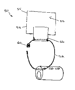
[0059] FIGS. 7A and 7B depict one embodiment of a catheter fastening
mechanism 50.
The mechanism 50 can be used with any of the catheters 10 described herein;
however, the
mechanism 50 is not limited to use with those specific catheters. The
mechanism 50 depicted
in FIGS. 7A and 7B is a suspension device for a nasogastric suction tube.
FIGS. 7C, 8A, and
8B depict alternative embodiments of the mechanism 50. Generally, the various
embodiments
of the mechanism 50 include a ring 52 (see, for example, FIGS. 9A and 9B) that
engages an
opening 14 in the tube 10 and is secured to the body by a fastening strap 54
(see, for example,
FIGS. 11A and 11B). The use of the ring 52 and strap 54 arrangement provides a
"floating"
connection of the catheter to the patient's body, allowing some minor movement
of the catheter
CA 02687860 2009-11-20
WO 2008/147842 PCT/US2008/064466
- 18 -
relative to the patient without the catheter becoming dislodged or being
subjected to excessive
force. In this particular embodiment, the tube 10 includes openings or micro-
tubes 14 spaced
about 1 cm apart and oriented approximately 90 degrees to the main lumen 20 of
the tube 10.
[0060] In practice, the nasogastric tube 10 with its openings 14 running
at right angles to
the main lumen is inserted into the body to an appropriate level. At least a
portion of the
fastening strap 54, which may include one or more layers/pieces of material,
is placed on the
bridge of the patient's nose 51. At least one ring 52 is passed through the
opening 14 at an
appropriate level for the patient and application, in this case about 1 cm to
about 3 cm from the
external nares 61. In the embodiment shown, the ring 52 is opened so that one
end thereof can
be passed through the opening 14 in the tube 10; however, the tube 10 can be
manufactured
with solid rings installed at predetermined locations on the tube 10. In the
embodiment shown,
the ring 52 includes a locking mechanism 60, as described in greater detail
hereinbelow.
Depending on the application, additional rings 52 and/or fastening straps 54
can be used (see,
for example, FIG. 13). The ring(s) 52 is secured to the patient via at least
another portion of the
fastening strap 54. The specific manner of attachment is described later with
respect to FIGS.
7A-7C, 8A, and 8B.
[0061] The ring 52 is shown in the closed configuration in FIG. 9A and
the open
configuration in FIG. 9B. In the embodiment shown, the ring 52 has a generally
circular
configuration and is biased into the open position by, for example, spring
tension in the body of
the ring 52 (i.e., the tendency of the bent ring to try to return to an unbent
configuration).
Alternatively, the ring 52 can be manufactured with a set gap between the ends
thereof, where
the gap is closed by the locking mechanism 60 or crimping the ends together.
In addition, the
term ring is used herein to include similar structures, such as loops or
hooks, which may
include shapes such as, for example, C, J, S, U, and Z. The ring 52 can be
made of a metal or
CA 02687860 2009-11-20
WO 2008/147842 PCT/US2008/064466
- 19 -
plastic material. In one embodiment, the ring 52 has an outside diameter of
about 1 cm to
about 5 cm and the body (e.g., wire diameter) of the ring 52 has a cross-
sectional dimension of
about 2 mm to about 8 mm.
[0062] The locking mechanism 60 for the ring 52 is intended to hold the
ends of the ring 52
together and prevent the inadvertent decoupling of the ring 52 and the tube
10. The locking
mechanism 60 can have various arrangements. As shown in FIG. 9B, the mechanism
60
includes an internally threaded collar 62 crimped on one end of the ring 52
and a threaded
portion 64 disposed on the other end of the ring 52. Alternative locking
mechanisms are also
possible, such as, for example, box clasps, toggle clasps, hook and eye
clasps, friction clasps,
and magnetic clasps. Essentially any locking mechanism can be used providing
that the portion
of the mechanism that passes through the opening 14 is smaller in
circumference than that of
the opening 14. In one embodiment, the ring 52 can include a sharpened tip
configured to
pierce through a wall of a catheter where no openings are provided; however,
due care should
be exercised to avoid breaching the lumen of the catheter and rendering the
catheter unfit for its
intended application.
[0063] FIGS. 9C and 9D depict alternative ring configurations. As shown
in FIG. 9C, the
ring 152 has a substantially oval or elliptical shape and includes a locking
mechanism 160 of
the types previously described. FIG. 9D depicts a ring 252 having a
rectangular configuration,
although other polygonal shapes are also contemplated and within the scope of
the invention.
The ring 252 includes a locking mechanism 260 having a simple bar and U
mechanism or
friction fit.
[0064] Generally, the size and shape of the fastening strap 54 will vary
to suit a particular
application and will depend, for example, on the type of catheter to be
secured and the location
where the catheter is to be secured. The strap 54 shown in FIGS. 7A, 7B, 8A,
and 8B has a
CA 02687860 2009-11-20
WO 2008/147842
PCT/US2008/064466
- 20 -
generally rectangular shape; however, the shape of the strap 54 may be
tailored by cutting with
scissors. The strap 54 can be manufactured from a porous or non-porous
material including,
for example, a woven cloth material of natural and/or synthetic fibers.
[0065] In
one embodiment, the strap 54 includes two layers 56, 58 with a lower or first
layer 58 usually placed first. The first layer 58 includes a lower surface 59
(see, for example,
FIG. 11B) that can include an adhesive material deposited thereon. The lower
surface 59 may
include a removable covering 53 to isolate the adhesive material from the
environment until
ready to use. The covering 53 can be removed from all or a portion of the
lower surface 59 and
the lower surface 59 applied to the patient's skin. The skin may be prepped
with Mastisol
brand liquid adhesive, as available from Ferndale Laboratories, Inc., prior to
applying the first
layer 58. The first layer 58 can include an adhesive or hook and loop type
fastener on its upper
surface 55. The upper surface 55 can also include a covering 53 to prevent
contamination of
the upper surface 55. The covering 53 can be removed from all or a portion of
the upper
surface 55 prior to use.
[0066] The upper or second layer 56 of the strap 54 can be bonded firmly or
otherwise
attached to the first layer 58. For example, as shown in FIG. 11B, the second
layer 56 can be
attached to the first layer 58 proximate a midline 65 of the first layer 58
over its vertical length
and for a width of about 1/2 cm to about 3/4 cm. Alternatively, and as shown
in FIGS. 7A and
7B, the first and second layers 58, 56 can be attached in an overlapping
manner. The second
layer 56 can have an adhesive or hook and loop type fastener disposed on a
lower surface 57
thereof to securely interface with the upper surface 55 of the first layer 58.
The adhesive or
hook and loop type fastener can extend over the entire lower surface 57,
except in the
embodiment of FIGS. 11A and 11B, where the second layer 56 is bonded to the
first layer 58 at
CA 02687860 2009-11-20
WO 2008/147842 PCT/US2008/064466
-21 -
the midline. The use of a hook and loop type fastener allows the first and
second layers 58, 56
to be repositioned relative to one another.
[0067] As shown in FIGS. 7A and 7B, the ring 52 is attached to the
second layer 56 of the
fastening strap 54, which acts as a holding strip for the ring 52. In the
embodiment shown, the
second layer 56 is slightly smaller than the first layer 58; however, the size
and shape of the
second layer 56 will vary to suit a particular application. In one embodiment,
the first layer
measures from about 4 cm to about 7 cm in width and about 3 cm to about 4 cm
in length and
the second layer measures from about 1/2 cm to about 3/4 cm in width and about
2 cm to about 4
cm in length for a nasogastric tube application. The second layer 56 depicted
in FIGS. 7A and
7B can include one side coated with an adhesive and optionally covered by a
peel off strip with
a tab (i.e., covering 53). In one embodiment, the second layer 56 is folded
over a portion of the
ring 52 and the side coated with the adhesive secured to itself, thereby
gripping the ring 52.
The opposing side, now the outer surface of the folded second layer 56, can
also include an
adhesive coating or a hook and loop type fastener on at least a portion
thereof. The second
layer 56 can now be secured to the first layer 58 of the fastening strap 54
previously secured to
the patient's skin via the adhesive coating or mating hook and loop type
fasteners, thereby
securing the tube 10 in place. Alternatively, the second layer 58 can be
applied to the patient's
skin at the same time as the first layer 58 is applied and can be sandwiched
between the first
layer 58 and the patient's skin. As shown in FIG. 7B, the second layer 56 does
not align evenly
with the first layer 58, but extends beyond the first layer by, for example,
about 1 to 2 cm.
[0068] In the alternative embodiment shown in FIG. 7C, the fastening
strap 54 includes
only a single layer (e.g., second layer 56). In this arrangement, the tube 10
is first inserted into
the patient at the appropriate position and then the fastening strap 54 and
ring 52 assembly is
secured to the patient. The fastening strap 54 can be trimmed as needed to
suit the application
CA 02687860 2009-11-20
WO 2008/147842 PCT/US2008/064466
- 22 -
site and the covering 53 removed to expose the adhesive coating. The patient's
skin can be
prepped as previously described. The fastening strap 54 is secured to the
patient's skin and the
attached ring 52 is passed through the appropriate opening 14.
[0069] The catheter fastening mechanism 50 shown in FIGS. 8A and 8B
utilizes the
fastening strap 54 shown in greater detail in FIGS. 11A and 11B. In addition
to the fastening
strap 54 having two substantially evenly aligned layers 56, 58, the mechanism
50 includes an
additional holding tab 70 (see FIG. 10). As shown in FIG. 10, the holding tab
70 is secured at
one end 72 to the ring 52. In one embodiment, the strap end 72 is folded over
the body of the
ring 52 and secured to itself by, for example, adhesive or stitching. In the
embodiment shown
in FIG. 10, the holding tab 70 measures about 3 cm to about 6 cm in length,
about 0.5 cm in
width, and about 2 mm in thickness, however, the size and shape of the tab 70
will vary to suit
a particular application. The holding tab 70 and ring 52 are sized and
configured such that the
tab 70 can be secured to the ring 52 prior to inserting the ring 52 through
the opening 14 in the
catheter 10 or thereafter. The holding tab 70 can include an adhesive or hook
and loop type
fasteners on one or both sides thereof.
[0070] As shown in FIGS. 8A and 8B, the first layer 58 of the fastening
strap 54 is secured
to the patient's nose 51. At least a portion of the holding tab 70 is
positioned over the first
layer 58. In the embodiment shown in FIG. 8A, the ring 52 is inserted through
the opening 14
in the tube 10 prior to positioning over the first layer 58 of the fastening
strap 54. The holding
tab 70 may be held in position by the adhesive or mating hook and loop
fasteners disposed on
the holding tab 70 and the upper surface 55 of the first layer 58. The second
layer 56 of the
fastening strap 54 is then positioned over the first layer 58, thereby
sandwiching the holding tab
70 between the two layers 56, 58.
CA 02687860 2009-11-20
WO 2008/147842 PCT/US2008/064466
-23 -
[0071] FIGS. 12A and 12B further depict the installation of the catheter
fastening
mechanism 50 of FIGS. 8A and 8B using the fastening strap 54 depicted on FIGS.
11A and
11B. As previously described, the fastening strap 54 includes first and second
layers 58, 56
connected proximate their midline 65. The strap 54 is positioned on the
patient's nose 51. The
first layer 58 is pressed onto the patient's nose 51 (arrows 67), allowing the
adhesive covered
lower surface 59 of the first layer 58 to contact the patient's skin.
[0072] Subsequently, the covering 53 can be removed from the side of the
upper surface 55
of the first layer 58 corresponding to the nostril in which the tube 10 is
inserted, thereby
exposing the adhesive or hook and loop type fastener. The holding tab 70 is
positioned over
the first layer 58 on the exposed side. The portion of the second layer 56
corresponding to the
side where the holding tab 70 is positioned is then attached (for example, by
adhesive or hook
and loop type fastener) to the upper surface 55 of the first layer 58, thereby
securing the tab 70
between the two layers 56, 58. Where the second layer 56 includes a covering
on its lower
surface 57, the covering 53 can be removed as needed to expose the adhesive or
hook and loop
type fastener for securing to the upper surface 55 of the first layer 58.
[0073] Alternatively, in an application not using the holding tab 70, at
least a portion of the
second layer 56 can be passed through the ring 52, which may or may not be
secured to the
tube 10 at this point. The portion of the second layer 56 that is passed
through the ring 52 will
depend on which nostril the tube 10 is inserted. The second layer 56 can then
be secured to the
upper surface 55 of the first layer 58, as previously described.
[0074] FIG. 13 depicts an alternative arrangement of a catheter
fastening mechanism in
accordance with one embodiment of the invention. As shown, the catheter 10
includes multiple
rings 52 secured along the length of the catheter 10. Multiple rings 52 can be
used to provide
more than one fastening point for securing the catheter 10. Additionally or
alternatively,
CA 02687860 2015-01-26
71495-97
- 24 -
sutures could be used to secure the rings 52 to the patient, either with or in
place of the
=
fastening strap.
[0075] Having described certain embodiments of the invention, it will be
apparent to those
of ordinary skill in the art that other embodiments incorporating the concepts
disclosed herein
may be used without departing from the scope of the invention as claimed. The
described
embodiments are to be considered in all respects as only illustrative and not
restrictive.
[0076] What is claimed is:
=