Note: Descriptions are shown in the official language in which they were submitted.
CA 02688341 2014-08-27
1
Micro-Fluidic Optical Trap Using Raman Spectrum
The present invention relates to Raman spectroscopy. In particular, the
invention
relates to the use of Raman spectroscopy for investigating biological
material, for
example, single cells.
Background of the Invention
Raman spectroscopy is a powerful technique that relies on collection of
inelastically
scattered laser light from a sample. This light exhibits a frequency shift
that reflects
the energy of specific molecular vibrations within the sample of interest.
Hence, it can
provide a detailed chemical composition of the sample, i.e. a chemical
fingerprint.
The technique has wide potential in biomedical science as it may be applied to
samples over a wide size range from single cells through to intact tissue.
One of the major challenges of Raman spectroscopy is the inherently weak
nature of
the signal. In addition, a Raman signal may be obtained from the local
environment
surrounding the sample, typically making it difficult to discern the molecular
signatures of interest. Thus, considerable effort has focussed on enhancing
the ratio
of signal to background noise. By increasing the acquisition time to several
minutes,
the signal to noise ratio can be improved. However, in some environments, long
acquisition times can cause damage due to extended irradiation by the
excitation laser
and the mechanism required to hold the particles under investigation in the
measurement position. These are particular problems when investigating live
cells or
tissue samples.
Some solutions to the problems with conventional Raman spectroscopy have been
proposed. Many of these involve the inclusion of additional material, for
example
nano-particles, in the samples that are being investigated. However, this is
not ideal
for the investigation of whole cells as the precise positional control of the
foreign
particles is difficult. Additionally, the enhancement achieved with the use of
foreign
particles is confined to the immediate surface of the particles (-10nm) making
the
measurement of the overall Raman signal impossible. One technique that does
not
require the addition of foreign particles uses wavelength modulation. This is
described in the article "Wavelength-Modulation Raman Spectroscopy" by Levin
et
CA 02688341 2014-08-27
2
al, Appl. Phys Letter 33(39), 1 Nov 1978. This technique increases the
sensitivity of a
Raman spectroscopic system by modulating the wavelength of the excitation
light,
and then using this to distinguish the sample's Raman response from background
radiation and/or noise. The system described uses a tuneable dye laser and
single
channel slowly scanning detection. A problem with this is that the scan takes
about
50 minutes for the whole spectra. Additionally the method relies on very
large,
expensive optics and is inappropriate for many practical applications, in
particular the
investigation of single cells.
One of the most promising areas of application for Raman spectroscopy is in
the
discrimination between sets of biomedical samples e.g. cancer diagnostics.
Here, it is
advantageous to have short acquisition times, especially if a live patient
rather than a
retrieved sample is being studied. It is also important to reduce the impact
of
fluorescence, as this has a high patient to patient and even cell to cell
variability that
can heavily reduce the performance of any subsequent diagnostic models. One of
the
most widely used tools for discriminating between the Raman spectra acquired
from
sets of biomedical samples is Principal Component Analysis (PCA).
Principal components analysis (PCA) is a statistical technique used to change
the
representation of a multidimensional data set. A new representation or
coordinate
system is constructed such that the variance of the data sets is biggest for
the first
coordinate component of the new representation. This is then called the first
principal
component. The second biggest variation lies the on the second coordinate of
the new
representation, and so on. Finally, the data set dimension is reduced by
retaining only
the first few principal components that account for most of the variance of
the original
data set. It is these low-order components that often contain the "most
important"
aspects of the data set. Using PCA to examine Raman spectra from sets of
biomedical
samples allows combinations of Raman peak fluctuations to be found that can
then be
used to discriminate between the Raman spectra from the sets of biomedical
samples.
Summary of the Invention
According to the present invention, there is provided a micro-fluidic system
comprising means for optically trapping a particle and means for obtaining a
Raman
spectrum from the particle whilst it is in the optical trap.
CA 02688341 2014-08-27
2a
According to an aspect of the invention, there is provided a system comprising
means
for optically trapping a particle and a radiation source for causing Raman
scatter from
the particle whilst in the optical trap, wherein the means for forming an
optical trap
comprise a dual beam arrangement, in which counter propagating optical beams
are
used to hold the particle, and wherein the source emits radiation orthogonal
to the
trapping beams, wherein the radiation source comprises two or more laser
sources
each independently switchable and operable to vary its intensity between
multiple
levels, each of the multiple levels of intensity being sufficient to cause
Raman scatter,
thereby to achieve intensity modulated multi wavelength excitation.
According to another aspect of the invention, there is provided a system
comprising
means for optically trapping a particle and a radiation source for causing
Raman
scatter from the particle whilst in the optical trap, wherein the radiation
source
comprises two or more laser sources each operable to output a different
wavelength,
and wherein each of the sources is independently switchable and operable to
vary its
intensity between multiple levels, each of the multiple levels of intensity
being
sufficient to cause Raman scatter, thereby to achieve intensity modulated
multi
wavelength excitation.
According to another aspect of the invention, there is provided a method for
obtaining
a Raman spectrum comprising:
exciting a sample or particle using radiation from two or more laser sources
that are operable to output a different wavelength and are independently
switchable
and operable to vary intensity between multiple levels of intensity, each of
the
multiple levels of intensity being sufficient to cause Raman scatter, thereby
to achieve
intensity modulated multi-wavelength excitation;
capturing light scattered from the sample or particle using a multi-channel
spectrometer comprising a CCD camera;
modulating the excitation radiation;
exciting the sample or particle using the modulated radiation;
capturing scattered radiation associated with the modulated radiation; and
identifying variations in the captured radiation associated with the
modulation,
thereby to obtain a Raman spectrum or a function thereof for the sample or
particle.
CA 02688341 2014-08-27
2b
According to another aspect of the invention, there is provided a method for
obtaining
a Raman spectrum comprising:
exciting a sample or particle using first radiation to cause emission of a
Raman signal;
capturing first light scattered from the sample or particle;
modulating the excitation radiation;
exciting the sample or particle using the modulated radiation to cause
emission of a Raman signal;
capturing scattered radiation associated with the modulated radiation;
forming a data set using the captured scattered first light and the captured
scattered modulated light; and
performing principal component analysis on the data set to identify a
differential Raman signal or a function thereof for the sample or particle.
CA 02688341 2009-11-25
WO 2007/141539
PCT/GB2007/002121
3
Typically, the means for forming an optical trap comprise a dual beam
arrangement,
in which counter propagating optical beams are used to hold the particle.
Because the
trapping beams are divergent, this arrangement reduces the chance of damage to
the
particle under investigation. This is particularly advantageous when the
particle is a
cell. The laser for exciting the Raman scatter may be placed orthogonal to the
trapping beams.
Means may be provided for modulating the Raman excitation signal. The
modulation
means may be operable to encode information onto one or more parameters of the
excitation signal. The modulation means may be operable to modulate one or
more of
the excitation laser driving current; intra-cavity or external cavity grating
position '
and/or orientation; change of the cavity length, using, for example mechanical
or
opto-electric means; polarisation variation; excitation mode variation and
variation of
the optical properties of any intra-cavity or external cavity non-linear
optical
elements.
Any suitable laser can be used to fonn the Raman excitation signal, although a
laser
diode in a Littrow or Littman-Metcalf configuration is preferred.
Alternatively, two or
more laser sources may be combined where each has a different wavelength. In
this
case, each of the sources can be independently switched and its intensity
varied to
achieve an efficient modulated multi wavelength excitation.
The Raman excitation can also be provided by a broadband light source such as
mode-locked pulsed lasers, delivering 100fs pulses, for example, or other
sources
such as a white light source. These sources can have their spectral
phase/chirp
specially engineered and/or modulated. This can be achieved, for example, by
passing
the pulse through a Fabry-Perot resonator giving a periodic spectral phase
modulation.
More complex spectral phase/chirp modulation can be obtained through the use
of a
Spatial Light Modulator (SLM) in conjunction with some spectral dispersion
elements
such as prisms or other photonic devices.
Means may be provided for doing a principal component analysis. In accordance
with
the invention, a single modulated measure from a cell consists of multiple,
short
CA 02688341 2009-11-25
WO 2007/141539
PCT/GB2007/002121
4
duration, spectra taken with the excitation laser at different wavelength. All
the
spectra together form a data set on which a principal component analysis can
be
performed. Contrary to conventional PCA, the value of the first principal
component
is not of interest. Instead, it is the associated eigen-spectra, which is the
basis vector
associated with the first principal component. It is these eigen-spectra
(basis-vector)
that are then the differential spectra. Here, the PCA is not used to reduce
the
dimensionality of the data set but to extract the element with the largest
variation.
According to the present invention, there is a method for obtaining a Raman
spectrum
comprising exciting a sample using radiation; capturing light emitted from the
sample;
modulating the excitation radiation; capturing light emitted in response to
the
modulated radiation and using the captured radiation to obtain the Raman
spectrum.
Preferably, the scattered radiation is captured using a multi-channel
spectrometer,
ideally a CCD camera.
The method may further involve correlating modulations in the excitation
radiation
with variations in the captured spectra. By doing this, the Raman peak can be
more
accurately identified, as background fluorescence, for example, should not
vary with
changes in the excitation signal.
By analysing the light emitted in response to both the initial excitation
radiation and
its modulated version using PCA, further improvements may be made. This
provides
a simple technique for pulling out variations in the acquired spectra. If the
modulated
spectra are fed into a PCA routine, this will look at the variation in the
spectra.
Because of the modulation, this variation is the moving Raman spectrum only,
as the
fluorescence remains steady. Thus the PCA routine outputs a spectrum, or
principal
component that is the differential Raman spectrum of the sample. For the
extraction of
the differential Raman signal a minimum of one modulation period is necessary.
Brief Description of the Drawings
Various aspects of the invention will now be described by way of example only
and
with reference to the accompanying drawings, of which:
CA 02688341 2009-11-25
WO 2007/141539
PCT/GB2007/002121
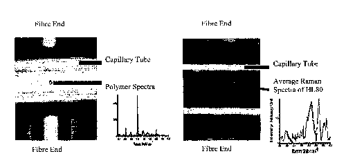
Figure 1 (a) is an image of a polymer microsphere that is optically trapped in
a
micro-fluidic channel and exposed to Raman excitation, together with the
resultant
Raman spectra;
Figure 1 (b) is an image of a HL60 cell, that is optically trapped in a micro-
5 fluidic channel and exposed to Raman excitation, together with the
resultant Raman
spectra;
Figure 2 (a) is a plot of laser intensity as a function of time;
Figure 2(b) is a plot of laser wavelength as a function of time;
Figure 3 is a complete Raman spectrum including background, noise,
excitation and Raman resonance peaks;
Figure 4 is a plot of the variance of 90 Raman Spectra of 0.5s each;
Figure 5 shows the average Raman spectra using the wavelength tracking and
signal renormalisation method;
Figure 6(a) is a plot of wavenumber versus intensity of the laser;
Figure 6(b) is a plot of binned and averaged spectra;
Figure 7 is a plot of integrated differential Raman signal as a function
wavelength;
Figure 8 is plot of simulated wavelength versus intensity in a multi-stable
lasing device;
Figure 9 is a diagram of an alternative optical arrangement for trapping a
single cell and obtaining Raman spectra from it;
Figure 10 is a single Raman spectrum recorded at one wavelength;
Figure 11 is a sequential recording of the Raman signal as the laser, and
hence
Raman spectra, was modulated between two fixed wavelengths;
Figure 12 is a plot of a differential Raman spectrum of a cell extracted from
the modulated Raman signal, and
Figure 13 shows the results of a PCA analysis carried out to compare the
effect of acquiring a Raman signal using conventional processes and a process
in
accordance with the invention.
Figure 1 shows a microfluidic device that is operable to form an optical trap
in a
micro-fluidic channel. Any suitable means can be used for causing fluid to
flow
through the device. The optical trap is formed using two counter propagating
diverging beams. In this example, the counter propagating beams are provided
via
CA 02688341 2009-11-25
WO 2007/141539
PCT/GB2007/002121
6
two optical fibres that are positioned on opposing sides of the micro-fluidic
channel.
In this case, the channel is a micro-capilliary. Radiation is directed via the
fibres into
the micro-fluidic channel, so that cells or other particles within the fluid
can be
trapped. Optical traps can be used to allow micrometer-sized particles to be
held,
moved and generally manipulated without any physical contact. This has been
well
documented, for example see Ashkin et al Optics Letters Vol. 11, p288 (1986).
Orthogonal to the fibre ends (not shown) is an objective lens for directing a
Raman
excitation beam onto the cell and capturing the emitted signal so that it can
be
recorded.
To test the arrangement of Figure 1, a flow system consisting of a capillary,
of square
cross-section size 80microns, was connected to a syringe or gravity feed pump.
Initially, 10micron polymer particles were flowed through the capillary tube,
and
trapped using the counter propagating beams, as and when desired. A 50mW Raman
examination beam was then introduced from below using a Nikon x50 NA 0.9 oil
immersion. Figure 1(a) shows a polymer microsphere trapped inside the
capillary,
together with the spectra obtained from the sphere. Figure 1(b) shows a HL60
cell
trapped, together with the spectra obtained from this.
By using optical trapping in a microfluidic environment, damage to the
particle/cell
that is under investigation can be minimised. However, to further reduce this,
a
statistical approach can be used to allow the Raman signals to be recorded
very
rapidly from a single cell. This method relies on modulation of the excitation
laser,
and in particular tuning of the laser wavelength. This can be done using
continuous or
discontinuous tuning. Statistical analysis of the resultant Raman scatter
allows a
significant reduction in the time needed to record the signals. This can be
done
without the addition of foreign particles, such as nanoparticles, specialist
surfaces,
and/or enhancement schemes.
The physical properties, such as wavelength and intensity, of the Raman
excitation
vary in time. The resulting Raman signal is then also subject to variations
but in a
complex way. Indeed, depending on their physical origin the different parts of
the
Raman spectra behave differently. If the wavelength is modulated then the
Raman
peaks in the spectra incur a shift in wavelength while the fluorescence
background
CA 02688341 2009-11-25
WO 2007/141539
PCT/GB2007/002121
7
remains constant. In the case of amplitude variation, both peaks and
background
change in amplitude.
The method of the present invention uses a general wavelength, frequency and
amplitude or other parameters variation of the excitation and correlates this
with the
measured Raman spectra to distinguish between the different components of the
spectrum, i.e. background, Raman peaks and noise. The input excitation is
encoded
with a variation which then is decoded at detection time distinguishing thus
between
signal, noise and background. Variation of the parameters is used to quantify
the
correlated variation of the Raman signal.
The encoding method is based on the variation of controlling the parameters of
the
Raman excitation source such as the laser or any device delivering the
necessary
excitation output. Examples of these parameters are: laser, diode or device
driving
current; intra-cavity or external cavity grating position and/or orientation;
mechanical
or opto-electric change of the cavity length; polarisation variation;
excitation mode
variation and variation of the optical properties of any intra-cavity or
external cavity
non-linear optical elements.
Another way to achieve source variation is by using bistable or multi stable
lasers that
naturally oscillate in a controlled or chaotic fashion between different
wavelength and
states. Alternatively, two or more laser sources can be combined where each
has a
different wavelength. Each of the sources can be independently switched and
its
intensity varied to achieve an efficient modulated multi wavelength
excitation.
Figure 2 shows the effect of varying the driving current of the diode laser
device.
Figure 2 (a) is a plot of laser intensity as a function of time, and Figure
2(b) is a plot
of laser wavelength as a function of time. As can be seen, varying the drive
current
induces a wavelength shift and an excitation intensity variation. Because of
the non-
linear properties of the laser, discrete wavelength jumps occur as the current
is varied.
These jumps correspond to laser mode hopping.
CA 02688341 2009-11-25
WO 2007/141539
PCT/GB2007/002121
8
To obtain the sample response, Raman spectra are repeatedly acquired in =as
short as
possible time slots whose duration is related to the speed of variation of the
excitation
parameters. Over this duration, the excitation parameters should not vary. For
practical reasons, the spectral snapshot can also contain the excitation
spectra suitably
attenuated in intensity. The excitation spectral information such as
wavelength,
amplitude and bandwidth can then be retrieved from this snapshot.
Alternatively
other measures can be used to deduce the excitation characteristics and their
variation
or the variation can be linked to the control parameters after suitable
calibration.
Every snapshot is then stored together with the excitation parameters for real-
time or
successive data treatment.
Figure 3 shows a Raman spectrum that was acquired with duration of 0.5s. The
excitation parameters can be retrieved from the furthest left peak (A), which
corresponds to the excitation laser. There are multiple ways to retrieve the
Raman
peaks from a family of short scans, each taken for different excitation
parameters.
Some methods cancel directly the background while others do not. A non-
exhaustive
list of possible methods includes statistical post processing (variance), real
time/post
processing signal tracking (spectral lock-in amplifier), and real time/post
processing
leading to differential signal (statistical approach).
Statistical post processing involves looking at the variation of a family of
spectra as a
function of wavelength. If the excitation wavelength variation is large enough
then
the variance of the family of spectra will show different levels of variance
for the
noise, background and Raman peaks. Indeed the shift of the excitation
wavelength
implies a shift of the peaks, which is equivalent to a large intensity
variation at a given
wavelength. The variance of the peak will thus be much higher than the
variance of
the surrounding region. Figure 4 shows the resulting Raman spectra after using
the
statistical post processing method that calculates the variance from 90 Raman
spectra
of 0.5s each.
Real time/post processing signal tracking (spectral lock-in amplifier)
involves using
the amplitude and wavelength position of the excitation laser peak to shift
and
normalise the individual 0.5s Raman spectra before averaging them. However,
this
CA 02688341 2009-11-25
WO 2007/141539
PCT/GB2007/002121
9
method does not cancel the background and is disadvantaged by the laser mode
hopping. It is similar to a lock-in amplifier as it locks-in onto the
reference excitation
wavelength and uses its shift to reconstruct the resonances. Figure 5 shows
the
processed Raman spectra using the excitation wavelength and amplitude tracking
method.
Real time/Post processing leading to differential signal (statistical
approach) involves
using a differential signal to eliminate the background. This can be achieved
by using
two laser states with different wavelengths. When plotting the amplitude
versus the
wavelength of the excitation laser while the driving currant is varied the
number of
modes accessed by the parameter variation can be recognised, as shown in
Figure
6(a), which is a plot of wavenumber versus intensity of the laser, and Figure
6(b),
which is a plot of binned and averaged spectra. In this case, the wavelength
position
of the laser peak is used to average only spectra where the excitation laser
has a
specific wavelength. The spectra in a bin are normalised with the amplitude of
the
laser intensity and then averaged. Because of the bi-stability there are only
two bins.
The differential signal corresponds in this case to the difference between the
red and
blue curve. When calculating this difference the background part of the signal
is
removed. The difference can then be integrated to retrieve original Raman
resonance
peaks, as shown in Figure 7. This method can be generalised to multi stable
lasing
devices. Figure 8 shows a simulated wavelength versus intensity in a multi-
stable
lasing device. In this multiple stabilities will increase the differential
signal as this
can be calculated using n-point differential formulas.
Figure 9 shows a more detailed system for providing a modulated Raman
excitation
signal in accordance with the invention. This has a laser that can be
modulated in
some form: mechanically, optically or by current. This is then reflected
against a
holographic notch filter that reflects a very narrow band around the
wavelength and
transmits all other wavelengths, into a microscope objective that focuses the
beam to
the sample. The Raman signal is collected by the same microscope objective and
transmitted through the notch filter onto a dichroic mirror, which reflects
the infrared
Raman scatter whilst allowing the visible incoherent light, which illuminates
the
sample, to pass to a viewing camera. This allows an image of the sample under
study
to be collected as well as its Raman spectrum. The collected Raman scatter is
then
CA 02688341 2009-11-25
WO 2007/141539
PCT/GB2007/002121
passed through an optional confocal aperture to reject any unwanted signal
surrounding the sample of interest. The signal is finally imaged onto a 550mm
spectrograph equipped with a 300 lines/mm grating to separate spatially all
the
collected Raman wavelengths that are imaged onto a multi-channel detector, for
5 example a CCD camera. The CCD camera is a liquid nitrogen cooled CCD that
has a
pixel array of 2048x512 with each pixel measuring 13.5m square, the array
having a
bandwidth of one pixel, i.e. about 0.15nm. The combined resolution of the
spectrograph is 0.078nm allowing the movement of the laser and hence Raman
spectrum to be captured.
In order to remove or reduce fluorescence in the acquired Raman spectra, as
well as
reduce the acquisition times the excitation wavelength is modulated and
multiple
spectra collected. The Raman spectra are then extracted from these multiple
spectra.
To improve extraction of the Raman spectrum from the modulated data, an
external
cavity laser diode was used in a Littman-Metcalf configuration. This
configuration
allows a significantly greater tuning range (-30nm) compared to the bandwidth
of
one pixel (0.15nm) of the detecting CCD mounted on the spectrometer, improving
the detection of the modulation significantly. This laser was used to switch
between
two wavelength positions that in turn modulated the Raman spectra between two
positions. A signal was acquired at each wavelength position as it was moved
between the two wavelengths. A single spectrum can be seen in Figure 10, which
shows single spectrum recorded at one wavelength position. Multiple signals
were
acquired at each wavelength position as it was modulated. Figure 11 shows the
sequential recording of the Raman spectra as the laser was modulated between
two
fixed wavelengths. The jumps in the spectra can be clearly seen. The laser is
on the
extreme left and the Raman peaks to the right of this.
To improve the detection of variations in the acquired spectra a modified
version of
conventional PCA can be used. This pulls out variation in the acquired
spectra. If the
modulated spectra are fed into a PCA routine, this will look at the variation
in the
spectra. Because of the modulation, this variation is the moving Raman
spectrum
only, as the fluorescence remains steady. Thus the PCA routine outputs a
spectrum,
or principal component that is the differential Raman spectrum of the sample.
For the
CA 02688341 2009-11-25
WO 2007/141539
PCT/GB2007/002121
11
extraction of the differential Raman signal a minimum of one modulation period
is
necessary.
Figure 11 shows an example of a differential spectrum after PCA processing.
This
differential spectrum can be integrated to reproduce the normal Raman spectrum
of
the sample or left as is for further statistical analysis. An advantage of
using PCA in
this way is that the output is the variation in the Raman spectrum. Thus,
there is no
need to track the laser line allowing points of interest in the spectra to be
identified
and giving much more flexibility in the choice of instrumentation such as
which
grating to use. This method also removes the fluorescence background. It
should be
noted that fluorescence is not always a problem in viewing the spectrum, but
is more
of a problem in subsequent statistical analysis where it can severely affect
the
efficiency of discrimination between two sample sets such as healthy and
diseased
cells.
In order to evaluate the ability of this technique to effectively remove
fluorescence
and potentially reduce acquisition times a comparison was made with
conventional
PCA Raman processing and the combined modulation/PCA processing of the
invention. This was done for sets of Raman spectra acquired from different
regions in
a biological cell, nucleus and cytoplasm. Ten Raman spectra were collected
from the
nucleus and cytoplasm. The spectra were acquired in two minutes for both
conventional PCA Raman processing and the combined modulation/PCA processing
of the invention. To test the acquisition time reducing potential of the
invention
spectra for the modulated/PCA were also acquired in one minute.
Figure 13 shows the results of the PCA analysis carried out to compare the
effect of
acquiring the signal normally to the modulated/PCA method of acquiring the
Raman
signal. Figure 13(a) shows the diagrammatic definition of the resolution used
in
Figures 13(b) to 13(a). From Figures 13(b) and (c), it can be seen that the
resolution
greatly increases when the Raman signal is acquired using the modulated/PCA
method. This is because it removes the fluorescence that has a negative impact
on the
diagnostic PCA model. This may be important in medical diagnostics as patient-
to--
patient variability in fluorescence may greatly affect any diagnostic models
based on
Raman spectroscopy. Furthermore, as shown in Figure 13(d) even when the
CA 02688341 2009-11-25
WO 2007/141539
PCT/GB2007/002121
12
acquisition time is halved, the modulated/PCA Raman spectra provides a much
better
resolution compared to the discrimination based on the normal acquisition
indicating
that the acquisition time could be reduced by a factor of at least two.
The present invention provides a system that allows single cells to be
optically
trapped and held, and Raman signals to be acquired from these cells in a very
short
time. Contrary to 1978 paper, where the Raman signal was acquired with a
slowly
scanning single channel detection system (2.4nm/min), the present invention
combines the advantages of acquiring the modulated Raman signals with modem
multi-channel CCD detection allowing a rapid acquisition whilst excluding the
fluorescence background. Additionally, the invention improves subsequent
statistical
analyses such as Principal Component Analysis (PCA) important medical
diagnostics
for example. Using excitation signal modulation, signals can be acquired in ¨
1/10 to
1/50 of the time that would normally be required. This means that damage to
cells
due to over exposure to the Raman excitation can be minimised.
A skilled person will appreciate that variations of the disclosed arrangements
are
possible without departing from the invention. For example, whilst a micro-
capilliary
is described in other embodiments, the microfluidic flow may be implemented
using
channels made using soft lithography in PDMS or similar and the size of the
channel
may naturally vary. Accordingly the above description of the specific
embodiment is
made by way of example only and not for the purposes of limitation. It will be
clear
to the skilled person that minor modifications may be made without significant
changes to the operation described.