Note: Descriptions are shown in the official language in which they were submitted.
CA 02688626 2009-11-27
-1-
METHOD FOR PREPARING AN ACELLULAR ORGANIC TISSUE FOR
REVITALISATION
BACKGROUND OF THE INVENTION
The invention relates to a method for preparing an acellular tissue for
revitalisation. It is
well known in the medical sector, and more specifically in the surgical
sector, that it is
becoming increasingly important to have tissues available for grafting into
living beings
to meet the growing need to replace parts of organs or whole organs.
The creation of biological substitutes that are prepared in the laboratory and
then
transplanted into animal or human recipients is a medical procedure known by
the name
of "tissue engineering".
According to a known technique, tissues for grafting are prepared in the
laboratory by
implanting cells into a matrix consisting of an inorganic supporting medium
generally
called a "scaffold".
The "scaffold", which is used to compensate for a loss of substance of the
organ being
treated, facilitates the three-dimensional organisation of the cells until the
formation of
new tissue has been completed.
The scaffold must naturally then undergo a process of degradation until it
disappears
completely and is replaced by the regenerated tissue, which is facilitated by
the cells
implanted in said scaffold.
Transplants can be obtained using this method with either artificial or
natural scaffolds
(i.e. from a "donor") obtainable from humans or animals, such as the
oesophageal wall.
To use a natural scaffold harvested from a donor for transplanting into
another human
being, the tissue must be treated first to eliminate all the cells existing
between the
fibres of the connective tissue, and then to reimplant human cells belonging
to the
intended recipient of the graft (the "host") in order to avoid rejection
phenomena.
CA 02688626 2009-11-27
-2-
The techniques used to create a scaffold, i.e. an acellular matrix, starting
from tissues
harvested from a donor, are well known and are consequently not described in
detail
here; briefly, they involve immerging the tissue to be treated in a fluid
containing
enzymatic substances capable of digesting and destroying the living cells
contained
within the tissue without damaging the tissue's connective fibres.
After creating an acellular tissue matrix, ready to receive the cells obtained
from the
host, said tissue, or scaffold, is prepared in a so-called "Petri dish" (or
similar container),
which is a tray commonly used in biological laboratories, on the bottom of
which the
tissue to revitalise is rested.
The tissue is revitalised by implanting stem cells from the future recipient
and nourishing
them with a cell culture broth that feeds the cells, keeping them alive and
enabling them
to multiply and become disseminated.
Basically, the stem cells initially placed on the upper surface of the tissue
move through
the natural interstitia in the tissue of the scaffold - interstitia that were
previously
occupied by the donor's cells.
After a given period of time, under controlled temperatures and in the
presence of the
nutritional substances contained in the culture broth, the living cells
reposition
themselves in the interstitia of the tissue, which is then ready for
transplantation into the
host organ.
It should be noted that the cells generally used to revitalise the scaffold
are stem cells,
which subsequently become differentiated (or may have already done so) and
acquire
the specific function of the organ in which the revitalised tissue is grafted.
The success or failure of the transplantation of the tissue treated in this
way depends on
a capillary diffusion of the cells through the tissue matrix.
CA 02688626 2009-11-27
-3-
If this diffusion proves difficult or occurs on the surface, but not in depth,
the
transplanted tissue is not adequately revitalised and a necrotic process
begins, leading
to the failure of the transplant.
From the above considerations, it is clear that it is essential and important,
not to say
indispensable to success, to ensure the in-depth revitalisation of every part
of the
tissue, particularly through its full thickness.
For the time being, even when the preparatory and revitalising treatments are
applied
for a sufficiently lengthy period of time, it is still impossible to ensure
results reliable
enough to guarantee against any graft failures.
This is due to the scarce penetration of the living cells being reimplanted in
the scaffold.
In practical terms, this drawback considerably restricts the opportunity to
prepare
tissues suitable for transplantation because thicker tissues are not fully
revitalised after
the transplant since they cannot be penetrated in sufficient depth.
It is consequently evident that the current technique is only suitable for the
transplantation of tissues of very limited thickness, e.g. not exceeding
approximately 0.1
mm.
US Patent 5,112,354 discloses the preparation of a bone allograft wherein
first all soft
tissue is removed and then the surface is textured to produce a pattern of
holes adapted
to facilitate the demineralization of the bone and to increase the surface for
interaction
with subsequently introduced mesenchymal cells. The holes are produced by
laser or
mechanical drilling.
SUMMARY OF THE INVENTION
In one aspect of the present invention there is disclosed a method for
preparing
acellular tissues that overcomes the above-mentioned drawbacks.
CA 02688626 2009-11-27
-4-
More specifically, there is disclosed a method for preparing acellular organic
tissue so
that, when said tissue is revitalised with stem cells, it is easier for said
cells to penetrate
and colonise every possible space in the network of connective tissue fibres,
so as to
substantially recreate the same conditions of the tissue before it was
devitalised.
Another aspect of the invention is to obtain a significant and important
reduction in the
treatment time needed to revitalise the acellular scaffold once the living
cells have been
added in order to prepare the tissue for transplantation.
The aspects of the invention are achieved by a method for preparing an
acellular
organic tissue for revitalisation by means of the reimplantation of living
cells, involving
the following stages: preparing the acellular tissue on an essentially flat
surface;
creating a plurality of holes on the surface of the tissue, distributed all
over the surface
and positioned so that they penetrate at least through a portion of the
thickness of the
tissue, the holes being suitable for containing the living cells when they are
reimplanted,
characterized in that the plurality of holes is created by means of one or
more metal
needles connected to an electric power supply that induces, on the tip of each
needle,
the passage of a current of such intensity and wave form as to provide
sufficient energy
to break the bonds between the molecules comprising the organic tissue in the
vicinity
of the tip of the needle, each hole being created by the passage of current
and being
large enough for the tip of the needle to enter the space created by the
opening of the
molecular bonds.
More precisely, this method consists in the creation of a plurality of holes
in the surface
of the tissue being prepared; these holes penetrate at least through a portion
of the
thickness, and preferably through the full thickness of the tissue concerned.
These holes are obtained by means of a device containing needles with a
suitable
current passing therethrough and without inducing any alteration (tearing,
necrosis,
reduction or increase in thickness, changes in fluid content, or coagulation)
in the
connective tissue surrounding the hole being created.
CA 02688626 2009-11-27
-5-
The holes can be made through the thickness of the tissue being treated using
various
devices and methods, provided that the preparation of these holes does not
cause any
deterioration of the connective tissue surrounding the hole and of the
scaffold in
general.
According to the description given below, the aspects of the invention and the
best
results in qualitative terms for the holes created in the tissue are achieved
by applying a
high-frequency voltage (generally 4 MHz) to the tip of each needle used to
create each
hole, so as to induce the passage of a weak electric current, but strong
enough to break
the bonds between the molecules in the connective tissue, thereby creating a
hole,
without inducing any breakage of the molecules.
This gives rise to narrow-diameter holes, essentially equating to the gauge of
the needle
being inserted.
It is important to use needles of very narrow gauge, e.g. in the order of 50
pm, but
sufficient to facilitate the penetration of the cells inside said holes to
revitalise the
surrounding tissue.
It is logical and evident that creating numerous holes means preparing new
routes for
grafting cells into the deepest parts of the tissue, thus ensuring the
complete
revitalisation of the tissue concerned.
Using the method according to the invention, there is practically no limit to
the thickness
of the tissues that can be prepared for transplantation, since the holes can
be made
throughout the thickness of the tissue and over its entire surface, enabling
its complete
revitalisation because the living cells reimplanted in the acellular scaffold
can penetrate
throughout the tissue.
Further characteristics and particular features of the invention will be
highlighted in
greater detail in the description of a preferred embodiment of the invention,
provided
here as a non-limiting example, and of a device usable in the method.
CA 02688626 2012-04-11
-6-
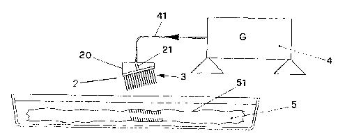
BRIEF DESCRIPTION OF THE DRAWINGS
The invention is described below with the aid of the attached drawings,
wherein:
- Figure 1 shows a schematic cross-section of the device comprising a holder
with an
array of needles resting on the thickness of the tissue being prepared for
revitalisation;
- Figure 2 shows one of the needles in the holder;
- Figure 3 shows the layout of the array of needles;
- Figure 4 shows a schematic view of the device for displacing the needle
holder.
DESCRIPTION OF THE INVENTION
According to the invention, a previously-treated acellular organic tissue, the
so-called
scaffold, is deposited on the bottom of a Petri dish (or similar container) so
that it lies
spread out on a flat surface.
A plurality of needles, such as the one indicated by the numeral 1 and shown
in Figure
2, is arranged in an array, e.g. to form a square, indicated as a whole by the
numeral 3
in Figure 3, so as to ensure an orderly arrangement of needles that are
preferably
separated by the same distance, i.e. they are equidistant from one another.
The needles are arranged essentially perpendicular with respect to the holder.
The head 11 of each needle 1 is connected electrically, e.g. by means of a
metal
conductor plate 2 attached to the holder 20 of said array of needles.
Said plate 2 is connected to an electric wire 21 that in turn receives the
output from a
generator 4.
Said generator 4 is a voltage generator, preferably generating 200-230 Volts,
but at a
wave frequency of 4 MHz, which is obtained by using electronic circuits, that
are well
known and consequently not described here for the sake of brevity.
CA 02688626 2009-11-27
-7-
The voltage sine wave available at the output 41 of the generator 4 is
preferably a
distorted sine wave and consequently with harmonics at least of the first,
second and
third order.
The power of the generator 4 is adjusted so that the current available at the
tip of each
electrode 1 comes between 2 and 2.5 mA.
When the tip of each needle is rested on the surface 51 of the organic tissue
5, contact
between the tip of each needle 1 and the organic tissue enables the passage of
a
current of around 2-2.5 mA, as mentioned previously.
Said current transmits an energy to the surrounding molecules that corresponds
(as
demonstrated experimentally) to what is called "molecular resonance".
This energy is just enough to break the bonds between the molecules affected
by the
passage of the current, while in the surrounding area it causes no breakdown,
tearing,
necrosis, reduction or increase in thickness, change in fluid content fluid,
coagulation or
other tissue degeneration.
Basically, this opening created in the molecular bonds equates to the creation
of a tiny
hole that, in practical terms, is the same diameter as each needle 1, i.e.
around 50-55
m.
Of course, needles of a different, larger or smaller gauge may be used,
provided that
the user bears in mind that the minimum gauge of the needle cannot be smaller
than
the diameter of the cells used for revitalisation.
The holder 20 of the array of needles 3 is then pushed in the direction in
which the
needles point and proceeds at a sufficiently slow pace such that, as the
needle moves
forward, the tip of the needle finds the hole already created by the flow of
current and
the consequent rupture of the molecular bonds.
CA 02688626 2012-04-11
-8-
It is easy to see that there is consequently no tearing of the connective
tissue, and that
a narrow hole corresponding to the gauge of the needle being inserted is
consequently
achieved.
As explained previously, this is particularly important and useful because the
cells that
are reimplanted on the tissue can thus penetrate in depth throughout the
tissue and
become grafted onto the walls of the holes, where they can multiply and very
quickly
revitalise the full thickness of the organic tissue.
As shown in Figure 1, the needles 1 penetrate preferably but not necessarily
obliquely
to the surface 51 of the scaffold 5, in order to increase the length of the
holes and
consequently obtain the maximum channelling effect in the scaffold.
Experiments have demonstrated that a 600 angle with respect to the vertical is
more
effective in the revitalisation process because the resulting holes are longer
than the
thickness of the scaffold.
The plurality of the holes may also lie in a direction essentially
perpendicular to
the surface of the tissue. _
Laboratory tests have shown that a useful dimension of the array of needles 3
containing the needles 1 is around 1 cm2, with said array comprising
approximately 200
needles; in this case, the current delivered by the generator 4 is no more
than 500 mA.
The perforation procedure must naturally be repeated all over the surface 51
of the
scaffold in order to obtain a homogeneous distribution of the holes throughout
the
thickness and over the entire useful surface of the tissue for
transplantation.
For this purpose, the invention uses a device for making the holes that is
advantageously provided with means 30 for displacing the holder 20 along three
Cartesian axes, i.e. along the vertical, or oblique axis Z, and along the
Cartesian axes X
and Y parallel to the plane of the surface 51 and shown schematically in
Figure 4.
CA 02688626 2009-11-27
-9-
Once the holes have been made in a given part of the scaffold 5, the holder 20
can be
moved and the procedure can be repeated in an orderly manner so as to cover
the
entire surface 51.
Clearly, if the holder 20 carrying the array of needles 3 is connected to
programmable
displacement means 30, e.g. with stepping motors governed by an electronic
control
unit, the procedure can be repeated automatically and sequentially, and with
the utmost
precision.
After completing the series of holes in the acellular tissue 5, as explained
above, it is
evident that said acellular tissue can be placed in a Petri dish, or similar
container,
where the living cells can then be added, which are generally stem cells from
the host
individual intended to receive the graft.
Suitably nourished with a culture broth, said stem cells can quickly and
easily occupy all
the holes made by the needles 1, thereby ensuring a complete and effective
revitalisation of the entire tissue for transplantation.
It is clear that the method of the invention achieves all the set aspects of
the invention,
since a perfect and effective revitalisation is ensured and any risk of
failure of the
subsequent transplantation is prevented.
Moreover, the revitalisation process takes place much more rapidly than when
the
known technique is used, and with extremely successful results.
Where technical features mentioned in any claim are followed by reference
signs, those
reference signs have been included for the sole purpose of increasing the
intelligibility of
the claims and accordingly such reference signs do not have any limiting
effect on the
interpretation of each element identified by way of example by such reference
signs.