Note: Descriptions are shown in the official language in which they were submitted.
CA 02689124 2009-11-30
WO 2008/151307 PCT/US2008/065992
T-CELL CYTOKINE-INDUCING SURFACE MOLECULES AND
METHODS OF USE
FIELD OF THE INVENTION
[0001] The invention relates to cytokine modulators and methods for using the
same
to modulate cytokine production in monocyte lineage-derived cells.
BACKGROUND OF THE INVENTION
[0002] A wide variety of clinical conditions are mediated by acute and/or
chronic
inflammation including, but not limited to, Rheumatoid Arthritis, Multiple
Sclerosis, Crohn's
Disease, Psoriasis, Psoriatic Arthritis, Graves Disease, Autoimmune
Polyendocrine
Syndromes, Hereditary Proteinuria Syndrome, Type I Diabetes, Systemic Lupus
Erythematosus, Primary Bilary Cirrhosis, Autoimmune Thyroiditis, Hepatitis,
Acquired
Immunodeficiency Disease (HIV), Graft versus Host Disease, Allograft Disease,
Asthma,
Cutaneous T-Cell Lymphoma, HTLV-I-Associated Cutaneous T-Cell Lymhoma, HTLV-II-
Associated Lymphoma, Hairy Cell Leukemia, Idiopathic CD4+ T-Lymphocytopenia,
or
Melanoma. It is believed that one of the mechanisms involved in inflammation
is up-
regulation of cytokines by activating monocyte lineage-derived macrophages
(Mt). For
example, proinflammatory cytokines such as Tumor Necrosis Factor-a (TNFa) and
Interleukin-11 (IL-1(3), produced in the lesion, have been shown to induce and
maintain
chronic lesional inflammation. Recent studies in relevant animal models
suggest that T-cells
(Ta) play a key role in M~ activation; however, T-cell cytokines, such as
Interleukin's-4, 10,
and 13 (IL-4, IL-10 and IL-13), have been shown to either play an anti-
inflammatory role or
only weakly induce TNFa/IL-11 up-regulation.
[0003] Therefore, there is a need to modulate inflammation to treat various
clinical
conditions associated with acute and/or chronic inflammation.
SUMMARY OF THE INVENTION
[0004] Some aspects of the invention relate to cytokine modulators and methods
for
using the same to modulate cytokine production in monocyte lineage-derived
cells. In some
1
CA 02689124 2009-11-30
WO 2008/151307 PCT/US2008/065992
particular embodiments, the invention provides proinflammatory cytokine
modulators and
methods for using the same to modulate proinflammatory cytokine production in
monocyte
lineage-derived cells, typically human monocyte lineage-derived cells. Without
being bound
by any theory, it is believed that monocyte lineage-derived cells become
activated once they
come in direct cell-cell contact with different effector T cell populations.
Controlling
adaptive (i.e., acquired) immunity at the level of TCISM Ligand and/or TCISM
Receptor
provides therapeutic intervention that still allow for innate immunity during
microorganism
infections (e.g., bacterial skin infections).
[0005] One aspect of the invention provides a method for modulating cytokine
production in monocyte lineage-derived cells of a subject comprising
administering a
cytokine modulator to said subject, wherein the cytokine modulator selectively
binds to a T-
cell cytokine-inducing surface molecule (TCISM)-ligand of T lymphocytes or the
corresponding TCISM-receptor of monocyte lineage-derived cells, whereby
selective binding
of the cytokine modulator to the TCISM-ligand or the TCISM-receptor modulates
cytokine
production in monocyte lineage-derived cells.
[0006] In some embodiments, TCISM-ligand comprises at least one of the TCISM-
ligand listed in Table 1 (Figure 19). Within these embodiments, in some
instances the
TCISM-ligand comprises CD81, CD21, CD316, a-Enolase, FKBP4, other members of
the
FKBP Multigene Family, or a combination thereof. Members of the FKBP Multigene
Family
include, but are not limited to, FKBP 12 (FKBP IA), FKBP 12.6 (FKBP I B), FKBP
13
(FKBP2), FKBP9 (FKBP11), FKBP22 (FKBP14), FKBP23 (FKBP7), FKBP25 (FKBP3),
FKBP36 (FKBP6), FKBP37 (AIP), FKBP38 (FKBP8), FKBP51 (FKBP5), FKBP52
(FKBP4), FKBP60 (FKBP9), and FKBP65 (FKBP10).
[0007] In other embodiments, monocyte lineage-derived cells comprise monocyte
lineage-derived macrophages, antigen-presenting cells (APC), dendritic cells,
Langerhans
cells, Kuppfler Cells, or a combination thereof.
[0008] Still in other embodiments, T lymphocytes are CD3+ T lymphocytes.
[0009] Yet in other embodiments, the modulated cytokine comprises Tumor
Necrosis
Factor-a (TNF-a), Interleukin-11 (IL-1(3), Interleukin-32 (IL-32), or a
combination thereof.
2
CA 02689124 2009-11-30
WO 2008/151307 PCT/US2008/065992
[0010] In other embodiments, TCISM-ligand is a TCISM-ligand that is present on
CD3+ lymphocytes. Within these embodiments, in some cases TCISM-ligand
comprises
CD81, CD21, CD315, CD316, a-enolase, a FKBP, or a combination thereof.
Exemplary
FKBPs include, but are not limited to, FKBP4, FKBP 12 (FKBP I A), FKBP 12.6
(FKBP I B),
FKBP 13 (FKBP2), FKBP9 (FKBP 11), FKBP22 (FKBP 14), FKBP23 (FKBP7), FKBP25
(FKBP3), FKBP36 (FKBP6), FKBP37 (AIP), FKBP38 (FKBP8), FKBP51 (FKBP5),
FKBP52 (FKBP4), FKBP60 (FKBP9), and FKBP65 (FKBP10).
[0011] Still in other embodiments, the TCISM-receptor comprises a TCISM-
receptor
that is present on CD68+ antigen-presenting cells. Within these embodiments,
in some
instances the TCISM-receptor comprises a receptor for CD8 1, a receptor for
CD2 1, a
receptor for CD315, a receptor for CD316, a receptor for a-enolase, a receptor
for FK
binding protein, or a combination thereof. In some cases, the TCISM-receptor
comprises a
receptor for CD19, CD21, CD225, CD315, CD316, C3dR, CD19, CD81, BCR, CD9,
CD81,
KAI1/CD82, FK506, Rapamycin (Sirolimus), Everolimus, Cyclosporin, Tacrolimus,
other
synthetic small molecule immunosuppressant agents, or a combination thereof.
In general,
the TCISM-receptor refers to a receptor that is present on monocyte lineage-
derived cells
which, when bound to a ligand, stimulates cytokine production in monocyte
lineage-derived
cells.
[0012] Another aspect of the invention provides a method for treating a
clinical
condition mediated by acute or chronic inflammation in a subject comprising
administering a
cytokine modulator to said subject, wherein the cytokine modulator selectively
binds to a T-
cell cytokine-inducing surface molecule (TCISM)-ligand of T lymphocytes or the
corresponding TCISM-receptor of monocyte lineage-derived cells, whereby
modulation of
cytokine production by the cytokine modulator is used to treat the clinical
condition mediated
by acute or chronic inflammation.
[0013] In some embodiments, the cytokine modulator binds selectively to TCISM-
ligand on CD3+ lymphocytes.
[0014] In other embodiments, the cytokine modulator binds selectively to TCISM-
receptor on CD68+ monocytic cells.
3
CA 02689124 2009-11-30
WO 2008/151307 PCT/US2008/065992
[0015] Yet in other embodiments, the clinical condition comprises an
autoimmune
disease. Within these embodiments, in some cases, the autoimmune disease
comprises
Rheumatoid Arthritis, Multiple Sclerosis, Crohn's Disease, Psoriasis,
Psoriatic Arthritis,
Graves Disease, Autoimmune Polyendocrine Syndromes, Hereditary Proteinuria
Syndrome,
Type I Diabetes, Systemic Lupus Erythematosus, Primary Bilary Cirrhosis,
Autoimmune
Thyroiditis, Hepatitis, Acquired Immunodeficiency Disease (HIV), Graft versus
Host
Disease, Allograft Disease, Asthma, Cutaneous T-Cell Lymphoma, HTLV-I-
Associated
Cutaneous T-Cell Lymphoma, HTLV-II-Associated Lymphoma, Hairy Cell Leukemia,
Idiopathic CD4+ T-Lymphocytopenia, or Melanoma, or a combination thereof.
[0016] Still another aspect of the invention provides a method for treating an
autoimmune disease in a subject comprising administering a therapeutically
effective amount
of an antagonist to a TCISM-ligand or an antagonist to the corresponding TCISM-
receptor to
the subject in need of such treatment.
[0017] Yet another aspects of the invention provides cytokine modulators that
can be
used to modulate cytokine production by selectively binding to a TCISM-ligand
and/or a
TCISM-receptor.
BRIEF DESCRIPTION OF THE DRAWINGS
[0018] Figure 1 is a graph showing that mitogen-stimulated human T-cells can
induce
monocytic THP-1 M~ to secrete proinflammatory cytokines through cell-cell
contact.
[0019] Figure 2 is a graph showing different activation stimuli induce
different T cell
"TCISM Ligand" activities.
[0020] Figure 3 is a graph showing human "cytokine cocktails" induce human
TCISM-ligand expression in PBMC-derived human T cells.
[0021] Figure 4 is a graph showing cytokine cocktail-activated human PBMC-
derived Pan CD3+ T cells do not activate human THP-1 cells to produce TNF-a.
[0022] Figure 5 is a graph showing that elevated levels of IL-12(p70) are
produced
by freshly obtained human blood monocytes after cell-cell contact with TCISM
ligand-
positive human CD3+ T cells.
4
CA 02689124 2009-11-30
WO 2008/151307 PCT/US2008/065992
[0023] Figure 6 is a graph showing that highly-purified stimulated human Hut-
78 T
cell membranes are more effective in activating human THP-1 M~ cells to
secrete TNF-a
then stimulated Hut-78 whole cells fixed with 1% paraformaldehyde.
[0024] Figure 7 is a graph of a microarray 2D cluster of genes encoding
membrane or
membrane associated proteins in PHA/PMA stimulated CD3+ T Cells, Hut-78, H9,
Molt4,
Jurkat & Raji Cells.
[0025] Figure 8 is a log-log "Signature" gene graph illustrating expression
levels of
potential human T-cell TCISM-ligand gene candidates.
[0026] Figure 9 is graphs of FACS analysis of PMA/PHA stimulated Hut-78 T-
cells
or non-stimulated Hut-78 T-cells measuring different known human T-cell
membrane
costimulation proteins.
[0027] Figure 10 Quantitative Real-time PCR (qRT-PCR) measurements of Total
RNA obtained from PMA/PHA stimulated Hut-78 human T-cells for 6h.
[0028] Figure 11 is graphs showing that neutralizing anti-human monoclonal
antibodies to IFN-y, CD40L, IFN-y + CD40L, IL-15, or IL-15 receptor (IL-15R)
do not
inhibit activated Hut-78 T cell purified membrane-driven THP-1 M~ cell
production of TNF-
a or IL-1(3.
[0029] Figures 12 is a graph of FACS analysis of hygromycin-resistant Flp-In
293
Cells.
[0030] Figure 13 is graphs showing that CD40L + IFN-y stimulates TNF-a
production, but not IL-1(3 production, using the flp-in molecular analysis
procedure.
[0031] Figure 14 is a graph showing inhibition of T-cell-mediated TNFa
production
from human M~ (i.e., adaptive immunity) in some instances, without having any
effect on
LPS-stimulated TNFa and IL-1(3 production (i.e., innate immunity).
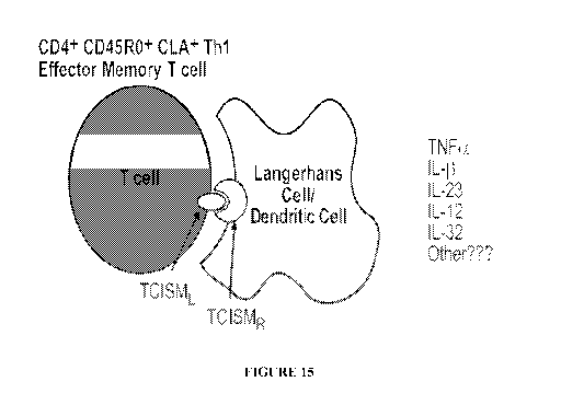
[0032] Figure 15 is a schematic illustration of one possible mechanism of p.
[0033] Figure 16 is a graph showing assessment of different small molecule
TNFa
inhibitors in Murine Collagen-Induced Arthritis in mice.
CA 02689124 2009-11-30
WO 2008/151307 PCT/US2008/065992
[0034] Figure 17 is a graph showing efficacy of anti-TNFa and IL- Ira treated
mice.
[0035] Figure 18 is a slide of joint histopathology of representative mice.
[0036] Figure 19 is Table 1 showing a list TCISM-ligands.
DETAILED DESCRIPTION OF THE INVENTION
[0037] It is believed that one of the mechanisms of activation for T,-induced
M~
activation is via direct cell-cell contact through an immune synapse
mechanism. Activated
T-cells express numerous known and unknown membrane-bound proteins. The
present
inventors have discovered that some of these molecules, which are referred
herein as T,
Cytokine Inducing Surface Molecules (i.e., TCISM or TCISM-ligand), are
involved in cell-
cell contact signaling cascades leading to proinflammatory cytokine induction
by selectively
binding to a corresponding TCISM-receptor that is present in M~.
[0038] A wide variety of clinical conditions are mediated by acute or chronic
inflammation including, but not limited to, Rheumatoid Arthritis, Multiple
Sclerosis, Crohn's
Disease, Psoriasis, Psoriatic Arthritis, Graves Disease, Autoimmune
Polyendocrine
Syndromes, Hereditary Proteinuria Syndrome, Type I Diabetes, Systemic Lupus
Erythematosus, Primary Bilary Cirrhosis, Autoimmune Thyroiditis, Hepatitis,
Acquired
Immunodeficiency Disease (HIV), Graft versus Host Disease, Allograft Disease,
Asthma,
Cutaneous T-Cell Lymphoma, HTLV-I-Associated Cutaneous T-Cell Lymphoma, HTLV-
II-
Associated Lymphoma, Hairy Cell Leukemia, Idiopathic CD4+ T-Lymphocytopenia,
and/or
Melanoma. The present inventors have also found that these clinical conditions
can be
treated by modulating cytokine production in monocyte lineage-derived
macrophages by
administering a cytokine modulator that can selectively bind to a TCISM-ligand
of T
lymphocytes and/or the corresponding TCISM-receptor of monocyte lineage-
derived cells or
by inhibiting the transcription of TCISM-ligands, e.g., by iRNA or siRNA.
[0039] Some aspects of the invention provide TCISM-ligand and methods for
modulating cytokine production in monocyte lineage-derived cells of a subject
by
administering a cytokine modulator that selectively binds to a T-cell cytokine-
inducing
surface molecule (TCISM)-ligand of T lymphocytes or the corresponding TCISM-
receptor of
monocyte lineage-derived cells or by inhibiting the transcription of a TCISM-
ligand.
6
CA 02689124 2009-11-30
WO 2008/151307 PCT/US2008/065992
[0040] In some embodiments, TCISM-ligand comprises at least one of the TCISM-
ligands listed in Table 1 (Figure 19). The corresponding TCISM-receptor(s) for
these
TCISM-ligands can be readily determined by one skilled in the art. For
example, by the use
of neutralizing monoclonal or polyclonal antibodies to inhibit TCISM-ligand to
TCISM
receptor interactions using the cell-cell contact bioassay. Another method is
Subtractive
Immunization using stimulated and non-stimulated Hut-78 and H9 subclone T cell
membranes to identify monoclonal antibodies that block cell-cell contact.
Without being
bound by any theory, it is believed that the contact-mediated activation of
monocyte-
macrophages is a major pathway inducing cytokine production. Accordingly, the
modulation
of this mechanism, e.g., the blockade of IL-1 and TNF-a production at the
triggering level or
the inhibition of expression of a TCISM-ligand or TCISM-receptor (e.g., via
siRNA), can be
used to treat clinical conditions mediated by cytokine production.
[0041] Some compositions and methods of the invention are useful in
selectively
binding a TCISM-receptor that is present in monocyte lineage-derived cells (or
by inhibiting
expression or transcription of such a receptor), thereby modulating cytokine
production in
these monocyte lineage-derived cells. Monocyte lineage-derived cells include
any cells that
when activated by T lymphocytes produce a cytokine. In some embodiments,
compositions
and methods of the invention modulate proinflammatory cytokine production.
[0042] Exemplary monocyte lineage-derived cells that produce a cytokine
include,
but are not limited to, monocyte lineage-derived macrophages, antigen-
presenting cells
(APC), dendritic cells, Langerhans cells, and Kuppfler Cells.
[0043] Compositions and methods of the invention include molecules that can
selectively bind to a TCISM-ligand or TCISM-receptor. In addition,
compositions and
methods of the invention also include molecules that can modulate translation,
transcription,
and/or expression of TCISM-ligand or TCISM-receptor. For example, siRNAs can
be
administered to T lymphocytes to modulate expression of TCISM-ligands. By
knowing
appropriate TCISM-ligands, one skilled in the art can readily identify
appropriate siRNAs
that can modulate the expression of TCISM-ligand. For example, siRNA's to the
messenger
RNA coding for a full-length protein can be designed with commercially
available computer
7
CA 02689124 2009-11-30
WO 2008/151307 PCT/US2008/065992
software which allows one to determine the sections of mRNA most susceptible
to
destabilization during transcription.
[0044] Controlling adaptive (acquired) immunity at the level of TCISM is
advantageous since therapeutic intervention allows for innate (natural)
immunity during
bacterial skin infections. Accordingly, some aspects of the invention provide
compositions
and methods for modulating adaptive or acquired immunity while substantially
maintaining
innate immunity.
[0045] Exemplary TCISM-ligand, TCISM-receptor and/or cytokine modulator
compounds of the invention include, but are not limited to, compounds having
the following
formula, analogs and derivatives thereof:
l
1 id
A (p38) B (p38) C (p38) D (p38)
41
E (p38) F (PKC) G (PKC)
O NH
IN ONH,
HN \f/
H (PKC) I (PKC)
[0046] A highly-reproducible and validated cell-cell contact bioassay was
established
using either primary human T-cells and autologous freshly obtained human blood
monocytes,
or human T-cell and monocyte cell lines. Figure 1 is a graph showing mitogen-
stimulated
8
CA 02689124 2009-11-30
WO 2008/151307 PCT/US2008/065992
human T-cells can induce monocytic THP-1 M~ to secrete proinflammatory
cytokines
through cell-cell contact. Figure 1 is a time course measurement of cytokines
at 24h and 48h
of cell-cell contact. TNF-a (left panel) and IL-1 (3 (right panel) production
in THP-1 M~
were incubated with different PMA/PHA stimulated human T-cell lines. Cells
from the
monocytic line THP-1 were incubated with highly-purified membrane preparations
from
stimulated ("s") or non-stimulated (ns) resting T cells; cytokine production
was measured
24h or 48 hours after incubation. A significant increase in TNF-a and IL-1(3
production was
detected from THP-1 M~ incubated with sHut-78 and sH9 T-cells at both time
periods.
Means +/- standard deviation with triplicate measurements are provided. As
shown in Figure
1, only a small induction of TNF-a and IL-1 (3 production was observed in
PHA/PMA
stimulated Molt4, Jurkat and Raji cells, while no detectable production was
observed in
resting primary human T-cells, unstimulated Hut-78, H9, Molt4, Jurkat or Raji
cells. It was
also observed that the PMA/PHA-stimulated H9 cells, and to a lesser extent,
the Hut-78 cells,
induced the greatest THP-1 M~ secretion of both TNF-a and IL-1(3, indicating
that these
cells are TCISML positive.
[0047] PMA/PHA stimulus provided a significant induction of TCISML on the
human T cells since elevated levels of TNF-a were produced in culture over the
24 h culture
period. See Figure 2. In Figure 2, Pan CD3+ T cells were isolated from healthy
human
donor blood, stimulated for 6 h at 37 C, washed, and fixed with 1%
paraformaldehyde (6 h
RT). Cells were then rinsed with PBS, and kept overnight at RT. PBMC-derived
CD14+
M~ were then added and incubated at 37 C 24 h. Cell culture supernatants were
centrifuged, filter sterilized and measured for cytokines by ELISA. Means
SEM are shown
(N=6 individual donors with triplicate measurements; P<0.05 in comparison to
non-
stimulated T cells). Comparable data were observed with T cells stimulated in
vitro with
aCD3/aCD28. Nearly twice as much TNF-a was produced in the T cell and M~ co-
culture
system in comparison to the whole T cell control cultures alone.
[0048] As shown in Figure 3, the results indicate that cytokine mixture #1
(IL2 + IL6
+ TNF-a) or #2 (IL 15 + IL6 + TNF-a) activated human T-cells, when mixed at a
ratio of 1:8
(blood monocyte:T-cells), can activate human monocytes to produce elevated
levels (100-
250 pg/ml) of TNF-a and IL-1(3. These cytokine levels were significantly
higher than the
9
CA 02689124 2009-11-30
WO 2008/151307 PCT/US2008/065992
levels of proinflammatory cytokines released by cytokine-activated, fixed T-
cells alone. In
Figure 3, Pan CD3+ T cells were isolated from healthy human donors and
incubated with
different cytokine cocktails (^ IL2 + IL6 + TNF-a; 1,4IL-15 +IL-6+ TNF-a; ^
unstimulated T cells) for 8 d at 37 C, washed, and then fixed with fresh 1%
paraformaldehyde (6h RT). Cells were then rinsed with PBS, and kept overnight
at RT.
Freshly obtained human blood monocytes were subsequently added and incubated
with the
T-cells (37 C for 24h). Cell culture supernatants were collected and measured
for cytokines
by ELISA. Means SEM are shown (N=4 individual donors with triplicate
measures). Two
separate experiments with purified Pan CD3+ T-cells from identical donors were
conducted
to confirm these findings.
[0049] Whether human PBMC-derived CD3+ T-cells stimulated with either cytokine
mixture #1 or #2 induces the expression of TCISM sufficient to activate human
THP-1 cells
to produce TNF-a and IL-11 was also tested. As shown in Figure 4, THP-1 cells
combined
with cytokine activated T-cells resulted in the production of elevated levels
of TNF-a,
however, the induced cytokine levels were not significantly higher than those
induced by the
paraformaldehyde-fixed, cytokine stimulated T-cells alone. In Figure 4, Pan
CD3+ T-cells
were isolated from healthy human donors and incubated with different cytokine
cocktails (^
IL2 + IL6 + TNF-a; IL-15 +IL-6+ TNF-a; ^ unstimulated T cells) for 8 d at 37
C,
washed, and then fixed with fresh 4% paraformaldehyde (6h RT). Cells were
subsequently
rinsed with PBS, kept overnight at RT, and then added to THP-1 cells (37 C
for 24h). Cell
culture supernatants were collected as above and measured for cytokines by
ELISA. Means
SEM are shown.
[0050] As shown in Figure 5, Pan CD3+ human T-cells obtained from the
peripheral
blood of healthy human volunteers were stimulated in vitro with either PMA/PHA
or
aCD3/aCD28 for 6h, then fixed with fresh I% paraformaldehyde overnight at room
temperature. Next, freshly obtained human blood monocytes were added to the
tissue culture
plates and incubated at 37 C with the fixed T-cells for either 6h or 24h of
culture.
Supernatants were then collected as described above and measured for various
Thl and Th2
cytokines by ELISA. The PMA/PHA stimulated T-cells were potent in activating
human
blood monocytes to produce IL-12(p70) at levels ranging between 1000 -
1250pg/ml,
CA 02689124 2009-11-30
WO 2008/151307 PCT/US2008/065992
especially after 6h of cell-cell contact (Figure 4). Similarly, aCD3/aCD28-
stimulated T
cells were also effective in activating human blood monocyte IL-12(p70)
release, although to
a lesser degree. Finally, both types of stimulated human T-cells were able to
activate human
blood monocytes to produce IL-1(3 and TNF-a at these two different time
periods.
[0051] Ability of PMA/PHA and aCD3/aCD28 stimulated Hut-78 T cells versus
PMA/PHA and aCD3/aCD28 stimulated Hut-78 T-cell membranes to activate THP-1
cells
to produce TNF-a was compared (see Figure 6). The PMA/PHA stimulated Hut-78 T-
cell
data is shown for illustrative purposes. These studies were conducted with the
same Hut-78
cell culture lot to reduce variability with the bioassay. Results indicate
that stimulated Hut-
78 T-cell purified membranes were more effective in inducing in vitro THP-1
cell TNF-a
production in comparison to the stimulated Hut-78 fixed with 1%
paraformaldehyde. Mix
and match "add back" experiments were also conducted to demonstrate that TCISM
was
predominantly present in the purified membrane fractions and not the Parbomb
cell
supernatant (e.g., cytosolic) fractions during the low and high centrifugation
steps (see Figure
6). In Figure 6, membranes were prepared as described above and combined with
THP-1
cells in the cell-cell contact bioassay. After 24h culture, supernatants were
removed,
centrifuged, filter-sterilized, and measured for cytokines by ELISA. Means +/-
SD are
shown.
[0052] A characteristic array profiling "heat map" is shown in Figure 7, which
is a
microarray 2-dimensional (2D) cluster of genes encoding membrane or membrane
associated
proteins in PHA/PMA stimulated CD3+ T Cells, Hut-78, H9, Molt4, Jurkat & Raji
Cells. At
least four experiments with fold change >2 & P value <0.01 were conducted. In
Figure 7,
red represents gene expression levels >2.0 (i.e.; above array profiling
background levels), and
green represents gene expression levels <2.0 (i.e.; below array profiling
background levels).
During the computational assessment stage of data review, only those genes
that were human
T-cell membrane associated and which were up-regulated in the TCISM (+) T-cell
lines and
down-regulated in the TCISM (-) T-cell lines were considered. Approximately
10,000 out of
50,000 genes resulting from microarray experiments were examined, where the
focus
centered on T-cell genes which were up regulated in Hut-78 and H9 cells but
not up-
regulated in human Molt-4 and Jurkat T-cells.
11
CA 02689124 2009-11-30
WO 2008/151307 PCT/US2008/065992
[0053] From this curated list of about 100 potential human T-cell TCISMs
candidates, log/log intensity plots were generated which were graphed on a
linear scale to
identify TCISM candidates. These plots (see, for example, Figure 8), describe
"unchanged",
"signature", "down-regulated", and "up-regulated" T-cell gene products, as
well as the
relative expression levels of the gene products compared to overall array
profiling results.
Five candidate human T-cell TCISM genes were identified: Diphtheria Toxin
Receptor, or
Heparin-Binding EGF (DTR, EGF module-containing Mucin-like hormone receptor 2
(EMR2), Adamlysin-17 (ADAM or A Disintegrin and Metalloprotease) TNFa-
converting
enzyme (TACE, TNF receptor Superfamily, member 9 (TNFRSF9 or LIGHT), and, for
cell-
cell contact positive control purposes driven by review of the published
scientific literature,
TNFRSF5, or CD40 Ligand (CD40L).
[0054] Real-time qRT-PCR was performed in order to confirm TCISM candidate
gene expression in PMA/PHA-stimulated Hut-78 and H9 subclone T-cells (see
Figure 9).
The TCISM candidates DTR and LIGHT followed preset criteria in that both genes
were
highly expressed in stimulated Hut-78 and H9 T cells, but were not up-
regulated in the Molt-
4 and Jurkat T cells or the Raji B-cells. However, both TACE and EMR2 were
highly up-
regulated in the TCISM (-) human Raji B-cell line. FACS was also used to
validate the
presence of these molecules on activated H9 cells (see Figure 10).
[0055] About 50 different commercially available neutralizing monoclonal
antibodies
to known human T-cell surface proteins were tested in the cell-cell contact
bioassay
(including mAb's directed to ALCAM, CD6, (32-integrins, CD69, CD23, CD40-CD40L
and
LAG-3). It was found that they do not appreciably inhibit more than about 30%
of the
proinflammatory cytokine production in this system. One of the more effective
polyclonal
antibody preparations observed was anti-ADAM-17 (TACE). These experiments were
conducted with available polyclonal antibodies. Use of highly-specific anti-
CD40L mAb's
in bioassay did not significantly block TNF-a or IL-1(3 cytokine production
(see Figure 11).
In Figure 11, Hut-78 cells were stimulated with PMA/PHA for 6h, at which time
purified
membranes were prepared as described earlier. Hut-78 membranes plus the
concentration of
anti-human mAb's shown were added to co-culture wells and allowed to incubate
at 37 C
for 2h. Recently passed resident THP-1 cells were then added to co-culture
wells, and kept at
12
CA 02689124 2009-11-30
WO 2008/151307 PCT/US2008/065992
37 C for 24h. Supernatants were collected for TNF-a and IL-1(3 ELISA. Figure
11 shows
two separate experiments with triplicate cytokine measurements at each mAb
concentration.
[0056] FACS analysis showed that transfected 293 cells (Figure 12)
consistently
expressed elevated levels of the TCISML gene product on their cell surface. In
Figure 12,
pcDNA5/FRT/DTR, EMR2-07 (isoform containing EGF-like domain 1, 2 & 5), or
CD40L
were co-transfected with p0G44 into Flp-In 293 cells and Jurkat cells,
respectively, and
colonies were selected in 500 mg/ml Hygromycin. Cells were detached with cell
disassociation buffer and analyzed by FACS. (Anti CD97 was cross-reacted to
EMR2-07 and
was used for detecting EMR2-07 expression.) Expression was stable after
several cell-
culture passages, indicating their suitability for use in the cell-cell
contact assay. Initial
experiments were conducted using the transfected 293 cells in the cell-cell
contact assay (see
Figure 13). The DTR and CD40L constructs are potent in augmenting sHut-78m
driven
THP-1 induction of TNF-a /IL-1(3 (Figure 13). The addition of exogenous IFN-y
to the
assay resulted in enhanced levels of CD40L-induced M~ activation and
subsequent TNF-a
release (Figure 13 left panel), but not IL-1 (3 release (Figure 13 right
panel). These effects
with the CD40L transfected 293 or Jurkat cells were highly reproducible.
[0057] Human T-cell TCISMs were identified in both healthy human T-cells and T-
cells obtained from patients with active Ps and PsA. As can be seen, the in
vitro cell-cell
contact bioassay is a highly reproducible human cytokine "readout system" to
identify
immunological synapse mechanisms between human T-cells and human M~ in co-
culture. It
was observed that the PMA/PHA-stimulated H9 cells and the Hut-78 cells induced
THP-1
secretion of both TNF-a and IL-1 (3 (indicating that these cells are TCISM
positive), while
PHA/PMA stimulated Molt4, Jurkat, Raji cells and resting primary human T-
cells,
unstimulated Hut-78, H9, Molt4, Jurkat or Raji cells are TCISM negative
(Figure 1).
[0058] TCISM candidates were determined using the following criteria: (A)
membrane-associated; (B) up-regulated by more than 2-fold in stimulated Hut-78
and H9
cells, but not in Molt4, Jurkat and Raji cells; and (C) high expression levels
must be
confirmed by qRT- PCR and FACS.
13
CA 02689124 2009-11-30
WO 2008/151307 PCT/US2008/065992
[0059] Primary T cells stimulated in vitro with aCD3/aCD28 or human cytokine
cocktails also augment proinflammatory cytokine (PIC) activity in the assay.
PMA/PHA or
aCD3/aCD28 stimulated T cells augmented M~ activation to produce IL-12. IL-12
has been
detected in psoriasis lesions. It is believed that IL-12 mainly stimulates IFN-
y production in
naive Th cells and would play a role in the expansion and stabilization of the
Thl response.
[0060] Microarray analysis was conducted from PBMC-derived T-cells from normal
healthy donors versus psoriasis patients to identify the human T-cell TCISML
molecule and
its signaling pathways through human M~ TCISMR that lead to inflammation and
cutaneous
skin diseases. Microarray analysis using human T cells from healthy donors,
PsA, and CPPs
patients have identified TCISM molecules, including Diphtheria toxin receptor
(DTR; HB-
EGF) and mucin-like Epidermal Growth Factor family members (EMR4; CD97). Up-
regulation of numerous TNF family members including 4-1BB (TNFSF14), OX-40,
LIGHT
(TNFSF9) and CD40L (CD154; TNFSF5) was also observed.
[0061] About 50 different commercially available neutralizing monoclonal
antibodies
were used to validate the human TCISM candidates by cell-cell contact assay.
In many cases,
it was found that these reagents inhibit no more than 30% of the
proinflammatory cytokine
production over a 24-96 hour time period in this system (see Fig 11). A "Flip-
in"
transfection system method was used to identify five initial candidate TCISML
gene
candidates from the microarray experiments, including: EMR2, DTR, 4-1BB,
LIGHT, and
CD40L. Full-length cDNA's of TCISM candidate genes transfected into TCISM
negative
293 and Jurkat cells, characterized in the cell-cell bioassay, showed that DTR
and CD40L
were potent in augmenting T cell-driven M~ TNF-a/IL-1(3. The addition of
exogenous IFN-
y to the assay resulted in enhanced levels of CD40L-induced M~ activation and
subsequent
TNF-a release, but not IL-1 (3 release (Figures 12a-b and 13).
[0062] Some of the identified human M~ TCISMRs (TCISM-receptors) that leads to
inflammation and cutaneous skin diseases include DTR, CD97, 4-1BB , OX-40,
LIGHT and
CD40L. Full-length cDNA's of TCISM candidate genes transfected into TCISM
negative
293 and Jurkat cells, characterized in the cell-cell bioassay, showed that DTR
and CD40L are
potent in augmenting T cell-driven M~ production of TNFa/IL-1(3.
14
CA 02689124 2009-11-30
WO 2008/151307 PCT/US2008/065992
Psoriasis
[0063] One particular aspect of the invention provides compositions and
methods for
treating psoriasis. Psoriasis (Ps) is a chronic skin disorder that affects
approximately 2% of
the US population. Without being bound by any theory, and as schematically
illustrated in
Figure 15, it is believed that the pathophysiology of Ps involves epidermal
proliferation and
differentiation, angiogenesis and hyperproliferation of keratinocytes and
infiltration of
activated T-cells (Ta), macrophages (Mt), dendritic cells (DC), Langerhans
cells (LC) and
neutrophils (PMN) into lesional skin. Proinflammatory cytokines (PIC),
including Tumor
Necrosis Factor-a (TNFa), Interleukin-11 (IL-1(3), and Interleukin-32 (IL-32),
produced in
the active lesion, are believed to induce and maintain chronic skin
inflammation in diseases
such as psoriatic arthritis (PsA) and Ps. Up-regulation of cytokines by
activating M~ is
believed to be responsible for the pathogenesis of these disorders. It has
been suggested that
T-cells play a key role in M~ activation; however, T, cytokines, such as
Interleukins-4, 10,
and 13 (IL-4, IL-10 and IL-13), have been shown to either play an anti-
inflammatory role or
only weakly induce TNFa/IL-11 up-regulation. It is believed that the mechanism
of
activation for T,-induced M~ activation is via direct cell-cell contact
through an immune
synapse mechanism in the skin.
[0064] An immunological synapse (IS) is formed at the interface between
antigen-
presenting cells (APCs) and T-cells, and is believed to be the structure
responsible for
antigen recognition and T-cell activation. The IS was initially found between
T-cells and B-
cells, or between T-cells and MHC-containing planar bilayers. It is believed
to be formed by
the accumulation of the T-cell receptor-major histocompatibility complex (TCR-
MHC) in
the IS central region, termed the central supramolecular activation cluster (c-
SMAC), and the
accumulation of leukocyte function-associated antigen 1 (LFA-1)-intercellular
adhesion
molecule-1 (ICAM-1) in external IS regions, termed the peripheral (p-) SMAC
(pSMAC).
The mature IS has been shown to contain a pSMAC that is enriched with LFA-1,
talin, VLA-
4, ADAP and transferring receptor. The pSMAC surrounds the cSMAC, which is
enriched
with the TCR, CD4 or CD8 co-receptors, CD28 co-stimulatory molecules, CD2,
PKCO, etc.
[0065] Ps skin is characterized by the hyperproliferation of keratinocytes,
resulting in
an exaggerated pattern of ridges and pegs. Keratinocytes, DC, and M~ in skin
have all been
CA 02689124 2009-11-30
WO 2008/151307 PCT/US2008/065992
shown to produce TNFa, IL-1(3, and IL-32. While the IS controls psoriatic
autoantigen-
specific cutaneous lymphocyte antigen (CLA)-positive T-cell activation, the
key molecular
components include the TCR and, surrounding it, a ring of adhesion molecules,
such as LFA-
1, which can bind to ICAM-1 expressed by the adjacent cell, e.g., a
keratinocyte or APC.
The IS is therefore a logical target for therapeutic approaches. Alefacept,
for example, is a
recombinant fusion protein that binds to CD2 on memory-effector T-cells,
inhibiting their
activation and reducing the number of these cells. Efalizumab is a humanized
monoclonal
antibody (Mab) against CD 11 a molecule. CD 11 a and CD 18 comprise subunits
of LFA- 1.
[0066] The present inventors have shown that there exist TCISMs on the surface
of
human T-cells which mediate skin inflammation by driving M~ activation to
produce
proinflammatory cytokines. Controlling adaptive (acquired) immunity at the
level of TCISM
is advantageous since therapeutic intervention allows for innate immunity
during bacterial
skin infections.
[0067] Psoriasis is a hereditary disorder of the skin with several clinical
expressions.
The most frequent type is psoriasis vulgaris (or Plaque Psoriasis [Ps]), which
occurs as
chronic, recurring, scaling papules and plaques in characteristic sites on the
body. Current
therapies for psoriasis are not satisfactory. Ps is characterized by the
infiltration of the skin
by activated T-cells and an abnormal proliferation of keratinocytes. As a
result of
overproduction by T-cells, keratinocytes, DC, and LC, it has been reported
that the
concentrations of TNFa are higher in Ps lesions than in uninvolved skin (in
both patients with
Ps and normal persons).
Autoimmune Diseases
[0068] Autoimmune diseases in humans, such as Ps and Psoriatic Arthritis
(PsA), are
chronic syndromes characterized by typical, often relapsing clinical symptoms
combined
with diagnostic results of adaptive Immoral (autoantibodies) or cellular
(autoreactive T-cells)
responses directed against autoantigen-expressing tissues. Important human
autoimmune
diseases often are co-morbid with, or are triggered by, viral or bacterial
infections and are
associated with certain MHC alleles. The innate immune system encompasses a
collection of
host defenses that range from non-specific barrier function of epithelia to
the highly selective
recognition of pathogens through the use of germline-encoded receptors. A
common feature
16
CA 02689124 2009-11-30
WO 2008/151307 PCT/US2008/065992
of these diverse elements is a rapid and blunt response to infection or tissue
destruction. On
the other hand, the adaptive immune system uses somatically rearranged antigen
receptor
genes to create receptors for virtually any antigen. The adaptive immune
response is slower
but more flexible and is able to combat infections that have evolved to evade
innate
responses.
[0069] The innate immune system responds by recognition of conserved motifs in
pathogens as well as a number of other indictors of cell stress or death. The
cellular
components of the innate immune system includes DC, monocytes, M~,
granulocytes and
natural killer T-cells (NKT), as well as the skin, pulmonary, and gut
epithelial cells that form
the interface between an organism and its environment. The non-cellular
elements of the
innate system are very diverse, and range from the simple barrier function of
the stratum
corneum to complex pathways such as the complement cascade. These elements
prevent
entry of pathogens through physical blockade, or, once cells are invaded,
allow them to
destroy pathogens directly or via phagocytic cells. The innate immune system
has also
evolved to recognize molecular patterns common to many classes of pathogens.
These are
termed pathogen-associated molecular patterns (PAMPs). PAMP recognition is
through
using a group of germ line-coded, evolutionary conserved pathogen-recognition
receptors
(PRR). The Toll-like receptors (TLR) are a very important group of pathogen
receptors, and
they are expressed on both innate immune cells and on cells in various
tissues, including
endothelial cells, epithelial cells, and fibroblasts. Ten TLR family members
specific for
various microbial molecules have been identified in humans. Binding of TLR to
their
microbial ligands leads to activation of phagocytes, as well as to the release
of
proinflammatory cytokines and anti-microbial peptides. These molecules are
believed to also
activate DC to initiate adaptive immune responses.
[0070] T-cells are important in immune response and can be divided into a
number of
distinctive subsets based on their migration patterns and functional
abilities. Naive T cells
recirculate primarily between the blood and lymph nodes, a pattern aided by
their expression
of the homing receptors L-selectin and CCR7. Naive T-cells are maintained in a
pluripotent
state and have a relatively quiescent effector program as they recirculate
from blood through
lymphoid organs, surveying DC for activating MHC-peptide complexes. Through
complex
17
CA 02689124 2009-11-30
WO 2008/151307 PCT/US2008/065992
mechanisms that integrate signals from activated DC and from the cytokine
milieu, naive T-
cells are driven through rapid rounds of division that are linked intimately
with the ability to
secrete effector cytokines necessary to confront distinct groups of pathogens.
CD4+ T helper
cells can be functionally divided into Thl (interferon [IFN] r - secreting)
and Th2
(interleukin [IL] - 4-secreting) subsets, as well as recently identified
additional Th subsets
which include Trl (IL-10-secreting), Th3 (transforming growth factor [TGF]13-
producing),
ThFH (follicular helper cells), peripherally - induced T regulatory (Treg;
FoxP3 - positive)
and Th17 (IL-17A - producing) cells. The discovery of additional subsets will
undoubtedly
fuel interest in identification of underlying regulatory transcription factors
that are likely to
be implicated in mechanisms that modify the signature cytokine genes involved
in effector
function.
[0071] An immunological synapse (IS) is formed at the interface between
antigen-
presenting cells and T-cells, and is believed to be the structure responsible
for antigen
recognition and T-cell activation. The IS was originally found between T-cells
and B-cells,
or between T-cells and MHC-containing planar bilayers. It is formed by the
accumulation of
T-cell receptor-major histocompatibility complex (TCR-MHC) in the central IS
region,
termed the central supramolecular activation cluster (c-SMAC), and the
accumulation of
leukocyte function-associated antigen 1 (LFA-1)-intercellular adhesion
molecule-1 (ICAM-
1) in external regions, called the peripheral (p-) SMAC (pSMAC). The mature
synapse
contains a pSMAC that is enriched with LFA-1, talin, VLA-4, ADAP and
transferring
receptor. The pSMAC surrounds the cSMAC, which is enriched with the TCR, CD4
or CD8
co-receptors, CD28 co-stimulatory molecules, CD2, PKCO, etc.
[0072] Ligands expressed on the surface of the APC are believed to recruit
specific
receptors to the IS contact site. The recruitment of co-stimulatory molecules
CD28 and
cytotoxic T lymphocyte antigen 4 (CTLA4) to the synapse is differentially
promoted by the
expression of their ligands, B7-1 and B7-2, on the APC. Although CD28 and
CTLA4 bind
either of these ligands, when expressed on the APC, B7-2 recruited CD28 and B7-
1 recruited
CTLA4 to the synapse. Stability of the ligand in the contact site on the APC
is also
important, as the recruitment of CD28, CTLA-4 and protein kinase C-O require
the presence
of the cytoplasmic domain of B7-1.
18
CA 02689124 2009-11-30
WO 2008/151307 PCT/US2008/065992
[0073] Psoriatic skin is characterized by the hyperproliferation of
keratinocytes,
resulting in an exaggerated pattern of ridges and pegs. Keratinocytes, DC, and
Mq in skin can
all produce TNFa. While the IS controls psoriatic autoantigen-specific
cutaneous lymphocyte
antigen (CLA) - positive T-cell activation, some of iJ Lcy olecu lar
components include
the TCR and, surrotandinw; it, a rind; of adhesion molecules, such as LFA -1,
,w"hich can kind to
ICAM_-.l c pressed by the taa~~,~ee it ce'l le .'('.. a keratMocy to or A-PC,
The 1,FA-1 conaponc.nt
of the synapse is believed to be important in pso iasi:s, as a t erapeutic
agent t anti---LFA--1
ra:_a 1~ody, e ralizunmab) blocking t _iis adhesive hiteract.ion has been
approved by the US Food
and Drug Administration (FDA) for the treatment of psoriasis. Additional cont~
ha.ato y
nnoleculcs i azla {lag other ; dhesion nmoiecules and" zo 3tiiana l,.tizaa_yr
anoleca l s also influence
' -cell respon'sivene'ss' e.g., the ell surface mo ecular pars CD2:LFA-3 and
CD'.-XCDSO CD86,
Rheumatoid Arthritis
[0074] Rheumatoid Arthritis (RA) is an inflammatory disease also related to IS
signaling. One of the potential approaches for the treatment of RA involves
the inhibition of
molecules present at the IS between T-cells and antigen-presenting cells. It
is believed that
the mechanism of cytokine up-regulation is contact-dependent. There are
multiple candidate
proteins on the cell surface that can mediate these functions.
[0075] It has been shown that T-cells activated through the T-cell receptor
complex
induce monocyte IL-10 synthesis. This is partially dependent on endogenous
TNFa and IL-1
levels, and T-cell membrane TNFa has been shown to be an important contact-
mediated
signal. However, IL-10 synthesis still occurs when TNFa and IL-1 are
neutralized, thus
indicating that there are TNF/IL-1-independent signals required for IL- 10
synthesis.
[0076] Of particular interest are members of the TNF/TNF-R family, which
include
CD40, CD27, CD30, OX-40, and LT(3. The ligands of these TNF-R molecules are
believed to
be upregulated upon T cell activation and, in addition, CD40L, 4-1BB, CD27L,
CD30 are
believed to be released as soluble mediators after activation. The interaction
between CD40L
and CD40 has been observed to be of importance for inducing both IL-1 and IL-
12 synthesis
following T-cell interaction with monocytes, and more recently, to mediate IL-
10 production
by human microglial cells upon interaction with anti-CD3-stimulated T cells.
19
CA 02689124 2009-11-30
WO 2008/151307 PCT/US2008/065992
T-cell Immunoglobulin Mucin Proteins
[0077] The T-cell immunoglobulin mucin (TIM) proteins are type I membrane
glycoproteins expressed on T-cells that contain common structural motifs. The
TIM gene
family is located on chromosome 11 in mice and 5q33 in humans. Genomic
analysis has
identified eight family members in mice (TIM-1 to TIM-8) and three in humans
(TIM-1,
TIM-3 and TIM-4). All members share a characteristic structure containing IgV,
mucin,
transmembrane, and cytoplasmic domains. This gene family plays a role in the
regulation of
immune responses.
[0078] TIM-1, previously identified as the hepatitis A virus receptor, co-
stimulates T-
cell expansion and cytokine production. TIM-1 is expressed on all activated T
cells and, upon
CD4+ T-cell polarization, at a higher level on Th2 than on Thl cells. An
agonistic
monoclonal anti-TIM-1 antibody (3B3) was shown to costimulate T-cells in vitro
when
cultured with either peptide and APCs or cross linking antibodies against CD3
and CD28.
When administered in vivo during an immune response, anti-TIM-1 antibody
augmented T-
cell proliferation in vitro, even in the absence of antigenic re-stimulation.
It also increased
the production of both Thl and Th2 prototypic cytokines compared with control
treatments.
In addition, anti-TIM-1 antibody abrogated the induction of high-dose
tolerance and could
also restore AHR when mice were immunized and challenged with antigen intra-
nasally.
TIM-1 is therefore surmised to act as a co-stimulatory molecule for all T-
cells, with possibly
stronger effects on Th2 than Thl cells.
[0079] It has been demonstrated that TIM-3 is preferentially expressed on in
vitro
polarized human CD4+ Thl cells as compared with Th2 cells. Thus, TIM-3
expression can
be used to identify human Thl cells. In addition, TIM-3 is believed to
contribute to
regulation of Thl cells in vivo. For example, administration of TIM-3-specific
antibody to
mice in an experimental autoimmune encephalitis (EAE) model resulted in the
acceleration
of a Thl-driven progression of EAE. Additionally, anti-TIM-3 antibodies
induced M~
activation and clonal T-cell expansion, for which a cognate interaction
between Mq and T-
cell was required. These data appear to show a role for TIM-3 in negatively
regulating the
activation of M~ by T-cells. TIM-3/TIM-3 ligand interactions also play a role
in tolerance.
Treatment of mice with both full-length and soluble TIM-3 Ig fusion proteins
abrogates
CA 02689124 2009-11-30
WO 2008/151307 PCT/US2008/065992
tolerance induced using high-dose aqueous antigen. Similarly, TIM-3-deficient
mice cannot
be tolerized. Indeed, both Ig fusion protein-treated mice and TIM-3-deficient
mice exhibit
increased T-cell proliferation and production of IL-2 after administration of
high-dose
aqueous antigen relative to controls.
[0080] TIM-4 is believed to be a natural ligand for TIM-1. Unlike the other
TIM
molecules, TIM-4 does not appeared to be expressed in T cells but is instead
appears to be
expressed in APCs, particularly in mature lymphoid DCs. A positively
regulating TIM-like
family molecule which mediates Thl T-cell-driven M~ activation has not been
discovered to
date. The present inventors have shown that neither TIM-1 nor TIM-3 is up
regulated in
PMA/ionomycin-activated H9 or primary human CD3+ T cells.
Proteomic Approaches to Identify Cytoplasmic, Membrane, and Nuclear proteins
involved in the Immunological Synapse
[0081] Proteomics technologies can be used to characterize biomarkers and
biosignatures of disease and to reveal information regarding functional
subproteomes and
networks. Although many proteomics applications provide general information
about
subsystems that change in response to disease, insult or drugs, proteomics can
also be used to
identify previously uncharacterized proteins involved in biochemical responses
such as
MAPK signaling, chemotaxis, melanoma oncogenesis and metastasis, and MHC Class
II-
induced cell death, etc. The combination of two-dimensional gel
electrophoresis (2DGE) and
mass spectrometry is one of the analytical techniques used for proteomics
applications. The
quantitative capability of 2DGE, coupled with the direct and unbiased
identification of
proteins via tandem mass spectrometry and advanced database searching
algorithms,
provides an excellent technical platform with which to address the profiling
of protein
expression changes in cell system, plasma, skin, etc. A complementary
discovery platform is
multi-dimensional chromatographic protein and peptide separations followed by
tandem
mass spectrometry and database searching for protein identification. Selected
reaction
monitoring is subsequently used to quantify relevant molecules.
Host Defense and Inflammatory Disease
[0082] The proinflammatory, pleotrophic cytokines TNFa and IL-1 (3 play key
roles in
host defense and inflammatory disease processes. TNFa and IL-1(3
overexpression has been
21
CA 02689124 2009-11-30
WO 2008/151307 PCT/US2008/065992
found in Ps disease target tissue as well as the circulation of patients with
inflammatory skin
diseases. One of the major functions of T-cells and monocyte-M4 is to release
various
cytokines, including IL-1I and/or TNFa. These molecules, in turn, participate
in the
induction and release of downstream moieties such as IL-32 (produced by
keratinocytes and
Mt), eventually leading to keratinocyte hyperproliferation and the development
of Ps.
Without being bound by any theory, it is believed that blocking the production
of these
cytokines at a more distal level (e.g., at the level of T-cell driven monocyte-
M4 activation,
perhaps at different time periods during the adaptive response and/or IS
formation), can lead
to new, molecular drug discovery targets designed to specifically inhibit
these cytokines,
resulting in safer therapeutics with less adverse events for Ps patients.
[0083] Additional objects, advantages, and novel features of this invention
will
become apparent to those skilled in the art upon examination of the following
examples
thereof, which are not intended to be limiting.
EXAMPLE S
Culture of cell lines
[0084] The human cell lines were cultured in a standard medium consisting of
RPMI
1640 (Biochrom, Berlin, Germany) supplemented with 10% (v/v) FCS serum, 2 mM L-
glutamine, 100 units/ml penicillin and 100 units/ml streptomycin. Hut78, H9,
Molt4, Jurkat,
Raji and THP-1 cell lines were obtained from the ATCC.
Cell isolation
[0085] Peripheral blood mononuclear cells (PBMCs). PBMCs were isolated by
Ficoll density gradient centrifugation. The viability of obtained PBMCs was
>95%, as
determined by trypan blue staining. The viable cells were quantified in a
Neubauer chamber
(Zeiss, Oberkochen, Germany) and stored in liquid nitrogen.
CD3+ T cells
[0086] Thawed PBMCs were centrifuged again at 1,500 rpm for 5 minutes.
Supernatant was discarded and pellets were resuspended in MACS buffer. The
cells were
disrupted into a single cell suspension at a concentration of 0.8 ml of buffer
per 108 cells.
About 0.2m1 of Hapten-Antibody Cocktail per 108 cells was added. The resulting
mixture
was mixed well and incubated for 20 minutes on ice. Cells were washed by
adding 20x the
22
CA 02689124 2009-11-30
WO 2008/151307 PCT/US2008/065992
labelling volume, centrifuging and supernatant removal. Cell pellets were
resuspended in
0.8m1 of buffer per 108 cells. About 0.2m1 of MACS Anti-Hapten MicroBeads per
108 cells
was added to label the cell magnetically. The mixture was incubated for 15
minutes on ice,
washed with 20x of the volume (frozen cells from Leukophoresis packs were
passed through
a 45 gm mesh filter to remove clustered dead cells), centrifuged and
supernatant discarded.
Cells were resuspended in lml of MACS buffer per 108 cells and the LS+ column
placed in
the magnetic field of an appropriate MACS separator. The column was prepared
by washing
with 3m1 of buffer. The cell suspension was applied to the column and the
unlabeled cells
were passed through. The effluent was collected as a negative fraction,
representing the
enriched T cell fraction. The column was rinsed with 4 x 3m1 of buffer and
effluent
collected. Following cell washing, the cell pellet was resuspended in 50 ml of
tissue culture
medium at a concentration of 106 cells/ml. T cell purity (>95%) was determined
by CD3-
FITC labelling and FACS analysis.
Monocytes
[0087] Method was the same as CD3+ T cell separation.
Immunophenotyping of the cells
[0088] Harvested cells were washed in FACS medium [phosphate buffered saline
(PBS) containing 1% bovine serum albumin (BSA)] and stained at 4 C for 20 min
by
antibodies directly conjugated with Fluorescein isothiocyanate (FITC) or
phycoerythrin (PE).
Thereafter cells were washed three times with PBS and analyzed by FACScan
(Becton
Dickinson, Heidelberg, Germany) using the CellQuest software (Becton
Dickinson).
Antibodies were the following: PE-labeled anti-mouse IgG, anti-human CD40L and
CD137.
Cell-cell contact assay
[0089] Primary T cells or H9 cells were washed with cold PBS and cell pellets
were
resuspended in freshly made I% paraformaldehyde at 5 x 10⁶ cells/ml. Cells
were fixed on
ice for 2 hours, and then washed with 20 x volume of cold PBS three times.
Following the
third wash, the cells were kept in PBS at 4 C overnight to allow diffusion of
paraformaldehyde. Cells were centrifuged and washed one more time. The fixed T
cells
were resuspended in medium at 1 x 10⁷ cells/ml and dispensed into a 96 well U
bottom plate
at about 1 X10⁶ /ml THP-1 cells l00gl per well. Primary T cells or H9 cells
(100gl per well)
23
CA 02689124 2009-11-30
WO 2008/151307 PCT/US2008/065992
were added at 8x, 4x, and 2x 10⁶ cells/ml. The plates were incubated at 37 C,
in humidified
5% CO2 for 48 hours. Plates were centrifuged at 1,500 rpm for 5 minutes and
supernatant
(l20 1) transferred into a fresh plate. The supernatant was stored at -20 C
until used.
Hut-78 Plasma Membrane Preparation
[0090] About 5 x 10⁸ stimulated or unstimulated Hut-78 cells were suspended in
10ml of hypertonic buffer containing protease inhibitors (50mM Tris-Cl (pH
7.4), 25mM
KC1, 5mM MgClz, 200uM PMSF, lx complete protease inhibitor) and homogenized
using a
dounce homogenizer by 20 strokes on ice. The nucleus and unbroken cell
fraction were
discarded by centrifugation at 4000 x g at 4 C for 15min and the supernatant
was
ultracentrifugated at 28K (100,000 x g) using a SW40 rotor at 4 C for 45min.
The
membrane pellet was resuspended in 9m1 of PBS with a 22G syringe needle and
was then
added to lml of 200mM CHAPS. The homogenate was incubated on ice for lhr.
Approximately 1 ml aliquots of suspended plasma membrane at 5 x 10⁷ cell
equivalent/ml
was stored at -80 T.
RNA sample preparation and hybridization
[0091] RNA was extracted and purified using the RNeasy MinElute kit (Qiagen)
and
Qiagen Mini RNeasy kit according to the manufacturer's protocol. cDNA
synthesis was
carried out as described in the Expression Analysis Technical Manual
(Affymetrix, two-cycle
protocol) using 100 ng of total RNA for each sample. The cRNA reactions were
carried out
using the BioArray High-Yield Transcript Labeling kit (Enzo). Fifteen
micrograms of
labeled cRNA was fragmented and sequentially hybridized to the GAPS Slides
(Coming)
following the manufacturer's instructions.
Flp-In transfection
[0092] Stable TCISM ligand (TCISML)-negative T-cell lines were established and
transfected with cDNA's of TCISML candidate genes identified from microarray
experiments, including: EMR2, DTR, 4-1BB, LIGHT, and CD40L. A transfection
system
known as the "Flp-In" method utilizing Flp-In recombinase was utilized
according to the
manufacturer's protocol. Eight different constructs (pcDNA5/ FRT/EMR-2-04
containing
EGF domains 2 and 5; pcDNA5/FRT/EMR-2-05 which contains EGF domains 1, 2 and
5;
pcDNA5/FRT/EMR-02-07 which contains EGF domains 1, 2 and 5; pcDNA/FRT/DTR;
24
CA 02689124 2009-11-30
WO 2008/151307 PCT/US2008/065992
pcDNA3/4-1BB; pcDNA5/FRT/LIGHT; pcDNA5/FRT/CD40L; and the pcDNA5/FRT
control) were prepared using adherent 293 cells (adherent cell controls) or
TCISM-negative
Jurkat cells.
Small Molecule Assay
[0093] Using the T cell membrane - M~ cell contact bioassay, small molecule
antagonists were identified that differentially block anti-CD3/anti-CD28
activated T-cell
mediated - but not LPS stimulated - TNFa and IL-11 production from peripheral
blood
resident CD14+ M~. Several kinase inhibitors were selected and assessed for
the effects of
these compounds in blocking TNFa and/or IL-11 production using a validated T -
cell
membrane -M~ contact bioassay. It was demonstrated that Compound C, a p38 MAP
kinase
inhibitor, appeared to completely inhibit T-cell-mediated TNFa production from
human M~,
without having any significant effect on LPS-stimulated TNFa and IL-11
production (see
Figure 14). Other Compound C analogs either inhibited TNFa and IL-1(3
production from
both activated T-cell membrane - and LPS-stimulated M~ to about the same
extent (about
50-100% inhibition), or showed less inhibition of cytokine production with LPS-
stimulated
M~ activation (about 30-50% inhibition of LPS-activated versus about 100%
inhibition of T
cell-mediated cytokine production). Therefore, the activated T-cell membrane-
Mt contact
bioassay using human T-cells and M~ can be used to establish high-throughput
screens with
recombinant TCISM (once identified and cloned) to identify orally-active,
small molecule
antagonists that specifically target adaptive, but not LPS-mediated, innate
immunity. Some
orally active, small molecule TNFa and/or IL-11 inhibitors which interfere
specifically with
T-cell mediated M~ activation, leading to enhanced cytokine production but not
LPS-
mediated M~ cytokine release, have a favorable therapeutic/side effect profile
in T-cell
mediated skin diseases such as Ps.
Data Analysis
[0094] The statistical evaluation was performed with the statistical software
"SIGMASTAT" - a tool of SIGMAPLOT V.9 (Systat Software, San Jose, CA). The
concentration of cytokine in the group of activated H9 cells knocked out using
siRNA and/or
small molecule compounds and in the group of activated H9 cells was compared
using the
CA 02689124 2009-11-30
WO 2008/151307 PCT/US2008/065992
Student T test. The significance level for all comparisons was set at the
common standard of
0.05.
Statistical Considerations
[0095] By comparing the readout of our bioassay between control cells,
stimulated
cells, and stimulated cells where the expression of potential TCISM candidates
has been
knocked out using siRNA and/or small molecule compounds, one can readily
validate the
candidate proteins.
[0096] One of the advantages of using small molecule compounds is the ability
to
elucidate the p38 signaling pathway activity of TCISM as well as being orally
active agents
to inhibit TCISM.
Identification of TCISM ligand candidates
[0097] The cell-cell contact bioassay indicated that TCISML is highly
expressed on
purified membranes obtained from PMA/ PHA stimulated primary T-cells, Hut-78
and H9
cells, but not in Molt4, Jurkat or RAJI B-cells. Expression profiling data
from activated
versus non-activated cells using eighteen separate TCISML(+) versus TCISML(-)
comparison conditions showed significant differences in gene expression
between these
different cell lines. Computational assessment indicated that TCISM molecules
were up-
regulated in the TCISML(+) T-cell lines and down-regulated in the TCISML(-) T-
cell lines.
Approximately 10,000 out of 50,000 genes resulting from microarray experiments
were
examined overall. TCISML candidates were determined using these criteria: (1)
they are a
membrane-associated; (2) they are up-regulated by more than 2-fold in
stimulated Hut-78
and H9 cells, but not in Molt4, Jurkat and Raji cells; and (3) their
expression levels are
confirmed by qRT-PCR. Subsequent data analysis reduced the overall list of
10,000 genes to
a list of just over 100 membrane-associated proteins. Log/log intensity plots
identified five
candidate human T-cell TCISML genes: Diphtheria Toxin Receptor, or Heparin-
Binding
EGF (DTR or HB-EGF), EGF module-containing Mucin-like hormone receptor 2
(EMR2;
CD97), 4-1BB (TNFRSF14), OX-40 (CD134), TNF receptor Superfamily member 9
(TNFRSF9 or LIGHT; CD248), and CD40 Ligand (CD40L; TNFRSF5). FACs was used to
validate the presence of these molecules on activated H9 cells. qRT-PCR was
also utilized to
determine if these genes were TCISM candidates.
26
CA 02689124 2009-11-30
WO 2008/151307 PCT/US2008/065992
[0098] Proteins from a membrane preparation of stimulated and unstimulated H9
cells were separated in the first dimension using an 11 cm IPG strip pH 4-6,
and in the
second dimension using a 10.5 - 14% gradient SDS-PAGE gel. The labeled spots,
representing a change in expression of at least 1.5 fold (by analysis with
ImageMaster), were
excised, digested using trypsin, then analyzed by nanoLC/MS/MS on an Agilent
Ultra high
capacity ion trap.
Gel Analysis
[0099] Imaging was performed on a Typhoon 9400 Fluorescent Scanner (GE
Healthcare) at 200 pixel resolution following destaining and rinse with water.
Protein spots
in the stimulated and unstimulated preparations were matched using IMAGE-
Master
platinum II software version 5.0 (GE Healthcare) as described below.
[0100] The intensity (3-dimensional volume) of each protein spot was
normalized to
the total intensity of all spots detected on a gel. A detection threshold
which resulted in an
average of 1500 protein spots per gel was individually adjusted before
comparing the gels.
Change in apparent spot density between the conditions was the criterion used
for excision of
spots with subsequent identification by mass spectrometry. Spots of interest
were excised
using a OneTouch Plus Spot Picker (The Gel Company) with 1.5 mm tips.
In-gel digestion
[0101] Proteins were digested in the gel spots using trypsin. Briefly, spots
from at
least 2 replicates were combined and destained once with 1/1 acetonitrile and
100 mM
ammonium bicarbonate, then contracted with 100% acetonitrile and vacuum dried.
Spots
were rehydrated with 25 ng/ l trypsin and incubated overnight at 37 C. The
supernatants
were collected and pooled with 2 additional extracts using I% formic acid
(aqueous) with
30% acetonitrile. Pooled extracts were vacuum-concentrated to approximately 10
L and
stored at -80 C until used.
LC/MS/MS analysis of trypsin digests
[0102] Approximately 30% of the in gel-digested sample was analyzed by reverse
phase nanospray LC-MS/MS (Agilent 1100 HPLC, 75 m ID x 15 cm column, Zorbax C
18).
Buffer A was 0.1 % formic acid. Peptides were eluted from the separating
column into the
27
CA 02689124 2009-11-30
WO 2008/151307 PCT/US2008/065992
mass spectrometer using a gradient of increasing buffer B (90% ACN, 0.1 %
formic acid) at a
flow rate of 300 nl/min. Spectra were collected over a m/z range of 350-1800
Da (Agilent
LC/MSD Trap XCT Ultra). Three MS/MS spectra were collected for the six most
abundant
m/z values, then those masses were excluded from analysis for 1 min and the
next six most
abundant m/z values were selected for fragmentation.
Protein Identification using Database Searching
[0103] Proteins were identified by searching the NCBInr, and SwissProt
databases
using both Mascot (Matrix Science) and Spectrum Mill (Agilent) programs. For
Mascot,
compound lists of the resulting spectra were generated using an intensity
threshold of 10,000
and a minimum of 0.2% relative abundance with grouping within 5 scans. The
compound
lists were exported as mgf files and searched against databases using a
taxonomy filter for
human. Parameters used in the database search were as follows: monoisotopic
mass, peptide
mass tolerance of 2.0 Da, fragment ion mass tolerance of 0.7 Da, tryptic
peptides only
allowing for 2 missed cleavages, carbamidomethylation of Cys as a fixed
modification and
deamidation (N,Q) and acetylation (K) as variable modifications. Similar
parameters were
used for the SpectrumMill search. SpectrumMill protein scores above 13, with
peptide
scores above 10 and scored percent intensity (SPI) above 70% were the cutoff
for initial hit
validation. Valid protein identifications required at least two peptide
matches. The
molecular weight and pI values were correlated from the gel to help
substantiate
identifications.
Identification of TCISM Related to Skin Inflammation
[0104] Human T-cell lymphoma H9 cells were stimulated with PMA/Ionomycin.
Cells were lysed using a chaotropic lysis buffer (7M urea, 2M thiourea, 4%
CHAPS) and
membrane proteins were isolated using a commercially available membrane
protein
extraction kit. Samples (200 g) were loaded onto an 11 cm pH 4-7 IPG strip
and focused
for 30,000 Vhr prior to separation by SDS page. Spots were matched and
relative
quantitation measured using ImageMaster software. Proteins showing greater
than 2-fold
change between stimulated and unstimulated samples were excised, in gel
digested with
trypsin, and analyzed by nanoLC/MS/MS using an Agilent Ultra ion trap.
Proteins were
identified using SpectrumMill and MASCOT algorithms. Western blots, siRNA
knockdown,
28
CA 02689124 2009-11-30
WO 2008/151307 PCT/US2008/065992
and a cell-cell contact bioassay were used to validate protein identification,
quantitation, and
function.
[0105] Approximately 30 protein spots from whole cell lysates or membrane
fractionated samples were observed to change at least 2-fold in expression in
our preliminary
experiments and were identified by nLC/MS/MS. Of these, 5 potential candidates
were
investigated further based on their potential functions in human T cells -
Annexin VI,
Enolase, FKBP4, CD8 1, CD316 and Ezrin. Western blots of these proteins showed
that the
expression of FKBP4 and Ezrin were significantly increased after activation of
T cells, while
the expression of enolase appears to be decreased. The expression of Annexin
VI, CD81 and
CD316 were unchanged after PMA/Ionomycin treatment. Of particular interest
were FKBP4
and Ezrin. FKBP, or FK506 binding protein, is believed to be an immunophilin
with prolyl
isomerase activity that functions as a protein folding chaperone for proteins
containing
proline residues. It also binds the immunosuppressant molecule tacrolimus
(originally
designated FK506), which is used to treat patients suffering from autoimmune
disorders. The
FBKP-tacrolimus complex inhibits calcineurin and blocks signal transduction in
the T-
lymphocyte transduction pathway, possibly by interfering with binding of FKBP4
to
Interferon Regulatory Factor-4 (IRF-4). Ezrin is believed to be an actin-
binding protein with
a proline-rich region. It is believed to be regulated by phosphoinositide
lipids and is believed
to be a substrate for Lek tyrosine kinase. Recently, phosphorylated Ezrin was
found to be
responsible for increased T cell polarization, adhesion and migration in
patients with SLE. It
is believed that Ezrin is also one of the key proteins mediating T cell
infiltration to the skin
resulting in cutaneous inflammation leading to psoriasis.
[0106] The present inventors have also shown that Alefacept (Amevive; Biogen-
Idec)
and Efalizumab (Raptiva; Genentech), two currently available psoriasis
treatments, do not
inhibit T-cell-driven M~ cytokine production in the above model system. The
mechanism of
these agents has been reported to selectively inactivate subpopulations of
human T cells by
mediating dysfunctional immune synapse (IS) formation. Without being bound by
any
theory, it is believed that in some instances immunophilins, including but not
limited to
FKBP4, in concert with Ezrin, can serve as a scaffolding protein to maintain
synapse
interaction and mediate effective signaling through the IS. The functional
roles of these T-
29
CA 02689124 2009-11-30
WO 2008/151307 PCT/US2008/065992
cell targets can be readily assessed by using siRNA knockdown and a cell-cell
contact
bioassay for cytokine readout using ECL.
In vivo Arthritis Study
[0107] Mice were immunized with CII+CFA on day 0 and boosted on day 21 with
CII+IFA. A total of 10 DBA female mice per group were used. Drugs were
administered as
shown and started on the first day of initial signs of paw swelling/arthritis.
Each group was
administered with a different TCISM modulator as shown in the Table below.
Mean arthritis
score (see data below) was assessed according to the procedure of Bendele et
al., Arthritis &
Rheumatism, 2000, 43(12), pp 2648-2659. Figure 16 shows a graph of the mean
arthritis
score as a function of time for various TCISM-ligand modulators. Figure 17
shows a graph
showing control (Rat IgG, HA), anti-TNFa treated mice, and IL- Ira treated
mice. In Figure
17, a rat anti-mouse TNFa monoclonal antibody (R&D Systems) was used as a
positive
control to demonstrate a therapeutic effect of inhibiting TNFa or an IL-1
inhibitor known as
IL- Ira (interleukin-1 receptor antagonist; Amgen). Mice were treated 8 days
after showing
signs of collagen induced arthritis.
[0108] Figure 18 is a joint histopathology of representative mice. In Figure
18, panel
A denotes a knee joint obtained from a DBA mouse with collagen-induced
arthritis treated
with an isotype-control rat-anti-mouse MAb (negative control) showing that no
inflammation
has occurred over the course of this control treatment. Panel B demonstrates
that there is
minimal inflammation that has occurred in the knee joint obtained from the
negative control
mouse. Panel C demonstrates severe inflammation, and monocytic cell and
synovial cell
infiltration into the knee joint obtained from a mouse treated with Compound H
(50 mg/kg
P.O. once per day beginning at day +1) at day +13 (i. e.; +13 days after
induction of collagen-
induced arthritis). Panel D demonstrates severe inflammation and monocytic and
synovial
cell infiltration into the knee joint obtained from a mouse treated with
Compound H (50
mg/kg P.O. once per day beginning at day +1) at day +13 (i.e., +13 days after
the induction of
collagen-induced arthritis). Panel E demonstrates reduced inflammation, and
virtually no
monocytic cell and synovial cell infiltration into the knee joint obtained
from a mouse treated
with Compound C (50 mg/kg P.O. once per day beginning at day +1) at day +13
(i.e.; +13
days after induction of collagen-induced arthritis). Panel F demonstrates
reduced
CA 02689124 2009-11-30
WO 2008/151307 PCT/US2008/065992
inflammation, virtually no monocytic and synovial cell infiltration, and
preservation of
cartilage and bone, obtained from a mouse treated with Compound H (50 mg/kg
P.O. once
per day beginning at day +1) at day +13 (i.e., +13 days after the induction of
collagen-induced
arthritis).
Table: Assessment of different small molecule TNFa inhibitors in Murine
Collagen-Induced
Arthritis
Control Cpd X BLX50 BLX25 Cpd D Cpd H
0 0 0 0 0 0
0.3 0.05 0 0.2 0.3 0
0.6 0.2 0.1 0.2 0.75 0.5
1 0.3 0.1 0.25 1 0.85
1.2 0.5 0.15 0.4 1.25 1
1.5 0.7 0.2 0.5 1.5 1.35
1.8 1.2 0.25 0.6 1.75 1.45
2 1.5 0.35 0.85 2.2 2
2.4 1.7 0.65 1.2 2.4 2.1
2.5 1.9 0.75 1.4 2.6 2.3
2.7 2.2 0.8 1.75 2.8 2.4
2.75 2.3 0.95 1.95 2.9 2.65
2.85 2.4 1.2 2 3 2.75
3.2 2.5 1.2 2 3.4 3
4 2.6 1.3 2.1 4.2 3.9
4.5 2.8 1.4 2.2 4.75 4
Control: PBS
Cpd X: 9-[(1R,3R)-trans-cyclopentan-3-ol]adenine (Adenosine A3 Antagonist; 10
mg/kg;
PO)
BLX50: BLX-WS1 (p38a MAPK Inhibitor; 50mg/kg; PO)
BLX25: BLX-WS1 (p38a MAPK Inhibitor; 25mg/kg; PO)
Cpd D: p38a/(3 MAPK Inhibitor; 50 mg/kg; PO;
Cpd H: PKC Inhibitor; 50 mg/kg; PO
[0109] The foregoing discussion of the invention has been presented for
purposes of
illustration and description. The foregoing is not intended to limit the
invention to the form
or forms disclosed herein. Although the description of the invention has
included description
of one or more embodiments and certain variations and modifications, other
variations and
modifications are within the scope of the invention, e.g., as may be within
the skill and
knowledge of those in the art, after understanding the present disclosure. It
is intended to
obtain rights which include alternative embodiments to the extent permitted,
including
alternate, interchangeable and/or equivalent structures, functions, ranges or
steps to those
31
CA 02689124 2009-11-30
WO 2008/151307 PCT/US2008/065992
claimed, whether or not such alternate, interchangeable and/or equivalent
structures,
functions, ranges or steps are disclosed herein, and without intending to
publicly dedicate any
patentable subject matter.
32