Note: Descriptions are shown in the official language in which they were submitted.
CA 02690065 2015-01-29
1
ENDOSCOPIC BITE BLOCK FOR USE WITH CANNULA
[0001] Cross Reference to Related Applications
[0002] The present application claims the priority benefit of United States
provisional patent
application, serial no. 60/941,707, filed on June 4, 2007.
[0003] Field of the Invention
[0004] The present invention relates, in general, to bite blocks for use in
endoscopic surgical
procedures, and in particular, to endoscopic bite blocks for use in procedures
involving sedation and analgesia systems.
[0005] Background of the Invention
[0006] During some medical procedures, specifically endoscopic procedures,
it is necessary
to insert medical instruments, such as an endoscope, into the mouth and down
the
trachea or esophagus of a patient. It is common to use in such procedures a
bite block
or mouthguard to protect both the patient's mouth from the endoscopc and the
endoscopc from the patient's mouth. The bite block or mouthguard essentially
maintains the patient's mouth in the open position, providing an opening
through
which the endoscopc can be passed, and prevents the patient from biting down
on the
endoscopic instruments, which are often quite expensive. Bite blocks capable
of such
function are generally known in the art; bite blocks designed for use with
sedation
and analgesia delivery and patient monitoring systems, however, are not.
[0007] In order to increase comfort and reduce patient resistance to the
advancing of the
scope, patients are often sedated during endoscopic procedures. In the case
when the
particular sedation drugs are respiratory depressants, there exist certain
well-known
risks related to patient respiration, including hypoventilation, oxygen
desaturation,
and apnea. In order to mitigate these risks, supplementary oxygen and
respiratory
monitoring are often utilized. Both the administration of supplementary oxygen
and
the sampling of respiratory gasses for monitoring require access to the
patient's
CA 02690065 2009-12-04
WO 2008/151180 PCT/US2008/065630
2
respiratory orifices, usually accomplished via oral-nasal cannula.
Difficulties
sometimes arise, however, when simultaneously managing the scope, delivering
supplementary oxygen, and sampling respiratory gasses via the oral cavity. If
the oral
cavity could be reserved for exclusive use by the endoscope and the nasal
passages
used for oxygen delivery and respiratory sampling, the difficulty would be
greatly
reduced. Unfortunately, this method would require that the patient inhale and
exhale
only through the nasal passages for the duration of the procedure; in a real-
world
scenario, however, this is not the case.
[0008] It is therefore desirable for endoscopic procedures that require
sedation to allow
maneuvering of an endoscope into the oral cavity simultaneous with oral and
nasal
oxygen delivery and expired gas sampling. It indeed requires little
imagination to see
that accommodating all three activities simultaneously through the oral cavity
with
instruments not designed to be used together would prove troublesome. It
follows
that, as the endoscopy is the main focus of the procedure, it would take
priority in use
of the oral cavity over the other two functions. While focusing on the
endoscope, an
oral-nasal cannula is rather easily bumped and relocated during the
maneuvering of
the scope, leaving its oral ports situated too far from the oral cavity and
occasionally
causing bruising internal to the nasal passages. The consequence is decreased
effectiveness in the administration of supplementary oxygen and sampling of
respiratory gasses, which in turn may compromise patient safety.
[0009] In addition, in current practice, some doctors use a finger to help
guide the endoscope
into the mouth and down the trachea or esophagus of the patient. To do so, a
doctor
may stick a finger inside a patient's mouth, outside of the bite block, in
order to
control the endoscope near the opening to the trachea or esophagus. This
requires
that the finger be inserted at least to the depth of the end of the bite
block, which may
cause the bite block to move around. This adds to the risk that, during all of
the
jostling of the bite block associated with the maneuvering of the endoscope
and
insertion of a finger, the oral ports of the cannula may be unintentionally
relocated
away from the oral cavity.
END6134W0PCT
CA 02690065 2015-01-29
3
[0010] It is therefore the object of the present invention to provide a
bite block with means
for locating and protecting the oral ports of an oral-nasal cannula and to
facilitate
simultaneous use of the oral cavity for an endoscopic diagnostic or surgical
procedure,
supplemental oxygen delivery, and respiratory sampling.
[0010A1 In one aspect, there is provided an endoscopy bite block defining a
front flange to
overlap a patient's mouth and an opening configured to be received between the
patient's lower and upper jaw and sized to provide access to the patient's
oral cavity,
the opening defines i) a first channel member extending into the patient's
oral cavity
and having a proximal end and a distal end, wherein the first channel member
comprises a lower surface and an upper surface and the upper surface defines,
ii) a
second channel member coincident with the proximal end and the distal end and
further defining a third surface substantially parallel to the lower surface
and
superior thereto and iii) a first support member positioned intermediate the
first
channel and the second channel and extending proximally from the front flange.
[0010BI In another aspect, there is provided a kit for performing an
endoscopic procedure
comprising:
a. a bite block defining a front flange to overlap a patient's mouth and an
opening
configured to be received between the patient's lower and upper jaw and for
communicating with the patient's oral cavity, the opening defines i) a first
channel
member extending into the patient's oral cavity and having a proximal end and
a distal
end, wherein the first channel member comprises a lower surface and an upper
surface,
ii) a second channel member coincident with the proximal end and the distal
end and
further defining a third surface substantially parallel to the lower surface
and superior
thereto, a first support member positioned intermediate the first channel and
the second
channel and extending proximally from the front flange; and
b. a cannula having a first port for receiving a first gas, a second port for
transmitting a
second gas, a third port for delivering the first gas to the patient and a
fourth port for
receiving the second gas from the patient, wherein the second channel member
is sized
to accommodate the third and fourth ports.
[0010C] In another aspect, there is provided an endoscopy bite block
defining a front flange to
overlap a patient's mouth and an opening configured to be received between the
patient's lower and upper jaw and sized to provide access to the patient's
oral cavity,
the opening defines i) a first channel member extending into the patient's
oral cavity
CA 02690065 2015-01-29
3a
and having a proximal end and a distal end, wherein the first channel member
comprises a lower surface and an upper surface and the upper surface defines,
ii) a
second channel member defining a third surface substantially parallel to the
lower
surface and superior thereto and iii) a first support member positioned
intermediate the
first channel and the second channel and extending proximally from the front
flange.
[0011] Brief Description of the Drawings
[0012] The novel features of the invention are set forth with particularity
in the appended
claims. The invention itself, however, both as to organization and methods of
operation, together with further objects and advantages thereof, may best be
understood by reference to the following description, taken in conjunction
with the
accompanying drawings in which:
[0013] FIG. 1 is a front perspective view of a bite block in accordance
with the present
invention;
[0014] FIG. 2 is back perspective view of a bite block in accordance with
the present
invention;
[0015] FIG. 3 is a side view of a bite block in accordance with the present
invention, shown
in a section view of a patient's mouth;
[0016] FIG. 4 is a perspective view of a bite block in accordance with the
present invention
and a typical oral-nasal cannula, shown together, interfaced as they would be
used
during a procedure;
[0017] FIG. 5 is a front view of a bite block in accordance with the
present invention;
[0018] FIG. 6 is a side section view of a bite block in accordance with the
present invention
and a typical oral-nasal cannula, shown together, interfaced as they would be
used
during a procedure; and
[0019] FIG. 7 is a rear perspective view of an alternate embodiment of the
present invention.
[0020] Detailed Description of the Invention
CA 02690065 2015-01-29
4
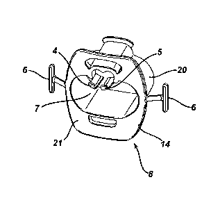
1(10211 Referring to Fig. 1 and Fig. 2, the bite block 8 of the present
invention consists of a
generally elliptical cylindrical main body 20. having a proximal end. which
sits
outside of a patient's mouth, and a distal end. which sits inside a patient's
mouth.
Main body 20 surrounds main oral passage 7, which is sized to allow for
passage or
an endoseope and ventilation of the patient. Integral to the proximal end of
main
body 20 is flange 14, which sits outside of patient's lips and serves both to
locate
bite block 8 relative to the patient's mouth and protect the patient's lips
and teeth
from an endoseope. Flange 14 is integral to main body 20 at distal surface 22.
Attached at each side of tlzmge 14 is strap attachment wing 6 for strap 19
that goes
around the patient's head and helps secure bite block 8.
100221 Referring to Fig. 3, extending from the proximal to distal end of
main body 20. arc a
raised top surtitee 17 and bottom surface 18 for seating patient's upper teeth
and
lower teeth. respectively. Located at the distal end of top surface 17 is
upper
protruding retention feature 9, protruding up generally perpendicular to top
surface
17. Upper protruding retention feature 9 serves as a stop to keep bite block 8
from
being expelled from a patient's mouth by requiring the mouth (or more
particularly,
the teeth) to be opened wide enough to get around retention feature 9.
Similarly, on
bottom surface 1 8 is lower protruding retention feature I I serving the same
purpose.
100231 Referring also now to Figs. 4 and 6. some features of the present
invention are
intended to interface with an oral-nasal cannula 1. generally known in the
art, with
oxygen outlet port 2 and CO! sampling inlet port 3. A representative oral-
nasal
cannula is described in pending application US-2006-0042636.
Oxygen outlet port 2 is the end of
the oxygen delivery fluid line that delivers oxygen into the patient's oral
cavity. and
CO., sampling inlet port 3 is the end of the fluid line of a eapnometry or
capnography
system through which expired CO2 enters from a patient's oral cavity. Oxygen
outlet
port 2 and CO2 sampling inlet port 3 consist of tubular extensions downward
from the
main body of cannula I. bent in a generally perpendicular fashion towards the
patient's mouth. In the absence of a bite block, the openings of oxygen outlet
port 2
and CO? sampling inlet port 3 would rest at the opening to the oral cavity.
CA 02690065 2009-12-04
WO 2008/151180 PCT/US2008/065630
[0024] Referring also now to Fig. 5, internal to main body 20, and
extending from the
proximal end to the distal end of main body 20, and adjacent to main oral
passage 7,
is internal gas channel 10. Internal gas channel 10 consists of two parallel
adjacently-
connected sub-channels, each of semi-circular cross section. Internal gas
channel 10
occupies the area under raised top surface 17. Internal gas channel 10 allows
the
exchange of gas from the proximal end (external to the patient's mouth) of
bite block
8 to the distal end (internal to the patient's mouth), and vice versa, without
using a
significant amount of the cross-sectional area of main oral passage 7, which
is
reserved for use by the endoscope. As seen in Fig. 4, internal gas channel 10
provides a path via one sub-channel for oxygen to flow from oxygen outlet port
2 of
an oral-nasal cannula 1 into the patient's mouth and, via the other sub-
channel, for
CO2 to flow from the patient's mouth into CO2 sampling inlet port 3 of cannula
1.
The sub-channels of internal gas channel 10 can be used interchangeably for
either
oxygen or CO2, depending on where the respective ports are located on cannula
1.
[0025] Oxygen port support 4 and CO2 port support 5 protrude from proximal
surface 21 of
flange 14, and proximal from internal gas channel 10. Oxygen port support 4
and
CO2 port support 5, each consist of a generally flat extension extending from
proximal surface 21, and generally symmetrical with respect to the vertical
plane
aligned longitudinally along main body 20. From their points of attachment
located
on the side away from the center axis of main body 20, oxygen port support 4
and
CO2 port support 5 slope slightly downward toward the center of main oral
passage 7.
Oxygen port support 4 and CO2 port support 5 also extend in the distal
direction for
approximately the thickness of flange 14, as best seen in Figs. 1 and 6,
partially
separating main oral passage 7 and internal gas channel 10. This arrangement
is
designed to allow the ends of oxygen outlet port 2 and CO2 sampling inlet port
3 of
cannula 1 to rest inside internal gas channel 10, as shown in Fig. 6. Oxygen
port
support 4 and CO2 port support 5 each terminate on their proximal ends in an
upward-
curving quarter-circular shaped feature, which provides a means for more
securely
locating near the oral cavity oxygen outlet port 2 of the oxygen delivery
system and
CO2 sampling inlet port 3 of a capnometry or capnography system. Oxygen port
support 4 and CO2 port support 5 are intended to provide a means for
protecting the
END6134W0PCT
CA 02690065 2015-01-29
6
location of oxygen outlet port 2 and CO2 sampling inlet port 3 against
jostling from
the movement of the scope. The terms "oxygen port support" and "CO2 port
support"
are used only illustratively in this description; since the supports are
generally
symmetrical, they could be used interchangeably, depending on which side of
cannula
1 each port was located.
[0026] An alternate embodiment of the present invention, shown in Fig. 7,
adds additional
functionality by allowing a doctor to insert a finger a short distance into
the patient's
mouth to help guide the endoscopc down into the trachea or esophagus, while
again
preventing excessive jostling of bite block 8 and cannula 1. In the alternate
embodiment, main body 20 has curved cutouts 23 on its distal end, on both of
its
sides. In addition, integral to flange 14, and extending out on both of its
sides, are
strap attachment wing extenders 24, each consisting of a thin arced, 'c'-
shaped
protrusion. Cutouts 23 and strap attachment wing extenders 24 are sized and
located
such that a finger may be inserted through the open side of the 'c' of strap
attachment
wing extender 24 and past cutout 23 into the patient's mouth. Strap attachment
wing
extenders 24 also locate the strap attachment wings 6 such that the strap is
not in the
way of a finger. In this manner, a doctor would be able to easily guide an
endoscope
with a finger without using any of the cross sectional area of main oral
passage 7, and
without too much jostling of bite block 8 and cannula 1.
[0027] While preferred embodiments of the present invention have been shown
and
described herein, it will be obvious to those skilled in the art that such
embodiments
are provided by way of example only. In addition, it should be understood that
every
structure described above has a function and such structure can be referred to
as a
means for performing that function. Numerous variations, changes, and
substitutions
will now occur to those skilled in the art. The scope of the claims may be
given the
broadest interpretation consistent with the description as a whole.