Note: Descriptions are shown in the official language in which they were submitted.
CA 02690304 2009-12-07
WO 2008/153930 PCT/US2008/007108
SENSORS FOR THE DETECTION OF DIOLS AND CARBOHYDRATES USING
BORONIC ACID CHELATORS FOR GLUCOSE
CROSS REFERENCE TO RELATED APPLICATIONS
This application claims the benefit of U.S. Provisional Patent Application
Serial No. 60/933,724 filed on June 8, 2007 and the U.S. Provisional Patent
Application entitled Sensors for the Detection of Diols and Carbohydrates by
inventor Heather Clark, filed on May 29, 2008. The teachings of all of the
referenced applications are incorporated by reference in their entirety.
BACKGROUND OF THE INVENTION
Diabetes has become a national health-care crisis. According to the 2005
National Diabetes Fact Sheet, an estimated 20.8 million people in the United
States
suffer from diabetes. The costs associated with diabetic care are also
astronomical,
with an estimated $132 billion dollars spent in 2002. As a result of a seminal
study
highlighting the benefits of tight glycemic control, the American Diabetes
Association recommends that patients with diabetes should try to control their
glucose levels to be as close to normal as possible. With tight glycemic
control, the
complications associated with diabetes, such as heart disease, blindness and
amputation are significantly reduced. Self-monitoring of glucose is essential
for
regulation, particularly for those with Type 1 diabetes. It is often performed
through
a finger-stick method three times or more per day. The need to draw blood,
even in
small quantities, multiple times a day is not desirable.
A continuous monitoring system would be highly advantageous for patients
and healthcare providers alike. It has become the goal of glucose sensor
research,
and continuous monitoring systems of many varieties are pursued by countless
researchers in the field. The benefits of continuous monitoring over the
finger-stick
method are numerous. First, the finger-stick method is both painful and
inconvenient
for the patient, which can lead to noncompliance. Second, a single-point
measurement gives static information on the concentration of blood glucose,
with no
knowledge of the trend, or in other words, whether the level is going up or
down.
Third, monitoring at night, a time when levels could dip dangerously low, is
either
I
CA 02690304 2009-12-07
WO 2008/153930 PCT/US2008/007108
not performed or especially inconvenient. Continuous monitoring systems have
been pursued in many different forms, and some are commercially available,
such as
the Guardian RT from Medtronic MiniMed (Northridge, California), and the
GlucoWatch Biographer from Animas (West Chester, PA). Both of these systems
work by sampling glucose from the interstitial space, the extracellular space
in the
dermis, rather than the blood. Currently, they are approved as monitors to
track
trends in glucose but highs and lows are verified by a finger-stick test. Some
reports
have shed doubt on the accuracy of nighttime monitoring in patients whose
glucose
is tightly controlled.
Commercially available systems for continuous or finger-stick measurements
rely on electrochemical biosensors. Glucose oxidase is the most well-known of
the
biological recognition units, and the enzyme provides a highly selective
sensor
platform. Enzyme-based sensors are difficult to implement as implantable
glucose
sensors, since the enzyme limits itself in a confined environment. Oxygen,
required
for function, regionally depletes, and hydrogen peroxide, a by-product of the
reaction, can lead to enzyme degradation. Most often the read-out is electrode-
based,
which is an added challenge for miniaturization and biological implantation.
Nano-
and microscale optical sensors have also been demonstrated, but typically lack
the
selectivity and robustness to replace traditional techniques.
There is still a need for a continuous, non-invasive method for glucose
monitoring, especially one that is easy to use, highly accurate and pain-free.
SUMMARY OF THE INVENTION
This invention discloses a sensor particle for detecting the presence of a
chelatable analyte, such as glucose, comprising a quantum dot, a polymer
matrix
comprising a polymer including moieties that bind the chelatable analyte and a
chromophore associated with the polymer matrix that binds to the moieties in
the
absence of the chelatable analyte. In some embodiments, photons emitted by the
quantum dot in an excited state are absorbed by the chromophore in an unbound
state but not by the chromphore in a bound state. The moieties may bind the
chelatable analyte and chromophore reversibly and competitively. In certain
embodiments, the moieties are boronic acids or boronic esters. In some
2
CA 02690304 2009-12-07
WO 2008/153930 PCT/US2008/007108
embodiments, one or more components of the sensor, such as the moieties and/or
chromophore, are covalently bound to or associated with the polymer matrix. In
some embodiments, the sensor particles further comprise a biocompatible layer.
In certain aspects, the invention comprises methods for detecting the
presence of a chelatable analyte in a medium using the sensor particles of the
invention. In certain embodiments, the chelatable analyte is glucose and the
medium
is selected from water, blood, plasma and urine. In certain embodiments, the
invention comprises a method for detecting the presence of a chelatable
analyte in an
animal. In certain such embodiments, the sensor particle is implanted in the
dermis
or epidermis and the chelatable analyte, such as glucose, is monitored.
BRIEF DESCRIPTION OF THE DRAWINGS
Figure 1. Sensor particle 3 with a. chromophore 2 bound to moiety 1, wherein
the
bound chromophore emits photons 4 at one wavelength and b. moiety 1 bound to
analyte 5 wherein the unbound chromophore 2 emits photons at a second
wavelength 6.
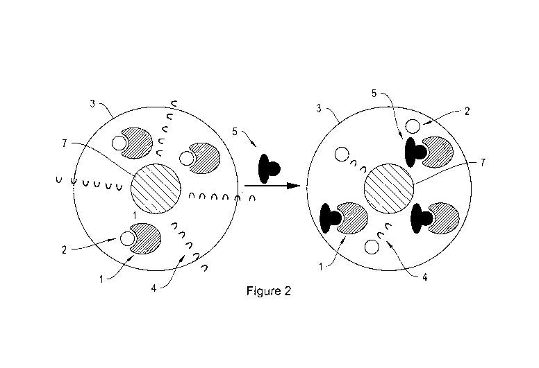
Figure 2. Sensor particle 3 with a. chromophore 2 bound to moiety 1, wherein
the
bound chromophore 2 does not absorb photons 4 emitted by the quantum dot
and/or
fluorescent dye 7 and b. moiety 1 bound to analyte 5 wherein unbound
chromophore
2 absorbs photons 4 emitted by quantum dot and/or fluorescent dye 7.
Figure 3. Sensor particle 3 with a. chromophore 2 bound to moiety 1, wherein
the
bound chromophore absorbs photons 4 emitted by the quantum dot and/or
fluorescent dye 7 and b. moiety 1 bound to analyte 5 wherein unbound
choromophore 2 absorbs photons 4 emitted by quantum dot and/or fluorescent dye
7.
Figure 4. An exemplary embodiment of the competitive interaction of a boronic
acid
(chelatable moiety) with alizarin (chromophore), or glucose (analyte).
3
CA 02690304 2009-12-07
WO 2008/153930 PCT/US2008/007108
Figure 5. Spectral signature of the components of a GSQD; a. overlap of
normalized
alizarin absorbance and quantum dot emission, b. individual contribution of
the two
components of the inner filter effect at high and low glucose concentration
and the
resulting overall fluorescence signal.
Figure 6. Wide field fluorescence microscopic image of a suspension of sensor
particles.
Figure 7. Nanometer-sized sensor particles demonstrating the inner filter
effect
wherein a. the absorbance changes from purple to yellow depending on the
binding
state of the chromophore, b. the same samples under UV excitation wherein the
sample that was visually purple does not absorb the 525 nm emission of the
quantum
dots and fluoresces brightly, while the yellow sample absorbs the fluorescence
emission of the quantum dot and has minimal emission.
Figure 8. Evaluating response to glucose, the sensor particles containing the
essential sensing components, alizarin, pyrene boronic acid and additive, was
immobilized to the bottom of a micro-well for calibration. Response to glucose
and
fructose was measured, the average SEM is shown, where n=6 and n=8 for
control
and monosaccharides, respectively.
Figure 9. Measuring the degree of cytotoxicity of sensor particles by
incubating the
particles overnight with HEK 293 calls and measuring the degree of cellular
injury
with an MTT assay. Results of particle sensors are compared to other
particles, e.g.,
gold, latex.
DETAILED DESCRIPTION
Disclosed are sensor particles for the detection of chelatable analytes, e.g.,
glucose. The sensor particles comprise a polymer matrix, moieties which bind a
chelatable analyte, and a component that emits or absorbs photons of a
particular
wavelength either in the presence of absence of the chelatable analyte. In an
exemplary embodiment, a chromophore absorbs photons of one wavelength when
4
CA 02690304 2009-12-07
WO 2008/153930 PCT/US2008/007108
bound to the moieties of the sensor and another wavelength when unbound from
the
moieties. When the chromophore-bound moieties are exposed to the chelatable
analyte, the chromophore is released and the chelatable analyte binds to the
moieties. The free chromophore appears as a different color than the bound
chromophore, a change which can be monitored visually or with
spectrophotometric
instrumentation. In an alternate exemplary embodiment, wherein the inner-
filter
effect is employed, the sensor particle of the preceding embodiment further
comprises a fluorescent dye and/or quantum dot. The fluorescent dye and/or
quantum dot absorbs a broad range of wavelengths and emits photons of a narrow
range of wavelengths. The fluorescence emitted by the fluorescent component is
either absorbed or not absorbed depending on the presence of the chelatable
analyte.
For example, when the chelatable analyte is bound to the moieties of the
sensor, the
fluorescence of the quantum dot is absorbed while no absorbance occurs in the
absence of the chelatable analyte.
In certain embodiments, the sensor particle for detecting the presence of
chelatable analytes comprises a polymer matrix comprising a polymer including
moieties that bind the chelatable analyte and a chromophore associated with
the
polymer matrix that binds to the moieties in the absence of the chelatable
analyte. In
certain embodiments, the chelatable anaylte is glucose and the moieties bind
glucose
and the chromophore reversibly and competitively. In an exemplary embodiment,
the sensor particle 3 comprises a polymer matrix with moieties 1 that can bind
both a
chromophore 2 and glucose 5 (Figure 1). In a first mode, the moieties 1 are
bound to
a chromophore 2 and the chromophore, in its bound mode, absorbs photons at a
first
wavelength 4. In a second mode, when the sensor particle 3 is contacted with
glucose 5, the glucose 5 binds to the moieties 1, displacing the chromophore 2
which, in its unbound state, absorbs photons at a second wavelength 6. In
certain
embodiments, the sensor 3 is monitored visually to determine a change in the
color
of the chromophore 2. In certain embodiments, the sensor 3 is monitored with
spectrophotometric instrumentation to determine the emission spectra of the
chromophore 2.
In certain embodiments, the sensor particle for detecting the presence of a
chelatable analyte comprises a fluorescent component, a polymer matrix
comprising
5
CA 02690304 2009-12-07
WO 2008/153930 PCT/US2008/007108
a polymer including moieties that bind the chelatable analyte and a
chromophore
associated with the polymer matrix that binds to the moieties in the absence
of the
chelatable analyte. In certain embodiments, the sensor particle emits photons
with an
inner filter effect. The inner-filter effect has been documented as a way to
increase
the signal intensity and concomitant sensitivity of ion-selective optical
sensors
(optode). In brief, a secondary, inert fluorescent component is added to the
polymer
matrix of the optode. When the concentration of analyte in the optode changes,
the
fluorescence intensity of the inert dye itself does not respond, however the
absorbance of the sensor does. Because the fluorescence emission has been
carefully
chosen to overlap with the absorbance spectrum of the sensor, the emission
from the
inert dye is then absorbed by the sensor. The attenuation of the fluorescence
output
of the inert dye is therefore directly related to the concentration of the ion
of interest
in solution.
In certain embodiments, the chelatable analyte is glucose and the moieties
bind glucose and the chromophore reversibly and competitively. In certain
embodiments, the fluorescent component is selected from one or more quantum
dots
and/or fluorescent dyes 7. In certain such embodiments, a sensor particle 3
comprises a fluorescent component 7, and a polymer matrix with moieties 1 that
can
bind both a chromophore 2 and glucose 5. In certain such embodiments, the
fluorescent component 7 absorbs a broad range of wavelengths of photons but
emits
a narrow range of wavelengths of photons. The fluorescent component 7 is
activated
by exciting with a light source, e.g., UV light. The fluorescence emitted from
the
excited fluorescent component 7 is either absorbed by a component of the
sensor,
e.g., the chromophore 2 or the glucose-moiety complex, or emitted from the
sensor 3
without being attenuated. In certain embodiments, photons 4 of the fluorescent
component 7 are absorbed when the chromophore 2 is bound to the moieties 1
(Figure 3, left). In certain such embodiments, the absence of fluorescence
emitted
from the sensor particle 3 indicates an absence of glucose molecules 5, i.e.
glucose
molecules are not bound to the moieties of the sensor. In such embodiments,
when
glucose 5 is introduced, the moieties 1 bind glucose 5, releasing the
chromophore 2.
The photons 4 of the fluorescent component 7 are no longer absorbed by a
6
CA 02690304 2009-12-07
WO 2008/153930 PCT/US2008/007108
component of the sensor, Figure 3, right. By detecting the emitted photons,
the
amount of bound glucose can be calculated relative to a standard.
In certain embodiments, a component of the sensor, e.g., the chromophore 2
or the glucose-moiety complex, absorbs photons 4 of the fluorescent component
7
when unbound from the moieties 2 (Figure 2, right). In certain such
embodiments,
the detection of photons 4 from the sensor 3 indicates the absence of glucose
5, i.e.
glucose molecules are not bound to the moieties of the sensor. In certain such
embodiments, when the sensor 3 is contacted with glucose 5, the moieties 1
release
the chromophore 2 and bind glucose 5. In such embodiments, the photons 4 of
the
fluorescent component 7 are not absorbed when glucose 5 is bound to the
moieties 1
such that the detection of photons 4 emitted from the sensor particle 3
indicates the
presence of glucose 5.
In certain embodiments, the sensors of the present invention may be used to
detect and measure the presence of a wide variety of chelatable analytes,
e.g., sugars
and related compounds, in a solution, in vitro or in vivo. The sensor may be
located
within a cell, i.e., intracellular, or exterior to a cell, i.e.,
extracellular. In certain
embodiments, the sensor is in contact with the cell membrane such as within a
cell
or exterior to a cell. Exemplary chelatable analytes for detection by the
sensor of the
present invention include sugars such as glucose, mannose, and other
monosaccharides, sialic acid, lactic acids, aminosugars, such as glucosamine,
disaccharides, trisaccharides, oligosaccharides, sugar-amino acids, sugar-
peptides
and glycoproteins. Other exemplary chelatable analytes include, but are not
limited
to, glycerol, dopamine, catechols, ascorbic acid, polyols, diols such as 1,4-
anhydroerythritol and ethylene glycol. The concentration range of chelatable
analytes which is typically of interest in biological samples is 0-25 mM, such
as
from 5-20 mM, such as from 5-10 mM, such as from 0-5 mM.
In certain embodiments, the moieties that bind the chelatable analytes
comprise a dihydroxide component, e.g., boron and alkali earth dihydroxides.
Complexation of sugars, for example, with boron and alkali earth dihydroxides
has
been reported in, among other sources, [S. A. Barker et al., Carbohydrate
Research,
26 (1973) 33-40; N. Roy et al., Carbohydrates Research, 24 (1972) 180-183]. A
variety of different boronic acids, having the structure RB(OH)2 may be used
to
7
CA 02690304 2009-12-07
WO 2008/153930 PCT/US2008/007108
chelate the analyte. R can be, for example, an aryl or a saturated or
unsaturated alkyl
moiety, either of which can be substituted or unsubstituted and can contain
one or
more heteroatoms, e.g., N, S, 0, P, B, F, Br. In certain embodiments, a
boronic ester
is used to chelate the analyte. Boronic esters have the molecular formula
RB(OR')2
wherein R' is typically an alkyl group and R can be defined as above. Under
aqueous
conditions, many boronic esters hydrolyze to form boronic acids. Therefore,
OR'
groups that hydrolyze to OH are of use in the present invention. The two R'
groups
of the ester may be linked to form a cyclic structure, e.g., -CH2CH2-. In
certain
embodiments, the moieties are selected from one ore more aromatic or aliphatic
boronic esters. In certain aspects, boronic acids are appended with
substituents that
affect the pKa such as electron withdrawing groups or electron donating
groups. In
certain embodiments the pKa of the boronic acid will change the dynamic range
of
the sensor. In certain embodiments the dynamic range of the sensor relates to
the
affinity for an analyte, such as glucose. In certain embodiments, the moieties
are
selected from one or more aromatic or aliphatic boronic acids. Exemplary
boronic
acid moieties of the invention include phenyl boronic acid, butyl boronic
acid, (3,5-
dichlorophenyl)boronic acid, [3,5-bis(trifluoromethyl)phenyl]boronic acid, and
(4-
bromophenyl)boronic acid.
In certain embodiments, the moieties of the sensor which chelate the analytes
comprise a metal ion. The ability of sugars, for example, and other molecules
to
form chelate complexes with metal ions in aqueous solution is well known
(general
review by: Whitfield, D. M. et al., "Metal coordination to carbohydrates.
Structure
and Function," Coord. Chem. Reviews 122, 171-225 (1993) and Angya, S.J.
Complexes of Metal Cations with Carbohydrates in Solution, in "Advances in
Carbohydrate Chemistry and Biochemistry," Academic Press, Inc. 1989, pp.1-4).
The complexation of Cu(II) with various sugar a-amino acids is described by M.
Angeles Diaz-Diez et al., Transition Met. Chem. 20, 402-405, 1995. Sugar-a-
amino
acid compounds will also form complexes with Co(II), Ni(II), Zn(II) and Cd(II)
(M.
Angeles Diaz-Diez et al., J. Inorg. Biochem. 56, 243-247, 1994). Additionally,
complexes of various sugars with vanadium, molybdenum, tungsten, aluminum,
iron, barium, magnesium, and strontium are known (Sreedhara, A. et al.,
Carbohydrate Res. 264, 227-235, 1994; Caldeira, M. M. et al., Inorg. Chim.
Acta.
8
CA 02690304 2009-12-07
WO 2008/153930 PCT/US2008/007108
221, 69-77, 1994; Tonkovic, M. and Bilinski, H., Polyhedron 14, 1025-1030,
1995;
Nagy, L. et al., Inorg. Chim. Acta. 124, 55-59, 1986; Tajmir-Riahi, H. A.,
Inorg.
Chim. Acta. 119, 227-232, 1986; and Tajmir-Riahi, H. A., J. Inorg. Biochem.,
24,
127-136, 1985.
In certain embodiments, the moieties that bind the chelatable analytes are
covalently conjugated to the polymer matrix. In certain embodiments, the
moieties
are covalently conjugated to the matrix, for example, through a linker
molecule. In
an exemplary embodiment, the moieties comprise aryl boronic acids which are
covalently conjugated to the polymer matrix through ester linkages originating
at an
aryl atom or the aryl boronic acid. Other exemplary linkages include amides,
ethers,
sulfonates, thioethers, thioesters and carbonates. In certain embodiments, the
moieties are covalently bound to the polymer matrix through a bond such as a
single
or double bond. In certain exemplary embodiments, the aryl boronic acids are
covalently bound to the polymer matrix through a single bond originating from
an
aryl atom or the aryl boronic acid.
In certain embodiments, the chromophore of the sensor is any molecule that
binds reversibly to the moieties of the sensor, e.g., the chromophore alizarin
binds
boronic acids, and absorbs photons of the fluorescent component in a first
state and
does not absorb photons of the fluorescent component in a second state. The
states
of the chromophore include bound to the moieties and unbound from the
moieties.
For example, the chromophore alizarin absorbs at a first wavelength when
unbound
and a second wavelength when bound to a boronic acid. In certain embodiments,
the
chromophore, e.g., alizarin, is selected from any dye that binds boronic acid
moieties, preferably having absorbance/fluorescence properties that differ in
the
bound vs. the free state. When a suitable chelatable analyte is present, the
boronic
acid releases the chromophore and binds the analyte. Additional FDA approved
dyes
and colored drugs are described in the Code of Federal Regulations (CFR) for
Food
and Drugs (see Title 21 of CFR chapter 1, parts 1-99). A wide variety of
chromophores and fluorescence sources may be used, e.g., paired so that the
absorbance wavelength of the unbound chromophore substantially matches the
wavelength of the fluorescent component's photon emissions, e.g., so as to
absorb
the emissions in an unbound state. The table below lists a number of suitable
9
CA 02690304 2009-12-07
WO 2008/153930 PCT/US2008/007108
chromophores, their Chemical Abstract Service (CAS) Registration Numbers,
colors
and absorption maxima. In certain embodiments, the chromophore is derivatized
in
such a manner that it can bind with the chelating moiety of the sensor.
Chromophore CAS Rea. No. Color Abs. Max.
Yellow No. 5 1934-21-0 yellow 428
(3-carotene 7235-40-7 orange 466
Rifampin 3292-46-1 red 475
Yellow No. 6 2783-94-0 yellow 480
Tetracycline 60-54-8 yellow N/A
Red No. 40 25956-16-6 red 502
Red No. 3 16423-68-0 red 524
Blue No. 2 860-22-0 blue 610
Evan's blue 314-13-6 blue 610
Green No. 3 2353-45-9 green 628
Blue No. 1 2650-18-2 blue 630
Methylene blue 7220-79-3 Blue 668/609
Indocyanine green 3599-32-4 Green 800 (mostly IR)
In certain embodiments, the chromophore is covalently conjugated to the
polymer matrix and comprises a reactive site that binds reversibly with the
chelatable analyte selective moieties. In an exemplary embodiment, the
chromophore is alizarin, and the alizarin is covalently bound to the polymer
matrix
through a linker or bond. In certain embodiments, the linker is an ester
amide, ether,
sulfonate, thioether, carbonate or thioester originating from an aromatic
carbon of
the alizarin. In certain embodiments, the chromophore is covalently conjugated
through a bond to the polymer matrix. In certain embodiments, the bond or
linkage
between the chromophore and the polymer matrix does not interfere with the
ability
of the chromophore to bind to the chelatable analyte. For example, in the case
of
alizarin, the linkage or bond to the polymer matrix originates from a ring of
the
CA 02690304 2009-12-07
WO 2008/153930 PCT/US2008/007108
polycyclic ring system that does not bear the hydroxy groups. In certain such
embodiments, the hydroxyl groups of the alizarin are unimpeded from
interacting
with the chelatable analyte.
In certain embodiments, the polymer matrix of the sensor comprises
poly(caprolactone) (PCL), ethylene vinyl acetate polymer (EVA), poly(lactic
acid)
(PLA), poly(L-lactic acid) (PLLA), poly(glycolic acid) (PGA), poly(lactic acid-
co-
glycolic acid) (PLGA), poly(L-lactic acid-co-glycolic acid) (PLLGA), poly(D,L-
lactide) (PDLA), poly(L-lactide) (PLLA), poly(D,L-lactide-co-caprolactone),
poly(D,L-lactide-co-caprolactone-co-glycolide), poly(D,L-lactide-co-PEO-co-D,L-
lactide), poly(D,L-lactide-co-PPO-co-D,L-lactide), polyalkyl cyanoacralate,
polyurethane, poly-L-lysine (PLL), hydroxypropyl methacrylate (HPMA),
polyethyleneglycol, poly-L-glutamic acid, poly(hydroxy acids), polyanhydrides,
polyorthoesters, poly(ester amides), polyamides, poly(ester ethers),
polycarbonates,
silicones, polyalkylenes such as polyethylene, polypropylene, and
polytetrafluoroethylene, polyalkylene glycols such as poly(ethylene glycol)
(PEG),
polyalkylene oxides (PEO), polyalkylene terephthalates such as poly(ethylene
terephthalate), polyvinyl alcohols (PVA), polyvinyl ethers, polyvinyl esters
such as
poly(vinyl acetate), polyvinyl halides such as poly(vinyl chloride) (PVC),
polyvinylpyrrolidone, polysiloxanes, polystyrene (PS), polyurethanes,
derivatized
celluloses such as alkyl celluloses, hydroxyalkyl celluloses, cellulose
ethers,
cellulose esters, nitro celluloses, hydroxypropylcellulose,
carboxymethylcellulose,
polymers of acrylic acids, such as poly(methyl(meth)acrylate) (PMMA),
poly(ethyl(meth)acrylate), poly(butyl(meth)acryl ate),
poly(isobutyl(meth)acrylate),
poly(hexyl(meth)acrylate), poly(i sodecyl(meth)acryl ate),
poly(lauryl(meth)acrylate),
poly(phenyl(meth)acryl ate), poly(methyl acrylate), poly(isopropyl acrylate),
poly(isobutyl acrylate), poly(octadecyl acrylate) (jointly referred to herein
as
"polyacrylic acids"), and copolymers and mixtures thereof, polydioxanone and
its
copolymers, polyhydroxyalkanoates, poly(propylene fumarate), pol yoxym
ethylene,
poloxamers, poly(ortho)esters, poly(butyric acid), poly(valeric acid),
poly(lactide-
co-caprolactone), trimethylene carbonate, polyvinylpyrrolidone, and the
polymers
described in Shieh et al., 1994, J. Biomed. Mater. Res., 28, 1465-1475, and in
U.S.
Patent No. 4,757,128, Hubbell et al., U.S. Pat. Nos. 5,654,381; 5,627,233;
11
CA 02690304 2009-12-07
WO 2008/153930 PCT/US2008/007108
5,628,863; 5,567,440; and 5,567,435. Other suitable polymers include
polyorthoesters (e.g. as disclosed in Heller et al., 2000, Eur. J. Pharm.
Biopharm.,
50:121-128), polyphosphazenes (e.g. as disclosed in Vandorpe et al., 1997,
Biomaterials, 18:1147-1152), and polyphosphoesters (e.g. as disclosed in
Encyclopedia of Controlled Drug Delivery, pp. 45-60, Ed. E. Mathiowitz, John
Wiley & Sons, Inc. New York, 1999), as well as blends and/or block copolymers
of
two or more such polymers. The carboxyl termini of lactide- and glycolide-
containing polymers may optionally be capped, e.g., by esterification, and the
hydroxyl termini may optionally be capped, e.g., by etherification or
esterification.
In certain embodiments, the polymer comprises or consists essentially of
polyvinyl
chloride (PVC), polymethyl methacrylate (PMMA) or decyl methacrylate or
copolymers or any combination thereof.
In certain embodiments, the polymer matrix of the sensor comprises a
biocompatible layer, e.g., selected from poly(caprolactone) (PCL), ethylene
vinyl
acetate polymer (EVA), poly(ethylene glycol) (PEG), poly(vinyl acetate) (PVA),
poly(lactic acid) (PLA), poly(glycolic acid) (PGA), poly(lactic-co-glycolic
acid)
(PLGA), polyalkyl cyanoacrylate, polyethylenimine,
dioleyltrimethyammoniumpropane/diol eyl-sn-gl ycerolphosphoethanol amine,
polysebacic anhydrides, polyurethane, nylons, or copolymers thereof. In
certain
embodiments, the biocompatible layer is disposed on the exterior of the sensor
such
as disposed around the polymer matrix and chromophore and optional component,
such as a fluorescent dye and/or quantum dot. In polymers including lactic
acid
monomers, the lactic acid may be D-, L-, or any mixture of D- and L- isomers.
In
certain aspects, the biocompatible layer of the sensor particle comprises a
PEG-lipid.
In certain embodiments, the lipid tail self-inserts into the lipophilic
polymer matrix
during fabrication, leaving the PEG headgroup on the surface of the sensor,
e.g., to
provide a hydrophilic, biocompatible coating that can be penetrated by the
analyte.
In certain embodiments, different chemical moieties, such as amines, can be
put on
the surface or further modified to attach antibodies or other recognition
units.
The terms "biocompatible polymer," "biocompatible layer" and
"biocompatibility" when used in relation to polymers are art-recognized. For
example, biocompatible polymers include polymers that are neither themselves
toxic
12
CA 02690304 2009-12-07
WO 2008/153930 PCT/US2008/007108
to the host (e.g., a cell or an animal such as a human), nor degrade (if the
polymer
degrades) at a rate that produces monomeric or oligomeric subunits or other
byproducts at toxic concentrations in the host. Consequently, in certain
embodiments, toxicology of a biodegradable polymer intended for intracellular
and/or in vivo use, such as implantation or injection into a patient, may be
determined after one or more toxicity analyses. It is not necessary that any
subject
composition have a purity of 100% to be deemed biocompatible. Hence, a subject
composition or layer may comprise 99%, 98%, 97%, 96%, 95%, 90% 85%, 80%,
75% or even less of biocompatible polymers, e.g., including polymers and other
materials and excipients described herein, and still be biocompatible.
The polymer matrix of the sensor may comprise a plasticizer, such as
dioctyl sebacate (DOS), o-nitrophenyl-octylether, dimethyl phthalate,
dioctylphenyl-
phosphonate, dibutyl phthalate, hexamethylphosphoramide, dibutyl adipate,
dioctyl
phthalate, diundecyl phthalate, dioctyl adipate, dioctyl sebacate, or other
suitable
plasticizers. In certain embodiments, the plasticizer is poly(glycerol
sebacate), PGS.
In certain embodiments, e.g., particularly where the polymer is
biocompatible, a biocompatible plasticizer is used. The term "biocompatible
plasticizer" is art-recognized, and includes materials which are soluble or
dispersible
in the relevant polymer, which increase the flexibility of the polymer matrix,
and
which, in the amounts employed, are biocompatible. Suitable plasticizers are
well
known in the art and include those disclosed in U.S. Pat. Nos. 2,784,127 and
4,444,933. Specific plasticizers include, by way of example, acetyl tri-n-
butyl
citrate (c. 20 weight percent or less), acetyltrihexyl citrate (c. 20 weight
percent or
less), butyl benzyl phthalate, dibutylphthalate, dioctylphthalate, n-butyryl
tri-n-hexyl
citrate, diethylene glycol dibenzoate (c. 20 weight percent or less) and the
like.
In certain embodiments, the sensor particle for detecting the presence of
glucose comprises: a quantum dot, a polymer matrix comprising a polymer
appended with moieties that selectively bind glucose, a chromophore associated
with
the polymer matrix that binds the moieties in the absence of glucose and a
biocompatible layer.
13
CA 02690304 2009-12-07
WO 2008/153930 PCT/US2008/007108
In certain embodiments, additives to the polymer matrix make the extraction
of the analyte (e.g., glucose) into the polymeric matrix more efficient. In
certain
embodiments, the addition of amine-based additives to the matrix lowers the
effective dynamic range of the sensor particles. In certain embodiments, the
addition of amines to the polymer matrix increases the affinity of the polymer
matrix
for the analyte, e.g., glucose.
In certain embodiments, the sensor comprises one or more quantum dots.
Quantum dots are fluorescent semiconductor nanocrystals having a
characteristic
spectral emission, which is tunable to a desired energy by selection of the
particle
size, size distribution and composition of the semiconductor nanocrystal. The
quantum yield of quantum dots is high, with reports of greater than 90%
efficiency
in cladded quantum dots, photobleaching is minimal, and a single quantum dot
can
be continuously tracked for minutes to hours. There is a wide range of colors
available, all with the same excitation wavelengths, and very narrow emission
bandwidths.
The emission spectra of a population of quantum dots have linewidths as narrow
as
25-30 nm, depending on the size distribution heterogeneity of the sample
population,
and lineshapes that are symmetric, gaussian or nearly gaussian with an absence
of a
tailing region. Advantageously, the range of excitation wavelengths of the
quantum
dots is broad. Consequently, this allows the simultaneous excitation of
varying
populations of quantum dots in a system having distinct emission spectra with
a
single light source, e.g., in the ultraviolet or blue region of the spectrum.
In certain embodiments, quantum dots of the sensor described herein are, for
example, inorganic crystallites between I nm and about 1000 nm in diameter,
preferably between about 2 nm and about 50 nm, more preferably about 5 nm to
20
nm, such as about 6, 7, 8, 9, 10, 11, 12, 13, 14, 15, 16, 17, 18, 19, or 20
nm. Such
quantum dots include a "core" of one or more first semiconductor materials,
and
which may be surrounded by a "shell" of a second semiconductor material. A
semiconductor nanocrystal core surrounded by a semiconductor shell is referred
to
as a "core/shell" semiconductor nanocrystal. The surrounded "shell" will most
preferably have a bandgap greater than the bandgap of the core material and
can be
chosen so to have an atomic spacing close to that of the "core" substrate. The
core
14
CA 02690304 2009-12-07
WO 2008/153930 PCT/US2008/007108
and/or the shell material can be a semiconductor material including, but not
limited
to, those of the group II-VI (ZnS, ZnSe, ZnTe, CdS, CdSe, CdTe, HgS, HgSe,
HgTe,
MgTe and the like) and III-V (GaN, GaP, GaAs, GaSb, InN, InP, InAs, InSb,
AlAs,
A1P, AlSb, A1S, and the like) and IV (Ge, Si, Pb and the like) materials, and
an alloy
thereof, or a mixture thereof.
In certain aspects, a sensor comprises exactly one quantum dot. In certain
embodiments, a sensor comprises more than one quantum dot, for example, 2, 3,
4,
or 5 quantum dots. In certain embodiments, wherein the sensor comprises more
than
one quantum dot, the sensor comprises two or more types of quantum dots, each
type having a distinct emission wavelength, e.g., independently selected from,
for
example, 490, 520, 545, 560, 580, 620, 655 nm. The availability of two
distinct
wavelength emissions (e.g., one or more quantum dots of wavelength 545 nm and
one or more quantum dots with emission wavelength of 655 nm) may allow
improvements in recording of changes in analyte concentration by using the
ratio of
the two distinct signals. Fluctuations in fluorescence that are common to both
signals
should theoretically cancel in a ratio. The detectable fluorescence emission
of the
quantum dot particles may fluctuate depending on variables including number of
quantum dots, quantum dot location within the cell, photobleaching, and
possible
changes in excitation light intensity, all effects that can occur slowly and
are not
related to analyte presence or concentration. Therefore, effects including
number of
quantum dots, quantum dot location within the cell, photobleaching, and
possible
changes in excitation light intensity, may be attenuated.
In certain embodiments, the fluorescence signal of the quantum dot may
trigger a detectable event within the cell. For example, fluorescence may in
turn
excite a secondary dye or quantum dot in the particle that easily generates
reactive
oxygen species (ROS). The ROS would then attack the cell, effectively
stimulating
necrosis (cell death), which may then be detected either visually or using
markers
sensitive to cell death. Alternatively, instead of including a secondary
component
within the particle, another particle may be added to the cell or cell
culture. This
additional particle may, for example, comprise a photo-degradable polymer
membrane. When the fluorescent component fluoresces, the emitted light will
rupture the secondary particle, releasing its contents. The contents may, for
CA 02690304 2009-12-07
WO 2008/153930 PCT/US2008/007108
example, be a drug that is therapeutic or apoptotic, e.g., triggering another
detectable event.
In certain aspects, the sensor of the application is a polymer film. In
certain
embodiments, the film comprises a polymeric matrix comprising a fluorescent
component, a chromophore and moieties that chelate analytes. In certain
embodiments, the film comprises multiple fluorescent components, chromophores
and moieties that chelate analytes. In certain aspects, the film is a polymer
matrix
comprising one or more sensor particles of the invention. A sensor film may be
deposited on any surface such as plastic, metal, paper or glass. The film may
be
deposited on an item such as a multi-well plate, a stirring rod, a Petri dish
or sample
cup. In certain embodiments, the film can be applied to a surface such as by
painting or spraying the surface with the polymer film, or by immersing the
surface
in a solution or dispersion of the elements of the polymer film. In certain
embodiments, the polymer film solidifies after the film has been applied to
the
surface. The polymer used in such films may be any one or more of the polymers
described herein or any other suitable polymer. In certain embodiments, the
film
further comprises a biocompatible coating. The fluorescent component of the
film
may be one or more quantum dots.
The quantum dot of the sensor particle may be modified with a surface
modifier, e.g., to alter one or more properties of the sensor particle, such
as
solubility, biocompatibility, or hydrophilicity/hydrophobicity. In certain
embodiments, the surface modifier comprises one or more ligands that can bind
reversibly with the quantum dot, while in other embodiments, the surface
modification may be essentially irreversible. In certain embodiments, the
surface
modifier improves the lipophilicity of the quantum dot. In certain such
embodiments, the ligand comprises an alkane such as decane-thiol.
In certain embodiments, the invention comprises methods of preparing
particles selective for a chelatable analyte, comprising contacting a quantum
dot
with a polymeric precursor mixture including moieties that bind the chelatable
analyte, and a chromophore. In certain embodiments, moieties are chosen which
chelate glucose. In certain embodiments, the moieties that bind the chelatable
16
CA 02690304 2009-12-07
WO 2008/153930 PCT/US2008/007108
analytes comprise boronic acids and/or boronic esters. In certain embodiments,
the
method further comprises coating the polymer matrix with a biocompatible
layer.
Chemical Vapor Deposition (iCVD), a coating technology, may be used to
deposit a layer that protects the sensors from the surrounding medium. The
solventless nature of iCVD particle coating may offer an advantage over
solution-
based methods that rely on drying of a wet polymer solution. In certain
embodiments, the iCVD particle coating employs a custom-designed rotating bed
reactor that has been demonstrated to provide conformal coating of
microspheres
and nanoparticles without inducing aggregation. In certain embodiments, the
primary monomer for the iCVD coatings of GSQDs is hydroxyethylmethacrylate
(HEMA) monomer.
In certain embodiments, the iCVD coatings of the nanoparticles are
pure polymer and no residual solvent is present, e.g., that may cause implant
rejection, irritation, or other unwanted side effects. The coatings can be
applied at
room temperature in a single step, taking only a few minutes of total time. In
certain
embodiments, the composition can be controlled systematically by changing the
gas
feed mix and thickness can be controlled by in situ monitoring
In certain embodiments, the invention includes methods for detecting the
presence of a chelatable analyte in a medium, comprising contacting a sensor
particle of the invention with a medium, exposing the quantum dot to light
energy
that causes the quantum dot to emit photons and using a detector to detect the
photons and determining the presence or absence of bound chelatable analyte
based
on the detected photons. In certain embodiments, the chelatable analyte is
glucose.
In certain embodiments, the light energy is selected from ultraviolet,
infrared, near
infrared or visible radiation. In certain embodiments, the light energy is
ultraviolet.
In certain embodiments, the medium comprises water, blood, plasma or urine. In
certain embodiments, the method of detecting glucose with a sensor particle of
the
invention is performed in vitro.
In certain aspects, the invention provides a method for detecting an analyte
in
an animal using any of the sensor particles of the invention. In certain
embodiments,
the invention provides a method for detecting the presence of a chelatable
analyte in
an animal, comprising the steps of: contacting a sensor particle of the
invention with
17
CA 02690304 2009-12-07
WO 2008/153930 PCT/US2008/007108
an animal cell or tissue, wherein the sensor particle comprises at least one
quantum
dot and/or fluorescent dye; a polymer matrix comprising a polymer matrix
including
moieties that bind a chelatable analyte and a chromophore associated with the
polymer matrix that binds to the moieties in the absence of the chelatable
analyte;
exposing the particles to light energy that causes the quantum dot and/or
fluorescent
dye to emit photons; using a detector to detect the photons; and determining
the
presence or absence of bound chelatable analyte based on the detected photons.
In
certain embodiments, the particle is implanted within the dermis or epidermis
of an
animal. In certain embodiments, the chelatable analyte is glucose.
In certain embodiments, the particle comprises a biocompatible layer. The
term "particle" may refer to one or more sensor particle of the invention. In
certain
embodiments, the particle comprises many sensor particles. In certain
embodiments,
the particle comprises a fluorescent dye and/or a quantum dot. In certain
embodiments, the particle comprises at least one quantum dot, a chromophore,
and a
polymer matrix. In certain embodiments, the photons emitted by the quantum dot
in
an excited state are absorbed by a chromophore in an unbound state but not
absorbed
by a chromophore in a bound state. In certain other embodiments, the photons
emitted by the quantum dot in an excited state are absorbed by a chromophore
in a
bound state but not absorbed by a chromophore in an unbound state.
In certain embodiment, the method for detecting an analyte in an animal
comprises implanting the particle below the surface of the epidermis or dermis
of the
animal. The particle may be implanted intracellularly, while in other
embodiments,
the sensors are implanted extracellularly. When implanted in tissues, the
composition may be taken into a cell or remain external to a cell. The
particle may
be implanted between about 0.05 mm and about 4 mm below the surface of the
epidermis or dermis of the animal. In certain embodiments, the particle is
injected or
surgically inserted within the dermis or epidermis of an animal. In certain
embodiments, the particle is injected within the dermis or epidermis of the
animal.
In certain embodiments, the particle is injected in a solution. In certain
embodiments, a particle solution comprises multiple particles. The particle
solution
may comprise particles with an average particle size between 10 nm and 10
microns.
In certain embodiments, the particle solution comprises particles with an
average
18
CA 02690304 2009-12-07
WO 2008/153930 PCT/US2008/007108
particle size between 10 microns and 500 microns such as between 50 microns
and
200 microns. In certain embodiments, the amount of signal decrease over time
due
to fouling and leaching for the implanted particle sensor is minimal.
In certain embodiments, the implanted particle produces an optical change
upon contact with a chelatable analyte. In certain embodiments, the optical
change is
the appearance of a color upon chelation of the moieties of the particle with
the
chelatable analyte, For example, in certain embodiments, when a colorless
particle
comes into contact with the chelatable analyte glucose, the chelatable
particle turns
red. In certain embodiments, wherein the particle is implanted in the dermis
or
epidermis, the color change can be seen from the surface of the skin. In
certain other
embodiments, the sensor turns yellow, green, blue, purple or orange.
In certain embodiments, the particle emits photons when contacted by a
chelatable analyte which can be detected spectrophotometri cally. The particle
may
emit photons immediately upon making contact with the chelatable analyte. In
certain embodiments, the particle may emit photons after a brief time such as
1-5
seconds upon making contact with the chelatable analyte. In an exemplary
embodiment, when a particle comprising a quantum dot contacts glucose, the
particle emits photons which can be detected with a spectrophotometer. In
certain
embodiments, the number of photons detected can be correlated with the amount
of
chelatable analyte present in a medium, e.g., blood. In certain embodiments,
where
the particle is implanted in the dermis or epidermis, the photons can be
detected
through the skin. In certain embodiments, the detector is a hand held unit
that can
be held near the skin to detect photons emitted from the sensor.
The epidermis may vary in thickness depending upon its location and the
animal, but is generally up to about 1 mm thick in a human. When implanted in
the
epidermis, it is preferred that the particle is placed or implanted of from
about 0.05
mm, about 0.06 mm, about 0.07 mm, about 0.08 mm, about 0.09 mm, about 0.10
mm, about 0.12 mm, about 0.14 mm, about 0.16 mm, about 0.18 mm, about 0.2 mm,
about 0.22 mm, about 0.24 mm, about 0.26 mm, about 0.28 mm, about 0.30 mm,
about 0.32 mm, about 0.34 mm, about 0.36 mm, about 0.38 mm, about 0.40 mm,
about 0.42 mm, about 0.44 mm, about 0.46 mm, about 0.48 mm, about 0.50 mm,
about 0.52 mm, about 0.54 mm, about 0.56 mm, about 0.58 mm, about 0.60 mm,
19
CA 02690304 2009-12-07
WO 2008/153930 PCT/US2008/007108
about 0.62 mm, about 0.64 mm, about 0.66 mm, about 0.68 mm, about 0.70 mm,
about 0.72 mm, about 0.74 mm, about 0.76 mm, about 0.78 mm, about 0.80 mm,
about 0.82 mm, about 0.84 mm, about 0.86 mm, about 0.88 mm, about 0.90 mm,
about 0.92 mm, about 0.94 mm, about 0.96 mm, or about 0.98 mm to about 1 mm
below the outer surface of the epidermis of an animal. In another preferred
aspect,
the particle is implanted between about 0.1 mm and about 0.15 mm below the
surface of the epidermis of the animal. Preferred animals include sheep,
goats, cats,
dogs, birds, cows, horses or pigs. A particularly preferred animal is a human.
When implanted in the epidermis of an animal, the particle may exist only
days or weeks before the cells containing or surrounding the particle are shed
from
the animal. In certain embodiments, the particle would remain in the position
in
which it was implanted for 1-4 weeks. In certain embodiments, the particle
will exist
up to about 2 weeks before removal through natural replacement of epidermal
layers.
In another embodiment, the particle is implanted in the dermis or dermal
layers of an animal. The dermis may very in thickness depending upon its
location
and the animal, but is generally from about 1 mm to about 4 mm thick in a
human.
The dermis is located beneath the epidermis, often generally beginning about 1
mm
beneath the epidermis, often generally beginning about 1 mm beneath the outer
surface of the epidermis. The dermis does not actively shed, so that a
particle may
exist semi-permanently or permanently in an animal, i.e., remain in the dermis
for
months or years. Depending on the thickness of the epidermis and dermis, in
certain
embodiments, the particle may be implanted or placed in the dermis of from
about 1
mm, about 1.1 mm, about 1.2 mm, about 1.3 mm, about 1.4 mm, about 1.5 mm,
about 1.6 mm, about 1.7 mm, about 1.8 mm, about 1.9 mm, about 2.0 mm, about
2.1
mm, about 2.2 mm, about 2.3 mm, about 2.4 mm, about 2.5 mm, about 2.6 mm,
about 2.7 mm, about 2.8 mm, about 2.9 mm, about 3.0 mm, about 3.1 mm, about
3.2
mm, about 3.3 mm, about 3.4 mm, about 3.5 mm, about 3.6 mm, about 3.7 mm,
about 3.8 mm, about 3.9 mm, about 4.0 mm, about 4.1 mm, about 4.2 mm , about
4.3 mm, about 4.4 mm, about 4.5 mm, about 4.6 mm, about 4.7 mm, about 4.8 mm,
or about 4.9 mm to about 5.0 mm beneath the outer surface of the epidermis. In
certain preferred embodiments, the particle would be implanted of from about 1
mm
CA 02690304 2009-12-07
WO 2008/153930 PCT/US2008/007108
to about 5 mm beneath the surface of the epidermis, with about 2 mm to about 3
mm
being particularly preferred.
In certain embodiments, the particle sensor is coupled with an optical readout
(e.g., placed over the implantation site). In certain embodiments, a small
insulin
pump is coupled to the optical readout device. The insulin pump may be
configured
such that the insulin pump is activated to deliver insulin if the optical
readout detects
a level of glucose above a predetermined value.
EXAMPLES
Nano-scale polymer-coated quantum dots: Commercially available quantum dots
(Evident Technologies, Troy, NY) were dispersed in a polymeric matrix. In
order to
make the dispersion homogeneous, a ligand exchange was performed to add a
decane-thiol to the surface of the quantum dot. The alkylated surface proved
more
miscible with the lipophilic polymer matrix. After a homogeneous distribution
was
obtained, nanoscale sensors were produced by sonicating the polymeric matrix
dissolved in THF, containing all of the sensing elements including quantum
dots, in
an aqueous solution of PEG-lipid surface modifier. The resulting nanosensor
solution was filtered to remove larger pieces of polymer. The resulting sensor
suspension fluoresced brightly when viewed in a wide-field fluorescence
microscope
(Figure 6).
Inner-filter effect: Nanometer-sized glucose-sensitive quantum dots (GSQDs) in
solution are shown in Figure 7. The absorbance changes from purple to yellow
are
easily seen by eye in Figure 7 (left). The same samples of nanosensors under
UV
excitation are shown in Figure 7 (right). The sample that was visually purple
does
not absorb the 525 nm emission of the quantum dots and fluoresces brightly.
The
yellow GSQD absorbs the fluorescence emission of the quantum dot and has
minimal emission.
Response to glucose: A polymer matrix containing the sensing components
alizarin,
pyrene boronic acid and additive, was immobilized to the bottom of a micro-
well for
21
CA 02690304 2009-12-07
WO 2008/153930 PCT/US2008/007108
calibration. Response to glucose and fructose was measured, the average SEM
is
shown in Figure 8, n = 6 and 8 for control and monosaccharides, respectively.
Biocompatibility: In vitro biocompatibility studies produced no indications of
cellular injury thus far. For instance, LIVE-DEAD assays showed no differences
from controls in the amount of cell death. In addition, the degree of
cytotoxicity was
determined by incubating the nanosensors overnight with HEK 293 cells and
measuring the degree of cellular injury with an MTT assay. These results were
compared to other nanoparticles and are shown in Figure 9. The ion-sensitive
quantum dot (ISQD) nanosensors show no cellular toxicity compared to controls
over the course of 72 hours after incubation. This result is also seen for 100
nm
diameter gold nanoparticles,
EQUIVALENTS
The present invention provides among other things sensor particles for
detecting chelatable analytes and methods of use thereof. While specific
embodiments of the subject invention have been discussed, the above
specification
is illustrative and not restrictive. Many variations of the invention will
become
apparent to those skilled in the art upon review of this specification. The
full scope
of the invention should be determined by reference to the claims, along with
their
full scope of equivalents, and the specification, along with such variations.
INCORPORATION BY REFERENCE
All publications and patents mentioned herein are hereby incorporated by
reference in their entirety as if each individual publication or patent was
specifically
and individually indicated to be incorporated by reference. In case of
conflict, the
present application, including any definitions herein, will control.
22