Note: Descriptions are shown in the official language in which they were submitted.
CA 02691558 2009-12-21
WO 2008/113493 PCT/EP2008/001931
- 1 -
Diagnostic camera and attachment for
the implementation thereof
The invention relates to a diagnostic camera and also to an
attachment for the implementation thereof.
A known dental diagnostic camera is provided for the remote
examination of an oral space of a patient. Its housing
exhibits a gripping portion and also a slender head region
connected to said gripping portion. The gripping portion
is provided for the grasping and guiding of the diagnostic
camera by a user. The head region, which is located in
prolongation of the gripping portion, contains a camera
unit with optical and electronic components such as lens
optics and an image-recording device.
With the camera unit a greatly magnifiable image of the
oral space to be examined or of the teeth can be recorded
and relayed in the form of electr_ical signals to a display
instrument, for example a monitor. Owing to the conditions
of working (stooped posture, poor direct visual contact
with the point of observation), it is difficult to record a
sharp and still, non-blurred diagnostic-camera image of the
oral space or of the teeth in the oral space.
By virtue of the present invention a way is to be
demonstrated in which a stiller image status and good image
sharpness is obtained with a diagnostic camera.
In accordance with the invention, this object is achieved
by a diagnostic camera according to the features of Claim 1
CA 02691558 2009-12-21
WO 2008/113493 PCT/EP2008/001931
- 2 -
and by a diagnostic-camera attachment with the features of
Claim 13.
The diagnostic camera according to the invention exhibits
at least one dimensionally stable spacer with a free end
portion which is at least predominantly arranged outside
the field of view. By means of the free end portion, an
object region defined by the optical components of the
diagnostic camera - that is to say, a location region
within which an object is sharply imaged by the diagnostic
camera - can, when use is made of the diagnostic camera, be
made visible to the user and/or made capable of being
experienced by the user by tactile means.
By this means, the user can position the diagnostic camera
in the oral space at a correct distance from the object to
be recorded, in particular a tooth, without for this
purpose having to view the monitor - which typically faces
towards the patient - on which the image of the object is
represented.
The spacer is preferentially configured in such a manner
that its free end is arranged in the object plane of the
diagnostic camera.
The free end portion of the spacer also predetermines a
bearing surface which, for example, may be placed onto the
surface of the tooth to be recorded, as a result of which a
stabilisation of the diagnostic-camera image recorded by
the diagnostic camera is obtained, the object being
situated simultaneously in the object plane of the
diagnostic camera.
CA 02691558 2009-12-21
WO 2008/113493 PCT/EP2008/001931
- 3 -
Advantageous further developments of the invention are
specified in the dependent claims.
With a diagnostic camera according to Claim 3, the free
spacer is designed as an at least substantially full-
perimeter sleeve and consequently defines a bearing
surface. As a result, a tilting of the diagnostic camera
in relation to the object to be recorded, in particular a
tooth, can be kept slight by planar seating of the
attachment on the object. As a result, distortions of the
image are reduced and the imaging quality for the object is
improved.
In a particularly advantageous embodiment of this variant,
the free end portion is constructed as a closed, full-
perimeter wall, so that a seating of the diagnostic camera
on the object to be imaged is guaranteed, irrespective of
the orientation of the gripping portion of the camera
housing.
The further development of the invention according to
Claim 4 is an advantage with regard to a reliable
connection between the spacer and the diagnostic camera.
In this way, during the utilisation of the diagnostic
camera the spacer is prevented from detaching from the head
region and falling into the oral space.
With the diagnostic camera accordirig to Claim 5, an
advantageous orientatiorl of the end face of the free end
portion in the object plane predetermined by the diagnostic
camera can be realised. Typically, a diagnostic camera
exhibits a direction of view which is tilted
(preferentially by 90 degrees) in relation to a central
CA 02691558 2009-12-21
WO 2008/113493 PCT/EP2008/001931
- 4 -
longitudinal axis of the gripping portion of the housing.
Hence the free end portion of the likewise angled
(preferentially by 90 degrees) channel, which is formed by
spacer and coupling portion, is situated at least almost in
the object plane.
The further development of the invention according to
Claim 6 guarantees that undesirable reflections on the
inner side of the channel in the region of the spacer,
which would impair the image quality, do not occur.
The inner side of the channel may, according to Claim 7,
exhibit an increased roughness in relation to the otherwise
smoothly-produced surfaces of the attachment. For example,
the inner side of the channel is provided with a mean
roughness height R, from 1_ pm to 40 ~am, preferentially from
2 pm to 10 pm, particularly preferentially less than 4 pm.
In supplement, or alternatively, grooves may also have been
provided on the inner side of the channel, which
preferentially run in the peripheral direction,
orthogonally relative to the direction of view. The
roughness brings about a diffuse scattering of incident
rays of light and consequently enables a reduction of
reflection.
The diagnostic camera according to Claim 8 enables, by
virtue of the preferentially conical or pyramidal widening
towards the free end, a compact design of the spacer
without the field of view of the diagnostic camera being
limited as a result.
The further development of the invention according to
Claim 9 guarantees an inexpensive production and also a
CA 02691558 2009-12-21
WO 2008/113493 PCT/EP2008/001931
- 5 -
robust design of the spacer attachment, since the coupling
portion and the spacer are produced integrally, in
particular in a plastics injection-moulding process.
The attachment is preferentially produced, according to
Claim 10, from a sterilisable plastic, for example a
polypropylene. Hence reusability can be guaranteed while
complying with the disinfection and/or sterilisation
regulations in force for medical and dental instruments.
In addition, the diagnostic camera can be realised with a
low weight.
The diagnostic camera according to Claim 11 is constructed
in such a manner that the camera housing and the coupling
portion exhibit co-operating latching means. Hence a
releasable fastening and a reliable fixing of the spacer
attachment to the diagnostic camera can be realised in
simple manner.
A spacer attachment according to Claim 13 and dependent
claims subordinate thereto permits the aforementioned
advantages to be obtained also with diagnostic cameras
already in use in this field.
According to the invention, a diagnostic camera is
consequently obtained (at the factory or by retrofitting)
that enables the diagnostic camera to be placed and
supported on the object to be recorded. Hence a low-blur
or blur-free image of the object can be achieved. In the
case of construction from plastic, in addition to a
favourable production cost a secure, preferentially
positive and hence precise and secure support of the spacer
attachment on the head region of the diagnostic camera is
CA 02691558 2009-12-21
WO 2008/113493 PCT/EP2008/001931
- 6 -
guaranteed. The elasticity properties of the plastic
material make it possible to avoid injuries to the gingiva
or to the mucous membrane of the mouth also in the case of
thin-walled design of the spacer attachment, since no hard
edges rest on the tooth or come into contact with the
gingiva.
The invention will be elucidated in more detail below on
the basis of exemplary embodiments with reference to the
drawinas. Shown therein are:
Fig. 1 a perspective representation of a dental diagnostic
camera with a gripping portion and with a head
region;
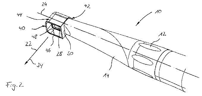
Fig. 2 a perspective representation of the dental
diagnostic camera according to Fig. 1 with a spacer
attachment;
Fig. 3 a sectional representation of the head region of
the dental diagnostic camera according to Fig. 2;
Fig. 4 a perspective representation of a spacer attachment
with short overall length;
Fig. 5 a perspective representation of a spacer attachment
with medium overall length;
Fig. 6 a perspective representation of a spacer attachment
with long overall length and with a pyramidally
widened end region; and
CA 02691558 2009-12-21
WO 2008/113493 PCT/EP2008/001931
- 7 -
Fig. 7 a side view of a dental diagnostic camera with
spacer moulded onto the camera housing.
Represented in Fig. 1 is a dental diagnostic camera 10
which exhibits a gripping portion 12 and a head region 14
designed substantially in the form of a conical portion
which is provided in prolongation of the gripping portion
12.
At a rear end of the gripping portion 12 facing away from
the head region 14 and not represented, the dental
diagnostic camera 10 exhibits a cable, not represented,
which is provided for the provision of electrical energy
and, in particular, for the communication of the electrical
image signals generated by the dental diagnostic camera 10.
The image signals are generated by a camera unit 16
integrated within the head region 14.
The camera unit 16 consists substantially, as represented
in more detail in Fig. 3, of an optical system 18 and an
image-recording device 20. A direction of view 22 of the
diagnostic camera 10 is oriented substantially orthogonally
relative to a central longitudinal axis 24 of the gripping
portion 21 and of the head region 14. As a result, a
particularly ergonomic handling of the diagnostic camera 10
is guaranteed, which is also particularly advantageous for
the examination of tooth surfaces in the cramped oral
space.
According to Fig. 2, an attachment 40 is attached onto the
diagnostic camera 10, which attachment facilitates
compliance with a defined spacing between the diagnostic
camera and the object to be examined and which will be
CA 02691558 2009-12-21
WO 2008/113493 PCT/EP2008/001931
- 8 -
described in more detail below on the basis of several
exemplary embodiments.
As represented in more detail in the sectional
representation shown in Fig. 3, rays of light emanating
from an illuminated object 41, for example from a tooth in
an oral space of a patient, are projected onto an image-
recording device 20 by means of the optical system 18. The
image-recording device 20 is, for example, a CCD (charge-
coupled device) and exhibits a photosensitive sensor
surface which effects, pixel by pixel, a conversion of
radiated rays of light into electrical signals.
The electrical output signals of the image-recording device
20 are then passed via the cable, which is not represented,
to an image-processing unit, likewise not represented,
which controls the representation of the object 41 on a
monitor.
The optical system 18 exhibits a transparent window 28
inserted tightly into the wall of the head region 16, a
deflecting mirror 26, and a lens system 30 with two lenses
31, 33. The transparent window 28 is tightly glued into
the head region 14, in order to be able to accommodate the
camera unit 16 in moisture-proof manner in the sleeve-
shaped head region 14 forming a part of the camera housing.
'The deflecting mirror 26 brings about a 90-degree
deflection of the rays of light, i.e. of the object rays
emanating from the object 41. A ray of light emanating
from the object 41 which is considered in exemplary manner
can, after deflection by the deflecting mirror 26, run
parallel to the central longitudinal axis 24 and impinges
CA 02691558 2009-12-21
WO 2008/113493 PCT/EP2008/001931
- 9 -
perpendicularly on the sensor surface of the image-
recording device 20. The direction of view 22 of the
camera unit 16 is consequently oriented orthogonally
relative to the central longitudinal axis 24 of the
diagnostic camera 10 by the action of the deflecting
mirror 26.
The angle of coverage y which is capable of being
registered by the image-recording device 20 is
substantially determined by the lens system 30. In
addition, the lens system 30 determines a depth-of-field
region or object region 36 within which an object is
sharply imaged onto the image-recording device 20.
Within the object region 36 an object plane 38
characterises that plane in which an object is imaged in
maximally sharp manner. The angle of coverage y can, where
appropriate, be limited by the size of the window 28, by
the size of the deflecting mirror 26, or by a field stop,
not represented, provided in the optical ray path or also
by the attachment 40.
Adjacent to the deflecting mirror 26 a light-source 32
constructed as a white-light LED is provided which serves
for illumination of the object to be imaged. The principal
direction of radiation 34 of the light-source 32 runs at
least substantially parallel to the direction of view 22 of
the camera unit 16.
The attachment 40 shown in the sectional representation of
Fig. 3 is pushed onto the end of the head region 14 of the
diagnostic camera 10 and is retained on the head region 14
by forced closure and also positively.
CA 02691558 2009-12-21
WO 2008/113493 PCT/EP2008/001931
- 10 -
The attachment 40 produced from a sterilisable plastic, for
example polypropylene, exhibits a coupling portion 42
designed as connection means and also a spacer portion 44.
As represented in more detail in Figs. 4 to 6, wall
portions of the coupling portion 42 and the spacer portion
44 delimit an opening 45, into which the head region 16 can
be introduced in positive manner.
The coupling portion 42 and the spacer portion 44 are both
sleeve-shaped and together form a right-angled channel.
The length of the channel in the direction of the central
longitudinal axis 24 is really short in the embodiments of
the attachment 40 according to Figs. 3 to 6 which are
represented. In a further embodiment of the invention
which is not represented, the channel may also be
significantly longer in the region of the coupling portion,
in order to achieve a greater axial overlap with the head
region 16.
The coupling portion 42 is matched to the cross-section of
the head region as regards the cross-section of the opening
45 in such a manner that a force-closed coupling of the
attachment 40 to the head region 14 is obtained. To this
end, the free inner cross-section of the coupling portion
42 is chosen to be slightly smaller than the outer cross-
section of the head region 14, so that when the attachment
40 is placed onto the head region 14 an elastic deformation
of the coupling portion 42 occurs. This elastic
deformation provides the frictional force necessary for the
desired force-closed fixing of the attachment 40 to the
diagnostic camera 10.
CA 02691558 2009-12-21
WO 2008/113493 PCT/EP2008/001931
- 11 -
In addition, the coupling portion 42 is provided with an
inward-projecting detent lug 56 which engages elastically
in a groove provided in the head region 14 and in this way
guarantees a reliable latching of the attachment 40 on the
diagnostic camera 10.
The spacer portion 44 designed in the form of a sleeve
exhibits a free end portion 46 projecting beyond the window
28 in the direction of view 22 of the camera unit 16, which
terminates in the object plane 38 and is at least closely
adjacent to the latter.
Hence the free end portion 46 indicates to the user how
closely the diagnostic camera 10 has to be advanced towards
the object 41 in order to provide a sharp image. If the
free end portion 46 is placed onto a surface of an object
41, the object surface is automatically situated within the
object region 36 of the diagnostic camera 10.
By virtue of the contiguity of the free end portion 46 on
the object surface, in addition an attitude stabilisation
of the diagnostic camera 10 is ensured. This facilitates,
on the one hand, the choice of the correct image detail.
On the other hand, jittering movements of the user, which
could arise without a placement of the free end portion 46
onto the object, are reduced or entirely avoided. Hence
the image recorded by the diagnostic camera 10 is still and
stable.
The attachment 40 represented in Fig. 4 exhibits a box-like
structure. A side wall of the attachment 40 is completely
recessed, as a result of which a viewing window 48 bordered
by the sleeve-shaped free end portion 46 is formed. On a
CA 02691558 2009-12-21
WO 2008/113493 PCT/EP2008/001931
- 12 -
side wall of the attachment 40 adjacent to the viewing
window 48 the opening 45 serving for coupling purposes is
provided, which exhibits a substantially circular edge
contour with a flat region 50 oriented parallel to the end
face of the end portion 46.
In the case of the attachment 40 according to Fig. 4, the
free end portion 46 is arranged approximately 5 mm away
from the flat region 50, so that the spacing between the
object 41 and the window 28 likewise amounts to
approximately 5 mm and the free end portion 46 is situated
at least almost in the object plane 38.
On an inner face of the attachment 40 a full-perimeter
roughened surface 54 is provided in the region of the
spacer portion 44. The roughening can be achieved, for
example, by a surface treatment provided in the injection
mould for the attachment 40, for example by sandblasting,
and is moulded onto the attachment 40 in the course of the
injection process.
The roughened surface 54 has the effect that rays of light
emanating from the light-source 32 or from the object are
diffusely scattered on the inner surface of the spacer
portion 44. Consequently these rays of light cannot be
scattered into the optical system 18 of the camera unit 16
in unhindered manner as rays of stray light, as a result of
which an improvement of the image quality of the diagnostic
camera 10 can be realised.
Alternatively, the inner face of the spacer portion 44 may
be constructed to be light-absorbing, in particular black,
for example it may be lacquered black.
CA 02691558 2009-12-21
WO 2008/113493 PCT/EP2008/001931
- 13 -
By virtue of the geometry of the attachment 40 which is
matched to the angle of coverage of the camera unit, in
addition a lateral escape of rays of light that were
radiated by the light-source 32 is reduced or prevented.
Hence more light is available for the illumination of the
object, as a result of which a further contribution for an
improved image quality is obtained.
In the case of the attachment 40 represented in Fig. 5,
wherein for functionally identical elements the reference
symbols already introduced are retained, the free end
portion 46 is arranged about 10 mm away from the flat
region 50 and is partly interrupted by a recess 58.
Hence in the course of placing the diagnostic camera 10
provided with the attachment 40 onto an uneven surface it
can be guaranteed that object regions protruding from the
uneven surface, which are to be represented by means of the
diagnostic camera 10, come to be situated in the object
plane 38 or at least in the object region 36 of the
diagnostic camera 10.
In addition, the recess 58, which faces towards the user of
the diagnostic camera 10, makes possible a direct view of
the surface to be recorded.
In the case of the attachment 40 represented in Fig. 6,
wherein for functionally identical elements the reference
symbols already introduced have been retained, the free end
portion 46 is arranged about 18 mm away from the flat
region 50 and is located at the end on an extension 52 of
CA 02691558 2009-12-21
WO 2008/113493 PCT/EP2008/001931
- 14 -
the box-shaped attachment 40 which is designed
substantially in the form of a truncated pyramid.
With the design of the extension 52 in the form of a
truncated pyramid it can be ensured that the attachment 40
is of compact construction and can be placed in simple
manner onto an object to be examined. The aperture angle
of the extension 52, accordingly the angle included between
the pyramidal faces arranged opposite in the given case, is
adapted to the angle of coverage y of the camera unit 16 in
such a manner that marginal rays which can still be
registered by the image-recording device 20 run
substantially parallel to the pyramidal faces of the
extension 52. Hence the attachment 40 does not act as a
limit stop for the image recorded by the camera unit 16.
In the exemplary embodiment according to Figure 7, a spacer
bar 44 is moulded onto the head region 16, the axis of
which runs perpendicular to the axis of gripping portion 14
and head region 16 and is situated laterally outside the
field of view or is closely adjacent to the edge of the
field of view.
Alternatively, the spacer bar 44 mav be detachably inserted
into a recess of the head region 16, which is advantageous
with regard to production as well as cleaning and
sterilisation of the diagnostic camera.
In this way, the same advantages are obtained as in the
exemplary embodiments according to Figures 1 to 6.
The diagnostic camera described above may also be used for
the purpose of examining other poorly accessible body parts
CA 02691558 2009-12-21
WO 2008/113493 PCT/EP2008/001931
- 15 -
of humans and animals in human medicine or in veterinary
medicine. It may also be employed in materials testing and
product testing for the purpose of monitoring poorly
accessible surface regions.
The free end porti.on 46 of the spacer 40 can be tightly
sealed by a transparent end plate 55, as indicated in
Figure 4. This constitutes an abutment surface for tissue
to be examined and in this way provides for a precise
positioning of the same in the object plane. At the same
time, curvatures of the tissue are prevented. In this way
the free erid of the spacer is also protected against
penetration of contaminants and germs. The smooth outer
surface at the end of the spacer 40 facilitates the
sterilisation and disinfection thereof.