Note: Descriptions are shown in the official language in which they were submitted.
CA 02691787 2009-12-22
WO 2009/005769 PCT/US2008/008119
SOFT GEL SYSTEMS IN MODULATING STEM CELL DEVELOPMENT
FIELD OF INVENTION
[001] This invention provides methods of modulating stem cell development
using soft-gels. Specifically, the
invention provides methods, compositions and devices for modulating the
development of stem cells, using
gels having optimized viscoelastic properties.
BACKGROUND OF THE INVENTION
[002] Adult mesenchymal stem cells have the ability to self-renew and
differentiate into multiple cell lineages
of mesenchymal tissues. Therefore, clinical applications of these cells, such
as replacement of damaged tissues
or carriers for anti-cancer agents, have been considered. Applications of
adult mesenchymal stem cells are still
limited to preclinical stage at this time, in part because of rapid aging of
these cells ex vivo, which limits their
expansion and engineering. Immortalizing mesenchymal stem cells by telomerase
transduction is reported to
overcome issues associated with accelerated aging. However, their ability of
unlimited self-renewal may lead to
an out-of-control growth, once they are implanted into tissues. In fact,
transformation of telomerase-transduced
mesenchymal stem cells was observed in in vitro settings.
[003] Thus, regulation of the growth of adult mesenchymal stem cells is one of
the key steps toward their
clinical applications.
SUMMARY OF THE INVENTION
[004] This invention provides methods of modulating stem cell development
using soft-gels. Specifically, the
invention provides methods, compositions and devices for modulating the
development of stem cells, using
gels having optimized viscoelastic properties.
[005] In one embodiment, the present invention provides a method of
manufacturing a coated polyacrylamide
gel having a rigidity in a range of 150-750 Pa, comprising the steps of
polymerizing a composition comprising
acrylamide and bisacrylamide, said composition having an acrylamide:
bisacrylamide mixture ratio of between
100:1 and 30:1; and coating said soft polyacrylamide gel with a composition
comprising a collagen type I and a
fibronectin.
[006] In another embodiment, the invention provide s a method of manufacturing
a fibrin matrix having a
rigidity in a range of 0.1-2.5 kPa, comprising the steps of polymerizing a
composition comprising a fibrin or
1
CA 02691787 2009-12-22
WO 2009/005769 PCT/US2008/008119
P-70022-PC
fibrinogen protein, thereby producing a soft fibrin matrix, wherein the
concentration of said fibrin or fibrinogen
protein in said soft fibrin matrix is 1-20 mg/ml, and coating said soft fibrin
matrix with a composition
comprising an adhesion protein.
[007] In another embodiment, the present invention provides a method of
preserving a mesenchymal stem
cell population, said method comprising the step of culturing said mesenchymal
stem cell population in a gel
matrix having a rigidity in a range of 0.1-2.5 kPa.
[008] In another embodiment, the present invention provides a method of
inducing differentiation of a
mesenchymal stem cell population into an adipocyte population, said method
comprising the step of culturing
said mesenchymal stem cell population in the presence of an apparatus
containing (a) a gel or matrix having a
rigidity in a range of 150-750 Pa and (b) an adipocyte induction medium,
thereby inducing differentiation of a
mesenchymal stem cell population into a cell type of interest.
[009] In another embodiment, the present invention provides an apparatus for
modulating growth of a
mesenchymal stem cell comprising: a gel matrix having a rigidity in a range of
150-750 Pa; and an adipocyte
induction medium, wherein said gel or matrix is coated with a type I collagen,
a fibronectin, or a combination
thereof.
[0010] In another embodiment, the present invention provides a gel matrix
comprising a gelling agent and an
acrylamide- bisacrylamide mixture wherein said gel matrix is coated with a
type I collagen, a fibronectin, or a
combination thereof and having a rigidity in a range of 150-750 Pa.
[0011] In one embodiment, the invention provides a method of modulating
development of a mesenchymal
stem cell, comprising the step of suspending the mesenchymal stem cell in a
gel matrix comprising a gelling
agent wherein said gel matrix is coated with a type I collagen, a fibronectin,
or a combination thereof and
wherein said gel matrix is maintained at a predetermined rigidity; and
exposing the gel matrix to a growth
modulating factor.
[0012] In another embodiment, the invention provides a method for inducing or
maintaining quiescence and
sustaining biological activity in a somatic stem cell ex vivo, comprising:
contacting the somatic stem cell with a
gel matrix comprising an extracellular material that bind to integrin on the
membrane of the somatic stem cell,
said gel matrix having a substantially similar elasticity to the elasticity of
the predominant in vivo biological
microenvironment of the somatic stem cell of the same type in vivo; and
providing the somatic stem cell with
2
CA 02691787 2009-12-22
WO 2009/005769 PCT/US2008/008119
P-70022-PC
nutrient material for sustaining biological activity of the somatic stem cell
ex vivo.
BRIEF DESCRIPTION OF THE FIGURES
[0013] Figure 1. A. Mechanical properties of polyacrylamide substrates. The
shear modulus of polyacrylamide
gels with a range of acrylamide (indicated as percents near data lines) to
bisacrylamide (indicated as
crosslinker) proportions was measured. The shear modulus (G'), expressed in
Pascal, increases at constant
polymer mass with increasing crosslinker. Increasing the concentration of
acrylamide from 3 to 12% also
creates a large stiffness range from 10 to 50,000 Pa. The solid line denotes
the theoretical stiffness of a
rubberlike network if every crosslink was elastically effective. B. Cell shape
and F-actin structure of hMSC on
stiff or soft matrices. C. Cell shape and F-actin structure of hMSC on soft
gels and glass.
[0014] Figure 2. BrdU incorporation into hMSC.
[0015] Figure 3. The effect of matrix rigidity on adipocyte differentiation.
A. Graph of percent positive cells.
First bar in each series: Oil Red 0-staining. Second bar: PPARy2 staining.
[0016] Figure 4. F-actin structure in astrocytes seeded either on stiff or
soft gels.
[0017] Figure 5. Quantification of increase in Rho activity from soft to hard
gels. Astrocytes were seeded on
polyacrylamide gels with various stiffness. GTP-loading level of Rho was
quantified.
[0018] Figure 6. Melanoma cells spread more on stiff matrices. Graphical
representation of area.
[0019] Figure 7. Melanoma cells adhered to soft and stiff gels with same
efficiency.
[0020] Figure 8. Larger population of melanoma cells on stiff gels.
[0021 ] Figure 9 show images of human MSCs on several substrates of different
elasticities according to
various embodiments of the present invention.
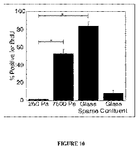
[0022] Figure 10 shows the amount of bromodeoxyuridine (BrdU) uptake into
human MSCs on substrates of
varying elasticities according to various embodiments of the invention.
[0023] Figure 11 shows (A-D) an illustration of the effect of a quasi 3D
environment on stem cell shape and
proliferation according to various embodiments of the invention.
3
CA 02691787 2009-12-22
WO 2009/005769 PCT/US2008/008119
P-70022-PC
[0024] Figure 12 shows the response of human MSCs to adipogenic induction
media according to various
embodiments of the invention.
[0025] Figure 13 shows calcium deposition visualized with Alizarin Red S after
stimulation of human MSCs
with osteoinduction media according to various embodiments of the invention.
[0026] Figure 14 shows a flow chart for preparing a system for inducing
quiescence, differentiation, and
proliferation in adult stem cells according to various embodiments of the
invention.
[0027] Figure 15 shows schematic illustrations of embodiments of systems of
the present invention.
DETAILED DESCRIPTION OF THE INVENTION
[0028] This invention provides gels and matrices having a rigidity in the
range of 0.01-50 kPa, methods of
manufacturing same, and method of preserving a mesenchymal stem cell
population or studying mesenchymal
stem cells, comprising same.
[0029] In one embodiment, provided herein is a method of manufacturing a
polyacrylamide gel with a rigidity
in a range of 150-750 Pa, comprising the steps of polymerizing a composition
comprising acrylamide and
bisacrylamide, thereby producing a soft polyacrylamide gel, and coating the
soft polyacrylamide gel with a
composition comprising a collagen type I and a fibronectin, thereby
manufacturing a polyacrylamide gel having
a rigidity in a range of 150-750 Pa. In another embodiment, the composition
has an acrylamide: bisacrylamide
mixture ratio of between 100:1 and 30:1. In another embodiment, the gel has an
acrylamide: bisacrylamide
mixture ratio of between 100:1 and 30:1. In another embodiment, the
composition a total acrylamide
concentration of 3-5%. In another embodiment, the gel or matrix has a total
acrylamide concentration of 3-5%.
In another embodiment, the composition is a solution. In another embodiment,
the composition is a suspension.
In another embodiment, the composition is any other type of composition known
in the art. Each possibility
represents a separate embodiment of the present invention.
[0030] In another embodiment, provided herein is a method of manufacturing a
fibrin matrix with a rigidity in
a range of 150-750 Pa, comprising the steps of polymerizing a composition
comprising a fibrin or fibrinogen
protein, thereby producing a soft fibrin matrix, wherein the concentration of
the fibrin or fibrinogen protein in
the soft fibrin matrix is 3-10 mg/mL, and coating the soft fibrin matrix with
a composition comprising an
adhesion protein, thereby manufacturing a fibrin matrix having a rigidity in a
range of 150-750 Pa.
4
CA 02691787 2009-12-22
WO 2009/005769 PCT/US2008/008119
P-70022-PC
[0031 ] In another embodiment, provided herein is a method of preserving a
mesenchymal stem cell population,
the method comprising the step of culturing the mesenchymal stem cell
population in a gel or matrix with a
rigidity in a range of 150-750 Pa, thereby preserving a mesenchymal stem cell
population. In another
embodiment, the step of culturing is performed in the absence of chemical
induction. In another embodiment,
the step of culturing is performed in the absence of an induction medium. Each
possibility represents a separate
embodiment of the present invention.
[0032] In another embodiment, provided herein is a method of preserving a
mesenchymal stem cell, the
method comprising the step of culturing the mesenchymal stem cell population
in a gel or matrix with a rigidity
in a range of 150-750 Pa, thereby preserving a mesenchymal stem cell. In
another embodiment, the step of
culturing is performed in the absence of chemical induction. In another
embodiment, the step of culturing is
performed in the absence of an induction medium. Each possibility represents a
separate embodiment of the
present invention.
[0033] In another embodiment, provided herein is a method of inducing
quiescence of a transformed cell,
comprising the step of culturing the transformed cell in a gel or matrix of
the present invention, thereby
inducing quiescence of a transformed cell. In another embodiment, the
transformed cell is a cancer cell. In
another embodiment, the transformed cell is a neoplastic cell. In another
embodiment, the transformed cell is
any other type of transformed cell known in the art. Each possibility
represents a separate embodiment of the
present invention.
[0034] In another embodiment of methods and compositions of the present
invention, the telomerase length of
the mesenchymal stem cell population is maintained. "Maintained" refers, in
another embodiment, to a lack of
substantial change in the length. In another embodiment, the term refers to a
lack of measurable change in the
length. In another embodiment, the term refers to a lack of sufficient change
in the length to affect proliferative
capacity. Each possibility represents a separate embodiment of the present
invention.
[0035] In another embodiment of methods and compositions of the present
invention, the mesenchymal stem
cell population is maintained in a quiescent state. "Quiescent" refers, in
another embodiment, to a lack of
significant replication. In another embodiment, the term refers to a
significantly reduced level of replication. In
another embodiment, the term refers to a large percentage of cells arrested in
the cell cycle. In another
embodiment, the cells are arrested at the GI phase. In another embodiment, the
cells are arrested in the G2
phase. In another embodiment, "quiescent" refers to any other art-accepted
definition of the term. Each
5
CA 02691787 2009-12-22
WO 2009/005769 PCT/US2008/008119
P-70022-PC
possibility represents a separate embodiment of the present invention.
[0036] In one embodiment, embodiment, when the stem cell is a bone marrow-
derived human mesenchymal
cell, the extracellular matrix (ECM) has an elasticity of about 250 Pa, and
comprises a mixture of collagen and
fibronectin. In another embodiment, the collagen is rat tail collagen, and the
fibronectin is human fibronectin.
The ratio of the collagen and fibronectin may vary, and in an embodiment, the
ratio of collagen to fibronectin is
approximately 5:1. Other ratios of collagen and fibronectin may be used. One
of ordinary skill in the art will
appreciate that collagen and fibronectin can be obtained from other sources,
and that substances other than
collagen and fibronectin may be used to present elasticity and bind to
integrins on the surface of the cell
membrane so that quiescence of the cell is induced.
[0037] According to embodiments of the present invention, the extracellular
material (ECM) is provided with
an appropriate apparent elasticity by coupling the ECM with a substrate such
that a stem cell contacting the
ECM senses the elasticity of the substrate. Correspondingly, the substrate may
be a material whose elasticity,
when coupled to the ECM, is sensed by a stem cell contacting the ECM. In some
embodiments, the substrate is
glass. In other embodiments, the substrate is a gel with elasticity of 250 Pa,
or a gel with elasticity.of 7500 Pa.
These gels may be polyacrylamide gels, and, as known to those with skill in
the art, the elasticity of
polyacrylamide gels may be varied, for example, by changing the concentrations
of acrylamide and
bisacrylamide in the gel formulation. The manufacture of gels of varying
elasticity that may be used in the
method of the present invention will be apparent to one of skill in the art in
light of this specification.
[0038] The elasticity of the in vivo biological environment of a stem cell may
be determined by extracting a
sample of physiological tissue from the immediate in vivo environment of the
stem cell, and then measuring the
shear modulus of that tissue sample. Exemplary procedures for preparing and
measuring the elasticity of rat
tissue and bovine tissue are described in this specification. Table I below
provides the elasticity of various
types of tissues:
Table 1
Species Tissue Elasticity (in Pa)
(mean SD)
Bovine Bone marrow 220 50
Rat Subcutaneous fat 160 70
6
CA 02691787 2009-12-22
WO 2009/005769 PCT/US2008/008119
P-70022-PC
Rat Visceral fat 130 70
Rat Liver 403 28
Rat Skeletal muscle 2251 166
[0039] In one embodiment, human MSCs on 250 Pa gels are in a quiescent state
awaiting a further signal to
determine their fate. In one embodiment, the hMSC's will undergo adipogenic
differentiation (induced by
chemical factors), or in other embodiments, a return to the cell cycle
(induced by coupling the cells to a stiff
surface), or osteogenic differentiation (which appears to require both
chemical induction and a stiff substrate).
Stimulating cells cultured on soft gels with adipogenic differentiation
factors results in one embodiment, in a
remarkably high number of cells accumulating lipid droplets. In one
embodiment, chemical induction is
required for osteoblast differentiation. The requirement for synchronized
mechanical and chemical stimulation
explains in one embodiment, how human MSCs can be compartmentalized into
compliant tissues such as bone
marrow and yet resist spontaneous differentiation.
[0040] Like matrix elasticity, in another embodiment the choice of
extracellular ligand strongly affects human
MSC adhesion and differentiation. Collagen type I is found in a variety of
tissues including bone and adipose,
and it is regularly used as a substrate for cell adhesion experiments. In one
embodiment, on 250 Pa gel,
collagen alone does not ensure efficient adhesion of a majority of cells. In
one embodiment, a mixture of
collagen type I and fibronectin at a ratio of 10:1 provides the best adhesion
of cells to the 250 Pa gels without
affecting differentiation potential.
[0041] Human MSCs have the capacity to remodel their microenvironment by
altering the expression of
matrix metalloproteases, and this helps in one embodiment to promote efficient
differentiation after an initial
strong adhesion is achieved.
[0042] In one embodiment, DNA synthesis in human MSCs decreases dramatically
when human MSCs are
cultured on soft gels, developing a round phenotype. This is in contrast with
other proliferating cell types such
as NIH 3T3 fibroblasts, bovine aortic endothelial cells, and NRK epithelial
cells which all continue to divide
when cultured on soft gels. Thus, stem cell quiescence on 250 Pa gels is not a
general shape-induced failure
of cytokinesis, but rather a specific sensitivity of these cells to substrate
compliance. Accordingly and in one
embodiment, provided herein is a method of maintaining stem cells in a
quiescent state, comprising suspending
the stem cells in a fibronectin/collagen gel having G' of 250 Pa.
7
CA 02691787 2009-12-22
WO 2009/005769 PCT/US2008/008119
P-70022-PC
[0043] In one embodiment, when nonproliferating human MSCs are presented with
a protein gel matrix-coated
glass substrate, the cells develop a spindle morphology and reenter the cell
cycle. In another embodiment, the
presence of a stiff substrate overrides the physical cues from a compliant
matrix. In one embodiment, no
significant population of cells exhibiting a neuronal phenotype with neurite-
like protrusions, are present on
soft 250 Pa gels without any chemical induction
[0044] In one embodiment, substrate elasticity regulates differentiation of
cells with specific phenotypes. In
another embodiment, mechanical properties alone do not direct stem cell
differentiation. This is because
several tissues in the body have similar elasticities. For example, brain,
fat, and bone marrow tissues all have a
storage modulus of approximately 200 Pa, yet all maintain unique populations
of cells. In another
embodiment, in vivo human MSCs are stored in an individual's bone marrow for
decades and yet retain
multipotency. In one embodiment, human MSCs are cultured ex vivo on stiff
tissue culture plastic and retain
multipotency for several passages. In one embodiment, both mechanical and
chemical stimuli are integrated by
the cell to determine its response. In another embodiment, while chemical
stimuli can override the influences
of substrate mechanics, in other embodiments, an inappropriate mechanical
environment prevents a normal
cellular response to chemical agonists. In one embodiment, quiescent cells
differentiate into osteoblasts only as
a result of changing both their physical and chemical environments to those
that stimulate osteogenesis.
Accordingly and in one embodiment, a matrix with appropriate elasticity has
the capability to maintain a
quiescent population of multipotent bone marrow mesenchymal stem cells that
respond to both mechanical and
chemical stimuli that drive proliferation and differentiation.
[0045] FIG. 14 illustrates a flow chart for preparing an embodiment of a
system for inducing quiescence,
differentiation, and proliferation in adult stem cells according to various
embodiments of the present invention.
Solutions of acrylamide and bisacrylamide are prepared in phosphate buffered
saline (PBS) to a total volume
of 500 l. In one embodiment, adjusting the concentration of acrylamide and
bisacrylamide enables obtaining a
wide range of rigidity. Polymerization is initiated with TEMED (N,N,N',N'-
Tetramethylethylenediamine) and
ammonium persulfate to form a gel. In step 601, Acrylamide/bisacrylamide
(polyacrylamide) solution, a
droplet (for example, about 200 l) of the polymerized gel is deposited on a
glass coverslip previously
modified with 3-aminopropyltrimethoxysilane and glutaraldehyde. In step 602,
overlay with N-
hydroxysuccinimide in tolulene, approximately 15 [t] of 2% acrylic acid N-
hydroxysuccinimide ester in toluene
is applied to the solution of step 601, and, in stem 603, Top Coverslip, a
chlorosilanized coverslip is placed on
top of the droplet. In step 604, Remove coverslip, the top coverslip is
removed after polymerization is
completed and, optionally, the gel is illuminated with ultraviolet light for
approximately 10 - 15 minutes (not
8
CA 02691787 2009-12-22
WO 2009/005769 PCT/US2008/008119
P-70022-PC
shown). In step 605, ECM ligand, N-succinimide acrylate on the top of the gel
is reacted with an extracellular
matrix ligand, which in an embodiment is a mixture of 0.1 mg/ml of collagen
type 1 and 0.02 mg/ml
fibronectin. In a further step (not depicted), gels are washed 3 times with
PBS and left in PBS until step 606,
Cells on gel, when stem cells are seeded on the cells. When bone marrow-
derived mesenchymal stem cells are
seeded on this material, cells become quiescent even in the presence of
chemical stimuli to cause proliferation
or differentiation.
[0046] Accordingly and in one embodiment, provided herein is a method for
inducing or maintaining
quiescence and sustaining biological activity in a somatic stem cell ex vivo,
comprising: contacting the somatic
stem cell with a gel matrix comprising an extracellular material that bind to
integrin on the membrane of the
somatic stem cell; said gel matrix having a substantially similar elasticity
to the elasticity of the predominant in
vivo biological microenvironment of the somatic stem cell of the same type in
vivo; and providing the somatic
stem cell with nutrient material for sustaining biological activity of the
somatic stem cell ex vivo.
[0047] In a method of the present invention, a stem cell may be contacted with
appropriate ECM in various
ways. For example, as described in this specification, the ECM may form a
layer coupled to the substrate, and
the stem cell may be placed on the ECM. Alternatively, the cell may be placed
on ECM coupled to the
substrate and additionally contacted by ECM placed on the cell, for example by
placing on the cell a structure
coupling ECM to a substrate that presents the appropriate apparent elasticity
to the stem cell.
[0048] In another embodiment, there may be two formulations of ECM: a first
formulation, which may or may
not include nutrient materials, that is coupled to the substrate; and a second
formulation that includes nutrient
materials and that is not coupled with the substrate. Structures and
configurations for contacting stem cells
with an appropriate ECM (including substrates and, optionally, linking
materials for linking the substrate to the
ECM), are described in this specification, including for example FIG. 6, and
are apparent to one of skill in the
art in light of this specification.
[0049] In embodiments of methods of the present invention, a stem cell that is
not in a quiescent state is
contacted with ECM according to methods of the present invention so that
quiescence is induced into the cell
and it transitions from a non-quiescent state to a quiescent state. In other
embodiments, a quiescent stem cell is
contacted with ECM according to methods of the present invention so that
quiescence is maintained in the cell
and it does not transition from a quiescent state.
[0050] Accordingly and in one embodiment, provided herein is a method of
modulating development of a
9
CA 02691787 2009-12-22
WO 2009/005769 PCT/US2008/008119
P-70022-PC
mesenchymal stem cell, comprising the step of suspending the mesenchymal stem
cell in a gel matrix
comprising a gelling agent wherein said gel matrix is coated with a type I
collagen, a fibronectin, or a
combination thereof and wherein said gel matrix is maintained at a
predetermined rigidity; and exposing the
gel matrix to a growth modulating factor, whereby exposure to the chemical or
physical factor results in an
increase in the rigidity of the gel matrix to coincide with the rigidity of
the ECM in the microenvironment the
mesenchymal stem cell is sought to differentiate into
[0051 ] In another embodiment, over 80% of the cells are cell cycle arrested.
In another embodiment, at least
80% of the cells are cell cycle arrested. In another embodiment, over 70% of
the cells are cell cycle arrested. In
another embodiment, at least 70% of the cells are cell cycle arrested. In
another embodiment, over 75% of the
cells are cell cycle arrested. In another embodiment, at least 75% of the
cells are cell cycle arrested. In another
embodiment, over 82% of the cells are cell cycle arrested. In another
embodiment, at least 82% of the cells are
cell cycle arrested. In another embodiment, over 85% of the cells are cell
cycle arrested. In another
embodiment, at least 85% of the cells are cell cycle arrested. In another
embodiment, over 87% of the cells are
cell cycle arrested. In another embodiment, at least 87% of the cells are cell
cycle arrested. In another
embodiment, over 90% of the cells are cell cycle arrested. In another
embodiment, at least 90% of the cells are
cell cycle arrested. In another embodiment, over 92% of the cells are cell
cycle arrested. In another
embodiment, at least 92% of the cells are cell cycle arrested. In another
embodiment, over 93% of the cells are
cell cycle arrested. In another embodiment, at least 93% of the cells are cell
cycle arrested. In another
embodiment, over 94% of the cells are cell cycle arrested. In another
embodiment, at least 94% of the cells are
cell cycle arrested. In another embodiment, over 95% of the cells are cell
cycle arrested. In another
embodiment, at least 95% of the cells are cell cycle arrested. In another
embodiment, over 96% of the cells are
cell cycle arrested. In another embodiment, at least 96% of the cells are cell
cycle arrested. In another
embodiment, over 97% of the cells are cell cycle arrested. In another
embodiment, at least 97% of the cells are
cell cycle arrested. In another embodiment, over 98% of the cells are cell
cycle arrested. In another
embodiment, at least 98% of the cells are cell cycle arrested. In another
embodiment, over 99% of the cells are
cell cycle arrested. In another embodiment, at least 99% of the cells are cell
cycle arrested. Each possibility
represents a separate embodiment of the present invention.
[0052] In another embodiment, replication is reduced by 50%, relative to
replication in a tissue culture dish. In
another embodiment, replication is reduced by 60% relative to a tissue culture
dish. In another embodiment,
replication is reduced by 65% relative to a tissue culture dish. In another
embodiment, replication is reduced by
70% relative to a tissue culture dish. In another embodiment, replication is
reduced by 75% relative to a tissue
CA 02691787 2009-12-22
WO 2009/005769 PCT/US2008/008119
P-70022-PC
culture dish. In another embodiment, replication is reduced by 80% relative to
a tissue culture dish. In another
embodiment, replication is reduced by 85% relative to a tissue culture dish.
In another embodiment, replication
is reduced by 90% relative to a tissue culture dish. In another embodiment,
replication is reduced by 95%
relative to a tissue culture dish. In another embodiment, replication is
reduced by 97% relative to a tissue
culture dish. In another embodiment, replication is reduced by over 97%
relative to a tissue culture dish. In
another embodiment, replication is reduced by over 98% relative to a tissue
culture dish. In another
embodiment, replication is reduced by over 99% relative to a tissue culture
dish. Each possibility represents a
separate embodiment of the present invention.
[0053] In another embodiment, a method of the present invention further
comprises the step of subsequently
(e.g. following culturing in the presence of a gel or matrix of the present
invention) plating the mesenchymal
stem cell population in a tissue-culture apparatus. In another embodiment, the
tissue culture apparatus contains
induction medium. In another embodiment, the step of subsequently plating is
performed with chemical
induction. Each possibility represents a separate embodiment of the present
invention.
[0054] In another embodiment, provided herein is a method of studying
proliferation or differentiation of a
mesenchymal stem cell, comprising the step of culturing the mesenchymal stem
cell in a gel or matrix with a
rigidity in a range of 150-750 Pa, thereby studying proliferation or
differentiation of a mesenchymal stem cell.
[0055] The adipocyte population of methods and compositions of the present
invention is, in another
embodiment, a population comprising adipocytes. In another embodiment, the
population is enriched for
adipocytes. In another embodiment, the population is a partially purified
adipocytes population. In another
embodiment, the adipocytes are isolated from a biological source, followed by
a purification or enrichment
step. In another embodiment, isolation from the biological source is followed
by culturing. In another
embodiment, isolation from the biological source is followed by culturing and
a purification or enrichment
step. Each possibility represents a separate embodiment of the present
invention.
[0056] In another embodiment, the cell population of methods and compositions
of the present invention is
cultured in the presence of a gel or matrix of methods and compositions of the
present invention. In another
embodiment, the cell population is cultured in the gel or matrix. In another
embodiment, the cell population is
cultured on the gel or matrix. In another embodiment, the cell population is
cultured in a tissue culture
apparatus containing the gel or matrix. Each possibility represents a separate
embodiment of the present
invention.
11
CA 02691787 2009-12-22
WO 2009/005769 PCT/US2008/008119
P-70022-PC
[0057] "Mesenchymal stem cell population" refers, in another embodiment, to a
population comprising
mesenchymal stem cells (MSC). In another embodiment, the population is
enriched for MSC. In another
embodiment, the population is a partially purified MSC population. In another
embodiment, the MSC are
isolated from a biological source, followed by a purification or enrichment
step. In another embodiment,
isolation from the biological source is followed by culturing. In another
embodiment, isolation from the
biological source is followed by culturing and a purification or enrichment
step. Each possibility represents a
separate embodiment of the present invention.
[0058] "Mesenchymal" cells of methods and compositions of the present
invention are isolated or purified, in
another embodiment, from bone marrow. In another embodiment, the cells are
bone marrow-derived
mesenchymal stem cell. In another embodiment, the cells are isolated or
purified from adipose tissue. In
another embodiment, the cells are isolated or purified from cartilage. In
another embodiment, the cells are
isolated or purified from any other tissue known in the art. Each possibility
represents a separate embodiment
of the present invention.
[0059] In another embodiment, a gel or matrix of methods and compositions of
the present invention has a
stiffness of 150-750 pascals (Pa). In another embodiment, a gel or matrix of
methods and compositions of the
present invention has a shear modulus of 150-750 Pa. Each possibility
represents a separate embodiment of the
present invention.
[0060] In another embodiment, a gel or matrix of methods and compositions of
the present invention has a
stiffness equivalent to a biological tissue. In another embodiment, the
biological tissue is bone marrow. In
another embodiment, the biological tissue is fat tissue. In another
embodiment, the biological tissue is any other
biological tissue known in the art. Each possibility represents a separate
embodiment of the present invention.
[0061] In one embodiment, the gel matrix described herein are capable of
forming gels of various strength,
depending on their structure and concentration as well as, in another
embodiment, environmental factors such
as ionic strength, pH and temperature. The combined viscosity and gel behavior
referred to as "viscoelasticity"
in one embodiment, are examined by determining the effect that an oscillating
force has on the movement of
the material. In another embodiment elastic modulus (G'), viscous modulus
(G"), and complex viscosity (q*)
are the parameters sought to be changed using the methods described herein,
and these are analyzed in another
embodiment by varying either stress or strain harmonically with time (Table
1). These parameters are derived
from the complex modulus (G*), which is the ratio of maximum stress to maximum
strain, and the phase angle
12
CA 02691787 2009-12-22
WO 2009/005769 PCT/US2008/008119
P-70022-PC
(0), which is the angle that the stress and strain are out of phase.
Table 2. Relationships between dynamic moduli, phase angle (8), and fre(luency
(c)).
Term Symbol Definition Information provided
Complex modulus G* [(G,)2 +(G")21o.g All viscoelastic characteristics
Elastic modulus, C: G, cos o Energy stored per deformation
storage modulus cycle; solid-like or elastic
behavior
Viscous modulus, G" G* sin o Energy dissipated per
loss modulus deformation cycle; gluid-like
or viscous behavior
Complex viscosity n* G*/w Viscoelastic flow
[0062] In one embodiment, in the gel matrices described herein, some of the
deformation caused by shear
stress is elastic and will return to zero when the force is removed. The
remaining deformation such as that
deformation created by the sliding displacement of the chains through the
solvent in one embodiment will not
return to zero when the force is removed. Under a constant force the elastic
displacement remains constant in
one embodiment, whereas the sliding displacement continues, so increasing.
[0063] In one embodiment, the term "elastic," or "elasticity," and like terms
refer to a physical property of the
gel matrices described herein, namely the deformability of the gel under
mechanical force and the ability of the
gel matrix to retain its original shape when the deforming force is removed.
In another embodiment, the term
"elastic modulus" refers to Young's Modulus and is a measure of the ratio of
(a) the uniaxial stress along an
axis of the material to (b) the accompanying normal strain along that axis.
[0064] The shear modulus (resulting from changing strain) is the ratio of the
shear stress to the shear strain. It
follows from the complex relationship similar to the above that:
G*=G'+iG"
[0065] where G* is the complex shear modulus, G' is the in-phase storage
modulus, i is a material-related
factor and G" is the out-of-phase similarly-directed loss modulus; G* = E(G'2
+ G"2). The frequency where
these parameters cross over corresponds to a relaxation time (T) specific for
the material.
[0066] In one embodiment, linear viscoelastic properties of the gel matrices
described herein are determined
by measurements in an oscillating shear flow at small amplitude and with
variable angular frequency. The
values for G' and G" are determined to a great extent here by the
concentration of the cellulose derivatives in
13
CA 02691787 2009-12-22
WO 2009/005769 PCT/US2008/008119
P-70022-PC
the aqueous solution and the magnitude of the representative viscosity value.
Therefore, hereinafter, only the
relative course of G' and G" with increasing angular frequency c?, is
considered. In another embodiment, at a
concentration of 1.5 to 2 % (w/w) of cellulose derivative of aqueous solution
and a temperature of
approximately 20 C, the behavior of G' and G" for the cellulose derivatives is
such that at a low angular
frequency (w, the storage modulus G' is less than the loss modulus G", but
with increasing angular frequency G'
increases more greatly than G". In another embodiment, G', above a certain
angular frequency, finally becomes
greater than G", and the solution at high values of angular frequency thus
predominantly reacts elastically. This
behavior is attenuated or changed using the modulating methods described
herein.
[0067] In another embodiment, the term "Elasticity" refers to the physical
property of a material that defines its
ability to deform by stress, whether or not the deformation is reversible. As
used in this specification, elasticity
and rigidity are inversely related, and the elasticity (rigidity) of a
material may be measured by using an RFS III
fluids spectrometer rheometer, available from Rheometrics, Piscataway, NJ,
using a 2% oscillatory shear strain
at a frequency of 10 radians per second. Elasticity and other 10 rheological
properties of cells and other
physiological tissues can be measured using any of a variety of methods known
to those skilled in the art. Such
methods may involve the use of rheometers or atomic force microscopes, as
examples. (See, e.g., Engler AJ,
Rehfeldt F, Sen S, Discher DE, "Microtissue elasticity: measurements by atomic
force microscopy and its
influence on cell differentiation," Methods Cell Biol. 2007;83:521-45; 15
Yeung T, Georges PC, Flanagan LA,
Marg B, Ortiz M, Funaki M, Zahir N, Ming W, Weaver V, Janmey PA, "Effects of
substrate stiffness on cell
morphology, cytoskeletal structure, and adhesion," Cell Motil Cytoskeleton.
2005 Jan;60(1):24-34.).
[0068] In one embodiment, the term "Intrinsic viscosity ([rl*]) refers to the
limit of the reduced viscosity
extrapolated to zero concentration. As with the reduced viscosity, it has
units of reciprocal concentration, for
example, mL g-I.
[0069] In one embodiment, rigidity or stiffness, refers to the G' values
observed or measured.
[0070] In another embodiment, a gel or matrix of methods and compositions of
the present invention is coated
with a solution comprising an adhesion protein. In another embodiment, the
adhesion protein is a collagen. In
another embodiment, the adhesion protein is a type I collagen. In another
embodiment, the adhesion protein is
a fibronectin. In another embodiment, the adhesion protein is any other
adhesion protein known in the art. In
another embodiment, the gel or matrix is coating with a solution comprising a
combination of adhesion
proteins. In another embodiment, the gel or matrix is coating with a solution
comprising a collagen and a
14
CA 02691787 2009-12-22
WO 2009/005769 PCT/US2008/008119
P-70022-PC
fibronectin. In another embodiment, the gel or matrix is coating with a
solution comprising a type I collagen
and a fibronectin. Each possibility represents a separate embodiment of the
present invention.
[0071] In another embodiment, the collagen of methods and compositions of the
present invention is a
recombinant collagen. In another embodiment, the collagen is purified from a
biological source. In another
embodiment, the collagen is a type I collagen. In another embodiment, the
collagen is any other type of
collagen known in the art. Each possibility represents a separate embodiment
of the present invention.
[0072] In another embodiment, the fibronectin of methods and compositions of
the present invention is a
recombinant fibronectin. In another embodiment, the fibronectin is purified
from a biological source. In another
embodiment, the fibronectin is a type 1 fibronectin. In another embodiment,
the fibronectin is any other type of
fibronectin known in the art. Each possibility represents a separate
embodiment of the present invention.
[0073] The gelling agent of methods and compositions of the present invention
is, in another embodiment, an
acrylamide. In another embodiment, the gelling agent is an acrylamide-
bisacrylamide mixture. In another
embodiment, the gelling agent comprises acrylamide. In another embodiment, the
gelling agent comprises an
acrylamide-bisacrylamide mixture. Each possibility represents a separate
embodiment of the present invention.
[0074] In another embodiment, an acrylamide gel of methods and compositions of
the present invention has an
acrylamide: bisacrylamide ratio of between 100:1 and 30:1. In another
embodiment, the acrylamide gel is
prepared from a solution having an acrylamide:bisacrylamide ratio of between
100:1 and 30:1. In another
embodiment, the ratio is between 100:1 and 20:1. In another embodiment, the
acrylamide:bisacrylamide ratio is
between 100:1 and 40:1. In another embodiment, the ratio is between 100:1 and
50:1. In another embodiment,
the ratio is between 100:1 and 60:1. In another embodiment, the ratio is
between 100:1 and 70:1. In another
embodiment, the ratio is between 120:1 and 30:1. In another embodiment, the
ratio is between 120:1 and 40:1.
In another embodiment, the ratio is between 120:1 and 50:1. In another
embodiment, the ratio is between 120:1
and 60:1. In another embodiment, the ratio is between 120:1 and 70:1. In
another embodiment, the ratio is
between 90:1 and 20:1. In another embodiment, the ratio is between 90:1 and
30:1. In another embodiment, the
ratio is between 90:1 and 40:1. In another embodiment, the ratio is between
90:1 and 50:1. In another
embodiment, the ratio is between 90:1 and 60:1. In another embodiment, the
ratio is between 80:1 and 20:1. In
another embodiment, the ratio is between 80:1 and 30:1. In another embodiment,
the ratio is between 80:1 and
40:1. In another embodiment, the ratio is between 80:1 and 50: 1.
[0075] In another embodiment, the ratio is 30:1. In another embodiment, the
ratio is 20:1. In another
CA 02691787 2009-12-22
WO 2009/005769 PCT/US2008/008119
P-70022-PC
embodiment, the ratio is 25:1. In another embodiment, the ratio is 35:1. In
another embodiment, the ratio is
40:1. In another embodiment, the ratio is 45:1. In another embodiment, the
ratio is 50:1. In another
embodiment, the ratio is 55:1. In another embodiment, the ratio is 60:1. In
another embodiment, the ratio is
65:1. In another embodiment, the ratio is 70:1. In another embodiment, the
ratio is 75:1. In another
embodiment, the ratio is 80:1. In another embodiment, the ratio is 85:1. In
another embodiment, the ratio is
90:1. In another embodiment, the ratio is 95:1. In another embodiment, the
ratio is 100:1.
[0076] Each acrylamide:bisacrylamide ratio represents a separate embodiment of
the present invention.
[0077] In another embodiment, an acrylamide gel of methods and compositions of
the present invention has a
total acrylamide concentration of 3-5%. In another embodiment, the acrylamide
gel is prepared from a solution
having a total acrylamide concentration of 3-5%. In another embodiment, the
total acrylamide concentration is
2%. In another embodiment, the concentration is 2.5%. In another embodiment,
the concentration is 3%. In
another embodiment, the concentration is 3.5%. In another embodiment, the
concentration is 4%. In another
embodiment, the concentration is 4.5%. In another embodiment, the
concentration is 5%. In another
embodiment, the concentration is 5.5%. In another embodiment, the
concentration is 6%. In another
embodiment, the concentration is 2-5%. In another embodiment, the
concentration is 2.5-5%. In another
embodiment, the concentration is 3.5-5%. In another embodiment, the
concentration is 2-4%. In another
embodiment, the concentration is 2-4.5%. In another embodiment, the
concentration is 2-5%. Each possibility
represents a separate embodiment of the present invention.
[0078] In another embodiment, the gelling agent of methods and compositions of
the present invention is a
fibrin protein. In another embodiment, the gelling agent is a fibrinogen
protein. In another embodiment, the
fibrinogen is depleted of clotting factors. Each possibility represents a
separate embodiment of the present
invention.
[0079] In another embodiment, the concentration of the recombinant fibrin or
fibrinogen protein in a gel or
matrix of methods and compositions of the present invention is 3-10 mg/mL. In
another embodiment, the
concentration is 3-12 mg/mL. In another embodiment, the concentration is 3-9
mg/mL. In another embodiment,
the concentration is 3-8 mg/mL. In another embodiment, the concentration is 3-
7 mg/mL. In another
embodiment, the concentration is 3-6 mg/mL. In another embodiment, the
concentration is 2-12 mg/mL. In
another embodiment, the concentration is 2-10 mg/mL. In another embodiment,
the concentration is 2-9
mg/mL. In another embodiment, the concentration is 2-8 mg/mL. In another
embodiment, the concentration is
16
CA 02691787 2009-12-22
WO 2009/005769 PCT/US2008/008119
P-70022-PC
2-7 mg/mL. In another embodiment, the concentration is 2-6 mg/mL. In another
embodiment, the concentration
is 4-12 mg/mL. In another embodiment, the concentration is 4-10 mg/mL. In
another embodiment, the
concentration is 4-9 mg/mL. In another embodiment, the concentration is 4-8
mg/mL. In another embodiment,
the concentration is 4-7 mg/mL. In another embodiment, the concentration is 5-
12 mg/mL. In another
embodiment, the concentration is 5-10 mg/mL. In another embodiment, the
concentration is 5-9 mg/mL. In
another embodiment, the concentration is 5-8 mg/mL. In another embodiment, the
concentration is 2 mg/mL. In
another embodiment, the concentration is 2.5 mg/mL. In another embodiment, the
concentration is 3 mg/mL. In
another embodiment, the concentration is 3.5 mg/mL. In another embodiment, the
concentration is 4 mg/mL. In
another embodiment, the concentration is 4.5 mg/mL. In another embodiment, the
concentration is 5 mg/mL. In
another embodiment, the concentration is 6 mg/mL. In another embodiment, the
concentration is 7 mg/mL. In
another embodiment, the concentration is 8 mg/mL. In another embodiment, the
concentration is 9 mg/mL. In
another embodiment, the concentration is 10 mg/mL. In another embodiment, the
concentration is 11 mg/mL.
In another embodiment, the concentration is 12 mg/mL. Each possibility
represents a separate embodiment of
the present invention.
[0080] In another embodiment, a fibrin or fibrinogen protein of methods and
compositions of the present
invention is a fibrin or fibrinogen protein of a heterothermic animal. In
another embodiment, the fibrin or
fibrinogen protein is a fibrin or fibrinogen protein of a homeothermic animal.
In another embodiment, the fibrin
or fibrinogen is from a fish. In another embodiment, the fibrin or fibrinogen
is from a salmon. In another
embodiment, the fibrin or fibrinogen is from any other fish known in the art.
In another embodiment, the fibrin
or fibrinogen is from any other heterothermic known in the art. In another
embodiment, the fibrin or fibrinogen
is from a mammal. In another embodiment, the fibrin or fibrinogen is human
fibrin or fibrinogen. In another
embodiment, the fibrin or fibrinogen is bovine fibrin or fibrinogen. In
another embodiment, the fibrin or
fibrinogen is from any other mammal known in the art. In another embodiment,
the fibrin or fibrinogen is from
any other homoeothermic known in the art. Each possibility represents a
separate embodiment of the present
invention.
[0081] In another embodiment, the gelling agent is agarose. In another
embodiment, the gelling agent is agar.
In another embodiment, the gelling agent is a glycosaminoglycan. In another
embodiment, the gelling agent is a
collagen. In another embodiment, the gelling agent is carrageen. In another
embodiment, the gelling agent is
carrageenan. In another embodiment, the gelling agent is locust bean gum. In
another embodiment, the gelling
agent is glycerine. In another embodiment, the gelling agent of methods and
compositions of the present
invention is any other gelling agent known in the art. Each possibility
represents a separate embodiment of the
17
CA 02691787 2009-12-22
WO 2009/005769 PCT/US2008/008119
P-70022-PC
present invention.
[0082] The gel or matrix of methods and compositions of the present invention
is, in another embodiment, a 2-
dimensional gel or matrix. In another embodiment, the gel or matrix is a 3-
dimensional gel or matrix. In
another embodiment, the gel or matrix is any other type of gel or matrix known
in the art. Each possibility
represents a separate embodiment of the present invention.
[0083] In another embodiment, a gel or matrix of methods and compositions of
the present invention further
comprises an animal serum. In another embodiment, the animal serum is a fetal
bovine serum. In another
embodiment, the animal serum is a bovine calf serum. In another embodiment,
the animal serum is a horse
serum. In another embodiment, the animal serum is any other type of growth
factor-containing animal serum
known in the art. Each possibility represents a separate embodiment of the
present invention.
[0084] In another embodiment, a gel or matrix of methods and compositions of
the present invention further
comprises a protease inhibitor.
[0085] In another embodiment, a protease inhibitor of methods and compositions
of the present invention
inhibits the function of a peptidase. In another embodiment, the protease
inhibitor is a protein. In some
embodiments, the protease inhibitor is a cysteine protease inhibitor, a serine
protease inhibitor (serpin), a
trypsin inhibitor, a threonine protease inhibitor, an aspartic protease
inhibitor, or a metallo-protease inhibitor. In
another embodiment, a protease inhibitor is a suicide inhibitor, a transition
state inhibitor, or a chelating agent.
[0086] In another embodiment, the protease inhibitor is soybean trypsin
inhibitor (SBTI). In another
embodiment, the protease inhibitor is AEBSF-HCI. In another embodiment, the
inhibitor is (epsilon)-
aminocaproic acid. In another embodiment, the inhibitor is (alpha) 1-
antichymotypsin. In another embodiment,
the inhibitor is antithrombin III. In another embodiment, the inhibitor is
(alpha) 1-antitrypsin ([alpha] 1-
proteinase inhibitor). In another embodiment, the inhibitor is APMSF-HCI (4-
amidinophenyl-methane
sulfonyl-fluoride). In another embodiment, the inhibitor is sprotinin. In
another embodiment, the inhibitor is
benzamidine-HCI. In another embodiment, the inhibitor is chymostatin. In
another embodiment, the inhibitor is
DFP (diisopropylfluoro-phosphate). In another embodiment, the inhibitor is
leupeptin. In another embodiment,
the inhibitor is PEFABLOC SC (4-(2-Aminoethyl)-benzenesulfonyl fluoride
hydrochloride). In another
embodiment, the inhibitor is PMSF (phenylmethyl sulfonyl fluoride). In another
embodiment, the inhibitor is
TLCK (1-Chloro-3-tosylamido-7-amino-2-heptanone HCl). In another embodiment,
the inhibitor is TPCK (1-
Chloro-3-tosylamido-4-phenyl-2-butanone). In another embodiment, the inhibitor
is trypsin inhibitor from egg
18
CA 02691787 2009-12-22
WO 2009/005769 PCT/US2008/008119
P-70022-PC
white (Ovomucoid). In another embodiment, the inhibitor is trypsin inhibitor
from soybean. In another
embodiment, the inhibitor is aprotinin. In another embodiment, the inhibitor
is pentamidine isethionate. In
another embodiment, the inhibitor is pepstatin. In another embodiment, the
inhibitor is guanidium. In another
embodiment, the inhibitor is alpha2-macroglobulin. In another embodiment, the
inhibitor is a chelating agent of
zinc. In another embodiment, the inhibitor is iodoacetate. In another
embodiment, the inhibitor is zinc. Each
possibility represents a separate embodiment of the present invention.
[0087] In another embodiment, the amount of protease inhibitor utilized in
methods and compositions of the
present invention is 0.1 mg/liter. In another embodiment, the amount of
protease inhibitor is 0.2 mg/liter. In
another embodiment, the amount is 0.3 mg/liter. In another embodiment, the
amount is 0.4 mg/liter. In another
embodiment, the amount is 0.6 mg/liter. In another embodiment, the amount is
0.8 mg/liter. In another
embodiment, the amount is I mg/liter. In another embodiment, the amount is 1.5
mg/liter. In another
embodiment, the amount is 2 mg/liter. In another embodiment, the amount is 2.5
mg/liter. In another
embodiment, the amount is 3 mg/liter. In another embodiment, the amount is 5
mg/liter. In another
embodiment, the amount is 7 mg/liter. In another embodiment, the amount is 10
mg/liter. In another
embodiment, the amount is 12 mg/liter. In another embodiment, the amount is 15
mg/liter. In another
embodiment, the amount is 20 mg/liter. In another embodiment, the amount is 30
mg/liter. In another
embodiment, the amount is 50 mg/liter. In another embodiment, the amount is 70
mg/liter. In another
embodiment, the amount is 100 mg/liter.
[0088] In another embodiment, the amount of protease inhibitor is 0.1-1
mg/liter. In another embodiment, the
amount of protease inhibitor is 0.2-1 mg/liter. In another embodiment, the
amount is 0.3-1 mg/liter. In another
embodiment, the amount is 0.5-1 mg/liter. In another embodiment, the amount is
0.1-2 mg/liter. In another
embodiment, the amount is 0.2-2 mg/liter. In another embodiment, the amount is
0.3-2 mg/liter. In another
embodiment, the amount is 0.5-2 mg/liter. In another embodiment, the amount is
1-2 mg/liter. In another
embodiment, the amount is 1-10 mg/liter. In another embodiment, the amount is
2-10 mg/liter. In another
embodiment, the amount is 3-10 mg/liter. In another embodiment, the amount is
5-10 mg/liter. In another
embodiment, the amount is 1-20 mg/liter. In another embodiment, the amount is
2-20 mg/liter. In another
embodiment, the amount is 3-20 mg/liter. In another embodiment, the amount is
5-20 mg/liter. In another
embodiment, the amount is 10-20 mg/liter. In another embodiment, the amount is
10-100 mg/liter. In another
embodiment, the amount is 20-100 mg/liter. In another embodiment, the amount
is 30-100 mg/liter. In another
embodiment, the amount is 50-100 mg/liter. In another embodiment, the amount
is 10-200 mg/liter. In another
embodiment, the amount is 20-200 mg/liter. In another embodiment, the amount
is 30-200 mg/liter. In another
19
CA 02691787 2009-12-22
WO 2009/005769 PCT/US2008/008119
P-70022-PC
embodiment, the amount is 50-200 mg/liter. In another embodiment, the amount
is 100-200 mg/liter.
[0089] In another embodiment, the amount of protease inhibitor utilized in
methods and compositions of the
present invention is 1000 k.i.u. (kallikrein inactivator units)/liter. In
another embodiment, the amount is 10
k.i.u./liter. In another embodiment, the amount is 12 k.i.u./liter. In another
embodiment, the amount is 15
k.i.u./liter. In another embodiment, the amount is 20 k.i.u./liter. In another
embodiment, the amount is 30
k.i.u./liter. In another embodiment, the amount is 40 k.i.u./liter. In another
embodiment, the amount is 50
k.i.u./liter. In another embodiment, the amount is 70 k.i.u./liter. In another
embodiment, the amount is 100
k.i.u./liter. In another embodiment, the amount is 150 k.i.u./liter. In
another embodiment, the amount is 200
k.i.u./liter. In another embodiment, the amount is 300 k.i.u./liter. In
another embodiment, the amount is 500
k.i.u./liter. In another embodiment, the amount is 700 k.i.u./liter. In
another embodiment, the amount is 1500
k.i.u./liter. In another embodiment, the amount is 3000 k.i.u./liter. In
another embodiment, the amount is 4000
k.i.u./liter. In another embodiment, the amount is 5000 k.i.u./liter.
[0090] Each amount of protease inhibitor represents a separate embodiment of
the present invention.
[00911 In another embodiment, the protease targeted by the protease inhibitor
of methods and compositions of
the present invention is a serine protease. In another embodiment, the
protease is trypsin. In another
embodiment, the protease is chymotrypsin. In another embodiment, the protease
is carboxypeptidase. In another
embodiment, the protease is aminopeptidase. In another embodiment, the
protease is any other protease that
functions in the duodenum or the small intestine. Each possibility represents
a separate embodiment of the
present invention.
[0092] The mesenchymal stem cell population of methods and compositions of the
present invention is, in
another embodiment, an adult mesenchymal stem cell population. In another
embodiment, the mesenchymal
stem cell population is ajuvenile mesenchymal stem cell population. In another
embodiment, the mesenchymal
stem cell population is an infantile mesenchymal stem cell population. In
another embodiment, the
mesenchymal stem cell population is a fetal mesenchymal stem cell population.
In another embodiment, the
mesenchymal stem cell population is a human mesenchymal stem cell population.
In another embodiment, the
mesenchymal stem cell population is from any animal known in the art. Each
possibility represents a separate
embodiment of the present invention.
[0093] In another embodiment, the cell type of interest is a stem cell. In
another embodiment, the cell type of
interest is a haematopoietic stem cell. In another embodiment, the cell type
of interest is an adipocyte. In
CA 02691787 2009-12-22
WO 2009/005769 PCT/US2008/008119
P-70022-PC
another embodiment, the cell type of interest is an endothelial progenitor
cell. In another embodiment, the cell
type of interest is a neural stem cell. In another embodiment, the cell type
of interest is an adult tissue-residing
progenitor cell. In another embodiment, the cell type of interest is an adult
tissue-residing pancreatic progenitor
cell. In another embodiment, the cell type of interest is a regenerating
native beta-cell. In another embodiment,
the cell type of interest is a gastrointestinal stem cell. In another
embodiment, the cell type of interest is a
hepatopancreatic epithelial stem cell. In another embodiment, the cell type of
interest is an epidermal stem cell.
In another embodiment, the cell type of interest is an intestinal epithelial
stem cell. In another embodiment, the
cell type of interest is a retinal stem cell. In another embodiment, the cell
type of interest is a neuronal epithelial
stem cell. In another embodiment, the cell type of interest is a muscle stem
cell. In another embodiment, the
cell type of interest is an endothelial stem cell. In another embodiment, the
cell type of interest is a peripheral
blood stem cell. In another embodiment, the cell type of interest is any other
type of stem cell known in the art.
[0094] In another embodiment, the cell type of interest is a progenitor cell.
In another embodiment, the cell
type of interest is a chrondrogenic progenitor cell. In another embodiment,
the cell type of interest is an
adipogenic progenitor cell. In another embodiment, the cell type of interest
is a marrow stroma progenitor cell.
In another embodiment, the cell type of interest is a myogenic progenitor
cell. In another embodiment, the cell
type of interest is an osteogenic progenitor cell. In another embodiment, the
cell type of interest is a tendon
progenitor cell. In another embodiment, the cell type of interest is any other
type of progenitor cell known in
the art.
[0095] In another embodiment, the cell type of interest is a progeny cell
type. In another embodiment, the cell
type of interest is a chondrocyte. In another embodiment, the cell type of
interest is a stromal cell. In another
embodiment, the cell type of interest is a myotube cell. In another
embodiment, the cell type of interest is an
osteocyte. In another embodiment, the cell type of interest is a tenocyte. In
another embodiment, the cell type of
interest is any other progeny cell type known in the art.
[0096] In another embodiment, a method of the present invention further
comprises the step of incubating the
mesenchymal stem cells in an induction medium. In another embodiment, the
induction medium is a stem cell
induction medium. In another embodiment, the induction medium is an osteoblast
induction medium. In
another embodiment, the induction medium is a haematopoietic stem cell
induction medium. In another
embodiment, the induction medium is an adipocyte induction medium. In another
embodiment, the induction
medium is an endothelial progenitor cell induction medium. In another
embodiment, the induction medium is a
neural stem cell induction medium. In another embodiment, the induction medium
is an adult tissue-residing
21
CA 02691787 2009-12-22
WO 2009/005769 PCT/US2008/008119
P-70022-PC
progenitor cell induction medium. In another embodiment, the induction medium
is an adult tissue-residing
pancreatic progenitor cell induction medium. In another embodiment, the
induction medium is a regenerating
native beta-cell induction medium. In another embodiment, the induction medium
is a gastrointestinal stem cell
induction medium. In another embodiment, the induction medium is a
hepatopancreatic epithelial stem cell
induction medium. In another embodiment, the induction medium is an epidermal
stem cell induction medium.
In another embodiment, the induction medium is an intestinal epithelia] stem
cell induction medium. In another
embodiment, the induction medium is a retinal stem cell induction medium. In
another embodiment, the
induction medium is a neuronal epithelial stem cell induction medium. In
another embodiment, the induction
medium is a muscle stem cell induction medium. In another embodiment, the
induction medium is an
endothelial stem cell induction medium. In another embodiment, the induction
medium is a peripheral blood
stem cell induction medium. In another embodiment, the induction medium is any
other type of induction
medium known in the art.
[0097] In another embodiment, the induction medium is a progenitor cell
induction medium. In another
embodiment, the induction medium is a chrondrogenic progenitor cell induction
medium. In another
embodiment, the induction medium is an adipogenic progenitor cell induction
medium. In another
embodiment, the induction medium is a marrow stroma progenitor cell induction
medium. In another
embodiment, the induction medium is a myogenic progenitor cell induction
medium. In another embodiment,
the induction medium is an osteogenic progenitor cell induction medium. In
another embodiment, the induction
medium is a tendon progenitor cell induction medium. In another embodiment,
the induction medium is any
other type of progenitor cell known in the art.
[0098] In another embodiment, the induction medium is any other type of
induction medium known in the art.
Each possibility represents a separate embodiment of the present invention.
[0099] The step of culturing of methods and compositions of the present
invention is performed, in another
embodiment, for at least 5 days. In another embodiment, the step of culturing
is performed for at least 4 days.
In another embodiment, the step of culturing is performed for at least 6 days.
In another embodiment, the step
of culturing is performed for at least 7 days. In another embodiment, the step
of culturing is performed for at
least 8 days. In another embodiment, the step of culturing is performed for at
least 10 days. In another
embodiment, the step of culturing is performed for at least 12 days. In
another embodiment, the step of
culturing is performed for at least 15 days. In another embodiment, the step
of culturing is performed for at
least 20 days. In another embodiment, the step of culturing is performed for
at least 25 days. In another
22
CA 02691787 2009-12-22
WO 2009/005769 PCT/US2008/008119
P-70022-PC
embodiment, the step of culturing is performed for at least 30 days. In
another embodiment, the step of
culturing is performed for at least 35 days. In another embodiment, the step
of culturing is performed for at
least 40 days. In another embodiment, the step of culturing is performed for
at least 50 days. In another
embodiment, the step of culturing is performed for at least 60 days. In
another embodiment, the step of
culturing is performed for over 4 days. In another embodiment, the step of
culturing is performed for over 6
days. In another embodiment, the step of culturing is performed for over 7
days. In another embodiment, the
step of culturing is performed for over 8 days. In another embodiment, the
step of culturing is performed for
over 10 days. In another embodiment, the step of culturing is performed for
over 12 days. In another
embodiment, the step of culturing is performed for over 15 days. In another
embodiment, the step of culturing
is performed for over 20 days. In another embodiment, the step of culturing is
performed for over 25 days. In
another embodiment, the step of culturing is performed for over 30 days. In
another embodiment, the step of
culturing is perfonned for over 35 days. In another embodiment, the step of
culturing is performed for over 40
days. In another embodiment, the step of culturing is performed for over 50
days. In another embodiment, the
step of culturing is performed for over 60 days. In another embodiment, the
step of culturing is performed for 4
days. In another embodiment, the step of culturing is performed for 6 days. In
another embodiment, the step of
culturing is performed for 7 days. In another embodiment, the step of
culturing is performed for 8 days. In
another embodiment, the step of culturing is performed for 10 days. In another
embodiment, the step of
culturing is performed for 12 days. In another embodiment, the step of
culturing is performed for 15 days. In
another embodiment, the step of culturing is performed for 20 days. In another
embodiment, the step of
culturing is performed for 25 days. In another embodiment, the step of
culturing is performed for 30 days. In
another embodiment, the step of culturing is performed for 35 days. In another
embodiment, the step of
culturing is performed for 40 days. In another embodiment, the step of
culturing is performed for 50 days. In
another embodiment, the step of culturing is performed for 60 days. In another
embodiment, the step of
culturing is performed for at over 60 days. Each possibility represents a
separate embodiment of the present
invention.
[00100] In another embodiment, the step of culturing the mesenchymal stem cell
population in a gel or matrix
of the present invention is preceded by a step of culturing the mesenchymal
stem cells in a tissue culture
apparatus. In another embodiment, the tissue culture apparatus is a dish. In
another embodiment, the tissue
culture apparatus is a plate. In another embodiment, the tissue culture
apparatus is a flask. In another
embodiment, the tissue culture apparatus is a bottle. In another embodiment,
the tissue culture apparatus is a
tube. In another embodiment, the tissue culture apparatus is any other type of
tissue culture apparatus known in
23
CA 02691787 2009-12-22
WO 2009/005769 PCT/US2008/008119
P-70022-PC
the art. In another embodiment, the step of culturing is preceded by a step of
culturing the mesenchymal stem
cells in tissue-culture media; e.g. not in the presence of a gel or matrix of
the present invention. In another
embodiment, the step of culturing the cells in a tissue culture apparatus or
in tissue culture media is performed
after isolation of the mesenchymal stem cell population from a biological
sample. In another embodiment, the
step of culturing is performed after purification of the mesenchymal stem cell
population from a biological
sample. In another embodiment, the step of culturing is performed after
enrichment of the mesenchymal stem
cell population in a biological sample. Each possibility represents a separate
embodiment of the present
invention.
[00101] In another embodiment, the step of culturing the mesenchymal stem cell
population in a gel or matrix
of the present invention is performed directly after isolation of the
mesenchymal stem cell population from a
biological sample. In another embodiment, the step of culturing is performed
directly after purification of the
mesenchymal stem cell population from a biological sample. In another
embodiment, the step of culturing is
performed directly after enrichment of the mesenchymal stem cell population in
a biological sample. "Directly"
refers, in another embodiment, to a culturing step in the absence of culturing
first in a tissue culture apparatus.
Each possibility represents a separate embodiment of the present invention.
[00102] The progenitor cell population of methods and compositions of the
present invention is, in another
embodiment, a hematopoietic stem cell population. In another embodiment, the
progenitor cell population is an
endothelial cell precursor population. In another embodiment, the progenitor
cell population is a satellite cell
population (e.g. muscle cell precursors). In another embodiment, the
progenitor cell population is a population
of transit-amplifying neural progenitors of the rostral migratory stream. In
another embodiment, the progenitor
cell population is a bone marrow stromal cell population. In another
embodiment, the progenitor cell
population is any other progenitor cell population known in the art. Each
possibility represents a separate
embodiment of the present invention.
[00103] "Progenitor cell population" refers, in another embodiment, to a
population comprising progenitor
cells. In another embodiment, the population is enriched for progenitor cells.
In another embodiment, the
population is a partially purified progenitor cell population. In another
embodiment, the progenitor cells are
isolated from a biological source, followed by a purification or enrichment
step. In another embodiment,
isolation from the biological source is followed by culturing. In another
embodiment, isolation from the
biological source is followed by culturing and a purification or enrichment
step. Each possibility represents a
separate embodiment of the present invention.
24
CA 02691787 2009-12-22
WO 2009/005769 PCT/US2008/008119
P-70022-PC
[00104] In another embodiment, provided herein is a method of differentiating
a transformed cell into a
differentiated cell type, comprising the step of culturing the transformed
cell in a gel or matrix of the present
invention, thereby differentiating a transformed cell into a differentiated
cell type. In another embodiment, the
differentiated cell type is a progenitor cell. In another embodiment, the
differentiated cell type is a progeny cell
type. In another embodiment, the differentiated cell type is a tissue cell
type. In another embodiment, the
differentiated cell type is one of the above cell types. In another
embodiment, the differentiated cell type is any
other type of differentiated cell type known in the art. Each possibility
represents a separate embodiment of the
present invention.
[00105] In another embodiment, a cell or cell population prepared by a method
of the present invention is used
for replacement of damaged tissues in a subject. In another embodiment, a cell
or cell population prepared by
a method of the present invention is used as carrier for anti-cancer agents
(Kassem M, Ann N Y Acad Sci.
2006 May; 1067:436-42). Each possibility represents a separate embodiment of
the present invention.
[00106] Methods for determining proliferative capacity and differentiation
potency of mesenchymal stem cells
are well known in the art, and are described, for example, in Baxter MA et al
(Study of telomere length reveals
rapid aging of human marrow stromal cells following in vitro expansion. Stem
Cells. 2004;22(5):675-82), Liu
L et al (Telomerase deficiency impairs differentiation of mesenchymal stem
cells. Exp Cell Res. 2004 Mar
10;294(1):1-8), and Bonab MM et al (Aging of mesenchymal stem cell in vitro.
BMC Cell Biol. 2006 Mar
10;7:14). Each possibility represents another embodiment of the present
invention.
[00107] In another embodiment, an advantage of methods and compositions of the
present invention is the lack
of immortalization of mesenchymal stem cells. In another embodiment, an
advantage is lack of evolution of
cancer cells from a mesenchymal stem cell population. In another embodiment,
an advantage is retention of
ability of the target cells to differentiate into multiple cell types. In
another embodiment, an advantage is
retention of proliferative capacity of the cells. In another embodiment, an
advantage is retention of ability of the
cells to support hematopoietic cell growth. In another embodiment, an
advantage is ability of the target cells to
differentiate without a requirement for contact inhibition. In another
embodiment, the differentiation is a
consequence of inhibition of proliferation. Each possibility represents
another embodiment of the present
invention.
[00108] In one embodiment, provided herein are methods for inducing
proliferation of a quiescent somatic stem
cell. It has been discovered that contacting a stem cell with a material that
is less elastic than the elasticity of
CA 02691787 2009-12-22
WO 2009/005769 PCT/US2008/008119
P-70022-PC
the naturally occurring in vivo microenvironment of the same type of stem cell
is effective to induce
proliferation of the stem cell. Embodiments of the present invention thus
include contacting the somatic stem
cell with a material that comprises compounds that attaches to integrins on
the surface of the cell membrane
and that has elasticity apparent to the stem cell less than the elasticity of
the predominant material in the
biological microenvironment of an in vivo somatic stem cell of the same type
as the somatic stem cell. For
example, the proliferation of quiescent stem cells may be induced by placing
them on a glass slide, which has a
rigidity of more than 1 gigaPascal (a small fraction of the elasticity of most
human or animal tissue types) and
which is coated with a material contacting the integrins on the cell. In other
embodiments, proliferation of a
stem cell may be induced by contacting the cell with a material apparent to
the cell having an elasticity of less
than 0.1 of the elasticity of the natural in vivo microenvironment of the stem
cell. In other embodiments,
proliferation may be induced by contacting the stem cell with a material
having an elasticity apparent to the
stem cell of less than about 0.5 times (e.g., about 0.4 to about 0.5 times)
the elasticity of the natural in vivo
microenvironment of the stem cell.
[00109] In embodiments, the cell may also be provided with nutrient-growth
material, for example, including
growth factors and serum, for promoting proliferation and sustaining
biological activity of the stem cell and its
progeny ex vivo. The formulation of the nutrient-growth material for
particular cell types is known to those of
skill in the art, and no variation in known nutrient-growth materials for
particular types of stem cells should be
needed to practice this aspect of the present invention.
[00110] As with other aspects of the present invention, embodiments of this
proliferation-inducing aspect may
be practiced with stem cells, including MSCs. The MSCs may be harvested from
living tissue, as described in
this specification, or derived from other sources such as in vitro cultures
and cryogenically frozen stem cells.
Such cells may be in a naturally-occurring quiescent state or an artificially-
induced quiescent state, and may for
example include cells in which quiescence has been induced or maintained using
methods of the present
invention. Accordingly, cells in which proliferation may be induced according
to methods of the present
invention may include bone marrow-derived mesenchymal stem cells (MSCs), renal
stem cells, hepatic stem
cells, skeletal muscle-derived stem cells, bone-derived stem cells, dental
pulp MSCs, cardiac muscle-derived
MSCs, synovial fluid-derived MSCs, umbilical cord MSCs, and other types of
cells that can be identified by
one of skill in the art in light of this specification.
[00111 ] The proliferation of stem cells according to the present invention is
reversible, enabling alternating
states of quiescence and proliferation to be induced into a cell. For example,
the methods of the present
26
CA 02691787 2009-12-22
WO 2009/005769 PCT/US2008/008119
P-70022-PC
invention may be used to induce and maintain quiescence in a stem cell, for
example by contacting the stem
cell with a material comprising compounds that bind to integrins on the cell
surface and having elasticity
apparent to the cell approximately the same as the elasticity of the in vivo
microenvironment of the cell. Then
proliferation may be induced in the cell by contacting the cell with a
material comprising compounds that
attach to integrins and having elasticity apparent to the cell substantially
less than the elasticity of the in vivo
microenvironment of the cell. Then quiescence may be induced and maintained by
contacting the resulting
daughter cells with a material including integrin-binding compounds and having
elasticity apparent to the cells
approximately the same as the in vivo microenvironment of the cells.
[00112] In another embodiment, proliferating stem cells are induced into a
quiescent state according to a
method of the present invention. These quiescent cells may then be induced to
proliferate, as described above.
[00113] The present invention further provides methods for inducing
differentiation of a somatic stem cell in
which biological activity is being sustained and quiescence has been induced
or is being maintained according
to the methods of the present invention. Embodiments of this aspect of the
present invention include the step
of contacting such cells with a differentiation material comprising chemical
stimuli selected to stimulate
differentiation of the cells to a predetermined cell type, and providing the
cells with a differentiated cell
nutrient material for sustaining biological activity of the differentiation-
stimulated cells. In some
embodiments, the contacting step may be preceded by a step comprising inducing
(or permitting) proliferation,
for example ex vivo, of the somatic stem cell by contacting the stem cell with
a material having elasticity less
than the elasticity of the natural microenvironment of the target cell of
intended differentiation.
[00114] Differentiation may be induced in quiescent or proliferating stem
cells sustained in biological activity
according to this invention, using methods known or apparent to those of skill
in the art in light of this
specification. For example, if the stem cell is a human bone marrow-derived
mesenchymal stem cell,
differentiation of the cell into adipocytes may be effectuated by contacting
the cell with an adipogenic medium
(as discussed in Example 1) on a substrate with an elasticity of approximately
250 Pa, as described in detail in
this specification. Using information that may be obtained from or as
described in this specification concerning
the rheology of various tissue types as well as the differentiation medium to
be used to induce stem cells to
differentiate into various types of cells, it would be apparent to those of
skill in the art that methods of the
present invention may be used to induce a human bone marrow-derived
mesenchymal stem cell to differentiate
into one or more of at least the following cell types: osteoblasts,
chondrocytes, myocytes, adipocytes, beta-
pancreatic islet cells, and neuronal cells. More generally, using information
on rheology of various tissue types
27
CA 02691787 2009-12-22
WO 2009/005769 PCT/US2008/008119
P-70022-PC
and differentiation media used to induce differentiation of various stem cell
types into various types of cells, it
would be readily apparent to those of skill in the art to induce
differentiation, in any of the stem cell types
identified in this specification, into one or more of an osteoblast, a
chondrocyte, a myocyte, an adipocyte, a
beta-pancreatic islet cell, a neuronal cell, or another cell type.
[00115] In embodiments of this aspect of the invention, the differentiated
cells are contacted with a medium
including nutrients to maintain biological activity of the cells. For example,
if a method of the present
invention is used to induce a human bone marrow-derived mesenchymal stem cell
to differentiate into
adipocytes, the resulting adipocytes may be contacted with a nutrient medium
in order to sustain their
biological activity. The nutrient medium may comprise DMEM (low glucose),
fetal bovine serum, and insulin.
Other nutrient media, and methods for contacting differentiated cells with
them, will be apparent to those of
skill in the art in light of this specification.
[00116] The present invention further provides an artificial system for
inducing or maintaining quiescence and
sustainable biological activity of a somatic stem cell. In embodiments, the
system includes an extracellular
material (ECM) ligand substance for contacting a stem cell, a substrate
material linked to the ECM ligand
substance, and a medium for providing nutrients to the stem cell and
sustaining its biological activity. The
ECM ligand substance, when linked to the substrate material, has elasticity
similar to the elasticity of the
predominant in vivo material in the biological microenvironment of an in vivo
stem cell of the same type.
Embodiments of the system may be adapted to induce quiescence in any of the
cell types in which quiescence
may be induced according to the methods of the invention described in this
specification. For example,
embodiments of the system may be adapted to induce quiescence in a somatic
stem cell or an embryonic stem
cell, a human stem cell or an animal stem cell, a mesenchymal somatic stem
cell (MSC), a bone marrow-
derived MSCs, a renal stem cell, a hepatic-derived stem cell, a skeletal
muscle-derived MSC, a bone-derived
MSC, a dental pulp MSC, a cardiac muscle-derived MSC a synovial-fluid derived
MSC or an umbilical cord
MSC.
[00117] In embodiments, when the ECM ligand substance is linked to the
substrate and contacts a stem cell, the
ECM material binds to integrins on the surface of the stem cell in a manner
that induces the stem cell to enter
quiescence, as described in detail in this specification.
[00118] In embodiments of such a system of the present invention, there may be
a linking material that links the
ECM ligand material with the substrate material. For example, when the
substrate material comprises a
28
CA 02691787 2009-12-22
WO 2009/005769 PCT/US2008/008119
P-70022-PC
polyacrylamide gel and the ECM comprises a collagen-fibronectin mixture, the
linking material may be NHS,
including specifically acrylic acid N-hydroxysuccinimide ester. Depending on
the nature of the substrate
material and the extracellular material, those of skill in the art can readily
ascertain linking materials, which
may also be characterized as cross-linkers, to be used to link the substrate
material and the ECM in various
embodiments of systems of the present invention.
[00119] In embodiments of the present invention, when the ECM ligand layer is
coupled to the substrate, the
ECM ligand layer has elasticity apparent to the stem cell substantially
similar to the elasticity of the
predominant material in the biological microenvironment of an in vivo stem
cell of the same type as the stem
cell contacting the ECM ligand layer. This apparent elasticity of the ECM
ligand layer may be independent of,
nearly independent of, or dependent on the elasticity of the substrate. In
embodiments, the elasticity of the
substrate is substantially similar to the elasticity of the predominant
material in the biological
microenvironment of an in vivo stem cell of the same type as the stem cell
contacting the ECM ligand layer,
and the ECM ligand layer presents to the stem cell substantially the same
elasticity as the elasticity of the
substrate to which it is coupled.
[00120] The artificial system of the present invention may be implemented
using a variety of structures. In
embodiments, the substrate material forms a matrix, and the ECM is dispersed
in the matrix. In embodiments,
a linking material may also be dispersed in the substrate matrix for linking
the material of the substrate matrix
with the ECM. For example, according to one embodiment, a 5:1 mixture of
collagen derived from rat tails
(0.5 mg/ml) and fibronectin derived from humans (0.1 mg/ml), and an acrylic
acid N-hydroxysuccinimide ester
(NHS) cross-linker, are dispersed in a polyacrylamide gel. The gel is
formulated so that the elasticity of the
structure is about 250 Pa. When bone marrow-derived MSCs are contacted with
the polyacrylamide-NHS-
fibronectin-collagen structure, they are induced to enter quiescence. When the
MSCs contacting the structure
are also provided with suitable nutrient material, their biological activity
in a quiescent state is maintained.
[00121 ] In embodiments, the substrate material forms a layer, and the ECM
forms a layer linked, directly or
indirectly, with the substrate layer. In other embodiments, the linking
material forms a linking layer that links
the ECM ligand layer with the substrate layer. The linking layer may serve to
present the elasticity of the
substrate to the ECM ligand layer in a manner that enables the ECM ligand
layer to present that elasticity to the
stem cell in which quiescence is to be induced. In an embodiment, the linking
layer includes appropriate cross-
linking compositions and other materials that serve these functions. For
example, the coupling layer may
comprise NHS or, in specific embodiments, acrylic acid N-hydroxysuccinimide
ester.
29
CA 02691787 2009-12-22
WO 2009/005769 PCT/US2008/008119
P-70022-PC
[00122] In embodiments, the substrate material is a polyacrylamide gel. As is
known in the art, the elasticity of
polyacrylamide gels may be adjusted by changing the concentrations of
acrylamide and bisacrylamide in the
gel. It would be apparent to those of skill in the art, in light of this
specification, how to make polyacrylamide
or other gels or substrate materials for use in the systems of the present
invention.
[00123] As is further apparent in light of this specification, embodiments may
utilize other structures. For
example, a quasi 3-D structure may be created by seeding stem cells on an ECM
layer as described above,
settling the cells onto the ECM layer by submerging the system with cells in
medium, and placing on the cells
another ECM layer with appropriate apparent elasticity, as described further
in Example 1.
[00124] In another embodiment, an advantage of methods and compositions of the
present invention is
resistance of the matrix or gel to proteolytic degradation. In another
embodiment, an advantage is resistance
to active remodeling by the cells. In another embodiment, an advantage is
resistance of heterothermic (e.g.
salmon) fibrin to proteases secreted by mammalian neurons compared to
mammalian (e.g. human or bovine)
fibrin. In another embodiment, an advantage is lower incidence of infectious
disease transfer of heterothermic
(e.g. salmon) fibrin compared to mammalian (e.g. human or bovine) fibrin. Each
possibility represents
another embodiment of the present invention.
[00125] In another embodiment, the target cell of methods and compositions of
the present invention is an
immortalized MSC.
[00126] In another embodiment, provided herein is a method of inducing growth
arrest of an immortalized
MSC, comprising the step of culturing the immortalized MSC in a gel or matrix
of the present invention,
thereby inducing growth arrest of an immortalized MSC. In another embodiment,
provided herein is a
method of inhibiting growth of an immortalized MSC population, comprising the
step of culturing the
immortalized MSC in a gel or matrix of the present invention, thereby
inhibiting growth of an immortalized
MSC population. Each possibility represents a separate embodiment of the
present invention.
[00127] In another embodiment, the target cell of methods and compositions of
the present invention is a
transformed cell. In another embodiment, the transformed cell is a melanoma
cell. In another embodiment,
the transformed cell is any other type of transformed cell known in the art.
Each possibility represents a
separate embodiment of the present invention.
[00128] As provided herein, M2 cells exhibited a larger size and larger cell
population on stiffer substrates. In
CA 02691787 2009-12-22
WO 2009/005769 PCT/US2008/008119
P-70022-PC
another embodiment, an increased cell population is a result of increased
growth of the cells on stiff
substrates. In another embodiment, an increased population is due to increased
death of cells on soft
substrates. Each possibility represents a separate embodiment of the present
invention.
[00129] In another embodiment, provided herein is a method of inducing growth
arrest of a transformed cell,
comprising the step of culturing the transformed cell in a gel or matrix of
the present invention, thereby
inducing growth arrest of a transformed cell. In another embodiment, provided
herein is a method of
inhibiting growth of a transformed cell population, comprising the step of
culturing the transformed cell in a
gel or matrix of the present invention, thereby inhibiting growth of a
transformed cell population. Each
possibility represents a separate embodiment of the present invention.
[00130] In another embodiment, a soft substrate of methods and compositions of
the present invention results in
an increase in inactive GDP-bound form of a GTP protein in the target cells.
In another embodiment, the GTP
protein is Rho. In another embodiment, the GTP protein is Rac. In another
embodiment, the GTP protein is
Cdc42. In another embodiment, the GTP protein is any other GTP protein known
in the art. Each possibility
represents a separate embodiment of the present invention.
[00131] In another embodiment, the Rho family member signals via formation of
focal adhesions. In another
embodiment, activation of Rho inhibits expression of p21WAF'/clPl. In another
embodiment, inhibition of p21
activates cyclin-dependent kinases (CDKs). In another embodiment, activation
of Rho induces downregulation
and degradation of p27KlPl , another CDK inhibitor. In another embodiment,
activation of Rho induces ROCK,
resulting in activation of the Ras-Raf-MEK-ERK pathway. In another embodiment,
this pathway induces Ras-
mediated cyclin-D 1 transcription. In another embodiment, this induces G1-
phase progression. Together, Rho
activation was shown to lead to G 1-phase progression. In another embodiment,
activation of Rac or Cdc42
upregulates cyclin-E1 and cyclin-D1, resulting in G1-phase progression. In
another embodiment, activation of
a Rho-family small GTP-binding protein results in signal transduction to
enhance cell cycle progression. Each
possibility represents a separate embodiment of the present invention.
[00132] In another embodiment, a soft substrate of methods and compositions of
the present invention
decreases the contractility of the actomyosin system of the target cell. In
another embodiment, this induces
target cells to cease proliferation. In another embodiment, this induces
target cells to become competent for
further stimuli to re-initiate proliferation. In another embodiment, this
induces target cells to become
competent for further stimuli to commit to terminal differentiation. Each
possibility represents a separate
31
CA 02691787 2009-12-22
WO 2009/005769 PCT/US2008/008119
P-70022-PC
embodiment of the present invention.
[00133] In another embodiment, a soft substrate of methods and compositions of
the present invention induces
activation of actomyosin. In another embodiment, the soft substrate induces
actomyosin regulated by Rho. In
another embodiment, the soft substrate induces myosin R. In another
embodiment, the soft substrate induces
myosin II regulated by Rho. Each possibility represents a separate embodiment
of the present invention.
[00134] In another embodiment, a composition of methods and compositions of
the present invention further
comprises an activator of a Rho family member. In another embodiment, the
composition further comprises an
inhibitor of a Rho family member. In another embodiment, the Rho family member
is Rho. In another
embodiment, the Rho family member is Rac. In another embodiment, the Rho
family member is Cdc42. In
another embodiment, the Rho family member is any other Rho family member.
known in the art. Each
possibility represents a separate embodiment of the present invention. In
another embodiment, the composition
further comprises an activator of actomyosin. In another embodiment, the
composition further comprises an
inhibitor of actomyosin. In another embodiment, the composition further
comprises an activator of myosin U.
In another embodiment, the composition further comprises an inhibitor of
myosin H. Each possibility represents
a separate embodiment of the present invention.
[00135] Methods for polymerizing acrylamide gels are well known in the art. In
another embodiment, 1.5 l
TEMED (FisherBiotech CAS no. 110189) and 5 p1 10% ammonium persulfate are
added with the appropriate
amount of H20 to yield a final volume of 1,000 pl , the solution is pipetted
onto a cover slip, and a top cover
slip is placed on top of the solution, then peeled away 10 minutes later. Each
method represents a separate
embodiment of the present invention.
[00136] In another embodiment, to crosslink adhesion proteins onto the gel or
matrix, a heterobifunctional
crosslinker is utilized. In another embodiment, the heterobifunctional
crosslinker is sulfo-SANPAH
(sulfosuccinimidyl6(4'-azido-2'-nitrophenyl-amino)hexanoate, Pierce no.
22589). In another embodiment, the
heterobifunctional crosslinker is any other heterobifunctional crosslinker
known in the art. In another
embodiment, sulfo-SANPAH is used as follows. I mg/mi sulfo-SANPAH is dissolved
in H20, and 200 l of
this solution is pipetted onto the gel surface. The polyacrylamide gel is then
placed 6 inches under an ultraviolet
lamp and irradiated for 10 min. It is then washed three times each with 3 mL
of 200 mM HEPES, pH 8.6. After
the last HEPES solution is aspirated, 200 p1 of a 0.14 mg/ml fish-fibronectin
solution (Sea Run Holdings,
South Freeport, ME) or 0.14 mg/ml type I collagen is pipetted on top of the
polyacrylamide gel. The multiwell
32
CA 02691787 2009-12-22
WO 2009/005769 PCT/US2008/008119
P-70022-PC
plate housing the gels is then incubated at 5 C for 4 h. Each method
represents a separate embodiment of the
present invention.
[00137] In another aspect of the present invention, a kit of components may be
provided for assembling a
system as described above or for otherwise carrying out the methods of the
present invention. In an
embodiment, such a kit includes as separate components: (1) a substrate, or
materials for making a substrate;
(2) optionally materials for making and/or applying a coupling layer; and (3)
materials for making and/or
applying ECM ligand material to form an ECM ligand layer. The substrate
component may, for example,
comprise a polyacrylamide gel of a predetermined elasticity, a polyacrylamide
gel with materials for adjusting
the elasticity of the gel to a predetermined elasticity or range of
elasticities, or materials for making a gel or
other suitable substrate with the desired elasticity. In embodiments, the kit
includes a device for mounting or
holding the gel in order to facilitate the application of the ECM ligand layer
and optionally a coupling layer for
coupling the substrate to the ECM ligand layer. As is apparent to those of
skill in the art in light of this
specification, various components of the kits of this invention may be
combined in order to facilitate storage,
shipment and assembly of the kits.
[00138] Embodiments of such a kit also may include materials for making and/or
applying the ECM ligand
layer. For example, the kit may include ECM ligand material for direct
application to the substrate. In other
embodiments, the kit may include individual components or ingredients of the
material of the ECM ligand
layer, with instructions for making and applying it to the other components of
the kit. Optionally, the kit may
include materials and instructions for assembling and applying a coupling
layer for coupling the substrate to the
ECM ligand layer. In a specific embodiment, a kit of a system of the present
invention for inducing quiescence
in a human bone marrow-derived MSC includes the following:
[00139] (a) a glass Petri dish (or other apparatus for supporting the
substrate and holding the rest of the
components);
[00140] (b) materials for making a polyacrylamide gel with about 250 Pa
hardness to serve as the substrate,
including appropriate amounts of acrylamide and bisacrylamide;
[00141 ](c) acrylic acid N-hydroxysuccinimide ester (NHS) cross-linker for
applying to the gel in order to
form a coupling layer; and
[00142] (d) materials for making an ECM ligand layer, including the materials
identified in Example 1, for
33
CA 02691787 2009-12-22
WO 2009/005769 PCT/US2008/008119
P-70022-PC
making a 1:5 fibronectin-collagen extracellular material.
[00143] In an embodiment, a kit of the present invention comprises a
polyacrylamide gel formulated with an
elasticity for inducing quiescence in stem cells of a specified type; a
solution including cross-linking
compositions for application to the gel; extracellular material formulated to
bind to integrins on the surface of
the specified type of cells; and, optionally, suitable nutrient material
(which may also be prepared by users) for
the cells to be induced into quiescence.
[00144] In some embodiments of the kit, the kit comprises one layer of
material that provides both elasticity and
ECM ligands. In another embodiment, the kit may comprise the following three
layers of materials:
[00145] a) a gel substrate that confers elasticity to the system;
[00146] b) ECM ligands; and
[00147] c) crosslinkers that connect ECM ligands to the substrate.
[00148] In embodiments of kits of the present invention, the components of the
kit are placed in a receptacle
such as a bag or plastic package containing phosphate-buffered saline (PBS).
In some embodiments, the
receptacle may also contain preservatives such as sodium azide. The components
may be hydrated in the kit,
for example, during storage. In some embodiments, the receptacle is sealed
and/or protected with appropriate
scaffolds to avoid damage to the system. The kit may be shipped at low
temperature, such as about 2 C to
about 8 C.
[00149] In embodiments of kits of the present invention, users can open the
receptacle and place the
components of the kit onto an appropriate material, such as a tissue culture
plate. Users can also remove any
hard material on top of the components to expose the ECM ligands and wash the
components with a buffer
such as PBS. Users can then seed cells onto the system by putting a cell
suspension that comprises cells,
medium, and serum, as necessary.
[00150] The systems and methods of the present invention may be practiced in
vivo as well as ex vivo. Such
systems may, for example, include porous structures for insertion in specific
tissues or the circulatory system
for maintaining a stem cell in quiescence within the body. For example, stem
cells, corresponding ECM and,
optionally, linking material, may be dispersed in a polymeric matrix that has
appropriate elasticity apparent to
the stem cells to induce or maintain quiescence, and that also has sufficient
porosity to permit in vivo nutrients
34
CA 02691787 2009-12-22
WO 2009/005769 PCT/US2008/008119
P-70022-PC
to reach the cell and to permit proteins and other factors expressed by the
cell to leave the matrix. Other
embodiments may include cassettes or other devices that induce or maintain
quiescence in stem cells, and that
may be implanted into a host.
[00151] A further aspect of the present invention encompasses a quiescent stem
cell sustained in biological
activity ex vivo. Examples of such a stem cell include a somatic stem cell or
an embryonic stem cell, a human
stem cell or an animal stem cell, a mesenchymal stem cell (MSC), a bone marrow-
derived MSCs, a renal stem
cell, a hepatic-derived stem cell, a skeletal muscle-derived MSC, a bone-
derived MSC, a dental pulp MSC, a
cardiac muscle-derived MSC a synovial-fluid derived MSC or an umbilical cord
MSC. In embodiments, the
systems or methods of the present invention are used to induce or maintain a
stem cell in a quiescent state, and
to sustain the biological activity of such quiescent stem cells.
[00152] Accordingly, in one embodiment, provided herein is an apparatus for
modulating growth of a
mesenchymal stem cell comprising: a gel matrix having a rigidity in a range of
150-750 Pa; and an adipocyte
induction medium, wherein said gel or matrix is coated with a type 1 collagen,
a fibronectin, or a combination
thereof.
[00153] In other embodiments, the gels and matrices of any of the methods
described above have any of the
characteristics of a gel or matrix of compositions of the present invention.
Each characteristic represents a
separate embodiment of the present invention.
EXPERIMENTAL DETAILS SECTION
EXAMPLE 1: MEASUREMENT OF THE RIGIDITY OF VARIOUS TISSUES AND
PREPARATION OF POLYACRYLAMIDE GELS APPROXIMATING THE RIGIDITIES OF
THE TISSUES
MATERIALS AND EXPERIMENTAL METHODS
Preparation of polyacrylamide gels
[00154] Acrylamide and bisacrylamide (Fisher Biotech, Loughborough,
Leicestershire, UK) solutions were
prepared to contain a constant polymer mass of 7.5% and bisacrylamide
concentrations of 0.01 %, 0.03% or
0.3% to alter stiffness. Acrylamide, bisacrylamide, ammonium persulfate, and
N,N,N',N'-
tetramethylethylenediamine (TEMED) under a nonaqueous layer of toluene
containing 0.5% acrylic acid N-
CA 02691787 2009-12-22
WO 2009/005769 PCT/US2008/008119
P-70022-PC
hydroxy succinimide ester (Sigma, St. Louis, MS) was polymerized between two
coverslips, chemically
modified as follows: 200 p 1 of 0.1 N NaOH was pipetted to cover the surface
of a 25-mm-diameter glass cover
slip (Fisherbrand, catalog no. 12-545-102; Fisher Scientific, Pittsburgh, PA)
for 5 min. The NaOH solution was
aspirated, and 200 l of 3-APTMS (3-Aminopropyltrimethoxysilane, Sigma no. 28-
1778, Sigma, St. Louis,
MO) was applied for 3 min. The glass cover slip was thoroughly rinsed with de-
ionized water to wash away
any remaining 3-APTMS solution, and 200 l of 0.5%v glutaraldehyde (Sigma no.
G7651) in H20 was added
onto the cover slip for 20 min. The glass cover slip was rinsed with water, an
18-mm-diameter glass cover slip
was placed on top of a piece of parafilm inside a tissue culture dish, and a
few drops of a 10% by volume
Surfasil solution (Pierce no. 42800, Pierce, Rockford, IL) in chloroform was
pipetted onto the parafilm near the
cover slip. The tissue culture dish with a half-closed lid was placed inside a
vacuum desiccator for 10 min.
[00155] The N-succinimidyl acrylate incorporated at the surface of the gel was
reacted with 0.2 mg/ml laminin
(Collaborative Biomedical, Bedford, MA) to produce a uniform coating of
adhesive ligands. After washing
with HEPES buffer to remove traces of un-polymerized solvent, wells containing
the polyacrylamide (PA) gels
were filled with culture medium and allowed to equilibrate overnight at 37 C.
Viscoelastic characterization of material scaffolds
[00156] The dynamic shear moduli of gels were measured on a strain-controlled
rheometrics fluids spectrometer
III (Rheometrics, Piscataway, NJ). A 500- L sample was polymerized between two
steel plates, and the shear
modulus G'(c.o), which describes elastic resistance, was calculated from the
shear stress in phase with a 2%
oscillatory (1 rad/s) shear strain. The dynamic shear moduli of tissues were
similarly measured. An 8-mm
diameter sample was cut using a stainless steel punch and placed between the
plates. Short-term G'((o) was
measured by oscillation at 2% strain and the long-term shear modulus G(t) was
measured by applying a 10%
steady strain and allowing the sample to relax for 30 s.
RESULTS
[00157] Polyacrylamide gels were prepared and coated with a mixture of 0.14
mg/ml type I collagen and 0.14
mg/ml fish fibronectin. By adjusting the concentration of acrylamide and
bisacrylamide, a wide range of
rigidity was achieved (Figure lA). The rigidity of polyacrylamide gels does
not affect the amount and the
distribution of the extracellular matrix on gels. In vitro rheometery was also
used to determine the elastic
properties of tissues relevant to mesenchymal stem cells (Table 1).
Polyacrylamide gels with G' of 200 Pa
(herein used as one example of "soft gels" and referred to as such) or 7500 Pa
(herein used as one example of
36
CA 02691787 2009-12-22
WO 2009/005769 PCT/US2008/008119
P-70022-PC
"stiff gels" and referred to as such) were prepared; the soft gels mimic the
rigidity of bone marrow and fat
tissues.
Table 1. Rillidity of tissues. Rat tissues were obtained from three Sprague-
Dawley rats.
Tissue Rigidity (Pa)
bovine bone marrow 225 25
rat subcutaneous fat 157 36
rat visceral fat 130 40
rat liver 403 28
rat skeletal muscle 2251 166
[00158] Thus, polyacrylamide gels were prepared that mimic the rigidity of
biological tissue.
EXAMPLE 2: CELL SHAPE AND F-ACTIN STRUCTURE OF hMSC ON SOFT AND STIFF
GELS
MATERIALS AND EXPERIMENTAL METHODS
[00159] hMSC were sparsely seeded on either stiff gels or soft gels coated
with collagen type I and fibronectin.
Cells were incubated for 24 hours in DMEM + 10% fetal calf serum, fixed, and
stained with Alexa Fluor 488
phalloidin.
RESULTS
[00160] The effect of extracellular matrix rigidity on the shape and F-actin
structure of hMSC (human
mesenchymal stem cells) was investigated. Cells were incubated in the presence
of serum on matrices with
various rigidities for 24 hours to allow adherence and spreading. hMSC seeded
on stiff gels or glass adopted a
spindle shape and exhibited stress fibers and cortical F-actin (as shown by
Alexa Fluor 488 phalloidin staining),
whereas cells seeded on soft gels exhibited a rounded appearance, lacked
stress fibers, and contained F-actin
aggregates (Figure 1 B-C).
[00161 ] Thus, hMSC sense the rigidity of the extracellular matrix, which
influences their shape and F-actin
structure.
EXAMPLE 3: INHIBITION OF hMSC PROLIFERATION ON SOFT GELS
37
CA 02691787 2009-12-22
WO 2009/005769 PCT/US2008/008119
P-70022-PC
MATERIALS AND EXPERIMENTAL METHODS
[00162] hMSC were incubated with BrdU (Invitrogen, Carlsbad, CA) overnight in
the presence of serum. Cells
were fixed and immunostained for BrdU (Invitrogen). More than 50 cells were
counted for three times in
randomly chosen fields
RESULTS
[00163] 5-bromo-2'-deoxyuri dine 5'-triphosphate (BrdU) incorporation was
measured in hMSC as a marker of
cell cycle progression (Figure 2). Cells were seeded sparsely on soft gels,
stiff gels or glass surfaces, all of
which were coated with collagen type 1 and fibronectin. As a control,
confluent cells on glass surface were also
prepared. As expected, hMSC sparsely seeded on glass surfaces efficiently
incorporated BrdU, indicating a
high level of proliferation. When cells were confluent on glass surface, very
few hMSC incorporated BrdU due
to a contact inhibition. 42% of hMSC on stiff gels incorporated BrdU,
indicating a large population of cells was
proliferating, although significantly less than that of sparsely seeded cells
on a glass surface. On the other hand,
no hMSC on soft gels incorporated BrdU, even though the cells were viable as
assessed by lack of Trypan Blue
staining. Further, continued incubation of sparsely seeded hMSC on matrices
yielded a different cell density
depending on the rigidity of matrices with higher density on stiffer matrices.
[00164] Thus, soft matrices inhibit proliferation of hMSC even in the presence
of serum.
EXAMPLE 4: hMSC ON SOFT GELS ARE COMPETENT TO DIFFERENTIATE INTO
ADIPOCYTES
MATERIALS AND EXPERIMENTAL METHODS
Adipocyte differentiation studies
[00165] hMSC from cell suspensions were seeded onto collagen type I plus
fibronectin-coated 96-well tissue
culture plates at a density of 3.5 x 10⁴ cells/well. After incubating cells in
DMEM containing 10% FCS
(growth medium, "GM") for 24 hours, cells were induced to differentiate into
adipocytes by incubating 3 for
days (2 cell cycles) in Adipogenic Induction Medium (AIM) (GM, I M
dexamethasone, 200 M
indomethacin, 10 pg/ml insulin, and 0.5 mM methylisobutylxanthine) then
maintaining in Adipogenic
Maintenance Medium (GM, 10 ( g/ml insulin). 8 days after switching to AIM,
adipocyte differentiation was
38
CA 02691787 2009-12-22
WO 2009/005769 PCT/US2008/008119
P-70022-PC
evaluated either by Oil Red 0 staining (Sigma-Aldrich, St. Louis, MO) or by
immuno-staining for
PPARy2 using anti-PPARy2 antibodies. More than 50 cells were counted for three
times in randomly chosen
fields. Anti-PPARy2 antibodies were provided by Dr. Mitchell A. Lazar,
University of Pennsylvania.
RESULTS
[00166] To confirm the viability of hMSC on soft gels, their ability to
differentiate into adipocytes was
measured in two ways; (a) immunostaining of peroxisome proliferators-activated
receptors gamma 2
(PPARy2), one of key transcription factors for adipogenesis, and (b) Oil Red 0-
staining to measure lipid
accumulation. When confluent hMSC on a glass surface were induced for
adipocyte differentiation by a
mixture of dexamethasone, indomethacin, 3-isobutyl-l-methyl-xanthine and
insulin in fetal calf serum-
containing medium, approximately 40% of cells exhibited an adipocyte phenotype
(Figure 3A). By contrast, on
soft gels with induction, the differentiation rate reached more than 80%,
significantly higher than on glass;
without induction, no adipocyte differentiation was observed. Further, hMSC
sparsely seeded on glass
exhibited a high level of proliferation (Figure 2) and did not differentiate
into adipocytes (Figure 3B). These
results further indicate that hMSC might need to leave the cell cycle as a
prerequisite for terminal
differentiation.
[00167] Thus, hMSC sparsely seeded on soft gels are fully viable and are
competent for adipocyte
differentiation.
EXAMPLE 5: F-ACTIN STRUCTURE IN ASTROCYTES SEEDED ON EITHER STIFF OR
SOFT GELS
[00168] To further characterize and quantify the response of cells to matrix
rigidity, the effect of extracellular
matrix stiffness on F-actin structure was also tested in astrocytes. Primary
astrocytes were isolated from
Sprague-Dawley rat embryos as follows: Embryos (E 17-E 19) were removed by
caesarean section from a
timed-pregnant Sprague-Dawley rat and the cortices were removed. Tissue was
digested in trypsin/DNase at 37
C, centrifuged (1000 g x 5 min), and filtered to derive a cell suspension. For
cultures containing both neurons
and glial cells, cells were plated directly onto substrates. Primary astrocyte
cultures were maintained for 14
days in culture with a series of trypsinizations to remove neurons. Cultures
used for experiments were >98%
astrocytes as determined by GFAP immunocytochemistry. Cells were grown in an
incubator at 37 C and 5%
COZ in Dulbecco's modified Eagle's medium (BioWhittaker, East Rutherford, NJ)
supplemented with Ham's
1712 (Sigma) and 5% fetal bovine serum (Hyclone, Logan, UT) for 7 days
followed by an additional 5 days
39
CA 02691787 2009-12-22
WO 2009/005769 PCT/US2008/008119
P-70022-PC
culture in Neurobasal (Gibco, Carlsbad, CA) also supplemented with 5% fetal
bovine serum, 2 mM 1-
glutamine, 50 mcg/mL streptomycin, and 50 units/mL penicillin.
[00169] Cells were incubated in the presence of serum on either stiff (11 kPa)
or soft (150 Pa) polyacrylamide
gels for 48 hours to allow adherence and spreading. Cells were fixed, and F-
actin structure was visualized
with phalloidin. As shown in Figure 4, stress fibers and cortical F-actin were
observed in astrocytes plated
on stiff gels. By contrast, in astrocytes plated on soft gels, stress fibers
were not present and only cortical
actin shells were observed. Thus, astrocytes on soft gels sensed the
flexibility of the matrix and consequently
did not exhibit stress fibers.
EXAMPLE 6: LOW LEVEL OF RHO GTP-LOADING IN ASTROCYTES ON SOFT GELS
MATERIALS AND EXPERIMENTAL METHODS
Rhotekin pulldown assay
[00170] Cells were washed with ice-cold Tris-buffered saline and lysed in RIPA
buffer (50 mM Tris, pH 7.2,
1% Triton X-100, 0.5% sodium deoxycholate, 0.1% SDS, 500 mM NaCI, 10 mM MgCl2,
10 ^g/ mI each of
leupeptin and aprotinin, and 1 mM PMSF). Cell lysates were clarified by
centrifugation at 13 000 x g at 4 C for
10 min, and equal volumes of lysates were incubated with GST-RBD (20 ^g) beads
at 4 C for 45 min. Beads
were washed 4 times with buffer B (Tris buffer containing 1% Triton X- 100,
150 mM NaCI, 10 mM MgCl2, 10
^g/ml each of leupeptin and aprotinin, and 0.1 mM PMSF). Bound Rho proteins
were detected by Western
blotting using a monoclonal antibody against RhoA (Santa Cruz Biotechnology).
Densitometry analysis was
performed using AlphalmagerTM system (Alpha Innotech). The amount of RBD-bound
Rho was normalized to
the total amount of Rho in cell lysates for the comparison of Rho activity
(level of GTPbound Rho) in different
samples.
RESULTS
[00171] To determine whether or not soft gel-induced loss of stress fibers in
astrocytes is associated with
inactivation of Rho, astrocytes were seeded on polyacrylamide gels with
various rigidities and incubated in the
presence of serum for 48 hours. Cell lysates were prepared, and GTP-loading of
Rho was assayed using
purified GST-Rhotekin and a Rhotekin pulldown assay. The ratio of GTP-bound
vs. total Rho was calculated.
Astrocytes on soft gels exhibited a low level of GTP-bound Rho (Figure 5),
indicating attenuation of Rho
activity in astrocytes on a soft matrix, results in absence of stress fibers
in the cells.
CA 02691787 2009-12-22
WO 2009/005769 PCT/US2008/008119
P-70022-PC
EXAMPLE 7: MELANOMA CELLS MODULATE THEIR SPREADING, BASED ON THE
RIGIDITY IN THE EXTRACELLULAR MATRIX
MATERIALS AND EXPERIMENTAL METHODS
[00172] M2 cells were sparsely seeded on matrices of various rigidities coated
with a mixture of collagen type 1
and fibronectin. After 24 hours of incubation, cell area was measured by
tracing cell boundaries. More than 30
cells were counted for 3 times in randomly chosen fields.
RESULTS
[00173] To test whether transformed cells modulate their behavior according to
the level of rigidity in the
extracellular matrix, the effect of rigidity on cell spreading was measured in
human melanoma cell lines,
termed M2 cells. As shown in Figure 6, M2 cells exhibited a larger size on
stiffer substrates. Thus, transformed
M2 cells have an ability to modulate their behavior (for example, cell
spreading) according to the mechanical
properties of the matrix.
EXAMPLE 8: REDUCED AMOUNT OF MELANOMA CELLS ON SOFT GELS
[00174] To determine the effect of matrix rigidity on the size of M2 cell
population, M2 cells were seeded
onto soft or stiff gels (the same number on each) and coated with a mixture of
type I collagen and fibronectin.
Efficiency of cell adherence to each substrate was evaluated after 24 hours of
incubation in the presence of
serum, when M2 cells were fully adhered and spread on both gels. As shown in
Figure 7, there was no
significant difference in the number of adhered cells between soft and stiff
gels, indicating the matrix rigidity
does not affect adherence of M2 cells. After a 72-hour incubation, although
the same number of cells adhered
to each substrate (Figure 7), the additional 48 hours of incubation caused a
significantly larger cell population
on stiff gels (Figure 8). No noticeable difference in the number of cells
floating in the medium was observed
between soft and stiff gels after 72 hours of incubation.
[00175] These results further demonstrate methods of quantify responses of
hMSC to soft substrates.
EXAMPLE 9: USE OF SOFT GELS FOR LONG-TERM PRESERVATION OF hMSC
WITHOUT ATTENUATING VIABILITY AND SELF-RENEWAL
MATERIALS AND EXPERIMENTAL METHODS
41
CA 02691787 2009-12-22
WO 2009/005769 PCT/US2008/008119
P-70022-PC
[00176] Soft polyacrylamide gels with G' of approximately 200 Pa coated with a
mixture of type I collagen
and fibronectin are prepared on glass coverslips as described for Example 1.
Polyacrylamide gels are
placed in 6-well plates covered with 1% agarose gel, to avoid cell adhesion
outside of polyacrylamide
gels, or glass coverslips. 5 x 10⁴ passage 2 hMSC are seeded either onto soft
polyacrylamide gels or
directly onto 6-well tissue culture plates coated with type 1 collagen plus
fibronectin, and are incubated in
DMEM supplemented with 10% fetal calf serum (FCS). Identical hMSC samples are
suspended in DMEM
containing 10% FCS and 10% dimethylsulfoxide (DMSO) and kept frozen in liquid
nitrogen vapor
according to conventional protocol (Gordon SL et al, Cryobiology. 2001
Sep;43(2):182-7). After reaching
90% confluence, cells that had been plated directly onto tissue culture plates
are trypsinized and
subcultured in new 6-well tissue culture plates at a density of 5 x 10⁴
cells/well. Cells on tissue culture
plates are maintained until they reach passage 10. Cells plated onto soft
polyacrylamide gels are re-fed
with fresh DMEM supplemented with 10% FCS twice per 7 days. When hMSC
maintained in tissue
culture plates reach passage 10, hMSC stocked in liquid nitrogen vapor are
thawed to generate a cell
suspension.
RESULTS
[00177] To determine the viability of hMSC subjected to long-ten-n
preservation in a quiescent state in soft gels,
hMSC are stored in soft gels until hMSC maintained on tissue culture plates
reach passage 10. Viability of
these cells is compared with cells stocked in liquid nitrogen vapor. Cells
adhered to either soft gels or tissue
culture plates are trypsinized, while cells stored in liquid nitrogen are
thawed, to generate a cell
suspension. Viability is determined by Trypan Blue staining of the cell
suspensions. Thus, soft gel storage is
an efficacious means of maintaining the viability of hMSC.
[00178] To measure the proliferation potency of hMSC stored long-term
incubation on soft gels, cells that have
been kept on soft gels, on tissue culture plates, or kept frozen in liquid
nitrogen vapor are prepared, and a
BrdU incorporation assay is conducted by re-plating cells from each source on
tissue culture plates coated
with type I collagen plus fibronectin and incubating in the presence of serum
and BrdU for 12 hours. Cells
are fixed and immuno-stained for BrdU by incubating cells with anti-BrdU
antibodies (Invitrogen).
EXAMPLE 10: USE OF SOFT GELS FOR LONG-TERM PRESERVATION OF hMSC
WITHOUT ATTENUATING DIFFERENTIATION
MATERIALS AND EXPERIMENTAL METHODS
42
CA 02691787 2009-12-22
WO 2009/005769 PCT/US2008/008119
P-70022-PC
Osteoblast differentiation assays
[00179] Passage 10 hMSC suspensions are seeded onto collagen type 1 plus
fibronectin-coated 96-well
tissue culture plates at a density of 103 cells/well. After incubating cells
in GM for 24 hours, cells are
induced to differentiate into osteoblasts by switching the medium to
Osteogenic Induction Medium (OIM)
(GM, 50 OM ascorbic acid-2-phosphate, 10 mM p-glycerophosphate, and 100 nM
dexamethasone), changing
the medium every 3 days for 3 weeks. Osteoblast differentiation is evaluated
by fixing the cells with
acetone/citrate and staining for alkaline phosphatase activity with Fast Blue
RR/naphthol (Sigma-Aldorich,
Kit# 85).
RESULTS
[00180] Next, differentiation potency of hMSC subjected to long-term
preservation in a quiescent state in soft
gels is measured. hMSC stored on soft gels or tissue culture plates are
trypsinized to make generated
suspensions; in parallel, cell suspensions are prepared by thawing hMSC kept
frozen in liquid nitrogen.
Adipocyte differentiation is assessed as described for Example 4.
[00181] In additional studies, adipocyte production is measured after
incubating hMSC directly on the soft gel,
without prior plating in tissue culture dishes and trypsinization.
[00182] In additional studies, osteoclast production is measured in cell
suspensions prepared from soft gels,
tissue culture plates, or frozen storage.
[00183] In additional studies, osteoclast production is measured after
incubating hMSC directly on the soft gel,
without prior plating in tissue culture dishes and trypsinization.
EXAMPLE 11: INVOLVEMENT OF RHO-FAMILY SMALL GTP-BINDING PROTEINS AND
ACTOMYOSIN SYSTEM IN REGULATING STEM CELL GROWTH BY MATRIX RIGIDITY
MATERIALS AND EXPERIMENTAL METHODS
Rho family assays
[00184] Polyacrylamide gels with G' of approximately 200 Pa (soft gels) and
with G' of approximately 7500 Pa
(stiff gels) are prepared on glass coverslips, and gels and coverslips are
coated with a mixture of type I
collagen and fibronectin. Polyacrylamide gels or glass coverslips are placed
in 6-well plates covered with 1%
43
CA 02691787 2009-12-22
WO 2009/005769 PCT/US2008/008119
P-70022-PC
agarose gel to avoid cell adhesion outside of polyacrylamide gels or glass
coverslips. 5 x 10' hMSC are seeded
onto either a polyacrylamide gel or a glass coverslip, then are incubated in
the presence of serum for 24 hours.
Cells are subjected to a pull-down assay to investigate GTP-loading level of
Rho, Rac and Cdc42 by using their
Activation Assay Kit (Upstate Biotech, Charlottesville, VA).
Transfection of cells with dominant negative or constitutively active Rho
forms
[00185] cDNA for the candidate Rho GTPase is cloned from a rat liver cDNA
library, and a mutation creating
a D/N-or C/A-form of a Rho protein (Qui RG et al, Proc Natl Acad Sci U S A.
1995 Dec 5;92(25):11781-5;
Lu X et al, Curr Biol. 1996 Dec 1;6(12):1677-84) is introduced, and a myc tag-
encoding sequence is added
to each cDNA. Recombinant adenovirus expressing mutant Rho-family proteins is
created and used to
overexpress the mutant proteins in hMSC.
RESULTS
[00186] To further study the role of Rho-family proteins in transmitting
information about the extracellular
matrix, activities of Rho-family proteins are assayed in MSC grown in soft or
stiff gels. Additional Rho-family
proteins involved in transmitting these signals are identified.
[00187] In additional experiments, the dominant negative (D/N) form or the
constitutively active (C/A) form of
a Rho-family protein of interest is overexpressed in hMSC. Recombinant
adenovirus expressing LacZ is
utilized as a negative control. hMSC from soft gels, stiff gels, or glass
coverslips are prepared, incubated for 24
hours, then left uninfected or infected with adenovirus that expresses LacZ or
mutant forms of Rho-family
proteins. After 36 hours of incubation, BrdU is added to the medium, and cells
are incubated for an additional
12 hours in serum-containing medium, then fixed and immunostained for both myc-
tag and BrdU by using anti-
myc antibodies (Cell Signaling Technology, Danvers, MA) and anti-BrdU
antibodies (Invitrogen, Carlsbad,
CA). Comparison of the number of cells positive for BrdU staining between
cells uninfected and cells infected
with LacZ adenovirus is used to confirm that adenovirus infection itself has
no effect on the growth of hMSC.
The number of cells positive for BrdU incorporation is compared to the number
positive for myc-tag staining.
Down-modulation of soft matrix-induced growth arrest or rigid matrix-induced
growth promotion by C/A- and
D/N-forms of Rho-family proteins, respectively, indicates involvement of the
overexpressed Rho-family
protein in growth regulation of hMSC by matrix rigidity.
EXAMPLE 12: DETERMINING THE ROLE OF ACTOMYOSIN IN REGULATING STEM
44 -
CA 02691787 2009-12-22
WO 2009/005769 PCT/US2008/008119
P-70022-PC
CELL GROWTH BY MATRIX RIGIDITY
[00188] To study the role of actomyosin in regulating hMSC growth on soft gels
before commitment to specific
cell lineages, hMSC are seeded on glass coverslips coated with a mixture of
collagen type I and fibronectin for
24 hours, then are incubated with BrdU in the presence or absence of 20 mM 2,3-
butanedione monoxime
(BDM), 100 OM blebbistatin or 0.25 GM/ml cytochalasin D (CD) for an additional
12 hours. Throughout the
experiment, cells are incubated in the presence of serum. After the
incubation, cells are fixed and immuno-
stained for BrdU. The effect on hMSC growth of inhibiting myosin 11 by BDM or
bebbistatin, or disrupting
actin filaments by CD, is evaluated.
EXAMPLE 13: GROWTH AND DIFFERENTIATION OF HMSC IN SOFT 3-DIMENSIONAL
FIBRIN GELS
MATERIALS AND EXPERIMENTAL METHODS
Preparation and seeding offibrin gels
[00189] Salmon fibrinogen (Searun Holdings, Freeport, ME) is re-hydrated in
H20 and diluted to 3 (for soft gel)
or 18 mg/mL (for stiff gel) in 50 mM Tris, 150 mM NaCI, pH 7.4, and 400
microliter (pl ) aliquots are
polymerized with 2 units/mL of fish thrombin (Searun Holdings) in tissue
culture wells. The rigidity of salmon
fibrin gels prepared from 3 and 18 mg/mL fibrinogen are 250 Pa and 2150 Pa,
respectively. 104 hMSC are
mixed with fibrinogen solution in DMEM containing 10% FCS before
polymerization, wherein cells are
incubated for 24 hours.
RESULTS
[00190] Analysis of proliferation of MSC in soft and stiff fibrin gels is
evaluated by incubating cells for an
additional 12 hours in the presence of serum and BrdU, followed by fixing and
immuno-staining for BrdU
incorporation.
[00191] Differentiation potency of hMSC in fibrin gels is evaluated by
efficiency of adipocyte differentiation.
MSC in fibrin gels are induced to differentiate into adipocytes by switching
the medium to Adipogenic
Induction Medium ("AIM" DMEM + 10% FBS, I micromolar (mcM) dexamethasone, 200
mcM
indomethacin, 10 microgram (mcg)/ml insulin, and 0.5 mM
methylisobutylxanthine) for 3 days, then
maintaining cells in Adipogenic Maintenance Medium (GM, 10 mcg /ml insulin). 8
days after switching to
CA 02691787 2009-12-22
WO 2009/005769 PCT/US2008/008119
P-70022-PC
Adipogenic Induction Medium, adipocyte differentiation is evaluated either by
Oil Red 0 staining or by anti-
PPARy2 immunostaining. Percentages of cells positive for Oil Red 0 staining or
PPAR72 staining are
compared between soft and stiff fibrin gels.
[00192] In other experiments, protease inhibitors are added to the matrix to
prevent or inhibit proteolytic
degradation or other active remodeling by the cells.
EXAMPLE 14: USE OF hMSC OF THE PRESENT INVENTION TO MAINTAIN VIABILITY
OF HEMATOPOIETIC STEM CELLS
[00193] Passage 10 MSC suspensions are seeded onto collagen type 1 plus
fibronectin-coated 96-well tissue
culture plates at a density of 103 cells/well. After incubating cells in GM
for 24 hours, cells are cultured in
the presence of a soft gel or matrix, as described in the above Examples. The
resulting mesenchymal stem
cell cultures are then added to hematopoietic stem cell cultures to maintain
viability of the latter cells.
[00] 94] In additional studies, hMSC are incubated directly on the soft gel,
without prior plating in tissue culture
dishes and trypsinization.
[00195] Having described preferred embodiments of the invention with reference
to the accompanying
drawings, it is to be understood that the invention is not limited to the
precise embodiments, and that various
changes and modifications may be effected therein by those skilled in the art
without departing from the scope
or spirit of the invention as defined in the appended claims.
46