Note: Descriptions are shown in the official language in which they were submitted.
CA 02692376 2009-12-22
WO 2009/006313 PCT/US2008/068606
IMPROVED ORTHOPEDIC IMPLANTS FOR USE WITH PRECISION BONE
RESURFACING INSTRUMENTATION
[0001] The present application claims priority to U.S. provisional application
60/947,254, filed June 29, 2007.
FIELD OF TECHNOLOGY
[0002] The present invention relates generally to orthopedics. More
specifically, the
present invention relates to implants for supporting and allowing the repair
and regeneration of
skeletal members in need thereof.
BACKGROUND
[0003] It is known in the art to implant a bone plate atop a bone surface and
across a
fracture site or other skeletal defect in need of repair. It is also known in
the art to secure (e.g.,
anchor) the bone plate to the underlying bone with bone screws. Bone plates
however are prone
to repulsion, due to the stresses imparted onto the bone plates and bone
screws. Such implant
failures in orthopedics is undesirable. That is, one of the most common
occurrences of implant
failure in orthopedics occurs when the bone screws back-out from the bone
plates, a
complication that may lead to serious consequences in a patient. Due to the
general cylindrical
shape of bone screws and the variety of forces acting thereon, during and
after the healing of a
bone, once a bone screw begins to dislodge from the bone or otherwise lose
purchase, there is
little to prevent a loosened bone screw from continuing to back out from the
bone and the bone
plate, potentially puncturing surrounding tissue, such as, has been the case
in cervical anterior
-1-
CA 02692376 2009-12-22
WO 2009/006313 PCT/US2008/068606
plate failures in which patients reportedly swallow or even cough up expulsed
anterior cervical
plate screws that puncture the esophageal lining. The bone plate from which
such a bone screw
has become dislodged becomes even less securely implanted and the chances of
additional
screws backing out and complete implant failure increases dramatically.
[0004] Implants, such as bone plates and bone screws, are less likely to fail
due to
screw back-out or implant repulsion when such implants are designed with
irregular, i.e., non-
rounded, shapes. In addition, implants, such as bone plates implanted
partially or entirely under
the surface of the bone are much less likely to fail due to repulsion.
However, bone resurfacing
technology, up until now, has not enabled implants such as bone plates to be
inserted under the
surface of the bone in part due to the difficulty with conventional mechanical
bone resurfacing
instrumentation, such as millers, rasps, and drills, in forming sharp edges or
precisely resurfaced
areas. Moreover, handling such instrumentation in the confined surgical areas
is difficult and
invasive for surgeons. Other difficulties include the possibility of breaching
the vascularized
bone underlying the cortical shell during the use of such instrumentation to
mill or otherwise
resurface a topical bone area, the risk of the resurfacing instrument slipping
off of the slippery
bone surface and causing damage to surrounding tissue or vessels, and the risk
of greatly
reducing the strength or integrity of the bone tissue immediately surrounding
the machined bone
surface. For example, it has conventionally been extremely difficult to form
slots or grooves in a
patient's bone using mechanical instruments, such as saws, due to the tendency
for the saw to
damage and/or destroy adjacent soft tissue in the process. These problems are
further
exacerbated when attempting to form a slot, groove or implant-receiving bed in
the skull bone
due to the relative thinness of the skull bone as well as the delicate tissue
structure underlying the
skull bone.
[0005] Recently, with the advent of improved bone resurfacing technologies,
such as,
for example, lasers, radio frequency RF and other electromagnetic bone
resurfacing instruments,
piezo-activated resurfacing instruments, piezoelectric cutting knives, water
jets, and other
precision bone milling instrumentation, etc. comes the opportunity to insert
implants, such as
bone plates, into partially and/or wholly implant-receiving slots, grooves, or
beds formed in the
surface of the bone in need of repair. Pulsed lasers, for example, have been
developed that are
capable of sending sensing signals between energy pulses that enable the laser
to cut, for
example, through the outer shell of a hard-boiled egg yet not damage the
delicate membrane
underlying the shell.
[0006] Such implantation would have reduced probability of implant repulsion.
Additional advantages of improved bone resurfacing technologies include
providing implants
-2-
CA 02692376 2009-12-22
WO 2009/006313 PCT/US2008/068606
having non-rounded edges and/or non-threaded bone anchors for use with such
implants that are
characterized as having noncircular cross-sectional anchor shafts.
[0007] A need exists to take advantage of improved bone resurfacing technology
to
provide orthopedic implants having reduced profiles and enhanced repulsion-
resistance
characteristics.
SUMMARY
[0008] Reduced height and zero-profile implants, such as bone plates, are
provided.
The implants are adapted for placement across a skeletal defect, such as a
fracture, in need of
repair. Further, the implants are provided in forms that provide increased
implant-repulsion
resistance.
[0009] In one embodiment, the implant may be in the form of a biocompatible
wire.
In a preferred embodiment, the wire is inserted into a curvilinear groove
formed using a bone
milling or resurfacing instrumentation such as, for example, a laser, radio
frequency RF
resurfacing instrument, other electromagnetic or mechanical resurfacing
instrument. The
curvilinear groove into which the wire is implanted preferably crosses the
fracture site, such that
upon implantation of the wire into the groove, the wire acts to secure the two
bone fragments.
[0010] In accordance with one aspect of the invention, the wire may have a
trapezoidal transverse cross section, where the distally implanted surface of
the wire has a width
that is larger than the proximally implanted surface, and may be implanted
into a groove that has
a substantially similar cross-sectional shape. In this manner, expulsion of
the wire is less likely.
Additionally, the wire may be formed with a material that is expandable once
introduced into the
patient's body.
[0011] In accordance with another aspect of the invention, the implanted wire
and/or
the surgically-formed groove may be covered with a biocompatible adhesive to
anchor the
implant with respect to the surrounding bone tissue. The adhesives may be
inserted into the
groove and/or around or on top of the wire implant either prior to, during, or
subsequent to the
implantation of the implant wire. Alternatively, the wire may be affixed to
the bone with bone
anchors.
[0012] In a preferred embodiment, the wire may have a diameter that is about
one
millimeter, and the groove into which the wire is implanted may have a similar
depth of one
millimeter, such that the proximal surface of the wire lies substantially
flush (e.g. even) with or
below the top surface of the bone and the depth of the groove does not extend
below the cortical
bone.
-3-
CA 02692376 2009-12-22
WO 2009/006313 PCT/US2008/068606
[0013] In another embodiment, the wire implant may also include extensions,
such
as, for example, barbs, filaments, or clips along the shaft of the wire. The
extensions may be
integrally formed with the wire implant. Alternatively, the extensions may be
formed
independently of the wire and attached thereto. Additionally, the wire implant
may be
configured with arrowheads at opposite ends of its length. In this manner, the
arrowheads may
assist in compressing the two bone fragments across the fracture site while
securing the wire
implant in place within the groove. Accordingly, the machining and/or lasering
of the implant-
accommodating groove may include surface cutting of one or more areas adjacent
to the
curvilinear groove to facilitate insertion of the implant wire and to
accommodate the additional
securing means.
[0014] In another embodiment, the implant may be in the form of a bone plate.
In a
preferred embodiment, the bone plate assumes a form having two enlarged end
portions with an
intermediary connecting or bridge portion located therebetween, wherein each
enlarged end
portion may include an optional bore hole for optional screw fixation. In use,
the bone plate may
be applied across a fracture site or other bone region in need of repair.
[0015] In accordance with another aspect of the invention, a plate-receiving
recess is
formed in the bone using a bone milling or resurfacing instrumentation. The
bone plate is
preferably inserted at least partially or wholly into the machined plate-
receiving recess. The
bone plate serves to hold the bone pieces across the fracture site securely
with respect to one
another to assist in fusion. Preferably, the thickness of the bone plate
substantially corresponds
to the depth of the machined plate-receiving recess or otherwise resurfaced
area of bone such
that, once implanted, the top surface of the bone plate lies substantially
flush with or below the
top surface of the bone, thus a zero-height implant is provided.
[0016] In accordance with one aspect of the invention, the plate may have a
trapezoidal transverse cross section, where the distally implanted surface of
the wire has a width
that is larger than the proximally implanted surface, and may be implanted
into a recess that has
a substantially similar cross-sectional shape. In this manner, expulsion of
the plate is less likely.
Additionally, the plate may be formed with a material that is expandable once
introduced into the
patient's body.
[0017] In accordance with another aspect of the invention, the implanted plate
and/or
the recess may be covered with a biocompatible adhesive to anchor the implant
with respect to
the surrounding bone tissue. The adhesives may be inserted into the recess
and/or around or on
top of the plate either prior to, during, or subsequent to the implantation of
the implant wire.
Alternatively, the plate may be affixed to the bone using bone anchors.
-4-
CA 02692376 2009-12-22
WO 2009/006313 PCT/US2008/068606
BRIEF DESCRIPTION OF THE DRAWINGS
[0018] The system is explained in even greater detail in the following
exemplary
drawings. The drawings are merely exemplary to illustrate the structure of
preferred devices and
certain features that may be used singularly or in combination with other
features. The invention
should not be limited to the embodiments shown.
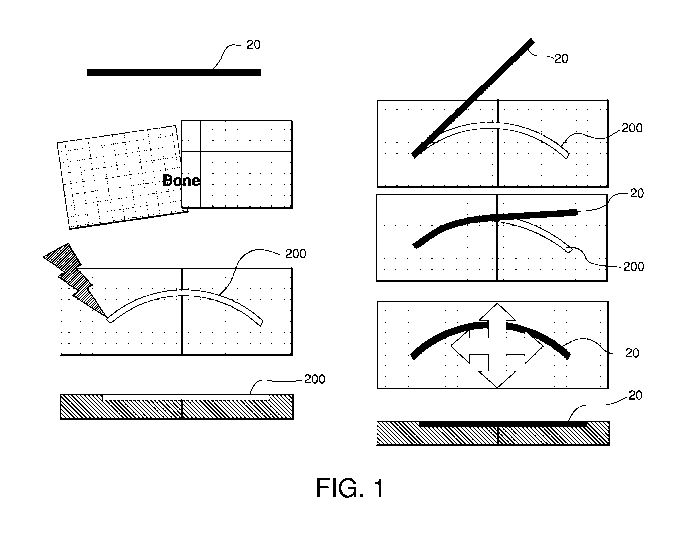
[0019] Figure 1 illustrates a wire-like implant and an associated orthopedic
fixation
method in accordance with one aspect of the present invention;
[0020] Figure 2 illustrates a wire-like implant having barb-like members and
an
associated orthopedic fixation method in accordance with another aspect of the
present
invention;
[0021] Figure 3 illustrates a wire-like implant having alternate anchoring
structures
and an associated orthopedic fixation method in accordance with another aspect
of the present
invention;
[0022] Figure 4 illustrates a wire-like implant having alternate anchoring
structures
and an associated orthopedic fixation method in accordance with another aspect
of the present
invention;
[0023] Figure 5 illustrates a zero profile bone plate and associated
implantation
method in accordance with another aspect of the present invention;
[0024] Figure 6 illustrates a zero profile bone plate and associated
implantation
method in accordance with another aspect of the present invention;
[0025] Figure 7 illustrates a variety of zero profile bone plate designs that
may be
used, for example, in cranio- and maxillofacial reconstruction, in accordance
with another aspect
of the present invention;
[0026] Figure 8 illustrates a human skull with skeletal reconstruction plates
in
accordance with another aspect of the present invention;
[0027] Figure 9 illustrates a variety of plates with respect to bone cross
sectional
profiles in accordance with another aspect of the present invention;
[0028] Figure 10 illustrates additional bone plate and bone anchor systems in
accordance with another aspect of the present invention;
[0029] Figure 11 illustrates a spring-biased cruciform spring clip for
orthopedic
fixation in accordance with another aspect of the present invention;
[0030] Figure 12 illustrates a wave blade for orthopedic fixation in
accordance with
another aspect of the present invention;
-5-
CA 02692376 2009-12-22
WO 2009/006313 PCT/US2008/068606
[0031] Figure 13 illustrates a wave blade for orthopedic fixation in
accordance with
another aspect of the present invention;
[0032] Figure 14 illustrates a staple-type implant having legs with square
cross-
sectional areas in accordance with another aspect of the present invention;
[0033] Figure 15 illustrates another embodiment of the staple-type implant in
accordance with another aspect of the present invention;
[0034] Figure 16 illustrates a skeletal fixation implant having a taper along
its depth
for reduced implant repulsion probability and an associated implantation
method in accordance
with another aspect of the present invention;
[0035] Figure 17 illustrates a variety of alternate implant designs in
accordance with
another aspect of the present invention;
[0036] Figure 18 illustrates a non-straight (e.g. snake-like or crooked) bone
cut,
which can be filled by an injectable material in accordance with another
aspect of the present
invention;
[0037] Figure 19 illustrates a non-straight (e.g. snake-like or crooked) bone
cut,
which can be filled with a soft and/or malleable, but non-liquid material in
accordance with
another aspect of the present invention;
[0038] Figure 20 illustrates a stencil or template-type instrument to guide
the bone
cutting tool; and
[0039] Figure 21 illustrates the use of a numerical controlled guiding system
for
controlled bone removal.
DETAILED DESCRIPTION OF ILLUSTRATIVE EMBODIMENTS
[0040] Certain exemplary embodiments of the invention will now be described
with
reference to the drawings. In general, such embodiments relate to a skeleton
fixation system 10
for securing bones across a fracture site. As generally understood by one of
ordinary skill in the
art, it should be understood that while the skeleton fixation system 10 may be
described in
connection with cranio or maxillofacial fixation, those skilled in the art
will appreciate that the
system as well as the components thereof may be used for fixation in other
parts of the body such
as, for example, in the long bones or bones in the hand, face, feet, etc.
[0041] As shown in Figure 1, a skeletal fixation member for placement across a
skeletal defect, such as a fracture, in need of repair may be in the form of a
biocompatible wire
20. The wire 20 preferably being at least partially embedded into the cortical
bone across the
skeletal defect to secure the skeletal area and/or to enable fusion. In a
preferred embodiment, the
wire 20 is inserted into a curvilinear groove 200 formed using a bone milling
or resurfacing
-6-
CA 02692376 2009-12-22
WO 2009/006313 PCT/US2008/068606
instrumentation such as, for example, a laser, radio frequency RF resurfacing
instrument, other
electromagnetic or mechanical resurfacing instrument. The curvilinear groove
200 into which
the wire 20 is implanted preferably crosses the fracture site at or near the
apex of its arc, such
that upon implantation of the wire 20 into the groove 200, the wire 20 acts to
secure the two bone
fragments and resists forces acting parallel to the top bone surface.
[0042] The wire 20 may have any cross-sectional shape and/or area known in the
art
including but not limited to cylindrical, rectilinear, trapezoidal, polygonal,
etc. Where the wire
20 has a trapezoidal shape, the distally implanted surface of the wire 20 may
have a width that is
larger than the proximally implanted surface, and may be implanted into a
groove 200 that has a
substantially similar cross-sectional shape and/or dimensions. In this manner,
expulsion of the
wire 20 is less likely. The wire 20 may be implanted by, for example, lacing
the wire 20 through
one end, inserting the implant down from above with some force, snapping the
implant into the
receiving bed, distracting the bone segments so that the groove 200 is
slightly enlarged as may
be practical in the case where there is a complete fracture, etc.
[0043] In addition, by selecting the appropriate choice of material, the wire
20 may
further be expandable once introduced into the patient's body or bone tissue.
Alternatively
and/or in addition, the wire 20 maybe drug-eluting and/or coated with a tissue-
ingrowth-
enhancing material, such as, for example, BGH or hydroxyapatite.
[0044] Alternatively and/or in addition, the implanted wire 20 and/or the
surgically-
formed groove 200 may be covered with a biocompatible adhesive such as, for
example, bone
putty, cyanoacrylates, polyurethanes, epoxies, acrylics, calcium phosphate
cement, etc. to
provide a more secure anchoring of the implant with respect to the surrounding
bone tissue. It is
envisioned that the adhesives may be inserted into the groove 200 and/or
around or on top of the
wire 20 implant either prior to, during, or subsequent to the implantation of
the implant wire 20.
[0045] The wire 20 may be formed of any biocompatible material known in the
art
meeting the strength and flexibility requirements of the particular
applications including but not
limited to stainless steel, titanium, Ni-Ti (nitinol), Elgiloy, other shape
memory alloys, polymers
such as PEEK, bioresorbable materials, etc.
[0046] In a preferred embodiment, the wire 20 may have a diameter that is
about one
millimeter, and the groove 200 into which the wire 20 is implanted may have a
similar depth of
one millimeter, such that the proximal surface 120 of the wire 201ies
substantially flush (e.g.
even) with or below the top surface of the bone and the depth of the groove
200 does not extend
below the cortical bone.
-7-
CA 02692376 2009-12-22
WO 2009/006313 PCT/US2008/068606
[0047] As shown in Figure 2, the wire 20 implant may also include extensions
22
such as, for example, barbs, filaments, or clips along the shaft of the wire
20. The extensions 22
may provide additional purchase into the surrounding cortical bone tissue and
provide a more
secure anchoring of the wire 20 implant with respect to the surrounding
tissue. The extensions
22 may be integrally formed with the wire 20 implant. Alternatively, the
extensions 22 may be
formed independently of the wire 20 and attached thereto. As such, the
extensions 22 may be
formed from the same material as the wire 20 implant, or they may be formed
from a different
material, such as, for example, of nitinol, Elgiloy, etc. The extensions 22
may also be
postoperatively or intraoperatively deployable. That is, for example, the
extensions 22 may be
mechanically or magnetically deployable. Alternatively, the extensions 22 may
be permanently
arranged on the exterior surface of the wire 20.
[0048] As shown in Figure 3, various additional securing means are
contemplated to
assist in anchoring the wire 20 implant with respect to the surrounding bone
tissue. In one
example, the wire 20 implant may be configured with arrowheads 24 at opposite
ends of its
length. In this manner, the arrowheads 24 may assist in compressing the two
bone fragments
across the fracture site while securing the wire 20 implant in place within
the groove 200. Figure
3 also shows various additional configurations for providing enhanced
securement of the wire 20
implant with respect to bone tissue.
[0049] As shown, the machining and/or lasering of the implant-accommodating
groove 200 may include surface-cutting of one or more areas 210 adjacent to
the curvilinear
groove 200 to facilitate insertion of the implant wire 20 to accommodate the
variously depicted
additional securing means.
[0050] Alternatively and/or in addition, as previously stated, the implanted
wire 20
and/or the surgically-formed groove 200 may be covered with a biocompatible
adhesive such as,
for example, bone putty, cyanoacrylates, polyurethanes, epoxies, acrylics,
calcium phosphate
cement, etc. to provide a more secure anchoring of the implant with respect to
the surrounding
bone tissue. It is envisioned that the adhesives may be inserted into the
groove 200 and/or
around or on top of the wire 20 implant either prior to, during, or subsequent
to the implantation
of the implant wire 20.
[0051] Alternatively and/or in addition, as shown in Figure 4, the groove 200
may
also include one or more circular machined areas 220. As shown, the circular
machined areas
220 maybe located at opposite ends of the curvilinear grooves, the circular
areas including a
hollow circular recess 222, the recess preferably having a depth similar to
that of the curvilinear
groove 200. The hollow circular recesses preferably surrounds one or more
cylindrical bone peg
-8-
CA 02692376 2009-12-22
WO 2009/006313 PCT/US2008/068606
224 that are formed by not machining or lasering, such that the cylindrical
bone pegs 224 lie
flush with the top bone surface that are unmachined or unlaser-treated. The
curvilinear groove
200 and hollow circular recesses are preferably sized and configured to
receive a wire 20 implant
having hollow rings or eyelets 26 disposed at opposite ends thereof so that
the eyelets 26
surround the cylindrical bone pegs 224 left during the machining or lasering
of the groove 200
thus facilitating a secure implantation with additional repulsion-resistance.
[0052] As shown, the eyelets 26 and corresponding bone pegs 224 may assume a
circular form. Alternatively, the eyelets 26 and corresponding bone pegs 224
may assume a non-
circular form such as, for example, a square or polygonal shape, which due to
their sharp edges
provided additional resistant to repulsion as compared to circular forms.
Alternatively and/or in
addition, the bone pegs 224 may assume a noncircular form, such as, for
example, a square or
polygonal shape for mating with a circular ring having a corresponding square
or polygonal
eyelet hole 26. Alternatively, the bone pegs 224 may assume a cylindrical form
while the ring
member may have a square or polygonal exterior surface with a circular eyelet
hole 26.
[0053] As will be appreciated by one of ordinary skill in the art the machined
areas
and corresponding eyelets 26 may be formed anywhere along the length of the
wire 20 and/or
groove 200.
[0054] As shown in Figures 5 and 6, a particularly well suited cranio or
maxillofacial
bone plate 30 is depicted. Although as will be appreciated by one of ordinary
skill in the art, the
bone plate 30 may be used in other parts of the body as well. The cranio or
maxillofacial bone
plate 30 is similar in design and geometry to conventional cranio or
maxillofacial bone plates in
that the bone plate 30 preferably has a thin profile including small-diameter
screw-receiving bore
holes 32 connected with a thin intermediary plate area 34.
[0055] That is, as shown, the cranio- or maxillofacial bone plate 30
preferably
assumes a form having two enlarged end portions 36 with an intermediary
connecting or bridge
portion 341ocated therebetween, wherein each enlarged end portion 36 may
include an optional
bore hole 32 for optional screw fixation. As such, the cranio or maxillofacial
bone plate 30 may
assume the general form of a barbell that includes two enlarged rounded lobes
36 at either end
connected by an intermediate linking portion 34 having a dimension smaller in
width than either
of the lobes 36. Each of the lobes 36 may include a bore hole 32 for optional
screw fixation.
The bore holes 32 may further be configured to fit over bone pegs similar to
those discussed
above with reference to Figure 5 instead of accommodating bone screws as seen
in Figure 7.
Alternatively, the lobes 36 may be free or devoid of any boreholes 32.
-9-
CA 02692376 2009-12-22
WO 2009/006313 PCT/US2008/068606
[0056] In use, as shown in Figure 8, the bone plate 30 may be applied across a
fracture site or other bone region in need of repair. A plate-receiving area
300 is formed in the
bone using a bone milling or resurfacing instrumentation such as, for example,
a pulsed laser, a
radio frequency RF resurfacing instrument, a mechanical resurfacing
instrument, etc. The bone
plate 30 is preferably inserted at least partially or wholly into the machined
plate-receiving area
300. The bone plate 30 serves to hold the bone pieces across the fracture site
securely with
respect to one another to assist in fusion. Preferably, as previously stated,
the thickness of the
bone plate 30 substantially corresponds to the depth of the machined plate-
receiving area 300 or
otherwise resurfaced area of bone such that, once implanted, the top surface
of the bone plate 30
lies substantially flush with or below the top surface of the bone, thus a
zero-height implant is
provided.
[0057] Moreover, as previously stated, the bone plate 30 implant may be
expandable
once introduced into the patient's body or bone tissue with the appropriate
choice of material.
The bone plate implant may also be drug-eluting and/or coated with a tissue-
ingrowth-enhancing
material, such as, for example, BGH or hydroxyapatite. The surface of the bone
plate implant
may also include texturing to assist with bone in-growth. Alternatively and/or
in addition, the
implanted bone plate 30 and/or the machined plate-receiving area 300 may be
covered with a
biocompatible adhesive. The bone plate 30 and/or the bone screws may further
be bioresorbable.
[0058] As shown in Figure 7, various alternate sized and shaped bone plates 30
are
depicted. Once again, these plates 30 are particularly well suited for cranio-
or maxillofacial
applications but as will be appreciated by one of ordinary skill in the art,
the bone plates 30 may
be used in other parts of the body as well. As shown, the bone plates 30 may
have generally
more complex plate designs, which are particularly suited for more complex
fracture reduction
and/or fusion. In one preferred embodiment, the zero-profile cruciform shaped
plate 30 has a
general X-shape in which connecting members 34 join boreholes 32 at opposite
ends 36 of each
connecting member 34 for receiving bone anchors or bone pegs 310. The
cruciform plate 30
may be oriented and/or inserted into a corresponding machined or lasered
groove 300 spanning a
fracture site such that two anchoring means are positioned on either side of
the fracture.
Similarly, the more complex plate designs illustrated in Figure 7 may be
implanted into
corresponding grooves 300 formed into the bone surfaces such that a plurality
of anchoring
means are situated on one or more sides of a bone fracture.
[0059] As shown in Figure 9, according to one aspect of the present invention,
the
implants (e.g., wire 20, plates, etc.) may be implanted into a corresponding
machined or lasered
area 300 of bone such that the top surface of the implant lies substantially
flush with or below the
-10-
CA 02692376 2009-12-22
WO 2009/006313 PCT/US2008/068606
top surface of the bone. Alternatively, the top surface of the implant may lie
slightly below (e.g.
recessed) with respect to the top surface of the bone or slightly above the
top surface of the bone.
Alternatively and/or in addition, a biocompatible adhesive such as, for
example, a bone putty,
can be applied atop the implant to further assist in fusing the two bone
segments in need of
repair. As previously stated, the bone plate 30 preferably has a thickness
designed for the
particular application and can be in the range of about 0.5 to about 5
millimeters (in some cases,
for example, approximately one millimeter in thickness is useful) and is
received in an implant-
receiving bed that is surgically formed into the top surface of the bone to a
depth of equal or
slightly greater depth (in some cases, for example, about one millimeter).
[0060] Figure 10 illustrates additional bone plate designs. As shown, the bone
plates
30 may include noncircular bore holes 32 for anchoring the implant with
respect to the
surrounding bone tissue. Preferably, the noncircular boreholes 32 are sized
and configured to
accommodate anchoring pins 600 having shafts characterized by a
correspondingly non-circular
cross-sectional area or alternately may house non-rounded bone pegs 3101eft
during the
machining of the implant-receiving recess. Anchoring pins 600 having
noncircular cross-
sectioned shafts provided additional resistance to expulsion as compared to
circular and
cylindrical threaded bone anchors, as the non-rounded bone anchors are not
susceptible to
rotating, and thus backing out, of the surrounding bone tissue.
[0061] If a circular cross section pin, nail, screw or anchor 600 is used, it
is preferred
that a minimum of two such pins, nails or anchors 600 are used to avoid
rotation of the bone
fragment around a single pin, nail, or anchor 600.
[0062] Preferably both the bone plate 30 and the heads of the bone anchoring
pins,
nails, and anchors 600, are sized and configured to lie substantially flush
with the top bone
surface after implantation. Alternatively, the bone plate 30 may lie atop a
non-machined bone
surface. In a preferred embodiment, the heads of the noncircular bone pins,
nails, anchors, etc.
600 are housed within the proximal portions of the boreholes through the plate
30 such that the
top surfaces of the noncircular bone pins 6001ie substantially flush with the
top surface of the
bone plate as well as the top surface of the bone. Alternatively, the top
surfaces of the
noncircular bone pins, nails, anchors, etc. 600 may lie above or below the top
surface of the bone
plate 30. In one embodiment, the distal cross-sectional area of the
noncircular bone pin, nail,
anchor, etc. 600 is larger than the proximal cross-sectional area of the
noncircular bone pin, nail,
anchor, etc. 600 thereby providing a slight taper along the length of the
shaft of the bone pin,
nail, anchor, etc. 600 such that the bone pin, nail, anchor, etc. 600 may be
snapped into the bone
-11-
CA 02692376 2009-12-22
WO 2009/006313 PCT/US2008/068606
and thereby provide additional securement of the bone pin, nail, anchor, etc.
600 and bone plate
30.
[0063] As shown in Figure 11, the bone fixation element may be in the form of
a
spring-biased fixation clip 40. The spring-biased fixation clip 40 including
two elongated
members 42 that may be connected at or near the centers of their lengths by a
connecting
member 44. The connection between the two elongated members 42 biases the
elongated
members such that rotation of one of the elongated members 42 with respect to
the other
elongated member 42 is permitted. Preferably, in the absence of any external
forces, the two
elongated connecting members 42 are sized and configured so that they are
positioned in a
cruciform or "X" shape. Thereafter, upon application of a rotational force to
one or both of the
elongated members 42, the connecting members 42 are permitted to rotate with
respect to one
another so that the two elongated members 42 may become aligned in parallel
form with respect
to each other.
[0064] In use, one of the elongated members 42 of the spring clip may be
inserted
into a machined or laser formed bone slot 400 and then, immediately upon
penetration into the
bone, the elongated member 42 is permitted to rotate (springs) approximately
90 degrees. That
is, in use, for example, the spring biased fixation clip 40, which is
particularly well suited for
cranio or maxillofacial applications such as, for example, cranial flap fusion
or fracture reduction
to lock translation of the bone pieces, may be applied across two skull bone
pieces in need of
fusion or reduction by resurfacing the bone(s), such as by forming a groove
400 across both bone
fragments in the vicinity of the fracture site. Preferably, the groove 400 is
formed all the way or
completely through the bone. The spring biased fixation clip 40 may then be
introduced into the
groove 400 in its parallel state, such as by using a grasping instrument or
inserter, such that the
distal elongated member 42 is introduced below the bottom surface of the bone,
while the
proximal elongated member 42 is positioned above the top surface of the bone,
with the
connecting member 44 spanning the depth of the bone. The instrument is then
released from the
implant and the fixation clip automatically reverts back to its natural state
in which the two
elongated members 42 assume a cruciform or X shape. In returning to its
cruciform state, the
two elongated members 42 position themselves with respect to the machined
groove 400 such
that implant repulsion is prohibited. In this manner, the spring biased
fixation clip 40 serves a
similar function as a conventional flap-fix, the goal being to keep the bone
flap on the same level
as the surrounding bone.
[0065] In addition, the elongated members 42 and/or the connecting member 44
of
the spring biased fixation clip 40 may include barbs or spikes or other
surface texturing that
-12-
CA 02692376 2009-12-22
WO 2009/006313 PCT/US2008/068606
assist in bone purchase and/or bone in-growth. The spring clip 40 may be
loaded into a groove
400 that is one millimeter deep and five millimeters in length. The spring
clip may be formed of
a bioresorbable material or non-resorbable material such as stainless steel,
titanium, nitinol, or
PEEK.
[0066] As shown in Figures 12 and 13, the implant may be in the form of a wave-
blade implant 50 that is characterized by two states, (1) a preloaded (e.g.
straight) and (2) non-
loaded (e.g. wavy) configuration. In this manner, the wave-blade implant 50
may be inserted
into a groove or rectilinear slot 500 formed in the bone surface in its
preloaded (e.g. straight)
configuration using a grasping type insertion instrument. Upon being seated in
the groove 500,
the wave-blade implant 50 is released from the grasping type insertion
instrument and reverts
back to its non-loaded (e.g. wavy) configuration. In the non-loaded
configuration, the wave
blade implant 50 may assume a sine-wave shape whose corners and sides contact
and/or engage
(e.g. dig into) the surrounding bone tissue to enhance implant seating.
[0067] The wave blade implant 50 may be formed of any biocompatible material
known in the art including but not limited to cold-worked titanium, cold-
worked steel, or any
other flexible biocompatible material. The grasping insertion instrument may
be in the form of a
pliers-type instrument having a straight slot into which the wave-blade
implant 50 is pre-loaded.
The wave-blade implant 50 may then be pushed out of the slot of the pliers-
type instrument and
simultaneously inserted into the bone slot 500.
[0068] As shown in Figure 14, the implant may be in the form of a staple-type
implant 60. The staple type implant may have a plurality of legs 62, the legs
62 may be
configured with a square cross-sectional area. The legs 62, preferably the
square legs, of the
staple type implant 60 provide angular stability and inhibit and/or prevent
the bone fragments
from rotating about the legs' axes. As shown, the staple type implant 60
preferably also includes
diverging legs 62, the diverging direction of the legs 62 secures the staple
to the bone, as they are
elastically bent during the insertion.
[0069] Alternatively and/or in addition, as shown in Figure 15, the staple
type
implant 60 may also include one or more screw holes 64 in combination with the
square cross
sectional area legs 60. The screws provide additional securement to keep the
staple type implant
atop of the bone.
[0070] As previously stated and as best shown in Figure 16, the implant may
further
include a slight taper 100. The taper may be provided along the height of the
implant such that
the surface area at the distal surface 110 of the implant is larger than the
surface area at the
proximal surface 120 of the implant. The tapered implant improves implant
retention with
-13-
CA 02692376 2009-12-22
WO 2009/006313 PCT/US2008/068606
respect to the surrounding bone tissue. The tapered height implant may be
implanted by, for
example, lacing the implant through one end, inserting the implant down from
above with some
force, snapping the implant into the receiving bed, distracting the bone
segments so that the
groove is slightly enlarged as may be practical in the case where there is a
complete fracture, etc.
[0071] Figure 17 illustrates a variety of alternate implant designs in
accordance with
another aspect of the present invention. As shown, the implants preferably
include a reduced
intermediary portion and enlarged end portions. The implants are preferably
sized and
configured to be received within corresponding receiving beds formed in the
cortical bone
surfaces. As such, the implants preferably have, once implanted, a zero height
or reduced height
profile with respect to the top bone surface as well as improved implant
retention.
[0072] Figure 18 illustrates a non-straight (e.g., snake-like or crooked) bone
cut,
which is subsequently filled by an injectable material that is hardenable in
situ. The injectable
material may include but is not limited to bone glue, cement, heated
resorbable or non-resorbable
polymer, etc. The injectable material, upon hardening, serves as a formable
implant that serves
the same skeletal repair purposes as the implants discussed above.
[0073] Figure 19 illustrates a non-straight (e.g., snake-like or crooked) bone
cut,
which is subsequently filled with a soft and/or malleable but non-liquid
material that may or may
not stiffen after implantation. The stiffening material may include but is not
limited to a
polymeric material which is heated over the glass transition temperature.
[0074] Figure 20 illustrates a stencil or template-type instrument to guide a
bone
cutting tool, such as, for example, a laser, water jet, piezoelectric cutting
knife, mechanical
milling instrument or other bone resurfacing instrument, etc.
[0075] Figure 21 illustrates the use of a numerical controlled guiding system
for
controlled bone removal that can assist in the precision bone resurfacing
step.
[0076] It is understood that the implants provided by the present invention
and
methods associated therewith may utilize additional securement means, such as
taking advantage
of biocompatible adhesives such as cyanoacrylates, polyurethanes, epoxies, and
acrylics with or
without ultrasonic energy application, or may take advantage of bone-welding
technology.
[0077] While the foregoing description and drawings represent the preferred
embodiments of the present invention, it will be understood that various
additions, modifications
and substitutions may be made therein without departing from the spirit and
scope of the present
invention as defined in the accompanying claims. In particular, it will be
clear to those skilled in
the art that the present invention may be embodied in other specific forms,
structures,
arrangements, proportions, and with other elements, materials, and components,
without
-14-
CA 02692376 2009-12-22
WO 2009/006313 PCT/US2008/068606
departing from the spirit or essential characteristics thereof. One skilled in
the art will appreciate
that the invention may be used with many modifications of structure,
arrangement, proportions,
materials, and components and otherwise, used in the practice of the
invention, which are
particularly adapted to specific environments and operative requirements
without departing from
the principles of the present invention. In addition, features described
herein may be used
singularly or in combination with other features. The presently disclosed
embodiments are
therefore to be considered in all respects as illustrative and not
restrictive, the scope of the
invention being indicated by the appended claims, and not limited to the
foregoing description.
- 15 -