Note: Descriptions are shown in the official language in which they were submitted.
CA 02693778 2013-05-28
METHOD AND APPARATUS FOR LARYNGEAL
ELEVATOR MUSCULATURE REHABILITATION
BACKGROUND
[0001] The current invention generally relates to a method of treating
decreased laryngeal
elevation. More specifically, this invention relates to the use of a neuro-
orthotic device, in
combination with electrical stimulation of the submandibular muscles, to treat
laryngeal elevator
musculature. This invention also relates to muscle re-education and
rehabilitation by using a neuro-
orthotic device, in combination with electrical stimulation of the
submandibular muscles, to
stimulate laryngeal elevator musculature.
[0002] People with dysphagia have difficulty swallowing, and may also
experience pain
while swallowing. A commonly encountered functional abnormality in individuals
with
dysphagia is a decrease in laryngeal elevation. Laryngeal elevation is
important in the
elongation of the pharyngeal-esophageal sphincter, and assistance with
epiglottic closure. Often,
the decrease in laryngeal elevation is due to atrophy of the laryngeal
elevator musculature.
[00031 The use of neuromuscular electrical stimulation (NMES) for
dysphagia treatment
has gained increased interest over several years. There have been a few
investigative studies into
NMES treatments of dysphagia. Some previous studies have focused on research
methods
involving the stimulation of open nerves in animals. Other studies have
focused on the use of
electrical stimulation with parameters adjusted to initiate the swallow
reflex.
10004] In the context of sleep apnea research, some researchers have
hypothesized that
electrical stimulation may improve laryngeal musculature and thereby decrease
apneic episodes.
There is, however, an absence of published research combining an orthotic or
neuro-orthotic in
1
CA 02693778 2013-05-28
combination with electrical stimulation to promote laryngeal elevator
musculature re-education,
rehabilitation, or regeneration.
[0005] The existing studies are not necessarily a best option for a
therapeutic treatment of
decreased laryngeal elevation. An evaluation of these techniques for their
significance in
swallowing rehabilitation and other treatments centered on the submandibular
and pharangyeal
regions, shows that the specific parameters and uses vary, and the results for
the research have
not been consistent.
[0006] One difficulty researchers face is finding the proper balance of
treatment and therapy
to overcome the decrease in laryngeal elevation. Major goals of treatment and
therapy include
being non-invasive to the patient, preventing disuse atrophy of the muscles,
increasing range of
motion, re-educating muscle functions, temporarily decreasing spasticity, and
increasing local
blood circulation.
100071 The present invention, as described herein, is directed to the
aforementioned
problems, deficiencies and goals.
SUMMARY
[0008] One embodiment is a method for muscle rehabilitation of laryngeal
elevator
musculature comprising placing a patient into a neuro-orthotic device for
elevation of the
laryngeal elevator musculature and applying a protocol of an electrical
stimulus to the
submandibular region for a sufficient period of time using a sufficient
electrical input.
[0009] Another embodiment is a method for non-invasive treatment of
laryngeal elevator
musculature. The method comprises fitting a prosthetic neuro-orthotic device
to a patient, and
applying at least one pair of electrodes to the submandibular region of the
patient. Once
positioned, a repeated pulsing of a pre-determined electrical current is sent
through the
2
CA 02693778 2013-05-28
electrodes. The stimulating of the patient's submandibular region muscles is
done for a pre-
determined period of time.
100101 Another embodiment of this invention is a method for re-educating
the laryngeal
elevator musculature of a patient. This method comprises placing a patient
into a neuro-orthotic
device which aligns the cervical spine of the patient in the orthotic device.
The method requires
applying at least one transcutaneous electrical muscle stimulator electrode to
the patient's
submandibular region. The positioning of the patient's submandibular region in
the neuro-
orthotic device increases the electrical signal efficiency of the electrode.
Once positioned, a
protocol is applied which has a sufficient electrical current that is applied
for a sufficient period
of time. The current further comprises, a sufficient frequency, a sufficient
pulse width, a
sufficient amplitude, a sufficient ramp period, and a sufficient waveform.
[0011] Another embodiment of this invention is a method for muscle
rehabilitation of
laryngeal elevator musculature. The inventive method comprises fitting a
patient into a neuro-
orthotic device for treatment. The neuro-orthotic device supports the
patient's submandibular
region for the treatment. The inventive method is to align a plurality of
transcutaneous electrical
muscle stimulator electrodes on the patient's submandibular with the neuro-
orthotic device. The
electrodes are applied and positioned to stimulate the laryngeal elevator
musculature. The
application of a protocol with a pre-determined pulsed electrical current to
the patient for a pre-
determined time with a pre-determined electrical input is accomplished.
[0012] Another embodiment of this invention is a removable electrode. The
electrode
comprises a pad, a series of electrically conductive elements affixed to the
pad and oriented to
align with the submandibular muscle fibers, a protective insulating cover
affixed to the
3
CA 02693778 2013-05-28
electrically conductive elements and the pad, and an electrical lead extending
from the pad,
which is in electrical communication with the series of electrically
conductive elements.
[0013] Numerous objects and advantages of the invention will become
apparent as the
following detailed description of the preferred embodiment is read in
conjunction with the
drawing, which illustrates such embodiment.
BRIEF DESCRIPTION OF THE DRAWINGS
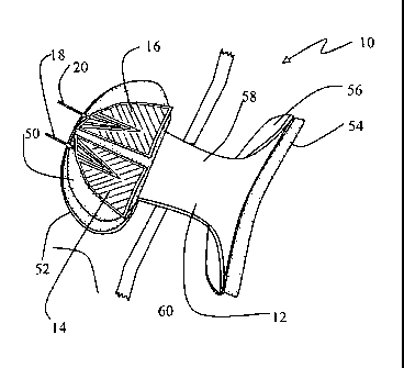
[0014] FIG. 1 ¨ Is a perspective view of a neuro-orthotic device with the
submandibular
electrodes placed upon it.
[0015] FIG. 2 ¨ Is a side elevation view of a neuro-orthotic device.
[0016] FIG. 3 ¨ Is a top view of a neuro-orthotic device.
[0017] FIG. 4 ¨ Is a perspective view of a neuro-orthotic device.
[0018] FIG. 5 ¨ Is a top view of a set of the inventive electrodes with
leads attached.
100191 FIG. 6 ¨ Is an exploded sectional view of the inventive electrodes
taken along
section line 6-6 of FIG. 5.
[0020] FIG. 7 ¨ Is a bottom view of a set of the inventive electrodes
affixed to a patient's
submandibular region.
[0021] FIG. 8 ¨ Is a table of a strength duration curve.
DETAILED DESCRIPTION
Overview
[0022] Muscle re-education, re-generation, and rehabilitation to improve
laryngeal
elevation using NMES are the primary goals of this inventive method and
apparatus. The rationale
is that the improved range of motion of the larynx and tongue base retraction
during swallowing
4
CA 02693778 2013-05-28
= ,
affects airway protection. This anterior motion of the hyolaryngeal complex is
essential to
improved swallow function.
[0023] Electronic stimulation of muscles has been practiced and is
understood for many
muscles and muscle groups. Electronic stimulation of the muscles forces a
specific muscle or
muscle group and often ancillary muscles to react to the stimulus. Electronic
stimulation in the
present invention is directed to muscle re-education, regeneration and
rehabilitation of the laryngeal
elevator musculature in order to promote laryngeal elevation, but is not
intended to, nor set at such a
level, to initiate the swallow reflex.
[0024] The use of electronic stimulation to promote laryngeal
elevation requires that the
correct muscles be exercised. The paired muscles of the mylohyoid, geniohyoid
and the anterior
belly of the digastric musculature are primarily responsible for anterior and
superior movement
of the hyoid bone during a swallow. This movement of the hyoid and laryngeal
elevation is vital
in airway protection during swallowing. The anterior/superior movement of the
larynx helps
bring the airway safely away from the path of the bolus. Techniques used to
accentuate and
prolong laryngeal elevation are used as indirect dysphagia treatment. These
techniques are based
on the anatomical relationship of the hyoid, larynx and cricopharyngeal
region.
[0025] Using a neuro-orthotic device or orthotic in combination with
electronic stimulation
of the laryngeal elevator musculature provides greater isolation of the
muscles, and allows proper
positioning and conduction of the electrodes. A neuro-orthotic device also
improves laryngeal
elevation while reducing complications associated with disorders and injuries
of the central
nervous system. Additionally, a neuro-orthotic device places the patient in
the proper anatomical
position to receive the most efficient electronic stimulation treatment. The
neuro-orthotic device
also prevents the adverse affect of moisture and saliva contacting the
electrodes. Provided that the
CA 02693778 2013-05-28
=
peripheral nervous system is intact, the protocol of this inventive method and
apparatus can be used
as an adjunct in the clinical treatment of a variety of neuromuscular and
musculoskeletal
problems. Neuromuscular electrical stimulation (NMES) used in combination with
the
electrodes of the present invention and a neuro-orthotic device shows an
increase of strength and
range of motion, facilitating weak contractions due to upper-motor neuron
lesions or disuse
atrophy, and to re-educate muscles.
[0026] FIG. 1 represents a first preferred embodiment of the invention.
Apparatus 10 is the
combination of neuro-orthotic device 12 and electrodes 14 and 16. Different
variations of apparatus
may be created by using a different neuro-orthotic device 12 or different
electrodes 14 and 16.
[0027] In FIGS. 1 and 5, electrodes 14 and 16 are shown with electrical
leads 18 and 20.
Also shown in FIGS. 1 and 5 are conductive elements 22 and 24. Electrical
leads 18 and 20 provide
electrical current to conductive elements 22 and 24. Electrical leads 18 and
20 are shown in FIG. 5
electrically connected to power source 26. Electrical leads 18 and 20 are
shown as separate lines,
but they may be combined into a single cable. The electrical current is
provided and regulated by
power source 26.
[0028] Power source 26 is preferably a muscle stimulator capable of
providing the protocol
parameters described herein. A known power source 26 is the Staodyne EMS +2
manufactured by
Compex Technologies, Inc. Other known power sources 26 include the IntelliSTIM
BE-28E
manufactured by EASYMED Instrument Co. Ltd; Respond Select manufactured by
Empi, Inc.;
BioStim NMS+ manufactured by BioMedical Life Systems, Inc.; and SYS*STIM 26
manufactured
by Mettler Electronics Corporation. However, any single or plural power source
26 may be used
that substantially meets the protocol requirements of the inventive method
described herein.
6
CA 02693778 2013-05-28
[0029] FIG. 6 is an exploded sectional view of electrodes taken along
section line 6-6 of
FIG. 5. FIG. 6 depicts electrodes 14 and 16 as subcomponents. Conductive
elements 22 and 24 are
shown positioned between protective insulating covers 28 and 29 and pads 30
and 31. Protective
insulating covers 28 and 29 may be any material that is non-conductive and
electrically
insulating. Preferably, protective insulating covers 28 and 29 are fabricated
out of a soft material
and also provide a cushion for protection. Conductive elements 22 and 24 are
affixed to pads 30
and 31. Conductive elements 22 and 24 are comprised of a series of small
fibers 32 and 34.
Preferably small fibers 32 and 34 are fabricated out of silver carbon.
Preferably, small fibers 32
and 34 are oriented on pads 30 and 31 parallel to the submandibular region 48
muscle fibers
when electrodes 14 and 16 are applied to patient 40. Pads 30 and 31 are
preferably fabricated
out of a material allowing conductive elements 22 and 24 to transmit
electrical current with
minimal electrical loss. In the preferred embodiment, a gel pad was used for
pads 30 and 31.
[0030] Pad 30 has pad first side 36 and 37 and pad second side 38 and 39.
Conductive
elements 22 and 24 are affixed to pad first side 36 and 37. Pad second side 38
and 39 is
preferably inherently tacky such that it will stick to patient 40, shown in
FIGS. 2 and 7.
However, pad second side 38 and 39 may be coated with a tacky substance.
[0031] Electrodes 14 and 16 are shown in FIGS. 1, 5 and 7 as geometric
shaped
segments. The geometric shaped segment of electrodes 14 and 16 is designed to
conformably
place electrodes 14 and 16 on submandibular region 48, and to properly orient
small fibers 32
and 34 in relation to the submandibular region 48 muscle fibers. Each segment
has first leg 42
and 43 having a length x, and second leg 44 and 45 having a length y connected
by arcuate
portion 46 and 47. Length "x" and length "y" may be produced in different
sizes to meet the
needs of differently sized patients 40. For the preferred embodiment, length
"x" is about 3.7
7
CA 02693778 2013-05-28
=
centimeters in length. For the preferred embodiment, length "y" is about 4.4
centimeters in
length. First leg 42 and 43 has a first leg first end 42a and 43a and a first
leg second end 42b and
43b. Second leg 44 and 45 has a first leg first end 44a and 45a and first leg
second end 44b and
45b. As seen in FIG. 5, first leg first end 42a and 43a is connected the
second leg first end 44a
and 45a. Arcuate portion 46 and 47 connects first leg second end 42b and 43b
and second leg
second end 44b and 45b.
[0032] Electrodes 14 and 16 are shown as mirror images of each other, and
each is sized
to substantially cover one-half of submandibular region 48 of patient 40. FIG.
7 depicts
electrodes 14 and 16 positioned upon submandibular region 48 of patient 40.
Only electrodes 14
and 16 are shown in FIGS. 1 and 5-7. Electrodes 14 and 16 are preferably used
in pairs with a
waveform that is biphasic. However, a single electrode 14 or 16 may be used in
combination with a
form of a manual probe.
[0033] Electrodes 14 and 16 are used in combination with neuro-orthotic
device 12.
Electrodes 14 and 16 are shown in FIG. 1 positioned upon chin pad 50 of
orthotic device 12.
Neuro-orthotic device 12 may be any orthotic or neuro-orthotic that properly
elevates the laryngeal
elevator musculature. The preferred neuro-orthotic device 12 is a device
previously marketed as
the "REST-EZZZTm with ESP (Enhanced Swallow Posture)" by Restorative Medical
Incorporated
headquartered in Brandenburg, Kentucky. The minimum criteria in selecting
neuro-orthotic device
12 are the proper positioning of the laryngeal elevator musculature and non-
interference with
electrodes 14 and 16. Once properly positioned, as shown in FIG. 2, neuro-
orthotic device 12
facilitates anterior and superior hyoidal movement while maintaining proper
postural alignment
with optimal contact of electrodes 14 and 16. Additionally, neuro-orthotic
device 12 facilitates
better contact between the submandibular region 48 musculature and electrodes
14 and 16. The
8
CA 02693778 2013-05-28
=
=
better contact is achieved by patient 40 resting chin 62 upon neuro-orthotic
device 12, which
improves contact with the submandibular region 48 musculature.
[0034] FIGS. 1-4 depict the preferred neuro-orthotic device 12. Neuro-
orthotic device 12
has chin pad 50, chin pad support structure 52, chest pad 54, chest pad
support structure 56,
connective support structure 58, and retention strap 60. Chin pad 50 is
designed to comfortably
support chin 62 of patient 40 without interfering with electrodes 14 and 16
affixed to submandibular
region 48.
[0035] Referring to FIG. 2, neuro-orthotic device 12 is shown with an
ergonomic design to
support chin 62 while keeping neuro-orthotic device 12 away from neck 64 of
patient 40. Chest 66
of patient 40 is used to provide a fulcrum to support chin 62 with neuro-
orthotic device 12.
Retention strap 60 is shown around the back of neck 64. In this position,
neuro-orthotic device 12
is held in position for treatment. Once positioned, as shown in FIG. 2, the
laryngeal elevator
musculature of patient 40 is properly positioned for treatment.
[0036] Patient 40 is depicted in FIG. 2 as a human. However, this
inventive method is
applicable to any animal having submandibular region 48. Usage of the term
animal is meant to
include all human and non-human species having a submandibular region 48.
100371 FIGS. 2 and 7 illustrate a preferred embodiment of the
inventive method. In
particular, electrodes 14 and 16 are affixed to submandibular region 48 of
patient 40. Second leg 44
and 45 of electrodes 14 and 16 are placed along a line between anterior
placement point 68 and
posterior placement point 70 as shown in FIG. 7. The unique shape of
electrodes 14 and 16 ensures
the proper alignment of small fibers 32 and 34 in relation to the
submandibular region 48 muscle
fibers. The placement of electrodes 14 and 16 is preferably non-invasive.
9
CA 02693778 2013-05-28
[0038]
With electrodes 14 and 16 in place, neuro-orthotic device 12 is placed under
chin 62
of patient 40. Chin pad 50 of neuro-orthotic device 12 comfortably raises the
submandibular region
48 of patient 40 to a proper position. In the proper position, the laryngeal
elevator musculature of
patient 40 is positioned for the maximum muscle re-education and
rehabilitation. Chin pad 50 may
be used to align electrodes 14 and 16 for initial treatment and for subsequent
treatments, thus
ensuring consistent or repeatable placement of electrodes 14 and 16 on
submandibular region 48.
[0039]
A proper protocol is used for treatment of the patient. The application of the
proper
protocol uses a pre-determined, or sufficient, pulsed electrical current sent
through electrodes 14
and 16 for a pre-determined, or sufficient, time with a pre-determined, or
sufficient, electrical
input.
Pre-determined, or sufficient, power comprises a sufficient voltage, a
sufficient
frequency, a sufficient pulse width, a sufficient amplitude, a sufficient ramp
period, and a
sufficient waveform. Pre-determination, or sufficiency, is based upon the
needs of patient 40
and what is tolerable to patient 40. The application of a sufficient frequency
produces a smooth
tetanic contraction in the muscles of a submandibular region 48 without
causing spasms.
[0040]
A pre-determined, or sufficient, protocol typically requires treatment twice a
day for
about 15 minutes. The duty cycle of the protocol starts about 5 seconds on and
about 25 seconds
off. Once patient 40 can tolerate the treatment, the duty cycle is changed to
about 5 seconds on
and about 15 seconds off. The maximum duty cycle is about a 1:1 ratio, or
about 5 seconds on
and about 5 seconds off
[0041]
The preferred protocol frequency is about 30 hertz (30 pulses per second). The
protocol pulse width is between about 240 microseconds to about 260
microseconds. The
preferred initial trial pulse width is about 250 microseconds. However, if
patient 40 finds the
treatment painful, and is still able to activate sensory and motor
recruitment, the pulse width may
CA 02693778 2013-05-28
be lowered between about 40 microseconds to about 60 microseconds. If the
pulse width is
lowered to between about 40 to about 60 microseconds, the amperage is
preferably doubled. The
protocol amperage is preferably about 10 milliamps to about 80 milliamps.
However, the
amperage is tied to the pulse width for maximum muscle stimulation. The
amplitude is between
about 10 milliamps to about 40 milliamps for a pulse width of about 250
microseconds. The
amplitude is between about 30 milliamps to about 80 milliamps for a pulse
width of about 50
microseconds. The protocol uses pulsed current. In one clinical trial the
amplitude had a range
of 14 milliamps to 35 milliamps for an input voltage range of 11 millivolts to
100 millivolts.
The average was 18.72 milliamps and 46.04 millivolts. The protocol ramp, or
rise in intensity, is
about 0.4 seconds. The waveform of the protocol is preferably a symmetrical
biphasic waveform
when pairs of electrodes 14 and 16 are utilized.
[0042] To stimulate the anterior digastric and mylohyoid muscles, the
current must pass
through the skin/fascia layer and platysma. The electrical current passes
through these layers to
the anterior digastric and mylohyoid musculature, and may overflow into other
musculature such
as the geniohyoid and hyoglossus. The benefits of stimulating this region are
that they
voluntarily assist the depression of the tongue via the hyoglossus and
contract the digastric
muscles, mylohyoid and geniohyoid muscles assisting in the anterior/superior
movement of the
larynx.
[0043] The goal of using these preferred parameters of the protocol is for
re-educating the
laryngeal elevator musculature without inducing a swallow reflex. To
accomplish this goal, the
anterior digastric muscle, which originates on the inferior border of the
mandible, is stimulated
since it is the most superficial suprahyoidal muscle. The anterior digastric
muscle insertion at
the cornu of the hyoid acts to elevate and pull the hyoid anteriorly. The
stimulation overflows to
11
CA 02693778 2013-05-28
= -
the mylohyoid for its origin on the mylohyoid line of the mandible and its
insertion at the body
of the hyoid, which acts to elevate and pull the hyoid anteriorly. The
stimulation overflows to
the hyoglossus for its origin on the hyoid bone and its insertion at the sides
of the tongue which
depresses the tongue. The stimulation may overflow to the geniohyoid which is
depresses the
jaw and elevates and protracts the hyoid. Origin of the geniohyoid from the
inferior mental spine
on the back of the symphysis menti and inserts to the hyoid bone.
[0044] Using a pulse rate of about 30 Hertz produces a tetanizing
muscle contraction
with minimal muscle fatigue and without causing spasm. Tetanizing is a
condition characterized
by twitching/contracting muscles. Using a pulse width of about 250
microseconds increases the
depth of penetration of the current. Initial trial of 250 microseconds is
recommended to achieve
muscle tetany. The higher the pulse width, the greater the penetration of
current and the lower
the intensity needed to make a contraction. The lower the pulse width, the
shallower the
penetration of current and the higher the intensity needed to make a
contraction. The strength
duration curve provided in FIG. 8 indicates that the large sensory nerves are
activated first,
motor nerves are activated second, and pain nerves are activated third.
[0045] The higher the amplitude, the greater the increase in the
number of muscle motor
units activated. The preferred amplitude is patient variable, ranging from 10
milliamps to 100
milliamps which is sufficient to elicit a comfortable motor/tetanic response
in patient 40. Ramp
up time, or ramp time, is the length of time it takes for the output stimulus
to reach maximum
strength for each muscle contraction. The ramp time aids in the comfort of the
treatment.
Typically, a ramp time of 0.4 seconds can be used to mimic normal recruitment
and is suggested
for the comfort of patient 40.
12
CA 02693778 2013-05-28
=
[0046] The duty cycle affects the fatigue rate of patient 40. Depending
upon patient 40,
initial treatments for muscle re-education may require longer on/off time in
the duty cycle. A
duty cycle of about 1:3, about 5 seconds on and about 15 seconds off, is
typically less fatiguing
to patient 40. However, it is recommended that treatment start at a duty cycle
of about 1:5, about
seconds on and about 25 seconds off The maximum duty cycle is about 1:1, about
5 seconds
on and about 5 seconds off.
[0047] The preferred waveform is a symmetrical biphasic waveform. The
symmetrical
biphasic waveform efficiently stimulates both electrodes 14 and 16. The size
and placement of
electrodes 14 and 16 must be chosen such that they provide the desired
response for patient 40
avoiding the carotid sinus. Any power source 26 that is a powered muscle
stimulator labeled for
"muscle re-education" can be used with neuro-orthotic device 12 as long as it
is capable of
meeting the protocol parameters and follows the manufacturers listed
contraindications.
[0048] The neuro-orthotic device 12 positions submandibular region 48 of
patient 40 into
a position such that the laryngeal elevator muscles are stimulated without
inducing a swallowing
action. This movement facilitates patterns leading to the reversal of disuse
atrophy, while
improved posture will enhance the ability to breathe and take in nutrition and
hydration. Neuro-
orthotic device 12 comfortably embraces submandibular region 48, and can be
used as an
orthosis to decrease pain from poor posture. An additional benefit is that
this inventive method
should increase the quality of life as patients will be able to take part in
activities which may also
improve their degree of orientation and cognition. Preferably, neuro-orthotic
device 12 can be
customized to allow for accurate and easy application/removal. By using heavy
gloves and
moving a heat gun in a small circular motion, neuro-orthotic device 12 can be
remolded to
13
CA 02693778 2013-05-28
change the angle of chin pad support structure 52 or chest pad support
structure 56 to lengthen or
shorten the overall height.
[0049] In a multi-center prospective clinical trial study, the principles
of this invention
were applied to a real world setting, using the protocol shown below on long
term care patients
who exhibited dysphagia due in part to poor or diminished laryngeal elevation.
The time of each
treatment included two 15-minute sessions daily, for a total of 30 minutes of
therapy time. This
protocol is accomplished 5 times per week.
[0050] The evaluated patients were from multiple long term care
facilities in Texas.
Group 1 included patients receiving at least 20 therapy days of the
Neuromuscular Electrical
Stimulation (NMES) protocol as well as traditional therapy (59 patients).
Group 2 included
patients receiving traditional dysphagia therapy only, as would have been
performed prior to any
NMES modality training, or for patients who refused the NMES therapy protocol
(46 patients).
[0051] A total of 105 patients were evaluated with the Modified Barium
Swallow Study
(MBSS) by a trained licensed speech language pathologist, and were found to
have impaired
laryngeal elevation as a primary or secondary dysfunction causing aspiration
or risk of aspiration
to the degree that diet changes were necessary. A swallow severity scale was
established to
determine the diet after the initial diagnosis of dysphagia using fluoroscopy.
The subset of
patients who were able to tolerate at least 20 days of traditional dysphagia
therapy while also
using the NMES established protocol were included in the analysis as patients
having successful
completion of the protocol. A comparison was made from a total of 46 patients
who received
only the traditional dysphagia therapy, but whose chart reviews noted these
patients exhibited
dysphagia with decreased laryngeal elevation as diagnosed from an MBSS. These
charts were
14
CA 02693778 2013-05-28
evaluated as to the number of patients who had an improved swallow severity
scale. The
severity scale is shown in Table 1.
Severity Scale
0 - NPO
1 - Therapeutic intake only.
2 - Pleasure feedings only, unsupervised > 2-3 times per day.
3 - Modified diet of either thickened liquids, puree or
mechanical soft with strategies (3 meals/day).
4 - Strategies only, no alternate method of intake.
- Normal swallow function.
Table 1
100521 The results of the study were promising. The swallow severity
scale improved
from a 2.25 level to a 3.6 level in the NMES subgroup receiving at least 20
days of the NMES
protocol. The swallow severity scale improved from a 2.52 level to a 2.60
level in the traditional
therapy group. A review of patients' charts revealed that not all of the
patients were able to
achieve a period of at least 20 days in the traditional therapy group. The
average number of
therapy visits in the NMES subgroup was 37.71 visits (range: 20-91). The
average number of
therapy days in the traditional study group was 19 days (range: 8-44). In this
study, 59% of the
patients who received at least 20 days of the NMES protocol had a diet
upgrade, while 41% of
the patients did not improve in diet upgrades. It should be noted that 4
patients were already at a
high swallow rating prior to beginning therapy (mechanical soft with thin
liquids), and therefore
did not have much room to improve. In the traditional therapy arm of this
review, 10% of
patients improved to achieve a diet upgrade, 80% of the patients did not
improve to a diet
upgrade, and 10% of the patients had a decline in ability. There were no
significant adverse
events that occurred during this study period. There were some patients who
refused therapy
both with the NMES, as well as the traditional methods. Some patients were
discharged back to
a hospital with ongoing medical illnesses which were not attributed to either
therapy group.
CA 02693778 2013-05-28
Most of these patients did not meet the intent to treat criteria of 20 days of
the NMES therapy
protocol.
[0053] The results of this clinical trial suggest that patients who
present with dysphagia
due in part to diminished laryngeal elevation and receive NMES to the
laryngeal elevators as an
adjunct to traditional methods of therapy improved in diet upgrades and
swallow function at a
higher percentage as compared to those patients who did not receive the NMES
protocol. There
were also more than 4,200 therapy visits using this protocol in this study,
which would suggest
that it is a safe adjunct to include in treating pharyngeal dysphagia with
impaired laryngeal
elevation. It was also evident, that due to the continued progress over weeks
of NMES therapy,
speech treatment was ongoing (ranging from 2-3 months). This was compared to
earlier
discharge from therapy (within 4-6 weeks) due to lack of progress noted with
traditional therapy
only.
[0054] Therefore, it will be seen that the apparatus and method of the
present invention
are well adapted to carry out the ends and advantages mentioned, as well as
those inherent
therein. While a presently preferred embodiment of the apparatus and method
have been
described for the purposes of this disclosure, numerous changes in the
arrangement and
construction of parts in the apparatus, and steps in the method may be made by
those skilled in
the art. All such changes are encompassed within the scope of the appended
claims.
16