Note: Descriptions are shown in the official language in which they were submitted.
CA 02694018 2015-07-28
MEASURING AMOUNT OF BOUND AND COMBINED
NITRIC OXIDE IN BLOOD
Technical Field
This invention is directed to determining amount of combined nitric oxide in
blood. There is a need for this determination in a clinical setting. For
example, blood
nitric oxide is depressed in patients with sickle cell disease and patients
with
pulmonary hypertension and determination of this is useful to confirm
diagnosis. See
Pawloski, J.R., et al., PNAS 102(7), 2531-2536 (2005) and McMahon, T.J., et
al.
PNAS 102(41), 14801-14806 (10/11/2005). Moreover, blood nitric oxide is
elevated
in those with sepsis; see Liu, L., et al., Cell 116, 617-628 (2004); and
determination of
this is useful to confirm diagnosis.
Background of the Invention
It is known that iron bound nitric oxide and S-nitrosothiols in blood samples
degrade and liberate free nitric oxide when the samples are irradiated with
ultraviolet
(UV) electromagnetic radiation, allowing detection of amount of free nitric
oxide.
In existing machines for detecting amount of nitric oxide bound to hemoglobin
and in nitrosothiols in blood, a 150 W mercury vapor lamp is used as a high
intensity,
broad spectrum UV source that irradiates liquid blood-containing samples as
they
flow through a Pyrex glass coil. The use of a 150 W mercury vapor lamp
requires a
large surface area sample. A flow-through process is necessary to provide the
large
surface area. A flow-through stream is aerated with a helium carrier gas
stream,
allowing free nitric oxide gas to be transported to a separate unit that
houses a nitric
oxide detector where the freed nitric oxide is reacted with ozone to generate
light
1
CA 02694018 2010-01-19
WO 2009/014616
PCT/US2008/008630
(chemiluminescence) which is detected by a photomultiplier tube. This method
is
described in Stamler et al. U.S. Patent No. 5,459,076 and in Stamler U.S.
Patent No.
5,891,735. This method while useful in a research setting is too cumbersome
for a
clinical setting. The narrow diameter of the tubing through which the samples
pass
and elevated temperatures encountered, prohibit measurement on turbid samples
in
the glass coil of the tubing. Furthermore, the glass coil needs to be reused,
requiring
cleaning between runs.
Lucht et al. U.S. Patent No. 6,982,426 teaches a nitric oxide sensor and
method comprising passing a signal beam from a laser in a crystal through a
sample
into a photomultiplier tube and detection of output ultraviolet radiation
which
indicates level of nitric oxide by comparison with control based on nitric
oxide
absorption of ultraviolet radiation. Measurements are made by photomultiplier
tubes.
The apparatus and method are not useful for biological samples and lack
sensitivity.
Sackner et al. U.S. Patent No. 7,090,648 teaches light/laser therapy in wound
healing and indicates this therapy releases nitric oxide from hemoglobin and
states
that this has the potential to enhance wound healing.
Summary of the Invention
It has been discovered herein that use of a low power laser electromagnetic
radiation beam or of a low power light-emitting diode electromagnetic
radiation to
liberate nitric oxide gas from a blood sample, allows use on a stationary
small
volume, small surface area sample which may constitute whole cells and use of
a
disposable sample container. As used herein the term "low power" means less
than
100 milliwatts, e.g. 30 to 60 milliwatts, e.g. 50 milliwatts.
A first embodiment herein is directed at a method for liberating nitric oxide
gas from combined nitric oxide in a blood sample, comprising directing low
power
electromagnetic radiation from a laser or a light-emitting diode at the blood
sample
for a period sufficient to release free nitric oxide from combined nitric
oxide which is
present in the blood sample.
As used herein the term "combined nitric oxide" means nitric oxide present as
nitrosothiols and as iron nitrosyls. As used herein the term iron nitrosyls
means
FeN0 and any other N-oxides bound to iron that liberate nitric oxide.
2
CA 02694018 2010-01-19
WO 2009/014616
PCT/US2008/008630
A second embodiment herein is directed at a method for determining amount
of nitric oxide present as combined nitric oxide in a blood sample, comprising
the
steps of:
(a) introducing a sample of the blood to be analyzed for amount of combined
nitric oxide therein, into a sample containing zone having a front side which
is
electromagnetic radiation transparent and a rear side which is porous to the
extent of
permitting nitric oxide gas to pass therethrough while preventing protein from
passing
therethrough;
(b) directing low power electromagnetic radiation at said front side to cause
liberation of nitric oxide gas from combined nitric oxide and passage of the
liberated
nitric oxide gas from said rear side;
(c) providing a solvent containing zone to dissolve the liberated nitric oxide
gas that has passed through said rear side where the solvent of the solvent-
containing
zone is one that dissolves nitric oxide gas;
(d) electrochemically detecting amount of dissolved nitric oxide gas in the
solvent which corresponds to the total amount of nitric oxide present as
combined
nitric oxide in the sample.
A third embodiment herein is directed at a method for determining amount of
nitric oxide present as combined nitric oxide in blood and also amount of
nitric oxide
present as iron nitrosyls in blood comprising the steps of
(a) obtaining two samples of blood from the same source (e.g., patient), each
comprising combined nitric oxide present as nitrosothiols and iron nitrosyls,
where
one of the samples is denoted as the first sample and the other of the samples
is
denoted as the second sample;
(b) treating the second sample with a nitrosothiols degrading agent, e.g. a
mercury compound, to cause decomposition of nitrosothiols therein to nitrous
acid;
(c) analyzing for amount of nitric oxide present as combined nitric oxide in
the first sample by the steps of
(i) introducing the first sample into a first sample containing zone
which has a front side which is electromagnetic radiation transparent and a
rear side
3
CA 02694018 2010-01-19
WO 2009/014616
PCT/US2008/008630
which is porous to the extent of permitting nitric oxide gas to pass
therethrough while
preventing protein from passing therethrough,
(ii) directing a low power electromagnetic radiation at said front side
of the first sample containing zone to cause liberation of nitric oxide gas
from
combined nitric oxide and passage of the liberated nitric oxide gas through
said rear
side,
(iii) providing a first solvent containing zone to dissolve the liberated
nitric oxide gas that has passed through said rear side where the solvent is
one that
dissolves nitric oxide gas,
(iv) electrochemically detecting amount of dissolved nitric oxide in
the first solvent containing zone which corresponds to the total amount of
nitric oxide
present as combined nitric oxide in said first sample,
(d) analyzing for amount of nitric oxide present as iron nitrosyls in the step
(b) treated second sample by the steps of
(i) introducing the step (b) treated second sample into a second sample
containing zone which has a front side which is electromagnetic radiation
transparent
and a rear side which is porous to the extent of permitting nitric oxide gas
to pass
therethrough while preventing protein from passing therethrough,
(ii) directing a low power electromagnetic radiation at said front side
of the second sample containing zone to cause liberation of nitric oxide gas
from said
iron nitrosyls and passing of the liberated nitric oxide gas through said rear
side,
(iii) providing a second solvent containing zone to dissolve the
liberated nitric oxide gas that has passed through said rear side where the
solvent is
one that dissolves nitric oxide,
4
CA 02694018 2010-01-19
WO 2009/014616
PCT/US2008/008630
(iv) electrochemically detecting amount of dissolved nitric oxide in
the second solvent containing zone which corresponds to the amount of nitric
oxide
present as iron nitrosyls in said second sample.
In a variation of the third embodiment, step (c) is omitted and only amount of
nitric oxide present as iron nitrosyls is analyzed for.
Brief Description of the Drawings
Figure 1 depicts a disposable phlebotomy cassette for holding the first sample
and treated second sample of the third embodiment;
Figure 2 depicts a reusable housing for holding solvent and for insertion of
electrode, for use in association with the cassette of Figure 1;
Figure 3 depicts an assembly of the cassette of Figure 1 and the housing of
Figure 2;
Figure 4 is an exploded view of the assembly of Figure 3 showing some
interior details;
Figure 5 is a schematic of apparatus for a method of the third embodiment.
Detailed Description
A low power radiation emitter is used in all embodiments herein because it has
been found that such a radiation emitter can be used to deliver a large dose
of
radiation to a stationary small sample of blood to liberate nitric oxide gas
therefrom.
The dose of energy delivered by the emitter is proportional to the power of
the emitter
and inversely proportional to the diameter of the emitter beam.
We turn now to the first embodiment.
CA 02694018 2010-01-19
WO 2009/014616
PCT/US2008/008630
The low power electromagnetic radiation can preferably be ultraviolet
radiation having a wavelength ranging from 300 to 400 nm, very preferably from
325
to 355 nm. This can be provided by a low power ultraviolet laser especially a
neodymium-doped yttrium aluminum garnet laser, i.e. a Nd:Y3A15012laser, which
emits ultraviolet radiation or by a tunable laser tuned, e.g. to provide 325
to 355 nm
radiation, commercially available from Opotek, Inc. (California) in the
specified
range. This low power ultraviolet radiation can also be provided by an
ultraviolet
light-emitting diode which is commercially available to emit ultraviolet
radiation in
these wavelengths.
The low power electromagnetic radiation can also be ultraviolet radiation
having a wavelength ranging from 210 to 220 nm, e.g. 220 nm. Light-emitting
diodes
emitting ultraviolet radiation down to 210 nm wavelength are available, e.g.
aluminum gallium indium nitride light emitting diodes emitting down to 210 nm
wavelength are available.
The low power electromagnetic radiation can also be low power visible
electromagnetic radiation having a wavelength ranging from 500 to 600 nm. This
can
be provided by a low power green LED lamp which is commercially available.
The low power electromagnetic radiation can also be low power near infrared
radiation (700-1400 nm wavelength). This can be provided by a near-infrared
light-
emitting diode which is commercially available.
The 210-220 nm, 300-400, 500-600 nm and near-infrared wavelength
emissions referred to above degrade nitrosothiols to gaseous nitric oxide and
to
provide adequate absorbance into iron nitrosyls (characteristic moiety for
nitric oxide
bound to heme) to liberate gaseous nitric oxide therefrom.
6
CA 02694018 2010-01-19
WO 2009/014616
PCT/US2008/008630
We turn now the blood sample. It has a small surface area and small volume.
For example, it can have a diameter ranging, for example, from 2 to 6 mm with
a
transverse dimension of, for example, 0.5 to 1 mm.
The blood sample is readily obtained by pricking a finger with a sharp and
may be loaded into a sample holder by capillary action.
If it is only desired to liberate nitric oxide from iron nitrosyls in blood,
the
blood sample is treated with metal ion (e.g., mercury (II) ion or Ag+ ion),
e.g. mercury
chloride, or organic mercury (e.g., methyl mercury) to degrade nitrosothiols
in the
sample to nitrous acid (which does not liberate nitric oxide on receiving
electromagnetic radiation energy). This can be carried out by providing
nitrosothiol
degrading agent in a sample containing (holding) zone before loading of blood
sample
therein. In this case the radiation emitter is directed at the blood sample
which has
been treated to degrade nitrosothiol and the term "blood sample" used in the
description of the first embodiment includes untreated blood sample as well as
nitrosothiols degraded treated (treated with nitrosothiols degrading agent)
blood
sample.
A laser or light-emitting diode is positioned, e.g., up to a foot, for
example, 6
to 10 inches from the sample. This distance can be reduced if fiber optic
transmission
of emitter beam is utilized.
An electromagnetic radiation beam is directed at the sample and preferably on
reaching the sample, has a cross-sectional area the same as and coextensive
with the
cross-sectional area of the sample.
The electromagnetic radiation treatment causes photolysis of nitrosothiols and
iron nitrosyls in a blood sample or a treated blood sample to release gaseous
nitric
oxide and is continued until nitric oxide gas emission is no longer noted.
We turn now to the second embodiment.
The sample containing zone has dimensions and volume described in
conjunction with the first embodiment.
7
CA 02694018 2010-01-19
WO 2009/014616
PCT/US2008/008630
The front side (wall) of the sample containing zone is electromagnetic
radiation transparent so the front wall of the sample containing zone does not
cause
attenuation of radiation energy emitting to the front side of the samples,
i.e. transmits
at least approximately 95% of the radiation energy directed thereat.
The front side of the sample holding zone can be, for example, Vycor glass
(Corning Glass Works), or quartz.
The rear side of the sample containing zone is preferably of a material of
construction which is porous to the extent of permitting passage of nitric
oxide gas but
not to the extent of permitting passage of protein, e.g., 40 micron pores, so
as to
separate liberated nitric oxide gas from protein so liberated nitric oxide gas
cannot
recombine with protein. The rear side of the sample containing zone is
preferably of
VycorOglass.
The solvent containing zone except adjacent the rear side gas passage
permitting portion of the sample holding zone, is constructed of an inert
material, e.g.
polytetrafluoroethylene and is preferably painted black except adjacent where
nitric
oxide gas is passing from the sample container (as explained later).
The solvent in the solvent containing zone is one that has a higher solubility
for nitric oxide gas than the sample and is preferably methanol.
The electrochemical detection is with nitric oxide selective electrode which
is
an ion selective electrode that generates a small voltage (e.g., in the
picovolt range)
which is quantitatively proportional to this concentration of nitric oxide
dissolved in
solvent when immersed in the solvent with nitric oxide dissolved therein.
We turn now to calibration of the response provided by the electrode with
amount of nitric oxide gas released and dissolved in the solvent.
Nitrosoglutathione
can be used to calibrate for photolysis of amount of nitric oxide from
nitrosothiols and
sodium nitroprusside can be used to calibrate for photolysis of amount of
nitric oxide
from iron nitrosyls and both cover the range of amounts of nitric oxide from
the
combined nitric oxide. Nitric oxide selective electrodes are commercially
available.
In a preferred method of the second embodiment, the blood sample is loaded
into the sample containing zone with and/or without nitrosothiol degrading
agent
therein, e.g. by pricking a finger with a lancet or other sharp and loading
the sample
into the sample containing zone, for example, by capillary action, solvent is
introduced into a solvent containing zone, the rear side of the sample
containing zone
is positioned adjacent the solvent containing zone, followed by positioning a
low
8
CA 02694018 2010-01-19
WO 2009/014616
PCT/US2008/008630
power electromagnetic radiation emitter (low power laser or low power light-
emitting
diode) up to 12 inches away from the sample containing zone and irradiating
sample
in the sample containing zone with the electromagnetic radiation emitter
emitting a
beam of cross-sectional area corresponding to the cross-sectional area of the
sample.
The electrode is lowered into the solvent containing zone and detects a
generated
voltage corresponding to the amount of nitric oxide in the solvent containing
zone.
Electromagnetic radiation beam is directed at the sample for as long as nitric
oxide
gas increase is detected, whereupon the electromagnetic radiation source is
turned off
and the electrode is raised out of contact with the solvent whereupon
apparatus
providing the sample containing zone may be discarded.
The generated voltage detected by the electrode is in the picovolt range and
is
amplified using a DC amplifier for measurement, e.g., using a voltmeter. A
signal
integrator can be present in the system to quantify the area under any peak.
Signal
from the amplifier and/or signal integrator may feed into an analog to digital
converter which passes a signal to a computer or volt meter or other digital
interface
to provide digital or graphical readout indicating amount of combined nitric
oxide,
that is total nitric oxide present as nitrosothiols and iron nitrosyls (no
nitrosothiol
destroying agent used), or amount of nitric oxide present as iron nitrosyls
(nitrosothiol
destroying agent used).
The electromagnetic radiation can be 300-400 nm wavelength ultraviolet
radiation provided by an ultraviolet laser or ultraviolet light-emitting diode
as
described in conjunction with the first embodiment or a 210-220 nm wavelength
ultraviolet radiation provided by an appropriate ultraviolet light emitting
diode as
described in conjunction with the first embodiment or visible light (500 to
600 nm
wavelength) radiation provided by a light-emitting diode as described in
conjunction
with the first embodiment or near infrared radiation provided by a mean
infrared
light-emitting diode as described in conjunction with the first embodiment,
and the
front side of the sample containing zone is transparent to whichever
electromagnetic
radiation is emitted in the direction of the sample containing zone to allow
passing of
the electromagnetic radiation into the sample containing zone and cause
liberation of
nitric oxide from the sample.
We turn now to the third embodiment herein.
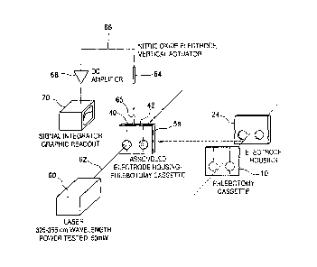
A preferred system for carrying out the method of the third embodiment is
depicted in Figures 1-5.
9
CA 02694018 2010-01-19
WO 2009/014616
PCT/US2008/008630
With reference to Figure 1, there is schematically depicted a disposable
phlebotomy cassette 10 which contains blood sample containing chambers 12 and
14
which both constitute sample containing zones. A front wall 16 of cassette 10
is of
Vycor glass or other electromagnetic radiation transparent material (i.e. to
whichever kind of electromagnetic radiation is used) and a rear wall 18 of the
cassette
is constituted, for example, of Vycor glass (40 micron pores) and reliance is
placed
on the rear wall's property of being porous to nitric oxide gas but preventing
passage
therethrough of protein. The blood sample containing chamber 12 is impregnated
with mercury (II) chloride or other nitrosothiols destroying agent so as not
to interfere
with absorption of electromagnetic radiation by blood sample. Blood sample
containing chambers 12 and 14 are, for example, 4 mm diameter and 5 mm
transverse
dimension. The chamber 12 contains nitrosothiols destroying agent, e.g.
mercury (II)
chloride, in large excess, compared to nitrosothiols that are present; the
chamber 14
does not contain nitrosothiols destroying agent.
Communicating with chamber 12 is a capillary blood sample containing
chamber/zone inlet 20. Communicating with chamber 14 is a capillary blood
sample
containing chamber/zone inlet 22.
With reference to Figure 2, there is schematically depicted a reusable solvent
reservoir/electrode introduction compartment or housing 24 which provides a
solvent
containing zone and which is constructed of inert material (i.e., inert to
solvent and
nitric oxide gas), e.g. polytetrafluoroethylene. The element 24 contains two
solvent
reservoir compartments 26 and 28 separated by a partition 30. The element 24
contains a solid back wall and a front wall with circular openings 32 and 34,
respectively, into each of the compartments 26 and 28. The opening 32 is
bounded by
a ring shaped upstanding protruding wall 36, and the opening 34 is bounded by
ring
shaped upstanding protruding wall 38. An upper wall of element 24 is provided
with
an opening 40 for introduction of an electrode into compartment 26 and an
opening 42
for introduction of electrode into compartment 28. Each of the upstanding
walls 36
and 38 is colored black or otherwise provided with electromagnetic radiation
shielding to minimize and guard against electromagnetic radiation scattering
to
inserted electrode (described later) since electromagnetic radiation affects
the voltage
detected by an electrode.
The disposable cassette 10 and solvent reservoir/electrode introduction
compartment 24 are assembled, for example, by clamping cassette 10 to solvent
CA 02694018 2010-01-19
WO 2009/014616
PCT/US2008/008630
reservoir/electrode introduction compartment 24 so the rear side of cassette
10
adjacent chambers 12 and 14 is contiguous with openings 32 and 34 (Figure 2).
Alternatively, cassette 10 can be attached to element 24 by providing
retaining
brackets on the front side of element 24 or by providing structure on the
front of
element 24 providing an insertion slot for assembling cassette 10 and element
24.
The assembly provided is schematically depicted in Figure 3 which is denoted
58.
Figure 4 depicts an exploded view of the assembly of Figure 3, and indicates
the cassette 10 positioned in front of a front wall 44 of element 24. The
front wall 44
is forward of rear wall 46 of element 24. Element 44 contains a left side wall
48, a
right side wall 50, an interior vertical wall 52 dividing element 24 into
compartments
26 and 28 (depicted in Figure 2), a bottom wall 54 and a top wall 56
containing
electrode insertion openings 40 and 42 (also depicted in Figure 2).
Figure 5 depicts phlebotomy cassette 10 (see also Figure 1), solvent
reservoir/electrode introduction compartment 24 (see also Figure 2), assembly
58 (see
also Figure 3) as well as an electromagnetic radiation source depicted as a
laser source
60 which directs a laser beam 62 of 325 ¨ 355 nm frequency and 50 milliwatts
power,
e.g. a Nd:Y3A15012 laser, at one of the sample containing compartments 12 and
14,
from a distance, for example, of 6 to 10 inches, providing a laser beam of
cross-
sectional area at assembly 58 coextensive with the inlet opening of a sample
containing compartment (12 or 14) to which it is directed.
Also depicted in Figure 5 is a nitric oxide selective electrode 64 provided
with
a vertical actuator (not shown) which can be operated by a computer to raise
or lower
the electrode 64 into the appropriate electrode insertion opening (40 or 42).
The
electrode 64 is shown inserted into compartment 26 (Figure 2) at 65 (Figure
5).
The electrode 64 is a nitric oxide selective electrode detecting voltage
generated by presence of nitric oxide gas in solvent in 24 and providing a
signal 66 in
picovolts to a DC amplifier 68 which in turn provides an amplified signal 70
to a
signal integrator, and to a graphic readout device 70 which provides readout
of
amount of nitric oxide dissolved in solvent corresponding to amount of nitric
oxide
present as combined nitric oxide.
In use, finger of patient for whom blood nitric oxide data is desired, is
pricked
with a lancet, e.g. at bedside, to provide blood flow by capillary action
through
channels 20 and 22 respectively into chambers 12 and 14 (Figure 1). Then
element 24
(Figure 2) laid flat with rings 36 and 38 facing up, is filled through
openings 32 and
11
CA 02694018 2010-01-19
WO 2009/014616
PCT/US2008/008630
34 with solvent that is of higher solubility for nitric oxide gas than the
blood sample,
preferably methanol. The cassette 10 is then assembled with element 24 so
compartments 12 and 14 are opposite openings 32 and 34 respectively, e.g. by
clamping cassette 10 to element 24 so that windows for compartments 12 and 14
are
in front of the cassette 10. Then laser 60 (Figure 5) is turned on to provide
beam of
ultraviolet laser irradiation of 325 ¨ 355 nm of intensity high enough to
maximize
nitric oxide that is liberated (i.e. is the intensity sufficient to break
bonds to nitric
oxide) but not so large as to break down or otherwise interfere with providing
liberated nitric oxide) into compartment 12 or compartment 14, e.g. 50
milliwatts
power. Electrode 64 (Figure 5) is lowered into solvent reservoir 26 after the
laser
beam of laser 60 is directed at sample compartment 12 and raised from
compartment
12 after electrochemical detection of released nitric oxide, and lowered into
solvent
reservoir 28 after the laser beam of laser 60 is directed at sample
compartment 14 and
raised from compartment 14 after electrochemical detection of released nitric
oxide.
The raising and lowering of electrode 64 into compartments 26 and 28 is
preferably
by computer activation of driving motor (not shown). Separate measurements are
obtained in succession in either order.
The nitrosothiol destroying agent in compartment 12 degrades (selectively
cleaves) the nitrosothiols therein to nitrous acid from which nitric oxide is
not
liberated by electromagnetic radiation.
When the laser beam 62 is aimed at compartment 12, electrode 64 is lowered
through opening 40 into solvent compartment 26. The laser treatment liberates
gaseous nitric oxide from iron nitrosyls in the sample in compartment 12 which
passes from compartment 12 to diffuse through the porous back wall of cassette
10
into compartment 26 where the liberated nitric oxide is dissolved in the
solvent in
chamber 26. Laser irradiation is continued for as long as reading on readout
at 70
increases. The readout indicates the amount of nitric oxide present as iron
nitrosyls in
the sample.
When the laser beam 62 is aimed at compartment 14, electrode 64 is lowered
through opening 42 into solvent compartment 28. The laser treatment liberates
nitric
oxide from iron nitrosyls and also from nitrosothiols. The liberated nitric
oxide
passes through the porous back wall of cassette 10 into solvent reservoir 28
whereby
amount of dissolved nitric oxide is detected to provide readout at 70 of total
nitric
oxide present as combined nitric oxide.
12
CA 02694018 2015-07-28
The porous back wall of cassette 10 allows passage of nitric oxide gas into
solvent containing zones but no protein so irradiation causes continuous
release of
nitric oxide without any rebinding to protein.
The determination of total nitric oxide present as combined nitric oxide and
of
nitric oxide present as iron nitrosyls allows computation of ratio of nitric
oxide
present as iron nitrosyls to total nitric oxide, i.e. present as combined
nitric oxide, and
by difference determination of amount of nitrosothiols in a sample thereby
providing
data allowing diagnosis and/or confirmation of diagnosis.
For the third embodiment, the laser 60 can be replaced by a light-emitting
diode that emits 210-220 nm wavelength ultraviolet radiation or 300-400 nm
wavelength ultraviolet radiation or 500-600 nm wavelength visible radiation or
700-
1400 nm wavelength near-infrared radiation with excellently comparable
results.
Variations
The foregoing description is the invention has been presented describing
certain operable and preferred embodiments. The scope of the claims should not
be
limited by the preferred embodiments set forth in the examples, but should be
given
the broadest interpretation consistent with the description as a whole.
13