Note: Descriptions are shown in the official language in which they were submitted.
CA 02694229 2010-01-20
WO 2009/024522 PCT/EP2008/060664
1
INJECTION APPARATUS AND SPECIAL NEEDLE FOR MAKING AN INJECTION AT A
PREDETERMINED DEPTH IN THE SKIN
The present invention relates to an injectinn apparatus
and to a method of injection.
Cutaneous injection is used in a number of applications.
It is advantageous to inject vaccines into the skin as antigen
which is then released into other tissues over a period of
time, promoting the response by antibodies and T-cells. Assay
sensors may also be injected into the skin, where they can be
interrogated optically through the skin. Such assays are
described for example in WO 00/02048 and W002/033550. They
may in particular be useful for glucose monitoring in
diabetes. Cutaneous injection is also used cosmetically in
wrinkle filling.
The depth at which material is injected is important, as
it determines the layer of the skin in which the material will
be deposited. The skin consists of two principal layers: the
epidermis (upper layer) and the dermis (lower layer), with an
overall thickness of 1.5 to 2 mm. The epidermis is overlaid
by the stratum corneum, a layer of dead cells approximately 10
to 25 pm thick. The upper cells of the stratum corneum are
continuously worn away. The epidermis and dermis are
separated by the basement membrane at a depth of approximately
150 }.tm. The cells at the top of the epidermis progressively
die and form the base of the stratum corneum, whilst the
basement membrane generates new cells at the base of the
epidermis. The dermis is vasculised, whereas the epidermis is
not.
The fluorophores commonly used in the competition assays
referred to above are illuminated transdermally with blue or
green light, which has a low penetration depth. Melanin,
which absorbs UV and visible radiation, is produced by the
basement membrane and transferred upwards into the epidermis
to protect the skin from UV radiation. This melanin absorbs
blue and green illumination used to interrogate the sensors
CA 02694229 2010-01-20
WO 2009/024522 PCT/EP2008/060664
2
and the resulting fluorescence, and accordingly penetration
through the skin is poor. Scattering in the skin and
absorption of light by blood contributes to this effect.
Therefore, the deeper the sensors are positioned in the skin,
the weaker the fluorescence detection will be. Accordingly,
for optimum sensitivity of the assay, the sensors should be as
close to the skin surface as possible.
However, there are disadvantages associated with
positioning the reagent particles within the epidermis or
basement membrane. In particular, the concentrations of
glucose within these layers may not correlato with the blood
glucose concentration which the assay is attempting to
measure. This is because the epidermis is not vasculised, and
the basement membrane uses glucose in the production of
epidermal cells which affects its glucose concentration. By
contrast, the concentration of glucose in the interstitial
fluid of the dermis is expected to correlate with blood
glucose concentration. Further, if the reagent particles were
positioned in the epidermis, they would move towards the skin
surface as the epidermal cells were renewed. Glucose
concentration in the epidermis is known to decrease towards
the skin surface (and is zero at the stratum corneum). This
will lead to an erroneous glucose estimate. Particles
injected into the dermis, on the other hand, will be retained
permanently, as seen in a conventional tattoo.
In the light of these considerations, the optimum
location for assay reagent particles is directly underneath
the basement membrane, at the top of the dermis.
In other assays, it may be desirable for sensor
particles to be deposited in the epidermis so that they will
be expelled from the body over time (W002/033550). Shallow
injection may be achieved using an array of short needles
coated with material to be injected. However, when injection
is carried out with an array of this type material is
deposited at every depth from the skin surface to the maximum
penetration depth of the needle.
CA 02694229 2010-01-20
WO 2009/024522 PCT/EP2008/060664
3
An apparatus or method that provides injection to a pre-
determined depth is consequently desirable.
An apparatus and method for injection at a desired
distance below the surface of the skin are described in
W003/072172. This document describes an injection apparatus
having a skin positioning member which lies or is moveable to
lie above or below the surrounding area of skin, and means for
inserting a needle parallel to the skin positioning member.
International standard nomenclature for needle point
geometry is described in IS07$64.
Injection needle types such as a lancet type or trocar
type needle are known. Examples of such injection needle types
are shown in Figures 1 and. 2. An injection needle is
generally formed from tubing having a lumen (315) and a shaft
(310), and a point (325) is formed at the distal end of the
needle by cutting across the tubing transversely to its
longitudinal axis (305) to form at least one bevel. A lancet
type needle (Figure 1) is formed by making a primary bevel 320
at an angle oc to the longitudinal axis 305 then making
secondary bevels (330) by increasing the grinding angle cx and
rotating the needle with respect to the grinding stone about
the longitudinal axis of the needle. The two secondary bevels
are formed with equal and opposite rotations of the needle
about its longitudinal axis with respect to the grinding
stone. The rotation angles used for the secondary bevels may
be altered depending on the intended use of the needle and are
less than 90 with respect to the primary bevel. Typically the
rotation with respect to the primary bevel is 55 . A trocar
type needle (Figure 2) may be formed by making three grindings
to the needle; the first forms the primary bevel (420), as
described above, and the secondary bevels (440) are formed at
a 120 rotational angle to each other and to the primary
bevel. Furthermore, the grinding angle with respect to the
longitudinal axis of the secondary bevels is steeper than the
grinding angle or,for the primary bevel. The secondary bevels
may result in a tip being formed at a position (450) along the
CA 02694229 2010-01-20
WO 2009/024522 PCT/EP2008/060664
4
radius of the cross-section of the needle perpendicular to the
longitudinal axis which lies between the outside of the lumen
and the outside of the shaft.
The angles of the primary bevel and of the secondary
bevels (if any) with respect to the longitudinal axis together
determine the length of the point of the needle, which is
defined as the length 325 from the very tip of the needle to
the heel (the edge formed by the bevel surface meeting the
outer surface of the shaft on the opposite side from the tip;
335).
In a first aspect, the present invention provides an
injection apparatus for making an injection at a predetermined
depth in skin comprising:
a skin positioning member for positioning on a patch of skin
within an area of skin to hold the patch of skin in a defined
position,
an injection needle comprising a point having a tip at a
distal end thereof and a shaft portion immediately proximal to
said point, the shaft portion having a longitudinal axis, and
means guiding said injection needle for movement from a
parking position above the skin beside said skin positioning
member to slide beneath said skin positioning member to an
injection position in which the distal end of the needle lies
at a predetermined distance below said skin positioning
member;
wherein:
the tip of the injection needle is closer to the longitudinal
axis of the shaft portion than is the outside of the shaft
portion.
We have found that, in a device as described in
w003/072172, it is sometimes difficult to insert the needle to
the correct depth in the skin due to the shape of the point of
the needle. This is particularly found to be a problem with
needles having a larger diameter, such as those used for
injecting sensor particles as described above. The diameter
of needles for such applications may be large compared with
the thickness of the skin (1.5 mm). Referring to Figure 3,
CA 02694229 2010-01-20
WO 2009/024522 PCT/EP2008/060664
using a conventional lancet-type point needle (300), the
sensor (360) may be injected too deeply when using the needle
with the primary bevel facing away from the skin surface (370)
(Figure 3b), or the needle may slide over the skin without
5 penetrating the surface when using the needle with the primary
bevel facing towards the skin surface (370) (Figure 3a). It
is therefore necessary to solve this problem in order to
ensure that a needle may reliably be inserted to the required
depth in the skin.
From the description of the known lancet and trocar type
needles, it may be seen that known patterns of grinding in
some cases do and in some cases do not result in the needle
tip being closer to the axis of the lumen than is the outside
wall of the surface of the needle. Where the grinding does
not provide this feature, we have found that modification of
the needle point in order that the tip of the injection needle
is closer to the longitudinal axis of the shaft portion than
is the outside of the shaft portion allows for more reliable
insertion of the needle to the required depth in skin.
Suitably, the needle tip may be made closer to the
longitudinal axis of the shaft portion than is the outside of
the shaft portion by modifying a lancet-type needle of the
type described above by bending the tip of the needle towards
the longitudinal axis of the shaft. Suitably, the bending of
the tip of the needle may be done before or after the
formation of the secondary bevels of the point are carried
out. Preferably, however, the bending of the tip is carried
out before the secondary bevels are formed in order to avoid
producing a sharp edge at the tip of the needle that may
damage the sensor to be injected.
Alternatively, the needle tip may be made closer to the
longitudinal axis of the shaft portion than is the outside of
the shaft portion by providing a suitable grinding or
combination of grindings at the needle tip. Suitably, a
trocar-type needle of the type described above may be used.
Such a trocar-type needle may be provided with at least one
further grinding at the tip of the needle which, without
CA 02694229 2010-01-20
WO 2009/024522 PCT/EP2008/060664
6
changing the position of the needle tip relative to the
longitudinal axis of the needle, provides a more gradual slope
from the outside of the shaft towards the tip on the back of
the needle (the side opposite the heel).
The inventors have proposed that the incorrect
positioning of the injected material resulting from using the
needle with the primary bevel facing away from the surface of
the skin (as in Figure 3b) may be due to bending of the needle
during insertion into the skin caused by a component of the
reaction force from the skin acting on at least the primary
bevel perpendicular to the injection path. This bending
results in deviation of the needle from the intended injection
path. When injecting material such as a solid sensor
particle, it is particularly important that the injection is
made at the correct depth to ensure correct placement of the
sensor in the skin, and a large gauge needle must be used in
order to accommodate the sensor within the lumen. Thus, the
inventors have devised a needle in which the component of the
reaction force acting perpendicular to the injection path is
reduced compared with standard designs of injection needle.
Accordingly, the present invention provides in a second
aspect an injection apparatus for making an injection at a
predetermined depth in skin comprising:
a skin positioning member for positioning on a patch of skin
within an area of skin to hold the patch of skin in a defined
position,
an injection needle comprising a tip, at least one bevel and a
heel together forming a point at a distal end thereof, and a
shaft portion immediately proximal of said heel having a
longitudinal axis, including a lumen extending along the
longitudinal axis, wherein the at least one bevel is formed
between said tip and said heel such that a lumen opening is
defined extending from the tip to a proximal end of the lumen
opening located distal of the heel; and
means guiding said injection needle for movement from a
parking position above the skin beside said skin positioning
member to slide beneath said skin positioning member to an
CA 02694229 2010-01-20
WO 2009/024522 PCT/EP2008/060664
7
injection position in which the distal end of the needle lies
at a predetermined distance below said skin positioning
member;
wherein:
the length of the lumen opening of the needle is in a range
from 5 to 15 times the diameter of the shaft of the needle.
For example, the length of the lumen opening of the needle may
be in the range from 8 to 12 times the diameter of the shaft
of the needle, such as ten times the diameter of the shaft of
the needle.
The present invention further provides in a third aspect
an injection needle comprising a tip, at least one bevel and a
heel together forming a point at a distal end thereof, and a
shaft portion immediately proximal of said heel having a
longitudinal axis, including a lumen extending along the
longitudinal axis, wherein the at least one bevel is formed
between said tip and said heel such that the lumen opening is
defined from the tip to a proximal end of the lumen opening
located distal of the heel,
characterised in that the length of the lumen opening of the
needle is in a range from 5 to 15 times the diameter of the
shaft of the needle. For example, the length of the lumen
opening of the needle may be in the range from 8 to 12 times
the diameter of the shaft of the needle, such as ten times the
diameter of the shaft of the needle.
Preferably, the needles of the second and third aspects
of the invention have a shaft diameter of from 0.5 mm to
1.5 mm, for example 0.8 to 1.3 mm or 1.0 to 1.2 mm,
particularly preferably 1.1 mm. Where the shaft diameter is
1.1 mm, the length of the lumen opening may be from 5.5 mm to
16.5 mm, for example 8.8 to 13.2 mm, such as 11 mm.
Preferably, at least a part of the point of a needle of
the second or third aspect of the invention is formed
substantially parallel to the longitudinal axis of the needle,
and most preferably, at least half of the point length is
formed parallel to the longitudinal axis of the needle. This
results in that part of the point having a part-cylindrical
CA 02694229 2010-01-20
WO 2009/024522 PCT/EP2008/060664
8
form with a constant cross-section. Preferably, the distance
perpendicular to the longitudinal axis of the needle from the
tip to the bevel face at the part-cylindrical point section is
at least 50% of the diameter of the shaft of the needle, such
as 60% or 70%. Preferably, the part-cylinder has a semi-
circular cross-section, i.e. is a hemi-cylinder. Preferably,
the section of the point distal of the part-cylindrical point
section is shaped into a desired needle tip geometry, suitably
a lancet or trocar tip geometry, and that geometry may
suitably be modified in accordance with the needles described
in the first aspect of the invention. Preferably, the section
of the point immediately distal of the heel and proximal of
the part-cylindrical point section forms a further bevel at an
angle to the longitudinal axis, suitably 8 to 12 , such as
10 . Suitably, the transition between the part-cylindrical
point section and the further bevel may be a rounded
transition or a chamfered transition.
Suitably, the needle tip may be bent or ground or
otherwise shaped such that the tip is closer to the
longitudinal axis of the needle, as described previously.
An additional benefit of using a needle according to the
second or third aspect of the invention is that, when using
needles having an outer diameter that is a significant
proportion of the thickness of the dermis (around 1.5 mm) for
intradermal injection, there is a reduction of the stress and
lesions in the dermis caused by the insertion of the needle
compared with that caused by a conventional needle. The
stress and lesions may be further reduced if the full diameter
of the needle tube is introduced only a short distance, such
as 1 mm, into the skin.
Preferably, the apparatus further comprises means for
attaching said skin positioning member to the skin.
Preferably, said skin positioning member is arranged
such that at least a portion of said skin positioning member
lies or is moveable to lie above or below said area of skin
CA 02694229 2010-01-20
WO 2009/024522 PCT/EP2008/060664
9
such that at least a part of said patch of skin is held
elevated above or depressed below said area of skin.
Preferably, the needle is guided for movement of the
distal end of the needle at a constant distance below the
surface of a lifted patch of skin attached to the skin
positioning member. This will ensure that the injection depth
is not dependent on the precise distance over which the needle
point is moved, as would be the case if the needle moved
obliquely with respect to the skin positioning member.
Preferably, the skin positioning member holds the surface of
the lifted area of skin flat (planar). The movement of the
needle is then preferably parallel to the skin positioning
member surface.
'rhe skin positioning member preferably has adhesive
thereon to secure the patch of skin to the skin positioning
member. Alternatively, the skin positioning member may be
porous or provided with bores through which vacuum may be
applied to hold the skin to the skin positioning member. In
an alternative embodiment, the skin positioning member may be
pressed against the patch of skin to depress the patch of
skin. Such depression of the patch of skin may be such that
the patch of skin lies obliquely slanted with respect to the
natural orientation, allowing the needle to penetrate
therebelow from its edge.
The skin positioning member may be plate-like, or may
form the surface of a non-plate-like member, for example a
cone, a pyramid, a triangular prism or a hemisphere.
Preferably, said skin positioning member is moveable
between a first position in which it lies on said area of skin
and a second position in which at least a portion of said skin
positioning member is elevated above or depressed below said
area of skin with said patch of skin. However, the skin
positioning member may be fixed in a position elevated above
the surface of the skin and the skin may be drawn up to the
skin positioning member by the application of vacuum and
retained there against the skin positioning member by vacuum
CA 02694229 2010-01-20
WO 2009/024522 PCT/EP2008/060664
or by adhesive as described, or the skin positioning member
may be fixed in a position depressed below said area of skin.
Preferably, means are provided for tilting said skin
positioning member to elevate an edge thereof with said patch
5 of skin attached thereto to lift said patch of skin.
Alternatively, however, the whole skin positioning member may
be elevated, with or without some tilting also, to raise the
patch of skin. To conveniently provide for the tilting
movement, said skin positioning member is preferably carried
10 by a support structure to which the skin positioning member
may be hinged at one edge of the skin positioning member.
The skin positioning member may be moved using by the
interaction of one or more cam followers carried by the skin
positioning member each engaging a cam groove in a cam plate
which is mounted for sliding movement with respect to the skin
positioning member.
The injection needle preferably is guided for movement
using one or more cam followers attached to the needle each
engaging in a cam groove in a cam plate mounted for sliding
movement with respect to the needle and the same cam plate may
control the movement of the skin positioning member and of the
injection needle.
The apparatus may comprise a lower portion which is left
on the skin after injection to define or mark the injection
site and an upper portion containing the injection needle
which is detachable after injection. Said upper portion may
further include said skin positioning member although this
could be mounted to the lower portion so that it is left
behind when the upper portion is removed. It could then
either remain as part of the lower portion or be removed
separately. If it were made transparent, it could remain
covering the injection site and optical interrogation of an
injected sensor could be made therethrough.
CA 02694229 2010-01-20
WO 2009/024522 PCT/EP2008/060664
11
The predetermined depth at which injection is made using
the apparatus is suitably in the range of 100 pm to 2mm and
may be fixed during manufacture or may be user adjustable.
Said injection needle is preferably carried by a syringe
comprising a chamber for injectable material and means for
dispensing said material through said needle. The syringe may
contain as said injectable material particles to be injected
and may contain in separate compartments said particles to be
injected and a liquid for suspending the particles.
As indicated above, desirably the particles are assay
sensor particles containing assay reagents. However, the
injectable material in the syringe may alternatively be a
medicament and may be an antigen for use in an immunisation.
The injectable material may be in the form of a liquid, paste,
emulsion, a single implant or sensor particle, a plurality of
implants or sensor particles, or a suspension of implants or
sensor particles in a liquid.
The invention includes in a fourth aspect injection
apparatus comprising a housing containing an injection needle
comprising a point having a tip at a distal end thereof and a
shaft portion immediately proximal to said point having a
longitudinal axis and mounted for guided movement from a
parked position to an operative position, a detachable marker
unit mounted to said housing and so positioned that said
needle passes therethrough to reach said operative position,
and means for securing said marker unit at an injection site
prior to the making of an injection, wherein said tip of the
injection needle is closer to the longitudinal axis of the
shaft portion than is the outside of the shaft portion of the
needle, and whereby said apparatus can in use be positioned at
an injection site, said marker unit can be secured at said
injection site, said needle can be moved to said operative
position to make an injection and said housing can be removed
leaving said marker unit at the injection site to mark the
position thereof.
CA 02694229 2010-01-20
WO 2009/024522 PCT/EP2008/060664
12
Said marker unit may comprise a plate having an aperture
therein through which the needle passes in use. Said aperture
preferably has a maximum dimension of 2 mm or less. Apparatus
according to this fourth aspect of the invention may have all
or any of the features described above in connection with the
first aspect of the invention.
In a fifth aspect, the present invention provides an
injection apparatus comprising a housing containing:
an injection needle comprising a tip, at least one bevel and a
heel together forming a point at a distal end thereof, and a
shaft portion immediately proximal of said heel having a
longitudinal axis, including a lumen extending along the
longitudinal axis, wherein the at least one bevel is formed
between said tip and said heel such that a lumen opening is
defined from the tip to a proximal end of the lumen opening
located distal of the heel, said injection needle being
mounted for guided movement from a parked position to an
operative position;
a detachable marker unit mounted to said housing and so
positioned that said needle passes therethrough to reach said
operative position; and
means for securing said marker unit at an injection site prior
to the making of an injection;
wherein the length of the lumen opening of the needle is in a
range from 5 to 15 times the diameter of the shaft of the
needle, and
whereby said apparatus can in use be positioned at an
injection site, said marker unit can be secured at said
injection site, said needle can be moved to said operative
position to make an injection and said housing can be removed
leaving said marker unit at the injection site to mark the
position thereof.
Said marker unit may comprise a plate having an aperture
therein through which the needle passes in use. Said aperture
preferably has a maximum dimension of 2 mm or less. Apparatus
according to the fourth and fifth aspects of the invention may
CA 02694229 2010-01-20
WO 2009/024522 PCT/EP2008/060664
13
have all or any of the features described above in connection
with the first, second or third aspects of the invention.
The invention further includes a method of fixed-depth
cutaneous injection comprising: holding the surface of a patch
of skin in a defined position against the surface of a skin
positioning member and guiding an injection needle beneath the
skin positioning member to bring a discharge opening of the
injection needle to a predefined location beneath the skin
positioning member, wherein the injection needle comprises a
point having a tip at a distal end thereof and a shaft portion
immediately proximal to said point having a longitudinal axis,
and wherein the tip of the needle is closer to the
longitudinal axis of the shaft portion than is the outside of
the shaft portion. This method may be carried out using
apparatus according to either or both of the first and fourth
aspects of the invention.
The invention includes a second method of fixed-depth
cutaneous injection comprising: holding the surface of a patch
of skin in a defined position against the surface of a skin
positioning member and guiding an injection needle beneath the
skin positioning member to bring a discharge opening of the
injection needle to a predefined location beneath the skin
positioning member, wherein the injection needle comprises a
tip, at least one bevel and a heel together forming a point at
a distal end thereof, and a shaft portion immediately proximal
of said heel having a longitudinal axis, including a lumen
extending along the longitudinal axis, wherein the at least
one bevel is formed between said tip and said heel such that
the lumen opening is defined from the tip to a proximal end of
the lumen opening located distal of the heel, and wherein the
length of the lumen opening of the needle is in a range from 5
to 15 times the diameter of the shaft of the needle. This
method may be carried out using apparatus according to either
or both of the second and fifth aspects of the invention.
Suitably, these methods can be carried out by a patient
on himself/herself without the need for assistance from
medical personnel.
CA 02694229 2010-01-20
WO 2009/024522 PCT/EP2008/060664
14
Preferably, said injection needle is guided such that
not more than 1 mm of the length of the shaft portion of the
needle is inserted beneath the surface of the skin.
The invention will be further described with reference
to the preferred embodiments shown in the accompanying
drawings, in which:
Figure 1 shows an example of a known lancet-type
injection needle. View (a) shows the primary bevel face of the
needle, view (b) shows the needle rotated by 900 about its
longitudinal axis compared with view (a), and view (c) shows
the needle rotated about its longitudinal axis by 180
compared with view (a).
Figure 2 shows an example of a needle for use in the
apparatus of the present invention. View (a) shows the primary
bevel face of the needle, view (b) shows the needle rotated by
90 about its longitudinal axis compared with view (a), and
view (c) shows the needle rotated about its longitudinal axis
by 180 compared with view (a).
Figure 3 shows the resulting position of a sensor
injected using the primary bevel of the needle (a) facing
towards the skin, and (b) facing away from the skin.
Figure 4 shows a vertical cross section through an
illustrative embodiment of the invention;
Figure 5 shows the same embodiment in perspective view;
Figure 6 shows the same perspective view but with some
upper components removed;
Figure 7 is an enlarged view of the syringe component of
the apparatus as shown in Figure 4;
Figure 8 is a plan view of the apparatus as shown in
Figure 6;
CA 02694229 2010-01-20
WO 2009/024522 PCT/EP2008/060664
Figure 9 shows an example of a needle for use in the
apparatus of the present invention. View (a) shows the primary
bevel face of the needle, view (b) shows the needle rotated by
90 about its longitudinal axis compared with view (a), and
5 view (c) shows the needle rotated about its longitudinal axis
by 180 compared with view (a).
Figure 10 shows another example of a needle for use in
the apparatus of the present invention. View (a) shows the
primary bevel face of the needle, view (b) shows the needle
10 rotated by 90 about its longitudinal axis compared with view
(a), and view (c) shows the needle rotated about its
longitudinal axis by 180 compared with view (a).
Figure 11 shows another example of a needle for use in
the apparatus of the present invention. View (a) shows the
15 primary bevel face of the needle, view (b) shows the needle
rotated by 90 about its longitudinal axis compared with view
(a), and view (c) shows the needle rotated about its
longitudinal axis by 180 compared with view (a).
Figure 12 shows an example of a needle according to the
present invention. View (a) shows the primary bevel face of
the needle, and view (b) shows the needle rotated by 90 about
its longitudinal axis compared with view (a).
Figure 13 shows a method of manufacturing a needle
according to the present invention.
Figure 14 shows another method of manufacturing a needle
according to the present invention.
Figure 15 shows a further method of manufacturing a
needle according to the present invention.
In a first variant of the injection apparatus according
to the present invention, as shown in Figure 4, the injection
apparatus 1 comprises an upper portion 2 and a lower portion
4. The lower portion comprises a circular plate 6 having a
central hole 8 defined by a cylindrical boss 10 with an
aperture 12. The upper portion 2 is dome-shaped, and has a
CA 02694229 2010-01-20
WO 2009/024522 PCT/EP2008/060664
16
lower surface 14 which lies on the upper surface 16 of the
lower portion 4. The upper portion 2 has a central
cylindrical boss 18 extending downwards inside the boss 10 of
the lower portion 4. The rim 20 of the upper portion boss 18
is attached by a pivot 22 to a skin positioning member
constituted by a base plate 24 of a bell crank 26. The base
plate 24 occupies the central hole 8 of the lower portion 4.
The lower surface 28 of the lower portion 4 and the lower
surface 30 of the base plate 24 have an adhesive covering 32,
which is covered with a release tape 34.
The upper portion 2 comprises a syringe 36 mounted in a
cylindrical sleeve 38 at an angle of approximately 20 to the
lower surface 14 of the upper portion 2. The sleeve 38 forms
an integral part of a wedge shaped block 39. The sleeve 38
has an axial slot 41 on its upper surface. The syringe 36
comprises a syringe body 40, a needle housing 42 and a plunger
44. The needle housing 42 extends from the lower end 46 of
the syringe body 40, and comprises a collapsible sleeve 48
housing a needle 50 which is attached to the syringe body 40.
At its distal end 52 the needle housing 42 passes through the
aperture 12 and lies inside a chamber 54 defined by the lower
portion boss 10, and is sealed with an end cap 56. The
plunger 44 lies in the upper end 58 of the syringe body 40.
The syringe body 40 contains material to be injected. In an
alternative variant, the double chamber syringes described
below may be used.
The upper portion 62 of the bell crank 26 forms a cam
follower 64. Cam followers 66, 68, 70 are also mounted on the
syringe body, the syringe plunger and the end cap respectively
and protrude through the slot 41 in the sleeve 38. Each of
the cam followers 64, 66, 68, 70 is constrained to radial
movement in the direction 71.
A grooved cam plate 72 engages the cam followers 64, 66,
68, 70 to form a box cam. A cam groove 74 engaging cam
follower 64 is initially angled to the left and then runs
straight outwards towards the periphery of the apparatus 1.
Cam grooves 76, 78 engaging cam followers 66, 68 initially run
CA 02694229 2010-01-20
WO 2009/024522 PCT/EP2008/060664
17
parallel to the final portion of the cam groove 74, then are
angled to the left with the cam groove 78 engaging cam
follower 68 more steeply angled, then parallel to the final
portion of the cam groove 74. A cam groove 80 engaging cam
follower 70 runs parallel to the final portion of the cam
groove 74. The cam grooves 76, 78, 80 engaging cam followers
66, 68, 70 terminate in a common lateral cam groove 82 which
is perpendicular to the final portions of the cam grooves 76,
78, 80. A spring (not shown) urges the cam followers 66, 68,
70 to the right. The cam plate 72 is mounted on runners 84
such that it is constrained to slide forwards and backwards in
the direction 85 only. The cam plate 72 is attached on its
upper surface 73 to a boss 87 which engages a manually
engageable slider 86 on the upper surface 88 of the upper
portion 2.
In use, the release tape 34 is removed from the adhesive
covering 32 of the lower portion lower surface 28 and the bell
crank base plate lower surface 30. The adhesive lower surface
28, 30 is applied to the skin. A small area of skin 90
becomes adhesively attached to the bell crank base plate 24,
and an annular area of skin 92 surrounding the small area of
skin 90 becomes adhesively attached to the lower portion 4.
To effect injection, the manually engageable slider 86
is pushed across the upper surface 88 of the upper portion 2
by the user. This causes the cam plate 72 to move forward
along the runners 84 from its initial position shown in Figure
6 to a final position. As the cam plate 72 moves, the cam
follower 64 of the bell crank 26 is immediately moved to the
left by the cam groove 74. This causes the bell crank 26 to
rotate around the pivot 22, such that the base plate 24 of the
bell crank 26 and the adhesively attached small area of skin
90 tilt relative to the lower surface 28 of the lower portion
4 to an angle of approximately 20 .
As the plate 72 continues to move forward, the cam
follower 66 on the syringe body 40 is moved to the left by its
cam groove 76. This causes the syringe needle 50 to move
through the end cap 56 and into the chamber 54 defined by the
CA 02694229 2010-01-20
WO 2009/024522 PCT/EP2008/060664
18
lower portion boss 10, collapsing the needle housing 42. The
needle 50 extends parallel to the lower surface 30 of the bell
crank base plate 24 at a defined distance from it, such that
it extends under the small area of skin 90 parallel to the
skin surface at a defined depth. The depth may for example be
100 gm, which lies in the dermis just below the junction with
the epidermis. In an alternative embodiment, the distance
between the bell crank base plate 24 and the needle 50 (and
hence the depth of injection) may not be preset in manufacture
but may be set by the user within a certain range, for example
using a dial coupled to a screw jack lifting the needle
assembly.
Simultaneously, the cam follower 68 on the syringe
plunger is moved to the left by its cam groove 78. The
steeper angle of this cam groove 78 compared with the cam
groove 76 for cam follower 66 means that the syringe plunger
44 moves to the left relative to the syringe body 40 and
travels down the syringe body 40. This causes the material 60
to be injected to be expelled through the needle 50 into the
skin.
When the plate 72 reaches its final position, the cam
followers 66, 68, 70 are forced to the right in the lateral
groove 80 by the spring (not shown), retracting the syringe 36
into its sleeve 38. The syringe 36 is now shorter in length
because the needle housing 42 has collapsed, and therefore the
syringe 36 does not protrude into the chamber 54 defined by
the boss 10. The upper portion 2 of the injection apparatus 1
can thus be removed from the skin surface. It is necessary to
remove the adhesive coating 32 from the lower surface 30 of
the bell crank base plate 24 to achieve this.
The lower portion 4 of the injection apparatus 1 is left
adhesively attached to the annular area of skin 92. Its
central hole 8 is used to define the site of injection. This
may be important, for example in the injection of assays which
need to be interrogated optically or otherwise at the site of
injection.
CA 02694229 2010-01-20
WO 2009/024522 PCT/EP2008/060664
19
The needle for use in the above-described injection
apparatus, in accordance with the first and fourth aspects of
the invention, has a tip which is closer to the longitudinal
axis of the needle than is the outside of the shaft of the
needle. One way of achieving this is to bend the needle tip
towards the longitudinal axis of the needle. Such a needle is
commercially available as a Huber tip needle, for example from
www.harvardapparatus.com. The bent needle may also be
provided with suitable shaping at the point. For example, as
shown in Figure 9, the needle 310 may be of the form of a
lancet-type needle, having a primary bevel face 320 and
secondary bevels 330 formed at equal and opposite rotational
angles about the longitudinal axis of the needle. The shaping
may be provided by any conventional means, such as grinding of
the needle with an abrasive surface such as a whetstone. The
tip of the needle may be bent towards the longitudinal axis
after the secondary bevels have been formed, as shown in
Figure 9. Alternatively, the tip may be bent before the
secondary bevels are formed, as shown in Figure 10.
A second method of making the needle tip closer to the
longitudinal axis than is the outside of the needle shaft is
to provide additional shaping of the tip of the needle by
suitable means such as grinding with an abrasive surface. The
known trocar-type needle is an example of the use of such
additional shaping, and the shape of the point of this type of
needle tip is shown in Figure 2. This type of needle has a
primary bevel 420, and two secondary bevels 440, each formed
at a rotational angle about the longitudinal axis of needle
410 of 120 to each other and to the primary bevel. 1t is
seen from the Figure that the tip 450 of the needle 410 is
thus formed closer to the longitudinal axis of the needle than
is the outside of the shaft of the needle.
Alternatively or additionally, at least one grinding may
be formed at the tip of the needle in order to make the tip
closer to the longitudinal axis. For example, a single
grinding 560 may be formed at a rotational angle of 180 to
the primary bevel 520, which grinding 560 is inclined from the
CA 02694229 2010-01-20
WO 2009/024522 PCT/EP2008/060664
outside of the needle shaft towards the longitudinal axis of
the needle.
An additional grinding as described above may be
advantageously be added to a trocar needle. While not
5 necessarily resulting in an adjustment of the tip position
relative to the longitudinal axis, the additional grinding
removes the sharp front edge of the tip of a standard trocar
needle that may split the skin surface when the needle is
inserted parallel thereto. An example of such a needle is
10 shown in Figure 11, and is described in US5,968,022.
An alternative needle type advantageous in the injection
methods and apparatuses of the second and fifth aspects of the
present invention is a needle according to the third aspect of
the invention in which the length of the lumen opening of the
15 needle is in a range from 5 to 15 times the diameter of the
shaft of the needle.
The intention in using such a needle is to reduce the
asymmetry and apparent gauge of the needle and thereby reduce
forces perpendicular to the skin surface during forming of the
20 injection channel. This in turn reduces the propensity of the
needle to bend when being inserted into the skin, and thus
minimises the deviation of the needle from the intended
injection path.
In order to achieve the above, the point of the needle,
is formed by further shaping in order to remove part of the
needle shaft and expose the lumen of the needle, resulting in
a part-cylindrical portion. This shaping also includes the
creation of a further bevel immediately distal of the heel of
the needle in order to facilitate the expansion of the channel
formed by the needle tip to accommodate the diameter of the
shaft of the needle. Thus, when the channel has been formed by
the part-cylindrical portion of the point, it is then expanded
to the full diameter of the needle tube by the insertion of
the further bevel in order that the injection of the desired
material may be carried out. Two planes at different angles to
the longitudinal axis (though at the same rotational angle
CA 02694229 2010-01-20
WO 2009/024522 PCT/EP2008/060664
21
about the longitudinal axis) may therefore be provided by the
further shaping in order to form a stepped bevel face. A
graduated transition may be provided between the part-
cylindrical section and the further bevel, such as a curved
transition, or there may be a sharp angle formed between the
part-cylindrical portion and the further bevel.
It is possible to envisage the manufacture of such a
needle in a number of ways. For convenience, the description
of the manufacture of the needle will be described starting
from a lancet needle of known type. However, it will be
appreciated that other types of needle, such as a trocar
needle or a plain needle with a single bevel and no further
shaping of the point, may be used as a suitable starting
point, or that the needle point may be shaped into the desired
form, such as a trocar or lancet point, after the shaping
described below has been carried out. The proximal end of the
lumen opening of the needle may additionally be shaped in a
known manner, such as by dulling the edge to prevent coring.
This needle design may in principle be applied to any size of
needle. It is envisaged that such needles for use in
intradermal injection will have a shaft diameter in the range
of 0.5 to 1.5 mm.
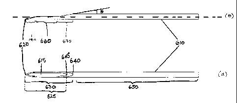
Referring to Figure 12, two depictions of a needle
according to the present invention are shown (elevation: Fig
12(b); plan: Fig 12(a)). The needle 610 depicted has a tip 620
shaped as for a known lancet needle, having a primary bevel
and two secondary bevels as described above. A standard
lancet needle has secondary bevels whose lengths 340 (Fig 1),
680 (Fig 12) are in the range from 2 to 2.5 times the diameter
of the needle shaft. The point 630 of the needle extends from
the tip 620 to the heel 640 and includes the whole of the tip
and the heel. The lumen opening 625 extends from the tip 620
to the proximal end of the lumen opening 645, distal of the
heel. The length of the lumen opening of the needle may be in
the range of from 5 to 15 times the diameter of the needle
shaft, for example 8 to 12 times, such as 10 times the
diameter of the needle shaft. Thus, for a needle for
CA 02694229 2010-01-20
WO 2009/024522 PCT/EP2008/060664
22
intradermal injection having a 1.1 mm shaft diameter (19G),
the length of the lumen opening may be from 5.5 mm to 16.5 mm,
for example 8.8 mm to 13.2 mm, such as 11 mm. The shaft 650
comprises the whole of the needle proximal of the heel. It can
be seen that a section of the needle has been removed from
point 630, forming a part-cylindrical portion, and exposing
the lumen of the needle at 660.
The shaping of the point at 660 and/or the bevel 670
(proximal of 660 and distal of heel 640) may be achieved by
grinding the needle using a rotating grinding stone having its
axis of rotation parallel to the longitudinal axis of the
needle. Such an arrangement is shown in Figure 13. Where
this method is used, the curvature of the surface of the
grinding stone determines the shaping of the needle at the
transition between the part-cylindrical portion and the
further bevel of the needle. For example, a rounded edge of
the grinding stone will result in a rounded transition,
whereas a chamfered edge will result in a corresponding
chamfered transition.
Alternatively, the shaping of the point at 660 and/or
the further bevel 670 may be achieved by grinding the needle
using a rotating grinding stone having its axis of rotation
perpendicular to the longitudinal axis of the needle. Such an
arrangement is shown in Figure 14. Where this method is used,
the diameter of the grinding stone is selected to achieve the
required shape of the transition between the part-cylindrical
portion and the further bevel 670. The grinding stone may also
be used to shape the further bevel 670 to the required angle 6
to the longitudinal axis of the needle.
As a further alternative, the shaping of the point at
660 and the further bevel 670 may be achieved by wire erosion
of the needle. Such an arrangement is depicted in Figure 15.
The shape of the point of the needle is defined by the path
cut by the wire through the needle. Suitably, the path of the
wire may be substantially in the plane of the longitudinal
axis at 660, and at a desired angle 0 to the longitudinal axis
to form the further bevel 670. At 660 it is envisaged that
CA 02694229 2010-01-20
WO 2009/024522 PCT/EP2008/060664
23
the wire may be moved in a path at an angle 6-x to the
longitudinal axis, so that the apparent diameter of the needle
increases gradually from the tip towards the heel. Suitably,
the angle 0 may be 10 , and the angle x may be between 0 and
0, such as 0 to 5 . Preferably, however, the part-cylindrical
portion of the needle is formed by moving the wire parallel to
the longitudinal axis of the needle (i.e. at an angle 6-x=0),
as this arrangement minimises the component of the reaction
forces acting perpendicular to the injection path during
insertion of the needle into the skin.
These preferred embodiments of the injection apparatus
allow injection to a fixed depth to be achieved accurately.
The system has several advantages over prior art methods of
injection. First, as the needle extends under the skin
surface the site of entry of the needle is not near the site
of injection. This may be important in optical interrogation
of assays. Secondly, the channel depth of the needle in the
skin is much larger than the injection depth. This means that
a seal is formed between the skin and the needle, so that the
material to be injected does not travel along the outside of
the needle to the outside of the skin. Thirdly, injected
material is often spread out because of the pressure of
injection and the possibility of migration through tissue.
This is particularly significant in vertical injection into
the skin, where material often reaches the fat tissue below
the skin which has a low resistance to flow. Using the
present injection apparatus, even if the injected material is
spread out, it will be spread horizontally at the same depth.
When the apparatus is used to inject assay sensors, this has
the advantage that there is no stray signal from sensors at
depths other than the required depth.
Further, the use of the needles described herein in
conjunction with the described injection apparatuses allows
the depth of injection to be reliably reproduced, particularly
when injecting substances or particles requiring a large lumen
diameter needle to be used. A particular application for
which this advantage is important is the implantation of
CA 02694229 2010-01-20
WO 2009/024522 PCT/EP2008/060664
24
sensor particles in the skin in order to carry out measurement
of blood glucose concentration based on fluorescence lifetime
spectroscopy, as the sensor must be placed immediately below
the basement membrane which separates the dermis from the
epidermis in order that the sensor may measure the glucose
concentration in a vasculised region of the skin and that the
sensor may be optically interrogated.
In an alternative embodiment, the tiltable base plate 24
may be replaced by an inclined surface which is pressed
against the skin surface to provide a fixed-depth injection
path parallel to the inclined surface. The inclined surface
may be the surface of a cone, the apex of the cone being
pressed against the skin surface, or may be the surface of a
flat plate pressed at an angle against the skin.
Figures 4 and 7 show a double chamber syringe 94
suitable for use with a preferred embodiment of the invention.
This syringe 94 is used for injecting powder suspended in a
liquid 98 which is kept separate from the powder 96 until the
moment of injection. In alternative embodiments, the syringe
may contain a liquid and two powders, two liquids and a
powder, a solid dose and a liquid, a solid dose and a plunger,
or other materials to be injected. Such a syringe is
described in detail in W003/072172.
Whilst the invention has been described with
reference to the illustrated embodiments, it is to be
appreciated that many modifications and variations are
possible within the scope of the invention.