Note: Descriptions are shown in the official language in which they were submitted.
CA 02694956 2010-01-28
WO 2009/048675
PCT/US2008/071775
PLURIPOTENT STEM CELL DIFFERENTIATION BY USING HUMAN FEEDER CELLS
FIELD OF THE INVENTION
[0001] The present invention relates to the field of pluripotent stem cell
differentiation. The
present invention provides methods for the differentiation of pluripotent stem
cells on a
human feeder cell layer. In particular, the present invention provides an
improved
method for the differentiation of pluripotent stem cells into pancreatic
endocrine cells
using a human feeder cell layer.
BACKGROUND
[0002] Pluripotent stem cells, such as, for example, embryonic stem cells
have the ability to
differentiate into all adult cell types. As such, embryonic stem cells may be
a source of
replacement cells and tissue for organs that have been damaged as a result of
disease,
infection, or congenital abnormalities. The potential for embryonic stem cells
to be
employed as a replacement cell source is hampered by the difficulty of
efficiently
differentiating the embryonic stem cells into the cell type of choice.
[0003] In one example, Hon i et al. (PNAS 99: 16105, 2002) disclose that
treatment of mouse
embryonic stem cells with inhibitors of phosphoinositide 3-kinase (LY294002)
produced
cells that resembled 13 cells.
[0004] In another example, Blyszczuk et al. (PNAS 100:998, 2003) reports
the generation of
insulin-producing cells from mouse embryonic stem cells constitutively
expressing Pax4.
[0005] Micallef et al. reports that retinoic acid can regulate the
commitment of embryonic stem
cells to form Pdxl positive pancreatic endoderm (Diabetes 54:301, 2005).
[0006] Skoudy et al. reports that activin A (a member of the TGF13
superfamily) upregulates the
expression of exocrine pancreatic genes (p48 and amylase) and endocrine genes
(Pdxl,
insulin, and glucagon) in mouse embryonic stem cells (Biochem. J. 379: 749,
2004).
CA 02694956 2010-01-28
WO 2009/048675
PCT/US2008/071775
100071 Shiraki et al. studied the effects of growth factors that
specifically enhance differentiation
of embryonic stem cells into Pdxl positive cells. They observed that TGFI32
reproducibly yielded a higher proportion of Pdxl positive cells (Genes Cells.
2005 Jun;
10(6): 503-16.).
[0008] Gordon et al. demonstrated the induction of brachyury AINF-3beta
endoderm cells from
mouse embryonic stem cells in the absence of serum and in the presence of
activin along
with an inhibitor of Wnt signaling (US 2006/0003446A1).
[0009] Gordon et al. (PNAS 103: 16806, 2006) states "Wnt and TGF-beta/
nodal/ activin
signaling simultaneously were required for the generation of the anterior
primitive
streak".
[0010] Thomson et al. isolated embryonic stem cells from human blastocysts
(Science 282:114,
1998). Concurrently, Gearhart and coworkers derived human embryonic germ (hEG)
cell
lines from fetal gonadal tissue (Shamblott et al., Proc. Natl. Acad. Sci. USA
95:13726,
1998). Unlike mouse embryonic stem cells, which can be prevented from
differentiating
simply by culturing with Leukemia Inhibitory Factor (LIF), human embryonic
stem cells
must be maintained under very special conditions (U.S. Pat. No. 6,200,806; WO
99/20741; WO 01/51616).
[0011] D'Amour etal. describes the production of enriched cultures of human
embryonic stem
cell-derived definitive endoderm in the presence of a high concentration of
activin and
low serum (Nature Biotechnology 2005). Transplanting these cells under the
kidney
capsule of mice resulted in differentiation into more mature cells with
characteristics of
some endodermal organs. Human embryonic stem cell-derived definitive endoderm
cells
can be further differentiated into Pdxl positive cells after addition of FGF-
10 (US
2005/0266554A1).
[0012] D'Amour et al. (Nature Biotechnology -24, 1392- 1401 (2006)) states:
"We have
developed a differentiation process that converts human embryonic stem (hES)
cells to
endocrine cells capable of synthesizing the pancreatic hormones insulin,
glucagon,
somatostatin, pancreatic polypeptide and ghrelin. This process mimics in vivo
pancreatic
2
CA 02694956 2016-01-28
organogenesis by directing cells through stages resembling definitive
endoderm, gut-tube
endoderm, pancreatic endoderm and endocrine precursor en route to cells that
express
endocrine hormones".
[0013] In another example, Fisk et al. reports a system for producing
pancreatic islet cells from
human embryonic stem cells (US2006/0040387A1). In this case, the
differentiation
pathway was divided into three stages. Human embryonic stem cells were first
differentiated to endoderm using a combination of sodium butyrate and activin
A. The
cells were then cultured with TGF13 antagonists such as Noggin in combination
with EGF
or betacellulin to generate Pdxl positive cells. The terminal differentiation
was induced
by nicotinamide.
[0014] In one example, Benvenistry et al. states: "We conclude that over-
expression of Pdxl
enhanced expression of pancreatic enriched genes, induction of insulin
expression may
require additional signals that are only present in vivo" (Benvenistry et al,
Stem Cells
2006; 24:1923-1930).
[0015] In another example, Condie et al. disclose: "feeder layers that
contain or express ligands
or other compounds that inhibit gamma-secretase or Notch signaling to enhance
the
maintenance of pluripotent cells in a pluripotent state feeder layers that
contain or express
ligands or other compounds that inhibit gamma-secretase or Notch signaling to
enhance
the maintenance of pluripotent cells in a pluripotent state" (W02004090110).
[0016] In another example, Mitalipova et al. disclose: "Human embryonic
stem cells are
cultured with human granulosa feeder cells, muscle cells, Fallopian ductal
epithelial cells,
bone marrow stromal cells, and skin fibroblasts and the embryonic stem cells
maintain
their pluripotent phenotype" (US20050037488).
[0017] In another example, Xu et al. disclose: "mesenchymal and fibroblast-
like cell lines
obtained from embryonic tissue or differentiated from embryonic stem cells.
Methods for
deriving such cell lines, processing media, and growing stem cells using the
feeder cells
or conditioned media are described" (US20020072117).
3
CA 02694956 2016-01-28
[0018] Therefore, there still remains a significant need to develop
conditions for establishing
pluripotent stem cell lines that can be expanded to address the current
clinical needs,
while retaining the potential to differentiate into pancreatic endocrine
cells, pancreatic
hormone expressing cells, or pancreatic hormone secreting cells. We have taken
an
alternative approach to improve the efficiency of differentiating pluripotent
stem cells
toward pancreatic endocrine cells.
SUMMARY
[0019] The present invention relates to the field of pluripotent stem cell
differentiation. The
present invention provides methods for the differentiation of pluripotent stem
cells on a
human feeder cell layer. In particular, the present invention provides an
improved
method for the differentiation of pluripotent stem cells into pancreatic
endocrine cells
using a human feeder cell layer.
[0020] In one embodiment, there is provided a method for differentiating
human pluripotent
stem cells, comprising the steps of:
a. plating human pluripotent stem cells onto a human feeder cell layer,
b. treating the pluripotent stem cells with at least one factor that promotes
the
differentiation of the pluripotent stem cells, and
c. differentiating the pluripotent stem cells on the human feeder cell layer,
wherein the
step of differentiating comprises differentiating the pluripotent stem cells
into cells
expressing markers characteristic of the definitive endoderm lineage.
[0020A] In another embodiment, there is provided a composition comprising
cells for use in
treating diabetes by transplanting the cells into a human patient having
diabetes,
wherein the cells are obtained by treating human pluripotent stem cells on a
human
feeder cell with at least one factor that promotes the differentiation of the
pluripotent
stem cells, wherein the cells are differentiated into cells expressing markers
characteristic of the 13-cell lineage or precursor cells thereof, wherein the
precursor
4
CA 2694956 2017-03-28
cells are cells expressing markers characteristic of definitive endoderm,
cells
expressing markers characteristic of pancreatic endoderm or cells expressing
markers
characteristic of pancreatic endocrine.
[0020B] In another embodiment, there is provided a method for differentiating
human
pluripotent stem cells comprising treating human pluripotent stem cells on a
human
feeder cell with at least one factor that promotes the differentiation of the
pluripotent
stem cells, wherein the cells are differentiated into cells expressing markers
characteristic of the p-cell lineage or precursor cells thereof, wherein the
precursor
cells are cells expressing markers characteristic of definitive endoderm,
cells
expressing markers characteristic of pancreatic endoderm or cells expressing
markers
characteristic of pancreatic endocrine.
[0020C] In another embodiment, there is provided a method for differentiating
human
pluripotent stem cells, comprising the steps of: a) treating the human
pluripotent stem
cells plated onto a human feeder cell layer with at least one factor that
promotes the
differentiation of the human pluripotent stem cells, and b) differentiating
the human
pluripotent stem cells on the human feeder cell layer, wherein the step of
differentiating
comprises differentiating the human pluripotent stem cells into cells
expressing
markers characteristic of the definitive endoderm lineage.
BRIEF DESCRIPTION OF THE DRAWINGS
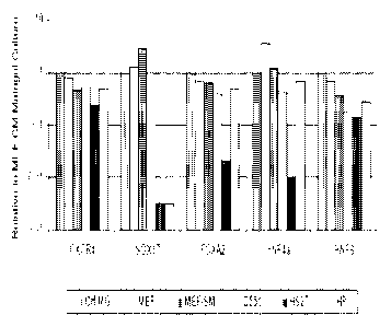
[0021] Figure 1 shows the expression of markers associated with
differentiation: CXCR4,
Sox-17, FoxA2, HNF-4a, HNF6 and AFP in populations of the human embryonic stem
cell line H9, at passage 46, cultured on MATRIGELTm with MEF conditioned media
and compared to cells transferred to mouse embryonic fibroblasts (MEF),
commercially available mouse embryonic fibroblasts (MEF-SM), human dermal
fibroblasts (D551), human foreskin fibroblasts (Hs27), and human pancreatic-
derived
stromal cells (HP).
CA 02694956 2010-01-28
WO 2009/048675
PCT/US2008/071775
[0022] Figure 2 shows the effects of human feeder cell layers on the
differentiation of human
embryonic stem cells into cells expressing markers characteristic of the
definitive
endoderm lineage. The figure shows expression of CXCR4, Sox-17, and FoxA2, as
determined by real-time PCR in populations of the human embryonic stem cell
line HI,
at passage 48, differentiated into cells expressing markers characteristic of
the definitive
endoderm lineage, cultured on mouse embryonic fibroblasts (MEF), commercially
available mouse embryonic fibroblasts (MEF-SM), human dermal fibroblasts
(D551),
human foreskin fibroblasts (Hs27), and human pancreatic-derived stromal cells
(HP).
[0023] Figure 3 shows the effects of human feeder cell layers on the
formation of cells
expressing markers characteristic of the pancreatic endoderm lineage. The
figure shows
expression of FoxA2, HNF-4a, HNF-6 and PDX-1, as determined by real-time PCR
in
populations of the human embryonic stem cell line H1, at passage 48,
differentiated into
cells expressing markers characteristic of the pancreatic endoderm lineage,
cultured on
mouse embryonic fibroblasts (MEF), commercially available mouse embryonic
fibroblasts (MEF-SM), human dermal fibroblasts (D551), human foreskin
fibroblasts
(Hs27), and human pancreatic-derived stromal cells (HP).
[0024] Figure 4 shows the effects of human feeder cell layers on the
formation of cells
expressing markers characteristic of the pancreatic endocrine lineage. The
figure shows
expression of FoxA2, HNF-4a, HNF-6, NeuroD1, Nkx 2.2, Pax-4, Nkx 6.1, PDX-1,
glucagon (GCG), and insulin (NS), as determined by real-time PCR in
populations of the
human embryonic stem cell line H1, at passage 48, differentiated into cells
expressing
markers characteristic of the pancreatic endocrine lineage, cultured on mouse
embryonic
fibroblasts (MEF), commercially available mouse embryonic fibroblasts (MEF-
SM),
human dermal fibroblasts (D551), human foreskin fibroblasts (Hs27), and human
pancreatic-derived stromal cells (HP).
[0025] Figure 5 shows the effects of human feeder cell layers on the
differentiation of human
embryonic stem cells into cells expressing markers characteristic of the
definitive
endoderm lineage. The figure shows expression of CXCR4, Sox-17, and FoxA2, as
determined by real-time PCR in populations of the human embryonic stem cell
line H9,
CA 02694956 2010-01-28
WO 2009/048675
PCT/US2008/071775
at passage 46, differentiated into cells expressing markers characteristic of
the definitive
endoderm lineage, cultured on mouse embryonic fibroblasts (MEF), commercially
available mouse embryonic fibroblasts (MEF-SM), human dermal fibroblasts
(D551),
human foreskin fibroblasts (Hs27), and human pancreatic-derived stromal cells
(HP).
[0026] Figure 6 shows the effects of human feeder cell layers on the
formation of cells
expressing markers characteristic of the pancreatic endoderm lineage. The
figure shows
expression of FoxA2, HNF-4a, HNF-6 and PDX-1, as determined by real-time PCR
in
populations of the human embryonic stem cell line H9, at passage 46,
differentiated into
cells expressing markers characteristic of the pancreatic endoderm lineage,
cultured on
mouse embryonic fibroblasts (MEF), commercially available mouse embryonic
fibroblasts (MEF-SM), human dermal fibroblasts (D551), human foreskin
fibroblasts
(Hs27), and human pancreatic-derived stromal cells (HP).
[0027] Figure 7 shows the effects of human feeder cell layers on the
formation of cells
expressing markers characteristic of the pancreatic endocrine lineage. The
figure shows
expression of FoxA2, HNF-4a, HNF-6, NeuroD1, Nkx 2.2, Pax-4, Nkx 6.1, PDX-1,
glueagon (GCG), and insulin (INS), as determined by real-time PCR in
populations of the
human embryonic stem cell line H9, at passage 46, differentiated into cells
expressing
markers characteristic of the pancreatic endocrine lineage, cultured on mouse
embryonic
fibroblasts (MEF), commercially available mouse embryonic fibroblasts (MEF-
SM),
human dermal fibroblasts (D551), human foreskin fibroblasts (Hs27), and human
pancreatic-derived stromal cells (HP).
DETAILED DESCRIPTION
[0028] For clarity of disclosure, and not by way of limitation, the
detailed description of the
invention is divided into the following subsections that describe or
illustrate certain
features, embodiments or applications of the present invention.
Definitions
[0029] Stem cells are undifferentiated cells defined by their ability at
the single cell level to both
self-renew and differentiate to produce progeny cells, including self-renewing
6
CA 02694956 2010-01-28
WO 2009/048675
PCT/US2008/071775
progenitors, non-renewing progenitors, and terminally differentiated cells.
Stem cells are
also characterized by their ability to differentiate in vitro into functional
cells of various
cell lineages from multiple germ layers (endoderm, mesoderm and ectoderm), as
well as
to give rise to tissues of multiple germ layers following transplantation and
to contribute
substantially to most, if not all, tissues following injection into
blastocysts.
[0030] Stem cells are classified by their developmental potential as: (1)
totipotent, meaning able
to give rise to all embryonic and extraembryonic cell types; (2) pluripotent,
meaning able
to give rise to all embryonic cell types; (3) multipotent, meaning able to
give rise to a
subset of cell lineages, but all within a particular tissue, organ, or
physiological system
(for example, hematopoietic stem cells (HSC) can produce progeny that include
HSC
(selfrenewal), blood cell restricted oligopotent progenitors and all cell
types and elements
(e.g., platelets) that are normal components of the blood); (4) oligopotent,
meaning able
to give rise to a more restricted subset of cell lineages than multipotent
stem cells; and (5)
unipotent, meaning able to give rise to a single cell lineage (e.g. ,
spermatogenic stem
cells).
[0031] Differentiation is the process by which an unspecialized
("uncommitted") or less
specialized cell acquires the features of a specialized cell such as, for
example, a nerve
cell or a muscle cell. A differentiated or differentiation-induced cell is one
that has taken
on a more specialized ("committed") position within the lineage of a cell. The
term
"committed", when applied to the process of differentiation, refers to a cell
that has
proceeded in the differentiation pathway to a point where, under normal
circumstances, it
will continue to differentiate into a specific cell type or subset of cell
types, and cannot,
under normal circumstances, differentiate into a different cell type or revert
to a less
differentiated cell type. De-differentiation refers to the process by which a
cell reverts to
a less specialized (or committed) position within the lineage of a cell. As
used herein, the
lineage of a cell defines the heredity of the cell, i.e., which cells it came
from and what
cells it can give rise to. The lineage of a cell places the cell within a
hereditary scheme of
development and differentiation. A lineage-specific marker refers to a
characteristic
specifically associated with the phenotype of cells of a lineage of interest
and can be used
to assess the differentiation of an uncommitted cell to the lineage of
interest.
7
CA 02694956 2010-01-28
WO 2009/048675
PCT/US2008/071775
[0032] Various terms are used to describe cells in culture. "Maintenance"
refers generally to
cells placed in a growth medium under conditions that facilitate cell growth
and/or
division, which may or may not result in a larger population of the cells.
"Passaging"
refers to the process of removing the cells from one culture vessel and
placing them in a
second culture vessel under conditions that facilitate cell growth and/or
division.
[0033] A specific population of cells, or a cell line, is sometimes
referred to or characterized by
the number of times it has been passaged. For example, a cultured cell
population that
has been passaged ten times may be referred to as a PIO culture. The primary
culture,
i.e., the first culture following the isolation of cells from tissue, is
designated PO.
Following the first subculture, the cells are described as a secondary culture
(P1 or
passage 1). After the second subculture, the cells become a tertiary culture
(P2 or
passage 2), and so on. It will be understood by those of skill in the art that
there may be
many population doublings during the period of passaging; therefore the number
of
population doublings of a culture is greater than the passage number. The
expansion of
cells (i.e., the number of population doublings) during the period between
passages
depends on many factors, including but not limited to the seeding density,
substrate,
medium, growth conditions, and time between passaging.
[0034] "13-cell lineage" refer to cells with positive gene expression for
the transcription factor
PDX-1 and at least one of the following transcription factors: NGN-3, Nkx2.2,
Nkx6.1,
NeuroD, Is1-1, HNF-3 beta, MAFA, Pax4, and Pax6. Cells expressing markers
characteristic of the fi cell lineage include 1 cells.
[0035] -Cells expressing markers characteristic of the definitive endoderm
lineage" as used
herein refer to cells expressing at least one of the following markers: SOX-
17, GATA-4,
HNF-3 beta, GSC, Cerl, Nodal, FGF8, Brachyury, Mixlike homeobox protein, FGF4
CD48, eomesodermin (EOMES), DKK4, FGF17, GATA-6, CXCR4, C-Kit, CD99, or
OTX2. Cells expressing markers characteristic of the definitive endoderm
lineage
include primitive streak precursor cells, primitive streak cells, mesendoderm
cells and
definitive endoderm cells.
8
CA 02694956 2016-01-28
[0036] "Cells expressing markers characteristic of the pancreatic endoderm
lineage" as used
herein refer to cells expressing at least one of the following markers: PDX-1,
HNF-lbeta,
HNF-3beta, PTF-1 alpha, fINF-6, or HB9. Cells expressing markers
characteristic of the
pancreatic endoderm lineage include pancreatic endoderm cells.
[0037] "Cells expressing markers characteristic of the pancreatic endocrine
lineage" as used
herein refer to cells expressing at least one of the following markers: NGN-3,
NeuroD,
Islet-1, PDX-1, NKX6.1, Pax-4, or PTF-1 alpha. Cells expressing markers
characteristic
of the pancreatic endocrine lineage include pancreatic endocrine cells,
pancreatic
hormone expressing cells, and pancreatic hormone secreting cells, and cells of
the 13-cell
lineage.
[0038] "Definitive endoderm" as used herein refers to cells which bear the
characteristics of cells
arising from the epiblast during gastrulation and which form the
gastrointestinal tract and
its derivatives. Definitive endoderm cells express the following markers:
CXCR4, HNF-3
beta, GATA-4, SOX-17, Cerberus, OTX2, goosecoid, c-Kit, CD99, and Mix11.
[0039] "Extraembryonic endoderm" as used herein refers to a population of
cells expressing at
least one of the following markers: SOX-7, AFP, and SPARC.
[0040] "Markers" as used herein, are nucleic acid or polypeptide molecules
that are differentially
expressed in a cell of interest. In this context, differential expression
means an increased
level for a positive marker and a decreased level for a negative marker. The
detectable
level of the marker nucleic acid or polypeptide is sufficiently higher or
lower in the cells
of interest compared to other cells, such that the cell of interest can be
identified and
distinguished from other cells using any of a variety of methods known in the
art.
[0041] "Mesendoderm cell" as used herein refers to a cell expressing at
least one of the following
markers: CD48, eomesodermin (EOMES), SOX-17, DKK4, HNF-3 beta, GSC, FGF17,
GATA-6.
9
CA 02694956 2010-01-28
WO 2009/048675
PCT/US2008/071775
[0042] "Pancreatic endocrine cell" or "pancreatic hormone expressing cell"
as used herein refers
to a cell capable of expressing at least one of the following hormones:
insulin, glucagon,
somatostatin, pancreatic polypeptide and ghrelin.
[0043] "Pancreatic hormone secreting cell" as used herein refers to a cell
capable of secreting at
least one of the following hormones: insulin, glucagon, somatostatin, and
pancreatic
polypeptide.
[0044] "Pre-primitive streak cell" as used herein refers to a cell
expressing at least one of the
following markers: Nodal, or FGF8.
[0045] "Primitive streak cell" as used herein refers to a cell expressing
at least one of the
following markers: Brachyury, Mix-like homeobox protein, or FGF4.
[0046] The present invention relates to the field of pluripotent stem cell
differentiation. The
present invention provides methods for the propagation of pluripotent stem
cells on a
human feeder cell layer. The present invention also provides methods for the
differentiation of pluripotent stem cells on a human feeder cell layer. In
particular, the
present invention provides an improved method for the differentiation of
pluripotent stem
cells into pancreatic endocrine cells using a human feeder cell layer.
[0047] In one embodiment, the present invention provides a method for
differentiating
pluripotent stem cells, comprising the steps of:
a. Culturing the pluripotent stem cells,
b. Plating the pluripotent stem cells onto a human feeder cell layer, and
c. Treating the pluripotent stem cells with at least one factor that promotes
the
differentiation of the pluripotent stem cells.
[0048] The methods of the present invention provides an improved method for
differentiating
pluripotent stem cells, wherein the pluripotent stem cells are plated onto a
human feeder
cell layer prior to differentiating the pluripotent stem cells. The
pluripotent stem cells
may be cultured by any suitable method in the art. Likewise, the pluripotent
stem cells
CA 02694956 2010-01-28
WO 2009/048675
PCT/US2008/071775
may be plated onto the human feeder cell layer by any suitable method in the
art. The
pluripotent stem cells may be treated with at least one factor that promotes
the
differentiation of the pluripotent stem cells immediately after plating onto
the human
feeder cell layer. Alternatively, the pluripotent stem cells may be treated
with at least one
factor that promotes the differentiation of the pluripotent stem cells after
the pluripotent
stem cells have been cultured in the presence of the human feeder cell layer
for a period
of time. For example, the pluripotent stem cells may be cultured in the
presence of the
human feeder cell layer for a period of time sufficient for the pluripotent
stem cells to
form a monolayer.
[0049] Pluripotent stem cells may be plated onto the human feeder cell
layer at any density.
Optimal density however, may be depend on factors, such as, for example, the
pluripotent
stem cell used, the cells comprising the human feeder cell layer, the
differentiated cell
type, the size of the culture vessel, and the like. In one embodiment, the
pluripotent stem
cells are plated at a density such that the pluripotent stem cells are about
60% to about
80% confluent following 5 days of culture on the human feeder cell layer.
[0050] Pluripotent stem cells suitable for use in the present invention
include, for example, the
human embryonic stem cell line H9 (N1H code: WA09), the human embryonic stem
cell
line H1 (N1H code: WA01), the human embryonic stem cell line H7 (NIH code:
WA07),
and the human embryonic stem cell line SA002 (Cellartis, Sweden). Also
suitable for use
in the present invention are cells that express at least one of the following
markers
characteristic of pluripotent cells: ABCG2, cripto, CD9, FoxD3, Connexin43,
Connexin45, Oct4, Sox2, Nanog, hTERT, UTF-1, ZFP42, SSEA-3, SSEA-4, Tral-60,
Tra1-81.
[0051] The cells comprising the human feeder cell layer may be any human
cell that is capable
of promoting the differentiation of pluripotent stem cells. The cells
comprising the
human feeder cell layer may be adult cells. Alternatively, the cells
comprising the human
feeder cell layer may be fetal or embryonic. In one embodiment, the human
feeder cell
layer is comprised of fibroblast cells. In one embodiment, the fibroblast
cells are dermal
fibroblasts. The dermal fibroblasts may be the human dermal fibroblast cell
line Detroit
11
CA 02694956 2010-01-28
WO 2009/048675
PCT/US2008/071775
551 (CCL-110 ATCC). In another embodiment, the fibroblasts cells are foreskin
fibroblasts. The human foreskin fibroblast may be the human foreskin
fibroblast line
Hs27 (CRL-1634 ATCC).
[0052] Alternatively, the human feeder cell layer is comprised of
pancreatic-derived stromal
cells. In one embodiment, the pancreatic-derived stromal cells are the cells
disclosed in
US20040241761. In an alternate embodiment, the pancreatic-derived stromal
cells are
the cells disclosed in Science 306: 2261-2264, 2004. In an alternate
embodiment, the
pancreatic-derived stromal cells are the cells disclosed in Nature
Biotechnology 22: 1115
¨ 1124, 2004. In an alternate embodiment, the pancreatic-derived stromal cells
are the
cells disclosed in US20030082155. In an alternate embodiment, the pancreatic-
derived
stromal cells arc the cells disclosed in U55834308. In an alternate
embodiment, the
pancreatic-derived stromal cells arc the cells disclosed in Proc Nat Acad Sci
97: 7999-
8004, 2000. In an alternate embodiment, the pancreatic-derived stromal cells
are the cells
disclosed in W02004011621. In an alternate embodiment, the pancreatic-derived
stromal cells are the cells disclosed in W003102134. In an alternate
embodiment, the
pancreatic-derived stromal cells are the cells disclosed in US2004015805. In
an alternate
embodiment, the pancreatic-derived stromal cells are the cells disclosed in
US6458593.
In an alternate embodiment, the pancreatic-derived stromal cells are the cells
disclosed in
W02006094286. In an alternate embodiment, the pancreatic-derived stromal cells
are of
the H513P6 cell line that have been assigned ATCC No. PTA-6974.
Generation of a Feeder Cell Layer
[0053] Human feeder cell layers described in this application are useful
for differentiating
pluripotent stem cells. It is recognized that other types of cells may benefit
from being
differentiated on these feeder cell layers, and the compositions of this
invention may be
used for such purposes without restriction.
[0054] In one aspect of the present invention, a feeder cell layer is
generated by a method which
essentially involves:
a. Culturing the cells that will form the feeder layer, and
12
CA 02694956 2010-01-28
WO 2009/048675
PCT/US2008/071775
b. Inactivating the cells.
[0055] The cells that will form the feeder cell layer may be cultured on a
suitable culture
substrate. In one embodiment, the suitable culture substrate is an
extracellular matrix
component, such as, for example, those derived from basement membrane or that
may
form part of adhesion molecule receptor-ligand couplings. In one embodiment, a
the
suitable culture substrate is MATRIGELO (Becton Dickenson). MATRIGELO is a
soluble preparation from Engelbreth-Holm-Swarm tumor cells that gels at room
temperature to form a reconstituted basement membrane. In another embodiment,
the
suitable culture substrate is gelatin (Sigma).
[0056] Other extracellular matrix components and component mixtures are
suitable as an
alternative. Depending on the cell type being proliferated, this may include
laminin,
fibronectin, proteoglycan, entactin, heparan sulfate, and the like, alone or
in various
combinations.
[0057] The cells used to form the feeder cell layer may be inactivated
(i.e., rendered incapable of
substantial replication) by, for example, radiation, treatment with a chemical
inactivator,
such as, for example, mitomycin c, or by any other effective method.
[0058] The medium used for culturing the cells used to form the feeder cell
layer can have any
of several different formulae. The medium must be able to support the
propagation of at
least the cell line used to form the feeder cell layer. It is convenient that
the medium also
support the propagation of the pluripotent stem cells. However, as an
alternative, the
medium can be supplemented with other factors or otherwise processed to adapt
it for
propagating the pluripotent stem cells.
[0059] In one embodiment, the pancreatic-derived stromal cells are the
cells disclosed in
W02006094286.
[0060] Isolation of pancreatic-derived cells: In one aspect of the present
invention, pancreatic
cells are isolated by a multi-stage method, which essentially involves:
13
CA 02694956 2016-01-28
a. Perfusion of a cadaver pancreas, living donor or autologous pancreas,
with an
enzymatic solution,
b. Mechanical dissociation of the perfused pancreas,
c. Layering the digested tissue over a polysucrose or FicollTM gradient,
followed by
centrifugation to yield three distinct interfaces,
d. Removing the tissues and cells at each interface,
e. Culturing the tissues and cells in standard tissue culture plates in a
nutrient rich
selection media containing less than 5% serum, and
f. Leaving the culture undisturbed for about 2 to 4 weeks without any media
changes.
[0061] Perfusion of a cadaver pancreas can be achieved with any of the
enzymatic solutions well
known to those skilled in the art. An example of an enzymatic solution
suitable for use in
the present invention contains LIBERASE HI TM (Roche - 0.5 mg/ml) and DNase
1(0.2
mg/ml).
[0062] Mechanical dissociation of the pancreatic tissue can be carried out
rapidly by the use of a
tissue processor. Alternatively, mechanical dissociation of the pancreatic
tissue can be
carried out using a Ricordi Chamber or other equivalent apparatus that enables
a less
destructive dissociation of the tissue, compared to other procedures.
[0063] The digested pancreatic tissues are then subjected to a polysuerose
or FicollTM gradient
centrifugation to yield three distinct interfaces, which are enriched in cells
from islets, the
ductal tissue and the acinar tissue, respectively. In one embodiment, the
tissues and cells
are removed from each interface and cultured separately. In an alternative
embodiment,
the tissues and cells from all the interfaces are combined and cultured. It
has been
determined in accordance with the present invention that pancreatic stromal
cells can be
derived from any of the three interfaces. Alternatively, a continuous gradient
can be
employed and the cell population of choice selected to generate the pancreatic
stromal
cells.
14
CA 02694956 2010-01-28
WO 2009/048675
PCT/US2008/071775
[0064] According to the present invention, the tissues and cells collected
from one or more of the
interfaces are cultured in a selection media to selectively enrich stromal
cells in the cell
population. The selection media is rich in nutrient and contains low levels of
glucose and
serum. Generally speaking, the selection media contains less than 5% serum,
alternatively 1-3% serum, alternatively about 2% serum; and less than 30 mM
glucose.
In one embodiment, the selection media is supplemented with 2% serum that is
derived
from the same mammalian species that the donor pancreas was harvested from.
Alternatively, fetal or calf scrum, scrum from other species, or other scrum
supplements
or replacements can be used to supplement the selection media. An example of a
suitable
selection media is composed of DMEM (5 mM glucose), 2% fetal bovine serum
(FBS),
100 U/[ig penicillin/streptomycin, insulin-transferrin-selenium (ITS), 2 mM L-
Glutamine,
0.0165 mM ZnSO4, and 0.38 tM 2-mercaptoethanol.
[0065] During the culture in a selection media ("the selection phase"), the
cells can be cultured
under hypoxic or normoxic conditions. Under hypoxic conditions, oxygen levels
are
lower than 20%, alternatively lower than 10%, alternatively lower than 5%, but
more
than 1%.
[0066] Preferably, the culture should be maintained in the selection media
undisturbed for about
2 to 4 weeks without any media changes, at which point the cells have
typically become
adherent to the culture substrate used. The selection phase is considered to
be complete
when there is no further increasein the number of adherent cells.
[0067] It has been discovered that the methods of tissue harvest and
culturing in accordance with
the present invention result in a cell population enriched in pancreatic
stromal cells. By
"enriched" is meant that pancreatic stromal cells account for at least about
30%,
alternatively about 40%, alternatively about 50% of all the cells in the
population.
[0068] Alternatively, the tissues and cells collected from one or more of
the interfaces arc
cultured in a selection media to selectively enrich stromal cells in the cell
population.
The selection media is rich in nutrient and contains low levels of glucose.
Generally
speaking, the selection media contains less than 20% serum, alternatively 10
to 5%
serum, alternatively about 10% serum; and less than 30 mM glucose. In one
CA 02694956 2010-01-28
WO 2009/048675
PCT/US2008/071775
embodiment, the selection media is supplemented with 10% serum that is derived
from
the same mammalian species that the donor pancreas was harvested from.
Alternatively,
fetal or calf serum, serum from other species, or other serum supplements or
replacements can be used to supplement the selection media. An example of a
suitable
selection media is composed of DMEM (5 mM glucose), 10% fetal bovine serum
(FBS),
100 U/[tg penicillin/streptomycin.
[0069] During the culture in a selection media ("the selection phase"), the
cells can be cultured
under hypoxic or normoxic conditions. Under hypoxic conditions, oxygen levels
are
lower than 20%, alternatively lower than 10%, alternatively lower than 5%, but
more
than 1%.
[0070] Under these culture conditions, the media is replaced regularly at 2-
4 day intervals.
[0071] It has been discovered that the methods of tissue harvest and
culturing in accordance with
the present invention result in a cell population enriched in pancreatic
stromal cells. By
"enriched" is meant that pancreatic stromal cells account for at least about
30%,
alternatively about 40%, or alternatively about 50% of all the cells in the
population.
[0072] Subsequent to the initial phase of selection and cell attachment,
the cells (enriched with
stromal cells) are expanded under conditions as further described hereinbelow.
[0073] If desirable, the cell population enriched in stromal cells can be
exposed, for example, to
an agent (such as an antibody) that specifically recognizes a protein marker
expressed by
stromal cells, to identify and select pancreatic stromal cells, thereby
obtaining a
substantially pure population of pancreatic stromal cells.
[0074] Characterization of the isolated pancreatic stronzal cells: Methods
for assessing
expression of protein and nucleic acid markers in cultured or isolated cells
are standard in
the art. These include quantitative reverse transcriptase polymerase chain
reaction (RT-
PCR), Northern blots, i hybridization (see, e.g., Current Protocols in
Molecular Biology
(Ausubel et al., eds. 2001 supplement)), and immunoassays, such as
immunohistochemical analysis of sectioned material, Western blotting, and for
markers
that are accessible in intact cells, flow cytometry analysis (FACS) (see,
e.g., Harlow and
16
CA 02694956 2010-01-28
WO 2009/048675
PCT/US2008/071775
Lane, Using Antibodies: A Laboratory Manual, New York: Cold Spring Harbor
Laboratory Press (1998)).
[0075] The pancreatic stromal cells isolated in accordance with the present
invention are
characterized as, inter alia, substantially lacking at least one of the
following protein
markers: CD117, NCAM, ABCG2, cytokeratin 7, 8, 18, or 19. In certain specific
embodiments, the pancreatic stromal cells isolated in accordance with the
present
invention are characterized as substantially positive for at least one of the
following
protein markers: CD44, CD73, CD90 and CD105.
[0076] Expansion of pancreatic stromal cells: In a further aspect, the
present invention provides
a method for expanding the pancreatic stromal cells obtained in accordance
with the
present invention. As described hereinabove, pancreatic digests, which may
contain a
heterogeneous mixture of islets, ductal fragments and exocrine tissue, are
cultured in a
low serum selection media for 2-4 weeks, preferably without any media changes,
to
selectively enrich the desired stromal cells. The resulting cell population,
now enriched
with pancreatic stromal cells, is then switched to a growth media to expand
the pancreatic
stromal cells in the cell population.
[0077] The growth media suitable for use in the present invention can be
composed of media
such as DMEM containing penicillin/streptomycin (P/S) and serum at a
concentration of
2% to 20%, alternatively about 5 to 10%. In one embodiment, the growth media
is
composed of DMEM (1000 mg/L D-glucose; 862 mg/L glutamine), and 10% fetal
bovine
scrum. In an alternate embodiment, the growth media is supplemented with scrum
that is
derived from the same mammalian species that the donor pancreas was harvested
from.
Alternatively, fetal or calf serum, or other serum supplements or
replacements, such as,
for example, serum albumin, may be used to supplement the growth media.
[0078] Furthermore, the stromal cells can be expanded by culturing in a
defined growth media
containing agent(s) that stimulate the proliferation of the cells of the
present invention.
These factors may include, for example, nicotinamide, members of TGF-13
family,
including TGF-131, 2, and 3, bone morphogenic proteins (BMP-2, -4,6, -7, -11, -
12, and ¨
13), serum albumin, fibroblast growth factor family, platelet-derived growth
factor-AA,
17
CA 02694956 2010-01-28
WO 2009/048675
PCT/US2008/071775
and ¨BB, platelet rich plasma, insulin growth factor (IGF-I, II) growth
differentiation
factor (GDF-5, -6, -8, -10, 11), glucagon like peptide-I and II (GLP-1 and
II), GLP-1 and
GLP-2 mimetobody, Exendin-4, retinoic acid, parathyroid hormone, insulin,
progesterone, aprotinin, hydrocortisone, ethanolamine, beta mercaptoethanol,
epidermal
growth factor (EGF), gastrin I and II, copper chelators such as triethylene
pentamine,
TGF-a, forskolin, Na-Butyrate, activin, betacellulin,
insulin/transferring/selenium (ITS),
hepatocyte growth factor (HGF), keratinocyte growth factor (KGF), bovine
pituitary
extract, islet neogenesis-associated protein (IN GAP), proteasome inhibitors,
notch
pathway inhibitors, sonic hedgehog inhibitors, or combinations thereof.
Alternatively,
the stromal cells may be expanded by culturing in conditioned media. By -
conditioned
media" is meant that a population of cells is grown in a basic defined cell
culture medium
and contributes soluble factors to the medium. In one such use, the cells are
removed
from the medium, while the soluble factors the cells produce remain. This
medium is
then used to nourish a different population of cells.
[0079] In certain embodiments, the pancreatic stromal cells are cultured on
standard tissue
culture plates. Alternatively, the culture plates may be coated with
extracellular matrix
proteins, such as, for example, MATRIGEL (R), growth factor reduced MATRIGEL
(R),
laminin, collagen, gelatin, tenascin, fibronectin, vitronectin,
thrombospondin, placenta
extracts or combinations thereof.
[0080] Furthermore, the pancreatic stromal cells can be expanded in vitro
under hypoxic or
normoxic conditions.
Differentiation of Pluripotent Stem Cells
[0081] In one embodiment of the present invention, pluripotent stem cells
are propagated in
culture, while maintaining their pluripotency. Pluripotent stem cells are then
transferred
onto human feeder cell layers prior to differentiation. Changes in
pluripotency of the
cells with time can be determined by detecting changes in the levels of
expression of
markers associated with pluripotency. Alternatively, changes in pluripotency
can be
monitored by detecting changes in the levels of expression of markers
associated with
differentiation, or markers associated with another cell type.
18
CA 02694956 2010-01-28
WO 2009/048675
PCT/US2008/071775
[0082] The pluripotent cells are treated with at least one factor that
promotes their differentiation
into another cell type. The other cell type may be a cell expressing markers
characteristic
of the definitive endoderm lineage. Alternatively, the cell type may be a cell
expressing
markers characteristic of the pancreatic endoderm lineage. Alternatively, the
cell type
may be a cell expressing markers characteristic of the pancreatic endocrine
lineage.
Alternatively, the cell type may be a cell expressing markers characteristic
of the 13-cell
lineage.
[0083] Pluripotent stem cells treated in accordance with the methods of the
present invention
may be differentiated into a variety of other cell types by any suitable
method in the art.
For example, pluripotent stem cells treated in accordance with the methods of
the present
invention may be differentiated into neural cells, cardiac cells, hepatocytes,
and the like.
[0084] For example, pluripotent stem cells treated in accordance with the
methods of the present
invention may be differentiated into neural progenitors and cardiomyocytes
according to
the methods disclosed in W02007030870.
[0085] In another example, pluripotent stem cells treated in accordance
with the methods of the
present invention may be differentiated into hepatocytes according to the
methods
disclosed in US patent 6,458,589.
Differentiation of Pluripotent Stem Cells into Cells Expressing Markers
Characteristic of the Pancreatic Endocrine Lineage
[0086] In one aspect of the present invention, cells expressing markers
characteristic of the
pancreatic endocrine linage are formed from pluripotent stem cells by a
multistage
method, comprising the steps of:
a. Culturing the pluripotent stem cells,
b. Plating the pluripotent cells on a human feeder cell layer,
c. Differentiating the pluripotent stem cells into cells expressing markers
characteristics
of the definitive endoderm lineage,
19
CA 02694956 2016-01-28
d. Differentiating the cells expressing markers characteristics of the
definitive endoderm
lineage into cells expressing markers characteristics of the pancreatic
endoderm lineage,
and
e. Differentiating the cells expressing markers characteristics of the
pancreatic
endoderm lineage into cells expressing markers characteristics of the
pancreatic
endocrine lineage.
[0087] Markers characteristic of the definitive endoderm lineage are
selected from the group
consisting of SOX-17, GATA4, Hnf-3beta, GSC, Cerl, Nodal, FGF8, Brachyury, Mix-
like homeobox protein, FGF4 CD48, eomesodermin (EOMES), DKK4, FGF17, GATA6,
CXCR4, C-Kit, CD99, and OTX2. Suitable for use in the present invention is a
cell that
expresses at least one of the markers characteristic of the definitive
endoderm lineage. In
one aspect of the present invention, a cell expressing markers characteristic
of the
definitive endoderm lineage is a primitive streak precursor cell. In an
alternate aspect, a
cell expressing markers characteristic of the definitive endoderm lineage is a
mesendoderm cell. In an alternate aspect, a cell expressing markers
characteristic of the
definitive endoderm lineage is a definitive endoderm cell.
[0088] Markers characteristic of the pancreatic endoderm lineage are
selected from the group
consisting of Pdxl, HNF-lbeta, PTFla, HNF-6, HB9 and PROX1. Suitable for use
in the
present invention is a cell that expresses at least one of the markers
characteristic of the
pancreatic endoderm lineage. In one aspect of the present invention, a cell
expressing
markers characteristic of the pancreatic endoderm lineage is a pancreatic
endoderm cell.
[0089] Markers characteristic of the pancreatic endocrine lineage are
selected from the group
consisting of NGN-3, NeuroD, Islet-1, Pdx-1 , NKX6.1, Pax-4, and PTF-1 alpha.
In one
embodiment, a pancreatic endocrine cell is capable of expressing at least one
of the
following hormones: insulin, glucagon, somatostatin, and pancreatic
polypeptide.
Suitable for use in the present invention is a cell that expresses at least
one of the markers
characteristic of the pancreatic endocrine lineage. In one aspect of the
present invention,
a cell expressing markers characteristic of the pancreatic endocrine lineage
is a pancreatic
endocrine cell. The pancreatic endocrine cell may be a pancreatic hormone
expressing
CA 02694956 2010-01-28
WO 2009/048675
PCT/US2008/071775
cell. Alternatively, the pancreatic endocrine cell may be a pancreatic hormone
secreting
cell.
[0090] In one aspect of the present invention, the pancreatic endocrine
cell is a cell expressing
markers characteristic of the 1 cell lineage. A cell expressing markers
characteristic of
the 13 cell lineage expresses Pdxl and at least one of the following
transcription factors:
NGN-3, Nkx2.2, Nkx6.1, NeuroD, Is1-1, HNF-3 beta, MAFA, Pax4, and Pax6. In one
aspect of the present invention, a cell expressing markers characteristic of
the 13 cell
lineage is a 13 cell.
[0091] For example, pluripotent stem cells may be differentiated into cells
expressing markers
characteristic of the pancreatic endocrine lineage according to the methods
disclosed in
D'Amour et al, Nature Biotechnology 24, 1392 - 1401 (2006).
[0092] Formation of cells expressing markers characteristic of the
definitive endoderm lineage:
Pluripotent stem cells may be differentiated into cells expressing markers
characteristic
of the definitive endoderm lineage by any method in the art or by any method
proposed in
this invention.
[0093] For example, pluripotent stem cells may be differentiated into cells
expressing markers
characteristic of the definitive endoderm lineage according to the methods
disclosed in
D'Amour et al, Nature Biotechnology 23, 1534 ¨ 1541 (2005).
[0094] For example, pluripotent stem cells may be differentiated into cells
expressing markers
characteristic of the definitive endoderm lineage according to the methods
disclosed in
Shinozaki et al, Development 131, 1651 - 1662 (2004).
[0095] For example, pluripotent stem cells may be differentiated into cells
expressing markers
characteristic of the definitive endoderm lineage according to the methods
disclosed in
McLean et al, Stem Cells 25, 29 - 38 (2007).
[0096] For example, pluripotent stem cells may be differentiated into cells
expressing markers
characteristic of the definitive endoderm lineage according to the methods
disclosed in
D'Amour et al, Nature Biotechnology 24, 1392 ¨ 1401 (2006).
21
CA 02694956 2010-01-28
WO 2009/048675
PCT/US2008/071775
[0097] For example, pluripotent stem cells may be differentiated into cells
expressing markers
characteristic of the definitive endoderm lineage by culturing the pluripotent
stem cells in
medium containing activin A in the absence of serum, then culturing the cells
with
activin A and serum, and then culturing the cells with activin A and serum of
a different
concentration. An example of this method is disclosed in Nature Biotechnology
23, 1534
- 1541 (2005).
[0098] For example, pluripotent stem cells may be differentiated into cells
expressing markers
characteristic of the definitive endoderm lineage by culturing the pluripotent
stem cells in
medium containing activin A in the absence of serum, then culturing the cells
with
activin A with serum of another concentration. An example of this method is
disclosed in
D' Amour et al, Nature Biotechnology, 2005.
[0099] For example, pluripotent stem cells may be differentiated into cells
expressing markers
characteristic of the definitive endoderm lineage by culturing the pluripotent
stem cells in
medium containing activin A and a Wnt ligand in the absence of serum, then
removing
the Wnt ligand and culturing the cells with activin A with scrum. An example
of this
method is disclosed in Nature Biotechnology 24, 1392 - 1401 (2006).
[0100] Formation of cells expressing markers characteristic of the
pancreatic endoderm
lineage: Cells expressing markers characteristic of the definitive endoderm
lineage may
be differentiated into cells expressing markers characteristic of the
pancreatic endoderm
lineage by any method in the art or by any method proposed in this invention.
[0101] For example, cells expressing markers characteristic of the
definitive endoderm lineage
may be differentiated into cells expressing markers characteristic of the
pancreatic
endoderm lineage according to the methods disclosed in D 'Amour et al, Nature
Biotechnology 24, 1392 - 1401 (2006).
[0102] For example, cells expressing markers characteristic of the
definitive endoderm lineage
are further differentiated into cells expressing markers characteristic of the
pancreatic
endoderm lineage, by treating the cells expressing markers characteristic of
the definitive
endoderm lineage with a fibroblast growth factor and the hedgehog signaling
pathway
22
CA 02694956 2010-01-28
WO 2009/048675
PCT/US2008/071775
inhibitor KAAD-cyclopamine, then removing the medium containing the fibroblast
growth factor and KAAD-cyclopamine and subsequently culturing the cells in
medium
containing retinoic acid, a fibroblast growth factor and KAAD-cyclopamine. An
example of this method is disclosed in Nature Biotechnology 24, 1392 - 1401
(2006).
[0103] Formation of cells expressing markers of the pancreatic endocrine
lineage: Cells
expressing markers characteristic of the pancreatic endoderm lineage may be
differentiated into cells expressing markers characteristic of the pancreatic
endocrine
lineage by any method in the art or by any method disclosed in this invention.
[0104] For example, cells expressing markers characteristic of the
pancreatic endoderm lineage
may be differentiated into cells expressing markers characteristic of the
pancreatic
endocrine lineage according to the methods disclosed in D 'Amour et al, Nature
Biotechnology 24, 1392 - 1401 (2006).
[0105] For example, cells expressing markers characteristic of the
pancreatic endoderm lineage
are further differentiated into cells expressing markers characteristic of the
pancreatic
endocrine lineage, by culturing the cells expressing markers characteristic of
the
pancreatic endoderm lineage in medium containing DAPT and exendin 4, then
removing
the medium containing DAPT and exendin 4 and subsequently culturing the cells
in
medium containing exendin 1, IGF-1 and HGF. An example of this method is
disclosed
in Nature Biotechnology 24, 1392- 1401 (2006).
[0106] For example, cells expressing markers characteristic of the
pancreatic endoderm lineage
are further differentiated into cells expressing markers characteristic of the
pancreatic
endocrine lineage, by culturing the cells expressing markers characteristic of
the
pancreatic endoderm lineage in medium containing exendin 4, then removing the
medium
containing exendin 4 and subsequently culturing the cells in medium containing
exendin
4, IGF-1 and HGF. An example of this method is disclosed in D' Amour et al,
Nature
Biotechnology, 2006.
[0107] For example, cells expressing markers characteristic of the
pancreatic endoderm lineage
are further differentiated into cells expressing markers characteristic of the
pancreatic
23
CA 02694956 2010-01-28
WO 2009/048675
PCT/US2008/071775
endocrine lineage, by culturing the cells expressing markers characteristic of
the
pancreatic endoderm lineage in medium containing DAPT and exendin 4. An
example of
this method is disclosed in D' Amour et al, Nature Biotechnology, 2006.
[0108] For example, cells expressing markers characteristic of the
pancreatic endoderm lineage
are further differentiated into cells expressing markers characteristic of the
pancreatic
endocrine lineage, by culturing the cells expressing markers characteristic of
the
pancreatic endoderm lineage in medium containing exendin 4. An example of this
method is disclosed in D' Amour et al, Nature Biotechnology, 2006.
Isolation, Expansion and Culture of Pluripotent Stem Cells
Characterization of Pluripotent Stem Cells
[0109] Pluripotent stem cells may express one or more of the stage-specific
embryonic antigens
(SSEA) 3 and 4, and markers detectable using antibodies designated Tra-1-60
and Tra-1-
81 (Thomson et al., Science 282:1145, 1998). Differentiation of pluripotent
stem cells in
vitro results in the loss of SSEA-4, Tra- 1-60, and Tra-1-81 expression (if
present) and
increased expression of SSEA-1. Undifferentiated pluripotent stem cells
typically have
alkaline phosphatase activity, which can be detected by fixing the cells with
4%
paraformaldehyde, and then developing with Vector Red as a substrate, as
described by
the manufacturer (Vector Laboratories, Burlingame Calif.). Undifferentiated
pluripotent
stem cells also typically express Oct-4 and TERT, as detected by real time
PCR.
[0110] Another desirable phenotype of propagated pluripotent stem cells is
a potential to
differentiate into cells of all three germinal layers: endoderm, mesoderm, and
ectoderm
tissues. Pluripotency of pluripotent stem cells can be confirmed, for example,
by
injecting cells into severe combined immunodeficient (SCID) mice, fixing the
teratomas
that form using a fixative such as 4% paraformaldehyde, and then examining
them
histologically for evidence of cell types from the three germ layers.
Alternatively,
pluripotency may be determined by the creation of embryoid bodies and
assessing the
embryoid bodies for the presence of markers associated with the three germinal
layers.
24
CA 02694956 2010-01-28
WO 2009/048675
PCT/US2008/071775
[0111] Propagated pluripotent stem cell lines may be karyotyped using a
standard G-banding
technique and compared to published karyotypes of the corresponding primate
species. It
is desirable to obtain cells that have a "normal karyotype," which means that
the cells are
euploid, wherein all human chromosomes are present and not noticeably altered.
Sources of Pluripotent Stein Cells
[0112] The types of pluripotent stem cells that may be used include
established lines of
pluripotent cells derived from tissue formed after gestation, including pre-
embryonic
tissue (such as, for example, a blastocyst), embryonic tissue, or fetal tissue
taken any time
during gestation, typically but not necessarily before approximately 10-12
weeks
gestation. Non-limiting examples are established lines of human embryonic stem
cells or
human embryonic germ cells, such as, for example the human embryonic stem cell
lines
HI, H7, and H9 (WiCell). Also contemplated is use of the compositions of this
disclosure during the initial establishment or stabilization of such cells, in
which case the
source cells would be primary pluripotent cells taken directly from the source
tissues.
Also suitable arc cells taken from a pluripotent stem cell population already
cultured in
the absence of feeder cells. Also suitable are mutant human embryonic stem
cell lines,
such as, for example, BGOlv (BresaGen, Athens, GA).
[0113] In one embodiment, human embryonic stem cells are prepared as
described by Thomson
et al. (U.S. Pat. No. 5,843,780; Science 282:1145, 1998; Curr. Top. Dev. Biol.
38:133 ff.,
1998; Proc. Natl. Acad. Sci. U.S.A. 92:7844, 1995).
Culture of Pluripotent Stem Cells
[0114] In one embodiment, pluripotent stem cells are typically cultured on
a layer of feeder cells
that support the pluripotent stem cells in various ways. Alternatively,
pluripotent stem
cells are cultured in a culture system that is essentially free of feeder
cells, but
nonetheless supports proliferation of pluripotent stem cells without
undergoing
substantial differentiation. The growth of pluripotent stem cells in feeder-
free culture
without differentiation is supported using a medium conditioned by culturing
previously
CA 02694956 2010-01-28
WO 2009/048675
PCT/US2008/071775
with another cell type. Alternatively, the growth of pluripotent stem cells in
feeder-free
culture without differentiation is supported using a chemically defined
medium.
[0115] For example, Reubinoff et al (Nature Biotechnology 18: 399 - 404
(2000)) and
Thompson et al (Science 6 November 1998: Vol. 282. no. 5391, pp. 1145 ¨1147)
disclose the culture of pluripotent stem cell lines from human blastocysts
using a mouse
embryonic fibroblast feeder cell layer.
[0116] Richards et al, (Stem Cells 21: 546-556, 2003) evaluated a panel of
11 different human
adult, fetal and neonatal feeder cell layers for their ability to support
human pluripotent
stem cell culture. Richards et al, states: "human embryonic stem cell lines
cultured on
adult skin fibroblast feeders retain human embryonic stem cell morphology and
remain
pluripotent".
[0117] US20020072117 discloses cell lines that produce media that support
the growth of
primate pluripotent stem cells in feeder-free culture. The cell lines employed
are
mesenchymal and fibroblast-like cell lines obtained from embryonic tissue or
differentiated from embryonic stem cells. US20020072117 also discloses the use
of the
cell lines as a primary feeder cell layer.
[0118] In another example, Wang et al (Stem Cells 23: 1221-1227, 2005)
discloses methods for
the long-term growth of human pluripotent stem cells on feeder cell layers
derived from
human embryonic stem cells.
[0119] In another example, Stojkovic et al (Stem Cells 2005 23: 306-314,
2005) disclose a
feeder cell system derived from the spontaneous differentiation of human
embryonic
stem cells.
[0120] In a further example, Miyamoto et at (Stem Cells 22: 433-440, 2004)
disclose a source of
feeder cells obtained from human placenta.
[0121] Amit et at (Biol. Reprod 68: 2150-2156, 2003) discloses a feeder
cell layer derived from
human foreskin.
26
CA 02694956 2010-01-28
WO 2009/048675
PCT/US2008/071775
[0122] In another example, Inzunza et al (Stem Cells 23: 544-549, 2005)
disclose a feeder cell
layer from human postnatal foreskin fibroblasts.
[0123] US6642048 discloses media that support the growth of primate
pluripotent stem (pPS)
cells in feeder-free culture, and cell lines useful for production of such
media.
US6642048 states: "This invention includes mesenchymal and fibroblast-like
cell lines
obtained from embryonic tissue or differentiated from embryonic stem cells.
Methods
for deriving such cell lines, processing media, and growing stem cells using
the
conditioned media are described and illustrated in this disclosure."
[0124] In another example, W02005014799 discloses conditioned medium for
the maintenance,
proliferation and differentiation of mammalian cells. W02005014799 states:
"The
culture medium produced in accordance with the present invention is
conditioned by the
cell secretion activity of murine cells, in particular, those differentiated
and immortalized
transgenic hepatocytes, named MMH (Met Murine Hepatocyte)."
[0125] In another example, Xu et al (Stem Cells 22: 972-980, 2004)
discloses conditioned
medium obtained from human embryonic stem cell derivatives that have been
genetically
modified to over express human telomerase reverse transcriptase.
[0126] In another example, US20070010011 discloses a chemically defined
culture medium for
the maintenance of pluripotent stem cells.
[0127] An alternative culture system employs serum-free medium supplemented
with growth
factors capable of promoting the proliferation of embryonic stem cells. For
example,
Cheon et al (BioReprod DOI:10.1095/biolreprod.105.046870, October 19, 2005)
disclose
a feeder-free, serum-free culture system in which embryonic stem cells are
maintained in
unconditioned serum replacement (SR) medium supplemented with different growth
factors capable of triggering embryonic stem cell self-renewal.
[0128] In another example, Levenstein et al (Stem Cells 24: 568-574, 2006)
disclose methods
for the long-term culture of human embryonic stem cells in the absence of
fibroblasts or
conditioned medium, using media supplemented with bFGF.
27
CA 02694956 2010-01-28
WO 2009/048675
PCT/US2008/071775
[0129] In another example, US20050148070 discloses a method of culturing
human embryonic
stem cells in defined media without serum and without fibroblast feeder cells,
the method
comprising: culturing the stem cells in a culture medium containing albumin,
amino
acids, vitamins, minerals, at least one transferrin or transferrin substitute,
at least one
insulin or insulin substitute, the culture medium essentially free of
mammalian fetal
serum and containing at least about 100 ng/ml of a fibroblast growth factor
capable of
activating a fibroblast growth factor signaling receptor, wherein the growth
factor is
supplied from a source other than just a fibroblast feeder layer, the medium
supported the
proliferation of stem cells in an undifferentiated state without feeder cells
or conditioned
medium.
[0130] In another example, US20050233446 discloses a defined media useful
in culturing stem
cells, including undifferentiated primate primordial stem cells. In solution,
the media is
substantially isotonic as compared to the stem cells being cultured. In a
given culture, the
particular medium comprises a base medium and an amount of each of bFGF,
insulin,
and ascorbic acid necessary to support substantially undifferentiated growth
of the
primordial stem cells.
[0131] In another example, US6800480 states "In one embodiment, a cell
culture medium for
growing primate-derived primordial stem cells in a substantially
undifferentiated state is
provided which includes a low osmotic pressure, low endotoxin basic medium
that is
effective to support the growth of primate-derived primordial stem cells. The
basic
medium is combined with a nutrient serum effective to support the growth of
primate-
derived primordial stem cells and a substrate selected from the group
consisting of feeder
cells and an extracellular matrix component derived from feeder cells. The
medium
further includes non-essential amino acids, an anti-oxidant, and a first
growth factor
selected from the group consisting of nucleosides and a pyruvate salt."
[0132] In another example, US20050244962 states: "In one aspect the
invention provides a
method of culturing primate embryonic stem cells. One cultures the stem cells
in a
culture essentially free of mammalian fetal serum (preferably also essentially
free of any
animal serum) and in the presence of fibroblast growth factor that is supplied
from a
28
CA 02694956 2010-01-28
WO 2009/048675
PCT/US2008/071775
source other than just a fibroblast feeder layer. In a preferred form, the
fibroblast feeder
layer, previously required to sustain a stem cell culture, is rendered
unnecessary by the
addition of sufficient fibroblast growth factor."
[0133] In a further example, W02005065354 discloses a defined, isotonic
culture medium that is
essentially feeder-free and serum-free, comprising: a. a basal medium; b. an
amount of
bFGF sufficient to support growth of substantially undifferentiated mammalian
stem
cells; c. an amount of insulin sufficient to support growth of substantially
undifferentiated
mammalian stem cells; and d. an amount of ascorbic acid sufficient to support
growth of
substantially undifferentiated mammalian stem cells.
[0134] In another example, W02005086845 discloses a method for maintenance
of an
undifferentiated stem cell, said method comprising exposing a stem cell to a
member of
the transforming growth factor-beta (TGFI3) family of proteins, a member of
the
fibroblast growth factor (FGF) family of proteins, or nicotinamide (NIC) in an
amount
sufficient to maintain the cell in an undifferentiated state for a sufficient
amount of time
to achieve a desired result.
[0135] The pluripotent stem cells may be plated onto a suitable culture
substrate. In one
embodiment, the suitable culture substrate is an extracellular matrix
component, such as,
for example, those derived from basement membrane or that may form part of
adhesion
molecule receptor-ligand couplings. In one embodiment, a suitable culture
substrate is
MATRIGELO (Becton Dickenson). MATRIGEL is a soluble preparation from
Engelbreth-Holm-Swarm tumor cells that gels at room temperature to form a
reconstituted basement membrane.
[0136] Other extracellular matrix components and component mixtures are
suitable as an
alternative. Depending on the cell type being proliferated, this may include
laminin,
fibroncctin, gelatin, protcoglycan, cntactin, hcparan sulfate, and the like,
alone or in
various combinations.
[0137] The pluripotent stem cells may be plated onto the substrate in a
suitable distribution and
in the presence of a medium that promotes cell survival, propagation, and
retention of the
29
CA 02694956 2010-01-28
WO 2009/048675
PCT/US2008/071775
desirable characteristics. All these characteristics benefit from careful
attention to the
seeding distribution and can readily be determined by one of skill in the art.
[0138] Suitable culture media may be made from the following components,
such as, for
example, Dulbecco's modified Eagle's medium (DMEM), Gibco # 11965-092;
Knockout
Dulbecco's modified Eagle's medium (KO DMEM), Gibco #10829-018; Ham's F12/50%
DMEM basal medium; 200 mM L-glutamine, Gibco # 15039-027; non-essential amino
acid solution, Gibco 11140-050; 13-mercaptoethano1, Sigma # M7522; human
recombinant basic fibroblast growth factor (bFGF), Gibco # 13256-029.
[0139] The present invention is further illustrated, but not limited by,
the following examples.
EXAMPLES
Example 1
The Establishment of Human Pancreatic Cell Lines
[0140] Pancreas Preparation - Human pancreata not suitable for clinical
transplantation were
obtained from The National Disease Research Interchange (Philadelphia, PA),
following
appropriate consent for research use. The pancreas was transferred with organ
preservation solution to a stainless steel pan on ice and trimmed of all
extraneous tissue.
The pancreatic duct was cannulated with an 18 gauge catheter and the pancreas
was
injected with an enzyme solution, which contained the LIBERASE HI TM enzyme
(Roche
- 0.5 mg/ml) and DNase 1(0.2 mg/m1), dissolved in Dulbecco's Phosphate
Buffered
Saline (DPBS).
[0141] Rapid Mechanical Dissociation Followed by Enzymatic Digestion ¨ The
enzyme infused
pancreata were homogenized in a tissue processor, pulsed 3-5 times for 3-5
seconds/pulse, and the dissociated tissue were transferred to two 500 ml
trypsinizing
flasks (Bellco) containing magnetic stir bars. Thereafter, 50-100 ml of the
enzyme
solution was added to each flask. The flasks were placed in a 37 C waterbath
on
submersible stir plates and allowed to incubate with an intermediate stir rate
for 10
minutes. The stirring was stopped and the fine digested tissue was removed
from the
CA 02694956 2010-01-28
WO 2009/048675
PCT/US2008/071775
flask and transferred into 250 ml tube containing DPBS, 5% Fetal Bovine Serum
(FBS)
and 0.1 mg/ml DNase I (DPBS+) at 4 C to quench the digestion process. The
flasks
were replenished with 50-100 ml of the enzyme solution and returned to the
waterbath
and the stirring was re-initiated for an additional ten minutes. Again, the
flasks were
removed and the fine digest was collected and transferred to the 250 ml tubes
on ice.
This process was repeated for additional 3-5 times until the pancreas was
completely
digested.
[0142] Gradual Mechanical Dissociation with Simultaneous Enzyme Digestion ¨
The enzyme
infused pancreata were processed according to methods as described in Diabetes
37:413-
420 (1988). Briefly, the pancreata were cleaned of extraneous tissue and
injected with
the enzyme solution as described above. The pancreata were then placed into a
Ricordi
Chamber with beads and covered with a screen with a mesh size of 400-600 [tm
to retain
larger clusters of tissue. The chamber was covered and the enzyme solution was
circulated through the chamber at approximately 37 C and the chamber was
shaken to
allow beads to disrupt pancreatic tissue while the enzyme digested the
pancreas. Once
adequate dissociation and digestion was achieved, the digestion was terminated
and the
tissue was collected.
[0143] Tissue Separation - The collected tissue was centrifuged at 150 x g
for 5 minutes at 4 C.
The supernatant was aspirated and the tissue was washed two additional times
in DPBS+.
Following the final wash, the tissue was applied to a discontinuous gradient
for
purification. The digested tissue was suspended in polysucrose (Mediatech, VA)
with a
density of 1.108 g/ml at a ratio of 1-2 ml tissue pellet per 10 ml of
polysucrose solution.
The tissue suspension was then transferred to round-bottom polycarbonate
centrifuge
tubes and polysucrose solutions with densities of 1.096 and 1.037 were
carefully applied
to the tubes. A final layer of DMEM completed the discontinuous purification
gradient.
The gradient tubes were centrifuged at 2000 rpm for 20 minutes at 4 C with no
brake
applied. Following centrifugation, the tissue was collected from each
interface (three
interfaces) and washed several times in DPBS+ as described above and collected
in a 50
ml test tube.
31
CA 02694956 2010-01-28
WO 2009/048675
PCT/US2008/071775
[0144] Further Cell Cluster Dissociation - Optionally, one can further
dissociate large cell
clusters obtained using the above protocol into smaller clusters or single
cell suspensions.
After the final wash, the tissue from each fraction was suspended in 10 ml IX
trypsin/EDTA solution containing 200U/m1DNase I. The tubes were placed in the
water
bath and repeatedly aspirated and discharged from a 10 ml serological pipette
for 5-6
minutes until a near single cell suspension is achieved. The digestion was
quenched with
the addition of 4 C DPBS+ and the tubes centrifuged at 800 rpm for 5 minutes.
The cell
suspensions were washed with DPBS+ and cultured as described below.
[0145] Pancreatic Cell Culture - Following the final wash, the cells from
each interface were
resuspended in DMEM, 2% FBS, 100 U/ tg penicillin/streptomycin, ITS, 2 mM L-
Glutamine, 0.0165 mM ZnSO4 (Sigma), and 0.38[M 2-mercaptoethanol (Invitrogen,
CA) (hereinafter "the selection media"). Six ml of the cell suspension was
seeded in T-
25 tissue culture flasks and 12 ml of the cell suspension was seeded into T-75
flasks. The
flasks were placed in 37 C incubators with 5% CO2. Following 2-4 weeks
culture, a
complete media change was performed and adherent cells were returned to
culture in
DMEM (2750 mg/L D-glucose, 862 mg/L glutamine) (Gibco, CA) with 5% FBS
(HyClone, UT), 1% PIS, 0.0165 mM ZnSO4 (hereinafter "the growth media") and
allowed to reach near confluence (this stage is referred to as "passage 0" or
"PO"), at
which point they were passaged. Subsequent culturing of the cells was at 5000
cell/cm2
in the growth media. Cultures were passaged every 7-10 days at approximately
70-90%
confluency. It was shown that stromal cells were isolated from each of the
three fractions
present following the purification gradient.
Example 2
Culturing of Stromal Pancreatic Cells
[0146] Pancreatic cells isolated according to Example 1 were either
cultured under hypoxic
conditions (5% CO2, 3% 02, and 92% N2) or normoxic conditions (5% CO2, 20% 02,
and
75% N2) for 2-4 weeks in the selection media. The cultures were then switched
to the
growth media and fed two to three times per week. After the initial culture
period,
adherent cells were observed in plates cultured under hypoxic and normoxic
conditions.
32
CA 02694956 2015-01-09
Furthermore, following the initial 2-4 wks of culturing, there were very few
remaining
islet-like or ductal structures in the plates.
Example 3
Expression of Genes Associated with Differentiation in Pluripotent Stem Cells
Cultured on Feeder Cells Layers
[0022] The human embryonic stem cell line H9 was obtained from WiCell
Research Institute,
Inc., (Madison, WI) and cultured according to instructions provided by the
source
institute. Undifferentiated H9 human embryonic stem cells that had been
maintained on
inactivated primary mouse embryonic fibroblasts (MEF) were cryopreserved in
60%
FBS, 20% DMSO, and 20% DMEM/F12 (Invitrogen/GIBCO) supplemented with 20%
knockout serum replacement, 100 nM MEM nonessential amino acids, 0.5 mM beta-
mercaptoethanol, 2mM L-glutamine with 4ng/m1 human basic fibroblast growth
factor
(bFGF) (all from Invitrogen/GIBCO) at a rate of -1 C/min and stored in vapor
nitrogen.
The cells were thawed and plated onto mitocycin c treated D551 human dermal
fibroblasts seeded at a density of 52,000 cell/ cm2. Following three passages
on the D551
cells, the pluripotent cells were harvested and then transferred on MATRIGELTm
in
conditioned medium from cultures of inactivated MEF supplemented with 8 ng/ml
bFGF.
Human embryonic stem cells plated on MATRIGELTm (1:30) were cultured at 37 C
in an
atmosphere of 5% CO2 within a humidified tissue culture incubator in 60 mm
tissue
culture plates. When confluent (approximately 5-7 days after plating), human
embryonic
stem cells were treated with 1mg/m1 dispaseTM (Invitrogen/GIBCO) for 25-40 mM.
Once
the cells were released from the plate, they were pipted repeatedly with a 2
ml serological
pipet until the desired colony size was achieved. Cells were spun at 1000 rpm
for 5 min,
and the pellet was resuspended and plated at a 1:3 to 1:4 ratio of cells in
fresh culture
medium onto MATRIGELTm coated cell culture plates. Following five to ten
passages
on MATRIGELTm, the pluripotent H9 cells were transferred to a number of
different
feeder cells. Briefly, cells were cultured on the feeders in ES cell medium
consisting of
DMEM/F12 (Invitrogen/GIBCO) supplemented with 20% knockout serum replacement,
100 nM MEM nonessential amino acids, 0.5 mM beta-mercaptoethanol, 2mM L-
33
CA 02694956 2010-01-28
WO 2009/048675
PCT/US2008/071775
glutamine with 4ng/m1 human basic fibroblast growth factor (bFGF) (all from
Invitrogen/GIBCO) in tissue culture treated 6 well plates. The plates are
prepared by
coating with 0.1% gelatin (Sigma) and incubating at 37 C for a minimum of 4
hrs prior to
seeding the feeders. Just prior to seeding the gelatin is aspirated and the
feeder cell
suspension delivered to each well of the 6 well plate. The cells were allowed
to expand
for 5 days prior to initiating the differentiation protocol.
[0148] The ability of the human dermal fibroblast cell line D551 (ATCC No.
CCL-110), the
human foreskin fibroblast cell line Hs27 (ATCC No. CRL-1634), and the human
pancreatic-derived stromal cell line (disclosed in W02006094286) to maintain
pluripotency were evaluated. The D551 human feeder cells were cultured in EMEM
(ATCC 30-2003) supplemented with 10% FBS. Once confluent, the cells were
inactivated by mitomycin-C treatment and cryopreserved in EMEM, 10% FBS, and
5%
DMSO at a rate of -1 C/min and stored in vapor nitrogen. The cells were thawed
at 37 C
and seeded onto gelatin-coated tissue culture plates at 52,000/cm2 in EMEM
with 10%
FBS. The Hs27 human feeder cells were cultured in DMEM (ATCC 30-2002)
supplemented with 10% FBS. Once confluent, the cells were inactivated by
mitomycin-
C treatment and cryopreserved in DMEM, 10% FBS, and 5% DMSO at a rate of -
1 C/min and stored in vapor nitrogen. The cells were thawed at 37 C and seeded
onto
gelatin-coated tissue culture plates at 55,000/cm2 in DMEM with 10% FBS. The
human
pancreatic-derived stromal cell line were cultured in DMEM and 10% FBS until
confluent and treated with mitomycin C. The cells were cryopreserved in 90%
FBS and
10% DMSO at a rate of -1 C/min and stored in vapor nitrogen. The cells were
thawed at
37 C and seeded onto gelatin-coated tissue culture plates at 43,000/cm2 in
DMEM with
10% FBS. Cultures of human embryonic stem cells, plated onto commercially
available
mouse embryonic fibroblasts (MEFSM), and freshly derived mouse embryonic
fibroblasts (MEF) were included as controls.
[0149] Plates of inactivated human feeder cells were washed with PBS and
seeded with
embryonic stem cells in ES medium. Embryonic stem cells were cultured on the
human
feeder cell layers for 5 days. After this time, the expression of CXCR4, Sox-
17, Fox-A2,
HNF-4a, HNF-6 and AFP was determined by real-time PCR from human embryonic
34
CA 02694956 2010-01-28
WO 2009/048675
PCT/US2008/071775
stem cells cultured on mouse embryonic fibroblasts (MEF, Figure 1),
commercially
available mouse embryonic fibroblasts (MEF-SM, Figure 1), human dermal
fibroblasts
(D551, Figure 1), human foreskin fibroblasts (Hs27, Figure 1), and the human
pancreatic-
derived stromal cell line disclosed in W02006094286 (HP, Figure 1). Results
from a
representative experiment are shown in Figure 1. Results were normalized to
human
embryonic stem cells cultured on MATRIGEL (Off MG, Figure 1). CXCR4, Sox-17,
Fox-A2, HNF-4a, HNF-6 and AFP arc markers associated with differentiation.
Culture
of human embryonic stem cells on the human feeder cell layer resulted in the
decrease in
expression of these markers. These data suggest that the human dermal
fibroblast cell
line D551, human foreskin fibroblasts Hs27, and the human pancreatic-derived
stromal
cell line disclosed in W02006094286 maintain the pluripotency of human
embryonic
stem cells.
Example 4
Differentiation of Human Embryonic Stem Cells into Cells Expressing Markers
Characteristic of the Pancreatic Endocrine Lineage on Human Feeder Cell
Layers.
[0150] The human embryonic stem cell lines HI and H9 was obtained from
WiCell Research
Institute, Inc., (Madison, WI) and cultured according to instructions provided
by the
source institute. Undifferentiated HI & H9 human embryonic stem cells that had
been
maintained on inactivated primary mouse embryonic fibroblasts (MEF) were
cryoprescrved in 60% FBS, 20% DMSO, and 20% DMEM/F12 (Invitrogen/GIBCO)
supplemented with 20% knockout scrum replacement, 100 nM MEM nonessential
amino
acids, 0.5 mM betamercaptoethanol, 2mM L-glutaminc with 4ng/m1 human basic
fibroblast growth factor (bFGF) (all from Invitrogen/GIBCO) at a rate of -1
C/min and
stored in vapor nitrogen. The cells were thawed and plated onto mitocycin C
treated
D551 human dermal fibroblasts seeded at a density of 52,000 cell/ cm2.
Following three
passages on the D551 cells, the pluripotent cells were harvested and then
transferred on
MATRIGEL in conditioned medium from cultures of inactivated MEF supplemented
with 8 ng/ml bFGF. Human embryonic stem cells plated on MATRIGEL (1:30) were
cultured at 37 C in an atmosphere of 5% CO2 within a humidified tissue culture
CA 02694956 2010-01-28
WO 2009/048675
PCT/US2008/071775
incubator in 60 mm tissue culture plates. When confluent (approximately 5-7
days after
plating), human embryonic stem cells were treated with 1mg/m1 dispase
(Invitrogen/GIBCO) for 25-40 min. Once the cells were released from the plate,
they
were pipted repeatedly with a 2 ml serological pipet until the desired colony
size was
achieved. Cells were spun at 1000 rpm for 5 min, and the pellet was
resuspended and
plated at a 1:3 to 1:4 ratio of cells in fresh culture medium onto MATRIGEL
coated cell
culture plates. Following eleven passages on MATRIGEL, the pluripotent H1 & H9
cells
were transferred to a number of different feeder cells described below.
Briefly, cells
were cultured on the feeders in ES cell medium consisting of DMEM/F12
(Invitrogen/GIBCO) supplemented with 20% knockout serum replacement, 100 nM
MEM nonessential amino acids, 0.5 mM beta-mercaptoethanol, 2mM L-glutamine
with
4ng/m1 human basic fibroblast growth factor (bFGF) (all from Invitrogen/GIBCO)
in
tissue culture treated 6 well plates. The plates are prepared by coating with
0.1% gelatin
(Sigma) and incubating at 37 C for a minimum of 4 hrs prior to seeding the
feeders. Just
prior to seeding the gelatin is aspirated and the feeder cell suspension
delivered to each
well of the 6 well plate. The cells were allowed to expand for 5 days prior to
initiating
the differentiation protocol.
[0151] The ability of human feeder cell layers to support the
differentiation of human embryonic
stem cells was evaluated. Embryonic stem cells were cultured on the mitomycin
C
inactivated human feeder cell layers for 5 days. Cultures of human embryonic
stem cells,
plated onto commercially available mouse embryonic fibroblasts (MEF-SM), and
freshly
derived mouse embryonic fibroblasts (MEF) were included as controls.
[0152] The ability of human dermal fibroblasts (D551, Figures 2 and 5),
human foreskin
fibroblasts (HS27, Figures 2 and 5), and the human pancreatic-derived stromal
cell line
disclosed in W02006094286 (HP, Figures 2 and 5) to support the differentiation
of
populations of the human embryonic stem cell line H9 (Figure 2) and H1 (Figure
5) into
cells expressing markers characteristic of the definitive endoderm lineage was
evaluated.
Populations of embryonic stem cells cultured on commercially available mouse
embryonic fibroblasts (MEF-SM, Figures 2 and 5), and freshly derived mouse
embryonic
fibroblasts (MEF, Figures 2 and 5) were included as controls. Activin A
(10Ong/m1) was
36
CA 02694956 2010-01-28
WO 2009/048675
PCT/US2008/071775
added to populations of human embryonic stem cells cultured on the feeder cell
layers.
Cells were cultured continuously in the presence of activin A and harvested
after 3 days.
The level of expression of markers characteristic of the definitive endoderm
lineage were
analyzed by real-time PCR (Figures 2 and 5). Results shown in Figures 2 and 5
are
normalized to the cells prior to the initiation of the differentiation
protocol (DO).
[0153] Activin A evoked an increase in the expression of CXCR4, Sox-17 and
Fox-A2, in cells
cultured on mouse embryonic fibroblasts and human feeder cell layers. These
data
suggest that human feeder cell layers are able to support the differentiation
of human
embryonic stem cells into cells expressing markers characteristic of the
definitive
endoderm lineage.
[0154] The ability of human dermal fibroblasts (D551, Figures 3 and 6),
human foreskin
fibroblasts (HS27, Figures 3 and 6), and the human pancreatic-derived stromal
cell line
disclosed in W02006094286 (HP, Figures 3 and 6) to support the differentiation
of
populations of cells expressing markers characteristic of the definitive
endoderm lineage,
derived from populations of the human embryonic stem cell line H9 (Figure 3)
and H1
(Figure 6) into cells expressing markers characteristic of the pancreatic
endoderm lineage
was evaluated. Populations of embryonic stem cells cultured on commercially
available
mouse embryonic fibroblasts (MEF-SM, Figures 3 and 6), and freshly derived
mouse
embryonic fibroblasts (MEF, Figures 3 and 6) were included as controls. 1 1iM
retinoic
acid, 0.25uIVI KAAD-Cyclopamine and FGF-10 (SOng/m1) was added to populations
of
cells expressing markers characteristic of the definitive endoderm lineage,
derived from
human embryonic stem cells cultured on the feeder cell layers. Cells were
harvested after
8 days. The level of expression of markers characteristic of the pancreatic
endoderm
lineage were analyzed by real-time PCR (Figures 3 and 6). Results shown in
Figures 3
and 6 are normalized to DO gene expression.
[0155] Retinoic acid 0.25 M KAAD Cyclopamine, and FGF-10 treatment evoked
an increase in
the expression of Fox-A2, HNF-4a, HNF-6 and PDX-1, in cells cultured on mouse
embryonic fibroblasts, and human feeder cell layers. These data suggest that
human
feeder cell layers are able to support the differentiation of cells expressing
markers
37
CA 02694956 2010-01-28
WO 2009/048675
PCT/US2008/071775
characteristic of the pancreatic endoderm lineage, derived from populations of
the human
embryonic stem cell line H9 (Figure 3) and H1 (Figure 6) into cells expressing
markers
characteristic of the pancreatic endoderm lineage.
[0156] The ability of human dermal fibroblasts (D551, Figures 4 and 7),
human foreskin
fibroblasts (HS27, Figures 4 and 7), and the human pancreatic-derived stromal
cell line
disclosed in W02006094286 (HP, Figures 4 and 7) to support the differentiation
of
populations of cells expressing markers characteristic of the pancreatic
endoderm lineage,
derived from populations of the human embryonic stem cell line H9 (Figure 4)
and HI
(Figure 7) into cells expressing markers characteristic of the pancreatic
endocrine lineage
was evaluated. Populations of embryonic stem cells cultured on commercially
available
mouse embryonic fibroblasts (MEF-SM, Figures 4 and 7), and freshly derived
mouse
embryonic fibroblasts (MEF, Figures 4 and 7) were included as controls. The y-
secretase
inhibitor DAPT at luM, exendin-4, 1GF-1 and HGF (all 50 ng/m1) were added to
populations of cells expressing markers characteristic of the pancreatic
endoderm lineage,
derived from human embryonic stem cells cultured on the feeder cell layers.
Following 9
days of culture, the level of expression of markers characteristic of the
pancreatic
endocrine cells were analyzed by real-time PCR (Figures 4 and 7). Results
shown in
Figures 4 and 7 are normalized to DO gene expression.
[0157] y-secretase inhibitor, exendin-4, 1GF-1 and HGF treatment evoked an
increase in the
expression of Fox-A2, HNF-4a, HNF-6, neuro-D1, Nkx2.2, Pax-4, Nkx6.1, PDX-1,
glucagon, and insulin, in cells cultured on mouse embryonic fibroblasts, and
human
feeder cell layers. These data suggest that human feeder cell layers are able
to support
the differentiation of cells expressing markers characteristic of the
pancreatic endocrine
lineage, derived from populations of the human embryonic stem cell line H9
(Figure 3)
and H1 (Figure 6) into cells expressing markers characteristic of the
pancreatic endoderm
lineage. The expression of insulin and glucagon was higher in cells cultured
on human
feeder cell layers than on mouse feeder cell layers. These data suggest that
human feeder
cell layers are more able to support the differentiation of human embryonic
stem cells.
38
CA 02694956 2016-01-28
[0158]
Although the various aspects of the invention have been illustrated above by
reference to
examples and preferred embodiments, it will be appreciated that the scope of
the
invention is defined not by the foregoing description, but by the following
claims properly
construed under principles of patent law.
39