Note: Descriptions are shown in the official language in which they were submitted.
CA 02696681 2010-02-17
WO 2009/024568 1 PCT/EP2008/060837
PERCUTANEOUS ABDOMINAL IMPLANT
The present invention relates to an implant for percutaneous
implantation through the abdominal wall for encircling and
engaging an externalised length of a body duct of a human or
animal patient. The implant is of the kind comprising an
exterior tubular section defined by an circumferential exterior
wall at least a part of which is adapted to protrude outwardly
from the abdominal wall with a free end which serves for
mounting of a detachable device, an interior section defined by
an circumferential interior wall adapted to extend through the
abdominal wall and inside the patient for internal fixation of
the implant, wherein the exterior tubular section and the
interior section have a common axis.
A detailed discussion of various diseases and known surgical
procedures involving ostomy are found in the applicants own
European patent application No. EP 0477475.4 and international
patent application PCT/IB2007/050646, not yet published.
A tubular implant with a flange is known from U.S. patent nr.
4,217,664. This implant is used as a permanent, closable stoma
and includes a flexible, pliable sleeve of biocompatible, soft,
mesh material, e.g. polypropylene. Since a flexible sleeve can
move in response to the peristaltic movements of the
externalised intestine there is a great risk that the
connection of the tissue growing into the sleeve is too weak at
the beginning of the healing process to resist peristaltic
movement. The fragile tissue bond may rupture in response to
movement of the sleeve during peristaltic and in response to
passage of substance. This prevents fast healing and protracts
patient recovery. In addition due to the required folding
technique of the intestine around the free edge of the implant
to allow the intestinal tissue to get attached to the flexible
mesh, infectious material is guided directly towards the
exterior skin surface, inducing a considerable risk of
CA 02696681 2010-02-17
WO 2009/024568 2 PCT/EP2008/060837
irritation, inflammation and last but not least bacterial
contamination with a.o. faecal matter on the surrounding skin
surface, in particular during healing.
The applicants own patent applications describes various
embodiments of implants provided with rigid mesh means along
various parts of the interior circumference of the implant.
Although theses implant have proven to be of particular
advantage for many ostomy patients it has turned out that in
some patient's uncontrollable infection and inflammation
occurs, in particular at the contact site between the exterior
surface of the implant and the access opening in the abdominal
skin of the patient. It is recognised that the reason for this
is the design of these known implants, in which the exterior
ring section merges directly into the interior section, for
example by means of connecting members. This design provides a
microbiological access route or wick for i.e. faecal bacteria
along the exterior surface of the implant. This exterior
surface also provides a propagating surface for bacteria.
In a first aspect according to the present invention is
provided an implant of the kind mentioned in the opening
paragraph, which enables fast ingrowth of an externalised
length of a body duct.
In a second aspect according to the present invention is
provided an implant of the kind mentioned in the opening
paragraph, which can be implanted with a minimum risk of
inflammation, infection and necrosis.
The novel and unique features, whereby this is achieved
according to the present invention, is the fact that the
circumferential exterior wall of the exterior tubular section
and the circumferential interior wall of the interior section
are arranged axially spaced apart at a distance from each other
to provide an axial gap between opposing free ends of the
CA 02696681 2010-02-17
WO 2009/024568 3 PCT/EP2008/060837
exterior tubular section and the interior section, said
exterior tubular section and interior section are connected by
means of distance means.
During the surgical implantation procedure an access opening is
made at a relevant site through the abdominal wall. The implant
is located in the abdominal opening with the exterior section
protruding from the patient and the exterior section anchored
inside the body.
The body duct, e.g. the colon, is then externalised through the
internal diameter of the implant so that the interior section
carefully encircles, guides and supports the externalised body
duct. The outmost tissue layer, e.g. the serosa or any other
exposed layer of the body duct's exterior wall is thereby
brought into engaging contact with interior faces of the
implant to trigger the gradual ingrowth of tissue, generation
of connective tissue and firm integration of body wall,
intestine and implant. This position of the externalised body
duct inside the implant may initially be secured using
appropriate mechanical means, such as sutures or a stent to
support the integration process.
Epithelial cells from the skin protect underlying tissue from
mechanical injury, harmful chemicals, invading bacteria and
from excessive loss of water. The epithelial cells are packed
tightly together, with almost no intercellular spaces and only
a small amount of intercellular substance. Epithelial tissue,
including skin epithelial tissue, is usually separated from the
underlying tissue by a thin sheet of connective tissue, the
basement membrane, which provides structural support for the
epithelium and also binds it to neighbouring structures. The
skin epithelial cells and epidermal cells will respond to the
injury resulting from the surgical creation of the access
opening for the externalised intestine or other body duct and
reunite with any exposed available neighbouring structure to
CA 02696681 2010-02-17
WO 2009/024568 4 PCT/EP2008/060837
try to fill the wound site, i.e. the access opening, and repair
the injury.
The exterior surface of the implant and the skin epithelium is
however contaminated with microbiological flora, other
contaminants or foreign bodies, which can induce infection
and/or inflammation if transferred inside the body during
repair of the wound site.
The axial gap between the exterior tubular section and the
interior section advantageously serves for interrupting the
skin epithelial cell-propagating route towards the interior
section. Any contaminating and infectious material associated
with the skin epithelial cells or at least the exterior face of
the exterior tubular section of the implant, is prevented from
getting into contact with the interior section inside the body,
which contact could provoke entrance of the undesired subject-
matter either directly or by encapsulation during healing. The
spaced apart exterior tubular and interior sections contribute
substantially to or completely prevent formation of a biofilm
at the implanted sections of the implant. Furthermore, skin
infections, necrosis of skin or body duct, rejection of implant
and many other side-effects of considerable annoyance to the
patient, such as skin irritation, redness and itchiness is
avoided to a greater extent than hitherto known.
In the most preferred embodiment the above-mentioned
advantageous effects may be further improved if the distance
means comprises a rigid ingrowth mesh, preferably solely is a
rigid ingrowth mesh.
It may further be preferred that the rigid ingrowth mesh is
shaped as a tubular body having a length of at least the
distance between the exterior tubular section and the interior
section so that the rigid ingrowth mesh serves at least for
keeping the exterior tubular section and the interior tubular
CA 02696681 2010-02-17
WO 2009/024568 5 PCT/EP2008/060837
section spaced apart in rigid relationship, connected to each
other so that their opposing free ends do not touch each other.
Furthermore, any of the exterior tubular section and the
interior section is provided with a rigid ingrowth mesh for
ingrowth of at least the exterior surface of the body duct wall
along at least a part of any of the internal circumference of
the exterior tubular section, the interior section or the
distance means.
A circumferential radial gap may advantageously be provided
between the distance means and the interior faces of any of the
exterior tubular section and the interior section to provide
sufficient space for any newly developed tissue formed during
healing after implantation, which tissue formation is required
to create a firm securing of the externalised body duct, for
example an intestine, to both implant and abdominal wall.
New tissue will develop initiated by natural healing processes,
and in this embodiment the abdominal wall, which has been
surgically cut and exposed, can be secured to the body by means
of new generated tissue, which penetrates from the abdominal
wall through the circumferential wall of interior section. The
externalised body duct located inside the body adjacent the
abdominal access opening is secured to the implant by means of
ingrowth of tissue originating from the body duct, such as
serosa, through the distance means, preferably the ingrowth
mesh. The new generated tissue originating from the abdominal
wall and the new generated tissue originating from the body
duct may meet and grow together in the radial gap inside the
body, although this is not required for complete securing of
the implant. The created new tissue bonds will completely
enclose the interior section to further enhance a very strong,
safe and reliable tissue anchorage of the implant inside the
body. The new tissue originating from the abdominal skin is not
able to, or only to a very limited degree able to, develop
CA 02696681 2010-02-17
WO 2009/024568 6 PCT/EP2008/060837
through the ingrowth mesh. As a result the ingrowth mesh is
mostly infiltrated with new tissue originating from the body
duct and the interior section with tissue originating from the
abdominal wall.
In the embodiment where the ingrowth mesh and the body duct
extends in parallel almost to the free end of the exterior
tubular section a major part, if not all, of the ingrowth mesh
is infiltrated with new tissue originating from the
externalised body duct tissue during healing. Thus, new tissue
may be created through all the openings of the ingrowth mesh to
infiltrate not only the parts of the ingrowth mesh situated
inside the exterior tubular section and the interior section,
but also the part of the ingrowth mesh extending in the axial
gap between the exterior tubular section and the interior
section.
Because an interior annular rim section at the free end of the
exterior tubular section is left free of ingrowth mesh, new
tissue, such as the body duct mucosa, serosa or any other kind
of new tissue, are prevented from propagating around the free
end of the exterior tubular section, so that this end is left
free for easy mounting of a detachable device, such as an
ostomy bag.
The exterior face of the exterior tubular section is free of
contact with the access opening and remains un-infiltrated with
any new tissue. As mentioned above new tissue which originates
from the body duct infiltrates the ingrowth mesh inserted in
the exterior tubular section and may occupy some of the space
of the radial gap, but bacteria from the skin cannot enter this
gap due to the closed exterior surface of the protruding part
of the exterior tubular section of the implant.
The exterior skin surface serves as a natural barrier against
pathological contaminants, infectants and foreign bodies which
CA 02696681 2010-02-17
WO 2009/024568 7 PCT/EP2008/060837
may induce destructive processes on the living but injured
tissue. In particular the skin constitutes a natural barrier to
bacteria. The bacteria and other microbiological material
cannot get through the abdominal skin surface and reach the
tissue, which encapsulates the interior section to thereby
cause deep infections. Any such pathological matter is
relegated to the inside face op the implant or to the intact
lumen of the body duct.
In an alternative embodiment the distance means may be
constituted by circumferentially spaced apart assembling
members arranged to provide the circumferential gap. What is
essential for a suitable shape of an assembling member is that
the ribs must have a suitable length to keep opposing free ends
of the exterior tubular section and the interior section spaced
apart at a defined and permanent suitable distance. The
assembling members must also be shaped to provide the radial
gap. Such fitting members may for example be shaped as
elongated U-shaped or L-shaped thin ribs, where the free ends
of the legs of the U's or L's are secured to the exterior
tubular section and the interior section, respectively, to
provide the radial gap distance, and the bottom of the U or
right angle of the L provide the axial gap distance.
In order to obtain a firm internal biological anchor the
interior section may advantageously extend into an anchoring
flange for anchoring the implant inside the body, said
anchoring flange extends radially of the interior section
opposite the exterior tubular section.
Tissue ingrowth is expedited if any of the interior section and
the anchoring flange comprises a plurality of through-opening
of the same or different sizes or combination of sizes to allow
infiltration of tissue and creation of a tissue bond.
CA 02696681 2010-02-17
WO 2009/024568 8 PCT/EP2008/060837
It has turned out that if the rigid ingrowth mesh has a
plurality of passageways, channels or openings, preferably of a
polygonal cross-section the infiltration of the ingrowth means
is facilitated. It is most preferred that the plurality of
passageways, channels or openings are of hexagonal cross-
section.
If the plurality of passageways, channels or openings of the
rigid ingrowth mesh is made by laser cutting they can be given
any appropriate size and shape. Moreover, it is very easy to
control and direct the laser beam so that the laser cutting
edges of the plurality of passageways, channels or openings of
the rigid ingrowth mesh is substantially 90 . It has been
realized that it is extremely difficult for skin epithelium
cells to pass over a squared edge, and as a consequence the
growth of new tissue originating from the abdominal wall can be
controlled and substantially limited to the interior section
and possibly the anchoring flange. Consequently, squared laser
cutting edges stops the skin epithelium cells growth inside the
body.
The laser cutting technique are known to the skilled persons
and further description can be found inter alia in the
aforementioned international patent application no.
PCT/IB2007/050646.
In a preferred embodiment the radial thickness of the interior
section is smaller than the radial thickness of the exterior
tubular section. This arrangement provides a thick exterior
tubular section, which keeps the body duct substantially
radially spaced apart from the circumference of the access
opening. Furthermore, the reduced thickness of the interior
section and the many through-openings of the implanted parts of
the implant according to the present invention reduce the
exposure of the body to large amounts of foreign implant
materials.
CA 02696681 2010-02-17
WO 2009/024568 9 PCT/EP2008/060837
The implant according to the present invention is described
below by way of examples in more details with reference to the
accompanying drawing, in which
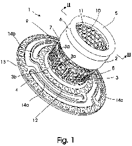
Fig. 1 shows a perspective view of a first embodiment of an
implant according to the present invention,
Fig. 2 shows a sectional view taken along line II-II of fig. 1,
Fig. 3 shows a perspective view of a second embodiment of an
implant according to the present invention, and
Fig. 4 shows a sectional view taken along line III-III of fig.
3.
The invention is described below for use in an ostomy in which
the body duct is an intestine. The skilled person would
appreciate that the implant according to the present invention
also can be used in other body accessing surgical procedure in
which a vessel is externalised for creation of a stoma.
Fig. 1 and 2 shows a first embodiment for a substantially
tubular implant 1 consisting of an exterior tubular section 2
arranged spaced apart from an interior section 3, which extends
radially into an anchoring flange 4 in an angle Oc of
approximately 90 . The exterior tubular section 2 has a free
end 5 provided with an annular mounting groove 6 for mounting
of a detachable device, such as an ostomy bag. The free end 5
of the exterior tubular section has an opposing free end 7
facing towards a free end 8 of the interior section 3 opposite
the anchoring flange 4.
The spaced apart relationship between the exterior tubular
section 2 and the interior section 3 provides an open gap 9
CA 02696681 2010-02-17
WO 2009/024568 10 PCT/EP2008/060837
delimited by the opposing free end 7 of the exterior tubular
section 2 and the free end 8 of the interior section 3.
The exterior tubular section 2 and the interior section 3 are
kept connected to each other by means of a rigid ingrowth mesh
having a plurality of laser cut hexagonal through openings
11.
The anchoring flange 4 consist of three concentric rings, a
10 first ring 12, extending from the interior section 3 opposite
the free end 8 of said interior ring 3. The first ring is
connected to a concentric second ring 13 by means of connection
members 14a,14b,14c, which may or may not be flexible. The
connection members 14a,14b,14c connect the concentric rings
12,13 at offsets points. The flange 4 has in its entirety a
slightly conical shape, i.e. the second ring is axial offset
from the first ring away from the interior section. The flange
also has a plurality of through-holes 15 for additional
securing the implant inside the body. The specific design of
the anchoring flange 4 shown in fig. 1 and 2 is described in
further detail in the applicants own international patent
application PCT/IB2007/050646. Other embodiments of anchoring
flanges can be used within the scope of the present invention
and the flange shown in the figures of the present application
should not be seen as limiting.
The axial gap 9 between the exterior tubular section 2 and the
interior section 3, which serves for closing the infection and
inflammation route to the interior of the body is seen more
clearly in the sectional view of fig. 2. Fig. 2 also shows that
the implant 1 has radial gaps 16 between the interior face 17
of the exterior tubular section 2 and the exterior face 18 of
the rigid ingrowth mesh 10, and between the interior face 19 of
the interior section 3 and the exterior face 18 of the rigid
ingrowth mesh 10. The radial gap 15 serves for accommodation of
new tissue, which infiltrate the rigid ingrowth mesh 10 via the
CA 02696681 2010-02-17
WO 2009/024568 11 PCT/EP2008/060837
through openings 11, and for the tissue bond between intestinal
tissue and abdominal wall tissue at the interior section 3
implanted inside the body.
The second embodiment for an implant 20 shown in fig. 3
corresponds substantially to the first embodiment 1 shown in
figs. 1 and 2 and for like parts same reference numerals are
used.
The implant 20 only differs from the implant 1 in that the
implementation of a different distance means. Instead of a
rigid ingrowth mesh 10 three circumferential spaced apart rigid
assembling members 21 are used as a distance means. Any other
suitable number of assembling members may be chosen.
As seen best in the sectional view of fig. 4 an assembling
member may be shaped as an U-bend or L-bend bracket 21. One two
brackets can be seen in fig. 4. One end 22 of the L-bracket 21
is secured to the interior face 17 of the exterior section 3
and the other bend end 23 of the L-bend bracket 21 is secured
to the interior face 19 of the interior section 2. In this
manner the required radial gap 16 is obtained.
The implant 20 also has an axial gap 18 between the free end 7
of the exterior tubular section 2 and the free end 8 the
interior tubular section 3. This embodiment is very open and is
particular suitable for patients demonstration a high
susceptibility to foreign implanted material.
The interior section 3 may for example be shaped as two
concentric rings, a first ring 3a facing the exterior section 2
and a second ring 3b facing extending into the anchoring flange
4. The concentric rings 3a and 3b may be axially interconnected
by e.g. a plurality of circumferential ribs 3c, crossbars or
any other appropriate rigid latticework or netting.
CA 02696681 2010-02-17
WO 2009/024568 12 PCT/EP2008/060837
The designs of a.o. the anchoring flange, the mounting groove
and the ingrowth mesh may vary within the scope of the appended
claims. Also, the length of the axial gap may be adapted to
individual needs as well as the radial gap may be adjusted
according to anatomic conditions. Examples of suitable designs
of the interior section and anchoring flange are given in
PCT/IB2007/050646.
The implant may be manufactured of any suitable biological
acceptable material, preferably a material that can be laser
cut.