Note: Descriptions are shown in the official language in which they were submitted.
CA 02696843 2010-02-18
WO 2009/023821 PCT/US2008/073282
METHOD, SYSTEM AND SOFTWARE ARRANGEMENT FOR COMPARATIVE
ANALYSIS AND PHYLOGENY WITH WHOLE-GENOME OPTICAL MAPS
FIELD OF THE INVENTION
[0001] The present invention relates generally to methods, systems and
software
arrangements for characterizing whole genomes of several species and strains
by comparing
and organizing their genomes in a searchable database.
BACKGROUND
[0002] A phylogenetic tree represents the evolutionary history among
organisms.
Constructing phylogenetic trees is a crucial step for biologists to find out
how today's extant
species are related to one another in terms of common ancestors. Numerous
computer tools
have been developed to construct such trees
[0003] Given DNA sequences of various taxa, the standard technique in
evolutionary
analysis is to first perform a multiple sequence alignment (on DNA sequences
or protein
sequences). From the resultant distance matrix, a phylogenetic tree is built
describing the
relationship of the various taxa with respect to one another. These distance-
based methods
compress sequence information into a single number and the two sequences with
shortest
distance are considered as closely related taxa. However, the high cost of
sequencing
techniques and the biological diversity among the genomes, make it impossible
to study
phylogeny using detailed sequences of many strains of large-number of related
species.
[0004] Standard methods for constructing phylogenetic trees, known to persons
having
ordinary skills in the art, include Unweighted Pair Group Method using
Arithmetic Average
(P. Sneath and R. Sokal. The principles and practice of numerical
classification. Numerical
Taxonomy, W. H. Freeman, San Francisco, 1973, incorporated herein by
reference),
Neighbor Joining (N. Saitou and M. Nei. The neighbor-joining method: a new
method for
reconstructing phylogenetic trees. Mol. Biol. Evol., 4:406-425, 1987,
incorporated herein by
reference), Fitch Margoliash (W. Fitch and E. Margoliash. The construction of
phylogenetic
trees - a generally applicable method utilizing estimates of the mutation
distance obtained
from cytochrome c sequences. Science, 155:279-284, 1967, incorporated herein
by
reference), Maximum Parsimony (J. Felsenstein. A likelihood approach to
character
weighting and what it tells us about parsimony and compatibility. Biological
Journal of
Linnean Society, 16:183-196, 1981, incorporated herein by reference), and
Maximum
Likelihood (J. Felsenstein. Evolutionary trees from DNA sequences: A maximum
likelihood
1
CA 02696843 2010-02-18
WO 2009/023821 PCT/US2008/073282
approach. Journal ofMolecular Evolution, 17:368-376, 1981, incorporated herein
by
reference).
[0005] The Unweighted Pair Group Method with Arithmetic Mean (UPGMA) method is
a sequential clustering algorithm. It works by constructing distance matrix,
amalgamating
two Operational Taxonomy Units (OTUs) at each stage and creating a new
internal node in
the tree at the same time. Whenever two nodes are merged into a new node, it
recalculates
the distances between the new nodes and other nodes, repeating the process
until all OTUs
are grouped in a single cluster. It produces a rooted tree containing all the
OTUs at the leaves
of the tree. It is suitable for constructing phylogenetic tree of taxa with a
relatively constant
rate of evolution. It has several advantages: The algorithm is simple and
fast. Its main
disadvantages are: (1) It implicitly assumes the existence of an ultrametric
tree: the total
branch lengths from the root to any leaf are all equal. In other words, there
is an assumed
"molecular clock," which ticks at a constant pace, and all the observed
species are at an equal
number of ticks from the root; the same evolution rate is assumed to apply to
all branches,
which is often not the case. (2) It assumes a stringent additive property.
[0006] The Neighbor Joining (NJ) method is a heuristic greedy algorithm. It
begins with
distance matrix and a star-like tree. At each stage two closest neighbors are
joined into a new
node, which becomes the root of the new tree. The branch lengths from the two
nodes to the
new node are calculated. The two nodes are replaced by the new node in the
distance matrix,
thus reducing the number of OTUs by 1. In the process, it updates the distance
matrix and
performs the node merging process again. The process repeats until there are
two OTUs left
and they are joined into a root node. Unlike UPGMA, which chooses the
neighbors with
minimum distance, NJ chooses the neighbors that minimize the sum of branch
lengths at each
stage. It has several advantages: (1) It is fast and well suited for data sets
of substantial size
and also for the postprocessing step of bootstrap analysis. (2) It is
especially suitable when
the rate of evolution of the separate lineages under consideration varies. Its
main
disadvantages are: (1) It depends heavily on the evolutionary model applied.
(2) Like
UPGMA, it assumes a stringent additive property.
[0007] Both UPGMA and NJ employ distance matrix to reflect evolutionary
relationship,
compressing sequence information into a single number, and thus cannot reflect
the changes
of character states of sequences. UPGMA and NJ are relatively fast, so they
are suitable for
analyzing large data set that is not very strongly similar. In general, NJ
gives better result
than UPGMA.
2
CA 02696843 2010-02-18
WO 2009/023821 PCT/US2008/073282
[0008] The Fitch Margoliash (FM) method assumes that the expected error is
proportional to the square root of the observed distances. It compares the two
most closely
related taxa to the average of all the other taxa. It then moves through the
tree sequentially to
calculate the distances between decreasingly related taxa until all the
distances are found. Its
advantages include the following: It does not assume a constant rate of
evolution and
therefore can produce varied branch lengths from a common ancestor. Its main
disadvantage
is that it requires longer computational execution time than UPGMA and NJ.
[0009] The Maximum Parsimony (MP) method is built upon the principle that
simple
hypotheses are more preferable than complicated ones. Consequently, the
construction of the
tree using this method requires the smallest number of evolutionary changes
among the
OTUs in order to explain the phylogeny of the species under study. This method
compares
different parsimonious trees and chooses the tree that has the least number of
evolutionary
steps (substitutions of nucleotides in the context of DNA sequence). MP is a
character-based
Maximum Parsimony algorithm. It starts with multiple alignment and construct
all possible
topologies. Based on evolutionary changes, it scores each of these topologies
and chooses a
tree with the fewest evolutionary changes as the final tree. An evolutionary
change is the
transformation from one character state to another. Character states can be
DNA bases, the
loss or gain of a restricted site, and the absence or presence of
morphological features. Its
advantages are enumerated as follows: (1) It allows the use of all known
evolutionary
information in tree building. (2) It produces numerous unrooted, "most
parsimonious trees."
Some of its disadvantages are listed below: (1) It requires long computation
time, although
faster than maximum likelihood. (2) It yields little information about branch
length. (3) It
usually performs well with closely related sequences, but often performs badly
with very
distantly related sequences.
[0010] The Maximum Likelihood (ML) method evaluates the topologies of
different trees
and chooses the best tree among all as measured with respect to a specified
model. Such a
model may be based on the evolutionary process that can account for the
conversion of one
sequence into another. It evaluates a hypothesis about evolutionary history in
terms of the
probability that the proposed model and the hypothesized history would give
rise to the
observed data set. The parameter considered in the topology is the branch
length. It starts
with a multiple alignment and lists all possible topologies of each data
partition. It then
calculates probability of all possible topologies for each data partition and
combines data
partitions. It identifies tree with the highest overall probability at all
partitions as most likely
3
CA 02696843 2010-02-18
WO 2009/023821 PCT/US2008/073282
phylogeny. Its advantages include the following: (1) It is more accurate than
other methods.
It is often used to test an existing tree. (2) All the sequence information is
used. (3)
Sampling errors have least effect on the method. Its main disadvantage is that
it is extremely
slow, and thus impractical for analyzing large data set.
SUMMARY OF THE INVENTION
[0011] The present invention provides a method for organizing genomic
information
from multiple organisms. In one embodiment of the invention, phylogenetic
trees can be
constructed for the organisms. The method of the present invention is termed
CAPO,
Comparative Analysis and Phylogeny with Optical-Maps. This method can be used
to
determine phylogeny among optical maps of multiple strains or genomes. The low
cost and
high speed of an Optical Mapping technique provides an elegant solution to the
problem
posed by the high cost procedures involved in sequence generation and
comparison.
[0012] In one aspect, the invention provides a method for comparative genomic
analysis,
the method includes comparing optical maps obtained from one or more organisms
in order
to obtain at least one pair-wise similarity value; and determining relatedness
of the organisms
based on said pair-wise similarity value. In a related embodiment, the method
further
includes constructing a phylogenetic tree based on the relatedness of the
organisms.
Exemplary organisms include a microorganism, a bacterium, a virus, and a
fungus.
[0013] Another aspect of the invention provides a method for identifying an
unknown
organism, the method includes comparing an optical map from an unknown
organism to a
plurality of optical maps from a phylogenetic tree of known organisms;
obtaining a pair-wise
similarity value for one or more comparisons between the unknown organism and
the known
organism in the phylogenetic tree; and identifying the unknown organism based
on the pair-
wise similarity values. In a related embodiment, the method further includes,
prior to the
comparing step, preparing an optical map from the unknown organism. In another
related
embodiment, the method further includes, prior to the comparing step,
constructing a
phylogenetic tree of known organisms.
[0014] Another aspect of the invention provides a method for constructing a
phylogenetic
tree, the method includes obtaining pair-wise distances among organisms by
comparing at
least one pair of optical maps from the organisms in order to generate a pair-
wise similarity
matrix; and constructing a phylogenetic tree based on the pair-wise similarity
matrix. In a
4
CA 02696843 2010-02-18
WO 2009/023821 PCT/US2008/073282
related embodiment, the method further includes, prior to said obtaining step,
preparing
optical maps of each organism.
[0015] Some of the steps of the methods can be accomplished by a computer
utilizing
various algorithms. Software instructions to perform embodiments of the
invention may be
stored on a computer readable medium such as a compact disc (CD), a diskette,
a tape, a file,
or any other computer readable storage device.
[0016] To begin the organization of genomic information, whole-genome physical
maps
or sequences of multiple organisms are obtained. These maps can either be
partially or fully
assembled. In one suitable embodiment the physical maps are optical maps.
Suitable optical
maps include, but are not limited to, restriction enzyme optical maps and
probe hybridization
optical maps. Once these maps are obtained, the maps of any two organisms are
compared.
[0017] In one embodiment this comparison is done by using pair-wise map
similarity
values found by comparing the optical maps of organisms. The distance between
the two
optical maps (labeled mapA and map B) is found by taking: (alignedLA +
alignedLB)/(LA +
LB), where aliginedLA is the length (in units of base pairs, bps) of aligned
restriction
fragments of mapA, and LA is the total length (also in bps) of restriction
fragments of mapA.
[0018] After the percentage similarity values are computed, these values are
fed into a
statistical package available in the language "R" and analyzed with a
clustering method,
which can be the nearest neighbor, furthest neighbor, or UPGMA
[0019] In another embodiment, the distance between the two optical maps is
computed by
a heuristic mer-based algorithm for pair-wise optical map comparison. After
choosing a mer
size k, the algorithm is used to generate all k-mers in an optical map for
both forward and
backward orientations. A k-mer is an optical map segment of length k
fragments. For each
genome, some k-mers occur much more, or less, frequently than chance predicts
(to within a
some sizing tolerance), and the distribution of k-mer frequencies comprises a
type of "species
signatures". The difference between k-mer distributions and profiles for two
species
increases as evolutionary distance increases, thus comparing k-mer profiles
can be used to
infer phylogenetic relationships.
[0020] To compare two optical maps i and j, the algorithm examines all common
k-mers
between them to count the number of common k-mers as c,j, and computes the
pair-wise map
similarity s,j, where s,j=( si+sj - 2c,j) /(s;+sj), where s; and sj are the
sizes (all measured in
terms of the numbers of restriction fragments) of the two optical maps. s,j =
0 if i = j. In one
embodiment the common mers are computed by accounting for the sizing error.
Given two
CA 02696843 2010-02-18
WO 2009/023821 PCT/US2008/073282
k-mers, ki =(fi, f2, ..., fk) in map 1 and k2 =(gi, g2, ..., gk) in map 2(f's
and g's are both
measured in units of base pairs, bps), it considers ki and k2 as a pair of
common k-mers if and
only if the following condition is true:
3 i ', f r,
~.; _. fr.
[0021] where F; is interval (f; - 6f,, fi + 6 fi), 6 fi is the standard
deviation for fragment f;; G;
is defined similarly. Threshold p is a cutoff determining the least overlap
degree between
two common intervals, deemed necessary to interpret them as equal modulo
statistical noise.
[0022] After the pair-wise distances among the organisms are found, a
plurality of
disjoint pairs of near neighbors among the organisms or their putative
ancestors is obtained.
In one embodiment a single pair of nearest neighbors is determined by
searching all pair-wise
possibilities. In another embodiment, multiple pairs of nearest neighbors are
determined by
using a stable marriage algorithm.
[0023] Once the nearest neighbors are determined, the plurality of pairs of
neighbors are
joined pair-wise to create a set of putative ancestral genomes. The
determination of the
plurality of disjoint pairs of near neighbors, and the pair-wise joining of
such neighbors are
repeated until no pair remains. These iterative steps organize the physical
maps in a
phylogenetic tree.
[0024] Another aspect of the invention provides a method for determining
similarity
among organisms, the method including, comparing optical maps from the
organisms to
determine relatedness of the organisms.
[0025] Other aspects of the invention will become apparent by consideration of
the
detailed description and accompanying drawings.
BRIEF DESCRIPTION OF THE DRAWINGS
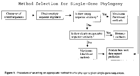
[0026] Figure 1 is a chart showing the procedure of selecting an appropriate
method to
infer phylogeny given single-gene sequences.
[0027] Figure 2 shows an example of building a bipartite graph given a
distance matrix.
A) A distance matrix M of four items (A, B, C, D). B) The corresponding
bipartite graph.
[0028] Figure 3 shows a first-degree polynomial fit for restriction fragment
sizing error.
(a) L vs. StdDev(L), cc=0.7428; (b) A vs. StdDev(L), cc=0.7562; (c) 1/A vs.
StdDev(L)/L,
cc=0.8290.
[0029] Figure 4 shows Data Set I: 11 Escherichia coli Strains.
6
CA 02696843 2010-02-18
WO 2009/023821 PCT/US2008/073282
[0030] Figure 5 shows view maps in Data set I using MapViewer. A pair-wise
alignment
between Escherichia coli 0157:H7 str. Sakai and Escherichia coli 0157:H7
EDL933 is
shown.
[0031] Figure 6 is a table showing data Set II: 28 Enterobacteriaceae Taxa.
[0032] Figure 7 shows view maps in Data set II using MapViewer
[0033] Figure 8 shows a Phylogenetic tree for data set I and II (k=2, p =0.9)
[0034] Figure 9 shows a Phylogenetic tree for data set I and II (k=3, p =0.8)
[0035] Figure 10 shows a Phylogenetic tree for data set I and II (k=4, p=0.7)
[0036] Figure 11 shows a number of clusters in the iterations of the
experiments of data
set I and II using CAPO SM-UPGMA/SM-NJ.
[0037] Figure 12 shows Phylogenetic trees constructed by CAPO for data set I
and II
using default setting and single merge mode.
[0038] Before any embodiments of the invention are explained in detail, it is
to be
understood that the invention is not limited in its application to the details
of construction and
the arrangement of components set forth in the following description or
illustrated in the
following drawings. The invention is capable of other embodiments and of being
practiced
or of being carried out in various ways. Also, it is to be understood that the
phraseology and
terminology used herein is for the purpose of description and should not be
regarded as
limiting. The use of "including," "comprising," or "having" and variations
thereof herein is
meant to encompass the items listed thereafter and equivalents thereof as well
as additional
items.
DETAILED DESCRIPTION OF THE INVENTION
[0039] A phylogenetic tree represents the evolutionary history among
organisms. Some
methods have been proposed and implemented for the construction of
phylogenetic trees.
They can be classified into two groups, the phenetic method (distance matrix
method, P.
Sneath and R. Sokal. The principles and practice of numerical classification.
Numerical
Taxonomy, W. H. Freeman, San Francisco, 1973, incorporated herein by
reference) and the
cladistic methods (maximum parsimony and maximum likelihood, J. Felsenstein. A
likelihood approach to character weighting and what it tells us about
parsimony and
compatibility. Biological Journal of Linnean Society, 16:183-196, 1981,
incorporated herein
by reference). Popular programs of constructing phylogenetic trees include
PHYLIP
(Available at evolution.genetics.washington.edu/phylip.html; phylogenetic
inference package
7
CA 02696843 2010-02-18
WO 2009/023821 PCT/US2008/073282
- J Felsenstein) and PAUP (Available at paup.csit.fsu.edu; phylogenetic
analysis using
parsimony - Sinauer Assoc.).
[0040] The phenetic methods use various measures of overall similarity for the
ranking of
species. They can use any number or type of characters, but the data has to be
converted into
a numerical value. The organisms are compared to each other for all of the
characters and
then the similarities are calculated. After this, the organisms are clustered
based on the
similarities. Such methods place a greater emphasis on the relationships among
data sets than
the paths they have taken to arrive at their current states. They do not
necessarily reflect
evolutionary relations.
[0041] The cladistic method is based on the notion that members of a group
share a
common evolutionary history and are more closely related to members of the
same group
than to any other organisms. This method emphasizes the need for large data
sets but differs
from phenetics in that it does not give equal weight to all characters.
Cladists are generally
more interested in evolutionary pathways than in relationships. FIG. 1 shows
how to select
an appropriate method to infer phylogeny given single-gene sequences.
[0042] Standard methods for constructing phylogenetic trees, known to persons
having
ordinary skills in the art, include Unweighted Pair Group Method with
Arithmetic Mean
(UPGMA), Neighbor Joining (NJ), Fitch Margoliash (FM), Maximum Parsimony (MP),
and
Maximum Likelihood (ML) methods, and can be combined with certain basic
methods
related to optical mapping to infer phylogeny using optical-map comparison.
[0043] In one embodiment of the present invention, a phylogenetic tree is
crafted by
using pair-wise map similarity values found by comparing the optical maps of
organisms.
To calculate the pair-wise map similarity value, a SOMA map aligner is used to
find all the
local alignments between the two strains above a certain score threshold.
Given two optical-
maps mapA and mapB, the percentage similarity is found by taking: (alginedLA +
alginedLB)/(LA + LB), where alginedLA is the length of aligned restriction
fragments of
mapA, and LA is the total length of restriction fragments of mapA.
[0044] After the percentage similarity values are computed, these values are
fed into a
statistical package available in the language "R" and analyzed with a
clustering method,
which can be the nearest neighbor, furthest neighbor, or UPGMA. As an example,
a pair-
wise alignment was performed between Escherichia coli 0157:H7 str. Sakai and
Escherichia
coli 0157:H7 EDL933 using SOMA map aligner with its default settings, shown in
Figure 5.
8
CA 02696843 2010-02-18
WO 2009/023821 PCT/US2008/073282
[0045] In another embodiment of the present invention, the distance between
the two
optical maps is computed by a heuristic mer-based algorithm for pair-wise
optical map
comparison is used to determine phylogeny among optical maps of multiple
strains or
genomes.
Optical maRpin~
[0046] Optical mapping is a single-molecule technique for production of
ordered
restriction maps from a single DNA molecule (Samad et al., Genome Res. 5:1-4,
1995).
During this method, individual fluorescently labeled DNA molecules are
elongated in a flow
of agarose between a coverslip and a microscope slide (in the first-generation
method) or
fixed onto polylysine-treated glass surfaces (in a second-generation method).
Id. The added
endonuclease cuts the DNA at specific points, and the fragments are imaged.
Id. Restriction
maps can be constructed based on the number of fragments resulting from the
digest. Id.
Generally, the final map is an average of fragment sizes derived from similar
molecules. Id.
[0047] Optical mapping and related methods are described in co-pending U.S.
patent
application serial number 12/120,586, co-pending U.S. patent application
serial number
12/120,592, U.S. Pat. No. 5,405,519, U.S. Pat. No. 5,599,664, U.S. Pat. No.
6,150,089, U.S.
Pat. No. 6,147,198, U.S. Pat. No. 5,720,928, U.S. Pat. No. 6,174,671, U.S.
Pat. No.
6,294,136, U.S. Pat. No. 6,340,567, U.S. Pat. No. 6,448,012, U.S. Pat. No.
6,509,158, U.S.
Pat. No. 6,610,256, and U.S. Pat. No. 6,713,263, each of which is incorporated
by reference
herein. Optical Maps are constructed as described in Reslewic et al., Appl
Environ
Microbiol. 2005 Sep; 71 (9):5511-22, incorporated by reference herein.
Briefly, individual
chromosomal fragments from test organisms are immobilized on derivatized glass
by virtue
of electrostatic interactions between the negatively-charged DNA and the
positively-charged
surface, digested with one or more restriction endonuclease, stained with an
intercalating dye
such as YOYO-1 (Invitrogen) and positioned onto an automated fluorescent
microscope for
image analysis. Since the chromosomal fragments are immobilized, the
restriction fragments
produced by digestion with the restriction endonuclease remain attached to the
glass and can
be visualized by fluorescence microscopy, after staining with the
intercalating dye. The size
of each restriction fragment in a chromosomal DNA molecule is measured using
image
analysis software and identical restriction fragment patterns in different
molecules are used to
assemble ordered restriction maps covering the entire chromosome.
9
CA 02696843 2010-02-18
WO 2009/023821 PCT/US2008/073282
[0048] A current issue with optical map comparison can be understood from the
following discussion: An optical map can be viewed as an ordered sequence of
"restriction
sites," or equivalently, "restriction fragment lengths." A vector of decimal
numbers, Hk =
(hi, h2, ..., h,,,), is used to represent a single map k, where h; with index
0 < i< m is the length
of the i-th restriction fragment. The size of an optical map k is defined as
sk= E h;, h; E Hk.
The input to the heuristic mer-based algorithm is an N by M matrix O=(o,j),
where each row
corresponds to an optical map of a strain or a genome. Each column corresponds
to a
position in that map. N is the total number of maps, and M is the number of
restriction
fragments in the longest map in that input. Because sequences of different
strains or genomes
vary in length, the final optical maps usually do not have the same number of
restriction
fragments. By using the present heuristic mer-based algorithm method, the
optical maps are
forced to have M fragments by appending zeros to the end of shorter map
vectors. Suitably,
all the restriction maps in the input must be digested by the same set of
restriction
endonucleases to make the map comparison meaningful in genome evolution study.
[0049] The heuristic mer-based algorithm is based on pair-wise optical map
comparison
and bipartite graph matching, combined with standard distance methods of
phylogeny tree
construction. It consists of two major phases. First, pair-wise optical map
comparison is
performed to generate a pair-wise similarity matrix S=(s,j), where s,j is the
map similarity
between the i-th and j-th map in the input matrix O. S is used as input to the
second phase of
CAPO, which determines phylogeny among input strains or genomes. The output is
in the
Phylip format, used by many phylogenetic analysis packages. This format
consists of a series
of nested parentheses describing the branching order with the sequence names.
Users can
display the phylogeny tree using the NJPLOT program distributed with the
ClustalX package
(The latest version of the ClustalX program is available at ftp://ftp-igbmc.u-
strasbg.fr/pub/ClustalX/). The details of the two algorithms implemented in
CAPO are
explained in the following sections.
Pair-wise Optical Map Comparison
[0050] In phase one of constructing a phylogenetic tree, a heuristic mer-based
algorithm
for pair-wise optical map comparison is used. A`mer' (or more elaborately
"restriction-
fragment-mer") in an optical map is an ordered sequence of restriction
fragment lengths. A
`k-mer' is a mer with k fragment lengths. Mathematically, a k-mer comprises k
decimal
numbers, and their positions reflect the sequence order of the corresponding
restriction
CA 02696843 2010-02-18
WO 2009/023821 PCT/US2008/073282
fragments. After choosing a mer size k, all k-mers in an optical map for both
forward and
backward orientations are generated. Each k-mer is indexed by its position in
the optical
map. To compare two optical maps i and j, all common k-mers between them are
examined
as follows: the number of common k-mers are counted as c,j, and the pair-wise
map similarity
s,j is computed by using the formula s,j==( s;+sj - 2c,j) /(s;+sj), where s;
and sj are the sizes of
the two optical maps. s,j = 0 if i = j. The computed pair-wise similarity
matrix S is used as
input to the next phase of inferring phylogeny.
[0051] Common mers are searched in a manner allowing for sizing errors. For
example,
given two k-mers, ki =(fi, f2, ..., fk) in map 1 and k2 =(gi, 92, ..., gk) in
map 2, ki and k2 are
considered as a pair of common k-mers if and only if the following condition
is true:
r~
-- <~. tr.r'<. --- ,. k"
[0052] where F; is interval (f; - 6f,, fi + 6fi 6fi is the standard deviation
for fragment f;; G;
is defined similarly. Threshold p is a cutoff determining the least overlap
degree between
two common intervals. The standard deviation of a restriction fragment is
estimated via
observations of experiment data. Details are given in a later section.
Inferring Ph ly ogeny
[0053] Given a matrix of distances among a set of taxa, both the UPGMA and NJ
methods are widely used in phylogenetic analysis to show how similar or
dissimilar they are.
The UPGMA method assumes equal rates of evolution, so that branch tips come
out equal.
The NJ method allows for unequal rates of evolution, so that branch lengths
are proportional
to amount of change. The present method combines the standard stable marriage
(SM)
algorithm for bipartite graph matching problem with either the UPGMA or the NJ
method for
inferring phylogeny.
[0054] Usually a phylogeny tree is constructed in stepwise manner. Every time
two most
similar sequences are clustered together, they are combined into a new node,
representing
their least common ancestor. The clustering process continues until there is
only one node
left. Therefore, given n taxa, traditional distance-based methods need O(n)
iterations to
construct a phylogenetic tree. In normal cases, the present method is capable
of constructing
a phylogenetic tree in log(n) iterations, though its worst-case number of
iterations is
comparable to traditional distance-based methods. It works as follows:
11
CA 02696843 2010-02-18
WO 2009/023821 PCT/US2008/073282
[0055] Initialization: Define T to be the set of leaf nodes, one for each
given optical map.
If the UPGMA method is used, the distance matrix D=(d,j)=(s,j), where s,j is
the map
similarity obtained from phase one. If the NJ method is used, u; Ej-in s,j/(n-
2) for each node
i in T, where n is the total number of nodes in T. The distance matrix D is
recomputed to be
D=(d;;)=(s;;-u;-u;).
[0056] Iteration: Build a bipartite graph. Partition D along diagonal line
into two parts:
the upper triangular part UT and the lower triangular part LT. Pairs in UT
form the left
column in the bipartite graph, and pairs in LT form the right column. Each
node i has a
preference list of nodes, ranked by d,j.
[0057] Apply the stable marriage algorithm and produce a set X of stable pairs
(B. Sun, J.
Schwartz, O. Gill, and B. Mishra. Combat: Search rapidly for highly similar
protein-coding
sequences using bipartite graph matching. In Computational Science - ICCS
2006: 6th
International Conf., pages 654-661, Reading, UK., 2006, incorporated herein by
reference).
Such a`stable pair' is a pair of nodes connected by the stable marriage
algorithm and is be
clustered into a new internal node if this pair passes the following cleaning
step.
[0058] Clean the set X: sort stable pairs in decreasing order of d,j and keep
only the first
m pairs in X that are disjoint. Note that two pairs (a, b) and (c, d) are
disjoint with each other
if and only if no two nodes in different pairs are the same.
[0059] Connect nodes and update the distance matrix D in a loop until X is
empty. In
each loop execute the following operations: I) extract the first pair (i, j)
in X; II) join them
with a new internal node v,j. The node v,j has its cluster size nij = n; + nj
(initially, n; = 1).}; III)
compute the distances between node v,j and the remaining nodes k; IV) delete
d,j in D and add
the new distances to D; V) connect nodes i and j in T with v,j.
[0060] Termination: When only two nodes i and j remain unconnected in T,
connect them
to the root node of the tree T.
[0061] An example of building a bipartite graph given a distance matrix is
shown in
Figure 2. Each node has a preference list (gray boxes) ordered by distances.
Left panel
contains pairs in the upper triangular part of M; right panel contains pairs
in the lower
triangular part of M. For example, the first row in the left panel means "item
A prefers to
pair with C, B, D, in the decreasing order of preferences."
Correction of Siziny Errors
12
CA 02696843 2010-02-18
WO 2009/023821 PCT/US2008/073282
[0062] Optical maps of different strains of the same species would vary due to
single
nucleotide differences (SNPs), small insertions and deletions (RFLPs) as well
as many
genomic rearrangement events that leave their footprints on restriction site
patterns. Further
variations are introduced by the noises in the experimental process. These can
be due to:
sizing errors, partial digestion, short missing restriction fragments, false
cuts, ambiguities in
the orientation, optical chimerisms, and so on (T. Anantharaman, B. Mishra,
and D.
Schwartz. Genomics via optical mapping II: Ordered restriction maps. Journal
of
Computational Biology, 4(2):91-118, 1997; B. Mishra. Optical mapping.
Encyclopedia of the
Human Genome, Nature Publishing Group, Macmillan Publishers Limited, London,
UK,
4:448-453, 2003, incorporated by reference). These error factors introduced by
the
experimental process are classified into three types -sizing errors, digestion
errors, and
orientation errors.
[0063] The sizing error statistics is estimated from observations of
experiments done by
OpGen, Inc. and NYU Bioinformatics Group. These observations (including
fragment
lengths and standard deviations) are what are reported in the output from the
GENTIG (T.
Anantharaman, B. Mishra, and D. Schwartz. Genomics via optical mapping III:
Contiging
genomic DNA and variations; B. Mishra. Optical mapping. Encyclopedia of the
Human
Genome, Nature Publishing Group, Macmillan Publishers Limited, London, UK,
4:448-453,
2003, incorporated herein by reference) software that OpGen and other
practitioners of
optical mapping have used to produces optical maps. A first-degree polynomial
fit for the
three pairs of variables: L- StdDev(L), ~(L) - StdDev(L), and 1N(L) -
StdDev(L)/L is
shown in Figure 3, where linear correlation coefficient is referred to as cc.
No apparent linear
relation is observed between any pair of them since none of these pairs have
linear correlation
coefficient close enough to one (e.g., > 0.95). These results indicate that it
may not be
appropriate to estimate standard deviations using any of these `linear
relations.' Therefore
data interpolation is used instead to estimate standard deviations StdDev(L)
for a restriction
fragment whose length is L. This data interpolation step is performed in the
following way:
given a fragment length L, find Li and Lr from the error plot shown in Figure
below (a) where
Li and Lr are the closest left neighbor and right neighbor of L, respectively
(Li < L < Lr);
compute StdDev(L) using StdDev(L) = ( StdDev(Li) + StdDev(Lr) ) / 2.
[0064] The invention having now been described, it is further illustrated by
the following
examples and claims, which are illustrative and are not meant to be further
limiting. Those
skilled in the art will recognize or be able to ascertain using no more than
routine
13
CA 02696843 2010-02-18
WO 2009/023821 PCT/US2008/073282
experimentation, numerous equivalents to the specific procedures described
herein. Such
equivalents are within the scope of the present invention and claims.
[0065] The contents of all references and citations, including issued patents,
published
patent applications, and journal articles cited throughout this application,
are hereby
incorporated by reference in their entireties for all purposes.
EXAMPLES
[0066] Creation of Data Set I
[0067] Eleven optical maps constructed commercially by OpGen (Website of OpGen
Inc.
is http://www.opgen.com/) for varying E. coli strains. Information describing
this data set is
listed in Fig. 4. All the organisms described in data set I are E. coli
bacteria, and are
identified by their individual strain names. Sequence data is not available
for most but four
of these E. coli strains, including Escherichia coli CFT073, Escherichia coli
K12,
Escherichia coli 0157:H7 str. Sakai, and Escherichia coli 0157:H7 EDL933.
[0068] The following procedure was used to produce this data: i) purified
chromosomal
DNA is deposited onto an optical mapping surface using a microfluidic device;
ii) the DNA
is encased in a thin layer of acrylamide and incubated with the restriction
enzyme BamHI (it
cleaves at every site containing the 6 bp long sequence GGATCC) in a
humidified chamber
at 37 C for 60 - 120 mins; iii) the digested DNA is labeled with fluorescent
YOYO-1 and the
individual molecules are imaged with fluorescence microscopy; iv) digital
images are
collected by an automated image-acquisition system and image files are
processed to create
single-molecule optical maps; v) individual molecule restriction maps are
overlapped by
using GENTIG (GENomic conTIG) map-assembly software.
[0069] Briefly, GENTIG works by comparing single-molecule restriction maps and
estimating the probability that these two molecules arose from overlapping
genomic
locations, where the probability is computed conditional to the likelihood of
possible
experimental errors resulting from incomplete digestion, spurious cuts, and
sizing errors.
Through repeated overlapping of molecules, the assembler reconstructs the
ordered
restriction map of the genome. This technique has been previously applied to
map many other
bacterial genomes.
[0070] A commercially available interface for viewing optical-maps, called
MapViewer
(available from OpGen, Inc.) is then used. MapViewer allows users to visualize
optical-
maps, to move maps around, pull up sequence information when available, and
change the
14
CA 02696843 2010-02-18
WO 2009/023821 PCT/US2008/073282
orientation of the maps. Figure 5 shows the optical maps for data set I using
MapViewer. A
pair-wise alignment between Escherichia coli 0157:H7 str. Sakai and
Escherichia coli
0157:H7 EDL933 is shown. Regions that match exactly once are colored green,
and regions
that match to more than one location are colored red.
[0071] Creation of Data Set II
[0072] Twenty-eight genomic sequences of Enterobacteriaceae taxa are
downloaded from
the NCBI database, and then cleaved "in silico" with the restriction enzyme
BamHI. Their
optical maps were constructed using the SilicoMap software provided by OpGen;
The
SilicoMap tool is built upon the BioPerl toolkit which is able to perform an
in silico
restriction digest, after which, it is straightforward to find the lengths of
each of the resulting
fragments and create the map. Information describing this data set is listed
in Figure 6.
Figure 7 shows the optical maps for data set I using MapViewer.
[0073] Analysis of Data Sets
[0074] Experimental results are provided in this section using CAPO on both
real optical
mapping data of eleven E. coli strains and simulated optical mapping data of
twenty-eight
entire genomes of Enterobacteriaceae taxa. All of the tests were run on a 2.4-
GHz Pentium
IV machine with 3GB of RAM.
[0075] Parameter Settings
[0076] Users have choices for two parameters in CAPO: k (mersize) and p
(cutoff value
involved in determining whether two restriction fragment lengths are `equal'
considering
sizing errors). The effect of parameter settings in CAPO is tested in the
following
experiments using the two data sets: k=2, p =0.9 (see Figure 6), k=3, p =0.8
(see Figure 7)
k=4, p =0.7 (see Figure 8). To adequately tolerate sizing errors it was found
reasonable to
use smaller cutoff value of p if a larger mer-size is chosen. Shown in Figure
8 - Figure 10,
the `best' results (whose phylogenetic trees are most biologically meaningful)
are produced
using k=3, p =0.8. k=3, p =0.8 was, therefore, subsequently used as the
default parameter
setting.
[0077] Phylogenetic Tree Evaluation
[0078] Since there are no `true' phylogenetic trees available for comparison
with the
results computed by the present method, the quality of these trees were
evaluated based on
optical map alignments, the taxonomy information given by the NCBI database,
and tree
topology overlap between the two different distance methods. Using the SOMA
map aligner
CA 02696843 2010-02-18
WO 2009/023821 PCT/US2008/073282
developed by OpGen, it was found that the map of Escherichia coli K12 is very
similar to
that of 886, and these two strains are clustered closely by the present method
with default
setting (see Figure 7, Al, A2). The present method also assigns the rest of
three known E.
coli strains close evolutionary distances. Using data set II, it was observed
that the present
method often clustered biologically closely related taxa together (the
Buchnera aphidicola
strains, the Candidatus Blochmannia strains, the E. coli strains, the
Salmonella strains, etc.),
as would be desired. Lastly, phylogenetic trees produced by the present method
for the same
data set using different distance methods were also found to share substantial
tree topology
overlap.
[0079] Cluster Sizes
[0080] The present method (CAPO) constructs phylogenetic trees in far fewer
iterations
than standard distance methods. For data set I, CAPO UPGMA-flavored trees and
NJ-
flavored trees were constructed in 5 and 6 iterations, respectively. For data
set II, CAPO
UPGMA-flavored trees and NJ-flavored trees were constructed in 8 and 9
iterations,
respectively. Number of remaining clusters in each iteration is shown in
Figure 11.
[0081] Impact of Single-Merge Mode and Multi-Merge Mode
[0082] To see if there was any effect on the phylogenetic tree topology by
merging more
than two clusters in a single iteration. Phylogenetic trees were generated for
both data sets
using `single-merge mode' (merge exactly two clusters at one iteration), as
shown in Figure
12. Compared with trees produced in `multi-merge mode' (merge multiple pairs
of disjoint
clusters found by the stable marriage procedure in a single iteration), as
shown in Figure 9,
some tree topology changes are shown, especially between Figure 12-A2 and
Figure 9-A2.
Because there is no reliable method for detecting the similarity level between
two trees and
because there is no prior knowledge about the `true' tree topology, at this
point, it remains
unclear what the impact of various merging mode could be. However, almost all
corresponding trees share substantial tree topology overlap, thus indicating a
strong measure
of consistency that can be achieved by the present method.
[0083] Implementation and Speed
[0084] The methods of the present invention are implemented in C++ and all
experiments
were performed on a Pentium IV PC with 3 GB memory. Experiments for data set I
and II
took - 4 sec. and - 18 sec., respectively. The computational efficiency of
CAPO indicates its
potential widespread usage in analyzing large genomic data sets.
16
CA 02696843 2010-02-18
WO 2009/023821 PCT/US2008/073282
Background References
S. Altschul, T. Madden, A. Schaffer, J. Zhang, Z. Zhang, W. Miller, and D.
Lipman. Gapped
blast and psi-blastla new generation of protein database search programs.
Nucleic Acids Res.,
25:3389-3402, 1997.
T. Anantharaman, B. Mishra, and D. Schwartz. Genomics via optical mapping III:
Contiging
genomic DNA and variations.
T. Anantharaman, B. Mishra, and D. Schwartz. Genomics via optical mapping II:
Ordered
restriction maps. Journal of Computational Biology, 4(2):91-118, 1997.
T. Anantharaman, V. Mysore, and B. Mishra. Fast and cheap genome wide
haplotype
construction via optical mapping. volume 10, pages 385-396. Pacific Symposium
on
Biocomputing, 2005.
C. Aston, B. Mishra, and D. Schwartz. Optical mapping and its potential for
large-scale
sequencing projects. Trends in Biotechnology, 17:297-302, 1999.
S. Batzoglou, L. Pachter, J. Mesirov, B. Berger, and E. Lander. Human and
mouse gene
structure: Comparative analysis and application to exon prediction. Genome
Res., 10:950-
958, 2000.
E. Bimey and R. Durbin. Using genewise in the drosophila annotation
experiment. Genome
Res., 10:547-548, 2000.
E. Bimey and et al. Ensembl. Nucleic Acids Res., 32:468-470, 2004.
N. Bray, I. Dubchak, and L. Pachter. Avid: A global alignment program. Genome
Res.,
13:97-102, 2003.
M. Brudno and B. Morgenstem. Fast and sensitive alignment of large genomic
sequences. In
Proc. of the IEEE Computer Society Bioinformatics Conference, pages 138-150,
2002.
C. Burge and S. Karlin. Prediction of complete gene structures in human
genomic dna. J.Mol.
Bio., 268:78-94, 1997.
W. Cai, J. Jing, B. Irvin, L. Ohler, E. Rose, H. Shizuya, U. Kim, M. Simon, T.
Anantharaman, B. Mishra, and D. Schwartz. High-resolution restriction maps of
bacterial
artificial chromosomes constructed by optical mapping. Proc. Natl. Acad. Sci.
U.S.A.,
95:3390-3395, 1998.
A. Delcher, S. Kasif, R. Fleischmann, J. Peterson, O. White, and S. Salzberg.
Alignment of
whole genomes. Nucleic Acids Res., 27:2369-2376, 1999.
A. Delcher, A. Phillippy, J. Carlton, and S. Salzberg. Fast algorithms for
large-scale genmoe
alignment and comparison. Nucleic Acids Res., 30(11):2478-2483, 2002.
17
CA 02696843 2010-02-18
WO 2009/023821 PCT/US2008/073282
J. Deogun, J. Yang, and F. Ma. Emagen: An efficient approach to multiple whole
genome
alignment. In the 2nd Asia Pacific Bioinformatics Conference (APBC2004),
volume 29,
Dunedin, New Zealand, 2004.
J. Felsenstein. Alternative methods of phylogenetic inference and their
interrelationship.
Systematic Zoology, 28:49-62, 1979.
J. Felsenstein. Evolutionary trees from DNA sequences: A maximum likelihood
approach.
Journal ofMolecular Evolution, 17:368-376, 1981.
J. Felsenstein. A likelihood approach to character weighting and what it tells
us about
parsimony and compatibility. Biological Journal of Linnean Society, 16:183-
196, 1981.
W. Fitch and E. Margoliash. The construction of phylogenetic trees - a
generally applicable
method utilizing estimates of the mutation distance obtained from cytochrome c
sequences.
Science, 155:279-284, 1967.
K. Frazer, L. Elnitski, D. Church, I. Dubchak, and R. Hardison. Cross-species
sequence
comparisons: A review of methods and available resources. Genome Res., 13:1-
12, 2003.
D. Gale and L. Shapley. College admissions and the stability of marriage. Am.
Math.
Monthly, 60(1):9-15, 1962.
M. Gelfand, A. Mironov, and P. Pevzner. Gene recognition via spliced sequence
alignment.
volume 93, pages 9061-9066, 1996.
A. Goldberg, S. Plotkin, D. Shmoys, and E. Tardos. Using interiorpoint methods
for fast
parallel algorithms for bipartite matchings and related problems. SIAM Journal
on
Computing, 21(1):140-150, 1992.
D. Gusfield. Algorithms on Strings, Trees and Sequences: Computer Science and
Computational Biology. Cambridge University Press, New York, 1997.
S. Henikoff and J. Henikoff. Amino acid substitution matrices from protein
blocks. Proc.
NatlAcad. Sci. USA, 89:10915-10919, 1992.
M. Hohl and E. Ohlebusch. Efficient multiple genome alignment. In Proceedings
of the 10th
Intervational Conference on Intelligent Systems for Molecular Biology, pages
312-320, 2002.
K. Iwama, D. Manlove, S. Miyazaki, and Y. Morita. Stable marriage with
incomplete lists
and ties. In Proc. ICALP '99, pages 443-452. 1999.
W. James Kent. Blat-the blast-like alignment tool. Genome Res., 12:656-664,
2002.
J. Jing, Z. Lai, C. Aston, J. Lin, D. Carucci, M. Gardner, B. Mishra, T.
Anantharaman, H.
Tettelin, L. Cummings, S. Hoffman, J. Venter, and D. Schwartz. Optical mapping
of
plasmodium falciparum chromosome 2. Genome Res., 9:175-181, 1999.
W. Kent and A. Zahler. Conservation, regulation, synteny, and introns in a
large-scale c.
briggsae - c. elegans genomic alignment. Genome Res., 10:1115-1125, 2000.
18
CA 02696843 2010-02-18
WO 2009/023821 PCT/US2008/073282
A. Krogh. Using database matches with for hmmgene for automated gene detection
in
drosophila. Genome Res., 11:817-832, 2000.
M. Kuhner and F. J. A simulation comparison of phylogeny algorithms under
equal and
unequal evolutionary rates. Mol. Biol. Evol., 11(3):459-468, 1994.
Z. Lai, J. Jing, C. Aston, V. Clarke, J. Apodaca, E. Dimalanta, D. Carucci, M.
Gardner, B.
Mishra, and et al. A shotgun optical map of the entire plasmodium falciparum
genome. Nat.
Genet., 23:309-313, 1999.
1. Lee, D. Westaway, A. Smit, K. Wang, J. Seto, L. Chen, C. Acharya, M.
Ankener, D.
Baskin, C. Cooper, and et al. Complete genomic sequence and analysis of the
prion protein
gene region from three mammalian species. Genome Res., 8:1022-1037, 1998.
A. Lim, E. Dimalanta, K. Potamousis, G. Yen, J. Apodoca, C. Tao, J. Lin, R.
Qi, J. Shiadas,
and et al. Shotgun optical maps of the whole Escherichia coli o157 :h7 genome.
Genome
Res., 11:1584-1593, 2001.
J. Lin, R. Qi, C. Aston, J. Jing, T. Anantharaman, B. Mishra, O. White, M.
Daly, K. W.
Minton, J. Venter, and D. Schwartz. Whole-genome shot-gun optical mapping of
deinococcus
radiodurans. SCIENCE, 285:1558-1562, 1999.
B. M., C. Do, G. Cooper, M. Kim, and E. Davydov. Lagan and multi-lagan:
Efficient tools
for large-scale multiple alignment of genomic DNA. Genome Res., 13:721-731,
2003.
E. McCreight. A space-economical suffix tree construction algorithm. J. ACM.,
23:262-272,
1976.
S. Melnik, H. Garcia-Molina, and E. Rahm. Similarity Flooding: A versatile
graph matching
algorithm and its application to schema matching. In Proc.l8th Intl. Conf. on
Data
Engineering (ICDE), San Jose CA, 2002.
B. Mishra. Optical mapping. Encyclopedia of the Human Genome, Nature
Publishing Group,
Macmillan Publishers Limited, London, UK, 4:448-453, 2003.
B. Morgenstem. Dialign 2: improvement of the segment-to-segment approach to
multiple
sequence alignment. Bioinformatics, 15(3):211-218, 1999.
B. Morgenstem, O. Rinner, S. AbdeddaAlm, D. Haase, K. Mayer, A. Dress, and H.
Mewes.
Exon discovery by genomic sequence alignment. Bioinformatics, 18(6):777-787,
2002.
C. Notredame, D. Higgins, and J. Heringa. T-coffee: A novel method for fast
and accurate
multiple sequence alignment. J. Mol. Biol., 302:205-217, 2000.
H. S. and H. J.G. Performance evaluation of amino acid substitution matrices.
Proteins,
17(1):49-61, 1993.
N. Saitou and M. Nei. The neighbor-joining method: a new method for
reconstructing
phylogenetic trees. Mol. Biol. Evol., 4:406-425, 1987.
19
CA 02696843 2010-02-18
WO 2009/023821 PCT/US2008/073282
S. Schwartz, L. Elnitski, M. Li, M. Weirauch, and et al. Multipipmaker and
supporting tools:
alignments and analysis of multiple genomic DNA sequences. Nucleic Acids
Research,
31(13):3518-3524, 2003.
S. Schwartz, W. Kent, A. Smit, Z. Zhang, R. Baertsch, R. Hardison, D.
Haussler, and W.
Miller. Human-mouse alignments with blastz. Genome Res., 13:103-107, 2003.
S. Schwartz, Z. Zhang, K. Frazer, A. Smit, C. Riemer, J. Bouck, R. Gibbs, R.
Hardison, and
W. Miller. Pipmaker-a web server for aligning two genomic DNA sequences.
Genome Res.,
10:577-586, 2000.
P. Sneath and R. Sokal. The principles and practice of numerical
classification. Numerical
Taxonomy, W. H. Freeman, San Francisco, 1973.
J. Stajich, D. Block, K. Boulez, S. Brenner, S. Chervitz, C. Dagdigian, and et
al. The bioperl
toolkit: Perl modules for the life sciences. Genome Res., 12(10):1611-1618,
2002.
B. Sun, J. Schwartz, O. Gill, and B. Mishra. Combat: Search rapidly for highly
similar
protein-coding sequences using bipartite graph matching. In Computational
Science - ICCS
2006: 6th International Conf., pages 654-661, Reading, UK., 2006.
W. Taylor. Protein structure comparison using bipartite graph matching and its
application to
protein structure classification. Mol. Cell Proteomics, 1(4):334-339, 2002.
J. Thompson, D. Higgins, and T. Gibson. Clustal w: improving the sensitivity
of progressive
multiple sequence alignment through sequence weighting, position-specific gap
penalties and
weight matrix choice. Nucleic Acids Research, 22(22):4673-4680, 1994.