Note: Descriptions are shown in the official language in which they were submitted.
CA 02697482 2010-02-23
WO 2009/030368
PCT/EP2008/006833
1
Combination therapy with type I and type II anti-CD20 antibodies
The present invention is directed to the use of two different anti-CD20
antibodies
for the manufacture of a medicament for the treatment of cancer, especially of
CD20 expressing cancers.
Background of the Invention
The CD20 molecule (also called human B-lymphocyte-restricted differentiation
antigen or Bp35) is a hydrophobic transmembrane protein with a molecular
weight
of approximately 35 kD located on pre-B and mature B lymphocytes (Valentine,
M.A., et al. J. Biol. Chem. 264(19) (1989) 11282-11287; and Einfield, D.A., et
al.
EMBO J. 7(3) (1988) 711-717). CD20 is found on the surface of greater than 90%
of B cells from peripheral blood or lymphoid organs and is expressed during
early
pre-B cell development and remains until plasma cell differentiation. CD20 is
present on both normal B cells as well as malignant B cells. In particular,
CD20 is
expressed on greater than 90% of B cell non-Hodgkin's lymphomas (NHL)
(Anderson, K.C., et al., Blood 63(6) (1984) 1424-1433) but is not found on
hematopoietic stem cells, pro-B cells, normal plasma cells, or other normal
tissues
(Tedder, T.F., et al., J, Immunol. 135 (2) (1985) 973- 979).
The 85 amino acid carboxyl-terminal region of the CD20 protein is located
within
the cytoplasm. The length of this region contrasts with that of other B cell-
specific
surface structures such as IgM, IgD, and IgG heavy chains or
histocompatibility
antigens class Ii a or 8 chains, which have relatively short intracytoplasmic
regions
of 3, 3, 28, 15, and 16 amino acids, respectively (Komaromy, M., et al., NAR
11
(1983) 6775-6785). Of the last 61 carboxyl-terminal amino acids, 21 are acidic
residues, whereas only 2 are basic, indicating that this region has a strong
net
negative charge. The GenBank Accession No. is NP-690605. It is thought that
CD20
might be involved in regulating an early step(s) in the activation and
differentiation
process of B cells (Tedder et al., Eur. J. Immunol. 25 Vol. 16 (1986) 881-887)
and
could function as a calcium ion channel (Tedder, T.F., et al., J. Cell.
Biochem. 14D
(1990) 195).
There exist two different types of anti-CD20 antibodies differing
significantly in
their mode of CD20 binding and biological activities (Cragg, M.S., et al,
Blood, 103
(2004) 2738-2743; and Cragg, M.S., et al, Blood, 101 (2003) 1045-1052). Type I
antibodies, as Rituximab, are potent in complement mediated cytotoxicity,
whereas
CA 02697482 2010-02-23
WO 2009/030368
PCT/EP2008/006833
- 2 -
type II antibodies, as Tositumomab (B1), 11B8 and AT80 or humanized
B-Lyl antibodies, effectively initiate target cell death via caspase-
independent
apoptosis with concomitant phosphatidylserine exposure.
The sharing common features of type I and type II anti-CD20 antibodies are
summarized in
Table 1:
type I anti-CD20 antibodies type II anti-CD20 antibodies
type I CD20 epitope type II CD20 epitope
Localize CD20 to lipid rafts Do not localize CD20 to lipid rafts
Increased CDC (if IgG1 isotype) Decreased CDC (if IgG1 isotype)
ADCC activity (if IgG1 isotype) ADCC activity(if IgG1 isotype)
Full binding capacity Reduced binding capacity
Homotypic aggregation Stronger homotypic aggregation
Apoptosis induction upon cross- Strong cell death induction without
linking cross-linking
Table 1: Properties of type I and type II anti-CD20 antibodies
W02004035607 relates to human monoclonal antibodies against CD20 and their
use for treatment of diseases associated with CD20 expressing cells.
Summary of the Invention
The invention comprises the use of a type I anti-CD20 antibody for the
manufacture of a medicament for the treatment of a CD20 expressing cancer
characterized in that said type I anti-CD20 antibody is co-administered with a
type
II anti-CD20 antibody.
The invention further comprises the use of a type I anti-CD20 antibody as
first anti-
CD20 antibody for the manufacture of a medicament for the treatment of a CD20
expressing cancer characterized in that said
CA 02697482 2010-02-23
WO 2009/030368
PCT/EP2008/006833
- 3 -
a) said first anti-CD20 antibody has a ratio of the binding capacities to
CD20 on
Raji cells (ATCC-No. CCL-86) of said first anti-CD20 antibody compared to
rituximab of 0.8 to 1.2,
b) said first anti-CD20 antibody is co-administered with a type II anti-
CD20
antibody as a second anti-CD20 antibody
c) said second anti-CD20 antibody has a ratio of the binding capacities to
CD20
on Raji cells (ATCC-No. CCL-86) of said second anti-CD20 antibody
compared to rituximab of 0.3 to 0.6.
The invention comprises the use of a type I anti-CD20 antibody for the
manufacture of a medicament for the treatment of a patient suffering from a
CD20
expressing cancer characterized in that said type I anti-CD20 antibody is co-
administered with a type II anti-CD20 antibody.
The invention further comprises the use of a type I anti-CD20 antibody as
first anti-
CD20 antibody for the manufacture of a medicament for the treatment of a
patient
suffering from a CD20 expressing cancer characterized in that said
a) said first anti-CD20 antibody has a ratio of the binding capacities to
CD20 on
Raji cells (ATCC-No. CCL-86) of said first anti-CD20 antibody compared to
rituximab of 0.8 to 1.2,
b) said first anti-CD20 antibody is co-administered with a type II anti-
CD20
antibody as a second anti-CD20 antibody
c) said second anti-CD20 antibody has a ratio of the binding capacities to
CD20
on Raji cells (ATCC-No. CCL-86) of said second anti-CD20 antibody
compared to rituximab of 0.3 to 0.6.
The invention further comprises a type I anti-CD20 antibody for the treatment
of a
CD20 expressing cancer characterized in that said type I anti-CD20 antibody is
co-
administered with a type II anti-CD20 antibody.
The invention further comprises a type I anti-CD20 antibody for the treatment
of a
patient suffering from a CD20 expressing cancer characterized in that said
type I
anti-CD20 antibody is co-administered with a type II anti-CD20 antibody.
CA 02697482 2010-02-23
WO 2009/030368
PCT/EP2008/006833
- 4 -
Preferably the CD20 expressing cancer is a B-Cell Non-Hodgkin's lymphoma
(NHL).
Preferably said first and second anti-CD20 antibodies (type I and type II) are
monoclonal antibodies.
Preferably said type I anti-CD20 antibody is rituximab.
Preferably said type II anti-CD20 antibody is a humanized B-Lyl antibody.
Preferably said type II anti-CD20 antibody has increased antibody dependent
cellular cytotoxicity (ADCC).
Preferably said type I anti-CD20 antibody has a ratio of the binding
capacities to
CD20 on Raji cells (ATCC-No. CCL-86) of said first anti-CD20 antibody compared
to rituximab of 0.9 to 1.1.
Preferably said type II anti-CD20 antibody has a ratio of the binding
capacities to
CD20 on Raji cells (ATCC-No. CCL-86) of said second anti-CD20 antibody
compared to rituximab of 0.35 to 0.55, more preferably of 0.4 to 0.5.
In one preferred embodiment of the invention said type I anti-CD20 antibody is
rituximab, said type II anti-CD20 antibody is a humanized B-Lyl antibody and
said
CD20 expressing cancer is a B-Cell Non-Hodgkin's lymphoma (NHL).
The invention further comprises a kit comprising a type II anti-CD20 antibody
and
a type I anti-CD20 antibody for the combination treatment of a patient
suffering
from a CD20 expressing cancer.
Preferably the kit is characterized in that said type I anti-CD20 antibody is
rituximab, said type II anti-CD20 antibody is a humanized 8-Ly1 antibody and
said
CD20 expressing cancer is a B-Cell Non-Hodgkin's lymphoma (NHL).
Detailed Description of the Invention
The term "antibody" encompasses the various forms of antibodies including but
not
being limited to whole antibodies, human antibodies, humanized antibodies and
genetically engineered antibodies like monoclonal antibodies, chimeric
antibodies
or recombinant antibodies as well as fragments of such antibodies as long as
the
characteristic properties according to the invention are retained.
CA 02697482 2010-02-23
WO 2009/030368
PCT/EP2008/006833
- 5 -
The terms "monoclonal antibody" or "monoclonal antibody composition" as used
herein refer to a preparation of antibody molecules of a single amino acid
composition. Accordingly, the term "human monoclonal antibody" refers to
antibodies displaying a single binding specificity which have variable and
constant
regions derived from human germline immunoglobulin sequences. In one
embodiment, the human monoclonal antibodies are produced by a hybridoma
which includes a B cell obtained from a transgenic non-human animal, e.g. a
transgenic mouse, having a genome comprising a human heavy chain transgene
and a light human chain transgene fused to an immortalized cell.
Preferably said first and second anti-CD20 antibodies (type I and type II) are
monoclonal antibodies.
The term "chimeric antibody" refers to a monoclonal antibody comprising a
variable region, i.e., binding region, from one source or species and at least
a
portion of a constant region derived from a different source or species,
usually
prepared by recombinant DNA techniques. Chimeric antibodies comprising a
murine variable region and a human constant region are especially preferred.
Such
murine/human chimeric antibodies are the product of expressed immunoglobulin
genes comprising DNA segments encoding murine immunoglobulin variable
regions and DNA segments encoding human immunoglobulin constant regions.
Other forms of "chimeric antibodies" encompassed by the present invention are
those in which the class or subclass has been modified or changed from that of
the
original antibody. Such "chimeric" antibodies are also referred to as "class-
switched
antibodies." Methods for producing chimeric antibodies involve conventional
recombinant DNA and gene transfection techniques now well known in the art.
See, e.g., Morrison, S.L., et al., Proc. Natl. Acad Sci. USA 81(1984) 6851-
6855;
US 5,202,238 and US 5,204,244.
The term "humanized antibody" refers to antibodies in which the framework or
"complementarity determining regions" (CDR) have been modified to comprise the
CDR of an immunoglobulin of different specificity as compared to that of the
parent immunoglobulin. In a preferred embodiment, a murine CDR is grafted into
the framework region of a human antibody to prepare the "humanized antibody."
See, e.g., Riechmann, L., et al., Nature 332 (1988) 323-327; and Neuberger,
M.S., et
al., Nature 314 (1985) 268-270. Particularly preferred CDRs correspond to
those
representing sequences recognizing the antigens noted above for chimeric and
bifunctional antibodies.
CA 02697482 2010-02-23
WO 2009/030368
PCT/EP2008/006833
- 6 -
The term "human antibody", as used herein, is intended to include antibodies
having variable and constant regions derived from human germline
immunoglobulin sequences. Human antibodies are well-known in the state of the
art (van Dijk, M.A., and van de Winkel, J.G., Curr. Opin. in Chemical Biology.
5
(2001) 368-374). Based on such technology, human antibodies against a great
variety of targets can be produced. Examples of human antibodies are for
example
described in Kellermann, S. A., et al., Curr Opin Biotechnol. 13 (2002) 593-
597.
The term "recombinant human antibody", as used herein, is intended to include
all
human antibodies that are prepared, expressed, created or isolated by
recombinant
means, such as antibodies isolated from a host cell such as a NSO or CHO cell
or
from an animal (e.g. a mouse) that is transgenic for human immunoglobulin
genes
or antibodies expressed using a recombinant expression vector transfected into
a
host cell. Such recombinant human antibodies have variable and constant
regions
derived from human germline immunoglobulin sequences in a rearranged form.
The recombinant human antibodies according to the invention have been
subjected
to in vivo somatic hypermutation. Thus, the amino acid sequences of the VH and
VL regions of the recombinant antibodies are sequences that, while derived
from
and related to human germline VI-I and VL sequences, may not naturally exist
within the human antibody germline repertoire in vivo.
As used herein, "specifically binding" or "binds specifically to" refers to an
antibody
specifically binding to the CD20 antigen. Preferably the binding affinity is
of KD-
value of 10-8 mo1/1 or lower, preferably 10-9 mo1/1 or lower (e.g. 10-10
mo1/1), more
preferably with a KD-value of 10-1 mo1/1 or lower (e.g. 10-12 mo1/1). The
binding
affinity is determined with a standard binding assay, such as surface plasmon
resonance technique (e.g. Biacore ) on CD20 expressing cells.
The term "nucleic acid molecule", as used herein, is intended to include DNA
molecules and RNA molecules. A nucleic acid molecule may be single-stranded or
double-stranded, but preferably is double-stranded DNA.
The "constant domains" are not involved directly in binding the antibody to an
antigen but are involved in the effector functions (ADCC, complement binding,
and CDC).
The "variable region" (variable region of a light chain (VL), variable region
of a
heavy chain (VH)) as used herein denotes each of the pair of light and heavy
chains
CA 02697482 2010-02-23
WO 2009/030368
PCT/EP2008/006833
- 7 -
which is involved directly in binding the antibody to the antigen. The domains
of
variable human light and heavy chains have the same general structure and each
domain comprises four framework (FR) regions whose sequences are widely
conserved, connected by three "hypervariable regions" (or complementarity
determining regions, CDRs). The framework regions adopt a b-sheet conformation
and the CDRs may form loops connecting the b-sheet structure. The CDRs in each
chain are held in their three-dimensional structure by the framework regions
and
form together with the CDRs from the other chain the antigen binding site. The
antibody heavy and light chain CDR3 regions play a particularly important role
in
the binding specificity/affinity of the antibodies according to the invention
and
therefore provide a further object of the invention.
The terms "hypervariable region" or "antigen-binding portion of an antibody"
when
used herein refer to the amino acid residues of an antibody which are
responsible
for antigen-binding. The hypervariable region comprises amino acid residues
from
the "complementarity determining regions" or "CDRs". "Framework" or "FR"
regions are those variable domain regions other than the hypervariable region
residues as herein defined. Therefore, the light and heavy chains of an
antibody
comprise from N- to C-terminus the domains FR1, CDR1, FR2, CDR2, FR3, CDR3,
and FR4. Especially, CDR3 of the heavy chain is the region which contributes
most
to antigen binding. CDR and FR regions are determined according to the
standard
definition of Kabat, E.A., et al., Sequences of Proteins of Immunological
Interest,
5th Ed. Public Health Service, National Institutes of Health, Bethesda, MD.
(1991)
and/or those residues from a "hypervariable loop".
The terms "CD20" and "CD20 antigen" are used interchangeably herein, and
include any variants, isoforms and species homologs of human CD20 which are
naturally expressed by cells or are expressed on cells transfected with the
CD20
gene. Binding of an antibody of the invention to the CD20 antigen mediate the
killing of cells expressing CD20 (e.g., a tumor cell) by inactivating CD20.
The
killing of the cells expressing CD20 may occur by one or more of the following
mechanisms: Cell death/apoptosis induction, ADCC and/or CDC.
Synonyms of CD20, as recognized in the art, include B-lymphocyte antigen CD20,
B-lymphocyte surface antigen B 1, Leu-16, Bp35, BM5, and LF5.
The term "anti-CD20 antibody" according to the invention is an antibody that
binds specifically to CD20 antigen. Depending on binding properties and
biological
CA 02697482 2010-02-23
WO 2009/030368
PCT/EP2008/006833
- 8 -
activities of anti-CD20 antibodies to the CD20 antigen, two types of anti-CD20
antibodies (type I and type II anti-CD20 antibodies) can be distinguished
according
to Cragg, M.S., et al , Blood 103 (2004) 2738-2743; and Cragg, M.S., et al
Blood 101
(2003) 1045-1052, see Table 2.
Table 2: Properties of type I and type II anti-CD20 antibodies
type I anti-CD20 antibodies type II anti-CD20 antibodies
type I CD20 epitope type II CD20 epitope
Localize CD20 to lipid rafts Do not localize CD20 to lipid
rafts
Increased CDC (if IgG1 isotype) Decreased CDC (if IgG1 isotype)
ADCC activity (if IgG1 isotype) ADCC activity(if IgG1 isotype)
Full binding capacity Reduced binding capacity
Homotypic aggregation Stronger homotypic aggregation
Strong cell death induction without
Apoptosis induction upon cross-linking
cross-linking
One essential property of type I and type II anti-CD20 antibody is their mode
of
binding. Thus type I and type II anti-CD20 antibody can be classified by the
ratio of
the binding capacities to CD20 on Raji cells (ATCC-No. CCL-86) of said anti-
CD20
antibody compared to rituximab.
The type I anti-CD20 antibodies have a ratio of the binding capacities to CD20
on
Raji cells (ATCC-No. CCL-86) of said anti-CD20 antibody compared to rituximab
of 0.8 to 1.2, preferably of 0.9 to 1.1. Examples of such type I anti-CD20
antibodies
include e.g. rituximab, 1F5 IgG2a (ECACC, hybridoma; Press, 0.W., et al.,
Blood
69/2 (1987) 584-591), HI47 IgG3 (ECACC, hybridoma), 2C6 IgG1 (as disclosed in
WO 2005/103081), 2F2 IgG1 (as disclosed and WO 2004/035607 and
WO 2005/103081) and 2H7 IgG1 (as disclosed in WO 2004/056312). Preferably
said type I anti-CD20 antibody is a monoclonal antibody that binds to the same
epitope as rituximab.
The type II anti-CD20 antibodies have a ratio of the binding capacities to
CD20 on
Raji cells (ATCC-No. CCL-86) of said anti-CD20 antibody compared to rituximab
of 0.3 to 0.6, preferably of 0.35 to 0.55, more preferably 0.4 to 0.5.
Examples of such
CA 02697482 2010-02-23
WO 2009/030368
PCT/EP2008/006833
- 9 -
type II anti-CD20 antibodies include e.g. tositumomab (B1 IgG2a), humanized
B-Lyl antibody IgG1 (a chimeric humanized IgG1 antibody as disclosed in
WO 2005/044859), 11B8 IgG1 (as disclosed in WO 2004/035607), and AT80 IgGl.
Preferably said type II anti-CD20 antibody is a monoclonal antibody that binds
to
the same epitope as humanized B-Ly1 antibody (as disclosed in WO 2005/044859).
The "ratio of the binding capacities to CD20 on Raji cells (ATCC-No. CCL-86)
of
an anti-CD20 antibodies compared to rituximab" is determined by direct
immunofluorescence measurement (the mean fluorescent intensities (MFI) is
measured) using said anti-CD20 antibody conjugated with Cy5 and rituximab
conjugated with Cy5 in a FACSArray (Becton Dickinson) with Raji cells (ATCC-
No. CCL-86), as described in Example No. 2, and calculated as follows:
Ratio of the binding capacities to CD20 on Raji cells (ATCC-No. CCL-86) =
MFI(Cy5- anti- CD20 antibody)x Cy5- labeling ratio (Cy5- rituximab)
MFI(Cy5- rituximab) Cy5-
labeling ratio(Cy5- anti - CD20 antibody)
MEI is the mean fluorescent intensity. The "Cy5-labeling ratio" as used herein
means number of Cy5-label molecules per molecule antibody.
Typically said type I anti-CD20 antibody has a ratio of the binding capacities
to
CD20 on Raji cells (ATCC-No. CCL-86) of said first anti-CD20 antibody compared
to rituximab of 0.8 to 1.2, preferably 0.9 to 1.1.
Typically said type II anti-CD20 antibody has a ratio of the binding
capacities to
CD20 on Raji cells (ATCC-No. CCL-86) of said second anti-CD20 antibody
compared to rituximab of 0.3 to 0.6, preferably 0.35 to 0.55, more preferably
0.4 to
0.5.
In a preferred embodiment said type II anti-CD20 antibody, preferably a
humanized B-Ly1 antibody, has increased antibody dependent cellular
cytotoxicity
(ADCC).
By "antibody having increased antibody dependent cellular cytotoxicity (ADCC)"
is
meant an antibody, as that term is defined herein, having increased ADCC as
determined by any suitable method known to those of ordinary skill in the art.
One
accepted in vitro ADCC assay is as follows:
CA 02697482 2010-02-23
WO 2009/030368
PCT/EP2008/006833
- 10 -
1) the assay uses target cells that are known to express the target antigen
recognized
by the antigen-binding region of the antibody;
2) the assay uses human peripheral blood mononuclear cells (PBMCs), isolated
from blood of a randomly chosen healthy donor, as effector cells;
3) the assay is carried out according to following protocol:
i) the PBMCs are isolated using standard density centrifugation procedures and
are
suspended at 5 X 10⁶ cells/ml in RPMI cell culture medium;
ii) the target cells are grown by standard tissue culture methods, harvested
from the
exponential growth phase with a viability higher than 90%, washed in RPMI cell
culture medium, labeled with 100 micro-Curies of "CI-, washed twice with cell
culture medium, and resuspended in cell culture medium at a density of 1 0'
cells/ml;
iii) 100 microliters of the final target cell suspension above are transferred
to each
well of a 96-well microtiter plate;
iv) the antibody is serially-diluted from 4000 ng/ml to 0.04 ng/ml in cell
culture
medium and 50 microliters of the resulting antibody solutions are added to the
target cells in the 96-well microtiter plate, testing in triplicate various
antibody
concentrations covering the whole concentration range above;
v) for the maximum release (MR) controls, 3 additional wells in the plate
containing the labeled target cells, receive 50 microliters of a 2% (VN)
aqueous
solution of non-ionic detergent (Nonidet, Sigma, St. Louis), instead of the
antibody
solution (point iv above);
vi) for the spontaneous release (SR) controls, 3 additional wells in the plate
containing the labeled target cells, receive 50 microliters of RPMI cell
culture
medium instead of the antibody solution (point iv above);
vii) the 96-well microtiter plate is then centrifuged at 50 x g for 1 minute
and
incubated for 1 hour at 4 C;
viii) 50 microliters of the PBMC suspension (point i above) are added to each
well
to yield an effector:target cell ratio of 25: 1 and the plates are placed in
an incubator
under 5% CO2 atmosphere at 37 C for 4 hours;
CA 02697482 2010-02-23
WO 2009/030368
PCT/EP2008/006833
- 11 -
ix) the cell-free supernatant from each well is harvested and the
experimentally
released radioactivity (ER) is quantified using a gamma counter;
x) the percentage of specific lysis is calculated for each antibody
concentration
according to the formula (ER-MR)/(MR-SR) x 100, where ER is the average
radioactivity quantified (see point ix above) for that antibody concentration,
MR is
the average radioactivity quantified (see point ix above) for the MR controls
(see
point V above), and SR is the average radioactivity quantified (see point ix
above)
for the SR controls (see point vi above);
4) "increased ADCC" is defined as either an increase in the maximum percentage
of
specific lysis observed within the antibody concentration range tested above,
and/or
a reduction in the concentration of antibody required to achieve one half of
the
maximum percentage of specific lysis observed within the antibody
concentration
range tested above. The increase in ADCC is relative to the ADCC, measured
with
the above assay, mediated by the same antibody, produced by the same type of
host
cells, using the same standard production, purification, formulation and
storage
methods, which are known to those skilled in the art, but that has not been
produced by host cells engineered to overexpress GnTIII.
Said "increased ADCC" can be obtained by glycoengineering of said antibodies,
that
means enhance said natural, cell-mediated effector functions of monoclonal
antibodies by engineering their oligosaccharide component as described in
Umana, P., et al., Nature Biotechnol. 17 (1999) 176-180 and US 6,602,684.
The term "complement-dependent cytotoxicity (CDC)" refers to lysis of human
tumor target cells by the antibody according to the invention in the presence
of
complement. CDC is measured preferably by the treatment of a preparation of
CD20 expressing cells with an anti-CD20 antibody according to the invention in
the presence of complement. CDC is found if the antibody induces at a
concentration of 100 nM the lysis (cell death) of 20% or more of the tumor
cells
after 4 hours. The assay is performed preferably with 51Cr or Eu labeled tumor
cells
and measurement of released 51Cr or Eu. Controls include the incubation of the
tumor target cells with complement but without the antibody.
Typically type I and type II anti-CD20 antibodies of the IgG1 isotype show
characteristic CDC properties. Type I anti-CD20 antibodies have and increased
CDC (if IgG1 isotype) and type II anti-CD20 antibodies have a decreased CDC
(if
CA 02697482 2010-02-23
WO 2009/030368
PCT/EP2008/006833
- 12 -
IgG1 isotype) compared to each other. Preferably both type I and type II anti-
CD20
antibodies are IgG1 isotype antibodies.
The "rituximab" antibody is a genetically engineered chimeric human gamma 1
murine constant domain containing monoclonal antibody directed against the
human CD20 antigen. This chimeric antibody contains human gamma 1 constant
domains and is identified by the name "C2B8" in US 5,736,137 (Andersen, et.
al.),
issued on April 17,1998, assigned to IDEC Pharmaceuticals Corporation.
Rituximab is approved for the treatment of patients with relapsed or
refracting low-
grade or follicular, CD20 positive, B cell non-Hodgkin's lymphoma. In vitro
mechanism of action studies have shown that rituximab exhibits human
complement--dependent cytotoxicity (CDC) (Reiff, M.E., et. al, Blood 83(2) 435-
445 (1994)). Additionally, it exhibits significant activity in assays that
measure
antibody-dependent cellular cytotoxicity (ADCC).
The term "humanized B-Lyl antibody" refers to humanized B-Lyl antibody as
disclosed in WO 2005/044859 and WO 2007/031875, which were obtained from the
murine monoclonal anti-CD20 antibody B-Lyl (variable region of the murine
heavy chain (VH): SEQ ID NO: 1; variable region of the murine light chain
(VL):
SEQ ID NO: 2- see Poppema, S. and Visser, L., Biotest Bulletin 3 (1987) 131-
139;)
by chimerization with a human constant domain from IgG1 and following
humanization (see WO 2005/044859 and WO 2007/031875). These "humanized
B-Lyl antibodies" are disclosed in detail in WO 2005/044859 and
WO 2007/031875.
Preferably the "humanized B-Lyl antibody" has variable region of the heavy
chain
(VH) selected from group of SEQ ID No.3 to SEQ ID No.20 (B-HH2 to B-HH9
and B-HL8 to B-HL17 of WO 2005/044859 and WO 2007/031875). Especially
preferred are Seq. ID No. 3,4, 7,9, 11, 13 and 15 (B-HH2, BHH-3, B-HH6, B-HH8,
B-HL8; B-HL11 and B-HL13 of WO 2005/044859). Preferably the "humanized
B-Lyl antibody" has variable region of the light chain (VL) of SEQ ID No. 20
(B-KV1 of WO 2005/044859. Furthermore the humanized B-Lyl antibody is
preferably an IgG1 antibody. Preferably such humanized B-Lyl antibodies are
glycoengineered (GE) in the Fc region according to the procedures described in
WO 2005/044859, WO 2004/065540, WO 2007/031875, Umana, P., et al., Nature
Biotechnol. 17 (1999) 176-180 and WO 99/154342. Such glycoengineered
humanized B-Lyl antibodies have an altered pattern of glycosylation in the Fc
region, preferably having a reduced level of fucose residues. Preferably at
least 40%
CA 02697482 2010-02-23
WO 2009/030368
PCT/EP2008/006833
- 13 -
or more (in one embodiment between 40% and 60%, in another embodiment at
least 50%, and in still another embodiment at least 70% or more) of the
oligosaccharides of the Fc region are non-fucosylated. Furthermore the
oligosaccharides of the Fc region are preferably bisected.
The invention comprises the use of a type I anti-CD20 antibody for the
manufacture of a medicament for the treatment of a CD20 expressing cancer
characterized in that said type I anti-CD20 antibody is co-administered with a
type
II anti-CD20 antibody.
The invention comprises the use of a type I anti-CD20 antibody for the
manufacture of a medicament for the treatment of a patient suffering from a
CD20
expressing cancer characterized in that said type I anti-CD20 antibody is co-
administered with a type II anti-CD20 antibody.
Preferably the use is characterized in that said type I anti-CD20 antibody is
rituximab, said type II anti-CD20 antibody is a humanized B-Lyl antibody and
said
CD20 expressing cancer is a B-Cell Non-Hodgkin's lymphoma (NHL).
The oligosaccharide component can significantly affect properties relevant to
the
efficacy of a therapeutic glycoprotein, including physical stability,
resistance to
protease attack, interactions with the immune system, pharmacokinetics, and
specific biological activity. Such properties may depend not only on the
presence or
absence, but also on the specific structures, of oligosaccharides. Some
generalizations between oligosaccharide structure and glycoprotein function
can be
made. For example, certain oligosaccharide structures mediate rapid clearance
of
the glycoprotein from the bloodstream through interactions with specific
carbohydrate binding proteins, while others can be bound by antibodies and
trigger
undesired immune reactions. (Jenkins, N., et al., Nature Biotechnol. 14 (1996)
975-
81).
Mammalian cells are the preferred hosts for production of therapeutic
glycoproteins, due to their capability to glycosylate proteins in the most
compatible
form for human application. (Cumming, D.A., et al., Glycobiology 1 (1991) 115-
30;
Jenkins, N., et al., Nature Biotechnol. 14 (1996) 975-81). Bacteria very
rarely
glycosylate proteins, and like other types of common hosts, such as yeasts,
filamentous fungi, insect and plant cells, yield glycosylation patterns
associated with
rapid clearance from the blood stream, undesirable immune interactions, and in
CA 02697482 2010-02-23
WO 2009/030368
PCT/EP2008/006833
- 14 -
some specific cases, reduced biological activity. Among mammalian cells,
Chinese
hamster ovary (CHO) cells have been most commonly used during the last two
decades. In addition to giving suitable glycosylation patterns, these cells
allow
consistent generation of genetically stable, highly productive clonal cell
lines. They
can be cultured to high densities in simple bioreactors using serumfree media,
and
permit the development of safe and reproducible bioprocesses. Other commonly
used animal cells include baby hamster kidney (BHK) cells, NSO- and SP2/0-
mouse
myeloma cells. More recently, production from transgenic animals has also been
tested. (Jenkins, N., et al., Nature Biotechnol. 14 (1996) 975-81.
All antibodies contain carbohydrate structures at conserved positions in the
heavy
chain constant regions, with each isotype possessing a distinct array of N-
linked
carbohydrate structures, which variably affect protein assembly, secretion or
functional activity. (Wright, A., and Morrison, S. L., Trends Biotech. 15
(1997) 26-
32). The structure of the attached N-linked carbohydrate varies considerably,
depending on the degree of processing, and can include highmannose, multiply-
branched as well as biantennary complex oligosaccharides. (Wright, A., and
Morrison, S. L., Trends Biotech. 15 (1997) 26-32). Typically, there is
heterogeneous
processing of the core oligosaccharide structures attached at a particular
glycosylation site such that even monoclonal antibodies exist as multiple
glycoforms. Likewise, it has been shown that major differences in antibody
glycosylation occur between cell lines, and even minor differences are seen
for a
given cell line grown under different culture conditions. (Lifely, M. R. et
al.,
Glycobiology 5(8) (1995) 813-22).
One way to obtain large increases in potency, while maintaining a simple
production process and potentially avoiding significant, undesirable side
effects, is
to enhance the natural, cell-mediated effector functions of monoclonal
antibodies
by engineering their oligosaccharide component as described in Umana, P., et
al.,
Nature Biotechnol. 17 (1999) 176-180 and US 6,602,684. IgG1 type antibodies,
the
most commonly used antibodies in cancer immunotherapy, are glycoproteins that
have a conserved N-linked glycosylation site at Asn297 in each CH2 domain. The
two complex biantennary oligosaccharides attached to Asn297 are buried between
the CH2 domains, forming extensive contacts with the polypeptide backbone, and
their presence is essential for the antibody to mediate effector functions
such as
antibody dependent cellular cytotoxicity
(ADCC)
(Lifely, M. R., et al., Glycobiology 5: 813-822 (1995); Jefferis, R., et al.,
Immunol.
CA 02697482 2010-02-23
WO 2009/030368
PCT/EP2008/006833
- 15 -
Rev. 163: 59-76 (1998); Wright, A. and Morrison, S. L., Trends Biotechnol. 15:
26-
32 (1997)).
It was previously shown that overexpression in Chinese hamster ovary (CHO)
cells
of 13(1,4)-N-acetylglucosaminyltransferase Ill ("GnTII17y), a
glycosyltransferase
catalyzing the formation of bisected oligosaccharides, significantly increases
the in
vitro ADCC activity of an antineuroblastoma chimeric monoclonal antibody
(chCE7) produced by the engineered CHO cells. (see Umana, P., et al., Nature
Biotechnol. 17 (1999) 176-180; and WO 99/154342, the entire contents of which
are
hereby incorporated by reference). The antibody chCE7 belongs to a large class
of
unconjugated monoclonal antibodies which have high tumor affinity and
specificity, but have too little potency to be clinically useful when produced
in
standard industrial cell lines lacking the GnTIII enzyme (Umana, P., et al.,
Nature
Biotechnol. 17 (1999) 176-180. That study was the first to show that large
increases
of ADCC activity could be obtained by engineering the antibody producing cells
to
express GnTIII, which also led to an increase in the proportion of constant
region
(Fc)-associated, bisected oligosaccharides, including bisected, non-
fucosylated
oligosaccharides, above the levels found in naturally-occurring antibodies.
The term "expression of the CD20" antigen is intended to indicate an
significant
level of expression of the CD20 antigen in a cell, preferably on the cell
surface of a
T- or B- Cell, more preferably a B-cell, from a tumor or cancer, respectively,
preferably a non-solid tumor. Patients having a "CD20 expressing cancer" can
be
determined by standard assays known in the art. E.g. CD20 antigen expression
is
measured using immunohistochemical (IHC) detection, FACS or via PCR-based
detection of the corresponding mRNA.
The term "CD20 expressing cancer" as used herein refers preferably to
lymphomas
(preferably B-Cell Non-Hodgkin's lymphomas (NHL)) and lymphocytic leukemias.
Such lymphomas and lymphocytic leukemias include e.g. a) follicular lymphomas,
b) Small Non-Cleaved Cell Lymphomas/ Burkitt's lymphoma (including endemic
Burkitt's lymphoma, sporadic Burkitt's lymphoma and Non-Burkitt's lymphoma)
c) marginal zone lymphomas (including extranodal marginal zone B cell lymphoma
(Mucosa-associated lymphatic tissue lymphomas, MALT), nodal marginal zone B
cell lymphoma and splenic marginal zone lymphoma), d) Mantle cell lymphoma
(MCL), e) Large Cell Lymphoma (including B-cell diffuse large cell lymphoma
(DLCL), Diffuse Mixed Cell Lymphoma, Immunoblastic Lymphoma, Primary
Mediastinal B-Cell Lymphoma, Angiocentric Lymphoma-Pulmonary B-Cell
CA 02697482 2010-02-23
WO 2009/030368
PCT/EP2008/006833
- 16 -
Lymphoma) 0 hairy cell leukemia, g ) lymphocytic lymphoma, waldenstrom's
macroglobulinemia, h) acute lymphocytic leukemia (ALL), chronic lymphocytic
leukemia (CLL)/ small lymphocytic lymphoma (SLL), B-cell prolymphocytic
leukemia, i) plasma cell neoplasms, plasma cell myeloma, multiple myeloma,
plasmacytoma j) Hodgkin's disease.
Preferably the CD20 expressing cancer is a B-Cell Non-Hodgkin's lymphomas
(NHL). Especially the CD20 expressing cancer a Mantle cell lymphoma (MCL),
acute lymphocytic leukemia (ALL), chronic lymphocytic leukemia (CLL), B-cell
diffuse large cell lymphoma (DLCL), Burkitt's lymphoma, hairy cell leukemia,
follicular lymphoma, multiple myeloma, marginal zone lymphoma, post transplant
lymphoproliferative disorder (PTLD), HIV associated lymphoma, waldenstrom's
macroglobulinemia, or primary CNS lymphoma.
The term "a method of treating" or its equivalent, when applied to, for
example,
cancer refers to a procedure or course of action that is designed to reduce or
eliminate the number of cancer cells in a patient, or to alleviate the
symptoms of a
cancer. "A method of treating" cancer or another proliferative disorder does
not
necessarily mean that the cancer cells or other disorder will, in fact, be
eliminated,
that the number of cells or disorder will, in fact, be reduced, or that the
symptoms
of a cancer or other disorder will, in fact, be alleviated. Often, a method of
treating
cancer will be performed even with a low likelihood of success, but which,
given the
medical history and estimated survival expectancy of a patient, is
nevertheless
deemed to induce an overall beneficial course of action.
The terms "co-administration" or "co-administering " refer to the
administration
of said first and second anti-CD20 antibody as one single formulation or as
two
separate formulations. The co-administration can be simultaneous or sequential
in
either order, wherein preferably there is a time period while both (or all)
active
agents simultaneously exert their biological activities. If one single
formulation is
used, both anti-CD20 antibodies are co-administered simultaneously. If two
separate formulations (one for the first anti-CD20 antibody and one for the
second
anti-CD20 antibody) are used, said first and second anti-CD20 antibody are co-
administered either simultaneously (e.g. through one single continuous
infusion or
through two separate continuous infusions at the same time) or sequentially.
When
both antibodies are co-administered sequentially the dose is administered
either on
the same day in two separate administrations, e.g. two separate continuous
infusions at different times, or one of the antibodies is administered on day
1 and
CA 02697482 2010-02-23
WO 2009/030368
PCT/EP2008/006833
- 17 -
the second antibody is co-administered on day 2 to day 7, preferably on day 2
to 4.
Thus the term "sequentially" means within 7 days after the dose of the first
antibody, preferably within 4 days after the dose of the first antibody; and
the term
"simultaneously" means at the same time. The terms "co-administration" with
respect to the maintenance doses of the anti-CD20 antibodies mean that the
maintenance doses can be either co-administered simultaneously, e.g. during
one
continuous infusion, if the treatment cycle is appropriate for both
antibodies, e.g.
every week. Or the maintenance doses are co-administered sequentially, either
within one or within several days, e.g. the maintenance dose of one of the
antibodies is administered approximately every week, and the maintenance dose
of
the second antibodies is co-administered also every 2 weeks. Also other
treatment
cycles /usually e.g. from 3 days up to several weeks, may be used for both
antibodies.
It is self-evident that the antibodies are administered to the patient in
therapeutically effective amount which is the amount of the subject compound
or
combination that will elicit the biological or medical response of a tissue,
system,
animal or human that is being sought by the researcher, veterinarian, medical
doctor or other clinician.
The amount of co-administration of said first and second anti-CD20 antibody
and
the timing of co-administration will depend on the type (species, gender, age,
weight, etc.) and condition of the patient being treated and the severity of
the
disease or condition being treated. Said first and second anti-CD20 antibody
are
suitably co-administered to the patient at one time or over a series of
treatments.
Depending on the type and severity of the disease, about 1 [ig /kg to 50 mg/kg
(e.g.
0.1-20 mg/kg) of said first or second anti-CD20 antibody is an initial
candidate
dosage for co-administration to the patient, whether, for example, by one or
more
separate administrations, or by continuous infusion. In one embodiment, the
initial
infusion time for said first or second anti-CD20 antibody may be longer than
subsequent infusion times, for instance approximately 90 minutes for the
initial
infusion, and approximately 30 minutes for subsequent infusions (if the
initial
infusion is well tolerated).
The preferred dosage of said first or second anti-CD20 antibody will be in the
range
from about 0.05mg/kg to about 30mg/kg. Thus, one or more doses of about
0.5mg/kg, 2.0mg/kg, 4.0mg/kg, 10 mg/kg or 30mg/kg (or any combination thereof)
may be co-administered to the patient. Depending on the on the type (species,
CA 02697482 2010-02-23
WO 2009/030368
PCT/EP2008/006833
- 18 -
gender, age, weight, etc.) and condition of the patient and on the type of
anti-CD20
antibody , the dosage of said first can differ from the dosage of the second
anti-
CD20 antibody. Such doses may be co-administered daily or intermittently, e.g.
every third to six day or even every one to three weeks. An initial higher
loading
dose, followed by one or more lower doses may be administered.
In a preferred embodiment, the medicament is useful for preventing or reducing
metastasis or further dissemination in such a patient suffering from CD20
expressing cancer. The medicament is useful for increasing the duration of
survival
of such a patient, increasing the progression free survival of such a patient,
increasing the duration of response, resulting in a statistically significant
and
clinically meaningful improvement of the treated patient as measured by the
duration of survival, progression free survival, response rate or duration of
response. In a preferred embodiment, the medicament is useful for increasing
the
response rate in a group of patients.
In the context of this invention, additional other cytotoxic, chemotherapeutic
or
anti-cancer agents, or compounds that enhance the effects of such agents may
be
used in the anti-CD20 antibody combination treatment of CD20 expressing
cancer.
Preferably the anti-CD20 antibody combination treatment is used without such
additional cytotoxic, chemotherapeutic or anti-cancer agents, or compounds
that
enhance the effects of such agents.
Such agents include, for example: alkylating agents or agents with an
alkylating
action, such as cyclophosphamide (CTX; e.g. cytoxan ), chlorambucil (CHL; e.g.
leukeran ), cisplatin (CisP; e.g. platinol ) busulfan (e.g. mylera",
melphalan,
carmustine (BCNU), streptozotocin, triethylenemelamine (TEM), mitomycin C,
and the like; anti-metabolites, such as methotrexate (MTX), etoposide (VP16;
e.g.
vepesid ), 6-mercaptopurine (6MP), 6-thiocguanine (6TG), cytarabine (Ara-C), 5-
fluorouracil (5-FU), capecitabine (e.g. Xeloda ), dacarbazine (DTIC), and the
like;
antibiotics, such as actinomycin D, doxorubicin (DXR; e.g. adriamyci",
daunorubicin (daunomycin), bleomycin, mithramycin and the like; alkaloids,
such
as vinca alkaloids such as vincristine (VCR), vinblastine, and the like; and
other
antitumor agents, such as paclitaxel (e.g. taxol ) and paclitaxel derivatives,
the
cytostatic agents, glucocorticoids such as dexamethasone (DEX; e.g. decadron )
and corticosteroids such as prednisone, nucleoside enzyme inhibitors such as
hydroxyurea, amino acid depleting enzymes such as asparaginase, leucovorin and
other folic acid derivatives, and similar, diverse antitumor agents. The
following
CA 02697482 2010-02-23
WO 2009/030368
PCT/EP2008/006833
- 19 -
agents may also be used as additional agents: arnifostine (e.g. ethyol ),
dactinomycin, mechlorethamine (nitrogen mustard),
streptozocin,
cyclophosphamide, lomustine (CCNU), doxorubicin lipo (e.g. dwdl ), gemcitabine
(e.g. gemzar ), daunorubicin lipo (e.g. daunoxome ), procarbazine, mitomycin,
docetaxel (e.g. taxotere ), aldesleukin, carboplatin, oxaliplatin, cladribine,
camptothecin, CPT 11 (irinotecan), 10-hydroxy 7-ethyl-camptothecin (SN38),
floxuridine, fludarabine, ifosfamide, idarubicin, mesna, interferon beta,
interferon
alpha, mitoxantrone, topotecan, leuprolide, megestrol, melphalan,
mercaptopurine,
plicamycin, mitotane, pegaspargase, pentostatin, pipobroman, plicamycin,
tamoxifen, teniposide, testolactone, thioguanine, thiotepa, uracil mustard,
vinorelbine, chlorambucil. Preferably the anti-CD20 antibody combination
treatment is used without such additional agents.
The use of the cytotoxic and anticancer agents described above as well as
antiproliferative target-specific anticancer drug like protein kinase
inhibitors in
chemotherapeutic regimens is generally well characterized in the cancer
therapy
arts, and their use herein falls under the same considerations for monitoring
tolerance and effectiveness and for controlling administration routes and
dosages,
with some adjustments. For example, the actual dosages of the cytotoxic agents
may
vary depending upon the patient's cultured cell response determined by using
histoculture methods. Generally, the dosage will be reduced compared to the
amount used in the absence of additional other agents.
Typical dosages of an effective cytotoxic agent can be in the ranges
recommended
by the manufacturer, and where indicated by in vitro responses or responses in
animal models, can be reduced by up to about one order of magnitude
concentration or amount. Thus, the actual dosage will depend upon the judgment
of the physician, the condition of the patient, and the effectiveness of the
therapeutic method based on the in vitro responsiveness of the primary
cultured
malignant cells or histocultured tissue sample, or the responses observed in
the
appropriate animal models.
In the context of this invention, an effective amount of ionizing radiation
may be
carried out and/or a radiopharmaceutical may be used in addition to the anti-
CD20
antibody combination treatment of CD20 expressing cancer. The source of
radiation can be either external or internal to the patient being treated.
When the
source is external to the patient, the therapy is known as external beam
radiation
therapy (EBRT). When the source of radiation is internal to the patient, the
CA 02697482 2010-02-23
WO 2009/030368
PCT/EP2008/006833
- 20 -
treatment is called brachytherapy (BT). Radioactive atoms for use in the
context of
this invention can be selected from the group including, but not limited to,
radium,
cesium-137, iridium-192, americium-241, gold-198, cobalt-57, copper-67,
technetium-99, iodine-123, iodine-131, and indium-111. Is also possible to
label
the antibody with such radioactive isotopes. Preferably the anti-CD20 antibody
combination treatment is used without such ionizing radiation.
Radiation therapy is a standard treatment for controlling unresectable or
inoperable
tumors and/or tumor metastases. Improved results have been seen when radiation
therapy has been combined with chemotherapy. Radiation therapy is based on the
principle that high-dose radiation delivered to a target area will result in
the death
of reproductive cells in both tumor and normal tissues. The radiation dosage
regimen is generally defined in terms of radiation absorbed dose (Gy), time
and
fractionation, and must be carefully defined by the oncologist. The amount of
radiation a patient receives will depend on various considerations, but the
two most
important are the location of the tumor in relation to other critical
structures or
organs of the body, and the extent to which the tumor has spread. A typical
course
of treatment for a patient undergoing radiation therapy will be a treatment
schedule
over a 1 to 6 week period, with a total dose of between 10 and 80 Gy
administered
to the patient in a single daily fraction of about 1.8 to 2.0 Gy, 5 days a
week. In a
preferred embodiment of this invention there is synergy when tumors in human
patients are treated with the combination treatment of the invention and
radiation.
In other words, the inhibition of tumor growth by means of the agents
comprising
the combination of the invention is enhanced when combined with radiation,
optionally with additional chemotherapeutic or anticancer agents. Parameters
of
adjuvant radiation therapies are, for example, contained in WO 99/60023.
The antibodies are administered to a patient according to known methods, by
intravenous administration as a bolus or by continuous infusion over a period
of
time, by intramuscular, intraperitoneal, intracerobrospinal, subcutaneous,
intra-
articular, intrasynovial, or intrathecal routes. Intravenous or subcutaneous
administration of the antibodies is preferred.
The invention further comprises a kit characterized in comprising a container,
a
composition within the container comprising said type I and type II anti-CD20
antibody , either in the form of one single or two separate formulations, and
a
package insert instructing the user of the composition to administer said type
I and
type II anti-CD20 antibody to a patient suffering from CD20 expressing cancer.
CA 02697482 2010-02-23
WO 2009/030368
PCT/EP2008/006833
- 21 -
Preferably the kit is characterized in that said type I anti-CD20 antibody is
rituximab, said type II anti-CD20 antibody is a humanized B-Lyl antibody and
said
CD20 expressing cancer is a B-Cell Non-Hodgkin's lymphoma (NHL).
The term "package insert" refers to instructions customarily included in
commercial packages of therapeutic products, which may include information
about the indications, usage, dosage, administration, contraindications and/or
warnings concerning the use of such therapeutic products.
In a preferred embodiment, the article of manufacture containers may further
include a pharmaceutically acceptable carrier. The article of manufacture may
further include a sterile diluent, which is preferably stored in a separate
additional
container.
As used herein, a "pharmaceutically acceptable carrier" is intended to include
any
and all material compatible with pharmaceutical administration including
solvents,
dispersion media, coatings, antibacterial and antifungal agents, isotonic and
absorption delaying agents, and other materials and compounds compatible with
pharmaceutical administration. Except insofar as any conventional media or
agent
is incompatible with the active compound, use thereof in the compositions of
the
invention is contemplated. Supplementary active compounds can also be
incorporated into the compositions.
Pharmaceutical Formulations
Therapeutic formulations of the antibodies used in accordance with the present
invention are prepared for storage by mixing an antibody having the desired
degree
of purity with optional pharmaceutically acceptable carriers, excipients or
stabilizers (Remington's Pharmaceutical Sciences 16th edition, Osol, A. Ed.
(1980)),
in the form of lyophilized formulations or aqueous solutions. Acceptable
carriers,
excipients, or stabilizers are nontoxic to recipients at the dosages and
concentrations employed, and include buffers such as phosphate, citrate, and
other
organic acids; antioxidants including ascorbic acid and methionine;
preservatives
(such as octadecyldimethylbenzyl ammonium chloride; hexamethonium chloride;
benzalkonium chloride, benzethonium chloride; phenol, butyl or benzyl alcohol;
alkyl parabens such as methyl or propyl paraben; catechol; resorcinol;
cyclohexanol;
3-pentanol; and m-cresol); low molecular weight (less than about 10 residues)
polypeptides; proteins, such as serum albumin, gelatin, or immunoglobulins;
CA 02697482 2010-02-23
WO 2009/030368
PCT/EP2008/006833
- 22 -
hydrophilic polymers such as polyvinylpyrrolidone; amino acids such as
glycine,
glutamine, asparagine, histidine, arginine, or lysine; monosaccharides,
disaccharides, and other carbohydrates including glucose, mannose, or
dextrins;
chelating agents such as EDTA; sugars such as sucrose, mannitol, trehalose or
sorbitol; salt-forming counter-ions such as sodium; metal complexes (e.g. Zn-
protein complexes); and/or non-ionic surfactants such as TWEENTm,
PLURONICSTM or polyethylene glycol (PEG).
The formulations according to the invention may be two separate formulations
for
each of the anti¨CD20 antibodies . Alternatively the formulation herein may
also
contain both antibodies in one formulation.
Additionally, the composition may further comprise a chemotherapeutic agent,
cytotoxic agent, cytokine, growth inhibitory agent or anti-angiogenic agent.
Such
molecules are suitably present in combination in amounts that are effective
for the
purpose intended.
The active ingredients may also be entrapped in microcapsules prepared, for
example, by coacervation techniques or by interracial polymerization, for
example,
hydroxymethylcellulose or gelatin-microcapsules and poly- (methylmethacylate)
microcapsules, respectively, in colloidal drug delivery systems (for example,
liposomes, albumin microspheres, microemulsions, nano- particles and
nanocapsules) or in macroemulsions. Such techniques are disclosed in
Remington's
Pharmaceutical Sciences 16th edition, Osol, A. Ed. (1980).
Sustained-release preparations may be prepared. Suitable examples of sustained-
release preparations include semipermeable matrices of solid hydrophobic
polymers containing the antibody, which matrices are in the form of shaped
articles, e.g. films, or microcapsules. Examples of sustained-release matrices
include
polyesters, hydrogels (for example, poly(2-hydroxyethyl-methacrylate), or
poly(vinylalcohol)), polylactides (US 3,773,919), copolymers of L-glutamic
acid
and gamma-ethyl-L-glutamate, non-degradable ethylene-vinyl acetate, degradable
lactic acid-glycolic acid copolymers such as the LUPRON DEPOTTm (injectable
microspheres composed of lactic acid-glycolic acid copolymer and leuprolide
acetate), and poly-D-(-)-3-hydroxybutyric acid.
The formulations to be used for in vivo administration must be sterile. This
is
readily accomplished by filtration through sterile filtration membranes.
CA 02697482 2015-06-29
=
- 23 -
The invention further comprises a type I anti-CD20 antibody for the treatment
of a
CD20 expressing cancer characterized in that said type I anti-CD20 antibody is
co-
administered with a type II anti-CD20 antibody.
The invention further comprises a type I anti-CD20 antibody for the treatment
of a
patient suffering from a CD20 expressing cancer characterized in that said
type I
anti-CD20 antibody is co-administered with a type II anti-CD20 antibody.
In one preferred embodiment of the invention said type I anti-CD20 antibody is
rituximab, said type II anti-CD20 antibody is a humanized B-Lyl antibody and
said
CD20 expressing cancer is a B-Cell Non-Hodgkin's lymphoma (NHL).
The invention further comprises a type! anti-CD20 antibody for the treatment
of a
CD20 expressing cancer or of a patient suffering from a CD20 expressing cancer
characterized in that
a) said type I anti-CD20 antibody has a ratio of the binding capacities to
CD20 on
Raji cells (ATCC-No. CCL-86) of said type I anti-CD20 antibody compared to
rituximab of 0.8 to 1.2,
b) said type I anti-CD20 antibody is co-administered with a type II anti-CD20
antibody, and
c) said type II anti-CD20 antibody has a ratio of the binding capacities to
CD20 on
Raji cells (ATCC-No. CCL-86) of said type II anti-CD20 antibody compared to
rituximab of 0.3 to 0.6.
Preferably the CD20 expressing cancer is a B-cell Non-Hodgkin's lymphoma
(NHL).
Preferably said type I anti-CD20 antibody is rituximab.
Preferably said type II anti-CD20 antibody is a humanized B-LyI antibody.
Preferably said type II anti-CD20 antibody has increased antibody dependent
cellular cytotoxicity (ADCC).
The following examples and figures are provided to aid the understanding of
the
present invention.
CA 02697482 2015-06-29
- 24 -
Sequence Listing
SEQ ID NO: 1 amino acid
sequence of variable region of the heavy chain
(VH) of murine monoclonal anti-CD20 antibody B-Lyl.
SEQ ID NO: 2 amino acid sequence of variable region of the light chain
(VL) of murine monoclonal anti-CD20 antibody B-Lyl .
SEQ ID NO: 3 -19 amino acid sequences of variable region of the heavy
chain
(VH) of humanized B-Lyl antibodies (B-HH2 to B-HH9, B-
HL8, and B-HL10 to B-HL17)
SEQ ID NO: 20 amino acid sequences of variable region of the light
chain
(VL) of humanized B-Lyl antibody B-KV1
Description of the Figures
Figure 1 Antitumor activity of
combined treatment of a type I anti-CD20
antibody (rituximab) having a ratio of the binding capacities to
CD20 on Raji cells (ATCC-No. CCL-86) of said type I anti-CD20
antibody compared to rituximab of 1.0, with a type II anti-CD20
antibody (B-HH6-B-KV1 GE) having a ratio of the binding
capacities to CD20 on Raji cells (ATCC-No. CCL-86) of said type
II anti-CD20 antibody compared to rituximab of 0.44, on OCI-
Ly18 human Non-Hodgkin-Lymphoma (NHL). Mean values of
tumor volume [mm3] plotted on the y-axis; number of days after
injection of tumor cells plotted on the x-axis. Legend: A)Vehicle
(circles ), B) rituximab 30 mg/kg i.v. once weekly (triangles). C)
humanized B-Iy1 (B-HH6-B-KV1 GE) 30 mg/kg once weekly
(squares) and D) rituximab co-administered with B-HH6-B-KV1
GE (each 30 mg,/kg once weekly) (crosses)
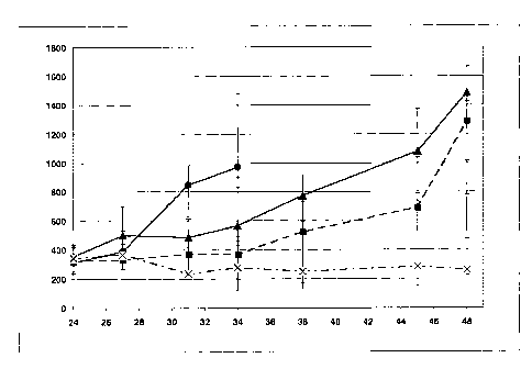
Figure 2 Mean Fluorescence
Intensity (MF1, left y-axis) of type I anti-
CD20 antibody (Cy5-rituximab = white bar) and type II anti-
CD20 antibody (Cy5 humanized B-Lyl B-HH6-B-KV1 GE =
black bar) on Raji cells (ATCC-No. CCL-86) ; Ratio of the
binding capacities to CD20 of type I anti-CD20 antibody
CA 02697482 2010-02-23
WO 2009/030368
PCT/EP2008/006833
- 25 -
(rituximab) and type II anti-CD20 antibody (B-HH6-B-KV1 GE)
compared to rituximab (scaled on right y-axis)
Figure 3 Antitumor activity of treatment of two type II anti-CD20
antibodies on the Z138 human Non-Hodgkin-Lymphoma
(NHL). Both antibodies are humanized B-Lyl anti-CD20
antibodies; 1) B-HH6-B-KV1 glycoengineered (GE) and 2) B-
HH6-B-KV1 wildtype (wt, non-glycoengineered). Mean values of
tumor volume [mm3] plotted on the y-axis; number of days after
injection of tumor cells plotted on the x-axis. Legend: A)Vehicle
(circles), B) humanized B-1y1 GE (B-HH6-B-KV1 GE) 30 mg/kg
once weekly (triangles) and C) humanized B-1y1 wt (B-HH6-B-
KV1 wt) 30 mg/kg once weekly (crosses)
Experimental Procedures
Example 1
Antitumor activity of combined treatment of a type I anti-CD20 antibody
(rituximab) with a type II anti-CD20 antibody (B-HH6-B-KV1 GE)
Test agents
Type I anti-CD20 antibody rituximab was provided as stock solution (c=10
mg/ml)
from Hoffmann La Roche, Basel, Switzerland. Buffer contains polysorbate 80,
Sodiumchloride and Sodiumcitrat.
Type II anti-CD20 antibody B-HH6-B-KV1 GE (= humanized B-Lyl,
glycoengineered B-HH6-B-KV1, see WO 2005/044859 and WO 2007/031875)) was
provided as stock solution (c=9.4 mg/kg) from GlycArt, Schlieren, Switzerland.
Antibody buffer included histidine, trehalose and polysorbate 20
Both solutions were diluted appropriately in PBS from stock for prior
injections.
Cell lines and culture conditions
OCI-Ly18 human Non-Hodgkin-Lymphoma (NHL) cells (Chang, H., et al, Leuk
Lymphoma. 1992 Sep;8(1-2):129-36) (diffuse large cell lymphoma-DLCL) was
used. Tumor cell line was routinely cultured in INDM medium (PAA,
Laboratories,
Austria) supplemented with 20 % fetal bovine serum (PAA Laboratories, Austria)
CA 02697482 2010-02-23
WO 2009/030368
PCT/EP2008/006833
- 26 -
and 2 mM L-glutamine, 25 nM HEPES and 0.05 mM mercaptoethanol at 37 C in a
water-saturated atmosphere at 5 % CO2. Passage 2 was used for transplantation.
Animals
Female SCID beige mice; age 4-5 weeks at arrival (purchased from Bomholtgard,
Ry, Denmark) were maintained under specific-pathogen-free condition with daily
cycles of 12 h light /12 h darkness according to committed guidelines (GV-
Solas;
Felasa; TierschG). Experimental study protocol was reviewed and approved by
local
government. After arrival animals were maintained in the quarantine part of
the
animal facility for one week to get accustomed to new environment and for
observation. Continuous health monitoring was carried out on regular basis.
Diet
food (Provimi Kliba 3337) and water (acidified pH 2.5-3) were provided ad
libitum.
Monitoring
Animals were controlled daily for clinical symptoms and detection of adverse
effects. For monitoring throughout the experiment body weight of animals was
documented two times weekly and tumor volume was measured by caliper after
staging.
Treatment of animals
Animal treatment started at day of randomisation, 24 days after cell
transplantation. Humanized type II anti-CD20 antibody B-HH6-B-KV1 GE
receiving groups and the corresponding vehicle group were treated i.v. q7d on
study day 24, 31, 38, 45 and 52 at the indicated dosage of 30 mg/kg. Type I
anti-
CD20 antibody rituximab treatment as single agent and in combination with type
II anti-CD20 antibody B-HH6-B-KV1 GE was performed on day 26, 33, 40, 47 and
54
Tumor growth inhibition study in vivo
Tumor bearing animals receiving vehicle control had to be excluded 10 days
after
treatment initiation due to tumor burden. Treatment of animals with weekly
Rituximab at 30 mg/kg as single agent inhibited xenograft growth for 10 days
(TGI
68%). Later on tumor xenografts progressed continuously despite further weekly
Rituximab single agent injections. In contrast single agent therapy with B-HH6-
B-
KV1 GE (30 mg/kg) once weekly controlled OCI-Ly18 tumor growth (TGI 100%).
Nevertheless, finally tumor xenografts started to progress under B-HH6-B-KV1
GE
single agent administration. However, combination of Rituximab and B-HH6-B-
CA 02697482 2010-02-23
WO 2009/030368
PCT/EP2008/006833
- 27 -
KV1 GE , both at 30 mg/kg, was obviously superiorly efficacious. Xenograft
tumors
were controlled and in contrast to each single agent antibody arm tumor stasis
maintained over time.
Example 2
Determination of the ratio of the binding capacities to CD20 on Raji cells
(ATCC-
No. CCL-86) of type II anti-CD20 antibody compared to rittudmab
Raji cells (ATCC-No. CCL-86) were maintained in culture in RPMI-1640 medium
(PanBiotech GmbH, Cat.-No. PO4-18500) containing 10% FCS (Gibco, Cat.-
No.10500-064). The type II anti-CD20 antibody B-HH6-B-KV1 (humanized B-Lyl
antibody) and ritwdmab were labeled using Cy5 Mono NHS ester (Amersham GE
Healthcare, Catalogue No. PA15101) according to the manufacturer's
instructions.
Cy5-conjugated ritwdmab had a labeling ratio of 2.0 molecules Cy5 per
antibody.
Cy5-conjugated B-HH6-B-KV1 had a labeling ratio of 2.2 molecules Cy5 per
antibody. In order to determine and compare the binding capacities and mode of
both antibodies, binding curves ( by titration of Cy5-conjugated Ritwdmab and
Cy5-conjugated B-HH6-B-KV1) were generated by direct immunofluorescence
using the Burkitt's lymphoma cell line Raji (ATCC-No. CCL-86). Mean
fluorescence intensities (MFI) were analyzed as EC50 (50% of maximal
intensity)
for Cy5-conjugated Rituximab and Cy5-conjugated B-HH6-B-KV1, respectively.
5*105 cells per sample were stained for 30 min at 4 C. Afterwards, cells were
washed in culture medium. Propidium iodide (PI) staining was used to exclude
dead cells. Measurements were performed using the FACSArray (Becton
Dickinson), Propidium iodide (PI) was measured at Far Red A and Cy5 at Red-A.
Figure 2 shows Mean Fluorescence Intensity (MFI) for binding at EC50 (50% of
maximal intensity) of Cy5-labeled B-HH6-B-KV1 (black bar) and Cy5-labeled
rituximab (white bar).
Then the ratio of the binding capacities to CD20 on Raji cells (ATCC-No. CCL-
86)
is calculated according to the following formula:
CA 02697482 2010-02-23
WO 2009/030368
PCT/EP2008/006833
- 28 -
Ratio of the binding capacities to CD20 on Raji cells (ATCC-No. CCL-86) =
MFI(Cy5- anti- CD20 antibody)x Cy5labeling ratio(Cy5- rituximab)
MFI(Cy5- rituximab)
Cy5labeling ratio(Cy5- anti - CD20 antibody)
MFI (B - HH6 - B - KV1 ) x Cy5labeling labeling ratio (Cy5 - rituximab)
=
MFI(Cy5- rituximab) Cy5labeling ratio (B - HH6- B - KV1 )
207 2.2 ,.., A A
= - X - = u...f.-1
433 2.0
Thus B-HH6-B-KV1 as a typical type II anti-CD20 antibody shows reduces binding
capacity compared to rituximab.
Example 3
Similar antitumor activity of glycoengineered (GE) and non-glycoengineered
(wildtype. wt) anti-CD20 antibody (B-HH6-B-KV1 GE and wt) against Z138 MCL
xenografts in SCID beige mice
Test agents
Type II anti-CD20 antibody B-I-1H6-B-KV1 (glycoengineered (GE) and wildtype
(wt)) were provided as stock solution (c=9.4 mg/ml and 12.5 mg/ml) from
GlycArt,
Schlieren, Switzerland. Antibody buffer included histidine, trehalose and
polysorbate 20.
Both solutions were diluted appropriately in PBS from stock for prior
injections.
Cell lines and culture conditions
Z138 human B-Cell Non-Hodgkin-lymphoma (NHL) cells were originally obtained
from Glycart (Mantle cell lymphoma-MCL). Tumor cell line was routinely
cultured
in DMEM medium (PAA, Laboratories, Austria) supplemented with 10 % fetal
bovine serum (PAA Laboratories, Austria) and 2 mM L-glutamine at 37 C in a
water-saturated atmosphere at 5 % CO2. Passage 2 was used for transplantation.
Animals
Female SCID beige mice; age 4-5 weeks at arrival (purchased from Bomholtgard,
Ry, Denmark) were maintained under specific-pathogen-free condition with daily
cycles of 12 h light /12 h darkness according to committed guidelines (GV-
Solas;
Felasa; TierschG). Experimental study protocol was reviewed and approved by
local
CA 02697482 2010-02-23
WO 2009/030368
PCT/EP2008/006833
- 29 -
government. After arrival animals were maintained in the quarantine part of
the
animal facility for one week to get accustomed to new environment and for
observation. Continuous health monitoring was carried out on regular basis.
Diet
food (Provimi Kliba 3337) and water (acidified pH 2.5-3) were provided ad
libitum.
Monitoring
Animals were controlled daily for clinical symptoms and detection of adverse
effects. For monitoring throughout the experiment body weight of animals was
documented two times weekly and tumor volume was measured by caliper
beginning at staging.
Treatment of animals
Animal treatment started at day of randomisation, 14 days after s.c. cell
transplantation. Humanized anti CD20 antibody (B-HH6-B-KV1 GE and wt)
receiving groups and the corresponding vehicle group were treated i.v. q7d on
study day 14, 20, 27 and 34 at the indicated dosage of 10 mg/kg.
Tumor growth inhibition study in vivo
Tumor bearing animals receiving vehicle control had to be excluded 19 days
after
treatment initiation due to tumor burden. Treatment of animals with weekly B-
HH6-B-KV1 as wt or glycoengineered (B-HH6-B-KV1 GE and wt) at 10 mg/kg
inhibited xenograft outgrowth shortly after start of treatment. At time of
control
termination all antibody tumors regressed and later most of Z138 tumor
xenografts
showed complete remission. No significant differences were observed between wt
and glycoengineered versions of anti CD20 antibody B-HH6-B-KV1 in this
xenograft model. This was not unlikely since mice do not express the correct
Fc
receptor on their NK cells and furthermore SCID beige mice are thought to be
incompetent for NK-mediated ADCC due to severe triple immunodeficiency.
Therefore s.c. xenografts models in SCID beige mice are not appropriate for
mimicking human ADCC mediated effect with glycoengineered modified
antibodies.