Note: Descriptions are shown in the official language in which they were submitted.
CA 02697933 2014-07-22
X-RAY DEVICE
Description
The invention relates to an x-ray system for examining a syringe cap having a
cannula.
X-ray systems of the type described here are well known. They are used when
inspecting
syringe caps that contain a cannula. This type of cap is placed on syringes
with a cannula on
the one hand in order to provide the syringe with the cannula with a sterile
cover, and on the
other hand as a protection against injuries. Syringe caps of this type
frequently include an
elastic stopper into which the cannula penetrates. A syringe cap of the type
described can be
placed on a syringe that has already been provided with a cannula. It can bend
when being
mounted on the syringe body or can be obliquely inserted thereon and then
already be
oblique when inserted into the stopper. But a syringe cap of this type can
also be provided
with a cannula, pre-mounted as-it-were, and then placed on a syringe. Also
when being pre-
assembled the cannula can be obliquely inserted into the stopper or bent when
being
inserted. X-ray systems of the type described here are used to detect cannulas
that are
positioned obliquely in the syringe cap. They have two x-ray sources that can
penetrate the
syringe cap from two directions, preferably from two directions positioned at
45 to 90 . Using
the two images produced it can be determined whether the cannula is obliquely
situated
inside the syringe cap. In order to prevent the tips of the cannulas from
possibly perforating
the wall of the syringe cap with the consequent loss of sterility on the one
hand, and on the
other hand would present the risk of injury, the syringe caps are separated
starting with a
specific angularity. X-ray devices of this type are expensive because on the
one hand two
radiation sources and on the other hand an image analysis circuit are required
for analyzing
the images from the two radiation sources.
It is desirable to provide an x-ray device that is simple to construct and can
be inexpensively
produced.
In one aspect, the present invention provides an x-ray device for examining
syringe caps with
a cannula, comprising an x-ray source, an x-ray detector, and a retaining
device for holding
the syringe cap in the beam path at an examination site, wherein the retaining
device
1
CA 02697933 2014-07-22
comprises a first receiving unit for the syringe cap, and wherein the syringe
cap is arranged
in the beam path so that its longitudinal axis coincides with the main axis of
the beam path.
The syringe cap is thus impinged from above and from below with x-rays, in
order to examine
the position of the cannula. If, with such an arrangement of the syringe cap
in the beam path
by the x-ray detector at the position, a point is detected at which the
cannula is expected,
then the cannula is oriented concentrically or coaxially to the beam path. It
is thus situated
also coaxial to the longitudinal axis of the syringe cap. It is unimportant to
this type of
examination whether the cannula is housed in a separate syringe cap and placed
together
with same on the syringe, or whether the syringe cap is placed on a syringe
that comprises
the cannula. If, however, the cannula is represented as a line in the x-ray
detector, then it
must be assumed that the cannula does not lie exactly in the longitudinal
axis, but at an
angle to it. In this instance the cannula is separated.
In a preferred exemplary embodiment of the x-ray device at least one
collimator is arranged
in the beam path between the x-ray beam source and the examination point. This
serves to
reduce to a minimum the stress of the beams on the area situated around the
examination
point.
A particularly preferred exemplary embodiment of the x-ray device is
characterized in that
additionally a first reference element is provided. This serves to provide a
first reference
signal when producing an image of the syringe cap in the x-ray detector. The
reference
element preferably has a circular opening.
Particularly preferred is an exemplary embodiment of the x-ray device that is
characterized
by a second reference element that is preferably annular in its configuration.
This, too,
serves to produce a reference signal in the x-ray detector. In virtue of the
two reference
signals it is particularly easy to decide, whether the cannula runs obliquely;
that is at an angle
to the longitudinal axis of the syringe cap, and whether the angularity is
still acceptable.
In one aspect, the invention provides a method for examining syringe caps
comprising a
cannula with an x-ray device comprising an x-ray source and an x-ray detector,
the method
comprising the following steps: arranging a syringe cap in a beam path between
the x-ray
source and the x-ray detector in a way that the longitudinal axis of the
syringe cap coincides
2
CA 02697933 2014-07-22
with the main axis of the beam path, taking an x-ray picture of the syringe
cap with the x-ray
device, and comparing the shape or the size of the cannula in the picture of
the syringe cap
with a reference picture to determine if the cannula has an acceptable degree
of
deviation/deflection with respect to the longitudinal axis of the syringe cap.
The invention will be more completely explained in the following using the
drawing, wherein:
Figure 1 represents a principle diagram of the x-ray device;
Figure 2 represents three syringe caps and their images taken at the
time of x-ray
examination;
Figure 3 represents a first exemplary embodiment of an x-ray device;
Figure 4 represents a second exemplary embodiment of an x-ray device;
Figure 5 represents a side view of a syringe with an obliquely seated
syringe cap;
Figure 6 represents a top view of the syringe with the syringe cap
according to Figure
5;
Figure 7 represents a side view of a syringe with a correctly seated
syringe cap; and
Figure 8 represents a top view of the syringe with the syringe cap
according to Figure
7.
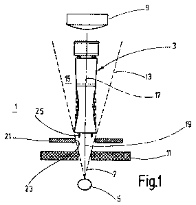
The basic diagram according to Figure 1 shows an x-ray device 1 that is used
for examining
a syringe cap 3 as well as an implied x-ray beam source 5, that emits x-ray
beams 7. An x-
ray detector 9 is provided at a distance from the x-ray beam source 5 which
evaluates the x-
ray beams impinging thereon. The x-ray beams 7 run a course towards the x-ray
detector 9
through a collimator 11, which shields against the radiation exiting from the
x-ray beam
source 5 and allows only a portion of the x-rays 7 to pass unobstructed toward
the x-ray
detector 9. There is thus an extended beam path 13 toward the x-ray detector
9. A syringe
cap 3 is arranged in same at an examination site 15 in such a fashion that its
longitudinal
axis 17 coincides with the main axis 19 of the beam path 13. In virtue of this
arrangement of
3
CA 02697933 2014-07-22
the syringe cap 3 on the examination site 15 it is possible to impinge it
vertically from above
or below with x-ray beams and so to obtain an x-ray image in the x-ray
detector 9.
A first reference element 21 is provided on the side of the collimator 11
facing away from the
x-ray radiation source, which like the collimator 11, limits the width of the
cone of the x-ray
beams 7 that run from the x-ray source 5 towards the examination site 15, in
order to
produce a defined conical beam path 13. The first reference element 21
preferably has a
circular opening 23 that is picked up by the x-ray detector and delimits the
image
represented there. The reference element 21 can be configured ring- or plate-
shaped, the
circular opening 23 is fundamental.
In Figure 1, a second reference element 25 is arranged between the x-ray
source 5 and the
x-ray detector 9¨here above the first reference element 21¨which, like the
collimator 11 and
the first reference element 21, is arranged in the region of the x-ray beams 7
emitted by the
x-ray source 5. Seen from the x-ray source 5, the second reference element 25
is situated
downstream of the beam relative to the first reference element. In the
preferred exemplary
embodiment represented here, it is arranged directly before the examination
site 15. In
another preferred exemplary embodiment (not shown here) the second reference
element 15
is arranged in the beam path opposite the examination site 15, preferably
directly in front of
the x-ray detector 9. The second reference element 25 is configured annular
and thin-walled
and is represented as a thin ring in the x-ray detector 9.
The external diameter of the second reference element 25 is smaller than the
inside diameter
of the opening 23. It is arranged concentric to the main axis 19 of the beam
path 13. This
applies also to the opening 23 of the first reference element 21.
Figure 2 shows three syringe caps 3 as are represented also in Figure 1. Here
in Figure 2
the cannula 27 that is situated on the inside of the syringe cap 3 is seen. In
the left
representation in Figure 2 the cannula 27 is not arranged concentric to the
longitudinal axis
17 of the syringe cap 3. Rather, it runs opposite same to the left so that it
includes an acute
angle opening downwards having the longitudinal axis 17 (Figure 2). The
deflection of the
cannula 27 can naturally lie also in a plane other than the image plane of
Figure 2.
4
CA 02697933 2014-07-22
Underneath the left tip cap an image a) is reproduced as it is detected by the
x-ray detector 9
shown in Figure 1. The wall of the syringe cap 3 transilluminated from the
upper side is
represented as an external ring 29. Concentric to the outside ring 29 a thin
inside ring 33 can
be seen, which represents the image of the second reference element 25.
A black line runs to the left from the center point 35 of the inside ring 33
and represents the
image 37 of the cannula 27 deflected to the left.
In the center representation in Figure 2 the cannula 27 runs concentrically to
the longitudinal
axis 17 of the syringe cap 3. In the illustration b) shown thereunder the
syringe cap 3 can be
seen that the cannula 27 in the x-ray detector 9 is represented in this case
as a point 37' that
coincides with the center point 35 of the inside ring 33, through which the
longitudinal axis 17
of the syringe cap 3 also runs.
The syringe cap 3 with a cannula 27 deflected to the right can be seen at the
upper right in
Figure 2. Thereunder the illustration c) of a syringe cap 3 of this type is
shown in the x-ray
detector 9: It can be seen that here a line running to the right between the
center point 25
and the inside ring represents the depiction 37 of the cannula 27.
Finally, it is clear that a correctly concentrically depicted cannula 27 is
depicted as point 37'
that coincides with the center point 35 of the inside ring 33. As soon as the
cannula 27 of the
longitudinal axis 17 of the syringe cap 3 is deflected¨that is, bent¨it is
depicted as a line.
A still acceptable measure of deflection of the cannula 27 can be defined in
virtue of the
inside diameter of the inside ring 33. If this is maximally bent vis-a-vis the
longitudinal axis 17
of the syringe cap 3 that it contacts the inside ring 33, the syringe cap 3 is
classified as
conforming. If, however, the depiction 37 of a deflected cannula 27 ends
outside of the inside
ring 33, the associated syringe cap 3 would be segregated as unacceptable.
The three illustrations a), b), and c) in Figure 2 of the syringe cap 3
depicted in this
illustration show that it is possible in simple fashion to differentiate
syringe caps 3 with
concentric cannula 27 from those, in which the cannula is bent or deflected
vis-a-vis the
longitudinal axis 17 of the syringe cap 3. Here it makes no difference whether
the cannula 27
5
CA 02697933 2014-07-22
=
is mounted obliquely in a syringe on which the syringe cap 3 is placed, or
whether the
cannula 27 is deflected from its originally correct position when placing the
syringe cap 3.
The x-ray device 1 is can thus be used for examining syringe caps 3 that are
pre-assembled
and already comprise a cannula 27. They can also be used for examining
syringes having a
cannula fixed on the syringe body and on which a syringe cap has been placed.
When this is
done, it is possible in both cases to x-ray the syringe cap 3 from its top
side 31 or from the
opposite side. It is critical only that the syringe cap 3, and with it also an
associated syringe,
if applicable, is arranged on the examination site 15 concentrically to the
main axis 19 of the
cone-shaped beam path 13, so that the longitudinal axis 17 of the syringe cap
3 coincides
with the main axis 19.
A first exemplary embodiment of an x-ray device 1 is shown in Figure 3 which
operates
according to the basic principle described using Figures 1 and 2 and used for
examining a
syringe cap 3 having a cannula 27, in order to establish whether the cannula
27 is or is not
deflected vis-a-vis the longitudinal axis 17 of the syringe cap 3.
The x-ray source 5 of the x-ray device can be seen in Figure 3; in addition
the collimator 11,
the first reference element 21, and the second reference element 25. Also
shown are the x-
ray beams leaving the x-ray source 5, which pass through the collimator 11 and
the first
reference element 21, and produce a conical beam path 13.
In this exemplary embodiment, on the side of the first reference element 21
facing away from
the x-ray source 5 the second reference element 25 is situated directly in
front of the syringe
cap 3 arranged at the examination site 15. It is likewise configured ring-
shaped, as shown in
Figure 1.
The x-ray detector 9 (not shown here) that delivers the image of the syringe
cap is situated
over the syringe cap 3.
In the exemplary embodiment of the x-ray device 1 shown in Figure 3, a
mounting device 29
installed in the beam path 13 is provided, which comprises a first receiving
unit 41. This
holds the syringe cap 3 in the desired orientation at the examination site 15.
It has a
cylindrical section 43 whose inside diameter is selected so that it receives
the syringe cap 3
6
CA 02697933 2014-07-22
in its inside as snugly as possible and whose middle axis coincides with the
main axis 19 of
the beam path 13. When this is done, the syringe cap 3 is so arranged vis-à-
vis the beam
path 13 that its longitudinal axis 17 coincides with the main axis 19 of the
beam path 13.
Here the syringe cap 3 is x-rayed from its upper side 31. It is also
conceivable that the first
receiving unit 41 is embodied so that the syringe cap 3, with an associated
syringe, if
necessary, is conversely held on the examination site 15 and thus is x-rayed
from below.
The first receiving unit 41 is embodied as an insert. It can thus be plugged
into an
appropriate recess 45 in the base 47 of the retaining device 39. In this
fashion it is also
possible to combine different first receiving units 41 using one and the same
retaining device
39.
Figure 3 shows that the retaining device 39 comprises a second receiving unit
49, on which
the syringe cap 3 can be slipped. In the exemplary embodiment of the retaining
device 39
represented here, the second receiving unit 39 is so embodied that with such a
mounting the
syringe cap 3 can be held with its upper side 31 facing upwards by the second
receiving unit
49. It is clear that the syringe cap 3 must be rotated 180 before
installation in the cylindrical
section 43 of the first receiving unit 42. This can be done manually or using
appropriate
manipulators.
The second receiving unit 49, too, can be embodied as an insert that can be
inserted into the
retaining device¨and thus interchangeably¨in order to be able to adapt the
mounting 39 to
the different syringe caps 3.
In the exemplary embodiment shown in Figure 3 the retaining device 39 has a
third receiving
unit 51 that is embodied differently than the other receiving units. It can be
used to receive
another type of syringe cap 3' or the like, that is to be examined using the x-
ray device 1 and
held at the examination site 15, when the x-ray examination is to be done.
Preferred is an exemplary embodiment of the retaining device 39 that has
several identical
receiving units that are embodied like the first receiving unit 41. This type
of receiving units
could be arranged circularly in the base 47 of the retaining device 39, for
example. It is then
possible to rotate the retaining device 39 after examining a first syringe cap
3, until the next
receiving unit is arranged with a syringe cap 3 at the examination site 15.
The receiving units
7
CA 02697933 2014-07-22
can also be arranged in a row in the base 47 of the retaining device 39. In
this instance, the
retaining device 39 is then subject to a transverse movement, in order to
bring the receiving
units with the syringe caps sequentially to the examination site 15 and
examine them.
The retaining device 39 is preferably provided with a data storage medium 53,
which is
-- merely implied here. When examining syringe caps 3 it is also possible to
supply them in
batches by means of a retention device 39 for examination using an x-ray
device 1. The
retaining device 39 can then be detected and stored for tracking a
manufacturing process.
In Figure 3 it can be seen that the collimator 11 is arranged under the
retaining device 39
and that the first reference element 21 is situated somewhat above the
underside of the
-- retaining device 39. It is quite possible to bring the first reference
element 21 closer to the
examination site 15 and to the position of the second reference element 25
represented in
Figure 3, for example. The results in the beam of the x-ray source 5 being
even better
focused on the examination site 15. In this case the second reference element
25 is then¨
viewed in the direction of irradiation¨placed on the side of the examination
site facing away
-- from the x-ray source 5, in front of the x-ray detector 9. Preferably it
can be arranged
immediately in front of the x-ray detector 9 so that a particularly sharp
image of the second
reference element 25 results and so that it can also be displayed as a very
thin ring.
Ultimately two annular reference elements of the type discussed here or in the
following and
that are concentric to each other can be used. A tolerance range can thus be
defined, within
-- which a deflection of the cannula 27 can still be accepted.
A transformed exemplary embodiment of the x-ray device 1 is shown in Figure 4.
Identical
and functionally analogous parts are provided with the same reference numbers.
Inasmuch,
reference is made here to the previous description.
The x-ray device 1 has an x-ray beam source 5, which emits x-ray beams 7
towards an x-ray
-- detector 9. A beam path 13 is merely implied here. Its main axis 19 runs
here in Figure 4
vertically upward from the x-ray source 5 through the examination site 15 to
the x-ray
detector 9.
8
CA 02697933 2014-07-22
Here a syringe 55 can be seen on the examination site 15 where it is held by a
suitable
retaining device 29 on the examination site 15 and so aligned that its
longitudinal axis 57
coincides with the main axis 19 of the beam path 13.
A syringe cap 3 is placed on the syringe 55 and said cap is seated here
obliquely on the
syringe 55 such that the longitudinal axis 17 of the syringe cap 3 exhibits an
angle to the
main axis 19 of the beam path 13.
It can be clearly seen that in the exemplary embodiment according to Figure 4
the syringe 55
and the syringe cap 3 are transradiated from below. The syringe cap 3 is thus
so arranged¨
differently than in the case of the representations according to Figures 1 to
3¨that its top side
31 faces upwards.
A deflection device 59, permeable to x-rays but deflecting light rays, and
having a mirror, is
arranged in the beam path 13A and¨viewed from the x-ray source 5¨on the other
side of the
examination site 15.
The x-ray device 1 has an image detection unit 61 that comprises a camera 63,
for example.
A broken line 65 is used to indicate that the image detection unit detects an
image from the
top side 31 of the syringe cap 3.
The x-ray detector 9 signals and those of the image detection unit 61 are
pooled and, as
indicated by a line 67, evaluated together; that is, superimposed. An image
analysis unit 69
is used for analyzing the x-ray detector 9 signals and the image detection
unit 61 signals,
which are not individually represented here.
A indicates the overlapping of the image signals of the x-ray detector 9 and
the image
detection unit 61: A target position lying concentrically to the main axis 19
is specified using
an external circle 71, said position corresponding to the syringe 55, for
example.
In the middle a concentric inner circle 73 can be seen in the case of A. This
indicates the
optical image of a syringe cap 3 with a concentric arrangement; that is, when
its longitudinal
axis 17 coincides with the main axis 19 of the beam path 13. In this instance,
the cannula 27
9
CA 02697933 2014-07-22
is depicted as a point 37' when it is arranged coaxial to the longitudinal
axis of the syringe
cap 3.
In the depictions according to A the external circle 71 can be seen to the
left. Inside the circle
71, the depiction can be seen of a syringe cap 3 arranged oblique to the main
axis 19 of the
beam path 13, which is seen as an ellipse 75 by the image detection unit 61.
It can also be
seen that a line is visible extending from the center point 35 of the external
circle 71, said line
representing the image 37 detected by the x-ray detector 9 of a
correspondingly deflected
cannula 27.
It is clear that the cannula 37 is deflected in the same direction as the
syringe cap 3. It is
thus assumed that the syringe cap 3 and the cannula 27 are bent in the same
direction, that
the cannula 27 does not anywhere penetrate the external wall of the syringe
cap 3, and that
accordingly this syringe cap 3 is acceptable.
Accordingly to the far right in the illustrations according to A, an external
circle 71 can be
seen. Here, too, the image detection unit 61 recognizes that the syringe cap 3
is deflected
upwards to the right. Here, too, this can be seen in virtue of the ellipse 75.
The depiction 37
of the cannula 27 lies on the central axis of the ellipse 75. It is therefore
assumed that the
syringe cap 3 and the cannula 27 are deflected in the same direction, and thus
the cannula
27, as in the left illustration according to A, is arranged concentrically to
the syringe cap 3.
The illustrations labeled with A thus show syringe caps 3 that all conform.
If one considers the illustration B in Figure 4, which results by an
overlapping of the images
of the x-ray detector 3 and the image detection unit 61, then here too the
external contour of
the syringe 55, which is detected by the x-ray detector 9, can be seen and is
indicated by an
external circle 71. In the case of the left illustration according to B the
image detection unit 61
recognizes that the syringe cap 3 is deflected to the right and the central
axis of the ellipse
lies on a conceptualized horizontal H. The x-ray detector 3 shows the
depiction 37 of the
cannula 27 as a line. It is obviously deflected vertically downwards; that is,
not in the
direction of the central axis of the ellipse 75. The image analysis unit 69
thus recognizes that
the syringe cap 3 does not conform.
CA 02697933 2014-07-22
A corresponding deviation of the central axis of the ellipse 75 and the
resulting deflection of
the syringe cap 3 from the deflection of the cannula 27, which is depicted by
the x-ray
detector 9 by the line 37, is shown also in the far right of illustration B.
This syringe cap 3,
too, is recognized by the image analysis unit 69 as being defective.
In the center of the illustration according to B the following can be seen:
Again a target
position or the wall of the syringe 55 recognized by the x-ray detector 9 is
in turn specified. A
point 37' coinciding with the center point of the circle 71 shows the cannula
27 aligned
concentrically to the main axis 19 of the beam path 13. The image analysis
unit 69 has
recognized that the syringe cap 3 is seated obliquely, which is indicated by a
circle 77
depicted eccentrically to the center point of the circle 77. It is clear that
the longitudinal axis
17 of the syringe cap 3 does not coincide with the cannula 27. The analysis of
the x-ray
image by means of the x-ray detector 9 and the detection of the syringe cap 3
by the image
detection unit 61 thus indicate that here there is an incorrect alignment of
the cannula 27
inside the syringe cap 3. Here, too, the image analysis unit 6 recognizes a
defective product
so that it can be segregated.
Figure 5 represents a side view of a syringe 55 with a syringe cap 3. Parts
that have already
been explained using the previous Figures are provided with the same reference
numbers.
The syringe 55 has a longitudinal axis 57, opposite which the longitudinal
axis 17 of the
syringe cap is pivoted. In Figure 5 the syringe cape 3 is tipped at an acute
angle to the left
because it is not correctly applied to the syringe 55. Therefore the needle 27
(only indicated
in Figure 5) situated in the syringe cap is also tipped vis-a-vis the central
axis 57 of the
syringe 55.
Figure 5 shows an alignment device 79. This serves to correctly align the
syringe cap 3 vis-a-
vis the syringe 55 so that alignment errors are prevented. With the aid of the
alignment
device 79 the syringe cap 3 is aligned so that its longitudinal axis 17 aligns
with the central
axis 57 of the syringe 55.
The alignment device 79 is embodied so that it embraces the syringe cap 3.
Here it has two
gripper elements 81 and 83 arranged at a distance from each other. There
separation is
11
CA 02697933 2014-07-22
selected so that the syringe 55 even with a crookedly seated syringe cap 3 can
be arranged
between the gripper elements 81, 83.
Figure 6 shows the syringe 55 according to Figure 5 viewed from the top. The
syringe cap 3
can be identified. Here not only its top side 31 but also parts of its
peripheral surface 85 can
be seen. Optical image detection can thus easily recognize the incorrect
seating of the
syringe cap 3.
If the syringe 55 illustrated here with the obliquely seated syringe cap 3 is
imaged from top to
bottom or from below upwards using the x-ray device 1 as described above, the
cannula 27
will be represented as a line. The depiction 37 of the cannula 27 is drawn in
Figure 6.
The x-ray device 1 is embodied so that the oblique seating of the cannula 27
can be
identified using the line depiction 37 of the cannula 27. It is also
conceivable to use the
depiction of the syringe cap 3 represented by the image detection unit 61 for
identifying its
oblique seating. Preferably, the signals of the x-ray detector 9 and of the
image detection unit
61 will be analyzed together, particularly overlapped.
The depiction 37 of the cannula 27 proceeds from the central axis 57 of the
syringe 55 and
runs from there essentially horizontally to the left. Accordingly a crescent-
shaped region of
the peripheral surface 85 can be identified to the right of the circularly
depicted top side 31 of
the syringe cannula 3. Using the depiction 37 of the cannula 27 detected by
means of the x-
ray detector 9 and using the position of the peripheral surface 85, which is
depicted crescent-
shaped in Figure 6, the direction in which the syringe cap 3 and the cannula
are pivoted can
be established.
The alignment device 79 can now specifically effect a counter-movement of the
syringe cap
3.
Preferably the gripper elements 81 and 83 of the alignment device 79 have V-
shaped inside
surfaces 87 and 89, whereby the intersection points of the surface segments of
the inside
surfaces 87 and 89 lie on a conceptual line 91 which runs through the central
axis 57 of the
syringe 55.
12
CA 02697933 2014-07-22
Figure 7 again represents the syringe 55 illustrated in Figure 5 in side view.
Arrows 93, 93'
indicate that the gripper elements 81 and 82 are moved towards each other;
that is, in the
direction towards the central axis 57 of the syringe 55. These act on the
syringe cap 3 so that
its longitudinal axis 17 aligned with the central axis 57, so that they are
arranged correctly on
the syringe 55. Accordingly the cannula 27, too, lies coaxial to the
longitudinal axis 17 and
thus to the central axis 55.
It can be seen in the top view according to Figure 8 that the inside surfaces
87 and 89 in this
position center the syringe cap 3 so that the cannula 27 is depicted as a
point 37' by the x-
ray device 1 or its x-ray detector 9. Optical detection of the syringe cap 3
would depict its top
side 31 as a circle. Any regions of the peripheral surface 85 of the syringe
cap 3 are no
longer visible here. The depiction of the cannula obtained by the x-ray
detector 9 and the
image of the syringe cap 3 generated by an image detection unit 61 show that
the cap is not
correctly aligned.
The syringe cap 3 is aligned exactly vis-a-vis the central axis 57 of the
syringe 55 by the
gripper elements 81, 83 moved toward each other and by their V-shaped inside
surfaces 87
and 89. The adjustment vis-à-vis the conceptual line 91 is made in that the V-
shaped inside
surfaces 87 and 89 are oriented to this line. If the gripper elements 81 and
83 are moved
symmetrically to a conceptual central plane 95, which runs through the central
axis 57 of the
syringe 55 and stands vertical to the line 91, an exact alignment of the
syringe cap 3 vis-a-vis
this central axis 95 is possible without further ado. Activation of the
alignment device can be
done automatically if the x-ray detector 9 and/or the image detection unit 61
indicate(s) an
incorrect position of the syringe cap vis-a-vis the syringe 55.
It should be reiterated that the embodiment of the alignment device 79 can
also be altered. It
is conceivable, for example, to align the syringe cap 3 by means of one or a
plurality of
optionally shaped grippers using the analysis of the image obtained from the x-
ray detector 9
or the image detection unit 61 that the surface 31 is aligned concentrically
to the central axis
57 of the syringe 55 or the cannula 27 is depicted as a point 37'.
If it is not possible to align the top side 31 concentrically to the central
axis 57 and
simultaneously to depict the cannula 27 as a point on the intersection of the
central plane 59
13
CA 02697933 2014-07-22
=
with the line 91, it must be assumed that the cannula 27 is not arranged
concentrically
relative to the syringe cap 3.
In Figures 6 and 8 the inside ring 33 is indicated by a line that is depicted
by using a second
reference element 25 in the x-ray detector 9. This serves to establish whether
or not, if the
cannula 27 is deflected out of the desired central position, it still lies in
the acceptable region.
Inasmuch reference is made to the statements relating to the image analysis
according to
Figure 2 and 4 (see illustrations A and B there).
Overall it can be seen that the imaging of a syringe cap 3 together with a
syringe 55, if
necessary, using an x-ray device 1 easily enables examination of whether the
cannula 27 is
correctly arranged inside the syringe cap 3, or lies at least within a
tolerance range, which
with the aid of a second reference element 25 or of the inside ring 33 can be
defined and
read.
At all events a single x-ray source 5 is sufficient for imaging the syringe
cap 1 from above or
from below, while it is arranged coaxially to the beam path 13 of the x-ray
source 5, while
thus its longitudinal axis 17 coincides with the main axis 19 of the beam path
13.
From the explanations relating to the x-ray device the following becomes
obvious: The x-ray
device images the syringe cap 3 along its longitudinal axis 17. It can
differentiate glass and
plastic from metal. In this fashion it is possible to detect a cannula 27 made
of metal inside
the syringe cap 3 and to do this regardless of whether the cannula is provided
as a pre-
assembled element of the syringe cap 3 or is mounted on the syringe, on which
the syringe
cap 3 is placed.
The critical criterion of the x-ray device 1 is the possibility of detecting
metal objects in an
environment that contains glass and/or plastic.
The x-ray device described using the Figures is characterized in that the
syringe cap 3 to be
imaged is impinged with beams along its longitudinal axis. In addition it can
also be provided
that the examination site 16 is impinged also laterally with x-rays, in order
to obtain additional
information on the alignment of the cannula 27 in the syringe cap 3. When this
is done the
examination direction and the number of examinations can be freely selected in
a wide
14
CA 02697933 2014-07-22
range, in order to achieve maximum reliability with regard to product quality.
As a rule, a
syringe or its syringe cap 3 arranged on the examination site will be imaged
laterally, from
the same height. But it is also possible to arrange the x-ray source so that
the syringe cap 3
can be examined laterally obliquely from below or above. The examinations can
also be
carried out at different angles vertically to the optimum course of the
cannula 27 so that in
the doubtful case exact information on the course of the cannula 27 inside the
syringe cap 3
can be obtained.
Therefore, all devices that examine the syringe cap 3 using beams, in order to
detect the
course of a cannula 27 inside the syringe cap 3 should be included in the term
"x-ray device".
Therefore, the invention is thus not particularly limited to x-ray but
includes all irradiation
types that enable a similar type of examination of the syringe cap 3.