Note: Descriptions are shown in the official language in which they were submitted.
CA 02699750 2010-03-12
WO 2009/036300 -1-
PCT/US2008/076211
ENDOS COPE WITH INTERNAL
LIGHT SOURCE AND POWER SUPPLY
BACKGROUND OF THE INVENTION
Endoscopes are commonly used to view within an actual or potential
space inside a subject (e.g., a human, or animal) or manufactured structure
(e.g., an engine or a pipe) while performing a therapeutic or diagnostic
procedure. Typically, an endoscope has a control handle coupled to an
elongated portion (e.g., a flexible elongated portion, a rigid elongated
portion,
a semi-rigid elongated portion). During use of the endoscope, the control
handle remains outside the subject while the elongated portion is at least
partially disposed inside the subject. Generally, the elongated portion has
one
or more optical components (e.g., one or more lenses, fiber optics, video
imager) to illuminate and view the region inside the subject, and the control
handle has one or more devices designed to control the optical components
(e.g., to control light intensity, focus an image, display and image, digitize
an
image) and the position of the elongated portion in the subject.
Typically the light source, the light source power supply and other
features and controls (such as irrigation and suction) are placed in a
separate
console that is connected to the endoscope handle by an umbilical cable that
includes a light guide. Traditional light sources for endoscopic use are
generally of two types: incandescent filament lamps and arc lamps. Both types
of lamps are very inefficient in converting electrical power to light, and
consequently produce large amounts of heat that must be dissipated. Because
of the heat generated and the need for a bulky light source power supply,
these
light sources typically reside outside of the endoscope itself
An illustrative endoscope according to the prior art is shown in FIG. 1,
which is simplified from FIG. 1 of U.S. Pat. No. 5,630,783. The endoscope 20
includes a housing or handle 22 having an elongated distal portion 24 that
supports a flexible tube 26. The handle 22 also includes an eyepiece 28 at the
proximal end of the handle 22. The eyepiece 28 can include a focus ring 30.
CA 02699750 2010-03-12
WO 2009/036300 -2-
PCT/US2008/076211
The tube 26 typically includes an outer covering 32 constructed from a low-
friction polymer. The distal end 34 of the tube 26 typically includes a
polished
metal end cap 35 having a plurality of exit ports. The handle 22 includes a
movable steering control 36 with a steering lever or knob 38. The steering
lever 38 moves (arrow 40) proximally and distally along the handle to control
a pair of steering cables (not shown) within the tube 26. The steering cables
enable the distal end 34 of the tube 26 to bend. The steering control 36 is
mounted on an enlarged ball 42 located between the proximal end and the
distal end of the handle 22.
A connector 50 links the ball 42 with a fiber optic light source cable 44
that communicates with a light source 46 according to the prior art. The light
source 46 is, typically, a variable-intensity incandescent or arc lamp that
draws
power from a conventional AC power source via an electrical cord 48. An
associated internal illumination light guide (not shown) is optically linked
within the ball 42 of the handle 22 with the light source cable 44. The
internal
illumination light guide extends through the distal end 24 of the handle 22
and
passes along the full length of the tube 26, and is exposed at the distal end
34
of the tube 26 to provide light at the distal end. According to the prior art,
an
irrigation/instrument assembly 52 or other fitting is optionally provided at
the
distal end 24 of the handle 22. The assembly 52 interconnects with an internal
tube (not shown) that exits at the distal end 34 of the tube 26. The assembly
52 includes an instrument port 53 that provides a conduit for guiding an
actuating mechanism of an instrument such as the grasper 55 located at the
tube's distal end 34. A remote plunger assembly 54 with a movable finger
control 59 controls the grasper 55. The irrigation/instrument assembly 52 also
includes a fluid inlet 57 that can comprise a Luer-style fitting connected
with a
sterile tubing 56 that can terminate in a source of sterile saline or water
used as
an irrigation fluid. Both instruments and irrigation fluid can pass down a
single internal tubing (not shown) to exit at the distal end 34.
Prior attempts to produce endoscopes that are not tethered by such a
fiber optic light source cable 44 to an external light source 46 have produced
CA 02699750 2010-03-12
WO 2009/036300 -3-
PCT/US2008/076211
light sources and battery power supplies that are external to and separate
from
the endoscope handle, yet still use inefficient light sources, such as halogen
lamps. An example of this form of prior art endoscope is illustrated by FIG.
2,
which is also simplified from FIG. 2 of U.S. Pat. No. 5,630,783. This prior
art
endoscope 120 includes a body or housing 122 having a distal end 124 and a
proximal eyepiece 128. The eyepiece 128 includes an adjustment or focus ring
130. The distal end 124 of the housing 122 supports a semi-rigid, flexible,
tube 126, which is covered with a flexible, low friction, sheathing 132, and
includes a distal end 134 having a polished metallic ring 135 with a series of
openings. A steering control 136 including a movable steering control lever
138 is mounted on an enlarged ball section 142 of the housing 122. A fitting
280 is provided adjacent the distal portion 124 of the housing 122 for the
introduction of instruments, such as graspers and biopsy forceps, medicaments
and small quantities of saline or water irrigation fluids.
This prior art endoscope has a self-contained illumination source 180
attached in the position of the fiber optic light source cable connector 50 of
FIG. 1. The source 180 utilizes a high intensity incandescent light bulb. The
bulb can be a halogen-type bulb for greater intensity and whiter light. The
battery and switch 204 are self-contained within a removable housing 208 that
is joined to the upper portion of the handle 122 by a threaded ring coupling
210. The battery can be a rechargeable nickel-cadmium or equivalent battery,
such as a lithium-ion battery. Other endoscopes with an add-on battery
powered light sources using an incandescent halogen bulb are known, for
example, at
http://www.pentaxmedical.com/Products/Bronchoscopy/PortableBronch.asp.
Such add-on battery powered light sources not only are inefficient and
clumsy, but the connection to the endoscope handle is an additional site for
leakage, corrosion and contamination. In general, after each use in a medical
procedure, the endoscope is cleaned to remove detritus, and subsequently
disinfected and/or sterilized. Standard cleaning and disinfection procedures
require that the entire endoscope, including the endoscope handle, is
CA 02699750 2010-03-12
WO 2009/036300 -4-
PCT/US2008/076211
completely immersible and watertight, and leak testing is performed routinely
as part of the cleaning and disinfection process. Fluids used during the
disinfection process (http://www.fda.gov/cdrh/ode/germlab.html), such as
activated alkaline glutaraldehyde, peracetic acid and orthophthaldehyde, are
harsh and corrosive chemicals. Fluid leakage into the interior of the
endoscope handle and contact with electronic circuits and components can
damage both a light source and its power supply if located within the
endoscope handle.
Thus, there is a need for a fully self-contained endoscope that is not
tethered to an external light and/or power source and that can withstand
multiple uses and cleanings while maintaining the integrity of the liquid
sensitive components of the endoscope.
SUMMARY OF THE INVENTION
The present invention provides an endoscope having a shaft and a
handle that are liquid and gas impermeable, having inside the handle an
internal solid-state light source and a further internal sealed compartment
that
is liquid impermeable and gas permeable and that contains at least one battery
and a light source control circuit that provides continuous control of the
light
intensity produced by the internal solid-state light source.
In preferred embodiments, the invention provides an endoscope having
a proximal end and a distal end comprising an elongated shaft at the distal
end
that is connected to an endoscope handle at the proximal end of the endoscope,
the endoscope handle having a proximal end and a distal end and an exterior,
an interior, and a longitudinal axis, wherein the shaft and the endoscope
handle
are sealed to be liquid and gas impermeable; optical components located at or
near the proximal end of the endoscope handle; a solid-state light source
contained within the interior of the endoscope handle; and a sealed
compartment contained within the interior of the endoscope handle, wherein
the sealed compartment contains at least one battery that powers a light
source
control circuit electrically connected to the solid-state light source.
Typically,
CA 02699750 2010-03-12
WO 2009/036300 -5-
PCT/US2008/076211
the optical components include an eyepiece, a still camera or a video camera.
In preferred embodiments, the eyepiece is interchangeable with a still camera
or a video camera. In other embodiments, a capacitor can be used in place of a
battery to store energy, and then discharged to power the light source and
electronics.
In other preferred embodiments, the present invention provides an
immersible endoscope having a proximal end and a distal end comprising an
elongated shaft at the distal end connected to an endoscope handle at the
proximal end that has a exterior and an interior, wherein the shaft and the
endoscope handle are sealed to be liquid and gas impermeable; a solid-state
light source contained within the interior of the endoscope handle; and a
liquid
impermeable and gas permeable sealed compartment that is contained within
the interior of the endoscope handle, wherein the sealed compartment contains
at least one battery that powers a light source control circuit electrically
connected to the solid-state light source, wherein the light intensity
produced
by the internal solid-state light source can be continuously varied. In
preferred
embodiments, the solid-state light source is a light emitting diode, more
preferably a high power light emitting diode. Preferably the light source
control circuit includes a linear Hall-effect sensor.
In further embodiments, the immersible endoscope has a proximal end
and a distal end comprising an elongated shaft at the distal end connected to
an
endoscope handle at the proximal end of the immersible endoscope, the
endoscope handle having a proximal end and a distal end and an exterior, an
interior, and a longitudinal axis, wherein the shaft and the endoscope handle
are sealed to be liquid and gas impermeable; imaging components located at or
near the proximal end of the endoscope handle; a solid-state light source
contained within the interior of the endoscope handle; and a sealed
compartment contained within the interior of the endoscope handle that is
oriented transversely to the longitudinal axis of the endoscope handle,
wherein
the sealed compartment has at least one liquid impermeable and gas permeable
vent and contains at least one rechargeable battery that powers a light source
CA 02699750 2010-03-12
WO 2009/036300 -6-
PCT/US2008/076211
control circuit electrically connected to the light emitting diode. Typical
imaging components include an eyepiece, a still camera or a video camera.
Preferably, the light produced by the internal solid-state light source can be
continuously varied in intensity. Typically the light source control circuits
include a linear Hall-effect sensor. Optimally, the sealed compartment is at
least partially surrounded by a movable structure, such as an articulation
drum.
In yet further embodiments, the endoscope has an elongated shaft at the
distal end connected to an endoscope handle at the proximal end of the
immersible endoscope, the endoscope handle having a proximal end and a
distal end and an exterior, an interior, and a longitudinal axis, wherein the
shaft and the endoscope handle are sealed to be liquid and gas impermeable;
imaging components located at or near the proximal end of the endoscope
handle; a light emitting diode contained within the interior of the endoscope
handle; and a sealed compartment contained within the interior of the
endoscope handle and oriented transversely to the longitudinal axis of the
endoscope handle, wherein the sealed compartment contains at least one
rechargeable battery that powers a light source control circuit electrically
connected to the light emitting diode. Preferably the light emitting diode is
in
thermal communication with an internal frame of the endoscope handle.
Generally, the shaft includes an illumination light guide having a
proximal end and a distal end, the proximal end of the illumination light
guide
being optically connected to the solid-state light source and the distal end
of
the illumination light guide extends to the distal end of the shaft.
Typically,
the shaft includes an image guide having a proximal end and a distal end,
where the proximal end of the image guide is optically connected to the
optical
components and the distal end of the image guide extends to the distal end of
the shaft. Alternatively, in a rigid or semi-rigid endoscope, such as a
laparoscope, the shaft can include imaging and relay lenses that optically
transmit the image to the eyepiece or a camera. Typically, at least part of
the
length of the shaft is flexible, usually near the distal end of the shaft. In
certain preferred embodiments, the shaft can also contain channels for the
CA 02699750 2010-03-12
WO 2009/036300 -7-
PCT/US2008/076211
introduction of gases, liquids, or surgical tools. In certain embodiments, the
image guide or relay lens system is replaced by a solid-state camera, such as
a
CCD or CMOS chip, at the distal end of the shaft.
The internal sealed compartment preferably includes a liquid
impermeable and gas permeable vent. In preferred embodiments, the liquid
impermeable and gas permeable vent comprises an expanded
polytetrafluoroethylene gas permeable membrane. In preferred embodiments,
the sealed compartment is at least partially surrounded by a movable
structure.
In certain preferred embodiments, the movable structure is an articulation
drum. Typically, the sealed compartment is oriented at an angle of about 80-
100 degrees, preferably about 85-95 degrees, to the longitudinal axis of the
endoscope handle.
In general, the endoscope handle has a distal portion attached to the
shaft, a proximal portion including an eyepiece at the proximal end of the
endoscope, and a transverse portion that separates the distal portion from the
proximal portion. In preferred embodiments, the transverse portion of the
endoscope handle contains the sealed compartment partially enclosed by an
articulation drum.
BRIEF DESCRIPTION OF THE DRAWINGS
The foregoing and other objects, features and advantages of the
invention will be apparent from the following more particular description of
preferred embodiments of the invention, as illustrated in the accompanying
drawings in which like reference characters refer to the same parts throughout
the different views. The drawings are not necessarily to scale, emphasis
instead being placed upon illustrating the principles of the invention.
FIG. 1 is a perspective view of a prior art endoscope having a separate
light source with an AC power incandescent lamp.
FIG. 2 is a perspective view of a prior art endoscope having an attached
battery power incandescent lamp light source.
CA 02699750 2010-03-12
WO 2009/036300 -8-
PCT/US2008/076211
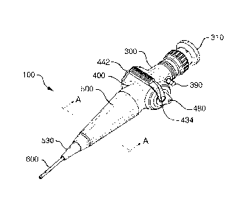
FIG. 3 is a perspective view of an embodiment of the endoscope 100 of
the present invention.
FIG. 4 is a section view through the longitudinal axis of an endoscope
handle 200 in plane A-A of FIG. 3 (the plane of the leak test vent 390) of an
embodiment of the endoscope of the present invention.
FIG. 5 is a section view through plane B-B of FIG. 2 of an articulation
assembly 430 of an embodiment of the endoscope of the present invention.
FIG. 6 is a section view through the longitudinal axis of a removable
eyepiece assembly 306 of an embodiment of the endoscope of the present
invention; the section is perpendicular to the plane of the section of FIG. 2.
FIG. 7A is a section view through a light source assembly 570 of an
embodiment of the endoscope of the present invention.
FIG. 7B is a view of the distal end 620 of the shaft 600 of an
embodiment of the endoscope of the present invention.
DETAILED DESCRIPTION OF THE PREFERRED EMBODIMENTS
The present invention provides an endoscope with a watertight and air-
tight endoscope handle that in preferred embodiments contains in its interior
a
solid-state light source and a gas permeable and liquid impermeable sealed
compartment holding a battery and an electronic circuit board, where the
sealed compartment is preferably transverse to the long axis of the endoscope
handle. In certain embodiments, the sealed compartment is at least partially
surrounded by a movable structure.
The present invention provides an endoscope having a watertight and
airtight endoscope handle that encloses an internal solid-state light source
and
power supply and a rigid or at least partially flexible shaft. In preferred
embodiments, the endoscope handle has three portions: a distal portion
attached to the shaft, a proximal portion including an eyepiece and imaging
optics and a transverse portion that separates the distal portion from the
CA 02699750 2010-03-12
WO 2009/036300 -9-
PCT/US2008/076211
proximal portion of the endoscope handle. In preferred embodiments, the
transverse portion includes at least one articulation drum(s), and a sealed
compartment that encloses a solid-state light source and a power supply for
the
solid-state light source that includes a battery, and electronic circuits for
controlling the light intensity produced by the solid-state light source. In
preferred embodiments, the solid-state light source is a light emitting diode
(LED).
In preferred embodiments the LED is mounted to a heat-conducting
internal frame of the endoscope handle. In certain embodiments, the heat-
conducting internal frame contacts a heat-conducting exterior of the endoscope
handle, thereby further dissipating heat produced by the operation of the LED.
In other embodiments, the exterior of the endoscope handle is not heat
conducting, providing a comfortable grip for the operator. In preferred
embodiments, the battery and electronic components of the LED power source
are contained within a fixed sealed compartment that is at least partially
surrounded by a movable structure, such as an articulation drum.
The sealed compartment is liquid impermeable but gas permeable,
preventing corrosive fluids from leaking into or out of the sealed
compartment,
but leaving the compartment in communication with the interior of the
endoscope handle for the increase in air pressure needed for leak testing. In
this way, all of the seals of the endoscope, including that of the battery
compartment, can be validated by the user. Since the endoscope shaft will
likely over time leak into the interior of the handle, the fluid intrusion
will not
reach the battery and electronics, thus avoiding corrosion of the electronics.
Similarly, if the battery leaks, the leaked fluid will not contaminate the
interior
of the endoscope handle, nor have a path to the patient. By placing the
electronics in the sealed battery compartment within the articulation drum,
previously unused space is used to house the battery and the electronics,
thereby minimizing the additional volume need to incorporate the solid state
light source and the light source power supply.
CA 02699750 2010-03-12
WO 2009/036300 -10-
PCT/US2008/076211
A Hall-effect sensor in the battery compartment senses the magnetic
field produced by a moveable permanent magnet located outside the sealed
scope body. The linear Hall-effect sensor has a response proportional to the
applied magnetic field, which then sends a control signal to the electronics,
setting the LED output light level by adjusting the LED's drive current. The
combination of the moveable permanent magnet and the linear Hall-effect
sensor thus serve both as an ON/OFF switch and as a continuously variable
light intensity control. Because the magnet is located outside the sealed
endoscope handle, the seal that would be needed for a moving conventional
switch is not needed. Currently, rubber boots are used to cover and seal
switches, typically ON/OFF or momentary pushbutton switches. In order to
produce variable light intensity, a potentiometer or rotary switch would have
to
be employed with a sealing mechanism (probably a rotating shaft seal
incorporating an 0-ring), which is a potential source of failure. This is
particularly problematic when the switch has electrical power applied to it.
While the articulation mechanism (rotating shaft) has a rotating seal, there
is
no electrical power associated with this moving part.
As shown in FIG. 3, an embodiment of the endoscope 100 of the
present invention has an eyepiece 310 at the end proximal to the user and at
the opposite (distal) end is a shaft 600 that includes one or more light
guides.
In preferred embodiments, the proximal portion 300 and the distal portion 500
of the endoscope handle are separated by a transverse portion 400. In certain
embodiments, the axis of the transverse portion 400 of the endoscope handle
may be at an angle of 80-100 degrees, more preferably about 85-95 degrees to
the longitudinal axis of the rest of the endoscope handle. In preferred
embodiments, the axis of the transverse portion 400 of the endoscope handle is
perpendicular to the longitudinal axis of the rest of the endoscope handle.
The
axes of the transverse portion 400 and of the rest of the endoscope handle may
or may not lie in the same plane. In certain preferred embodiments, the axis
of
the transverse portion 400 and the longitudinal axis of the rest of the
endoscope handle are approximately co-planar.
CA 02699750 2010-03-12
WO 2009/036300 -11-
PCT/US2008/076211
FIG. 3 also illustrates the position of leak test vent 390 in the distal
portion 300 of the endoscope handle. The articulation lever 434, the magnetic
illumination control lever 480, and the battery compartment cap 442 are
external structures of the transverse portion 400 of the endoscope handle. The
shaft boot 530 is disposed on the distal end of the endoscope handle.
FIG. 4 is a drawing of a section at "A-A" in FIG. 3 in a plane that
passes through the longitudinal axis of an endoscope handle 200 in the plane
of axis of the leak test vent 390 in an embodiment of the endoscope of the
present invention. The approximate extents of the proximal portion 300, the
transverse portion 400 and the distal portion 500 are indicated below the
drawing. Structures of the proximal portion 300 of the endoscope handle
include the eyepiece 310, the eyepiece lens assembly 320, the focusing ring
330, the image light guide 360, the image light guide tip adapter 370, body
380
of the endoscope handle 200, the backing plate 386, the leak test vent 390,
and
the leak test vent cap 394. An alternative embodiment of a removable
eyepiece assembly is illustrated in FIG. 6.
When not in use, described below, the leak test vent 390 is normally
covered, preferably with a removable cap. In preferred embodiments, the leak
test vent 390 is a poppet valve. When the leak test vent 390 is a poppet
valve,
the leak test vent cap 394 holds the leak test vent 390 open when it is in
place
to aeration, shipping and ethylene oxide sterilization. Removal of the leak
test
vent cap 394 allows the poppet valve to close, preventing fluid invasion. In
embodiments in which the leak test vent 390 is a poppet valve, the leak test
vent cap 394 should be removed for patient use, leak testing, cold-soak
disinfection (e.g., with a 2% glutaraldehyde solution), or disinfection using
a
endoscope disinfection processor, such as the STERIS SYSTEM 18 processor
(STERIS Corporation, Mentor, OH).
FIG. 4, a section view through the transverse portion of the endoscope
handle 400, shows the battery compartment 440, the battery compartment cap
442, the battery 450, and the battery chassis 454 that includes the Hall-
effect
sensor 482 and battery connections 460 to the light source power and control
CA 02699750 2010-03-12
WO 2009/036300 -12-
PCT/US2008/076211
circuitry. The battery compartment 440 is disposed within the articulation
drum 432, which is attached to the articulation wires 436. The articulation
wires 436 pass through the distal portion 500 of the endoscope handle into the
shaft 600. Rotation of the articulation drum 432 using the articulation lever
434 (see FIG. 3 and FIG. 5) takes up or pays out the articulation wires 436,
providing controlled movement of the distal end of the shaft. Further details
of the transverse portion 400 of the endoscope handle are shown in FIG. 5,
which is a section view in the plane "B-B."
As illustrated in FIG. 4, in preferred embodiments, the distal portion
500 of the endoscope handle includes a housing 510, a subframe support beam
542 and a shaft boot 530. The subframe 540 includes a subframe base
assembly 550 that is connected to at least one subframe wall 544 and a
subframe tip 546 by at least one subframe support beam 542. In preferred
embodiments, the subframe base assembly 550 is secured to the housing of the
transverse portion 400 of the endoscope handle by the use of a backing plate
556. Alternatively, the subframe base assembly 550 may be secured directly to
the housing of the transverse portion 400 of the endoscope handle. In certain
preferred embodiments, the subframe base assembly 550 includes a base plate
554 and a wall 552 disposed approximately perpendicular to the base plate
554. The base plate 554 contains one or more openings to accommodate the
articulation wire 436 and the image guide 360.
The light source assembly 570 is preferably disposed within the
subframe base assembly 550, and optimally coupled to the subframe base
assembly 550 to provide a heat sink for the light source. In preferred
embodiments, the light source is solid-state light source such as a light
emitting diode (LED) 572 or a laser diode. The illumination light guide 590 is
mounted at its proximal end, crosses the distal portion of the endoscope
handle, and distally ends at the end cap of the shaft (FIG. 7B). Further
details
of the lights source assembly 570 are shown in FIG. 7A.
A preferred embodiment of the endoscope of the present invention uses
a high power LED (Luxeon K2 Model LXK2-PWC4-0160, Lumileds Lighting,
CA 02699750 2010-03-12
WO 2009/036300 -13-
PCT/US2008/076211
LLC, 370W. Trimble Road, San Jose, CA. 95131) with atypical forward
voltage of 3.72V and operating current at 1000 mA. This device can be safely
operated up to a current of 1.5A with a corresponding typical forward voltage
of 3.85V. This white LED has a typical color temperature of 6500K. The
LED chip has an emitting surface of approximately 1 mm x 1 mm, and is
coated with a wavelength conversion phosphor (and/or fluorophor) that emits a
broadband continuum of visible white light between about 470-700 nm.
Suitable ways of coupling the light-emitting surface of such a high power LED
to a light guide are disclosed in U.S. Patent No. 7,229,201. Briefly, the dome
lens of the high power LED is removed, along with the index-matching gel.
The illumination light guide fiber bundle is polished flat and placed directly
onto the LED (or the LED'S phosphor coating) in order to maximize the
amount of light coupled into the fiber optic light guide.
Generally, the light emitting area of the LED is coupled to a small 1
mm square or round bundle of light guide fibers. This is a typical light guide
bundle size used in endoscopes. The light guide bundle is typically composed
of hundreds of individual glass (or plastic) fibers grouped together to form a
single bundle at the light source, and either a single bundle or multiple
bundles
at the distal end of the endoscope. Such bundles can take on a variety of
shapes at the distal end depending upon the design of the particular
endoscope:
one or more round bundles, a circular halo, a crescent, or the like. Small
diameter fibers, typically 30-50 micrometers in diameter, are employed
because these small fibers are flexible (necessary for flexible endoscopes
that
bend during use), or because they are required to fit into the narrow spaces
around the optics either in the shaft or distal head of the endoscope.
Other types of light guides can be coupled to the LED in the manners
described herein, including: liquid light guides, plastic or glass fibers,
plastic
or glass rods, and tapers made from fibers (glass and plastic) or solid tapers
(glass and plastic). Single glass or plastic fibers may comprise the light
guide.
Such fibers around 1 mm in diameter are typically flexible. In order to
accommodate a small light guide bundle of less than the LED emitting area, a
CA 02699750 2010-03-12
WO 2009/036300 -14-
PCT/US2008/076211
fiber optic or solid plastic or glass taper may be placed between the LED
emitting surface and the bundle, acting as an adapter that captures
substantially
all of the light emitted from the LED and efficiently couples it into the
fiber
bundle that delivers the light to the distal end of the instrument.
FIG. 5 is a drawing of a section through plane B-B of FIG. 4 of an
articulation assembly 430 of an embodiment of the endoscope of the present
invention. The plane B-B of section passes through the articulation lever 434
(see FIG. 1) and is roughly perpendicular to the longitudinal axis of the
endoscope handle. The articulation assembly 430 is a major part of the
transverse portion of the endoscope handle 400. The articulation assembly
430 includes the articulation drum 432, the articulation stop 452, and the
articulation wire 436 in addition to the articulation lever 434. The battery
compartment 440 is disposed within the articulation drum 432, and maintains
a fixed position in the endoscope handle when the articulation drum 432
rotates around it as the shaft tip is moved. The battery compartment 440 has a
gas permeable, liquid impermeable vent 444 that includes a gas permeable
membrane (not shown), such as an expanded polytetrafluoroethylene gas
permeable membrane. Suitable expanded polytetrafluoroethylene gas
permeable membranes include Gore protective vents, preferably QPE quick
pressure equalization vents (W.L. Gore & Associates, Newark, DE;
http://www.gore.com/en_xx/products/venting/technical/qpe.html).
The battery compartment 440 is sealed by the battery compartment cap
442, and contains a battery 450 and the battery chassis 454. The battery
chassis 454 supports the battery 450 within the battery compartment 440 and is
attached to the electrical circuit board 462, which includes the light source
power and control circuitry and components. The openings through the battery
compartment 440 for the wires 452 connecting the electrical circuit board 462
and the LED 752 (FIG. 4 and FIG. 7A) are sealed with epoxy, silicone
adhesive or other suitable adhesive after assembly. Also shown in FIG. 5 are
0-ring seals 420, a gasket seal 422 and bushings 424.
CA 02699750 2010-03-12
WO 2009/036300 -15-
PCT/US2008/076211
The battery is a rechargeable battery, preferably a lithium ion
rechargeable battery. When the solid-state light source is a high power LED, a
preferred battery is the Konica Minolta NP700 lithium ion battery, 3.6V
nominal voltage, 1000 mAHr typical capacity, or equivalent. This battery is
used in several digital cameras and is commercially available.
In some embodiments, the battery can be charged in place in the
endoscope handle, using inductive coupling to a battery charger and venting
the handle through the leak test vent 390. Alternatively, the battery is
charged
in an external battery charger plugged into a wall outlet.
In a preferred embodiment, the Hall-effect sensor 482 (FIG. 4) is
attached to the end of the battery chassis 454 that is farthest from battery
compartment cap 442. The linear Hall-effect sensor 482 has an electrical
response that is proportional to the applied magnetic field generated by the
magnetic illumination control lever 480. A change in the position of the
magnetic illumination control lever 480 produces a corresponding change in
the electrical response of the linear Hall-effect sensor 482, resulting in a
change in the light intensity produced by the LED. The combination of the
external magnetic illumination control lever 480 and the internal linear Hall-
effect sensor 482 thus provides ON/OFF switching and continuously variable
light intensity control of the solid-state light source. Suitable linear Hall-
effect
sensors are available from Allegro Micro Systems, Worcester, MA, such as
Models A1391, A1392, A1393, and A1395. When the solid-state light source
is a high power LED, a preferred linear Hall-effect sensor is Allegro
MicroSystems Model A1391. In other embodiments, variable light intensity
can be produced using a pushbutton switch having a suitable cover and seal,
e.g., a rubber boot, wherein the amount of power applied to a LED is
proportional to the length of time that the switch is held closed.
FIG. 6 is a drawing of a section through the longitudinal axis of a
removable eyepiece assembly 306 of an embodiment of the endoscope of the
present invention; the section is perpendicular to the plane of the section of
FIG. 4. Shown are the eyepiece 310, the eyepiece lens assembly 320, the
CA 02699750 2010-03-12
WO 2009/036300 -16-
PCT/US2008/076211
focusing ring 330, the image light guide tip adapter 370, body 380 of the
endoscope handle 200, the backing plate 386 and the leak test vent mounting
hole 392. The J fitting connector 340 allows the replacement of the eyepiece
assembly 306 with a video camera or a single frame digital camera, avoiding
the need for an optical coupler to connect the eyepiece to a digital or video
camera. Alternatively, an adapter can be fitted to the eyepiece and used to
couple the endoscope eyepiece to a multi-use electronic device, such as a
personal digital assistant (PDA) as disclosed in published international
patent
application WO 2006/055949. In another embodiment, a suitable digital
camera and coupler kit can be used that is commercially available from Optim,
Incorporated (Sturbridge, MA).
FIG. 7A is a drawing of a section through a light source assembly 570
of an embodiment of the endoscope of the present invention. In preferred
embodiments, a solid-state light source 572, such as a high power LED
(Luxeon K2 Model LXK2-PWC4-0160, Lumileds Lighting, LLC, 370 W.
Trimble Road, San Jose, CA 95131) is mounted on a LED backing plate 574
and placed in a LED housing 576 that is mounted in the subframe base
assembly (550 in FIG. 4) using thermally conductive electrically isolating
epoxy 575. The light source assembly 570 is preferably configured with at
least one mounting hole 580 adapted to engage a fastener to mount the light
source assembly 570 to the subframe base assembly. The light source
assembly 570 preferably also includes at least one receptacle fastener hole
582
adapted to stabilize the proximal end of the illumination light guide in the
illumination light guide receptacle 577. Suitable ways of coupling the light-
emitting surface of such a high power LED to a light guide are disclosed in
U.S. Patent No. 7,229,201. After mechanical attachment of the light guide to
the light source assembly, the light guide is sealed to the housing with
epoxy,
silicone adhesive or other suitable adhesive to prevent fluid intrusion.
FIG. 7B is a drawing of a view of the distal end 620 of the shaft 600 of
an embodiment of the endoscope of the present invention, showing the distal
CA 02699750 2010-03-12
WO 2009/036300 -17-
PCT/US2008/076211
end of the image light guide 360 and the distal end(s) of at least one
illumination light guide(s) 590.
Leak Testing
The endoscope is tested to ensure that there are no leaks before high-
level disinfection using a disinfectant such as a 2% glutaraldehyde solution
or
peracetic acid, or sterilization using ethylene oxide (Et0). The leak test
vent
cap 394 is removed from the leak test vent 390 and a leak tester is attached
to
the leak test vent. Suitable leak testers are commercially available, for
example from Optim Incorporated (Part No. 004918) or from Surgical Repairs
International (Tonawanda, NY,
http://vvww.sfirepairs.com/products_leaktesters.asp). Using the leak tester,
the
interior of the endoscope is pressurized to about 140-180 mmHg as measured
on the leak tester. The pressure measurement is observed for about 10 seconds
to determine if the connection between the leak tester and the leak test vent
is
loose. If the pressure drops, the connection between the endoscope leak test
vent and the leak tester may be loose. The attachment and pressurization
procedure is repeated to verify the connection. If the pressure drops again,
the
endoscope may have a damaged seal and should not be immersed in any
liquid. The endoscope should be repaired prior to use or cleaning.
If the pressure does not drop, the entire endoscope, while pressurized,
is immersed in water. The endoscope is observed for about 30 seconds. The
distal end of the shaft is articulated up and down during this period; since
holes in the soft covering of the distal end of the shaft may not be evident
in a
relaxed position. In addition, the battery cap seal should also be inspected
during this period for leaks, since the interior of the battery chamber
becomes
pressurized through the gas permeable, liquid impermeable vent. A
continuous stream of bubbles indicates a leak. The endoscope should be kept
under pressure while removing it from the water to avoid corrosion caused by
infiltration of the water into the leak site.
CA 02699750 2015-09-04
- 1 8-
Typically, the leak test should be performed right after using the scope
to ensure there are no leaks prior to immersing the scope in disinfection
liquid.
It would be wise to check the scope for leaks after replacing the battery
before
use. If the seal is compromised leak testing will prevent contamination of the
battery and its compartment within the scope by the entry of any patient fluid
or debris.
The claims should not be read as limited to the described order or
elements unless stated to that effect. The scope of the claims should not be
limited by the preferred embodiments or the examples but should be given
the broadest interpretation consistent with the description as a whole.