Note: Descriptions are shown in the official language in which they were submitted.
CA 02700309 2010-03-19
WO 2009/039366 PCT/US2008/077008
METHOD OF ENHANCING IONTOPHORETIC DELIVERY OF A PEPTIDE
RELATED APPLICATION
This application claims the benefit of Provisional Application No.
60/973,956 filed on September 20, 2007. The entire teachings of the above
application are incorporated herein by reference.
BACKGROUND OF THE INVENTION
An iontophoretic delivery system is an example of a drug delivery system
that releases drug at a controlled rate to the target tissue upon application.
The
advantages of systems wherein drug is delivered locally via iontophoresis are
the
ease of use, being relatively safe, and affording the interruption of the
medication by
simply stopping the current and/or peeling off or removing it from the skin or
other
body surface whenever an overdosing is suspected. The total skin surface area
of an
adult is about 2 m2. In recent years iontophoretic delivery of drugs has
attracted
wide attention as a better way of administering drugs for local as well as
systemic
effects. The design of iontophoretic delivery systems can usually be such that
the
side effects generally seen with the systemic administration of conventional
dosage
forms are minimized.
lontophoresis has been employed for many years as a means for applying
medication locally through a patient's skin and for delivering medicaments to
the
eyes and ears. The application of an electric field to the skin is known to
greatly
enhance the ability of the drugs to penetrate the target tissue. The use of
iontophoretic transdermal delivery techniques has obviated the need for
hypodermic
injection for some medicaments, thereby eliminating the concomitant problems
of
trauma, pain and risk of infection to the patient.
lontophoresis involves the application of an electromotive force to drive or
repel ions into a target tissue, such as through the stratum comeum and into
the
epidermaUdermal layers of the skin. Particularly suitable target tissues
include those
adjacent to the delivery site for localized treatment. Uncharged molecules can
also
be delivered using iontophoresis via a process called electroosmosis.
Regardless of the charge of the medicament to be administered, an
iontophoretic delivery device employs two electrodes (an anode and a cathode)
in
Page 1 of 16
CA 02700309 2010-03-19
WO 2009/039366 PCT/US2008/077008
conjunction with the patient's body to form a closed circuit between one of
the
electrodes (referred to herein alternatively as a"working" or "application" or
"applicator" electrode) which is positioned at the site of drug delivery and a
passive
or "grounding" electrode affixed to a second site on the body surface to
enhance the
rate of penetration of the medicament into the tissue adjacent to the
applicator
electrode.
U.S. Patent No. 6,477,410 issued to Henley et al. describes the use of
iontophoresis for drug delivery. It would be advantageous to improve the
permeation of high molecular weight drugs such as proteins by iontophoretic
delivery.
SUMMARY OF THE INVENTION
It has now surprisingly been found that microporation combined with
iontophoretic administration of a protein resulted in improved transdermal
delivery
of the protein. As shown in Example 1 below, in the hairless rat model, the
combination of microneedle treatment with iontophoretic administration of
salmon
calcitonin increased the amount of protein that permeated the skin by about
four
times compared to the use of iontophoresis alone.
The present invention provides methods for the administration of a peptide to
a body surface of the patient comprising microporating the body surface and
iontophoretically administering the peptide to said body surface.
The invention is also directed to methods of administering a peptide to the
body surface of a patient in need thereof comprising microporating the body
surface
with one or more microneedles and iontophoretically administering the peptide
to
said body surface.
In another embodiment, the present invention is directed to a method of
transdermally administering a peptide to the skin of the patient comprising
microporating the body surface with one or more microneedles and
iontophoretically
administering the peptide into the skin of the patient. In one embodiment, the
skin is
pretreated with microporation using a microneedle followed by administration
of the
drug using iontophoresis.
The present invention also encompasses a method of transdermally
administering a peptide to the skin of a patient comprising microporating the
skin of
Page 2 of 16
CA 02700309 2010-03-19
WO 2009/039366 PCT/US2008/077008
said patient with one or more microneedles while concurrently
iontophoretically
administering said peptide into the skin.
BRIEF DESCRIPTION OF THE DRAWINGS
FIG. lA is a drawing of a titanium microneedle array bent out of plane.
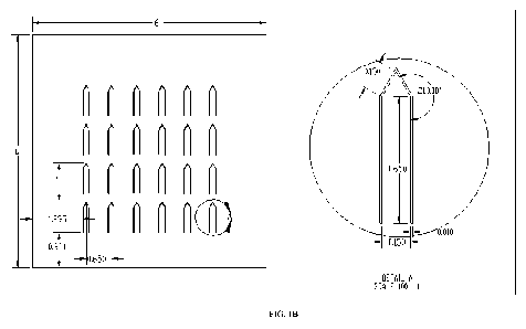
FIG. 1B shows the dimensions ( m) of a titanium microneedle array and of
each microneedle.
FIG. 1C is a plot of the plasma concentration (ng/ml) over time (min) of
salmon calcitonin delivered using microneedles alone, iontophoresis alone or
the
combination of microneedles and iontophoresis in the hairless rat model.
DETAILED DESCRIPTION OF THE INVENTION
As used herein, the word "a" or "an" is meant to encompass one or more
unless otherwise specified. For example, "a microneedle" is intended to
encompass
one or more microneedles.
The invention is directed to methods of administering a peptide to a body
surface comprising microporating the body surface and iontophoretically
administering said peptide to the body surface. In one embodiment, the body
surface is microporated using one or microneedles. In another embodiment, the
body surface is the skin. In one embodiment, the body surface is microporated
prior
to iontophoretic administration of the peptide. In yet other embodiment, the
body
surface is microporated using one or more hollow or porous microneedles while
concurrently iontophoretically administering the peptide.
As used herein, the term "peptide" is meant to encompass proteins, peptide
drugs as well as amino acid drugs (such as the beta lactam antibiotics
including the
penicillins and the cephalosporins). Peptides have a molecular weight of at
least
about 500 Daltons (Da). The term "peptide" is also meant to include proteins
or
peptide drugs which have been chemically modified. Such chemical modifications
include, for example, replacement of an amino acid with a different amino acid
or
other group and/or addition of a functional group and/or a chemical modifier.
In one
embodiment, the peptide administered according to a method of the invention
has a
molecular weight of at least about 500 Da. In another embodiment, the peptide
administered according to the inventive method has a molecular weight of at
least
about 1000 Da. In a further embodiment, the peptide administered according to
a
Page3of16
CA 02700309 2010-03-19
WO 2009/039366 PCT/US2008/077008
method of the invention has a molecular weight of at least about 3000 Da. In
another embodiment, the molecular weight of the peptide is at least about
10,000
Da. In yet another embodiment, the molecular weight of the peptide is at least
about
100,000 Da.
In one embodiment, the peptide administered according to a method of the
invention is a therapeutic protein. Therapeutic proteins, include but are not
limited
to, cytokines, hormones and antibodies. In another embodiment, the peptide
administered according to a method of the invention is selected from the group
consisting of a fusion protein and an antibody.
Proteins and peptide drugs that may be used in the method of the present
invention include, but are not limited to, Luteinizing hormone-releasing
hormone
(LHRH), Somatostatin, Bradykinin, Goserelin, Somatotropin, Buserelin, Platelet-
derived growth factor, Triptorelin, Gonadorelin, Asparaginase, Nafarelin,
Bleomycin sulfate, Leuprolide Chymopapain, Growth hormone-releasing factor,
Cholecystokinin, Chorionic gonadotropin, Insulin, Corticotropin (ACTH),
Calcitonin (e.g., eel, salmon, Erythropoietin human), Glucagon, Calcitonin
gene
related peptide, Hyaluronidase Interferons (e.g., alpha, beta and gamma),
Endorphin
(alpha, beta, and and gamma), Interleukins (e.g., IL-l, IL-4, IL-6, IL-2 and
IL-10),
Thyrotropin-releasing hormone, CSIF (cytokine synthesis inhibitory factor), NT-
36
(N-[[(s)-4-oxo-2-azetidinyl]carbonyl]-L-histidyl-L-prolinamide, Liprecin,
Menotropins, Pituitary hormones (e.g., HGH, HMG, HCG, desmopressin acetate,
etc.), Urofollitropin (Follicle Stimulating Hormone), desmo- pressin acetate,
etc.,
Leutinizing hormone (LH), aANF growth factor releasing factor, leutinizing
hormone (LH), LH releasing hormone, Melanocyte-stimulating hormone (alpha,
beta and gamma), Vasopressin, Streptokinase, ACTH analogs, Tissue plasminogen
activator, Atrial natriuretic peptide, ANP clearance inhibitors, Urokinase,
Angiotensin II antagonists, Bradykinin potentiator B, Bradykinin antagonists,
Bradykinin potentiator C, CD4, Ceredase, Brain-derived neutrotrophic factor,
Colony stimulating factors, Cystic fibrosis transmembrane conduce regulator
(CFTR), Enkephalins, Fab fragments, IgE peptide suppressors, Chorionic
gonadotoropin, Insulin-like growth factors, Ciliary neutrotrophic factor,
Neurorophic factors, Parathyroid hormone, Corticotropin releasing factor,
Prostaglandin antagonists, Granulocyte colony stimulating factor, Pentigetide,
Protein C, Protein S, Thymosin a-l, Thrombolytics, Tumor necrosis factor alpha
Page 4 of 16
CA 02700309 2010-03-19
WO 2009/039366 PCT/US2008/077008
(TNF-a), Multilineage colony stimulating factor, Macrophage-specific colony
stimulating factor, Vaccines, Vasopressin antagonist, Colony stimulating
factor 4, a-
1 Anti-trypsin, Adenosine deaminase, Epidermal growth factor, Amylin, Atrial
natriuretic peptide, Enkephalin leu, B-Glucocerebrosidase, Enkephalin met,
Bone
morphogenesis protein 2, Factor IX, Bombesin, Factor VIII,
BactericidaUPermeability increasing protein, Follicular gonadotropin releasing
peptide, Hirudin, G- 1128, IEV inhibitor peptide, Gastrin-releasing peptide,
Inhibin-
like peptide, Glucagon, Insulin, Insulinotropin, Growth hormone releasing
factor,
Lipotropin, Macrophage-derived neutrophil chemotaxis factor, Heparin binding
neurotrophic factor, Melatonin, Tryptophan hydroxylase, Fibroblast growth
factor,
Midkine, Neurophysin, Somatostatin, Neurotrophin-3, Nerve growth factor,
Oxytocin, Phospholipase A2, Soluble IL-1 receptor, Thymidine kinase, Thymosin
alpha one, soluble TNF receptor, Tissue plasminogen activator, Transforming
growth factor beta, TSH-releasing hormone, Thyroid stimulating hormone (TSH),
Vasopresssin and Vasotocin. Proteins that may be used according to the present
invention include antibodies. In the present invention, antibodies include,
but are
not limited to, polyclonal, monoclonal, chimeric, single-chain, humanized and
human antibodies, as well as various fragments thereof such as Fab fragments
and
fragments produced from specialized expression systems.
In one embodiment, a current density sufficient for permeation into a body
surface is applied. In another embodiment, a current density sufficient for
permeation through the stratum comeum is applied. In one embodiment, a current
density of about 0.001 mA/em2 to about 2.0 mA/em2 is applied. In yet another
embodiment, a current density of about 0.01 mA/cm2 to about 1 mA/cm2 is
applied.
In a further embodiment, a current density of about 0.05 mA/em2 to about 0.5
mA/em2 is applied. In an additional embodiment, a current density from about
0.1
mA/cm2 to about 0.5 mA/cm2 is applied.
The iontophoresis can be applied for a sufficient time to achieve an effective
amount of permeation. For example, a sufficient time for application is a time
from
about 1 minute to about 4 hours. In one embodiment, iontophoresis is applied
for a
time from about 5 minutes to about 2 hours. In yet another embodiment,
iontophoresis is applied for a time from about 10 minutes to about 90 minutes.
In a
further embodiment, iontophoresis is applied from about 10 minutes to about 1
hour.
Page5ofl6
CA 02700309 2010-03-19
WO 2009/039366 PCT/US2008/077008
In one embodiment, the peptide is formulated with a pharmaceutically
acceptable carrier or excipient. As used herein, the term "pharmaceutically
acceptable carrier or excipient" means any non-toxic diluent or other
formulation
auxiliary that is suitable for use in iontophoresis. Examples of
pharmaceutically
acceptable carriers or excipients include, but are not limited to, solvents,
cosolvents,
solubilizing agents (such as sorbitol and glycerin), buffers, pharmaceutically
acceptable bases, alcohols such as benzyl alcohol and viscosity modulating
agents
such as cellulose and its derivatives. The formulation may further comprise a
chemical permeation enhancer. A "permeation enhancer" is a material which
achieves permeation enhancement or an increase in the permeability of the body
surface to a pharmacologically active agent. Examples of such permeation
enhancers
include, but are not limited to, N-acetylcysteine, urea, salicylic acid,
linoleic acid,
benzoic acid, cyclodextrin, dimethyl sulfoxide, dimyristoyl
phosphatidylserine, and
the like. In another embodiment, the formulation may contain stabilizers such
as
antioxidants (EDTA, sodium sulfites, ascorbic acid, vitamin E, BHT, etc.)
and/or an
alcohol. In another embodiment, the formulation comprising the protein may
contain a preservative such as benzalkonium chloride, parabens, etc. In a
further
embodiment, the formulation may contain an agent that affects protein binding
including, but not limited to, linolenic acid, dimyristoyl phosphatidyl
glycerol
(DPMG), a polysorbate and dimyristoyl phosphatidyl choline (DPMC). The peptide
can be administered in a therapeutically effective amount. A "therapeutically
effective amount" is an amount of peptide that is sufficient to prevent
development
of or alleviate to some extent one or more of a patient's symptoms of a
disease being
treated or to elicit a desired biological or medical response in a subject.
In one embodiment, the peptide is iontopheretically administered using an
iontophoretic delivery device. Examples of iontophoretic delivery devices
useful
with the compositions and methods of the invention include, but are not
limited to,
those described in U.S Pat. Nos. 6,148,231, 6,385,487, 6,477,410, 6,553,253,
6,792,306, 6,895,271, 7,016,724 and 7,127,285, all incorporated herein by
reference.
An example of an applicator which can be used with a formulation of the
invention
comprises an active electrode adhered to an open cell polymer foam or
hydrogel.
Another applicator which has been developed for use with a device for
iontophoretic
delivery of an agent to a treatment site comprises an applicator head having
opposite
faces and including an active electrode and a porous pad (such as a woven or
non-
Page 6 of 16
CA 02700309 2010-03-19
WO 2009/039366 PCT/US2008/077008
woven polymer, for example, a polypropylene pad); a margin of the applicator
head
about the active electrode having a plurality of spaced projections there
along; the
porous pad and the applicator head being ultrasonically welded to one another
about
the margin of the head with the electrode underlying the porous pad; and a
medicament or a medicament and an electrically conductive carrier therefor
carried
by the porous pad in electrical contact with the electrode. In one embodiment,
the
formulation is iontophoretically administered using carbon electrodes, silver-
silver
chloride electrodes or silver coated carbon electrodes.
In one embodiment, the body surface is selected from the group consisting of
the skin, the nail plate, the eyes, the ears and a mucous membrane.
Microporation refers to the formation of micropores on a body surface. A
micropore in the skin means a small breach or pore formed in the stratum
comeum
within a selected area of the skin to decrease the barrier properties of the
stratum
corneum. Microporation may be achieved using any suitable method including,
but
not limited to, the use of a microneedle, thermal poration, radiofrequency
ablation,
laser ablation, and sonophoresis (with or without the use of dyes or other
energy
absorbing materials to assist in the ablation and removal of the stratum
corneum).
In one embodiment, microporation of the body surface is achieved using one
or more microneedles. The length and density of the microneedle as well as the
thickness or diameter of the needles can vary depending on the location of the
targeted treatment site underlying the skin surface. In one embodiment, the
microneedle has a height of about 2 millimeters (mm) or less and/or are about
50 to
about 300 m in diameter when such structures are cylindrical in nature. In an
additional embodiment, the microneedle has a diameter of about 100 to about
200
m. Non-cylindrical structures are also encompassed by the term microneedle;
such
microneedles are of comparable cross-sectional length or cross-sectional area
and
include pyramidal, rectangular, octagonal, wedged, and other geometrical
shapes.
Microneedles have been described, for example, in U.S. Pat. Nos. 6,256,533;
6,312,612; 6,334,856; 6,379,324; 6,451,240; 6,471,903; 6,503,231; 6,511,463;
6,533,949; 6,565,532; 6,603,987; 6,611,707; 6,663,820; 6,767,341; 6,790,372;
6,815,360; 6,881,203; 6,908,453; 6,939,311; all of which are incorporated by
reference herein. In another embodiment, the microneedle may protrude from a
substrate by the height of 2 mm or less. In another embodiment, the
microneedle
has a height of about 1 mm or less. In yet another embodiment, the microneedle
has
Page 7 of 16
CA 02700309 2010-03-19
WO 2009/039366 PCT/US2008/077008
a height from about 100 to about 1 mm. In yet an additional embodiment, the
microneedle has a height from 150 to 900 m. In another embodiment, the
microneedle has a height of about 300 to 800 m. In one embodiment, the
microneedle is of sufficient height to penetrate beyond the stratum corneum to
an
underlying layer of skin. In another embodiment, the microneedle is of
sufficient
height to pass into the dermis but not a height great enough to stimulate
nerves in
deeper tissue and/or cause pain when applied or inserted into the body
surface. In
another embodiment, the ratio length to width (at the base of the microneedle)
is
from about 0.5 to about 16Ø
The number of microneedles that can be used in the inventive method is one
or more. In one embodiment, the method employs more than one microneedle. In
another embodiment, the method employs more than five microneedles. In a
further
embodiment, the method employs more than ten microneedles. In yet another
embodiment, the method employs more than about one hundred microneedles. In
other embodiments, a microneedle array is used. A microneedle array has more
than
two microneedles and can include tens, hundreds, or thousands of needles. The
density of microneedles in the microneedle array may be from about 1 to about
1000
needles per cm2. The microneedles can be attached and/or arranged in a pattern
or
randomly over the surface of a substrate. As used herein the "substrate" of a
microneedle device includes the base to which the microneedles are attached or
integrally formed. Such substrates can be constructed from a variety of
materials,
including, for example, metals, ceramics, semiconductors, organics, polymers,
and
composites. In one embodiment, the substrate and/or microneedles, as well as
other
components, are formed from flexible materials to allow the device to fit the
contours of the body surface. Microneedles include solid microneedles, hollow
microneedles and porous microneedles.
A microneedle can be made of any suitable material allowing it to penetrate
the body surface. Suitability of the material can be determined by considering
the
compatibility of the material with the body surface or any agent that is in
contact
with the microneedle, such as the drug or protein to be administered or the
formulation comprising the drug as well as the mechanical properties of the
material
as they pertain creating mechanically robust structures. The microneedles can
be
formed of a non-conductive material (e.g., a plastic material or a metal
material
coated with a non-conductive material). The microneedles can also be formed of
Page 8 of 16
CA 02700309 2010-03-19
WO 2009/039366 PCT/US2008/077008
conductive materials and coated with a non-conductive layer. Suitable
materials
include, for example, glassy materials, metals, ceramics, semiconductors,
organics
(such as sugars), polymers including biodegradable polymers and plastics,
composites, and combinations of such materials. Sugars include, for example,
maltose (Miyano et al. (2005), Biomedical Microdevices, 7(3): 185-8). Metals
include pharmaceutical grade stainless steel, gold, titanium, nickel, iron,
gold, tin,
chromium, copper, alloys of these or other metals, silicon, silicon dioxide,
and
polymers. Biodegradable polymers include polymers of hydroxy acids such as
lactic
acid and glycolic acid polylactide, polyglycolide, polylactide-co-glycolide,
and
copolymers with PEG, polyanhydrides, poly(ortho)esters, polyurethanes,
poly(butyric acid), poly(valeric acid), poly(lactide-co-caprolactone) and the
like.
Non-biodegradable polymers include polycarbonate, polymethacrylic acid,
ethylenevinyl acetate, polytetrafluorethylene (TEFLON), polyesters and the
like.
Suitable polymeric materials include acrylonitrile-butadiene-styrenes,
polyphenyl
sulfides, polycarbonates, polypropylenes, acetals, acrylics, polyetherimides,
polybutylene terephthalates, polyethylene terephthalates and the like.
One aspect of the invention is directed to a method of transdermally
administering a peptide to the skin of a patient comprising microporating the
skin
with one or more microneedles and iontophoretically administering the peptide.
Microneedles that may be used in a method of the invention include solid
microneedles as well as microneedles possessing one or more orifices through
which
drug can be delivered into the skin. Microneedles with one or more orifices
include
hollow and porous microneedles. A hollow microneedle can have one or more
substantially annular bores or channels through the interior of the
microneedle
structure, having a diameter sufficiently large to permit passage of fluid
and/or solid
materials through the microneedle. The annular bores may extend throughout all
or a
portion of the needle in the direction of the tip to the base, extending
parallel to the
direction of the needle or branching or exiting at a side of the needle, as
appropriate.
The diameter of the bore of the hollow microneedle can be about 5 m to about
100
m. Porous microneedles have pores or voids throughout at least a portion of
the
microneedle which are sufficiently large and sufficiently interconnected to
permit
passage of fluid and/or solid materials through the microneedle. The diameter
of the
pore of the porous microneedle can be about 5 m to about 20 m.
Page 9 of 16
CA 02700309 2010-03-19
WO 2009/039366 PCT/US2008/077008
Another embodiment of the invention is directed to a method of
transdermally administering a peptide to the skin of a patient comprising
microporating the skin with a microneedle while concurrently administering the
peptide into the skin using iontophoresis. According to this aspect of the
invention,
microporation and peptide administration occur concurrently. Hollow or porous
microneedles can be used to create micropores in the skin while at the same
time
administering a peptide into the skin (and through the microneedle).
Solid microneedles may also be used to concurrently microporate the skin
and iontophoretically administer the peptide if the solid microneedles are
fabricated
of a material that dissolves upon contact with fluid within and contains the
peptide.
An example of a material that dissolves upon contacting the skin and can
contain a
peptide is a bioresorbable polymer such as polylactic acid. Solid microneedles
can
also be used according to this aspect of the invention when they have one or
more
indentations along their surface which create a channel or trough on the
needle
surface along which fluid could flow. For example, a solid microneedle can
have a
"C" shaped indentation that runs along the length of the needle through which
fluid
flows. The diameter of the indentation of the solid microneedle can be about 5
m
to about 100 m.
In another embodiment, concurrent drug delivery and microporation are
achieved with a microneedle in contact with the skin, a drug reservoir in
contact
with the microneedles and an electrode in contact with the drug reservoir,
wherein
the drug reservoir comprises a peptide. In a further embodiment, concurrent
drug
delivery and microporation are achieved.
In one embodiment, an iontophoretic patch is utilized. The patch may
include a rigid boundary surrounding an array of microneedles enabling, upon
application, the skin surrounded by the boundary to present itself. In another
embodiment, a microneedle is attached to a slightly concave-shaped elastomeric
backing attached to the iontophoretic patch and acts as a suction cup. Upon
actuation by the user, the target skin area is pulled into the concavity and
against the
microneedles attached to the more rigid backing material.
In a further embodiment, the substrate upon which the needles are attached
may be combined with a delivery device. For example, the finger mounted
devices
disclosed in U.S. Pat. Nos. 6,792,306 and 6,735,470 may be provided with
substrates containing needles of selected sizes and configurations to
penetrate
Page 10 of 16
CA 02700309 2010-03-19
WO 2009/039366 PCT/US2008/077008
through the high electrically resistant layers of the skin to supply
medicament to the
targeted treatment site. Alternatively, the device disclosed in U.S. Pat. No.
RE37796, may also use substrates comprising microneedles described herein. In
all
instances, by forming a multiplicity of low electrically resistant micropores
through
the higher electrically resistant layer or layers of the skin, the peptide can
be driven
from the supply matrix or drug reservoir through the microneedles directly to
the
targeted treatment site bypassing the high electrically resistant layers of
skin.
Additional devices that can be used according to a method of the invention
include those disclosed in U.S. Pat. Publication No. 2007185432, the contents
of
which are incorporated by reference herein.
The following Examples further illustrate the present invention but should not
be
construed as in any way limiting its scope.
EXEMPLIFICATION
Example 1: In Vivo lontophoretic Delivery of Salmon Calcitonin across
Microporated Skin
Purpose: To determine the effect of iontophoresis and its combination with
microneedles on the in vivo delivery of salmon calcitonin (SCT) as a model
peptide.
Methods: Microneedles, iontophoresis and the combination were investigated for
their effect on the transdermal delivery of SCT in vivo using the hairless
rat. SCT
(350 l of a 1 mg/ml solution in 50mM citrate buffer, pH 4.0) was placed in a
cartridge designed for iontophoresis. Maltose microneedles (500 micron, Texmac
Inc.), stacked in three layers, were used to porate the skin prior to the
application of
the drug with or without iontophoresis. Since SCT (pI 10.4) was positively
charged
at pH 4, constant current iontophoresis (0.2mA/cm~, 1 hr) was conducted with
the
anode connected to the cartridge, and the cathode connected to a TransQ
(IOMED,
Inc.) inactive electrode. Transport of drug across the skin was assessed by
collecting
blood samples at regular intervals via the tail vein which were analyzed for
serum
SCT using ELISA.
Results: The maximum concentrations of SCT in the serum were 41.45 pg/ml,
605.21 pg/ml, and 2374.06 pg/ml under microneedles alone, 1 hr iontophoresis
alone, and the combination, respectively. When compared to the delivery with
microneedles alone, the increase in concentration with iontophoresis alone was
15-
fold (p < 0.05) and with the combination of microneedles the increase was 57-
fold
Page 11 of 16
CA 02700309 2010-03-19
WO 2009/039366 PCT/US2008/077008
(p < 0.05). The total amount of SCT delivered by iontophoresis and its
combination
with microneedles in the hairless rat was 648.67 ng/kg and 3075.96 ng/kg,
respectively, as calculated by WinNonlin.
Conclusion: lontophoresis or a disruption of the skin barrier by microneedles
enabled the transdermal delivery of SCT. A combination of iontophoresis and
microneedles resulted in the highest delivery flux.
Example 2: In Vivo Delivery of Salmon Calcitonin using Iontophoresis in
Combination with Microporation using Titanium Needle Arrays
Titanium needles with a width, thickness and height of 150 um, 75 um and
750 um, respectively, in arrays of 24 needles (6x4) with 0.65" center to
center
spacing were used to porate the skin prior to application of SCT. SCT was
measured after application of microporation alone, iontophoresis alone and
microporation in combination with iontophoresis. SCT was delivered and
measured
as described above in Example 1.
FIG. lA is a drawing of the array bent out of the plane and FIG. 1 B shows
the dimensions of the needle and the array. AS shown in FIG. 1 C,the plasma
concentration of SCT 0.5 minutes after administration using microporation in
combination with iontophresis was about 10-fold greater than the concentration
of
SCT after administration using either microporation or iontophoresis, alone.
While this invention has been particularly shown and described with
references to preferred embodiments thereof, it will be understood by those
skilled
in the art that various changes in form and details may be made therein
without
departing from the scope of the invention encompassed by the appended claims.
Page 12 of 16