Note: Descriptions are shown in the official language in which they were submitted.
CA 02700389 2010-03-23
WO 2009/129437 PCT/US2009/040910
1
METHODS AND COMPOSITIONS FOR TREATING POST-OPERATIVE PAIN
COMPRISING CLONIDINE
[0001] This application claims the benefit of the filing date of U.S. Patent
Application No.
12/421,144 filed Apri19, 2009 and entitled "Methods and Compositions for
Treating Post-
Operative Pain Comprising Clonidine" and U.S. Provisional Application No.
61/046,277
filed April 18, 2008 and entitled "Methods and Compositions for Treating Post-
Operative
Pain Comprising Clonidine," both of which are hereby incorporated by reference
thereto.
BACKGROUND OF THE INVENTION
[0002] Pain relief is of prime importance to anyone treating patients
undergoing surgery.
Proper pain relief imparts significant physiological and psychological
benefits to the
patient. Not only does effective pain relief mean a smoother more pleasant
post-operative
course (e.g., mood, sleep, quality of life, etc.) with earlier discharge from
medical/surgical/outpatient facilities, but it may also reduce the onset of
chronic pain
syndromes (e.g., fibromyalgia, myalgia, etc.).
[0003] Pain serves a biological function. It often signals the presence of
damage or
disease within the body and is often accompanied by inflammation (redness,
swelling,
and/or burning). In the case of post-operative pain, it may be a result of the
surgery, or
other treatments such as, for example, management of acute pain following
burns or non-
surgical trauma. The goal for post-operative pain management is to reduce or
eliminate
pain and discomfort with medication that cause minimum or no side effects.
[0004] The site of the surgery has a profound effect upon the degree of post-
operative
pain a patient may suffer. In general, operations on the thorax and upper
abdomen are
more painful than operations on the lower abdomen, which in turn are more
painful than
peripheral operations on the limbs. However, any operation involving a body
cavity, large
joint surfaces, the spine or deep tissues should be regarded as painful. In
particular,
operations on the thorax or upper abdomen may produce widespread changes in
pulmonary function, an increase in abdominal muscle tone and an associated
decrease in
diaphragmatic function. The result will be an inability to cough and clear
secretions,
which may lead to lung collapse and pneumonia. Prolonged pain can reduce
physical
activity and lead to venous stasis and an increased risk of deep vein
thrombosis and
CA 02700389 2010-03-23
WO 2009/129437 PCT/US2009/040910
2
consequently pulmonary embolism. In addition, there can be widespread effects
on gut
and urinary tract motility, which may lead in turn to post-operative ileus,
nausea, vomiting
and urinary retention. These problems are unpleasant for the patient and may
prolong
hospital stay. Many patients that experience moderate to severe post-operative
pain, post-
traumatic pain and burning pains, often require pain control at least in the
first 3 days after
trauma or surgery.
[0005] One known class of pharmaceuticals to treat post-operative pain is
opioids. This
class of compounds is well-recognized as being among the most effective type
of drugs for
controlling post-operative pain. Unfortunately, because opioids are
administered
systemically, the associated side effects raise significant concerns,
including disabling the
patient, depressing the respiratory system, constipation, and psychoactive
effects such as
sedation and euphoria, thereby instituting a hurdle to recovery and regained
mobility.
Further, because of these side-effects, physicians typically limit the
administration of
opioids to within the first 24 hours post-surgery. Thus, it would be
preferable to use non-
narcotic drugs that deliver direct, localized pain control at a surgical site.
[0006] One pharmaceutical that is known to the medical profession is
clonidine, which is
widely recognized as an antihypertensive agent that acts as an agonist on the
alpha-2-
adrenergic receptor and as a neural receptor agonist. In general, clonidine,
also referred to
as 2,6-dichloro-N-2-imidazolidinyldenebenzenamine (C9H9C12N3) may be
represented by
the following chemical structure:
CI
H
N N
(t:(Ci
[0007] However, to date it has not been widely appreciated as an effective
treatment for
pain including post-operative pain and/or inflammation. Thus, there is a need
to develop
effective formulations of this compound for this application.
SUMMARY OF THE INVENTION
[0008] New compositions and methods are provided that effectively prevent,
treat or
reduce post-operative pain or inflammation. In various embodiments,
compositions and
methods are provided that have long acting analgesic and anti-inflammatory
effects over
CA 02700389 2010-03-23
WO 2009/129437 PCT/US2009/040910
3
periods of at least 3 days in a single drug depot or multiple drug depots. New
compositions and methods are provided, which can easily allow accurate and
precise
implantation of a drug depot including an antihypertensive agent with minimal
physical
and psychological trauma to a patient. The drug depot can now be easily
delivered to the
target tissue site (e.g., abdomen, synovial joint, at or near the spinal
column, etc.) and
alleviate and/or treat pain for at least 3 to 10 days. In this way, accurate
and precise
implantation of the drug depot in a minimally invasive procedure can be
accomplished.
[0009] In one exemplary embodiment, an implantable drug depot useful for
reducing,
preventing or treating post-operative pain or inflammation in a patient in
need of such
treatment is provided. The implantable drug depot comprises a therapeutically
effective
amount of clonidine or pharmaceutically acceptable salt thereof and a polymer.
The depot
is implantable at a site beneath the skin to reduce, prevent or treat post-
operative pain.
The depot is capable of releasing (i) about 5% to about 45% of the clonidine
or
pharmaceutically acceptable salt thereof relative to a total amount of the
clonidine or
pharmaceutically acceptable salt thereof loaded in the drug depot over a first
period of up
to 48 hours, a first period of up to 24 hours, or a first period of about 24
to 48 hours and
(ii) about 55% to about 95% of the clonidine or pharmaceutically acceptable
salt thereof
relative to a total amount of the clonidine or pharmaceutically acceptable
salt thereof
loaded in the drug depot over a subsequent period of at least 3 days, at least
7 days, 3 to 30
days, or 3 to 10 days. The polymer comprises one or more of poly(lactide-co-
glycolide),
polylactide, polyglycolide, polyorthoester, D-lactide, D,L-lactide, poly(D,L-
lactide), L-
lactide, poly(D,L-lactide-co-caprolactone), poly(D,L-lactide-co-glycolide-co-
caprolactone), polycaprolactone or a combination thereof. The polymer may be
biodegradeable. In various embodiments, when the first period is up to 24
hours or about
24 to 48 hours, the depot is capable of releasing about 5% to about 30% of the
clonidine or
pharmaceutically acceptable salt thereof.
[0010] In another exemplary embodiment, a method of making an implantable drug
depot
is provided. The method comprises combining a biocompatible polymer and a
therapeutically effective amount of clonidine or pharmaceutically acceptable
salt thereof
and forming the implantable drug depot from the combination.
[0011] In still yet another exemplary embodiment, a method of treating,
preventing or
reducing post-operative pain in a patient in need of such treatment is
provided. The
CA 02700389 2010-03-23
WO 2009/129437 PCT/US2009/040910
4
method comprises delivering one or more biodegradable drug depots comprising a
therapeutically effective amount of clonidine or pharmaceutically acceptable
salt thereof
to a target tissue site beneath the skin before, during or after surgery,
wherein the drug
depot is capable of releasing an initial bolus dose of an effective amount of
clonidine or
pharmaceutically acceptable salt thereof at a site beneath the skin followed
by a sustained
release dose of an effective amount of clonidine or pharmaceutically
acceptable salt
thereof over a period of at least 3 days, at least 7 days, 3 to 30 days, 3 to
10 days, or 5 to 7
days. The drug depot may comprise a polymer and the polymer may comprise one
or
more of poly(lactide-co-glycolide), polylactide, polyglycolide,
polyorthoester, D-lactide,
D,L-lactide, poly(D,L-lactide), L-lactide, poly(D,L-lactide-co-caprolactone),
poly(D,L-
lactide-co-glycolide-co-caprolactone), polycaprolactone or a combination
thereof. The
drug depot is capable of releasing about 40 to 90% of the clonidine or
pharmaceutically
acceptable salt thereof relative to a total amount of clonidine or
pharmaceutically
acceptable salt thereof loaded in the drug depot over the sustained release
period of 3 to 10
days after the drug depot is administered to the target tissue site. The
initial bolus dose of
the clonidine may be about 15% to about 45% of the clonidine or
pharmaceutically
acceptable salt thereof relative to a total amount of clonidine loaded in the
drug depot.
[0012] In another exemplary embodiment, an implantable drug depot is provided.
The
implantable drug depot comprises: (i) a therapeutically effective amount of
clonidine or
pharmaceutically acceptable salt thereof; and (ii) a polymer. The depot is
capable of
releasing an initial bolus dose of clonidine or pharmaceutically acceptable
salt thereof at a
site beneath the skin, and the depot is capable of releasing a sustained
release dose of an
effective amount of clonidine or pharmaceutically acceptable salt thereof over
a
subsequent period of 3 to 30 days, 3 to 10 days, or 7 to 10 days. The drug
depot is capable
of releasing about 55% to about 85% of the clonidine or pharmaceutically
acceptable salt
thereof relative to a total amount of clonidine loaded in the drug depot over
the sustained
release period of 3 to 30 days, 3 to 10 days, or 7 to 10 days after the drug
depot is
administered. The polymer comprises one or more of poly(lactide-co-glycolide),
polylactide, polyglycolide, polyorthoester, D-lactide, D,L-lactide, poly(D,L-
lactide), L-
lactide, poly(D,L-lactide-co-caprolactone), poly(D,L-lactide-co-glycolide-co-
caprolactone), polycaprolactone or a combination thereof. The initial bolus
dose of the
CA 02700389 2010-03-23
WO 2009/129437 PCT/US2009/040910
clonidine may be about 15% to about 45% of the clonidine or pharmaceutically
acceptable
salt thereof relative to a total amount of clonidine loaded in the drug depot.
[0013] Clonidine in the various embodiments may be in the form of a salt. One
example
of a salt is a hydrochloric salt. In various embodiments, clonidine may be in
the form of a
5 base. Further, clonidine or a pharmaceutically acceptable salt thereof may
be encapsulated
in a plurality of depots comprising microparticles, microspheres,
microcapsules, and/or
microfibers which could be suspended in a gel. The drug depot may be a ribbon-
like strip.
The drug depot can also be a gel formulation.
[0014] The polymer in the various embodiments may comprise about 60% to about
90%
of the total wt.% of the drug depot. The polymer is capable of degrading or
degrades in 30
days or less after the drug depot is implanted at the site. In various
embodiments, the
polymer may comprise poly(lactic-co-glycolic acid) and the poly(lactic-co-
glycolic acid)
comprises a mixture of polyglycolide and polylactide. The mixture comprises
more
polylactide than polyglycolide.
[0015] The drug depot in various embodiments may comprise a radiographic
marker
adapted to assist in radiographic imaging. The radiographic marker may
comprise barium,
bismuth, tungsten, tantalum, iodine, calcium phosphate, and/or metal beads.
[0016] The drug depot in various embodiments may comprise at least one
additional anti-
inflammatory or analgesic agent, at least one anabolic or an anti-catabolic
growth factor or
a combination thereof.
[0017] The drug depot is capable of releasing between 0.05 microgram (ug) and
3
milligram (mg) per day of clonidine or pharmaceutically acceptable salt
thereof to reduce
post-operative pain.
[0018] The target tissue site comprises at least one muscle, ligament, tendon,
cartilage,
spinal disc, spinal foraminal space near the spinal nerve root, facet or
synovial joint, or
spinal canal.
[0019] The pain may be associated with surgical amputation, hernia repair,
orthopedic or
spine surgery or a combination thereof. The surgery may be arthroscopic
surgery, an
excision of a mass, hernia repair, spinal fusion, thoracic, cervical, or
lumbar surgery, an
amputation, pelvic surgery or a combination thereof.
[0020] One or more drug depots of the present invention may be used to treat
conditions
of pain and/or inflammation in chronic conditions including rheumatoid
arthritis,
CA 02700389 2010-03-23
WO 2009/129437 PCT/US2009/040910
6
osteoarthritis, sciatica, carpal tunnel syndrome, lower back pain, lower
extremity pain,
upper extremity pain, pain associated with an amputation which is sometimes
referred to
as "phantom pain," cancer, tissue pain and pain associated with injury or
repair of cervical,
thoracic, and/or lumbar vertebrae or intervertebral discs, rotator cuff,
articular joint, TMJ,
tendons, ligaments, muscles, or the like.
[0021] Additional features and advantages of various embodiments will be set
forth in part
in the description that follows, and in part will be apparent from the
description, or may be
learned by practice of various embodiments. The objectives and other
advantages of
various embodiments will be realized and attained by means of the elements and
combinations particularly pointed out in the description and appended claims.
BRIEF DESCRIPTION OF THE DRAWINGS
[0022] In part, other aspects, features, benefits and advantages of the
embodiments will be
apparent with regard to the following description, appended claims and
accompanying
drawings where:
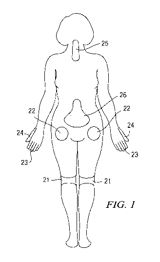
[0023] Figure 1 illustrates a number of common locations within a patient that
may be
sites where surgery is conducted and locations where the drug depot containing
an
antihypertensive agent or clonidine can be administered thereto.
[0024] Figure 2 illustrates a schematic dorsal view of the spine and sites
where a drug
depot containing an antihypertensive agent or clonidine can be administered
thereto.
[0025] Figure 3 is a graphic representation of a study of the average
cumulative release in
ug of clonidine for clonidine strip implants described in Example 1.
[0026] Figure 4 is a graphic representation of a study of the average
percentage
cumulative release of clonidine for clonidine strip implants described in
Example 1.
[0027] Figure 5 is a graphic representation of the thermal paw withdrawal
threshold in
grams per days post-surgery for clonidine implants from Example 1.
[0028] Figure 6 is a graphic representation of the average percentage
cumulative release
of clonidine for several irradiated clonidine HC1 strip or ribbon implants
from Example 2
during days 1-8.
[0029] Figure 7 is a graphic representation of the calculated average daily
release of
clonidine in micrograms during days 1-8 for the clonidine HC1 strip or ribbon
implants
from Example 2.
CA 02700389 2010-03-23
WO 2009/129437 PCT/US2009/040910
7
[0030] Figure 8 is a graphic representation of the average percentage
cumulative release
of clonidine for certain clonidine HC1 strip or ribbon implants illustrated in
Figure 6.
[0031] Figure 9 is a graphic representation of the average daily release of
clonidine in
micrograms during days 1-8 for the certain clonidine HC1 strip or ribbon
implants
illustrated in Figure 8.
[0032] Figure 10 is a graphic representation of the average percentage
cumulative release
of clonidine during days 1-14 for certain clonidine HC1 strip or ribbon
implants illustrated
in Figure 6.
[0033] Figure 11 is a graphic representation of the average daily release of
clonidine
during days 1-14 for the certain clonidine HC1 strip or ribbon implants
illustrated in Figure
10.
[0034] Figure 12 is a graphic representation of the average percentage
cumulative release
of clonidine during days 1-14 for certain clonidine HC1 strip or ribbon
implants illustrated
in Figure 6.
[0035] Figure 13 is a graphic representation of the average daily release of
clonidine
during days 1-14 for the certain clonidine HC1 strip or ribbon implants
illustrated in Figure
12.
[0036] Figure 14 is a graphic representation of the average cumulative in
vitro release
profile for clonidine strip implants from a study described in Example 3.
[0037] Figure 15 shows the average cumulative in vitro release profile for
clonidine strip
implants from a study described in Example 4.
[0038] Figure 16 is a graphic representation of the percentage cumulative
release of
clonidine for three clonidine strip implants from a study described in Example
5.
[0039] Figure 17 is a graphic representation of the average percentage
cumulative release
of clonidine for the clonidine strip implants shown in Figure 16.
[0040] Figure 18 is a graphic representation of the cumulative in vitro
release of clonidine
in ug for the three clonidine strip implants described in Example 5.
[0041] Figure 19 is a graphic representation of the average cumulative in
vitro release of
clonidine in ug for the clonidine strip implants shown in Figure 18.
[0042] Figure 20 is a graphic representation of pain scores of clonidine
depots implanted
post-operatively at the surgical incision.
CA 02700389 2010-03-23
WO 2009/129437 PCT/US2009/040910
8
[0043] It is to be understood that the figures are not drawn to scale.
Further, the relation
between objects in a figure may not be to scale, and may in fact have a
reverse relationship
as to size. The figures are intended to bring understanding and clarity to the
structure of
each object shown, and thus, some features may be exaggerated in order to
illustrate a
specific feature of a structure.
DETAILED DESCRIPTION
[0044] For the purposes of this specification and appended claims, unless
otherwise
indicated, all numbers expressing quantities of ingredients, percentages or
proportions of
materials, reaction conditions, and other numerical values used in the
specification and
claims, are to be understood as being modified in all instances by the term
"about."
Accordingly, unless indicated to the contrary, the numerical parameters set
forth in the
following specification and attached claims are approximations that may vary
depending
upon the desired properties sought to be obtained by the present invention. At
the very
least, and not as an attempt to limit the application of the doctrine of
equivalents to the
scope of the claims, each numerical parameter should at least be construed in
light of the
number of reported significant digits and by applying ordinary rounding
techniques.
[0045] Notwithstanding that the numerical ranges and parameters setting forth
the broad
scope of the invention are approximations, the numerical values set forth in
the specific
examples are reported as precisely as possible. Any numerical value, however,
inherently
contains certain errors necessarily resulting from the standard deviation
found in their
respective testing measurements. Moreover, all ranges disclosed herein are to
be
understood to encompass any and all subranges subsumed therein. For example, a
range
of "1 to 10" includes any and all subranges between (and including) the
minimum value of
1 and the maximum value of 10, that is, any and all subranges having a minimum
value of
equal to or greater than 1 and a maximum value of equal to or less than 10,
e.g., 5.5 to 10.
[0046] It is noted that, as used in this specification and the appended
claims, the singular
forms "a," "an," and "the," include plural referents unless expressly and
unequivocally
limited to one referent. Thus, for example, reference to "a drug depot"
includes one, two,
three or more drug depots.
[0047] Reference will now be made in detail to certain embodiments of the
invention,
examples of which are illustrated in the accompanying drawings. While the
invention will
CA 02700389 2010-03-23
WO 2009/129437 PCT/US2009/040910
9
be described in conjunction with the illustrated embodiments, it will be
understood that
they are not intended to limit the invention to those embodiments. On the
contrary, the
invention is intended to cover all alternatives, modifications, and
equivalents, which may
be included within the invention as defined by the appended claims.
[0048] The headings below are not meant to limit the disclosure in any way;
embodiments
under any one heading may be used in conjunction with embodiments under any
other
heading.
[0049] New compositions and methods are provided that effectively prevent,
treat or
reduce post-operative pain or inflammation. In various embodiments,
compositions and
methods are provided that have long acting analgesic and anti-inflammatory
effects over
periods of at least 3 days in a single drug depot or multiple drug depots. New
compositions and methods are provided, which can easily allow accurate and
precise
implantation of a drug depot including clonidine with minimal physical and
psychological
trauma to a patient. The drug depot can now be easily delivered to the target
tissue site
(e.g., abdomen, synovial joint, at or near the spinal column, etc.) and
alleviate and/or treat
pain for at least 3 to 10 days. In this way, accurate and precise implantation
of the drug
depot in a minimally invasive procedure as well as an open procedure can be
accomplished.
Clonidine
[0050] Clonidine may be contained in a drug depot. A drug depot comprises a
physical
structure to facilitate implantation and retention in a desired site (e.g., a
synovial joint, a
disc space, a spinal canal, abdominal area, a tissue of the patient, etc.).
The drug depot
also comprises the drug. The term "drug" as used herein is generally meant to
refer to any
substance that alters the physiology of a patient. The term "drug" may be used
interchangeably herein with the terms "therapeutic agent", "therapeutically
effective
amount", and "active pharmaceutical ingredient" or "API". It will be
understood that a
"drug" formulation may include more than one therapeutic agent, wherein
exemplary
combinations of therapeutic agents include a combination of two or more drugs.
The drug
depot provides a concentration gradient of the therapeutic agent for delivery
to the site. In
various embodiments, the drug depot provides an optimal drug concentration
gradient of
CA 02700389 2010-03-23
WO 2009/129437 PCT/US2009/040910
the therapeutic agent at a distance of up to about 1 cm to about 10 cm from
the implant
site.
[0051] A "therapeutically effective amount" or "effective amount" is such that
when
administered, the drug results in alteration of the biological activity, such
as, for example,
5 inhibition of inflammation, reduction or alleviation of pain, improvement in
the condition,
etc. In various embodiments, the therapeutically effective amount of clonidine
comprises
from about 0.1 ug/day to 100 mg/day. In some embodiments, the therapeutically
effective
amount of clonidine comprises from about 30 ug to 1 mg of clonidine per day.
In some
embodiments, the therapeutically effective amount of clonidine comprises from
about 30
10 ug to 2.4 mg of clonidine per day. In some embodiments, the therapeutically
effective
amount of clonidine comprises from about 0.1 mg to 0.3 mg of clonidine per
day. In some
embodiments, the therapeutically effective amount of clonidine comprises 0.1
ug, 0.2 ug,
0.3 ug, 0.4 ug, 0.5 ug, 0.6 ug, 0.7 ug, 0.8 ug, 0.9 ug, 1 ug, 10 ug, 20 ug, 30
ug, 40 ug, 50
ug, 60 ug, 70 ug, 80 ug, 90 ug, 0.1 mg, 0.2 mg, 0.3 mg, 0.4 mg, 0.5 mg, 0.6
mg, 0.7 mg,
0.8 mg, 0.9 mg, 1 mg, 1.1 mg, 1.2 mg, 1.3 mg, 1.4 mg, 1.5 mg, 1.6 mg, 1.7 mg,
1.8 mg,
1.9 mg, 2 mg, 3 mg, 4 mg, 5 mg, 6 mg, 7 mg, 8 mg, 9 mg, 10 mg, 11 mg, 12 mg,
13 mg,
14 mg, 15 mg, 16 mg, 17 mg, 18 mg, 19 mg, 20 mg, 21 mg, 22 mg, 23 mg, 24 mg,
25 mg,
30 mg, 35 mg, or 40 mg (and all ranges and subranges therebetween) of
clonidine per day.
In one embodiment, the dosage to a human is between 0.1mg and 0.3mg of
clonidine per
day. It will be understood that the dosage administered to a patient can be as
single depot
or multiple depots depending upon a variety of factors, including the drug's
administered
pharmacokinetic properties, the route of administration, patient conditions
and
characteristics (sex, age, body weight, health, size, etc.), extent of
symptoms, concurrent
treatments, frequency of treatment and the effect desired. For example, lower
daily doses
of clonidine may be needed when there is concurrent treatment with an opioid
(e.g.,
morphine), alternatively, the patient may require higher doses of clonidine as
the dosage of
the opioid (e.g., morphine) is reduced or eliminated to control post-operative
pain.
[0052] In various embodiments, a therapeutically effective amount of clonidine
is
provided to inhibit, reduce, treat and/or prevent post-operative pain or
inflammation. In
general, the chemical name of clonidine is 2,6-dichloro-N-2-
imidazolidinyldenebenzenamine (C9H9C12N3). Clonidine has a molecular weight of
230.09 and exhibits the following general structure:
CA 02700389 2010-03-23
WO 2009/129437 PCT/US2009/040910
11
CI
H
N~ N N
I / NH~
CI
[0053] Unless otherwise specified or apparent from context, where this
specification and
the set of claims that follows refer to clonidine, it is understood that the
inventors are also
referring to pharmaceutically acceptable salts. One well-known commercially
available
salt for clonidine is its hydrochloride salt. Some other examples of
potentially
pharmaceutically acceptable salts include those salt-forming acids and bases
that do not
substantially increase the toxicity of a compound, such as, salts of alkali
metals such as
magnesium, potassium and ammonium, salts of mineral acids such as hydriodic,
hydrobromic, phosphoric, metaphosphoric, nitric and sulfuric acids, as well as
salts of
organic acids such as tartaric, acetic, citric, malic, benzoic, glycollic,
gluconic, gulonic,
succinic, arylsulfonic, e.g., p-toluenesulfonic acids, and the like.
[0054] Further, when referring to clonidine, the active ingredient may not
only be in the
salt form, but also in the base form (e.g., free base). In various
embodiments, if it is in the
base form, it may be combined with polymers under conditions in which there is
not
severe polymer degradation, as may be seen upon heat or solvent processing
that may
occur with PLGA or PLA. By way of a non-limiting example, when formulating
clonidine with poly(orthoesters), it may be desirable to use the clonidine
base formulation.
By contrast, when formulating clonidine with PLGA, it may be desirable to use
the HC1
salt form. In various embodiments, clonidine may be in the form of a
combination of a
salt and a base.
[0055] In addition to clonidine, the drug depot may comprise one or more
additional
therapeutic agents. Examples of therapeutic agents include, those that are
direct- and
local-acting modulators of pro-inflammatory cytokines such as TNF-a and IL-1
including,
but not limited to, soluble tumor necrosis factor a receptors, any pegylated
soluble tumor
necrosis factor a receptor, monoclonal or polyclonal antibodies or antibody
fragments or
combinations thereof. Examples of suitable therapeutic agents include receptor
antagonists, molecules that compete with the receptor for binding to the
target molecule,
antisense polynucleotides, and inhibitors of transcription of the DNA encoding
the target
protein. Suitable examples include but are not limited to Adalimumab,
Infliximab,
CA 02700389 2010-03-23
WO 2009/129437 PCT/US2009/040910
12
Etanercept, Pegsunercept (PEG sTNF-R1), sTNF-R1, CDP-870, CDP-571, CNI-1493,
RDP58, ISIS 104838, 1-3-(3-D-glucans, Lenercept, PEG-sTNFRII Fc Mutein, D2E7,
Afelimomab, and combinations thereof. In other embodiments, a therapeutic
agent
includes metalloprotease inhibitors, glutamate antagonists, glial cell-derived
neurotropic
factors (GDNF), B2 receptor antagonists, Substance P receptor (NK1)
antagonists such as
capsaicin and civamide, downstream regulatory element antagonistic modulator
(DREAM), iNOS, inhibitors of tetrodotoxin (TTX)-resistant Na+ -channel
receptor
subtypes PN3 and SNS2, inhibitors of interleukins such as IL-1, IL-6 and IL-8,
and anti-
inflammatory cytokines, TNF binding protein, onercept (r-hTBP-1), recombinant
adeno-
associated viral (rAAV) vectors encoding inhibitors, enhancers, potentiators,
or
neutralizers, antibodies, including but not limited to naturally occurring or
synthetic,
double-chain, single-chain, or fragments thereof. For example, suitable
therapeutic agents
include molecules that are based on single chain antibodies called
NanobodiesTM (Ablynx,
Ghent Belgium), which are defined as the smallest functional fragment of a
naturally
occurring, single-domain antibody. Alternatively, therapeutic agents include,
agents that
effect kinases and/or inhibit cell signaling mitogen-activated protein kinases
(MAPK), p38
MAPK, Src or protein tyrosine kinase (PTK). Therapeutic agents include, kinase
inhibitors such as, for example, Gleevec, Herceptin, fressa, imatinib
(STI571), herbimycin
A, tyrphostin 47, erbstatin, genistein, staurosporine, PD98059, SB203580, CNI-
1493, VX-
50/702 (Vertex/Kissei), SB203580, BIRB 796 (Boehringer Ingelheim), Glaxo P38
MAP
Kinase inhibitor, RWJ67657 (J&J), U0126, Gd, SCIO-469 (Scios), R03201195
(Roche),
Semipimod (Cytokine PharmaSciences), or derivatives thereof.
[0056] Therapeutic agents, in various embodiments, block the transcription or
translation
of TNF-a or other proteins in the inflammation cascade. Suitable therapeutic
agents
include, but are not limited to, integrin antagonists, alpha-4 beta-7 integrin
antagonists,
cell adhesion inhibitors, interferon gamma antagonists, CTLA4-Ig
agonists/antagonists
(BMS-188667), CD40 ligand antagonists, Humanized anti-IL-6 mAb (MRA,
Tocilizumab,
Chugai), HMGB-1 mAb (Critical Therapeutics Inc.), anti-IL2R antibodies
(daclizumab,
basilicimab), ABX (anti IL-8 antibodies), recombinant human IL-10, or HuMax IL-
15
(anti-IL 15 antibodies).
[0057] Other suitable therapeutic agents include IL-1 inhibitors, such Kineret
(anakinra)
which is a recombinant, non-glycosylated form of the human inerleukin-1
receptor
CA 02700389 2010-03-23
WO 2009/129437 PCT/US2009/040910
13
antagonist (IL-1Ra), or AMG 108, which is a monoclonal antibody that blocks
the action
of IL-1. Therapeutic agents also include excitatory amino acids such as
glutamate and
aspartate, antagonists or inhibitors of glutamate binding to NMDA receptors,
AMPA
receptors, and/or kainate receptors. Interleukin-1 receptor antagonists,
thalidomide (a
TNF-a release inhibitor), thalidomide analogues (which reduce TNF-a production
by
macrophages), bone morphogenetic protein (BMP) type 2 and BMP-4 (inhibitors of
caspase 8, a TNF-a activator), quinapril (an inhibitor of angiotensin II,
which upregulates
TNF-a), interferons such as IL-11 (which modulate TNF-a receptor expression),
and
aurin-tricarboxylic acid (which inhibits TNF-a), for example, may also be
useful as
therapeutic agents for reducing inflammation. It is contemplated that where
desirable a
pegylated form of the above may be used. Examples of other therapeutic agents
include
NF kappa B inhibitors such as glucocorticoids, clonidine; antioxidants, such
as
dithiocarbamate, and other compounds, such as, for example, sulfasalazine.
[0058] Specific examples of therapeutic agents suitable for use include, but
are not limited
to an anti-inflammatory agent, analgesic agent, or osteoinductive growth
factor or a
combination thereof. Anti-inflammatory agents include, but are not limited to,
salicylates,
diflunisal, sulfasalazine, indomethacin, ibuprofen, naproxen, tolmetin,
diclofenac,
ketoprofen, fenamates (mefenamic acid, meclofenamic acid), enolic acids
(piroxicam,
meloxicam), nabumetone, celecoxib, etodolac, nimesulide, apazone, gold,
sulindac or
tepoxalin; antioxidants, such as dithiocarbamate, and other compounds such as
sulfasalazine [2-hydroxy-5-[-4-[C2-pyridinylamino)sulfonyl]azo]benzoic acid],
steroids,
such as fluocinolone, cortisol, cortisone, hydrocortisone, fludrocortisone,
prednisone,
prednisolone, methylprednisolone, triamcinolone, betamethasone, dexamethasone,
beclomethasone, fluticasone or a combination thereof.
[0059] Suitable anabolic growth or anti-catabolic growth factors include, but
are not
limited to, a bone morphogenetic protein, a growth differentiation factor, a
LIM
mineralization protein, CDMP or progenitor cells or a combination thereof.
[0060] Additional analgesic agents may also be included in the depot. Suitable
analgesic
agents include, but are not limited to, acetaminophen, bupivacaine, lidocaine,
opioid
analgesics such as buprenorphine, butorphanol, dextromoramide, dezocine,
dextropropoxyphene, diamorphine, fentanyl, alfentanil, sufentanil,
hydrocodone,
hydromorphone, ketobemidone, levomethadyl, mepiridine, methadone, morphine,
CA 02700389 2010-03-23
WO 2009/129437 PCT/US2009/040910
14
nalbuphine, opium, oxycodone, papaveretum, pentazocine, pethidine,
phenoperidine,
piritramide, dextropropoxyphene, remifentanil, tilidine, tramadol, codeine,
dihydrocodeine, meptazinol, dezocine, eptazocine, flupirtine or a combination
thereof.
[0061] Suitable analgesics also include agents with analgesic properties, such
as for
example, amitriptyline, carbamazepine, gabapentin, pregabalin, or a
combination thereof.
[0062] The depot may contain a muscle relaxant. Exemplary muscle relaxants
include by
way of example and not limitation, alcuronium chloride, atracurium bescylate,
baclofen,
carbolonium, carisoprodol, chlorphenesin carbamate, chlorzoxazone,
cyclobenzaprine,
dantrolene, decamethonium bromide, fazadinium, gallamine triethiodide,
hexafluorenium,
meladrazine, mephensin, metaxalone, methocarbamol, metocurine iodide,
pancuronium,
pridinol mesylate, styramate, suxamethonium, suxethonium, thiocolchicoside,
tizanidine,
tolperisone, tubocuarine, vecuronium, or combinations thereof.
[0063] The depot comprises the therapeutic agent or agents and may also
contain other
non-active ingredients. These non-active ingredients may have a multi-
functional purpose
including the carrying, stabilizing and controlling of the release of the
therapeutic agent(s).
The sustained release process, for example, may be by a solution-diffusion
mechanism or
it may be governed by an erosion-controlled process. Typically, the depot will
be a solid
or semi-solid formulation comprised of a biocompatible material, which can be
biodegradable. The term "solid" is intended to mean a non-gel like material,
while, "semi-
solid" is intended to mean a gel-like material that has some degree of
flowability, thereby
allowing the depot to bend and conform to the surrounding tissue requirements.
The term
"gel" is intended to mean a material that is soft and deformable at any point
in its
application to the surgical site.
[0064] In various embodiments, the depot material will be durable within the
tissue site
for a period of time similar to (for biodegradable components) or greater than
(for non-
biodegradable components) the planned period of drug delivery. For example,
the depot
material may have a melting point or glass transition temperature close to or
higher than
body temperature, but lower then the decomposition or degradation temperature
of the
therapeutic agent. However, the pre-determined erosion of the depot material
can also be
used to provide for slow release of the loaded therapeutic agent(s).
[0065] In various embodiments, the drug depot may be designed to release the
clonidine
when certain trigger points are reached (e.g., temperature, pH, etc.) after
implantation in
CA 02700389 2010-03-23
WO 2009/129437 PCT/US2009/040910
vivo. For example, the drug depot may comprise polymers that will release more
drug as
the body temperature reaches greater than, for example, 102 F, particularly if
the drug
possesses antipyretic properties. In various embodiments, depending on the
site of
implantation, the drug depot may release more or less drug as a certain pH is
reached. For
5 example, the drug depot may be designed to release the drug as the bodily
fluid having a
certain pH contact the drug depot (e.g., CSF having a pH of about 7.35 to
about 7.70,
synovial fluid having a pH of about 7.29 to about 7.45; urine having a pH of
about 4.6 to
about 8.0, pleural fluids having a pH of about 7.2 to about 7.4, blood having
a pH of about
7.35 to about 7.45, etc.).
10 [0066] In various embodiments, the depot may have a high drug loading, such
that the
clonidine and/or other therapeutic agent comprises about 0.5-90 wt.% of the
depot, or 1-50
wt.% of the depot, or 1-25 wt.% of the depot, or 1-10 wt.% of the depot. In
various
embodiments, the amount of clonidine and/or other therapeutic agent are
present in the
depot in a range from about 0.1 Io to about 40 Io by weight of the depot
(including 0.1 Io,
15 0.2%, 0.3%, 0.4%, 0.5%, 0.6%, 0.7%, 0.8%, 0.9%, 1%, 2%, 3%, 4%, 5%, 6%, 7%,
8%,
9%, 10%, 11%, 12%, 13%, 14%, 15%, 16%, 17%, 18%, 19%, 20%, 21%, 22%, 23%,
24%, 25%, 26%, 27%, 28%, 29%, 30%, 31%, 32%, 33%, 34%, 35%, 36%, 37%, 38%,
39%, 40%, and ranges between any two of these points, for instance, 0.5-5%, 5-
10% and
10-20%, etc.).
[0067] In various embodiments, the drug depot may release 0.1 ug, 0.2 ug, 0.3
ug, 0.4 ug,
0.5 ug, 0.6 ug, 0.7 ug, 0.8 ug, 0.9 ug, 1 ug, lOug, 20ug, 30ug, 40ug, 50ug,
60ug, 70ug,
80ug, 90ug, 0.1mg, 0.2mg, 0.3mg, 0.4mg, 0.5mg, 0.6mg, 0.7mg, 0.8mg, 0.9mg,
1mg,
1.1mg, 1.2mg, 1.3mg, 1.4mg, 1.5mg, 1.6mg, 1.7mg, 1.8mg, 1.9mg, 2mg, 3mg, 4mg,
5mg,
6mg, 7mg, 8mg, 9mg, 10mg, 11mg, 12mg, 13mg, 14mg, 15mg, 16mg, 17mg, 18mg,
19mg, 20mg, 21mg, 22mg, 23mg, 24mg, 25mg, 30mg, 35mg, or 40mg, 45mg, or 50mg
of
clonidine per day for a total of at least 3 days, at least 7 days, at least 8
days, 3 to 30 days,
3 to 10 days, 3 to 8 days, 5 to 7 days or 7 to 10 days. In various
embodiments, the drug
depot may release 0.5mg to 1mg of clonidine per hour for a total of at least 3
days, 3 to 10
days, 5 to 7 days or 7 to 10 days to reduce, treat or prevent post-operative
pain. In various
embodiments, the drug depot releases 5 Io, 10%, 15 Io, 20%, 25 Io, 30%, 35 Io,
40%, 45 Io,
50 Io, 55 Io, 60%, 65 Io, 70%, 75 Io, 80%, 85 Io, 90%, 95 Io, or 99% of the
clonidine over a
period of 3 to 10 days after the drug depot is administered to the target
tissue site or 5 to 7
CA 02700389 2010-03-23
WO 2009/129437 PCT/US2009/040910
16
days. The drug depot may have a "release rate profile" that refers to the
percentage of
active ingredient that is released over fixed units of time, e.g., mg/hr,
mg/day, 10% per day
for ten days, etc. As persons of ordinary skill know, a release rate profile
may be but need
not be linear. By way of a non-limiting example, the drug depot may be a strip
or a
ribbon-like strip or fiber that releases the clonidine over a period of time.
[0068] In various embodiments, the drug depot comprises from about 1% to 10%
by
weight clonidine, 75% to 94% by weight of a polymer and 5% to 15% by weight of
an
excipient. mPEG may be used as an excipient or plasticizer for a polymer as it
imparts
malleability to the resulting formulation. PEG 300 may also be used as an
excipient. In
addition, a combination of PEG 300 and NMP may be used as the excipient.
[0069] Exemplary excipients that may be formulated with clonidine in addition
to the
biodegradable polymer include but are not limited to MgO (e.g., 1 wt.%), 5050
DLG 6E,
5050 DLG 1A, mPEG, TBO-Ac, mPEG, Span-65, Span-85, pluronic F127, TBO-Ac,
sorbital, cyclodextrin, maltodextrin and combinations thereof. In some
embodiments, the
excipient or excipients may comprise from about 0.001 wt.% to about 50 wt.% of
the
formulation. In some embodiments, the excipient(s) comprise from about 0.001
wt. Io to
about 40 wt.% of the formulation. In some embodiments, the excipient(s)
comprise from
about 0.001 wt.% to about 30 wt.% of the formulation. In some embodiments, the
excipient(s) comprise from about 0.001 wt.% to about 20 wt.% of the
formulation. In
some embodiments, the excipient(s) comprise from about 0.5 wt.% to about 20
wt.% of
the formulation. In some embodiments, the excipient(s) comprise from about
0.001 wt.%
to about 10 wt. Io of the formulation. In some embodiments, the excipient(s)
comprise
from about 0.001 wt.% to about 2 wt.% of the formulation.
[0070] In some embodiments, the drug depot may not be biodegradable. For
example, the
drug depot may comprise polyurethane, polyurea, polyether(amide), PEBA,
thermoplastic
elastomeric olefin, copolyester, and styrenic thermoplastic elastomer, steel,
aluminum,
stainless steel, titanium, metal alloys with high non-ferrous metal content
and a low
relative proportion of iron, carbon fiber, glass fiber, plastics, ceramics or
combinations
thereof. Typically, these types of drug depots may need to be removed after a
certain
amount of time.
[0071] In some instances, it may be desirable to avoid having to remove the
drug depot
after use. In those instances, the drug depot may comprise a biodegradable
material.
CA 02700389 2010-03-23
WO 2009/129437 PCT/US2009/040910
17
There are numerous materials available for this purpose and having the
characteristic of
being able to breakdown or disintegrate over a prolonged period of time when
positioned
at or near the target tissue. As function of the chemistry of the
biodegradable material the
mechanism of the degradation process can be hydrolytical or enzymatical in
nature, or
both. In various embodiments, the degradation can occur either at the surface
(heterogeneous or surface erosion) or uniformly throughout the drug delivery
system depot
(homogeneous or bulk erosion).
[0072] The drug depot may comprise a polymeric or non-polymeric material as
well as a
synthetic or naturally occurring material, or a combination thereof. Non-
polymeric
materials include, for example, cholesterol, stigmasterol, glycerol,
estradiol, sucrose,
distearate, sorbitan, sorbitan monooleate, sorbitan monopalmitate, sorbitan
tristearate, or
the like.
[0073] In various embodiments, the drug depot comprises a polymer and the
polymer will
degrade in vivo over a period of less than a year, with at least 50% of the
polymer
degrading within six months or less. In some embodiments, the polymer is
capable of or
will degrade in two months, one month or less. In some embodiments, the
polymer will
degrade significantly within a month, with at least 50% of the polymer
degrading into non-
toxic residues which are removed by the body, and 100% of the drug being
released within
a two week period. Polymers should also degrade by hydrolysis by surface
erosion, rather
than by bulk erosion, so that release is not only sustained but also linear.
Polymers which
meet this criteria include some of the polyanhydrides, co-polymers of lactic
acid and
glycolic acid wherein the weight ratio of lactic acid to glycolic acid is no
more than 4:1
(i.e., 80% or less lactic acid to 20% or more glycolic acid by weight), and
polyorthoesters
containing a catalyst or degradation enhancing compound, for example,
containing at least
1% by weight anhydride catalyst such as maleic anhydride. Other polymers
include
protein polymers such as gelatin and fibrin and polysaccharides such as
hyaluronic acid.
[0074] A "depot" includes but is not limited to capsules, microspheres,
microparticles,
microcapsules, microfibers particles, nanospheres, nanoparticles, coating,
matrices,
wafers, pills, pellets, emulsions, liposomes, micelles, sheets, strips, ribbon-
like strips or
fibers, mesh, a paste, a slab, pellets, gels, or other pharmaceutical delivery
compositions.
Suitable materials for the depot are ideally pharmaceutically acceptable
biodegradable
and/or any bioabsorbable materials that are preferably FDA approved or GRAS
materials.
CA 02700389 2010-03-23
WO 2009/129437 PCT/US2009/040910
18
These materials can be polymeric or non-polymeric, as well as synthetic or
naturally
occurring, or a combination thereof.
[0075] The term "biodegradable" includes that all or parts of the drug depot
will degrade
over time by the action of enzymes, by hydrolytic action and/or by other
similar
mechanisms in the human body. In various embodiments, "biodegradable" includes
that
the depot (e.g., microparticle, microsphere, gel, etc.) can break down or
degrade within the
body to non-toxic components after or while a therapeutic agent has been or is
being
released. By "bioerodible," it is meant that the depot and/or gel will erode
or degrade over
time due, at least in part, to contact with substances found in the
surrounding tissue, fluids
or by cellular action. By "bioabsorbable," it is meant that the depot will be
broken down
and absorbed within the human body, for example, by a cell or tissue.
"Biocompatible"
means that the depot will not cause substantial tissue irritation or necrosis
at the target
tissue site.
[0076] In various embodiments, the depot may comprise a bioabsorbable, a
bioabsorbable,
and/or a biodegradable biopolymer that may provide immediate release,
sustained release
or controlled release of the drug. Examples of suitable sustained release
biopolymers
include but are not limited to poly(alpha-hydroxy acids), poly(lactide-co-
glycolide)
(PLGA or DLG) (which includes poly(lactide-co-glycolide, poly(D-lactide-co-
glycolide),
poly(L-lactide-co-glycolide) and poly(D,L-lactide-co-glycolide)), polylactide
(PLA),
poly(D,L-lactide), poly(D-lactide), poly(L-lactide), polyglycolide (PG),
polyethylene
glycol (PEG), PEG 200, PEG 300, PEG 400, PEG 500, PEG 550, PEG 600, PEG 700,
PEG 800, PEG 900, PEG 1000, PEG 1450, PEG 3350, PEG 4500, PEG 8000, conjugates
of poly (alpha-hydroxy acids), polyhydroxybutyrate, poly(glycolide-co-
trimethylenecarbonate), poly(lactic acid-co-lysine), poly(lactide-co-
urethane), poly(ester-
co-amide), polyorthoesters (POE), polyaspirins, polyphosphazenes,
polyanhydrides;
polyketals, collagen, starch, pre-gelatinized starch, hyaluronic acid,
chitosans, gelatin,
alginates, albumin, fibrin, vitamin E analogs, such as alpha tocopheryl
acetate, d-alpha
tocopheryl succinate, D-lactide, D,L-lactide, L-lactide, ^-caprolactone,
poly(D,L-lactide-
co-caprolactone) (DL-CL or DLCL), poly(D,L-lactide-co-glycolide-co-
caprolactone) (DL-
G-CL), polycaprolactone (PCL), dextrans, vinylpyrrolidone, polyvinyl alcohol
(PVA),
PVA-g-PLGA, PEGT-PBT copolymer (polyactive), methacrylates, poly (N-
isopropylacrylamide), PEO-PPO-PEO (pluronics), PEO-PPO-PAA copolymers, PLGA-
CA 02700389 2010-03-23
WO 2009/129437 PCT/US2009/040910
19
PEO-PLGA, PEG-PLG, PLA-PLGA, poloxamer 407, PEG-PLGA-PEG triblock
copolymers, SAIB (sucrose acetate isobutyrate) hydroxypropyl cellulose,
hydroxypropyl
methylcellulose, hydroxyethyl methylcellulose, carboxymethylcellulose or salts
thereof,
Carbopol, poly(hydroxyethylmethacrylate), poly(methoxyethylmethacrylate),
poly(methoxyethoxy-ethylmethacrylate), polymethylmethacrylate (PMMA),
methylmethacrylate (MMA), gelatin, polyvinyl alcohols, propylene glycol, or
combinations thereof.
[0077] In various embodiments, the molecular weight of the polymer can be a
wide range
of values. The average molecular weight of the polymer can be from about 1000
to about
10,000,000; or about 1,000 to about 1,000,000; or about 5,000 to about
500,000; or about
10,000 to about 100,000; or about 20,000 to 50,000.
[0078] In some embodiments, the polymer comprises PLGA or POE or a combination
thereof. The PLGA may comprise a mixture of polyglycolide and polylactide and
in
some embodiments, in the mixture, there is more polylactide than
polyglycolide. In some
embodiments, the molar ratio of polylactide to polyglycolide is between 50:50
and 100:0.
In various embodiments, there is 100% polylactide and 0% polyglycolide; 95%
polylactide and 5% polyglycolide; 90% polylactide and 10% polyglycolide; 85%
polylactide and 15% polyglycolide; 80% polylactide and 20% polyglycolide; 75%
polylactide and 25% polyglycolide; 70% polylactide and 30% polyglycolide; 65%
polylactide and 35% polyglycolide; 60% polylactide and 40% polyglycolide; 55%
polylactide and 45% polyglycolide; 50% polylactide and 50% polyglycolide; 45%
polylactide and 55% polyglycolide; 40% polylactide and 60% polyglycolide; 35%
polylactide and 65% polyglycolide; 30% polylactide and 70% polyglycolide; 25%
polylactide and 75% polyglycolide; 20% polylactide and 80% polyglycolide; 15%
polylactide and 85% polyglycolide; 10% polylactide and 90% polyglycolide; 5%
polylactide and 95% polyglycolide; and 0% polylactide and 100% polyglycolide.
[0079] In various embodiments that comprise both polylactide and
polyglycolide; there is
at least 95% polylactide; at least 90% polylactide; at least 85% polylactide;
at least 80%
polylactide; at least 75% polylactide; at least 70% polylactide; at least 65%
polylactide; at
least 60% polylactide; at least 55%; at least 50% polylactide; at least 45%
polylactide; at
least 40% polylactide; at least 35% polylactide; at least 30% polylactide; at
least 25%
CA 02700389 2010-03-23
WO 2009/129437 PCT/US2009/040910
polylactide; at least 20% polylactide; at least 15% polylactide; at least 10%
polylactide; or
at least 5% polylactide; and the remainder of the biopolymer is polyglycolide.
[0080] In some embodiments, the polymer comprises DL-CL or a combination
thereof.
The DL-CL may comprise a mixture of lactide and caprolactone. The molar ratio
of
5 lactide to caprolactone can be 10:90 to 90:10 and all subranges therebetween
(e.g., 20:80,
30:70, 45:55, 65:35, 67:33, 89:11, etc.).
[0081] In some embodiments, the polymer comprises DL-G-CL or a combination
thereof.
The DL-G-CL may comprise a mixture of lactide, glycolide and caprolactone. In
some
embodiments, the molar ratio of lactide to glycolide to caprolactone may be
30:20:50. In
10 some embodiments, the mixture may comprise 5-50% lactide, 5-50% glycolide,
and 20-
80% caprolactone.
[0082] In various embodiments, when the drug depot comprises a polymer, it is
employed
at about 10 wt.% to about 90 wt.%, 10 wt.% to about 50 wt.%, or about 20 wt.%
to about
40 wt.% based on the weight of the drug depot.
15 [0083] In some embodiments, at least 75% of the particles have a size from
about 1
micrometer to about 200 micrometers. In some embodiments, at least 85% of the
particles
have a size from about 1 micrometer to about 200 micrometers. In some
embodiments, at
least 95% of the particles have a size from about 1 micrometer to about 200
micrometers.
In some embodiments, all of the particles have a size from about 1 micrometer
to about
20 200 micrometers.
[0084] In some embodiments, at least 75% of the particles have a size from
about 20
micrometer to about 100 micrometers. In some embodiments, at least 85% of the
particles
have a size from about 20 micrometers to about 100 micrometers. In some
embodiments,
at least 95% of the particles have a size from about 20 micrometer to about
100
micrometers. In some embodiments, all of the particles have a size from about
20
micrometer to about 100 micrometers.
[0085] The depot may optionally contain inactive materials such as buffering
agents and
pH adjusting agents such as potassium bicarbonate, potassium carbonate,
potassium
hydroxide, sodium acetate, sodium borate, sodium bicarbonate, sodium
carbonate, sodium
hydroxide or sodium phosphate; degradation/release modifiers; drug release
adjusting
agents; emulsifiers; preservatives such as benzalkonium chloride,
chlorobutanol,
phenylmercuric acetate and phenylmercuric nitrate, sodium bisulfite, sodium
bisulfate,
CA 02700389 2010-03-23
WO 2009/129437 PCT/US2009/040910
21
sodium thiosulfate, thimerosal, methyl and other paraben, polyvinyl alcohol
and
phenylethyl alcohol; solubility adjusting agents; stabilizers; and/or cohesion
modifiers.
Typically, any such inactive materials will be present within the range of 0-
75 wt.%, and
more typically within the range of 0-30 wt.%. If the depot is to be placed in
the spinal
area or joint area, in various embodiments, the depot may comprise sterile
preservative
free material.
[0086] The depot can have many different sizes, shapes and configurations.
There are
several factors that can be taken into consideration in determining the size,
shape and
configuration of the drug depot. For example, both the size and shape may
allow for ease
in positioning the drug depot at the target tissue site that is selected as
the implantation or
injection site. In addition, the shape and size of the system should be
selected so as to
minimize or prevent the drug depot from moving after implantation or
injection. In
various embodiments, the drug depot can be shaped like a sphere, a cylinder
such as a rod
or fiber, a flat surface such as a disc, film, strip, ribbon, or sheet, a
paste, a slab,
microparticles, nanoparticles, pellets, mesh or the like. Flexibility may be a
consideration
so as to facilitate placement of the drug depot. In various embodiments, the
drug depot
can be different sizes, for example, the drug depot may be a length of from
about 0.5 mm
to 100 mm and have a diameter or thickness of from about 0.01 to about 5 mm.
In various
embodiments, the drug depot may have a layer thickness of from about 0.005 to
5.0 mm,
such as, for example, from 0.05 to 2.0 mm. In some embodiments, the shape may
be a
strip or a ribbon-like strip and the strip or ribbon-like strip has a ratio of
width to thickness
in the range of 2 to 20 or greater.
[0087] Radiographic markers can be included on or in the drug depot to permit
the user to
accurately position the depot into the target site of the patient. These
radiographic markers
will also permit the user to track movement and degradation of the depot at
the site over
time. In this embodiment, the user may accurately position the depot in the
site using any
of the numerous diagnostic imaging procedures. Such diagnostic imaging
procedures
include, for example, X-ray imaging or fluoroscopy. Examples of such
radiographic
markers include, but are not limited to, barium, bismuth, iodine, tantalum,
tungsten,
calcium, and/or metal beads or particles. Where present, the radiographic
marker is
typically present in an amount of from about 10% to about 40% (including 10%,
11 Io,
12 Io, 13 Io, 14 Io, 15 Io, 16 Io, 17 Io, 18 Io, 19 Io, 20 Io, 21 Io, 22 Io,
23 Io, 24 Io, 25 Io, 26 Io,
CA 02700389 2010-03-23
WO 2009/129437 PCT/US2009/040910
22
27%, 28%, 29%, 30%, 31%, 32%, 33%, 34%, 35%, 36%, 37%, 38%, 39% and 40%, as
well as ranges between any two of these values, e.g., 10-15%, 15-20%, 20-25%,
25-30%,
30-35%, 35-40%, and so forth, with 15-30% being more typical, even more
typically 20-
25%). In various embodiments, the radiographic marker could be a spherical
shape or a
ring around the depot.
[0088] In some embodiments, the drug depot has pores that allow release of the
drug from
the depot. The drug depot will allow fluid in the depot to displace the drug.
However, cell
infiltration into the depot will be prevented by the size of the pores of the
depot. In this
way, in some embodiments, the depot will not function as a tissue scaffold and
will not
allow tissue growth. Rather, the drug depot will solely be utilized for drug
delivery. In
some embodiments, the pores in the drug depot will be less than 250 to 500
microns. This
pore size will prevent cells from infiltrating the drug depot and laying down
scaffolding
cells. Thus, in this embodiment, drug will elute from the drug depot as fluid
enters the
drug depot, but cells will be prevented from entering. In some embodiments,
where there
are little or no pores, the drug will elute out from the drug depot by the
action of enzymes,
by hydrolytic action and/or by other similar mechanisms in the human body. In
other
embodiments, the drug depot may have pore sizes above 500 microns to allow
influx of
cells and drug release and the drug depot may function, in this embodiment, as
a tissue
scaffold.
[0089] In one exemplary embodiment, a drug depot for delivering a therapeutic
agent to a
target tissue site beneath the skin of a patient is provided, the drug depot
comprising an
effective amount of clonidine, wherein the target tissue site comprises at
least one muscle,
ligament, tendon, cartilage, spinal disc, spinal foraminal space near the
spinal nerve root,
facet or synovial joint, or spinal canal.
[0090] In various embodiments, the drug depot comprises a gel, which includes
a
substance having gelatinous, jelly-like, or colloidal properties at room
temperature. The
gel, in various embodiments, may have the clonidine and optionally one or more
additional therapeutic agents dispersed throughout it or suspended within the
gel. The
dispersal of the therapeutic agent may be even throughout the gel.
Alternatively, the
concentration of the therapeutic agent may vary throughout it. As the
biodegradable
material of the gel or drug depot degrades at the site, the therapeutic agent
is released.
CA 02700389 2010-03-23
WO 2009/129437 PCT/US2009/040910
23
[0091] When the drug depot is a gel, in contrast to a sprayable gel that
typically employs a
low viscosity polymer, a gel with a higher viscosity may be desirable for
other
applications, for example, a gel having a putty-like consistency may be more
preferable
for bone regeneration applications. In various embodiments, when a polymer is
employed
in the gel, the polymeric composition includes about 40 wt.% to about 99 wt.%
or about
90 wt.% to about 99 wt.% of the gel.
[0092] In another exemplary embodiment, the gel is in viscous form is loaded
with one or
more drug depots (e.g., microspheres loaded with a therapeutic agent), wherein
the viscous
gel is positioned into a synovial joint, disc space, a spinal canal, or a soft
tissue
surrounding the spinal canal of a subject. The gel can also be used, in
various
embodiments, to seal or repair tissue. In yet another exemplary embodiment,
the gel is
injectable, and/or an adherent gel that solidifies upon contact with tissue.
For example, the
gel may be administered as a liquid that gels in situ at the target tissue
site. In various
embodiments, the gel can comprise a two part system where a liquid is
administered and a
gelling agent is added subsequently to cause the liquid to gel or harden.
[0093] In various embodiments, the gel is a hardening gel, where after the gel
is applied to
the target site, it hardens and the drug can be released as the bodily fluid
contacts the gel.
[0094] In various embodiments, the drug depot is loaded with clonidine and
optionally
one or more additional therapeutic agents, and delivered to the desired target
tissue site
(e.g., surgical wound site, inflamed tissue, degenerative tissue, etc.) and,
in various
embodiments, the drug depot may be held in place by a suture, barb, staple,
adhesive gel,
etc. which prevents the drug depot from being removed from that site by the
venous
systemic circulation or otherwise dispersed too widely, which reduces the
desired
therapeutic effect. For example, after hours or days, the drug depot may
degrade, thereby
allowing the drug depots (e.g., strips, ribbon-like strips, etc.) to begin
releasing the
therapeutic agent. The strips may be formed from an insoluble or inert
substance, but
soluble or active once it comes into contact with the target tissue site.
Likewise, the drug
depot may comprise a substance that dissolves or disperses within the tissue.
As the drug
depot begins to dissolve within hours to days, the drug depots (e.g., strips)
are exposed to
body fluids and begin releasing their contents. The drug depot can be
formulated to
optimize exposure time of the drug depot and release of the therapeutic agent
from the
drug depot.
CA 02700389 2010-03-23
WO 2009/129437 PCT/US2009/040910
24
[0095] In various embodiments, the drug depot (e.g., gel) is flowable and can
be injected,
sprayed, instilled, and/or dispensed to, on or in the target tissue site.
"Flowable" means
that the gel formulation is easy to manipulate and may be brushed, sprayed,
dripped,
painted, injected, shaped and/or molded at or near the target tissue site as
it coagulates.
"Flowable" includes formulations with a low viscosity or water-like
consistency to those
with a high viscosity, such as a paste-like material. In various embodiments,
the
flowability of the formulation allows it to conform to irregularities,
crevices, cracks,
and/or voids in the tissue site. For example, in various embodiments, the gel
may be used
to fill one or more voids in an osteolytic lesion.
[0096] In various embodiments, the drug depot comprises poly (alpha-hydroxy
acids),
PLGA, PLA, D,L-lactide-glycolide-s-caprolactone, PG, polyhydroxybutyrate,
poly(glycolide-co-trimethylenecarbonate), poly(lactic acid-co-lysine),
poly(lactide-co-
urethane), poly(ester-co-amide), PEG conjugates of poly (alpha-hydroxy acids),
polyorthoesters, polyaspirins, polyphosphazenes, polyanhydrides; polyketals,
collagen,
starch, pre-gelatinized starch, hyaluronic acid, chitosans, gelatin,
alginates, albumin,
fibrin, vitamin E analogs, such as alpha tocopheryl acetate, d-alpha
tocopheryl succinate,
s-caprolactone, D,L-lactide, D-lactide, L-lactide, D,L-lactide-caprolactone,
D,L-lactide-
glycolide-caprolactone, dextrans, vinylpyrrolidone, polyvinyl alcohol (PVA),
PVA-g-
PLGA, PEGT-PBT copolymer (polyactive), methacrylates, poly (N-
isopropylacrylamide),
PEO-PPO-PEO (pluronics), PEO-PPO-PAA copolymers, PLGA-PEO-PLGA, PEG-PLG
(poly(d,l-lactide-co-glycolide), PLA-PLGA, poloxamer 407, PEG-PLGA-PEG
triblock
copolymers, SAIB (sucrose acetate isobutyrate) or combinations thereof. These
one or
more components allow the therapeutic agent to be released from the drug depot
in a
controlled and/or sustained manner. For example, the drug depot containing the
therapeutic agent and a polymer matrix can be injected at the target tissue
site and the
polymer matrix breaks down over time (e.g., hours, days) within the target
tissue site
releasing clonidine and optionally additional therapeutic agents. Thus, the
administration
of the drug depot can be localized and occur over a period of time (e.g., at
least one day to
about 2, 3, 4, 5, 6, 7, 8, 9, 10, 11, 12, 13, 14, 15, 16, 17, 18, 19, 20, 21,
22, 23, 24, 25, 26,
27, 28, 29, 30, 40, 50, 60, 70, 80 and 90 days).
[0097] The terms "sustained release" or "sustain release" (also referred to as
extended
release or controlled release) are used herein to refer to one or more
therapeutic agent(s)
CA 02700389 2010-03-23
WO 2009/129437 PCT/US2009/040910
that is introduced into the body of a human or other mammal and continuously
releases a
stream of one or more therapeutic agents over a predetermined time period and
at a
therapeutic level sufficient to achieve a desired therapeutic effect
throughout the
predetermined time period. Reference to a continuous release stream is
intended to
5 encompass release that occurs as the result of biodegradation in vivo of
drug depot, or a
matrix or component thereof, or as the result of metabolic transformation or
dissolution of
the therapeutic agent(s) or conjugates of therapeutic agent(s).
[0098] The phrase "immediate release" is used herein to refer to one or more
therapeutic
agent(s) that is introduced into the body and that is allowed to dissolve in
or become
10 absorbed at the location to which it is administered, with no intention of
delaying or
prolonging the dissolution or absorption of the drug.
[0099] The two types of formulations (sustain release and immediate release)
may be used
in conjunction. The sustained release and immediate release may be in or more
of the
same depots. In various embodiments, the sustained release and immediate
release may be
15 part of separate depots. For example, a bolus or immediate release
formulation of
clonidine may be placed at or near the target site and a sustain release
formulation may
also be placed at or near the same site. Thus, even after the bolus becomes
completely
accessible, the sustain release formulation would continue to provide the
active ingredient
for the intended tissue.
20 [00100] In various embodiments, the drug depot is designed to cause an
initial burst dose
of therapeutic agent within the first 48 hours or 24 hours after implantation.
"Initial burst"
or "burst effect" or "bolus dose" refers to the release of therapeutic agent
from the drug
depot during the first 48 hours or 24 hours after the drug depot comes in
contact with an
aqueous fluid (e.g., synovial fluid, cerebral spinal fluid, etc.). In some
embodiments, the
25 drug depot is designed to avoid this initial burst effect.
[00101] In various embodiments, the drug depot contains one or more different
release
layer(s) that releases a bolus dose of clonidine or pharmaceutically
acceptable salt thereof
(e.g., 100ug to 300ug at a target site beneath the skin) and one or more
sustain release
layer(s) that releases an effective amount of clonidine or pharmaceutically
acceptable salt
thereof over a period of 3 to 30 days, 3 to 10 days, or 7 to 10 days. In
various
embodiments, the one or more immediate release layer(s) comprise PLGA, which
CA 02700389 2010-03-23
WO 2009/129437 PCT/US2009/040910
26
degrades faster than the one or more sustain release layer(s), which comprises
PLA, which
degrades at a slower rate than the PLGA.
[00102] In various embodiments, when the drug depot comprises a gel, the gel
may have
a pre-dosed viscosity in the range of about 1 to about 2,000 centipoise (cps),
1 to about
500 cps, 1 to about 200 cps, or 1 to about 100 cps. After the gel is
administered to the
target site, the viscosity of the gel will increase and the gel will have a
modulus of
elasticity (Young's modulus) in the range of about 1 x 10² to about 6 x 10⁵
dynes/cm2, or 2
x 104 to about 5 x 10⁵ dynes/cm2, or 5 x 10⁴ to about 5 x 10⁵ dynes/cm2.
[00103] In one embodiment, the gel may be an adherent gel, which comprises a
therapeutic agent that is evenly distributed throughout the gel. The gel may
be of any
suitable type, as previously indicated, and should be sufficiently viscous so
as to prevent
the gel from migrating from the targeted delivery site once deployed; the gel
should, in
effect, "stick" or adhere to the targeted tissue site. The gel may, for
example, solidify
upon contact with the targeted tissue or after deployment from a targeted
delivery system.
The targeted delivery system may be, for example, a syringe, a catheter,
needle or cannula
or any other suitable device. The targeted delivery system may inject or spray
the gel into
or on the targeted tissue site. The therapeutic agent may be mixed into the
gel prior to the
gel being deployed at the targeted tissue site. In various embodiments, the
gel may be part
of a two-component delivery system and when the two components are mixed, a
chemical
process is activated to form the gel and cause it to stick or adhere to the
target tissue.
[00104] In various embodiments, for those gel formulations that contain a
polymer, the
polymer concentration may affect the rate at which the gel hardens (e.g., a
gel with a
higher concentration of polymer may coagulate more quickly than gels having a
lower
concentration of polymer). In various embodiments, when the gel hardens, the
resulting
matrix is solid but is also able to conform to the irregular surface of the
tissue (e.g.,
recesses and/or projections in bone).
[00105] The percentage of polymer present in the gel may also affect the
viscosity of the
polymeric composition. For example, a composition having a higher percentage
by
weight of polymer is typically thicker and more viscous than a composition
having a lower
percentage by weight of polymer. A more viscous composition tends to flow more
slowly.
Therefore, a composition having a lower viscosity may be preferred in some
instances, for
example, when applying the formulation via spray.
CA 02700389 2010-03-23
WO 2009/129437 PCT/US2009/040910
27
[00106] In various embodiments, the molecular weight of the gel can be varied
by many
methods known in the art. The choice of method to vary molecular weight is
typically
determined by the composition of the gel (e.g., polymer, versus non-polymer).
For
example, in various embodiments, when the gel comprises one or more polymers,
the
degree of polymerization can be controlled by varying the amount of polymer
initiators
(e.g. benzoyl peroxide), organic solvents or activator (e.g. DMPT),
crosslinking agents,
polymerization agent, and/or reaction time.
[00107] Suitable gel polymers may be soluble in an organic solvent. The
solubility of a
polymer in a solvent varies depending on the crystallinity, hydrophobicity,
hydrogen-
bonding and molecular weight of the polymer. Lower molecular weight polymers
will
normally dissolve more readily in an organic solvent than high-molecular
weight
polymers. A polymeric gel, which includes a high molecular weight polymer,
tends to
coagulate or solidify more quickly than a polymeric composition, which
includes a low-
molecular weight polymer. Polymeric gel formulations, which include high
molecular
weight polymers, also tend to have a higher solution viscosity than a
polymeric gel, which
include a low-molecular weight polymer.
[00108] In various embodiments, the gel has an inherent viscosity (abbreviated
as "I.V."
and units are in deciliters/gram), which is a measure of the gel's molecular
weight and
degradation time (e.g., a gel with a high inherent viscosity has a higher
molecular weight
and longer degradation time). Typically, a gel with a high molecular weight
provides a
stronger matrix and the matrix takes more time to degrade. In contrast, a gel
with a low
molecular weight degrades more quickly and provides a softer matrix. In
various
embodiments, the gel has a molecular weight, as shown by the inherent
viscosity, from
about 0.10 dL/g to about 1.2 dL/g or from about 0.10 dL/g to about 0.40 dL/g.
Other IV
ranges include but are not limited to about 0.05 to about 0.15 dL/g, about
0.10 to about
0.20 dL/g, about 0.15 to about 0.25 dL/g, about 0.20 to about 0.30 dL/g, about
0.25 to
about 0.35 dL/g, about 0.30 to about 0.35 dL/g, about 0.35 to about 0.45 dL/g,
about 0.40
to about 0.45 dL/g, about 0.45 to about 0.50 dL/g, about 0.50 to about 0.70
dL/g, about
0.60 to about 0.80 dL/g, about 0.70 to about 0.90 dL/g, and about 0.80 to
about 1.00 dL/g.
[00109] In various embodiments, the gel can have a viscosity of about 300 to
about 5,000
centipoise (cp). In other embodiments, the gel can have a viscosity of from
about 5 to
CA 02700389 2010-03-23
WO 2009/129437 PCT/US2009/040910
28
about 300 cps, from about 10 cps to about 50 cps, from about 15 cps to about
75 cps at
room temperature, which allows it to be sprayed at or near the target site.
[00110] In various embodiments, the drug depot may comprise a material to
enhance
viscosity and control the release of the drug. Such material may include, for
example,
hydroxypropyl cellulose, hydroxypropyl methylcellulose, hydroxyethyl
methylcellulose,
carboxymethylcellulose and salts thereof, Carbopol,
poly(hydroxyethylmethacrylate),
poly(methoxyethylmethacrylate), poly(methoxyethoxy-ethylmethacrylate),
polymethyl-
methacrylate (PMMA), methylmethacrylate (MMA), gelatin, polyvinyl alcohols,
propylene glycol, PEG 200, PEG 300, PEG 400, PEG 500, PEG 550, PEG 600, PEG
700,
PEG 800, PEG 900, PEG 1000, PEG 1450, PEG 3350, PEG 4500, PEG 8000 or
combinations thereof. For example, in various embodiments, the drug depot
comprises
from about 2.5% to 5% by weight clonidine, about 85% to 87.5% by weight PLGA,
and
about 10% by weight of mPEG.
[00111] The drug depot release profile can also be controlled, among other
things, by
controlling the particle size distribution of the components of the drug
depot. In various
embodiments, the particle size distribution of the components of the drug
depot (e.g.,
clonidine, gel, etc.) may be in the range of from about 10 M to 200 M so
that the drug
depot can easily be delivered to or at or near the target site by injection,
spraying,
instilling, etc. In various embodiments, the particle size may be 10 M, 13
M, 85 M,
100 M, 151 M, 200 M and all subranges therebetween.
[00112] In various embodiments, the drug depot may comprise a hydrogel made of
high
molecular weight biocompatible elastomeric polymers of synthetic or natural
origin. A
desirable property for the hydrogel to have is the ability to respond rapidly
to mechanical
stresses, particularly shears and loads, in the human body.
[00113] Hydrogels obtained from natural sources are particularly appealing
since they are
more likely to be biodegradable and biocompatible for in vivo applications.
Suitable
hydrogels include natural hydrogels, such as for example, gelatin, collagen,
silk, elastin,
fibrin and polysaccharide-derived polymers like agarose, and chitosan,
glucomannan gel,
hyaluronic acid, polysaccharides, such as cross-linked carboxyl-containing
polysaccharides, or a combination thereof. Synthetic hydrogels include, but
are not
limited to those formed from polyvinyl alcohol, acrylamides such as
polyacrylic acid and
poly (acrylonitrile-acrylic acid), polyurethanes, polyethylene glycol (e.g.,
PEG 3350, PEG
CA 02700389 2010-03-23
WO 2009/129437 PCT/US2009/040910
29
4500, PEG 8000), silicone, polyolefins such as polyisobutylene and
polyisoprene,
copolymers of silicone and polyurethane, neoprene, nitrile, vulcanized rubber,
poly(N-
vinyl-2-pyrrolidone), acrylates such as poly(2-hydroxy ethyl methacrylate) and
copolymers of acrylates with N-vinyl pyrolidone, N-vinyl lactams,
polyacrylonitrile or
combinations thereof. The hydrogel materials may further be cross-linked to
provide
further strength as needed. Examples of different types of polyurethanes
include
thermoplastic or thermoset polyurethanes, aliphatic or aromatic polyurethanes,
polyetherurethane, polycarbonate-urethane or silicone polyether-urethane, or a
combination thereof.
[00114] In various embodiments, rather than directly admixing the therapeutic
agent into
the gel, microspheres may be dispersed within the gel, the microspheres being
loaded with
clonidine. In one embodiment, the microspheres provide for a sustained release
of the
clonidine. In yet another embodiment, the gel, which is biodegradable,
prevents the
microspheres from releasing the clonidine; the microspheres thus do not
release the
clonidine until they have been released from the gel. For example, a gel may
be deployed
around a target tissue site (e.g., a nerve root). Dispersed within the gel is
a plurality of
microspheres that encapsulate the desired therapeutic agent. Certain of these
microspheres
degrade once released from the gel, thus releasing the clonidine.
[00115] Microspheres, much like a fluid, may disperse relatively quickly,
depending
upon the surrounding tissue type, and hence disperse the clonidine. In some
situations,
this may be desirable; in others, it may be more desirable to keep the
clonidine tightly
constrained to a well-defined target site. The present invention also
contemplates the use
of adherent gels to so constrain dispersal of the therapeutic agent. These
gels may be
deployed, for example, in a disc space, in a spinal canal, or in surrounding
tissue.
Drug Delivery
[00116] It will be appreciated by those with skill in the art that the depot
can be
administered to the target site using a "cannula" or "needle" that can be a
part of a drug
delivery device e.g., a syringe, a gun drug delivery device, or any medical
device suitable
for the application of a drug to a targeted organ or anatomic region. The
cannula or needle
of the drug depot device is designed to cause minimal physical and
psychological trauma
to the patient.
CA 02700389 2010-03-23
WO 2009/129437 PCT/US2009/040910
[00117] Cannulas or needles include tubes that may be made from materials,
such as for
example, polyurethane, polyurea, polyether(amide), PEBA, thermoplastic
elastomeric
olefin, copolyester, and styrenic thermoplastic elastomer, steel, aluminum,
stainless steel,
titanium, metal alloys with high non-ferrous metal content and a low relative
proportion of
5 iron, carbon fiber, glass fiber, plastics, ceramics or combinations thereof.
The cannula or
needle may optionally include one or more tapered regions. In various
embodiments, the
cannula or needle may be beveled. The cannula or needle may also have a tip
style vital
for accurate treatment of the patient depending on the site for implantation.
Examples of
tip styles include, for example, Trephine, Cournand, Veress, Huber, Seldinger,
Chiba,
10 Francine, Bias, Crawford, deflected tips, Hustead, Lancet, or Tuohey. In
various
embodiments, the cannula or needle may also be non-coring and have a sheath
covering it
to avoid unwanted needle sticks.
[00118] The dimensions of the hollow cannula or needle, among other things,
will depend
on the site for implantation. For example, the width of the epidural space is
only about 3-5
15 mm for the thoracic region and about 5-7 mm for the lumbar region. Thus,
the needle or
cannula, in various embodiments, can be designed for these specific areas. In
various
embodiments, the cannula or needle may be inserted using a transforaminal
approach in
the spinal foramen space, for example, along an inflammed nerve root and the
drug depot
implanted at this site for treating the condition. Typically, the
transforaminal approach
20 involves approaching the intervertebral space through the intervertebral
foramina.
[00119] Some examples of lengths of the cannula or needle may include, but are
not
limited to, from about 50 to 150 mm in length, for example, about 65 mm for
epidural
pediatric use, about 85 mm for a standard adult and about 110 mm for an obese
adult
patient. The thickness of the cannula or needle will also depend on the site
of
25 implantation. In various embodiments, the thickness includes, but is not
limited to, from
about 0.05 to about 1.655. The gauge of the cannula or needle may be the
widest or
smallest diameter or a diameter in between for insertion into a human or
animal body. The
widest diameter is typically about 14 gauge while the smallest diameter is
about 22 gauge.
In various embodiments, the gauge of the needle or cannula is about 18 to
about 22 gauge.
30 [00120] In various embodiments, like the drug depot and/or gel, the cannula
or needle
includes dose radiographic markers that indicate location at or near the site
beneath the
skin, so that the user may accurately position the depot at or near the site
using any of the
CA 02700389 2010-03-23
WO 2009/129437 PCT/US2009/040910
31
numerous diagnostic imaging procedures. Such diagnostic imaging procedures
include,
for example, X-ray imaging or fluoroscopy. Examples of such radiographic
markers
include, but are not limited to, barium, bismuth, tantalum, tungsten, iodine,
calcium,
and/or metal beads or particles.
[00121] In various embodiments, the needle or cannula may include a
transparent or
translucent portion that can be visualizable by ultrasound, fluoroscopy, X-
ray, or other
imaging techniques. In such embodiments, the transparent or translucent
portion may
include a radiopaque material or ultrasound responsive topography that
increases the
contrast of the needle or cannula relative to the absence of the material or
topography.
[00122] The drug depot, and/or medical device to administer the drug may be
sterilizable.
In various embodiments, one or more components of the drug depot, and/or
medical
device to administer the drug are sterilized by radiation in a terminal
sterilization step in
the final packaging. Terminal sterilization of a product provides greater
assurance of
sterility than from processes such as an aseptic process, which require
individual product
components to be sterilized separately and the final package assembled in a
sterile
environment.
[00123] Typically, in various embodiments, gamma radiation is used in the
terminal
sterilization step, which involves utilizing ionizing energy from gamma rays
that
penetrates deeply in the device. Gamma rays are highly effective in killing
microorganisms, they leave no residues nor have sufficient energy to impart
radioactivity
to the device. Gamma rays can be employed when the device is in the package
and
gamma sterilization does not require high pressures or vacuum conditions,
thus, package
seals and other components are not stressed. In addition, gamma radiation
eliminates the
need for permeable packaging materials.
[00124] In various embodiments, electron beam (e-beam) radiation may be used
to
sterilize one or more components of the device. E-beam radiation comprises a
form of
ionizing energy, which is generally characterized by low penetration and high-
dose rates.
E-beam irradiation is similar to gamma processing in that it alters various
chemical and
molecular bonds on contact, including the reproducing cells of microorganisms.
Beams
produced for e-beam sterilization are concentrated, highly-charged streams of
electrons
generated by the acceleration and conversion of electricity. E-beam
sterilization may be
used, for example, when the drug depot is included in a gel.
CA 02700389 2010-03-23
WO 2009/129437 PCT/US2009/040910
32
[00125] Other methods may also be used to sterilize the depot and/or one or
more
components of the device, including, but not limited to, gas sterilization,
such as, for
example, with ethylene oxide or steam sterilization.
[00126] In various embodiments, a kit is provided that may include additional
parts along
with the drug depot and/or medical device combined together to be used to
implant the
drug depot (e.g., ribbon-like strips). The kit may include the drug depot
device in a first
compartment. The second compartment may include a canister holding the drug
depot and
any other instruments needed for the localized drug delivery. A third
compartment may
include gloves, drapes, wound dressings and other procedural supplies for
maintaining
sterility of the implanting process, as well as an instruction booklet. A
fourth
compartment may include additional cannulas and/or needles. Each tool may be
separately packaged in a plastic pouch that is radiation sterilized. A cover
of the kit may
include illustrations of the implanting procedure and a clear plastic cover
may be placed
over the compartments to maintain sterility.
[00127] In various embodiments, a method for delivering clonidine into a
target tissue
site of a patient is provided. The method comprises inserting a cannula or
needle at or
near a target tissue site and implanting the drug depot containing the
clonidine at the target
site beneath the skin of the patient. In various embodiments, to administer
the drug depot
to the desired site, first the cannula or needle can be inserted through the
skin and soft
tissue down to the target tissue site and the drug depot administered (e.g.,
injected,
implanted, instilled, sprayed, etc.) at or near the target site. In those
embodiments where
the drug depot is separate from the gel, first the cannula or needle can be
inserted through
the skin and soft tissue down to the site of injection and one or more base
layer(s) of gel
can be administered to the target site. Following administration of the one or
more base
layer(s), the drug depot can be implanted on or in the base layer(s) so that
the gel can hold
the depot in place or reduce migration. If required, a subsequent layer or
layers of gel can
be applied on the drug depot to surround the depot and further hold it in
place.
Alternatively, the drug depot may be implanted or injected first and then the
gel placed
(e.g., brushed, dripped, injected, or painted, etc.) around the drug depot to
hold it in place.
By using the gel, accurate and precise implantation of a drug depot can be
accomplished
with minimal physical and psychological trauma to the patient. In various
embodiments,
the drug depot can be sutured to the target site or alternatively the drug
depot can be
CA 02700389 2010-03-23
WO 2009/129437 PCT/US2009/040910
33
implanted, without suturing. For example, in various embodiments, the drug
depot can be
a strip-shaped or ribbon-shaped depot and placed at the target site, before,
during or after
surgery. As another example, the drug depot can be delivered in the form of a
gel via a
syringe or other injectable delivery directly to the target site, before,
during or after
surgery.
[00128] In various embodiments, when the target tissue site comprises a spinal
region, a
portion of fluid (e.g., spinal fluid, etc.) can be withdrawn from the target
site through a
cannula or needle first and then the depot administered (e.g., placed,
dripped, injected, or
implanted, etc.). The target site will re-hydrate (e.g., replenishment of
fluid) and this
aqueous environment will cause the drug to be released from the depot.
[00129] Treating or treatment of a disease or condition refers to executing a
protocol,
which may include administering one or more drugs to a patient (human, other
normal or
otherwise), in an effort to alleviate signs or symptoms of the disease.
Alleviation can
occur prior to signs or symptoms of the disease or condition appearing, as
well as after
their appearance. Thus, "treating" or "treatment" may include "preventing" or
"prevention" of disease or undesirable condition. In addition, "treating" or
"treatment"
does not require complete alleviation of signs or symptoms, does not require a
cure, and
specifically includes protocols that have only a marginal effect on the
patient. "Reducing
pain" includes a decrease in pain and does not require complete alleviation of
pain signs or
symptoms, and does not require a cure. In various embodiments, reducing pain
includes
even a marginal decrease in pain. By way of example, the administration of one
or more
effective dosages of clonidine may be used to prevent, treat or relieve the
symptoms of
post-operative pain incidental to surgery.
[00130] "Localized" delivery includes delivery where one or more drugs are
deposited
within, at or near a tissue. For example, localized delivery includes delivery
to a nerve
root of the nervous system or a region of the brain, or in close proximity
(within about 10
cm, or preferably within about 5 cm, for example) thereto. "Targeted delivery
system"
provides delivery of one or more drugs depots (e.g., gels or depot dispersed
in the gel, etc.)
having a quantity of therapeutic agent that can be deposited at or near the
target tissue site
as needed for treatment of pain and/or inflammation incidental to surgery.
[00131] Figure 1 illustrates a number of common locations within a patient
that may be
sites at which surgery took place. It will be recognized that the locations
illustrated in
CA 02700389 2010-03-23
WO 2009/129437 PCT/US2009/040910
34
Figure 1 are merely exemplary of the many different locations within a patient
that may be
at which surgery took place. For example, surgery may be required at a
patient's knees 21,
hips 22, fingers 23, thumbs 24, neck 25, and spine 26. Thus, during or
following these
surgeries, the patient may be experiencing post-operative pain and/or
inflammation.
[00132] The term "pain" includes nociception and the sensation of pain, both
of which
can be assessed objectively and subjectively, using pain scores and other
methods well-
known in the art. In various embodiments, pain may include allodynia (e.g.,
increased
response to a normally non-noxious stimulus) or hyperalgesia (e.g.,increased
response to a
normally noxious or unpleasant stimulus), which can in turn be thermal or
mechanical
(tactile) in nature. In some embodiments, pain is characterized by thermal
sensitivity,
mechanical sensitivity and/or resting pain. In other embodiments, pain
comprises
mechanically-induced pain or resting pain. In still other embodiments, the
pain comprises
resting pain. The pain can be primary or secondary pain, as is well-known in
the art.
Exemplary types of pain reducible, preventable or treatable by the methods and
compositions disclosed herein include, without limitation, post-operative
pain, for
example, in the back in the lumbar regions (lower back pain) or in the
cervical region
(neck pain), leg pain, radicular pain (experienced in the lower back and leg
from lumber
surgery and in the neck and arm from cervical surgery), abdominal pain from
abdominal
surgery, and neuropathic pain of the arm, neck, back, lower back, leg, and
related pain
distributions resulting from disk or spine surgery. Neuropathic pain may
include pain
arising from surgery to the nerve root, dorsal root ganglion, or peripheral
nerve.
[00133] In various embodiments, the pain results from "post-surgical pain" or
"post-
operative pain" or "surgery-induced pain", which are used herein
interchangeably, and
refer to pain arising in the recovery period of seconds, minutes, hours, days
or weeks
following a surgical procedure (e.g., hernia repair, orthopedic or spine
surgery, etc.).
Surgical procedures include any procedure that penetrates beneath the skin and
causes
pain and/or inflammation to the patient. Surgical procedure also includes
arthroscopic
surgery, an excision of a mass, spinal fusion, thoracic, cervical, or lumbar
surgery, pelvic
surgery or a combination thereof.
[00134] The term "pain management medication" includes one or more therapeutic
agents that are administered to reduce, prevent, alleviate or remove pain
entirely. These
CA 02700389 2010-03-23
WO 2009/129437 PCT/US2009/040910
include anti-inflammatory agents, muscle relaxants, analgesics, anesthetics,
narcotics, etc.,
or combinations thereof.
[00135] In various embodiments, the post-surgical pain or post-operative pain
or surgery-
induced pain, is accompanied by inflammation. Inflammation can be an acute
response to
5 trauma or surgery. When tissues are damaged, TNF-a attaches to cells to
cause them to
release other cytokines that cause inflammation. The purpose of the
inflammatory cascade
is to promote healing of the damaged tissue, but once the tissue is healed the
inflammatory
process does not necessarily end. Left unchecked, this can lead to degradation
of
surrounding tissues and associated pain. Thus, pain can become a disease state
in itself.
10 That is, when this pathway is activated, inflammation and pain ensue. Often
a vicious and
seemingly endless cycle of insult, inflammation, and pain sets in.
[00136] One exemplary embodiment where the depot is suitable for use in pain
and/or
inflammation management (e.g., post-operative pain and/or inflammation
management) is
illustrated in Figure 2. Schematically shown in Figure 2 is a dorsal view of
the spine and
15 sites where the drug depot may be inserted using a syringe, cannula or
needle beneath the
skin 34 to a spinal site 32 (e.g., spinal disc space, spinal canal, soft
tissue surrounding the
spine, nerve root, etc.) and one or more drug depots 28 and 32 are delivered
to various
sites along the spine. In this way, when several drug depots are to be
implanted, they are
implanted in a manner that optimizes location, accurate spacing, and drug
distribution.
20 [00137] Although the spinal site is shown, as described above, the drug
depot can be
delivered to any site beneath the skin, including, but not limited to, at
least one muscle,
ligament, tendon, cartilage, spinal disc, spinal foraminal space, near the
spinal nerve root,
or spinal canal. In various embodiments, the drug depot containing clonidine
can be
administered to the patient intra-operatively, intravenously, intramuscularly,
continuous or
25 intermittent infusion, intraperitoneal, intrasternal, subcutaneously,
intrathecally,
intradiskally, peridiskally, epidurally, perispinally, intraarticular
injection, parenterally, or
via combinations thereof. In some embodiments, the injection is intrathecal,
which refers
to an injection into the spinal canal (intrathecal space surrounding the
spinal cord). An
injection may also be into a muscle or other tissue.
30 [00138] In some embodiments, it is preferable to co-administer clonidine
with an
antagonist to counteract undesirable effects, for example the blood pressure
decrease that
can be caused by clonidine. Exemplary antagonists include but are not limited
to
CA 02700389 2010-03-23
WO 2009/129437 PCT/US2009/040910
36
phentolamine, yohimbine, tolazoline and piperoxane. Additionally, compounds
such as 5-
fluorodeoxyuridine (FUDR) and 3,4 dehydroprolene may also be included. These
compounds may prevent or reduce glial and fibroblastic scar formation
associated with
some types of surgeries.
[00139] The clonidine-based formulations of the present application may be
used as
medicaments in the form of pharmaceutical preparations. The preparations may
be formed
in an administration with a suitable pharmaceutical carrier that may be solid
or liquid and
organic or inorganic, and placed in the appropriate form for parenteral or
other
administration as desired. As persons of ordinary skill are aware, known
carriers include
but are not limited to water, gelatine, lactose, starches, stearic acid,
magnesium stearate,
sicaryl alcohol, talc, vegetable oils, benzyl alcohols, gums, waxes, propylene
glycol,
polyalkylene glycols and other known carriers for medicaments.
[00140] Parenteral administration may additionally include, for example, an
infusion
pump that administers a pharmaceutical composition (e.g., analgesic and anti-
inflammatory combination) through a catheter near the spine or one or more
inflamed
joints, an implantable mini-pump that can be inserted at or near the target
site, an
implantable controlled release device or sustained release delivery system
that can release
a certain amount of the statin per hour or in intermittent bolus doses. One
example of a
suitable pump for use is the SynchroMed (Medtronic, Minneapolis, Minnesota)
pump.
This pump has three sealed chambers. One contains an electronic module and
battery.
The second contains a peristaltic pump and drug reservoir. The third contains
an inert gas,
which provides the pressure needed to force the pharmaceutical composition
into the
peristaltic pump. To fill the pump, the pharmaceutical composition is injected
through the
reservoir fill port to the expandable reservoir. The inert gas creates
pressure on the
reservoir, and the pressure forces the pharmaceutical composition through a
filter and into
the pump chamber. The pharmaceutical composition is then pumped out of the
device
from the pump chamber and into the catheter, which will direct it for deposit
at the target
site. The rate of delivery of pharmaceutical composition is controlled by a
microprocessor. This allows the pump to be used to deliver similar or
different amounts
of pharmaceutical composition continuously, at specific times, or at set
intervals between
deliveries.
CA 02700389 2010-03-23
WO 2009/129437 PCT/US2009/040910
37
[00141] In various embodiments, where the target tissue site comprises blood
vessels, a
vasoconstrictor may be employed in the drug depot. When the vasoconstrictor is
released,
it lengthens the duration of the analgesic response and reduces the systemic
uptake of the
agent. The analgesic may be, for example, clonidine, and the vasoconstrictor
may be, for
example, epinephrine or phenylephrine.
[00142] The term "patient" refers to organisms from the taxonomy class
"mammalian,"
including but not limited to humans, other primates such as chimpanzees, apes
orangutans
and monkeys, rats, mice, cats, dogs, cows, horses, etc.
Method of Making Clonidine Depots
[00143] In various embodiments, the drug depot comprising the clonidine can be
made by
combining a biocompatible polymer and a therapeutically effective amount of
clonidine or
pharmaceutically acceptable salt thereof and forming the implantable drug
depot from the
combination.
[00144] Various techniques are available for forming at least a portion of a
drug depot
from the biocompatible polymer(s), therapeutic agent(s), and optional
materials, including
solution processing techniques and/or thermoplastic processing techniques.
Where
solution processing techniques are used, a solvent system is typically
selected that contains
one or more solvent species. The solvent system is generally a good solvent
for at least
one component of interest, for example, biocompatible polymer and/or
therapeutic agent.
The particular solvent species that make up the solvent system can also be
selected based
on other characteristics, including drying rate and surface tension.
[00145] Solution processing techniques include solvent casting techniques,
spin coating
techniques, web coating techniques, solvent spraying techniques, dipping
techniques,
techniques involving coating via mechanical suspension, including air
suspension (e.g.,
fluidized coating), ink jet techniques and electrostatic techniques. Where
appropriate,
techniques such as those listed above can be repeated or combined to build up
the depot to
obtain the desired release rate and desired thickness.
[00146] In various embodiments, a solution containing solvent and
biocompatible
polymer are combined and placed in a mold of the desired size and shape. In
this way,
polymeric regions, including barrier layers, lubricious layers, and so forth
can be formed.
If desired, the solution can further comprise one or more of the following:
clonidine and
CA 02700389 2010-03-23
WO 2009/129437 PCT/US2009/040910
38
other therapeutic agent(s) and other optional additives such as radiographic
agent(s), etc.
in dissolved or dispersed form. This results in a polymeric matrix region
containing these
species after solvent removal. In other embodiments, a solution containing
solvent with
dissolved or dispersed therapeutic agent is applied to a pre-existing
polymeric region,
which can be formed using a variety of techniques including solution
processing and
thermoplastic processing techniques, whereupon the therapeutic agent is
imbibed into the
polymeric region.
[00147] Thermoplastic processing techniques for forming the depot or portions
thereof
include molding techniques (for example, injection molding, rotational
molding, and so
forth), extrusion techniques (for example, extrusion, co-extrusion, multi-
layer extrusion,
and so forth) and casting.
[00148] Thermoplastic processing in accordance with various embodiments
comprises
mixing or compounding, in one or more stages, the biocompatible polymer(s) and
one or
more of the following: clonidine, optional additional therapeutic agent(s),
radiographic
agent(s), and so forth. The resulting mixture is then shaped into an
implantable drug
depot. The mixing and shaping operations may be performed using any of the
conventional devices known in the art for such purposes.
[00149] During thermoplastic processing, there exists the potential for the
therapeutic
agent(s) to degrade, for example, due to elevated temperatures and/or
mechanical shear
that are associated with such processing. For example, clonidine may undergo
substantial
degradation under ordinary thermoplastic processing conditions. Hence,
processing is
preferably performed under modified conditions, which prevent the substantial
degradation of the therapeutic agent(s). Although it is understood that some
degradation
may be unavoidable during thermoplastic processing, degradation is generally
limited to
10% or less. Among the processing conditions that may be controlled during
processing
to avoid substantial degradation of the therapeutic agent(s) are temperature,
applied shear
rate, applied shear stress, residence time of the mixture containing the
therapeutic agent,
and the technique by which the polymeric material and the therapeutic agent(s)
are mixed.
[00150] Mixing or compounding the biocompatible polymer with therapeutic
agent(s)
and any additional additives to form a substantially homogenous mixture
thereof may be
performed with any device known in the art and conventionally used for mixing
polymeric
materials with additives.
CA 02700389 2010-03-23
WO 2009/129437 PCT/US2009/040910
39
[00151] Where thermoplastic materials are employed, a polymer melt may be
formed by
heating the biocompatible polymer, which can be mixed with various additives
(e.g.,
therapeutic agent(s), inactive ingredients, etc.) to form a mixture. A common
way of
doing so is to apply mechanical shear to a mixture of the biocompatible
polymer(s) and
additive(s). Devices in which the biocompatible polymer(s) and additive(s) may
be mixed
in this fashion include devices such as single screw extruders, twin screw
extruders,
banbury mixers, high-speed mixers, ross kettles, and so forth.
[00152] Any of the biocompatible polymer(s) and various additives may be
premixed
prior to a final thermoplastic mixing and shaping process, if desired (e.g.,
to prevent
substantial degradation of the therapeutic agent among other reasons).
[00153] For example, in various embodiments, a biocompatible polymer is pre-
compounded with a radiographic agent (e.g., radio-opacifying agent) under
conditions of
temperature and mechanical shear that would result in substantial degradation
of the
therapeutic agent, if it were present. This pre-compounded material is then
mixed with
therapeutic agent under conditions of lower temperature and mechanical shear,
and the
resulting mixture is shaped into the clonidine containing drug depot.
Conversely, in
another embodiment, the biocompatible polymer can be pre-compounded with the
therapeutic agent under conditions of reduced temperature and mechanical
shear. This
pre-compounded material is then mixed with, for example, a radio-opacifying
agent, also
under conditions of reduced temperature and mechanical shear, and the
resulting mixture
is shaped into the drug depot.
[00154] The conditions used to achieve a mixture of the biocompatible polymer
and
therapeutic agent and other additives will depend on a number of factors
including, for
example, the specific biocompatible polymer(s) and additive(s) used, as well
as the type of
mixing device used.
[00155] As an example, different biocompatible polymers will typically soften
to
facilitate mixing at different temperatures. For instance, where a depot is
formed
comprising PLGA or PLA polymer, a radio-opacifying agent (e.g., bismuth
subcarbonate),
and a therapeutic agent prone to degradation by heat and/or mechanical shear,
in various
embodiments, the PGLA or PLA can be premixed with the radio-opacifying agent
at
temperatures of about, for example, 150 C to 170 C. The therapeutic agent is
then
combined with the premixed composition and subjected to further thermoplastic
CA 02700389 2010-03-23
WO 2009/129437 PCT/US2009/040910
processing at conditions of temperature and mechanical shear that are
substantially lower
than is typical for PGLA or PLA compositions. For example, where extruders are
used,
barrel temperature, volumetric output are typically controlled to limit the
shear and
therefore to prevent substantial degradation of the therapeutic agent(s). For
instance, the
5 therapeutic agent and premixed composition can be mixed/compounded using a
twin
screw extruder at substantially lower temperatures (e.g., 100-105 C), and
using
substantially reduced volumetric output (e.g., less than 30% of full capacity,
which
generally corresponds to a volumetric output of less than 200 cc/min). It is
noted that this
processing temperature is well below the melting points of clonidine, because
processing
10 at or above these temperatures will result in substantial therapeutic agent
degradation. It is
further noted that in certain embodiments, the processing temperature will be
below the
melting point of all bioactive compounds within the composition, including the
therapeutic
agent. After compounding, the resulting depot is shaped into the desired form,
also under
conditions of reduced temperature and shear.
15 [00156] In other embodiments, biodegradable polymer(s) and one or more
therapeutic
agents are premixed using non-thermoplastic techniques. For example, the
biocompatible
polymer can be dissolved in a solvent system containing one or more solvent
species. Any
desired agents (for example, a radio-opacifying agent, a therapeutic agent, or
both radio-
opacifying agent and therapeutic agent) can also be dissolved or dispersed in
the solvents
20 system. Solvent is then removed from the resulting solution/dispersion,
forming a solid
material. The resulting solid material can then be granulated for further
thermoplastic
processing (for example, extrusion) if desired.
[00157] As another example, the therapeutic agent can be dissolved or
dispersed in a
solvent system, which is then applied to a pre-existing drug depot (the pre-
existing drug
25 depot can be formed using a variety of techniques including solution and
thermoplastic
processing techniques, and it can comprise a variety of additives including a
radio-
opacifying agent and/or viscosity enhancing agent), whereupon the therapeutic
agent is
imbibed on or in the drug depot. As above, the resulting solid material can
then be
granulated for further processing, if desired.
30 [00158] Typically, an extrusion process may be used to form the drug depot
comprising a
biocompatible polymer(s), therapeutic agent(s) and radio-opacifying agent(s).
Co-
extrusion may also be employed, which is a shaping process that can be used to
produce a
CA 02700389 2010-03-23
WO 2009/129437 PCT/US2009/040910
41
drug depot comprising the same or different layers or regions (for example, a
structure
comprising one or more polymeric matrix layers or regions that have
permeability to fluids
to allow immediate and/or sustained drug release). Multi-region depots can
also be
formed by other processing and shaping techniques such as co-injection or
sequential
injection molding technology.
[00159] In various embodiments, the depot that may emerge from the
thermoplastic
processing (e.g., ribbon, pellet, strip, etc.) is cooled. Examples of cooling
processes
include air cooling and/or immersion in a cooling bath. In some embodiments, a
water
bath is used to cool the extruded depot. However, where the therapeutic agent
is water-
soluble, the immersion time should be held to a minimum to avoid unnecessary
loss of
therapeutic agent into the bath.
[00160] In various embodiments, immediate removal of water or moisture by use
of
ambient or warm air jets after exiting the bath will also prevent re-
crystallization of the
drug on the depot surface, thus controlling or minimizing a high drug dose
"initial burst"
or "bolus dose" upon implantation or insertion if this is release profile is
not desired.
[00161] In various embodiments, the drug depot can be prepared by mixing or
spraying
the drug with the polymer and then molding the depot to the desired shape. In
various
embodiments, clonidine is used and mixed or sprayed with PLGA, poly(D,L-
lactide-co-
caprolactone) polymer, and/or poly(D,L-lactide-co-glycolide-co-caprolactone)
polymer
and the resulting depot may be formed by extrusion and dried.
[00162] In an exemplary formulation, there is 0.1-10% clonidine, 75-94% PLGA
and 5-
15% mPEG. Some of these formulations will release between 10 and 30% of the
active
ingredient on day 1 and all or substantially all of the active ingredient by
day 10. Some of
these formulations will release between 15 and 25% of the active ingredient on
day 1 and
all or substantially all of the product by day 10.
[00163] In another exemplary embodiment, an implantable drug depot useful for
reducing, preventing or treating post-operative pain in a patient in need of
such treatment
is provided. The implantable drug depot comprises a therapeutically effective
amount of
clonidine or pharmaceutically acceptable salt thereof and the depot is
implantable at a site
beneath the skin to reduce, prevent or treat post-operative pain. The drug
depot comprises
(i) one or more immediate release layer(s) that is capable of releasing about
15% to about
45% of the clonidine or pharmaceutically acceptable salt thereof relative to a
total amount
CA 02700389 2010-03-23
WO 2009/129437 PCT/US2009/040910
42
of the clonidine or pharmaceutically acceptable salt thereof loaded in the
drug depot over a
first period of up to 48 hours and (ii) one or more sustain release layer(s)
that is capable of
releasing about 55% to about 85% of the clonidine or pharmaceutically
acceptable salt
thereof relative to a total amount of the clonidine or pharmaceutically
acceptable salt
thereof loaded in the drug depot over a subsequent period of up to 4 to 10
days.
[00164] In yet another exemplary embodiment, an implantable drug depot is
provided in
which the depot comprises: (i) a therapeutically effective amount of clonidine
or
pharmaceutically acceptable salt thereof; (ii) one or more immediate release
layer(s) that is
capable of releasing a bolus dose of clonidine or pharmaceutically acceptable
salt thereof
at a site beneath the skin; and (iii) one or more sustain release layer(s)
that is capable of
releasing an effective amount of clonidine or pharmaceutically acceptable salt
thereof over
a period of 4 to 10 days. The one or more immediate release layer(s) comprise
one or
more of poly(lactide-co-glycolide), polylactide, polyglycolide,
polyorthoester, D-lactide,
D,L-lactide, poly(D,L-lactide), L-lactide, poly(D,L-lactide-co-caprolactone),
poly(D,L-
lactide-co-glycolide-co-caprolactone), polycaprolactone or a combination
thereof, and the
one or more sustain release layer(s) comprise one or more of poly(lactide-co-
glycolide),
polylactide, polyglycolide, polyorthoester, D-lactide, D,L-lactide, poly(D,L-
lactide), L-
lactide, poly(D,L-lactide-co-caprolactone), poly(D,L-lactide-co-glycolide-co-
caprolactone), polycaprolactone or a combination thereof.
[00165] Having now generally described the invention, the same may be more
readily
understood through the following reference to the following examples, which
are provided
by way of illustration and are not intended to limit the present invention
unless specified.
EXAMPLES
Example 1
[00166] Implants comprising clonidine were prepared according to the following
procedures:
[00167] Materials: Poly(D,L-lactide-co-glycolide) having a 50:50 lactide to
glycolide
molar ratio (PLGA50501A), a molecular weight of 8 kDa, an intrinsic viscosity
of 0.12
dL/g and acid end capped polymer chain ends was purchased from Lakeshore
Biomaterials (Birmingham, AL). Clonidine HC1 was purchased from Spectrum
Chemicals
(Gardena, CA). Methoxy polyethylene glycol (mPEG) having an average molecular
CA 02700389 2010-03-23
WO 2009/129437 PCT/US2009/040910
43
weight of 550 was purchased from Sigma-Aldrich. Methanol and acetone were also
purchased from Sigma-Aldrich.
[00168] Methods:
[00169] Preparation of Spray-Dried Clonidine HC1: Clonidine HC1 was dissolved
in
methanol to yield a 12% (w/w) solution. The solution was spray-dried in a
Buchi B-290
Mini Spray Dryer (Buchi Laboratorium AG, Switzerland) using a 120kHz Sono-Tek
ultrasonic nozzle (Sono-Tek Corp., Milton, NY). The processing parameters were
set as
follows: inlet temp. (70 C), aspirator (80%), nitrogen inlet (50 mm), spray
flow rate (80
mL/hr) and ultrasonic generator (0.8 watts). The spray-dried powder was
collected and
dried for an additiona124 hours at 70 C and 15 mmHg vacuum.
[00170] Preparation of Melt Extruded Rods: Two formulations were prepared for
melt
extrusion. Both formulations contained PLGA50501A ground into powder using a
Retsch
(Retsch GmbH, Germany) rotor mill with an 80 micrometer sieve filter. The
first such
formulation contained 85% (w/w) ground PLGA50501A, 5% (w/w) spray-dried
clonidine
HC1, and 10% (w/w) mPEG. The second formulation contained 87.5% (w/w) ground
PLGA50501A, 2.5% (w/w) spray-dried clonidine HC1, and 10% (w/w) mPEG. Both
formulations were dry mixed with a spatula prior to being fed into a Haake
Mini-Lab twin
screw extruder (Thermo Fischer Scientific, Waltham, MA). The extruder settings
were as
follows: 70 C and 30 RPM for the 2.5% and 5% clonidine formulations. Both
formulations were extruded out of a 1.5mm diameter dye.
[00171] Strip Preparation: Extruded formulations were pressed into sheets of a
desired
thickness using a Carver Laboratory Heat Press (Carver, Inc., Wabash, IN) set
at 50 C.
The sheets were cut by razor blades to form strips or ribbons of the desired
dimensions.
The dimensions of the strips or ribbons were as follows (L x W x H which is
length by
width by height): the strips or ribbons comprising the 2.5% clonidine
formulation were
9mm x 1.5mm x 0.5mm, and the strips or ribbons comprising the 5.0% clonidine
formulation were 9mm x 1.5mm x 0.5mm. It should be noted that the size of the
strips or
ribbons was selected for a rat paw implant.
[00172] In Vitro Drug Elution Testing: Each strip or ribbon implant was tested
in
triplicate and placed in 20mL scintillation vials for drug elution testing.
The 5% clonidine
and 2.5% clonidine strips or ribbons were incubated in 5mL of phosphate
buffered saline
pH 7.4 (Hyclone) at 37 C under mild agitation. At pre-selected time points,
the buffer
CA 02700389 2010-03-23
WO 2009/129437 PCT/US2009/040910
44
was removed for analysis and replaced with fresh buffer medium. The drug
content was
quantified at 226 nm for clonidine by a Molecular Devices SpectraMax M2
(Sunnyvale,
CA) plate reader. Figures 3 and 4 show the average release rate of clonidine
in
micrograms and percentages for strip or ribbon implants. In particular, in
Figure 3, the 5%
clonidine strips released faster (over 450 mcg in 9 days) than the 2.5%
clonidine strips
(over 200 mcg over 9 days). From Figure 3, it is apparent that the more wt.%
drug load,
the greater the release of drug. In Figure 4, the 2.5% clonidine strips
released faster than
the 5% clonidine strips, however, the 5% clonidine strips had a steadier
release than the
2.5% clonidine strips. Table 1 below summarizes the elution profile for the 5%
clonidine
and 2.5% clonidine strips.
[00173] In vivo data: These implants of clonidine were tested in Brennan rats
to
determine their in vivo performance. The results are summarized below in Table
1:
Table 1
Implant Polymer Active Excipient Handling In vitro In vivo
Number (wt.%) wt.% of (wt.%) Property elution data
Clonidine profile
clonidine 1 85% 5% 10% mPEG Malleable Day 1 Statistically
PLGA (clonidine release of significant
5050 1A HC1) 18%; by reduction in
Day 9, mechanical
100% hyperalgesia
release on Days 2
and 3
clonidine 2 85% 2.5 % 10% mPEG Malleable Day 1 Statistically
PLGA (clonidine release of significant
5050 1A HC1) 22%; by reduction in
Day 9, mechanical
100% hyperalgesia
release on Days 2-4
[00174] For all of the clonidine 1 and 2 implants, the polymer degraded in
less than one
month and the handling was of a malleable and formable product that could be
extruded to
a strip or ribbon-like dosage form. The efficacy of the clonidine 1 and 2
implant
formulations was tested in the Brennan rat model of post-incisional pain.
Mechanical
hyperalgesia was used as the behavioral endpoint to assess the
presence/absence of pain in
the animal model following treatment with these drug formulations. Clonidine 1
implants
showed statistically significant reduction in mechanical hyperalgesia on days
2 and 3
following administration compared to Brennan rats receiving no treatment.
Clonidine 2
CA 02700389 2010-03-23
WO 2009/129437 PCT/US2009/040910
implants, though, showed statistical reversal in mechanical hyperlagesia on
days 2, 3, and
4. This preliminary in vivo study has demonstrated both clonidine 1 and 2
implant
formulations are effective in treating post-incisional pain in the Brennan rat
as assessed by
rats' behavioral response to mechanical stimuli following treatment with the
clonidine
5 implants. Figure 5 shows the thermal paw withdrawal threshold in grams per
day post-
surgery (as measured at day -1, day 1, day 2, day 3, day 4, day 6, day 8 and
day 10 post-
surgery) for the clonidine 1 and 2 implants. In Figure 5, "n" represents the
number of
animals. These measurements are indicative of mechanical hyperalgesia in
clonidine
treated animals. Starting two days after the depot was implanted, there was a
significant
10 decrease in pain (indicated by the *).
Example 2
[00175] A number of strip or ribbon implants comprising clonidine were
prepared in
which the polymer type, drug load, and excipient (including some formulations
in which
15 there was no excipient) were varied. Representative formulations for the
strip or ribbon
implants are described below in Table 2. A number of tests were performed on
these strip
or ribbon implants, including in vitro release tests in which the number of
micrograms
released was measured, as well as the cumulative percentage release of
clonidine. The
results of these tests appear in Figures 6-13.
20 [00176] Materials: Poly(D,L-lactide-co-glycolide) having a 50:50 lactide to
glycolide
molar ratio having a molecular weight of 18 kDa, an inherent viscosity of 0.2
dL/g and
acid end capped polymer chain ends (5050DLG 2A) was purchased from Lakeshore
Biomaterials (Birmingham, AL). Poly(D,L-lactide-co-caprolactone) having a
10:90
lactide to caprolactone molar ratio, a molecular weight of 149 kDa, an
inherent viscosity
25 of 1.0 dL/g and acid end capped polymer chain ends (1090 DLCL 10A) was
purchased
from Lakeshore Biomaterials (Birmingham, AL). Poly(D,L-lactide-co-
caprolactone)
having a 65:35 lactide to caprolactone molar ratio, a molecular weight of 51
kDa, an
inherent viscosity of 0.4 dL/g and acid end capped polymer chain ends (6535
DLCL 4A)
was purchased from Lakeshore Biomaterials (Birmingham, AL). Poly(D,L-lactide-
co-
30 caprolactone) having a 25:75 lactide to caprolactone molar ratio, a
molecular weight of 62
kDa, an inherent viscosity of 0.5 dL/g and acid end capped polymer chain ends
(2575
DLCL 5A) was purchased from Lakeshore Biomaterials (Birmingham, AL). Clonidine
CA 02700389 2010-03-23
WO 2009/129437 PCT/US2009/040910
46
HC1 was purchased from Spectrum Chemicals (Gardena, CA). Pluronic F127 which
is a
nonionic surfactant, polyoxyethylene polyoxypropylene block copolymer (also
known as
Poloxamer 407) was purchased from BASF. Methanol and acetone were also
purchased
from Sigma-Aldrich.
[00177] Method of Preparation of Strip Implants: Several formulations were
made
according to the following method and the composition of these formulations is
provided
in Table 2 below associated with a batch number. For each formulation, the
polymer,
clonidine, and excipient (where included) were weighed into a glass
lyophilization bottle.
Glacial acetic acid was added to the bottle and sonicated for approximately 45
minutes to
dissolve all of the components (approximately 5 grams of solids per 80 mL of
acetic acid).
The solution was then shell-frozen in an isopropyl alcohol/dry ice bath. The
frozen
material was then lyophilized for 24-72 hours to remove the glacial acetic
acid. The
resulting bulk material intermediate was then pressed into a thin film or
sheet using a
Carver Laboratory Heat Press (Carver, Inc., Wabash, IN). The film or sheet was
prepared
using a 0.25mm shim at 65 C and pressed for 1 minute at 5000 psi pressure. The
sheet for
each formulation was cut by razor blades to form strips or ribbons of a
desired dimension.
The dimensions of all of the strips or ribbons was 10mm in length by 2mm in
width by 0.4
mm thick which was sized for a rat paw. The composition of the formulation
used to
make strips or ribbons is provided below in Table 2 associated with a batch
number and
the average amount of clonidine released daily by strips or ribbons made from
each
formulation is provided in Table 2 below.
Table 2
Batch Clonidine 5050 1090 6535 2575 Excipient Amount of Amount of
Number (wt.%) DLG 2A DL-CL DL-CL DL-CL (Pluronic Release of Daily Release
for Strips (wt.%) 10A 4A 5A F127) Clonidine of Clonidine for
or (wt.%) (wt.%) (wt.%) (wt.%) for Day 1 Days 2-10 (ug)
Ribbons (ug)
00268-15 5 - 95 - - - 60 30-80
00268-22 5 15 - 75 - 5 25 10-80
00268-23 5 15 - - 75 5 130 40-5
00268-24 2.5 16 - 38 38 5 35 30-50
00268-31 5 - 47.5 47.5 - - 45 15-20
00268-32 5 25 - 60 - 5 30 20-70
00268-33 7 14.7 - 73.4 - 4.9 40 120-50
00268-34 5 15 - 60 20 - 15 5-20
CA 02700389 2010-03-23
WO 2009/129437 PCT/US2009/040910
47
00268-35 1 20 - 37.1 37.1 4.8 10 10-20
00268-36 2.5 25.5 - 33.5 33.5 5 25 20-40
[00178] The handling properties of strips or ribbons from the batch numbers
identified in
Table 2 were noted. In particular, the strips or ribbons of batch numbers
00268-15,
00268-22, 00268-23 and 00268-24 were all found to be very flexible. Strips of
batch
number 00268-15 were firm, strips of batch number 00268-22 were soft and
strips of batch
numbers 00268-23 and 00268-24 were sticky. The strips of batch numbers 00268-
31,
00268-32, 00268-33, 00268-34 and 00268-35 were all found to be very flexible.
[00179] In Vitro Drug Elution Testing: Each strip from the batch numbers from
Table 2
was tested in triplicate and placed in 4mL scintillation vials for drug
elution testing. Each
strip or ribbon was incubated in 2mL of phosphate buffered saline pH 7.4
(Hyclone) at
37 C under mild agitation. At pre-selected time points, the buffer was removed
for
analysis and replaced with fresh buffer medium. The drug content was
quantified by
HPLC. Figures 6 and 7 show the average release rate of clonidine in
percentages and
micrograms, respectively, for strips of the batch numbers from Table 2 during
days 1-8.
Figures 8 and 9 show the average release rate of clonidine in percentages and
micrograms,
respectively, for strips of batch numbers 00268-15, 00268-24, 00268-31 and
00268-35
from Table 2 during days 1-8. Figures 10 and 11 show the average release rate
of
clonidine in percentages and micrograms, respectively, for strips of batch
numbers 00268-
15, 00268-22, 00268-31, 00268-32 and 00268-33 from Table 2 during days 1-14.
Figures
12 and 13 show the average release rate of clonidine in percentages and
micrograms,
respectively, for strips of batch numbers 00268-24, 00268-34, 00268-35 and
00268-36
from Table 2 during days 1-14.
[00180] The inventors were able to achieve a wide range of release profiles
including
some formulations with burst release initially and some formulations with
linear constant
release. These formulations were successful in achieving drug release for at
least 14 days.
Example 3
[00181] Several clonidine HC1 implants were prepared in which the drug load
was varied.
Representative formulations for the strip or ribbon implants are described
below in Table
3. A number of tests were performed on the strip or ribbon implants including
in vitro
CA 02700389 2010-03-23
WO 2009/129437 PCT/US2009/040910
48
release tests in which the number of micrograms released was measured as well
as the
cumulative percentage release of clonidine. The results of these tests appear
in Figure 14.
[00182] Materials: Poly(D,L-lactide-co-glycolide) having a 50:50 lactide to
glycolide
molar ratio, a molecular weight of 8 kDa, an inherent viscosity of 0.12 dL/g
and acid end
capped polymer chain ends (5050 DLG 1A) was purchased from Lakeshore
Biomaterials
(Birmingham, AL). Clonidine HC1 was purchased from Spectrum Chemicals
(Gardena,
CA). Methoxy polyethylene glycol (mPEG) having an average molecular weight of
550
was purchased from Sigma-Aldrich. Methanol and acetone were also purchased
from
Sigma-Aldrich.
[00183] Method of Preparation of Spray-Dried Clonidine HC1: Clonidine HC1 was
dissolved in methanol to yield a 12% (w/w) solution. The solution was spray-
dried in a
Buchi B-290 Mini Spray Dryer (Buchi Laboratorium AG, Switzerland) using a
120kHz
Sono-Tek ultrasonic nozzle (Sono-Tek Corp., Milton, NY). The processing
parameters
were set as follows: inlet temp. (70 C), aspirator (80%), nitrogen inlet (50
mm), spray
flow rate (80 mL/hr) and ultrasonic generator (0.8 watts). The spray-dried
powder was
collected and dried for additiona124 hours at 70 C and 15 mmHg vacuum.
[00184] Preparation of Melt Extruded Rods: Several formulations were prepared
for melt
extrusion. All of the formulations contained 5050 DLG 1A ground into powder
using a
Retsch (Retsch GmbH, Germany) rotor mill with an 80 micrometer sieve filter.
All of the
formulations contained 10% (w/w) mPEG. The rest of each of the formulations
contained
5050 DLG 1A polymer and clonidine HC1 with the weight percentages shown in
Table 3
below. The formulations were dry mixed with a spatula prior to being feed into
a Haake
Mini-Lab twin screw extruder (Thermo Fischer Scientific, Waltham, MA). The
extruder
settings were as follows: 70 C and 30 RPM for all of the formulations. All of
the
formulations were extruded out of a 1.5mm diameter die.
[00185] Strip Preparation: Extruded formulations were pressed into sheets of a
desired
thickness using a Carver Laboratory Heat Press (Carver, Inc., Wabash, IN) set
at 50 C.
The sheets were cut by razor blades to form strip or ribbon implants of the
desired
dimensions for a rat paw. The dimensions of strips or ribbons made from the
formulations
are provided in Table 3 below.
Table 3
Formulation Polymer Drug Excipient Strip Size (mm) (L x W
CA 02700389 2010-03-23
WO 2009/129437 PCT/US2009/040910
49
ID Load (%) x H)
13335-76-4a 5050 DLG 1A 5 10% mPEG 9 x 1.5 x 0.5
13335-76-4d 5050 DLG 1A 5 10% mPEG 9 x 3 x 0.25
13335-76-5a 5050 DLG 1A 2.5 10% mPEG 9 x 3 x 0.5
13335-76-5d 5050 DLG 1A 2.5 10% mPEG 9 x 3 x 0.25
13335-76-6 5050 DLG 1A 10 10% mPEG 9 x 3 x 0.25
[00186] In Vitro Drug Elution Testing: Clonidine strip or ribbon implants made
from the
formulations from Table 3 were tested in triplicate and placed in 20mL
scintillation vials
for drug elution testing. The clonidine strip or ribbon implants were
incubated in 5mL of
phosphate buffered saline pH 7.4 (Hyclone) at 37 C under mild agitation. At
pre-selected
time points, the buffer was removed for analysis and replaced with fresh
buffer medium.
The drug content was quantified at 226 nm for clonidine by a Molecular Devices
SpectraMax M2 (Sunnyvale, CA) plate reader. The drug load for strips from the
formulations is shown in Table 3. Figure 14 shows the average percentage
release rate of
clonidine for strips from formulation ID Nos. 13335-76-4a, 13335-76-4d, 13335-
76-5a,
13335-76-5d and 13335-76-6 from Table 3 during days 1-12. Clonidine
formulations
having an in vitro release profile for the clonidine was successfully
formulated for at least
11 days. (Figure 14).
Example 4
[00187] A clonidine HC1 gel formulation was prepared. The average in vitro
cumulative
percentage release of clonidine was measured and is shown in Figure 15.
[00188] Preparation of PLA Gel: Depolymerization of Polylactic Acid with
Dodecanol
[00189] Polylactic acid (intrinsic viscosity of 5.71 and weight of 15.0
grams), 4-
dimethylaminopyridine (weight of 9.16 grams), and dodecanol (weight of 5.59
grams)
were added into a lOOmL round bottom flask, charged, capped with a rubber
septum and
placed in an oil bath at 140 C. The materials were heated at that temperature
for 30
minutes after everything was melted and was stirred freely with a magnetic
stir bar. After
cooling, 15 mL of tetrahydrofuran was added into the flask to dissolve the
materials and
precipitated by adding heptane. After decanting off the solvents, the material
was
dissolved in chloroform (30 mL) and washed with hydrochloride (1 molar, 20 mL,
three
times) and brined once. The solution was dried over anhydrous sodium sulfate.
Yellow
CA 02700389 2010-03-23
WO 2009/129437 PCT/US2009/040910
oil was obtained after solvent removal by rota-evaporation. (Mn about 800
g/mol by end
group analysis by H-NMR)
[00190] Method of Preparation of Clonidine HC1 Gel Formulation: The
formulation was
prepared to contain 99% (w/w) PLA gel and 1% (w/w) spray-dried clonidine HC1.
The
5 two components were added to a 2cc transfer cup and mixed in a Flacktek,
Inc.
Speedmixer DAC 150 FVZ for 2 minutes. The mixed formulation was then back
loaded
into a lmL BD syringe with a 18G 1.5 inch blunt tip needle.
[00191] In Vitro Drug Elution Testing: 100uL of the gel formulation was
injected in a
20mL scintillation vial for drug elution testing. The formulation was tested
in triplicate
10 and incubated in lOmL of phosphate buffer with 0.5% (w/w) sodium dodecyl
sulfate pH
7.4 at 37 C under mild agitation. At pre-selected time points, the buffer was
removed for
analysis and replaced with fresh buffer medium. The drug content was
quantified at 226
nm for clonidine by a Molecular Devices SpectraMax M2 (Sunnyvale, CA) plate
reader.
The resulting formulation (Formulation ID 13699-13-2) included 1% clonidine
HCL.
15 Figure 15 shows the average in vitro cumulative percentage release of
clonidine per day
for the 3 samples of the formulation that were tested.
Example 5
[00192] A clonidine HC1 formulation was prepared and a number of tests were
performed
20 on strips or ribbons made from the formulation including in vitro release
tests in which the
number of micrograms released was measured as well as the cumulative
percentage
release of clonidine. The results of these tests appear in Figures 16-19.
[00193] Materials: Poly(D,L-lactide-co-glycolide) having a 50:50 lactide to
glycolide
molar ratio, a molecular weight of 8 kDa, an inherent viscosity of 0.12 dL/g
and acid end
25 capped polymer chain ends (5050 DLG 1A) was purchased from Lakeshore
Biomaterials
(Birmingham, AL). Poly(D,L-lactide-co-glycolide) having a 50:50 lactide to
glycolide
molar ratio, a molecular weight of 58 kDa, an inherent viscosity of 0.43 dL/g
and acid end
capped polymer chain ends (5050 DLG 4A) was purchased from Lakeshore
Biomaterials
(Birmingham, AL). Clonidine HC1 was purchased from Spectrum Chemicals
(Gardena,
30 CA). Methoxy polyethylene glycol (mPEG) having an average molecular weight
of 550
was purchased from Sigma-Aldrich. Methanol was also purchased from Sigma-
Aldrich.
CA 02700389 2010-03-23
WO 2009/129437 PCT/US2009/040910
51
[00194] Method of Preparation of Spray-Dried Clonidine HC1: Clonidine HC1 was
dissolved in methanol to yield a 12% (w/w) solution. The solution was spray-
dried in a
Buchi B-290 Mini Spray Dryer (Buchi Laboratorium AG, Switzerland) using a
120kHz
Sono-Tek ultrasonic nozzle (Sono-Tek Corp., Milton, NY). The processing
parameters
were set as follows: inlet temp. (70 C), aspirator (80%), nitrogen inlet (50
mm), spray
flow rate (80 mL/hr) and ultrasonic generator (0.8 watts). The spray-dried
powder was
collected and dried for an additiona124 hours at 70 C and 15 mmHg vacuum.
[00195] Preparation of Melt Extruded Rods: The formulation containing 25 wt.%
5050
DLG 1A and 64.5 wt.% 5050DLG 4A was ground into powder using a Retsch (Retsch
GmbH, Germany) rotor mill with an 80 micrometer sieve filter. The polymer
powder was
dry mixed with 10 wt.% mPEG with a spatula prior to being fed into a
Laboratory Mixer
Molder (Dynisco, Franklin, MA) set at 70 C and max RPM. The polymer mixture
was
melt mixed for 5 minutes. Next, 0.5 wt.% spray-dried clonidine HC1 was added
to the
polymer melt and mixed for 3 minutes in the mixer molder at 70 C and max RPM.
[00196] Strip Preparation: The mixed formulation was pressed into sheets of a
0.5mm
thickness using a Carver Laboratory Heat Press (Carver, Inc., Wabash, IN) set
at 50 C.
The sheets were cut by razor blades to form strips/ribbons of a desired
dimension. The
strip implants comprising 25 wt.% 5050 DLG 1A, 64.5 wt.% 5050DLG 4A, 10 wt.%
mPEG and 0.5% spray-dried clonidine HC1 were then tested for their in vitro
release.
[00197] In Vitro Drug Elution Testing: Three clonidine strip implants prepared
according
to the procedure described in this example having the dimensions 20mm x 5mm x
0.5mm
were placed in 20mL scintillation vials for drug elution testing. The
clonidine strips were
incubated in 5mL of phosphate buffered saline pH 7.4 (Hyclone) at 37 C under
mild
agitation. At pre-selected time points, the buffer was removed for analysis
and replaced
with fresh buffer medium. The drug content was quantified at 226 nm for
clonidine by a
Molecular Devices SpectraMax M2 (Sunnyvale, CA) plate reader.
[00198] Figures 16 and 17 are in vitro graphic representations of the
percentage
cumulative release of three sterilized clonidine strip implants. As is readily
apparent in
these figures, each strip released between 90% and 100% of the clonidine over
14 days
with an average of 5 Io-10 Io of drug released every day. The average
cumulative drug
release of the three strips is shown in Figure 17, where 95 Io of the drug
released in 14
days.
CA 02700389 2010-03-23
WO 2009/129437 PCT/US2009/040910
52
[00199] Figures 18 and 19 are in vitro graphic representations of the daily
release profile
of the three sterilized clonidine strip implants and their cumulative average
daily release in
micrograms per day. As is readily apparent in these figures, each drug depot
had an initial
burst effect with a release of clonidine HC1 at a dose of about 45 to 60 mcg
within about 1
day. After the first day, each drug depot released about 5-35 mcg per day
until the drug
depot was exhausted at day 14.
Example 6: In vivo efficacy evaluation of clonidine implants in the pig
surgical model
[00200] Induction of Post-Operative Pain in piglets: Piglets were anesthetized
by an
Isoflurane/Oxygen mixture, which was delivered through a face mask. A 5 cm
long skin
and fascia incision was made to the right femur at the groin keeping the
muscle intact.
The skin incision was closed with metal clamps. The duration of the anesthesia
was less
than 10 minutes. Immediately after the incision, the animals were administered
with either
control or drug implants into the incisional space. Morphine (Mor) was
administered
subcutaneously in the animals in a morphine group as a positive control.
[00201] Analgesia evaluation: The analgesic effect of the clonidine implants
was
assessed using pain behavior scoring. The pain scoring system was the
summation of 3
major categories:
1. Animal solitary performance (walking and vocalization)
2. Animal social behavior
3. The length of time in which the pigs stayed on a sling
[00202] All animals were observed at baseline (3 days prior to surgery) and at
1 and 3
hours post surgery (study day 0). Pain behavior was then assessed daily for 4
more days
(study days 1, 2, 3 and 4). The implants were administered into the surgical
wound bed on
study day 0 immediately right after surgery. Morphine was administered one
hour prior to
pain assessment in animals in the morphine group (Mor).
[00203] Results: Figure 20 shows an in vivo efficacy evaluation of clonidine
high dose
(1500 mcg loaded in the depot, designed to release 150 mcg/day) implants and
clonidine
low dose (750 mcg loaded in the depot, designed to release 75 mcg/day)
implants as
measured by pain scores at 0 hours (baseline), 1 hour, 3 hours, day 1, day 2,
day 3 and day
4, post-treatment. Pigs receiving the clonidine low dose implant exhibited
significantly
reduced pain scores on day 0 at 1 hour and 3 hours post-treatment and on days
1 and 2
CA 02700389 2010-03-23
WO 2009/129437 PCT/US2009/040910
53
post-treatment compared to pigs receiving the control polymer. The effect of
the low dose
clonidine implant at days 3 and 4 could not be determined as the post-
operative pain in
this model dissipated around day 3 or day 4. The low dose clonidine implant
showed
statistically significant reduction in pain on days 1 and 2 compared to the
control polymer.
Figure 20 also shows the effect of morphine at all tested time points.
[00204] Pigs receiving the clonidine high dose implant exhibited significantly
reduced
pain scores on day 0 at 1 hour and 3 hours post-treatment and on days 1 and 2
compared to
pigs receiving the control polymer. It is also readily apparent from Figure 20
that the pain
score was reduced at all time points for pigs receiving the clonidine high
dose implant
compared to the control polymer.
[00205] Conclusion: In view of the findings obtained under the conditions of
this study,
treatment with clonidine high dose and low dose implants was effective in
reducing post-
operative pain in pigs.
[00206] It will be apparent to those skilled in the art that various
modifications and
variations can be made to various embodiments described herein without
departing from
the spirit or scope of the teachings herein. Thus, it is intended that various
embodiments
cover other modifications and variations of various embodiments within the
scope of the
present teachings.