Note: Descriptions are shown in the official language in which they were submitted.
CA 02700390 2012-09-27
DETERMINATION OF SITE OF ORIGIN FOR A NATURAL ELECTRICAL
PULSE IN A LIVING BODY
BACKGROUND OF THE INVENTION
I. Field of the Invention
[0002] The present invention relates to detecting a site of origin of a
natural electrical
pulse inside a living body, such as a ventricular tachycardia arrhythmia (VT).
2. Description of the Related Art
[0003j Sudden cardiac death (SCD) afflicts an estimated 450,000 people
annually in
United States alone. Ninety percent of these events are related to structural
heart disease,
of which ischemic heart disease represents the majority. Loss of functioning
myocardium
through infarction leads to a decline in ventricular function and congestive
heart failure,
and provides the substrate for malignant ventricular tachyarrhythmias.
100041 The recognition that depressed left ventricular systolic function
secondary to
myocardial infarction dramatically increases the risk of SCD led to the design
and
execution of several, large, multicenter, randomized trials over the past 15
years the
results of which collectively showed a survival benefit conferred by the
implantation of
an implantable cardioverter-defibrillator (ICD) compared to optimal medical
therapy
alone. The 1CD is now indicated for the primary prevention of SCD in patients
with
depressed left ventricular systolic function and symptoms of heart failure,
and for
secondary prevention in patients who have been resuscitated from an episode of
SCD.
100051 Ventricular tachycardia (VT) is a frequently-lethal arrhythmia
arising from the
ventricles that is most commonly associated with cardiac disease, mainly
ischemic heart
disease and idiopathic cardiomyopathy. With the advent and widespread use of
the ICD,
many patients are successfully treated for such malignant ventricular
tachyarrhythmias.
which would have been otherwise fatal. However, as such patients survive these
events,
both the incidence and prevalence of patients with recurrent ICD shocks for VT
are
-1-
CA 02700390 2010-03-22
WO 2009/045852
PCT/US2008/077708
increasing. Strategies to control VT include anti-arrhythinic medications and
ablative
therapy. The findings of the classic drug trials, specifically CAST, where
anti-arrhythmic
drugs were administered to suppress complex ventricular ectopy in post-
myocardial
infarction patients, were disturbing. Such drugs, namely the class I anti-
arrhythmic drugs,
were associated with increased, not decreased, mortality. It is now
contraindicated to use
this class of drugs in patients with structural heart disease. Therefore,
there is a restricted
choice of anti-arrhythmic drugs to use, with limited efficacy and considerable
side effect
profiles, in an increasing population of patients with VT who are receiving
recurrent ICD
shocks. Trial results have shown that ICD shocks are associated with increased
patient
morbidity, hospitalizations, and mortality.
[0006] The mechanical interruption of VT circuits in the left ventricular
myocardium
was first practiced by surgeons guided by cardiac electrophysiologists as
subendocandial
resection of scarred tissue and aneurysmectomy. Catheter-based techniques soon
evolved,
due to increasing demand. Currently the ablation of VT is almost solely
performed in the
electrophysiology laboratory by a cardiac electrophysiologist using a variety
of energy
sources, such as chemical, thermal, electrical and optical, and mainly by
radiofrequency
waves and low-temperature (cryo-ablation). However, myriad factors contrive to
make
catheter ablation of VT the most challenging electrophysiological procedure
for a patient
to undergo and an electrophysiologist to undertake. In its current state,
catheter ablation
for VT is indicated as important adjunctive therapy in patients with
symptomatic VT in
combination with the 1CD and anti-arrhythmic drugs.
[0007] The most time-consuming step in the VT ablation procedure is the
identification of its site of origin (SO). Considerable experience is required
to conduct the
rapid visual inspection and comparison of multiple electrocardiographs (ECGs)
followed
by rapid catheter manipulation to successive sites during pace-mapping. In
pace-mapping,
a stimulated electric pulse is introduced to the myocardium at a specific site
using a
catheter and the depolarization pulse propagation is monitored on 12 leads of
a standard
ECG. Automated matching of pace-maps and the VT ECG can be performed by
existing
software to determine when the myocardium has been stimulated at the VT SO.
But,
when the myocardium is stimulated at a site other than the VT SO, the matching
software
provides no data on the VT SO or any guidance as to where to stimulate or
otherwise
-2-
CA 02700390 2010-03-22
WO 2009/045852
PCT/US2008/077708
direct attention next to bracket or converge on the VT SO. Currently, there is
no available
automated technique that would guide the operator toward the VT SO.
-3-
CA 02700390 2010-03-22
WO 2009/045852
PCT/US2008/077708
SUMMARY OF THE INVENTION
[0008] Techniques are provided for determining a site of origin of a
natural electrical
pulse in a living body.
[0009] In one set of embodiments, a method includes determining a first
vector of
temporal changes in electrical data measured at multiple electrical sensors
positioned at
corresponding locations on a surface of a living body due to a natural
electrical pulse. A
different vector of temporal changes in electrical data measured at the same
electrical
sensors is determined due to each stimulated signal of multiple stimulated
signals within
the living body. Stimulated position data is received, which indicates a
different
corresponding position within the living body where each of the stimulated
signals
originates. The site of origin of the natural electrical pulse is determined
based on the first
vector and the multiple different vectors and the stimulated position data.
[0010] In other sets of embodiments, an apparatus or system or computer
readable
medium is configured to perform one or more steps of the above method.
-4-
CA 02700390 2010-03-22
WO 2009/045852
PCT/US2008/077708
BRIEF DESCRIPTION OF THE DRAWINGS
[0011] The present invention is illustrated by way of example, and not by
way of
limitation, in the figures of the accompanying drawings and in which like
reference
numerals refer to similar elements and in which:
[0012] FIG. 1 is a block diagram that illustrates an example system for
determining
VT SO in a living subject, according to an embodiment;
[0013] FIG. 2 is a block diagram that illustrates leads and placement of
electrodes for
standard electrocardiograph (ECG) measurements;
[0014] FIG. 3 is a graph that illustrates example stimulated signals for
pace mapping
a ventricle, according to an embodiment;
[0015] FIG. 4 is a graph that illustrates example measurements of a natural
VT,
according to an embodiment;
[0016] FIG. 5 is a flow diagram that illustrates at a high level a method
for
determining site of origin for VT, according to an embodiment;
[0017] FIG. 6, is a block diagram that illustrates example mapping of
vectors
produced from lead measurements to positions in a ventricle, according to an
embodiment; and
[0018] FIG. 7 is a block diagram that illustrates a computer system upon
which an
embodiment of the invention may be implemented.
-5-
CA 02700390 2010-03-22
WO 2009/045852
PCT/US2008/077708
DETAILED DESCRIPTION
[0019] Techniques are described for determining the site of origin for a
natural
electrical pulse inside a living body. In the following description, for the
purposes of
explanation, numerous specific details are set forth in order to provide a
thorough
understanding of the present invention. It will be apparent, however, to one
skilled in the
art that the present invention may be practiced without these specific
details. In other
instances, well-known structures and devices are shown in block diagram form
in order to
avoid unnecessarily obscuring the present invention.
[0020] Some embodiments of the invention are descried below in the context
of
determining a site of origin for VT using conventional ECG leads and an
electrical
ablating probe at the tip of a catheter. However, the invention is not limited
to this
context. In other embodiments the site of origin of other electrical pulses
inside a living
body are determined using the same or different surface electrical sensors and
probe or
probes. For example, in some embodiments, more or fewer ECG electrodes placed
at
standard or non-standard positions on the surface of a human body are used.
1.0 Structural Overview
[0021] FIG. 1 is a block diagram that illustrates an example system 100 for
determining VT SO in a living subject. The system 100 includes an
electrocardiograph
(ECG) system 120, a probe system 140 and a computer system 150. The system 100
operates on a patient 190, who is a living subject, such as an animal or
human. Although
depicted for purposes of illustration, the patient 190 is not part of the
system 100.
[0022] Like most ECG systems, ECG system 120 includes lead electrodes 122
that
provide electrically conducting contact to a surface of a living body. The
lead electrodes
are connected by electrically conducting wires to an ECG recorder 124. The ECG
recorder 124 records traces (on paper called electrocardiograms, or in digital
files, or
both) that indicate electrical signals received at or between the lead
electrodes 122. A
standard ECG system generates twelve traces, called leads, based on six uni-
polar lead
electrodes 122 and three bi-polar lead electrodes 122. A bipolar lead
determines a
difference in electrical voltage between two electrodes. By convention, a
positive
electrode is one in which the ECG records a positive (upward) deflection when
the
measured electrical impulse flows toward it and a negative (downward)
deflection when it
flows away from it. For a uni-polar lead, the electrical potential at an
exploring electrode
-6-
CA 02700390 2010-03-22
WO 2009/045852
PCT/US2008/077708
is compared to a reference point that averages electrical activity, rather
than to that of
another electrode. The single electrode of a uni-polar lead, termed the
exploring
electrode, is the positive electrode. In some embodiments, one or more steps
of ECG
recorder 124 are performed by an ECG process, not shown, on computer system
150.
[0023] The support table 110 supports the patient 190. The patient 190
includes a
heart ventricle 192 part of a heart in the patient 190.
[0024] Probe system 140 includes a probe 142, a catheter 143 and a probe
controller
144. In the illustrated embodiment, the probe system 140 includes probe
position sensor
146a and probe position sensor 146b (collectively referenced hereinafter as
probe
positions sensors 146), and probe measurement process 154 on computer system
150.
[0025] The probe 142 is any device that is inserted into a living body for
any reason,
such as an ablating electrophysiological tip, well known in the art, for
measuring voltage
in the heart and generating lesions in the heart to change electrical
conductance associated
with arrhythmia. For example, the probe 142 is depicted in the heart ventricle
192 of
patient 190. The probe 142 includes a probe electrode for introducing an
electrical
stimulation signal to tissue in contact with the probe electrode. An
electrical pulse
propagates from the probe in response to such a stimulation signal. For
example, a
direction of pulse propagation 193 as a result of a stimulation signal from
probe 142 in
contact with a wall of the heart ventricle 192 is depicted in FIG. 1.
[0026] The probe controller 144 is any device that is used to control
operation of the
probe, such as hand held manipulators that control the movement of the probe
and control
probe operations, such as stimulation, measurement and ablation.
[0027] The catheter 143 is a tube inserted into a lumen of the living
subject, such as a
blood vessel, through which the probe is passed to a particular location in
the patient.
Inside the catheter 143 are one or more control lines for connecting the probe
to the probe
controller 144. In other embodiments, the catheter is replaced by any tether
that ties the
probe to a device located outside the living subject and used to control the
probe. In some
embodiments the catheter is replaced by a wireless communication link between
the
probe 142 inside the patient and the probe controller 144 outside the patient.
[0028] In some embodiments, the probe system includes one or more probe
positioning sensors, such as probe positioning sensors 146. Probe positioning
sensors 146
determine the three dimensional position of probe 142 using any method known
in the art,
-7-
CA 02700390 2010-03-22
WO 2009/045852
PCT/US2008/077708
such as measuring strength of electromagnetic induction from an electrical
source in the
probe 142. A probe positioning process, such as a process executing on probe
controller
144 or computer system 150, uses triangulation or other algorithms to deduce
probe
position from the measurements made at position sensors 146. Well known probe
positioning systems for an electrophysiological catheter tip include CARTOTh
provided
by Biosense Webster, Inc. of Diamond Bar, California and NAVXTm provided by
St. Jude
Medical of Sylmar, California.
[0029] A probe measurement process, such as probe measurement process 154
on
computer system 150, determines conditions in patient 190 based on
measurements made
by probe 142. In some embodiments, probe measurement process 154 includes the
probe
positioning process, described above. For example, in some embodiments, probe
measurement process 154 determines the action potential on an inner surface of
the heart
based on voltage measurements made over one or more heart cycles at probe 142,
a probe
position determined based on sensors 146, patient position (e.g., based on
markers
attached to the patient) and a model of the heart of patient 190 based on
generic features
or pre-operative internal scans of the patient. In some embodiments, such
action potential
is stored as a three dimensional (3D) electro-anatomic map of all or a portion
of the heart
and is presented as a colored area on a cartoon representation of a heart in a
two
dimensional screen image displayed to a human operator of probe controller
144. The
probe position relative to the model heart is estimated using any of several
estimation
processes that are well known in the art.
[0030] According to an illustrated embodiment, a process 160 executing on
computer
system 150 combines information about current probe position and probe
measurements,
if any, from probe measurement process 154 with ECG data from ECG recorder 124
to
determine VT SO with reference to the 3D electro-anatomic map of the heart
wall
(myocardium). Although process 154 is depicted on the same computer system 150
as
the VT SO process 160 for purposes of illustration, in various other
embodiments, one
process executes on a different computer in communication with computer system
150,
directly or indirectly via a communications or data network.
2.0 ECG Overview
[0031] FIG. 2 is a block diagram that illustrates leads and placement of
electrodes for
standard electrocardiograph (ECG) measurements, For reference, a patient 290
is indicted
-8-
CA 02700390 2010-03-22
WO 2009/045852
PCT/US2008/077708
by a drawing with a mid-clavicular line 291, an anterior axillary line 292 and
a mid-
axillary line 293. Electrodes for bipolar leads are placed at the upper right
arm (RA)
210a, the upper left arm (LA) 210b and the left foot LF 210c. These same
electrodes are
also processed as uni-polar leads, as described below. Electrodes for uni-
polar leads are
placed at six locations on the chest indicated by V1 210d, V2 210e, V3 210f,
V4 210g on
mid-clavicular line 291, V5 210h on anterior axillary line 292 and V6 210i on
the mid-
axillary line 293. In some embodiments, the surface electrodes are placed as
depicted in
FIG. 2. In other embodiments, more or fewer electrodes are placed at zero or
more of the
positions depicted in FIG. 2.
[0032] The standard 12-lead ECG provides spatial information about the
heart's
electrical activity in 3 approximately orthogonal directions: patient right to
left; patient
head to toe (superior to inferior); and patient front to back (anterior to
posterior). Bipolar
lead I is based on the difference between electrode RA 210a and electrode LA
210b; and
indicates the propagation 211a of pulses from patient right to left. Bipolar
lead His based
on the difference between electrode RA 210a and electrode LF 210c; and
indicates the
propagation 211b of pulses from superior to inferior (with minor influence for
right to
left).. Bipolar lead III is based on the difference between electrode LA 210b
and
electrode LF 210c; and indicates the propagation 211c of pulses from superior
to inferior
(with minor influence for left to right). Augmented uni-polar limb leads
(frontal plane)
are designated lead aVR, lead aVL and lead aVF; and, are based on average
measurements at RA 210a, LA 210b and LF 210c. Lead aVR indicates the rightward
propagation 211d of pulses perpendicular to lead III. Lead aVL indicates the
leftward
propagation 211e of pulses perpendicular to lead II. Lead aVF indicates the
inferior-ward
propagation 211f of pulses perpendicular to lead I. The positive uni-polar
chest leads
indicate propagation from the heart in a cross-sectional (horizontal) plane
through the
heart. Leads V1, V2, V3 from electrodes V1 210d, V2 210e, V3 210f,
respectively,
indicate propagation in the posterior to anterior direction (negative changes
indicate the
opposite direction). Leads V4, V5, V6 from electrodes V4 210g, V5 210h, V6
210i,
respectively, indicate propagation in the lateral right to left direction
(negative changes
indicate the opposite direction).
[0033] Actual measurements at the standard 12 lead configuration of
electrodes vary
from patient to patient, depending on the location and direction of the
electrical pulses
-9-
CA 02700390 2010-03-22
WO 2009/045852
PCT/US2008/077708
inside the patient, and the size and location and electrical properties of the
tissues in the
patient.
[0034] In an ECG of a normal patient, heart beat (pulse rate) lies between
60 and 100
beats/minute. Rhythm is regular except for minor variations with respiration.
A P-R
interval is the time required for completion of aerial depolarization,
conduction through
the heart tissue, and arrival at the ventricular myocardial cells. The normal
P-R interval is
0.12 to 0.20 seconds. The QRS interval represents the time required for
ventricular cells
to depolarize. The normal duration is 0.06 to 0.10 seconds. The Q-T interval
is the time
required for depolarization and repolarization of the ventricles. The time
required is
proportional to the heart rate. The faster the heart rate, the faster the
repolarization, and
therefore the shorter the Q-T interval. With slow heart rates, the Q-T
interval is longer.
The Q-T interval represents about 40% of the total time between the QRS
complexes. In
most cases, the Q-T interval lasts between 0.34 and 0.42 seconds.
[0035] Ventricular tissue is capable of spontaneous depolarization. When
this occurs,
a premature ventricular contraction (PVC) is initiated. Because the
depolarization wave
arises in the myocardium, it usually does not follow the normal path of
ventricular
depolarization. Therefore, the QRS complex is prolonged and unusual in shape.
Ventricular Tachycardia (VT) is defined as a run of 3 or more PVCs.
[0036] To determine the source of VT, a probe is used to stimulate the
heart once per
heartbeat for one or more heartbeats at each of several locations in the
ventricle of
interest. This process is called pace-mapping. The 12-lead ECG of the VT is
compared
to each pace-mapped 12 lead ECG. When a match is found, it is determined that
the
stimulated site is the VT SO. When there is no match, however, there is no
current
process for determining where to stimulate next. It can take an
eleetrophysiology tens to
hundreds of pace-mapping locations and several hours to find the VT SO.
[0037] FIG. 3 is a graph that illustrates example stimulated signals for
pace mapping
a ventricle, according to an embodiment. The horizontal axis 302 indicates
time, with the
large tick marks separated by 0.1 seconds and the small tick marks by 0.01
seconds.
Figure 3 includes plots of multiple traces, each offset vertically by a
different amount to
avoid confusion, and all sharing the same horizontal time axis 302. Vertical
axis 304
indicates the change in a measurable physical phenomenon, such as voltage,
pressure,
from some fixed value.
-10-
CA 02700390 2010-03-22
WO 2009/045852
PCT/US2008/077708
[0038] Trace 310, at the bottom, indicates a stimulation signal input to a
probe, e.g.,
probe 142, to cause a depolarization at a location on a ventricle wall. The
stimulation
pulse is repeated at a rate indicated by beat interval 333.
[0039] Trace 311 indicates patient blood pressure during the stimulation.
Horizontal
line 312 provides a vertical origin for the blood pressure trace 311.
[0040] Trace 313 indicates electrical voltage measured at the probe tip,
e.g. at the tip
of probe 142. Horizontal Line 314 indicates a voltage measured at a proximal
bipolar
electrode. Trace 313 indicates that the ventricle wall is depolarized upon
stimulation and
then gradually reestablishes polarization after a few tenths of a second.
[0041] Traces 315 are the 2 local bipolar electrogram channels from the
right
ventricular chamber-a distal pair at the tip of the probe and a more proximal
pair father
up on the shaft of the catheter (e.g., on catheter 143 father from the probe
142).
[0042] The remaining traces indicate the 12 standard lead measurements.
Traces
320a, 320b, 320c, 320d, 320e, 320f, 320g, 320h, 3201, 320j, 320k, 3201
(collectively
referenced hereinafter as traces 320) depicted voltage measurements at leads
I, II, III,
aVR, aVL, aVF, VI, V2, V3, V4, V5, V6, respectively, of a standard 12-lead
ECG.
[0043] The time of the stimulated pulse is indicated by vertical line tO
330a. Also
depicted is a time tl 330b, shortly after time tO 330a. In the illustrated
embodiment, time
tl 330b is 0.08 seconds after time tO 330a. It can be seen that in the
interval from time tO
to time tl, some leads present a large increase in voltage (e.g., lead V1
320g), some leads
present a large decrease in voltage (e.g., leads V2 320h, V3 320i and V4 320j)
and some
leads express little change (e.g., lead II 320b and lead aVF 320f).
[0044] FIG. 4 is a graph that illustrates example measurements of a natural
VT,
according to an embodiment. The horizontal axis 402 indicates time, with the
large tick
marks separated by 0.1 seconds and the small tick marks by 0.01 seconds.
Figure 4
includes plots of multiple traces, each offset vertically by a different
amount to avoid
confusion, and all sharing the same horizontal time axis 402. Vertical axis
404 indicates
the change in a measurable physical phenomenon, such as voltage, from some
fixed
value. The natural heart beat is indicated by beat interval 433.
[0045] The traces indicate the 12 standard lead measurements for the
natural VT.
Traces 420a, 420b, 420c, 420d, 420e, 420f, 420g, 420h, 420i, 420j, 420k, 4201
(collectively referenced hereinafter as traces 420) depicted voltage
measurements at leads
-11-
CA 02700390 2010-03-22
WO 2009/045852
PCT/US2008/077708
I, H, III, aVR, aVL, aVF, V1, V2, V3, V4, V5, V6, respectively, of a standard
12-lead
ECG.
[0046] The time of the QRS start is indicated by vertical line tO 430a.
Also depicted is
a time ti 430b, shortly after time ti 430a. In the illustrated embodiment,
time tl 430b is
0.08 seconds after time tO 430a. It can be seen that in the interval from time
tO to time tl,
some leads present a large increase in voltage (e.g., lead aVL 420e), some
leads present a
large decrease in voltage (e.g., leads aVF 420f, V2 420h and V3 420i) and some
leads
express little change (e.g., lead aVR 420d). These expressions differ at
several leads from
those expressed in FIG. 3.
[0047] Because the two 12-lead ECGs do not match, the site of the pace map
for
traces 320 is not the VT SO. There is no objective procedure in the prior art
to determine
where to move the probe to obtain a better match with the traces 420.
3.0 Method to determine VT SO
[0048] According to embodiments of the invention, a site of origin of a
natural
electrical pulse inside a living body is derived from surface measurements of
the natural
pulse and multiple measurements of surface pulses from stimulated pulses at
known
locations. The three dimensional coordinates of the site of origin constitute
three
unknown quantities to be derived. Thus it is anticipated that at least three
equations
involving three known positions are useful in making the derivation. With
additional
equations involving additional known positions, uncertainty in the derived
position can be
reduced. Such solutions involve the minimization of square differences, called
least-
squares techniques. In an illustrated embodiment, digitized 12-lead ECG data
of the
induced VT and those created by pace-mapping at a number of distinct
endocardial sites
such as the left ventricular apex, inferior base, superior base, mid-septum
and lateral wall
of the ventricle are collected.
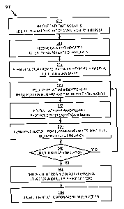
[0049] FIG. 5 is a flow diagram that illustrates at a high level a method
500 for
determining site of origin for VT, according to an embodiment. Although steps
in FIG. 5
are shown in a particular order for purposes of illustration, in other
embodiments, one or
more steps may be performed in a different order or overlapping in time, in
series or in
parallel, or one or more steps may be omitted or added, or changed in some
combination
of ways. In other embodiments, a different site related to a different
electrical pulse
inside a living body is determined by a similar method.
-12-
CA 02700390 2010-03-22
WO 2009/045852
PCTPUS2008/077708
[0050] In step 502, data is received that indicates a 3D electro-anatomic
map of an
organ of interest, such as a ventricle. Any method may be used to receive this
data. For
example, in various embodiments, the data is included as a default value in
software
instructions, is received as manual input from a network administrator on the
local or a
remote node, is retrieved from a local file or database, or is sent from a
different node on
the network, either in response to a query or unsolicited, or the data is
received using
some combination of these methods.
[0051] For example, during step 502, an interventional electrophysiologist
executes
multiple touches of a ventricle wall with probe 142, positioned by virtue of
probe
positioned sensors 146. This data is fed to a commercially available software
package,
such as CARTOTm or NAVXTm A model of a standard heart is combined with this
data to
determine the shape and polarization values of the particular ventricle 192 of
particular
patient 190. The result is the 3D electro-anatomic map of the ventricle of
interest. In
some embodiments, a different anatomical model is used for a different type of
natural
electrical pulse. In some embodiments, step 502 is omitted.
[0052] In step 510 data is received that indicates surface electrical
measurements of
the natural electrical pulse. For example, the 12 lead measurements associated
with the
natural VT are received, such as traces 420 depicted in FIG. 4.
[0053] In step 512 a natural vector is formed from the surface electrical
measurements of the natural electrical pulse in a particular time interval.
For example, a
VT vector is formed from traces 420 in the time interval from tO 430a to ti
430b. The size
of the time interval is selected to give a good indication of the direction of
propagation of
the pulse of interest. For example, in the case of a VT vector, the time
interval starts at
the start of the QRS interval, is a short time compared to the heart beat but
sufficiently
long to characterize the direction (positive or negative) and proximity of the
pulse (as
indicted by the magnitude of the measured voltage change). It is assumed for
purposes of
illustration that the time interval duration is 0.08 seconds. In other
embodiments, other
time interval durations are selected
[0054] In the illustrated embodiment, a 12 element vector is produced based
on the
traces 420 and the time interval tO 430a to tl 430b. The first element of the
vector is
based on the direction and magnitude of the voltage change during the selected
interval of
the trace 420a of lead I by using a signed numeric value. Similarly, the
second through
-13-
CA 02700390 2010-03-22
WO 2009/045852
PCT/US2008/077708
12th elements of the vector are based on the direction and magnitude of the
voltage
change during the selected interval of the traces 420b through 4201,
respectively. For
purposes of illustration it is assumed that the VT vector is a 12 element
vector represented
by the twelve values (0,-2, -2, 0, 1, -1, -1, -2, -2, -2, -1, -1), based on
the changes in the
selected 0.08 second intervals beginning at the start of QRS.
[0055] This vector captures the propagation of a surface pulse that is
based on the
propagation of the natural pulse inside the living body. In some embodiments,
the 12
element vector is reduced to a 3 element vector in the patient coordinate
system (right to
left, superior to inferior, anterior to posterior).
[0056] In some embodiments that involve periodic pulses, such as in a
beating heart,
each vector element is based on the average of several time intervals all
during the same
phase of multiple periodic pulses. Thus, each of the twelve values in the
illustrated VT
vector represents the average change over 0.08 seconds after QRS onset for
several heart
beats. Averaging serves to increase signal to noise ratio and produce vectors
that are
more stable in time.
[0057] In some embodiments, the change is determined by the signed temporal
gradient over the selected interval (e.g., in milliVolts per millisecond). In
some
embodiments, more than one statistic of the change during the selected
interval is
characterized, such as both the signed gradient and signed curvature of the
change in the
selected interval. In this case the vector has twice as many elements, e.g.,
24 instead of
12. As further statistics of the change are characterized, the number of
elements in the
vector increases.
[0058] In some embodiments, not all lead traces are used. For example, in
some
embodiments leads I, II and III are excluded and the vector includes only 9
elements, one
for each electrode.
[0059] FIG. 6 is a block diagram 600 that illustrates example mapping of
vectors
produced from lead measurements to positions in a ventricle, according to an
embodiment. Diagram 600 includes an ellipse that represents 12 dimensional
lead space
620 and a second ellipse that represents 3 dimensional ventricle wall space
610. The
origin 611 of the 3-D ventricle wall space is represented by the center of the
diamond
inside 3-D space 610. Locations in the ventricle are represented by points in
this ellipse,
such as point 612a, point 612b, point 612c, point 612d, point 612d, point
612e, inferred
-14-
CA 02700390 2010-03-22
WO 2009/045852 PC
T/US2008/077708
VT SO point 650, among others, collectively referenced hereinafter as
ventricle space
points 612. The origin 621 of the 12-D lead space is represented by the center
of the
diamond inside 3-D space 620. Particular lead measurements are represented by
points in
this ellipse, such as point 622a, point 622b, point 622c, point 622d, point
622d, point
622e, and VT lead vector point 624, among others, collectively referenced
hereinafter as
ventricle space points 622. Each dimension in lead space corresponds to a
different lead
of the 12 standard ECG leads.
[0060] A point in each space can also be represented by an arrow that
starts at the
origin and ends at the point. For example, point 612b can be represented by
the arrow
613 from the origin 611 to the point 612b. The point 622h can be represented
by the
arrow from the origin 621 to the point 622b.
[0061] It is assumed for purposes of illustration that the VT vector (0,-2,
-2,0, 1, -1, -
1, -2, -2, -2, -1, -1), formed during step 512, is represented by the VT lead
vector point
624.
[0062] In step 520, data is received that indicates the next position of a
stimulating
probe and the time of the stimulation. For example, during step 520 the
location is
received of the tip of probe 142 in ventricle 192 as expressed in the
coordinates of the 3D
electro-anatomic model received in step 502. It is further assumed that this
position
corresponds to point 612a in the 3-D ventricle wall space 610
[0063] In step 522, data is received that indicates surface electrical
measurements of
the stimulation. For example, during step 522, an interventional
electrophysiologist
moves the probe 142 to the depicted position in the heart ventricle 192 and
depolarizes
the ventricle wall. The 12 lead measurements associated with the pace mapping
are
received, such as traces 320 depicted in FIG. 3.
[0064] In step 530 a stimulated vector is formed from the surface
electrical
measurements of the stimulation in a particular time interval. The vector
elements are
formed in the same manner as the elements of the natural pulse vector is
formed, from the
same surface electrical sensors at the same locations. The size of the time
interval is
selected to match that used to form the natural pulse vector. For example, a
pace vector is
formed from 12 traces 320 in the time interval from tO 330a to ti 330b. It is
assumed for
purposes of illustration that the time interval duration is 0.08 seconds. In
other
embodiments, other time interval durations are selected
-15-
CA 02700390 2010-03-22
WO 2009/045852
PCT/US2008/077708
[0065] In the illustrated embodiment, a 12 element vector is produced based
on the
traces 320 and the time interval tO 330a to ti 330b. For purposes of
illustration it is
assumed that the VT vector is a 12 element vector represented by the twelve
values
(-1, 0, 0, 1, -1, 0, 2, -2, -2, -2, -1, -1), based on the changes in the
selected 0.08 second
interval beginning at the stimulation voltage spike.
[0066] This vector captures the propagation of a surface pulse that is
based on the
propagation of the stimulated pulse inside the living body. In some
embodiments, the 12
element vector is reduced to a 3 element vector in the patient coordinate
system (right to
left, superior to inferior, anterior to posterior).
[0067] In some embodiments that involve periodic pulses, such as in a
beating heart,
each vector element is based on the average of several time intervals all
during the same
phase of multiple periodic pulses. Thus, each of the twelve values in the
vector
represents the average change over 0.08 seconds after the stimulation spike
for several
stimulated heart beats. In some embodiments, more or fewer vector elements are
determined to match the vector elements in the natural pulse vector.
[0068] In step 540, it is determined whether another pace stimulation is to
be
performed. If so, control passes back to step 520 to receive data that
indicates the time
and location of the next stimulation signals. For purposes of illustration, it
is assumed
that steps 520 through 540 are repeated sufficiently to have enough
information to deduce
the 3D position of the site of origin.
[0069] For purposes of illustration, it is assumed that steps 520 through
540 are
repeated five times. As a result of repeating these steps five times, five 12-
D vectors are
obtained, represented by point 622a, point 622b, point 622c, point 622d, point
622e in
FIG. 6. Associated with each is a 3-D position on a wall of the ventricle of
interest,
where depolarization pulses were stimulated, represented by point 612a, point
612b, point
612c, point 612d and point 612e, respectively.
[0070] In step 550 a site of origin is determined based on the natural
vector and the
multiple stimulated vectors with associated locations. Any method may be used.
For
example, inferred VT SO 650 is determined based on the associated points
(point 622a
associated with point 612a; point 622b associated with point 612b; point 622c
associated
with point 612c; point 622d associated with point 612d; point 622e associated
with
point 612e) and the VT lead vector 624..
-16-
CA 02700390 2010-03-22
WO 2009/045852
PCT/US2008/077708
[0071] In some embodiments, a single vector transform is determined that
best
converts every stimulated vector to the different corresponding position
within the body.
Any method may be used to determine the transform. In some embodiments, an
electrical
propagation model is used to produce a model of surface electrical values tied
to a site of
origin and parameters that describe electrical properties of intervening
tissues. In some
embodiments, a parametric equation of a particular or arbitrary polynomial or
other form
is used to relate the 12-D vectors to the 3-D vectors. The parameters of the
propagation
model or arbitrary form are fit to the observations of surface electrical
quantities, for
example using a least squares approach in some embodiments.
[0072] When the vector transform operates on any 12-D vector used in its
derivation,
the output is a 3-D vector that is close to the associated 3-D point. Thus
when the vector
transom operates on point 622b it outputs a 3-D coordinate close to 612b, as
represented
by the arrow 640a. The same vector transform operates on VT lead vector 624 to
produce
an inferred VT SO point 650, as represented by arrow 640b.
[0073] In some embodiments a linear combination of the different stimulated
vectors
is determined to produce the natural vector. For example, a linear combination
of he
vectors represented by points 622a, 622b, 622c, 622d, 622e, is determined that
produces
the VT lead vector 624. That same linear combination is used to deduce the
inferred VT
SO point 650 from the 3-D positions represented by points 612a, 612b, 612c,
612d, 612e.
In essence, the vectors 622a through 622e form a vector basis set for
describing any
arbitrary point in 12-D space 620, while the corresponding vectors 612a
through 612e
form a basis set for describing any point in 3-D space 610.
[0074] In some embodiments, inferred VT SO point 650 is taken as the final
VT SO
and control passes to step 560. In some embodiments, the inferred VT SO point
650 is
used as the next stimulation location and control passes back to step 520.
[0075] In some embodiments, a 3-dimensional (3D) vector is derived from the
VT
12-lead ECG as well as from each of the pace-map 12-lead ECGs. A quantitative
comparison between the 3D vector derived from the VT and those vectors derived
from
the pace-maps is used to guide the catheter movement to the SO of VT. In some
embodiments, vector analysis is used to determine an angle between the pace-
map-
derived vector and the VT-derived vector.
-17-
CA 02700390 2010-03-22
WO 2009/045852
PCT/US2008/077708
[0076] In some embodiments, paired analysis of each of the created pace-map-
derived vectors with the VT-derived vector provide multiple correction angles,
resulting
in a final direction for a vector that intersects with the surface grid of the
previously
created electro-anatomic map. For example, an angle formed between arrow 623
and
arrow 640a is the vector transform. In some embodiments, an angle formed
between
arrow 623 and arrow 613 is the vector transform. That same angle is applied to
a vector
from origin 621 to VT lead vector point 624 to produce the derived vector
(transform
640b). The derived vector (transform 640b) intersects with the 3D electro-
anatomic map
at a minimum of one and a maximum of two points, including point 650. In the
case of
two intersection points, one of the points is rejected based upon data derived
from
concurrent paired analyses. The result is the identification of a single point
(e.g., inferred
VT SO point 650) which represents the predicted VT SO. In some embodiments,
the
probe is directed to the next pacing site by on-line vector analysis and the
VT SO is
inferred with subsequent iterations, when enough data has been acquired.
[0077] As mentioned above, in some embodiments, validation by pace-mapping
at
that specific point and its immediate vicinity confirms this point as the SO
of VT. Using
this method, one can rapidly focus on a specific site rather than performing
extensive,
time-consuming pace-mapping throughout the ventricle in search of the SO of
VT.
[0078] In some embodiments, the least squares method is also used to find
the least
distance between two vectors thus detecting vector coincidence of the VT-
derived vector
and the pace-map-derived vectors. This is used as an adjunct technique either
as an initial
step to align the VT-derived vector with one of the pace-map-derived vectors
to guide
subsequent vector analysis or after completion of vector analysis to further
qualify the
predicted VT SO, where one is dealing with a much more circumscribed area, for
more
accurate VT SO localization
[0079] If it is determined, in step 540, that another pace stimulation is
not to be
performed, control passes to step 560. In step 560, treatment is administered
based on the
site of origin. For example, the VT SO is ablated with electrical, chemical or
other source
of energy to form a lesion that inhibits depolarization at the location of
that lesion.
4. Hardware Overview
[0080] FIG. 7 is a block diagram that illustrates a computer system 700
upon which
an embodiment of the invention may be implemented. Computer system 700
includes a
-18-
CA 02700390 2010-03-22
WO 2009/045852
PCT/US2008/077708
communication mechanism such as a bus 710 for passing information between
other
internal and external components of the computer system 700. Information is
represented
as physical signals of a measurable phenomenon, typically electric voltages,
but
including, in other embodiments, such phenomena as magnetic, electromagnetic,
pressure, chemical, molecular atomic and quantum interactions. For example,
north and
south magnetic fields, or a zero and non-zero electric voltage, represent two
states (0, 1)
of a binary digit (bit). A sequence of binary digits constitutes digital data
that is used to
represent a number or code for a character. A bus 710 includes many parallel
conductors
of information so that information is transferred quickly among devices
coupled to the
bus 710. One or more processors 702 for processing information are coupled
with the bus
710. A processor 702 performs a set of operations on information. The set of
operations
include bringing information in from the bus 710 and placing information on
the bus 710.
The set of operations also typically include comparing two or more units of
information,
shifting positions of units of information, and combining two or more units of
information, such as by addition or multiplication. A sequence of operations
to be
executed by the processor 702 constitutes computer instructions.
[0081] Computer system 700 also includes a memory 704 coupled to bus 710.
The
memory 704, such as a random access memory (RAM) or other dynamic storage
device,
stores information including computer instructions. Dynamic memory allows
information
stored therein to be changed by the computer system 700. RAM allows a unit of
information stored at a location called a memory address to be stored and
retrieved
independently of information at neighboring addresses. The memory 704 is also
used by
the processor 702 to store temporary values during execution of computer
instructions.
The computer system 700 also includes a read only memory (ROM) 706 or other
static
storage device coupled to the bus 710 for storing static information,
including
instructions, that is not changed by the computer system 700. Also coupled to
bus 710 is
a non-volatile (persistent) storage device 708, such as a magnetic disk or
optical disk, for
storing information, including instructions, that persists even when the
computer system
700 is turned off or otherwise loses power.
[0082] Information, including instructions, is provided to the bus 710 for
use by the
processor from an external input device 712, such as a keyboard containing
alphanumeric
keys operated by a human user, or a sensor. A sensor detects conditions in its
vicinity
-19-
CA 02700390 2010-03-22
WO 2009/045852
PCT/US2008/077708
and transforms those detections into signals compatible with the signals used
to represent
information in computer system 700. Other external devices coupled to bus 710,
used
primarily for interacting with humans, include a display device 714, such as a
cathode ray
tube (CRT) or a liquid crystal display (LCD), for presenting images, and a
pointing
device 716, such as a mouse or a trackball or cursor direction keys, for
controlling a
position of a small cursor image presented on the display 714 and issuing
commands
associated with graphical elements presented on the display 714.
[0083] In the illustrated embodiment, special purpose hardware, such as an
application specific integrated circuit (IC) 720, is coupled to bus 710. The
special
purpose hardware is configured to perform operations not performed by
processor 702
quickly enough for special purposes. Examples of application specific ICs
include
graphics accelerator cards for generating images for display 714,
cryptographic boards for
encrypting and decrypting messages sent over a network, speech recognition,
and
interfaces to special external devices, such as robotic arms and medical
scanning
equipment that repeatedly perform some complex sequence of operations that are
more
efficiently implemented in hardware.
[0084] Computer system 700 also includes one or more instances of a
communications interface 770 coupled to bus 710. Communication interface 770
provides a two-way communication coupling to a variety of external devices
that operate
with their own processors, such as printers, scanners and external disks. In
general the
coupling is with a network link 778 that is connected to a local network 780
to which a
variety of external devices with their own processors are connected. For
example,
communication interface 770 may be a parallel port or a serial port or a
universal serial
bus (USB) port on a personal computer. In some embodiments, communications
interface 770 is an integrated services digital network (ISDN) card or a
digital subscriber
line (DSL) card or a telephone modem that provides an information
communication
connection to a corresponding type of telephone line. In some embodiments, a
communication interface 770 is a cable modem that converts signals on bus 710
into
signals for a communication connection over a coaxial cable or into optical
signals for a
communication connection over a fiber optic cable. As another example,
communications interface 770 may be a local area network (LAN) card to provide
a data
communication connection to a compatible LAN, such as Ethernet. Wireless links
may
-20-
CA 02700390 2010-03-22
WO 2009/045852
PCT/US2008/077708
also be implemented. Carrier waves, such as acoustic waves and electromagnetic
waves,
including radio, optical and infrared waves travel through space without wires
or cables.
Signals include man-made variations in amplitude, frequency, phase,
polarization or other
physical properties of carrier waves. For wireless links, the communications
interface 770
sends and receives electrical, acoustic or electromagnetic signals, including
infrared and
optical signals, that carry information streams, such as digital data.
[0085] The term computer-readable medium is used herein to refer to any
medium
that participates in providing information to processor 702, including
instructions for
execution. Such a medium may take many forms, including, but not limited to,
non-
volatile media, volatile media and transmission media. Non-volatile media
include, for
example, optical or magnetic disks, such as storage device 708. Volatile media
include,
for example, dynamic memory 704. Transmission media include, for example,
coaxial
cables, copper wire, fiber optic cables, and waves that travel through space
without wires
or cables, such as acoustic waves and electromagnetic waves, including radio,
optical and
infrared waves.
[0086] Common forms of computer-readable media include, for example, a
floppy
disk, a flexible disk, a hard disk, a magnetic tape, or any other magnetic
medium, a
compact disk ROM (CD-ROM), a digital video disk (DVD) or any other optical
medium,
punch cards, paper tape, or any other physical medium with patterns of holes,
a RAM, a
programmable ROM (PROM), an erasable PROM (EPROM), a FLASH-EPROM, or any
other memory chip or cartridge, a carrier wave, or any other medium from which
a
computer can read.
[0087] Network link 778 typically provides information communication
through one
or more networks to other devices that use or process the information. For
example,
network link 778 may provide a connection through local network 780 to a host
computer
782 or to equipment 784 operated by an Internet Service Provider (ISP). ISP
equipment
784 in turn provides data communication services through the public, world-
wide packet-
switching communication network of networks now commonly referred to as the
Internet
790. A computer called a server 792 connected to the Internet provides a
service in
response to information received over the Internet. For example, server 792
provides
information representing video data for presentation at display 714.
-21-
CA 02700390 2010-03-22
WO 2009/045852
PCTMS2008/077708
[0088] The invention is related to the use of computer system 700 for
implementing
the techniques described herein. According to one embodiment of the invention,
those
techniques are performed by computer system 700 in response to processor 702
executing
one or more sequences of one or more instructions contained in memory 704.
Such
instructions, also called software and program code, may be read into memory
704 from
another computer-readable medium such as storage device 708. Execution of the
sequences of instructions contained in memory 704 causes processor 702 to
perform the
method steps described herein. In alternative embodiments, hardware, such as
application
specific integrated circuit 720, may be used in place of or in combination
with software to
implement the invention. Thus, embodiments of the invention are not limited to
any
specific combination of hardware and software.
[0089] The signals transmitted over network link 778 and other networks
through
communications interface 770, carry information to and from computer system
700.
Computer system 700 can send and receive information, including program code,
through
the networks 780, 790 among others, through network link 778 and
communications
interface 770. In an example using the Internet 790, a server 792 transmits
program code
for a particular application, requested by a message sent from computer 700,
through
Internet 790, ISP equipment 784, local network 780 and communications
interface 770.
The received code may be executed by processor 702 as it is received, or may
be stored in
storage device 708 or other non-volatile storage for later execution, or both.
In this
manner, computer system 700 may obtain application program code in the form of
a
signal on a carrier wave.
[0090] Various forms of computer readable media may be involved in carrying
one or
more sequence of instructions or data or both to processor 702 for execution.
For
example, instructions and data may initially be carried on a magnetic disk of
a remote
computer such as host 782. The remote computer loads the instructions and data
into its
dynamic memory and sends the instructions and data over a telephone line using
a
modem. A modem local to the computer system 700 receives the instructions and
data on
a telephone line and uses an infra-red transmitter to convert the instructions
and data to a
signal on an infra-red a carrier wave serving as the network link 778. An
infrared detector
serving as communications interface 770 receives the instructions and data
carried in the
infrared signal and places information representing the instructions and data
onto bus 710.
-22-
CA 02700390 2015-04-21
Bus 710 carries the information to memory 704 from which processor 702
retrieves and
executes the instructions using some of the data sent with the instructions.
The
instructions and data received in memory 704 may optionally be stored on
storage device
708, either before or after execution by the processor 702.
5.0 Extensions and Modifications
100911 In the
foregoing specification, the invention has been described with reference
to specific embodiments thereof. It will, however, be evident that various
modifications
and changes may be made thereto without departing from the scope of the
invention as
described herein.
-23-