Note: Descriptions are shown in the official language in which they were submitted.
CA 02700942 2010-03-26
WO 2009/040413 PCT/EP2008/062909
An ex vivo, fast and efficient process to obtain activated antigen-
presenting cells that are useful for therapies against cancer and
immune system-related diseases
This invention refers to an ex vivo, fast and efficient process to obtain
activated antigen-presenting cells that are useful for therapies against
cancer
and immune system-related diseases. At the same time, it is related to a
cellular
composition that contributes to stimulate the activated antigen-presenting
cells
to induce a specific immune response against tumors in patients with cancer or
other pathologies involving immune responses.
STATE OF THE ART
With the improvement of new medical technologies and the upgrading of
material conditions, life expectancy for world population has increased,
especially in developed countries. This has lead to an increase incidence of
various tumors and cancer in the population, showing an overall augmented
number of patients suffering from cancer, as well as immune system-associated
disorders
Cancer is a pathology in which cells with an uncontrolled capacity for
growth and spreading are able to invade their originating organs or tissues
and
spread towards the body through the blood or lymphatic tissues. Its aberrant
expansion destroys healthy tissues, producing metabolic unbalances and
altering the function of organs, many times causing death. In the light of
recent
developments, the treatment for this disease has been improved. However, this
pathology still remains one of the primary causes of death worldwide.
Over the last thirty years, great progress has been achieved in
understanding the contribution of the immune system, regarding tumor cell
recognition and destruction, so the manipulation of the immune system as an
antitumoral tool has become a potential alternative for cancer treatment. The
so-
called antitumoral immune therapy may be used as a complement for usual
treatments of ontological conditions, such as surgery, chemotherapy and
radiotherapy.
Although some typesof immune therapy are already a part of the usual
treatment of some types of cancer, there are others in a preclinical or
clinical
CA 02700942 2010-03-26
WO 2009/040413 PCT/EP2008/062909
2
trial stage. Among the strategies employed in immune therapy, the use of
immune molecules, such as interferons, interleukins, colony-stimulating
factors
and monoclonal antibodies has been of vital importance. A different strategy
is
the active immunization against tumors, which is commonly known as cancer
vaccines.
Therapeutic vaccines for cancer disorders are a form of specific immune
therapy, whose purpose is stimulating or strengthening a direct response of
the
patient against the tumor through immunization, for instance with inactivated
or
radiation exposed tumor cells, or by administering tumor antigen-containing
(Ag)
vaccines.
Tumor-associated antigens or tumor-specific antigens are protein-origin
molecules mainly, which are differentially expressed in the tumor and normal
tissue, where they become a target for immunological responses.
Cancer vaccines are generally provided after the onset of the disease; to
this effect complete attenuated cells may be used, as well as cellular
compounds or specific antigens with the purpose of stimulating the patient's
immune system. These vaccines may be commonly classified as complete
tumor cell vaccines or vaccination preparations from tumor antigens. The
former
may be divided in complete autologous cell vaccines coming from the subject
itself and in complete allogeneic cells consisting in a combination of tumor
cells
of the same histological type but from different patients. These preparations
are
manufactured in laboratory facilities and they are usually combined with
adjuvant.
The tumor-associated Ags can be obtained from complete tumor cells,
from tumor-purified proteins or peptides, from artificially synthesized
peptide
sequences or genetic material obtained of the tumors.
In regard to this, vaccines of specific proteins/peptides are designed from
tumor-associated antigens, which are recognized by T lymphocytes. The
antigenic peptide or protein may be administered purified or synthesized as a
part of the vaccine composition or by inducing the synthesis of the tumor
peptide
or antigen into the target cell by transfection.
To introduce genetic material into the body, viral vectors, such as
adenoviruses may be used. Although, adenovirus is the most commonly used
virus, retroviruses have also been used with successful results. These viral
CA 02700942 2010-03-26
WO 2009/040413 PCT/EP2008/062909
3
vectors might also encode additional cytokine genes beside the tumor-
associated antigen.
The DNA vaccines consisting in plasmids coding tumor Ag have the
advantage of acting independently from the subject's MHC haplotype. New
strategies are currently being developed for this kind of vaccines involving
the
fusion of genes, such as coding agents for idiotypic determinants of the
immunoglobulin molecule with a sequence of the titanic toxoid antigen, which
enables the activation of the immune system's effector mechanisms.
Another therapeutic alternative corresponds to the dendritic cell vaccines
or professional antigen-presenting cells (APC), which is a technique recently
incorporated to clinical practice and seems to be interestingly effective for
generating a specific CTL response against tumors and infectious agents.
Notwithstanding the multiplicity of developing alternatives for the
treatment of tumors, the success of active immunotherapy in cancer treatment
may be affected by multiple factors such as the heterogeneity existing among
tumor cells, for instance, the low immunogenicity of tumor antigens and the
immune evasion mechanisms developed by tumors to avoid the immunological
response. The tumor-associated antigens -potentially immunogenic molecules -
may be effective targets for cancer vaccines, but they may also be present in
normal cells and not be recognized by the immunological system for different
reasons, such as the cryptic expression due to the physical orientation or
configuration of Ag on the cell surface, the physical separation, the
separation
by cell membranes or masking by other cell components; lower antigenic
expression than the required for immune recognition or a different surface
distribution regarding tumor cells.
Recently, the existence of regulatory lymphocytes (Treg) has been
described. Tregs are able to inhibit immune responses and their main role is
keeping tolerance in order to avoid autoimmune responses. There is evidence
that these cells may exert a deleterious effect on the generation of
antitumoral
responses in patients with cancer, which would enhance the tumor growth.
Consequently, objectives pursued by the active immune therapy against
cancer would be: overcoming the immune suppression produced by tumoral
deriving factors, increasing the immunogenicity of antigens that may help
eliminating tumors and metastasis and the clinical recovery of patients when
treated with any antitumoral vaccine.
CA 02700942 2010-03-26
WO 2009/040413 PCT/EP2008/062909
4
The development of dendritic cell (DC) vaccines is an explored alternative
with promissory results. The DCs are originated in the bone marrow from
pluripotential progenitors and about 0.5% of total blood mononuclear cells
correspond to DC in circulation and they are very hard to maintain in culture
conditions (Fearnley D.B. et al. 1999). DCs are a subgroup of leukocytes with
a
great antigen-presentation capacity and the potential to induce and regulate
the
immune response (Svane IM et al. 2003, Banchereau J et al. 2003). DCs have
proven to be the most effective antigen-presenting cells (APC); this is why
they
are called professional APCs. By presenting intra- and extra-cellular
antigens,
they are able to induce a T lymphocyte CD4* and CD8*- mediated specific
immune response. DCs are strategically positioned in peripheral tissues in
possible antigen-entering areas, where they are able to capture process and
present them associated with histocompatibility molecules (HCM). DCs
comprise a heterogeneous population with different surface markers
(phenotype) associated with their maturation tempo. It is thought that
different
stimuli would be able to trigger qualitatively different maturation processes,
thus
suggesting that DCs could interpret environment signals, which depend on the
stimulus nature and then develop to mature DCs which are able to polarize a
the
LT immune response into Thl or Th2 (cellular or humoral immune response
respectively) or to a tolerogenic type of response (Moser M and Murphy KM.
2000). During the maturing transition, the phenotype of DCs changes,
cytoplasmatic prolongations increase as well as the characteristic markers of
immature DCs (DCi) decrease; at the same time the expression of co-
stimulating molecules begin to increase, such as CD40, CD80 and CD86, CD83,
class I and II MHC molecules and the chemokine receptor CCR-7, which
recognizes chemokines CCL19 and CCL21, which guide migration of DCs to the
T zone of secondary lymphoid organs, where the naive antigen-specific LT
clone may be found (Mellman I et al. 2001, Delamarre L et al. 2003).
Since obtaining these cells from peripheral blood is difficult and laborious
intense, different methods have been developed during the last decade for
their
in vitro generation from monocytes, thus allowing a greater quantity of DCs to
be
available for study and use in immune therapies as an alternative treatment
for
cancer. During the last two decades, different clinical trials of vaccination
with
autologous DCs have been published in relation to the treatment of advanced
cancer. In most of them researchers use DCs generated from CD14+
CA 02700942 2010-03-26
WO 2009/040413 PCT/EP2008/062909
monocytes or CD34+ progenitor cells cultured for seven to ten days in a
culture
medium supplemented with granulocytes and macrophages colony of
stimulating factors (GM-CSF) and interleukine-4 (IL-4), adding the alpha tumor
necrosis factor (TNF-a) as a maturing stimulus (Sallusto F. et al. 1994, Svane
5 1G. et al. 2003). It has been proposed, however, that generating mature in
vitro
DCs is possible in shorter periods. FASTDC are obtained in 48 hours, using a
combination of proinflammatory factors as IL-6, ILA P, prostaglandin E2 and
TNF-a (Dauer et al. 2003) as maturing stimulus. Three days (3 days APC) are
generated by mixing of macrophages and Langerhans cells obtained by a
combination of GM-CSF and TNF-a described in the international application
W02004/050855. Although these protocols shorten the DC-production times,
their drawbacks are the high cost derived from the use of human recombinant
cytokines, further necessary addition of antigens able to arouse immunological
responses against tumors and in some cases, the lower levels of DCs maturing
capacity, which limits their clinical use due to the potential risk of tumor
evasion
or tolerance induction.
Other recent studies state that several population of DC on the periphery
derives from monocytes that infiltrate tissues due to inflammatory stimuli
probably mediated by innate immunity (Palucka KA et al. 1998). This
differentiation process of in vivo monocytes is performed in early stages of
the
immunological response in order to allow in less than a week, an effective T
lymphocyte mediated response (Gwendalyn J. et al. 1998). The current DC
production methods involve in vitro incubation periods of several days, which
lessens the strength, viability and quality of DCs or use a complex set of pro-
inflammatory recombinant factors that generate a type of APC that is
questioned for some of its phenotypic features associated with activated
monocytes, immature DCs and macrophages; this is why its therapeutic use
against infections or tumors is limited due to possibility of tolerance
induction (J
lmmunol. 2004, Dauer M, et al. 2003). There is evidence in literature that
activated monocytes and immature DCs have the capacity to react to stimulus
from molecules termed pathogen-associated molecular patterns, PAMPs,
through pattern recognitions receptors, PRRs (Steinman RM et al. 2006). There
are several PRRs ligands and they exist not only in pathogens, but as
endogenous molecules expressed mainly in transformed, infected or stressed
cells and are able to activate PRRs.
CA 02700942 2010-03-26
WO 2009/040413 PCT/EP2008/062909
6
Among the experiences developed to obtain antigen-loaded DCs, the
method described in US20020155108 consists in an ex vivo DC co-culture
performed along with soluble antigens, without physical contact, an antibody
is
included against the soluble antigen in order to form immune complexes which
the DCs are able to absorb, process and present on the cell surface.
In the Japanese patent application JP2000143534, a method is disclosed
to obtain DC vaccines with antigen-presenting activity. The mentioned method
consists in incubating a DC with antigen-presenting activity with the
following
components: a suspension of cells containing a DC cellular precursor, for
instance bone marrow cells, blood cells from umbilical cord or peripheral
blood
monocytes; a differentiation-inducing agent, for instance combinations of GM-
CSF, IL-4, TNF-a, stem cells factor and TNF-a; and a chemokine.
In addition, W002053176 describes a method to produce autologous
APCs loaded with a mixture of at least two lysate of allogeneic melanoma tumor
cells. Maturation of DC is induced with TNF, E2 prostaglandin and/or
polyribocytidilic acid.
The US 2007/0014795 describes in turn a method for activation of
antigen presenting cells, which might be DCs.
In published results from L6pez M: et al (Rev Med Chile 2004; 132: 1115-
1126) and Escobar et al. (Clin. Exp. Immunology 2005; 142(3): 555-568) the
authors demonstrate a procedure for DCs production from monocytes by 7-day
incubation and a later incubation with TNF-a and with tumor lysate of three
lines
of allogeneic melanoma. In addition, Nestle FO et al. (Nature Medicine N 4,
328
332 (1998) discloses a clinical study where DCs are induced by monocyte
culture for also during seven days with GM-CSF, IL-4 and a lysate of tumor
cells
or a group of known peptides identified through recognition by T cytotoxic
lymphocytes.
The state of the art allows us identify some weaknesses and limitations
resulting in drawbacks for the development and practical application of the
technology disclosed in those inventions. On one hand, the methods described
require a laborious and time consuming preparation of these cells, up to 8 to
9
days in total. Moreover, these technologies result in the aging of cells,
which
may shorten their survival in the body after their injection or may affect
their
functionality. In addition to the above, these technologies also involve a
slow
differentiation of cells which does not reflect a natural process, since it is
known
CA 02700942 2010-03-26
WO 2009/040413 PCT/EP2008/062909
7
that monocytes differentiating in vivo into DCs are able to do it in hours and
not
within 3 or more days (Randolph GJ, Science 1998).
From the methodological point of view, the methods described in the art
provide both operating disadvantages and inefficiencies as compared with this
invention. First, classic DCs require a longer in vitro incubation time (7-9
days)
which increases the production cost, increasing the use of reagents acting as
differentiation factors and culture media, in addition to an increased risk of
infection or cell's death. Second, in the processes described in the state of
the
art, the rate of DCs obtained from blood mononuclear cells (PBMC) is at least
three times lower than the obtained by using the method proposed in this
invention. In addition, the APCs obtained according to this invention have
characteristics that make them more effective to be used as anti-tumor therapy
in patients with cancer.
Consequently, in order to optimize the in vitro generation of APCs, this
invention refers to an extract or lysate of tumor tissue or cells and to a
fast an
effective method to produce APCs, from pre activated peripheral blood
monocytes by differentiation cytokines and matured with components of cell
lysate of tumor tissue. Lysate obtained through our treatment have a double
function: on one hand, they are able to induce differentiation and maturation
of
activated monocytes into APCs highly similar to mature DCs and they are also
able to provide a wide range of tumor antigens able to induce the activation
of T
lymphocytes with the potential to recognize and destroy tumor cells.
DESCRIPTION OF FIGURES
The figures described below are proposed in order to show background
information to back-up and describe the invention; therefore, they are not
intended to restrict and must by no means be understood as limiting the scope
of the development proposed.
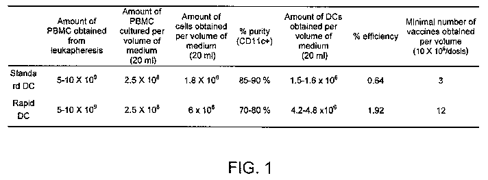
Figure 1 corresponds to a table showing a comparative evaluation of the
efficiency between the method proposed herein of rapid differentiation of
dendritic cells (Rapid DC) as compared with the traditional method of seven-
day
DC production (DC standard). It is noted that from the same number of
peripheral blood cells (PBMC), nearly 4 times the quantity of DCs is obtained
when using the method proposed in this invention, which in turn allows
obtaining
a greater quantity of doses for vaccination of patients. Also, the use of
fewer
CA 02700942 2010-03-26
WO 2009/040413 PCT/EP2008/062909
8
differentiation factors and culture medium, the facility and cost of
production is
reduced to half of the value.
Figure 2 shows the expression of melanoma-associated antigens
expressed in some lines of melanoma used to obtain an extract or lysate of
tumor cells comprising part of the invention. The expression of melanoma-
associated antigens was determined by Immunohistochemistry (*), flow
cytometry (#) or RT-PCR ( ). The combination of these lines altogether is able
to express a wide range of melanoma antigens.
Figure 3 shows the morphology of rapid differentiation dendritic cells
(Rapid DC) belonging to this invention, which does not differ from the
morphology of standard DCs (7 days). DCs are identified with arrows.
Figure 4 shows monocytes incubated with GM-CSF and IL-4 and
stimulated with a lysate obtained from the mixing of melanoma lines Mel 1, Mel
2 and Mel 3 called TRIMEL and TNF-a. These cells develop the characteristic
1.5 phenotype of mature DCs within 48 hours (Rapid DC). (a) As described
hereinafter in this invention, Rapid DCs were generated and stained with
monoclonal antibody (MAb) anti-CD11 c (myeloid DCs marker) conjugated with
PE to be then read through flow cytometry. The gated population represents the
percentage of positive CD11 c cells from the total cells obtained after
culture
(figure is representative staining of 5 different patients). (b) CD11 c+ DCs
were
analyzed for the expression of CD83, CD86, CCR7, CD40, class I MHC and
class II MHC. The expression analysis of these markers indicated that Rapid
DCs had the characteristic phenotype of mature dendritic cells.
Figure 5 shows images illustrating that the cells obtained through the
Rapid DC method had a similar phenotype to cells obtained by the short FastDC
protocol, as well as traditional 7-day DCs. Monocytes incubated with GM-CSF
and IL-4 for 24 hours were cultured for additional 24 hours with culture
medium
alone, TNF-a, TRIMEL alone, TNF-a and tumor lysate TRIMEL or IL-1 p + IL-6 +
TNF-a + PG-E2 (Fast DC cells). (a) The expression of myeloid DCs markers,
CD11c+ and DCs maturation markers, such as CD86 and CD83 was
determined by flow cytometry. (b) As described hereinafter in this invention
and
in the state of the art, Rapid DCs and traditional 7-day DCs were generated
and
the following markers were determined by flow cytometry: CD11 c, class I MHC,
class Ii MHC and CD83. Histograms representative of 2 independent
experiments show CD11c+ cell. Bars represent the MFl of positive CD11c cells.
CA 02700942 2010-03-26
WO 2009/040413 PCT/EP2008/062909
9
These results show that with a shorter method and using fewer factors DCs are
obtained with similar characteristics to more complex protocols described in
the
state of the art.
Figure 6 shows that the combination of melanoma lysate called TRIMEL
with TNF-a induces Rapid DC to a powerful maturation process. The evaluation
is done by flow cytometry analysis for CD11 c, CD86 and CD83 markers of
monocytes treated for 24 hours with IL-4 and GM-CSF and without a later
stimulus, only stimulated with TRIMEL or TNF-a or with TRIMEL and TNF-a.
The bars represent the MFI percentage of positive CD11 c cells. This result
also
indicates that the tumor lysate called TRIMEL is able by itself and without
TNF-a
to induce the expression of markers associated with mature DCs.
Figure 7 shows that lysate from normal cells are not able to induce Rapid
DC maturation. Lysate were prepared from autologous and allogeneic PBL and
they were used to stimulate the Rapid DCs. The expression of CD11c, CD86
and CD83 markers was measured by flow cytometry. The bars represent the
fluorescence percentage as regards the maximum fluorescence of positive
CD11 c cells. (PBL: peripheral blood lymphocytes). This result indicates that
monocytes maturation process depends of factors that are present in the tumor
lysate and not in normal cells.
Figure 8 shows that tumor lysate other than TRIMEL, made of other three
melanoma cell lines is also able to induce Rapid DC maturation process. A
tumor lysate called NO TRIMEL lysate was prepared from 3 cell lines, FM 55
(skin melanoma), OCM-1 and OCM-3 (eye melanoma) and the maturation
inducing effect was evaluated on monocytes. The expression of CD11c, CD83
and MHC 11 markers was determined by flow cytometry. Levels of maturation
markers are similar to those obtained using TRIMEL. This result indicates that
the combination of different melanoma lysate obtained from different
individuals
is able to induce the maturation process of monocytes into mature DCs.
This proves that diverse components present in tumor cells, such as melanoma,
are useful for the proper execution and performance of this invention.
Figure 9 shows that the Rapid DCs have a low capacity of endocytosis,
similar to traditional mature DCs, which is an indication that Rapid DCs are
in a
final phase of differentiation, that is, optimal for the induction of T
lymphocyte
activation. A phagocytosis assay was performed with FITC-linked Dextran and
CA 02700942 2010-03-26
WO 2009/040413 PCT/EP2008/062909
the results were measured by flow cytometry. As a control of passive
endocytosis cells were kept at 4 C,
Figure 10 shows that Rapid DC secretes IL-12 and IL-10 cytokines. (a)
Different ratios of Rapid DC cells were co-cultured with fibroblasts which
5 express CD40L -- constitutively - for 12 hours. An ELISA assay was performed
in the supernatant of co-culture to determine the concentration of secreted IL-
12
p70. (b) Peripheral blood monocytes were incubated for 24 hours with GM-CSF
and IL-4 and then stimulated with TNF-a, TRIMEL or TNF-a and TRIMEL for
further 24 hours. An ELISA assay was performed in the culture supernatant to
10 determine the concentration of secreted IL-10. The secretion of these
cytokines,
especially IL-12, indicates that Rapid DCs are able to induce Th1-type
responses, described as very effective against tumors.
Figure 11 shows that LTs stimulated with Rapid DC recognize melanoma
cells. Autologous PBL were co-cultured for 12 hours with Rapid DC, allogeneic
melanoma cells (Mel 1, Mel 2 and 0505 Mel), the NK-sensitive prototype called
K562 and rat fibroblast (NIH 3T3). The secretion of IFN-y was determined by
ELISPOT. This result shows that the Rapid DCs are able to stimulate T
lymphocytes in vitro with anti-tumor activity.
Figure 12 shows the results of a Phase I clinical trial using Rapid DC for
the treatment of 9 patients with malign advanced melanoma, two patients with
pulmonary carcinoma, one with ovarian cancer, one with colorectal carcinoma
and one with prostate cancer. None of the patients treated showed important
adverse effects, and only in some patients, redness of the injection area and
local rash was observed, which provides evidence that the treatment is
biologically safe and well tolerated. In addition, 70% of patients develop a
type
IV delayed hypersensitivity in vivo response (DTH) specific against the tumor
antigens, which exceeds the studies published before (Escobar et al. Clin. and
Exp. Immunol. 2005) where standard DCs produced immunological response in
50% of patients.
DESCRIPTION OF THE INVENTION
On one hand, this invention refers to an extract of cells and/or tumor
tissues with the capacity to induce differentiation and activation of APCs.
Another aspect of the invention, in turn, is related to a method to produce
DCs
ex vivo from peripheral blood monocytes in a shorter time, as compared with
the
CA 02700942 2010-03-26
WO 2009/040413 PCT/EP2008/062909
11
state of the art, where the extract mentioned before is used. DCs produced in
this way are useful to make up a therapeutic composition as a vaccine, which
is
useful in the treatment of cancer and other related diseases.
The method uses common blood cells obtained from patients, donors or
blood banks, among other sources, from which mononuclear cells are
separated. Then, monocytes are selected and incubated with growth factors and
cytokines to be then exposed to a tumor lysate, preferably in the presence of
a
growth factor. Under these conditions, and in less than three days after the
ex
vivo cultivation, preferably within 48 hours of ex vivo culture, these cells
express
markers associated with traditional mature dendritic cells and acquire the
capacity of inducing responses from in vitro anti-tumor cytotoxic lymphocytes
and generate in vivo immunological responses in patients vaccinated with these
cells.
The lysate of tumor cells might be obtained by different means. In one
approach to the invention, the lysate of tumor cells contains a mixture of at
least
two extracts of tumor cells kept under culture. In another approach to the
invention, the lysate of tumor cells is obtained from fresh tumor tissue taken
from patients with different types of cancer, such as melanoma and uveal
melanoma, prostate, kidney, colorectal, gastric, pulmonary, breast, ovarian,
testicle carcinomas and other types of neoplasm.
In another approach to the invention, the lysate of tumor cells is obtained
from fresh tumor tissue taken from patients with different types of cancer
combined with lysate of allogeneic tumor cell lines of the same tumor type.
DETAILED DESCRIPTION OF THE INVENTION
In the context of this invention, a rapid, efficient and cost-effective method
has been developed, to allow the training of antigen presenting cells similar
to
DCs, from monocytes of peripheral blood, so that they may in a short time
express surface markers consistent with their function. They are also able to
trigger an immune response when they become in contact with the other
components of the immune system of an organism.
On one hand, this invention uses cells obtained from blood of patients,
donors or blood banks which are separated from the other components of the
blood through traditional methods of the art; preferably leukapheresis. In
particular leukocytes are selected through the usual methods known in the art,
CA 02700942 2010-03-26
WO 2009/040413 PCT/EP2008/062909
12
such as density gradient for example. From the fraction of leukocytes,
monocytes are separated through traditional methods known by any expert in
the art. In a preferred embodiment, the capacity of monocytes to adhere to
plastic surfaces is used. In another embodiment, monocytes selection can also
be performed by separation kits which use antibodies against the C 14
molecule coupled to magnetic beads for magnetic selection of the desired
cellular type.
In a preferred embodiment of the invention, the peripheral blood
mononuclear cells are incubated at 13x106 cells per ml, although
concentrations
between 104 and 1010, preferably between 106 and 107 are also allowed in a
culture medium free from bovine fetal serum. The culture may take place in
proper containers, such as different well number plates, bottles, cell
reactors
and others. Temperatures between 30 and 40 C are tolerated; preferentially
37 C in an atmosphere of about 5% CO2 should be used for 1 to 4 hours, with
an ideal time of about 2 hours.
Cells that remained attached to the container (well) correspond to
monocytes, and are kept under culture in the presence of 100 to 800 U/ml,
preferably between 400 and 600 and with an ideal concentration of 500 U/ml of
cytokines such as interleukins preferentially IL-4.; and in the presence of
500 to
1,100, preferably between 700 and 900 and more preferably as an ideal
concentration around 800 U/ml of at least one growth factor, most preferably
GM-CFS. The incubation can be extended for at least 10 hours, although
incubation times of more than 18 hours are preferred reaching and ideal time
of
about 22 hours.
Then, the cells can be incubated for at least 10 more hours, ideally 18
hours, and preferentially for about 24 hours. In this second incubation cycle,
the
cells are kept in culture medium alone or ideally supplemented with a growth
factor, like TNF-a, or with the mixture of tumor cells lysate described above
or
with both components at the same time. In another embodiment of the invention,
the mixture of tumor cells lysate described above may be combined with other
pro-inflammatory cytokines such as IFN-y, IL-6, IL-13 or other factors like
prostaglandin E2, CpG, thermal shock proteins, Toll-like receptors (TLR)
ligands
or other factors that activate DCs maturation.
Regarding the use of growth factors, TNF-a might be used at a
concentration between 100 pglmI to 100 nglml, ideally between 1 ng/ml to 50
CA 02700942 2010-03-26
WO 2009/040413 PCT/EP2008/062909
13
nglml, more preferably between 2 ng/ml to 20 ng/ml and ideally around 10
ng/ml.
An integral and essential part of this invention is the mixture of lysate or
extracts of tumor cells. This is a mixture made up by at least two cell lines
of
tumors from metastatic tissue deriving from patients with cancer. In a
preferred
embodiment of the invention, the tumor cells are selected from malign
melanomas and correspond to three cell lines, preferably deriving from gland
metastasis. Another alternative provided by the invention, the lysate of tumor
cells is obtained from fresh tumor cell derived from patients with different
kinds
of cancer combined or not with lysate of allogeneic tumor cell lines of the
same
tumor type. The phenotype of used cells is confirmed through conventional
techniques in order to determine the expression of tumor-associated antigens.
The cells or tissues are then incubated between 15 minutes and 4 hours, with a
preferred timing of 1 and 3 hours ideally around 2 hours at a temperature that
range between 39 and 44 C, more preferably between 40 and 43 C and
preferentially near 42 C in a serum-free culture medium. Later, the cells
and/or
tissues are placed at physiological temperature again, that is, around 37 C
for 1
to 6 hours, ideally between 2 and 4 hours preferentially 3 hours before being
lysate.
Cells treated in this way are subject to I to 6 freezing and thawing cycles,
preferably 2 to 4 cycles, and ideally 3 cycles are used. For each freezing
cycle,
the cells are introduced into a tank containing liquid nitrogen, which freezes
them instantly and then thawed to 35 to 40 C.
The lysate or extract obtained is subject to homogenization by ultrasound
for 30-second 2 to 10 cycles at 30 to 40 KHz in a standard sonicator. Finally,
the
lysate or extract of each tissue is irradiated at doses ranging between 40 and
120 Gy, preferably between . 70 and 90 Gy and preferentially around 80 Gy.
Later, the lysate may be mixed or not on equal parts or individually used
depending on the type of tumor to be treated. The lysate or extract obtained
is
used in the culture of dendritic cells at a concentration between 1 p,glml and
1
mg/ml and ideally around 100 g/ml.
A quite outstanding development of this invention is that the extract of
tumor cell lysate described is able to stimulate the differentiation of
dendritic
cells from preactivated monocytes with differentiation cytokines. This
maturation
induction and differentiation occurs even in the absence of other cytokines or
CA 02700942 2010-03-26
WO 2009/040413 PCT/EP2008/062909
14
maturation factors existing in the state of the art. In these cases, it was
noted
that after 24 hours of treatment with the lysate, monocytes showed a
morphology equivalent to DCs classically incubated for 7 days (Figure 3),
which
confirms the advantages of the method proposed and the prominent qualities of
the extract developed. Also, the monocytes activated with tumor cells extracts
showed the CD11c membrane marker expression, which is characteristic of the
myeloid-type DCs in addition to the expression of a number of membrane
markers characteristic of mature DCs, such as MHC I and MHC II, CD83, CD86,
CD40 and CCR7 (Figures 4 to 6).
Of pivotal importance is that most tumor lysate and not lysate from normal
cells are able to induce this differentiation and maturation, which is a
property
that has not been described for tumor cells (Figure 7). A key feature of this
invention is indeed related to the role played by the components of tumor
cells in
the differentiation of monocytes to DCs and their later maturation. There is
indeed some background information in the state of the art on the capacity of
some necrotic tumor cells of inducing DC maturation (Bhardwaj N. et al 2000, J
Exp Med. 191:411-6; Escobar et al. 2005, Clin. Exp. lmmunol. 142:555-68), but
there is no evidence regarding the effect of these cells and their components
in
inducing also differentiation of monocytes to DCs. In this invention, it is
described that tumor lysate and/or a mixture of them are able to act on
monocytes inducing the differentiation thereof to professional antigen-
presenting
cells, similar to DCs and giving them the capacity of activating the T
lymphocyte-
mediated immune response against tumor cells, thus having a great therapeutic
potential.
Another aspect of the invention refers to the pharmaceutical composition
or vaccine obtained with DCs produced under the methods described above.
This invention provides evidence that DCs obtained under the method hereby
invented, corresponding to rapid differentiation DCs has the power of inducing
potent immune anti-tumor responses. This quality is reflected in the fact that
T
lymphocytes co-cultured with rapid DCs are able to produce inflammatory
cytokines such as interferon-y and TNF-a and recognize and destroy lines of
allogeneic melanomas through cytolysis (Figure 11). Also the cells obtained
through the method described herein are able of inducing the proliferation of
specific T lymphocytes against tumor cells.
CA 02700942 2010-03-26
WO 2009/040413 PCT/EP2008/062909
Another fundamental outcome of the invention corresponds to the use of
dendritic cells obtained under method of the invention in patients with
melanoma, other kinds of cancer or another type of immune response-
associated diseases (Figure 12).
5
EXAMPLE 1
The method of this invention allows obtaining DCs that may be
incorporated to vaccines to treat individuals suffering from different kinds
of
cancer. To this effect, in order to treat patients suffering from these
diseases,
10 blood is obtained through a standard method to obtain blood by-products
called
leukapheresis. A volume equal to 2 blood volemia is obtained from each
patient.
Blood is processed in a biohazard laboratory. The leukapheresis product is
diluted in PBS in a 1:1 dilution. Then, this product is separated by a density
gradient called LymphoprepTM as described in the state of the art. The white
15 fraction of blood consisting in the peripheral blood mononuclear cells
(PBMC) is
washed three times with PBS and then placed in culture bottles (Nunc T75) at a
concentration that ranges between 10 and 40 x 106 of PBMC/ml of a serum-free
culture medium, concentrations between 20 and 30 x 106 of PBMC/mI of a
medium are used and ideally 25 x 106 of PBMC/ml of a medium (serum free). In
another protocol allowed within the parameters of the invention, the PBMCs are
cultivated in cell reactors or in roller-type bottles or cultivation bags,
keeping the
concentration indicated above. The cultivation is supplemented with cytokines
such as IL-4 and GM-CSF as already described. Twenty-two hours after
cultivation, the maturation factors are added, which correspond to tumor
lysate
alone or in presence of cytokines and/or differentiation factors, preferably
TNF-a
as already described. After further 24 hours of incubation and about 48 hours
after culture start, DCs are harvested, washed and frozen in 1 ml of freezing
medium in cryovials at doses between 1 and 50 x 106 of DCs, preferably
between 20 and 30 x 106 in 500 l of freezing medium. The, freezing medium
consists in 90% de-supplemented autologous plasma treated at 56 C for
inactivation of complement for 20 minutes and 10% dimethylsulfoxide (DMSO).
Vials are then frozen using isopropanol freezing chambers and kept in liquid
nitrogen. For vaccination, the vial is thawed at 37 C and mixed with 150 p.I
of
KLH adjuvant (hemocyanin deriving from the Keyhole limpet mollusk) at a
concentration of 1 gg/ml and intradermally injected into one of the patient's
CA 02700942 2010-03-26
WO 2009/040413 PCT/EP2008/062909
16
limbs. This process can be repeated between 2 and 10 times, preferably
between 3 and 5 times and ideally 4 times, at 7 to 30 day intervals,
preferably 10
days. Each immune therapy consists in 4 immunization cycles that may be
repeated every 6 months or every year according to the decision of the
attending physician. Most patients immunized under this method show the
presence of specific T lymphocytes against tumors detected through cytokine
secretion assays and develop after immunization a delayed hypersensitivity
reactions type IV in the skin against tumor lysate, which shows the memory
immunological response against tumor cells.
EXAMPLE 2
The production process of the antigen presenting cells called Rapid DC is
described above. The method is rapid, efficient and cost-effective, thus
allowing
training antigen presenting cells similar to DCs from peripheral blood
monocytes, so that in a short time they may express surface markers according
to their function and are able to trigger an immune response.
Under this method, leukocytes are obtained from the blood through
leukapheresis. These cells are separated through density gradient using
LymphoprepTM in order to eliminate red cells excess. From the fraction of
leukocytes, monocytes are separated using their characteristic capacity of
adhering to plastic.
Then peripheral blood mononuclear cells are incubated at a concentration
of 13 x 106 of cells by ml, in a culture medium free from bovine fetal serum
called AIM-V (Life Technologies, USA). Culture is done in wells at 37 C in an
atmosphere of about 5% CO2 for 2 hours.
The cells remaining adhered to the well correspond to monocytes, and
are kept under culture in the presence of 500 Ulml of IL-4; and 800 U/mI of GM-
CFS. The cells remain under the above mentioned culture conditions for about
22 hours.
Then, the cells are incubated for at least further 24 hours. In this second
incubation cycle, the cells are kept in a medium supplemented with 10 ng/ml of
TNF-a, and with the mixture of tumor cells lysate as described in this
invention.
After 48 hours of culture start, the cells obtained are separated. Their
morphology is equivalent to that of DCs cells obtained through other methods.
These cells are washed and frozen for their use afterwards.
CA 02700942 2010-03-26
WO 2009/040413 PCT/EP2008/062909
17
EXAMPLE 3
Under this invention, it has been described that a mixture of lysate or
extracts of tumor cells may be used in this invention in order to induce DCs.
This
mixture is manufactured from three melanoma cell lines obtained from
metastatic tissue from patients with malign melanoma, which will be called
TRIMEL. The cells used are checked through conventional techniques in order
to determine the expression of melanoma-associated antigens. Cells or tissues
are then incubated for 2 hours at a temperature of 42 C in a serum-free
culture
medium. Later, the cells and/or tissues are placed at physiological
temperature
again, at near 37 C for 3 hours before being lysate.
The cells treated in this way are subject to 3 cycles of freezing and
thawing. For each freezing cycle, cells are introduced to a tank containing
liquid
nitrogen, being instantly frozen and they are then thawed at 37 C.
The lysate or extract obtained is subject to a homogenization of 4 cycles
of 30-second ultrasound (40 to 40 KHz) in a standard sonicator. Finally the
lysate or extract of each tissue is irradiated to 80 Gy doses. Lysate are
mixed in
equal parts and used for the in vitro activation of monocytes of patients with
melanoma. The lysate or extract obtained may be used for the culture of
dendritic cells.
EXAMPLE 4
In subjects with prostate and colon cancer, an APC production protocol
similar to the one described above is used. The melanoma lysate is replaced
with another one made up by two lines of prostate carcinoma and a lysate of
autologous prostate tissue or cell lines and tissue of colon carcinoma.
Following
the same vaccination scheme as described above, a DTH response was
induced against the prostate and colon tumor lysate. In clinical evaluations,
a
reduction of the PSA prostate antigen levels was noted after treatment.
Considering that the levels of plasmatic PSA always correlate with the
progress
of disease, these results indicate that the procedure performed in this
invention
allows obtaining high quality and efficient DCs for immune therapy. It also
provides evidence that mixing lysate or extracts of tumor cells, as well as
their
obtaining process under this invention, are useful for obtaining DCs.