Note: Descriptions are shown in the official language in which they were submitted.
CA 02701329 2010-03-30
WO 2009/052293 PCT/US2008/080177
1
PATENT APPLICATION
DOCKET 07-19PC
COMBINATION OF BLyS INHIBITION AND ANTI-CD 20 AGENTS FOR
TREATMENT OF AUTOIMMUNE DISEASE
FIELD OF THE INVENTION
[1] The invention relates to novel combination therapies involving BLyS or
BLyS/APRIL inhibition and anti-CD 20 agents for the treatment of autoimmune
diseases.
BACKGROUND OF THE INVENTION
[2] Lymphocytes are one of several populations of white blood cells; they
specifically
recognize and respond to foreign antigen. The three major classes of
lymphocytes are B lymphocytes
(B cells), T lymphocytes (T cells) and natural killer (NK) cells. B
lymphocytes are the cells
responsible for antibody production and provide humoral immunity. B cells
mature within the bone
marrow and leave the marrow expressing an antigen-binding antibody on their
cell surface. When a
naive B cell first encounters the antigen for which its membrane-bound
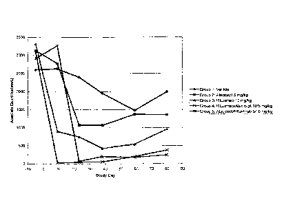
antibody is specific, the cell
begins to divide rapidly and its progeny differentiate into memory B cells and
effector cells called
plasma cells. Memory B cells have a longer life span and continue to express
membrane-bound
antibody with the same specificity as the original parent cell. Plasma cells
do not produce membrane-
bound antibody but instead produce secreted form of the antibody. Secreted
antibodies are the major
effector molecules of humoral immunity.
131 A group of tumor necrosis factor (TNF) receptors found on the
surface of B cells
under various conditions are among the cellular regulators of B cell function
in the immune system.
In particular, three TNF receptors: transmembrane activator and CAML
interactor (TACI), B cell
activator belonging to the TNF family receptor (BAFF-R), and B cell maturation
protein (BCMA) are
known to bind one or both TNF ligands ¨ B Lymphocyte stimulator (BLyS also
known as BAFF,
TALL-1, ztnf4 and THANK) and a proliferation-inducing ligand (APRIL).
Specifically, TACI and
BCMA are known to bind both BLyS and APRIL and BAFF-R binds only BLyS.
[4] A number of BLyS antagonists have been developed in order to block
the various
functions of BLyS, which include but should not be limited to B cell co-
stimulation, plasmablast and
plasma cell survival, Ig class switching, enhanced B-cell antigen presenting
cell function, survival of
malignant B cells, development of B-1 cell function, B cell development beyond
the T-1 stage, and
complete germinal centre formation Some of these molecules can also bind to
and block the effect of
APRIL on B cells and other components of the immune system (Dillon et al.
(2006) Nat. Rev. Drug
Dis. 5, 235-246). Molecules that have been developed to affect B cell function
by interfering with
BLyS and/or APRIL binding include BLyS antibodies such as Lymphostat-B
(Belimumab) (Baker et
CA 02701329 2010-03-30
WO 2009/052293 PCT/US2008/080177
2
al, (2003) Arthritis Rheum, 48, 3253-3265 and WO 02/02641); receptor-
extracellular domain/Fc
domain fusions proteins such as TACI-Ig, including one particular embodiment,
atacicept (U.S. Patent
Application No. 20060034852), BAFF-R-Fc (WO 05/0000351), and BCMA-Ig or other
fusion
proteins utilizing receptor extracellular domains. A further class of BLyS
antagonists include other
molecules relying on BLyS binding ability to block binding to its receptors
such as AMG 623,
receptor antibodies, and other molecules disclosed in WO 03/035846 and WO
02/16312.
151 The CD20 antigen (also called human B-lymphocyte-restricted
differentiation
antigen, Bp35) is a hydrophobic transmembrane protein with a molecular weight
of approximately 35
kD located on pre-B and mature B lymphocytes (Valentine et al. J. Biol. Chent.
264 (19): 11282-
11287 (1989) ; and Einfeld et al. EMBO J. 7 (3): 711-717 (1988) ). The antigen
is also expressed on
greater than 90% of B cell non-Hodgkin's lymphomas (NHL) (Anderson et al.
Blood 63 (6): 1424-
1433 (1984)), but is not found on hematopoietic stem cells, pro-B cells,
normal plasma cells or other
normal tissues (Tedder et al. J. Immunol. 135 (2): 973-979 (1985)). CD20
regulates an early step (s)
in the activation process for cell cycle initiation and differentiation
(Tedder et al., supra) and possibly
functions as a calcium ion channel (Tedder et al. J. Cell. Biochem. 14D: 195
(1990)).
[6] Given the expression of CD20 on B cells, this antigen can serve as
a candidate for
"targeting" of those cells to treat autoimmune diseases. In essence, such
targeting can be generalized
as follows: antibodies specific to the CD20 surface antigen of B cells are
administered to a patient.
These anti-CD20 antibodies specifically bind to the CD20 antigen of
(ostensibly) both B cells
producing normal antibodies and detrimental autoantibodies; the antibody bound
to the CD20 surface
antigen may lead to the destruction and depletion of these B cells.
Irrespective of the approach, a
primary goal is to destroy the cells producing the autoantibodies; the
specific approach can be
determined by the particular anti-CD20 antibody which is utilized and, thus,
the available approaches
to targeting the CD20 antigen can vary considerably.
171 The rituximab (RITUXANO) antibody is a genetically engineered
chimeric
murine/human monoclonal antibody directed against the CD20 antigen. Rituximab
is the antibody
called"C2B8"in US Patent No. 5,736, 137 issued April 7, 1998 (Anderson et
al.).
[8] RITUXANO has been approved for the treatment of patients with
relapsed or
refractory low-grade or follicular, CD20 positive, B cell non-Hodgkin's
lymphoma. In vitro
mechanism of action studies have demonstrated that RITUXANO binds human
complement and lyses
lymphoid B cell lines through complement-dependent cytotoxicity (CDC) (Reff et
al. Blood 83 (2):
435- 445 (1994)). Additionally, it has significant activity in assays for
antibody-dependent cellular
cytotoxicity (ADCC). More recently, RITUXANO has been shown to have anti-
proliferative effects
in tritiated thymidine incorporation assays and to induce apoptosis directly,
while other anti-CD19
and CD20 antibodies do not (Maloney et al. Blood 88 (10): 637a (1996)).
Synergy between
RITUXAN and chemotherapies and toxins has also been observed experimentally.
CA 02701329 2016-09-14
3
[9] In particular, RITUXAN sensitizes drug-resistant human B cell lymphoma
cell lines
to the cytotoxic effects of doxorubicin, CDDP, VP-16, diphtheria toxin and
ricin (Demidem et at.,
Cancer Chemotherapy & Radiopharmaceuticals 12 (3): 177-186 (1997)). In vivo
preclinical studies
have shown that RITUXAN depletes B cells from the peripheral blood, lymph
nodes, and bone
marrow of cynomolgus monkeys, presumably through complement and cell-mediated
processes (Reff
et al. Blood 83(2): 435-445 (1994)).
[10] Patents and patent publications concerning CD20 antibodies include US
Patent Nos.
5,776, 456; 5,736,137; 6,399, 061; and 5,843, 439, as well as US patent appin
nos. US
2002/0197255A1, US 2003/0021781A1, US 2003/0082172 Al, US 2003/0095963 Al, US
2003/0147885 Al (Anderson et al.) ; US Patent No. 6,455,043B1 and W000/09160
(Grillo- Lopez, A.
) ; W000/27428 (Grillo-Lopez and White); W000/27433 (Grillo-Lopez and
Leonard); W000/44788
(Braslawsky et al.) ; W001/10462 (Rastetter, W.) ; W001/10461 (Rastetter and
White); W001/10460
(White and Grillo-Lopez); US appin no. US2002/0006404 and W002/04021 (Hanna
and Hariharan);
US appin no. US2002/0012665 Al and W001/74388 (Hanna, N.); US appin no. US
2002/0058029 Al
(Hanna, N.); US appin no.US 2003/0103971 Al (Hariharan and Hanna); US appin
no.
US2002/0009444A1, and W001/80884 (Grillo-Lopez, A.); W001/97858 (White, C.) ;
US appin no.
US2002/0128488A1 and W002/34790 (Reff, M.) ; W002/060955 (Braslawsky et al.) ;
W02/096948
(Braslawsky et al.) ; W002/079255 (Reff and Davies); US Patent No. 6,171,
58681, and W098/56418
(Lam et al) ; W098/58964 (Raju, S.) ; W099/22764 (Raju, S.) ; W099/51642, US
Patent No. 6,194,
551B1, US Patent No. 6,242, 195B1, US Patent No. 6,528, 624B1 and US Patent
No. 6, 538, 124
(Idusogie et al.) ; W0/42072 (Presta, L.) ; W000/67796 (Curd et al.) ;
W001/03734 (Grillo-Lopez et
al.) ; US appin no. US 2002/0004587A1 and W001/77342 (Miller and Presta); US
appin no.
US2002/0197256 (Grewal, I); US Appin no. US 2003/0157108 Al (Presta, L.) ; US
Patent Nos. 6,090,
365B1, 6,287, 53781, 6,015, 542,5, 843,398, and 5,595, 721, (Kaminski et al.)
; US Patent Nos.
5,500, 362; 5,677, 180; 5,721, 108; and 6,120,767 (Robinson et al.) ; US Pat
No. 6,410, 39181
(Raubitschek et al.); US Patent No. 6,224,866B1 and W000/20864 (Barbera-
Guillem, E.);
W001/13945 (Barbera-Guillem, E.) ; W000/67795 (Goldenberg); US Appl No. US
2003/01339301
Al and W000/74718 (Goldenberg and Hansen); W0/76542 (Golay et al.); W001/72333
(Wolin and
Rosenblatt); US Patent No. 6,368,596B1 (Ghetie et al.); US Appin no.
US2002/0041847 Al,
(Goldenberg, D.) ; US Appin no. US2003/0026801A1 (Weiner and Hartmann);
W002/102312
(Engleman, E.); US Patent Application No. 2003/0068664 (Albitar et al.) ;
W003/002607 (Leung, S.),
W0049694 (Wolin et al.); W003/061694 (Sing and Siegall). See, also, US Patent
No. 5,849,898 and EP appin no. 330,191 (Seed et al.); US Patent No. 4,861, 579
and EP
332, 865A2 (Meyer and Weiss); USP 4, 861,579 (Meyer et al.) and W095/03770
(Bhat et
al.).
CA 02701329 2010-03-30
WO 2009/052293 PCT/US2008/080177
4
[11] Publications concerning therapy with Rituximab include: Perotta and
Abuel"Response of chronic relapsing ITP of 10 years duration to
Rituximab"Abstract # 3360 Blood
(1) (part 1-2): p. 88B (1998); Stashi et al."Rituximab chimeric anti-CD20
monoclonal antibody
treatment for adults with chronic idopathic thrombocytopenic purpura"Blood 98
(4): 952-957 (2001);
Matthews, R."Medical Heretics"New Scientist (7 April, 2001); Leandro et al.
"Clinical outcome in 22
patients with rheumatoid arthritis treated with B lymphocyte depletion"Ann
Rheum Dis 61: 833-888
(2002); Leandro et al."Lymphocyte depletion in rheumatoid arthritis: early
evidence for safety,
efficacy and dose response. Arthritis and Rheumatism 44 (9): S370 (2001);
Leandro et al."An open
study of B lymphocyte depletion in systemic lupus erythematosus", Arthritis &
Rheumatism 46 (1) :
2673-2677 (2002); Edwards and Cambridge "Sustained improvement in rheumatoid
arthritis
following a protocol designed to deplete B lymphocytes"Rhematology 40: 205-211
(2001); Edwards
et al. "B-lymphocyte depletion therapy in rheumatoid arthritis and other
autoimmune disorders"
Biochem. Soc. Trans. 30 (4): 824-828 (2002); Edwards et al."Efficacy and
safety of Rituximab, a B-
cell targeted chimeric monoclonal antibody: A randomized, placebo controlled
trial in patients with
rheumatoid arthritis. Arthritis and Rheumatism 46 (9): S197 (2002); Levine and
Pestronk "IgM
antibody-related polyneuropathies: B-cell depletion chemotherapy using
Rituximab" Neurology 52:
1701-1704 (1999); DeVita et al. "Efficacy of selective B cell blockade in the
treatment of rheumatoid
arthritis"Arthritis & Rheum 46: 2029-2033 (2002); Hidashida et al. "Treatment
of DMARD-
Refractory rheumatoid arthritis with rituximab."Presented at the Anfzual
Sciehtific Meeting of the
American College of Rheumatology ; Oct 24-29; New Orleans, LA 2002; Tuscano,
J. "Successful
treatment of Infliximab-refractory rheumatoid arthritis with rituximab"
Presented at the Annual
Scientific Meeting of the American College of Rheumatology ; Oct 24-29; New
Orleans, LA 2002.
SUMMARY OF THE INVENTION
[12] One aspect of the present invention is a method of reducing B cell
levels in a mammal
comprising administering a BLyS antagonist and an anti-CD20 agent. One
preferred method is where
the BLyS antagonist is a Fc-fusion protein which can be a TACI-Fc-fusion
protein comprising the
extracellular domain of TACI or a functional fragment thereof, a BAFF-R-Fc-
fusion protein
comprising the extracellular domain of BAFF-R or a functional fragment
thereof, or a BCMA-Fc-
fusion protein comprising the extracellular domain of BCMA or a functional
fragment thereof. In
particular, the Fc-fusion protein comprises the polypeptide sequences of SEQ
ID NO: 19, SEQ ID
NO: 21, SEQ ID NO: 23, SEQ ID NO: 25, or SEQ ID NO: 26.
[13] In another embodiment, the BLyS antagonist is a BLyS antibody,
preferably that
binds BLyS within a region comprising amino acids 162-275 of SEQ ID NO: 8, or
the BLyS antibody
known as LymphoStat-B. In further embodiment, the BLyS antagonist is a TACI
antibody, preferably
that binds TACI within a region comprising 72-109 of SEQ ID NO:2 or 82-222 of
SEQ ID NO:2.
CA 02701329 2010-03-30
WO 2009/052293 PCT/US2008/080177
[14] In the methods of the present invention some of the anti-CD20 agents
contemplated
include RITUXANO, although any drug that targets the CD-20 antigen would be
suitable. The B cell
level that is reduced by the present method can be at least one of measurement
of whole B cell counts
(such as circulating B cell counts), memory B cells counts, and spleen B cell
counts (such as geminal
center B cell counts).
[15] The present invention also encompasses a method of alleviating a B-
cell regulated
autoimmune disorders comprising administering to a patient suffering from the
disorder, a
therapeutically effective amount of a anti-CD-20 agent and of a BLyS
antagonist. In one
embodiment, the autoimmune disorder is selected from the group consisting of
rheumatoid arthritis,
juvenile rheumatoid arthritis, systemic lupus erythematosus (SLE), lupus
nephritis (LN), Wegener's
disease, inflammatory bowel disease, idiopathic thrombocytopenic purpura
(ITP), thrombotic
throbocytopenic purpura (TTP), autoimmune thrombocytopenia, multiple
sclerosis, psoriasis, IgA
nephropathy, IgM polyneuropathies, myasthenia gravis, vasculitis, diabetes
mellitus, Reynaud's
syndrome, Sjorgen's syndrome and glomerulonephritis. One disease specifically
treated in this
manner is systemic lupus erythematosus (SLE).
[16] Particularly when the autoimmune disorder is rheumatoid arthritis,
systemic lupus
erythematosus, or lupus nephritis, in one embodiment, the BLyS antagonist and
the anti-CD-20 agent
can be administered in further conjunction with therapy using an
immunosuppressive drug such as
nonsteroidal anti-inflammatory drugs (NSAIDs), glucocorticoid, prednisone, and
disease-modifying
antirheumatic drug (DMARD).
[17] The methods of the present invention can also utilize
immunosuppressive drugs
which have been shown to be effective in treating autoimmune disease.
Among the
immunosuppressive drugs contemplated for use in the methods of the present
invention are
cyclophosphamide (CYC), azathioprine (AZA), cyclosporine A (CSA), and
mycophenolate mofetil
(MMF).
[18] In any of the methods of treatment or alleviation of a disorder where
the anti-CD 20
agent and the BLyS antagonist are administered to a patient, the anti-CD 20
agent and BLyS
antagonist can be administered concurrently or sequentially. In a specific
embodiment, the anti-CD 20
agent is administered before BLyS antagonist
[19] A composition comprising an anti-CD 20 agent and a BLyS antagonist is
also
provided.
[20] Further provided by the invention is an article of manufacture
comprising an anti-CD
20 agent , a BLyS antagonist, and a label wherein the label indicates that the
composition is for
treating a B cell regulated autoimmune disorder.
CA 02701329 2010-03-30
WO 2009/052293 PCT/US2008/080177
6
[21] In any of the embodiments of the methods, compositions and articles of
manufacture
of the invention, the BLyS antagonist or the anti-CD20 agent, if an antibody,
includes chimeric and
humanized antibodies.
[22] In any of the embodiments of the methods, compositions and articles of
manufacture
of the invention, the BLyS antagonist, in one embodiment, is a fusion protein
between the
extracellular domain of a receptor that binds BLyS and the Fc domain of an
immunoglobulin, or an
Fc-fusion protein. In specific embodiments, the Fc-fusion protein selected
from the group consisting
of TACI Fc-fusion protein comprising the extracellular domain of TACI, in
particular atacicept,
BAFF-R Fc-fusion protein comprising the extracellular domain of BAFF-R, in
particular BR3-Ig, and
BCMA Fc-fusion protein comprising the extracellular domain of BCMA. In other
embodiments, the
BLyS antagonist is a BLyS antibody, in particular, a BLyS antibody that binds
BLyS within a region
of BLyS comprising residues 162-275, in particular Lymphostat B. In another
embodiment, the BLyS
antagonist is a BAFF-R antibody including one that binds in a region
comprising residues 23-38 of
human BAFF-R. In another embodiment, the BLyS antagonist is a TACI antibody, a
BCMA
antibody, or an antibody that binds both molecules as described in U.S. Patent
Application No. 2003-
0012783.
BRIEF DESCRIPTION OF THE FIGURES
[23] FIGURE 1 illustrates exemplary FACS results utilizing the gating
strategy of
Example 3 followed for the analysis of the cellular populations, in
particular, B cell levels in
cynomogous monkey spleen. This particular example is a control, e.g., spleen
treated with vehicle for
RITUXANO. This example shows total spleen lymphocytes (FIGURE 1A), CD40+/CD3-
B
lymphocytes (FIGURE 1B), and CD40+/CD3-/CD21+/CD27+ Memory B cells (FIGURE
1C). The
results reported in Example 4 are mean values of FACS analysis performed in
the same way as the
example shown here.
[24] FIGURE 2 graphs the absolute count of total B cells (CD45+/B220+) in
mice through
study day 80 for the combination experiments described in Example 5.
[25] FIGURE 3 graphs the absolute count of human CD20+ B cells
(B220+/huCD20+) in
human CD20 transgenic mice through study day 80 for the combination
experiments described in
Example 5.
[26] FIGURE 4 illustrates an exemplary FACS result utilizing the gating
strategy of
Example 5 for the analysis of the cellular populations, in particular, B cell
levels in mouse peripheral
blood. This particular example is a control, e.g., a human CD20 transgenic
mouse treated with
vehicle. In this example, Figure 4A shows total white blood cells selecting
for CD45+ lymphocytes,
then in Figure 4B, selecting CD45+/B220+ B cells from the CD45+ lymphocyte
population. Figure
4C shows selection for lymphocytes from peripheral blood, Figure 4D graphs the
selection of B220+
CA 02701329 2010-03-30
WO 2009/052293 PCT/US2008/080177
7
and CD3+ cells from the lymphocyte population, and Figure 4E shows that the
majority of the B220+
lymphocyte population is huCD20+.
DETAILED DESCRIPTION OF THE PREFERRED EMBODIMENTS
[27] While anti-CD 20 agent treatment appears useful in the treatment of
autoimmune
disease, it was discovered from the experiments described herein that
administration of a combination
of an anti-CD 20 agent with a BLyS antagonist is a method of treatment that
will block multiple
signal pathways in B cells believed responsible for the production of
antibodies directed at self-
antigens, thereby triggering and/or perpetuating the autoimmune condition.
This results in a reduction
of B cell numbers, sequentially, a reduction in the circulating immunoglobulin
in a mammal
undergoing such treatment. Without being bound by theory, as such circulating
immunoglobulin is
believed at least partially responsible for triggering the negative symptoms
of autoimmune disease,
the combination of anti-CD 20 agents and therapies directed against the BLyS
pathway therefore
provides a novel method of treating B cell-mediated diseases such as B cell-
based autoimmune
diseases. The combination therapy of anti-CD 20 agents with a BLyS antagonist
may offer more
effective alternatives to existing treatments for certain diseases, e. g.,
SLE.
[28] An "autoimmune disease" herein is any non-malignant disease or
disorder arising
from antibodies that are produced directed against an individual's own (self)
antigens and/or tissues.
[29] As used herein, "B cell depletion" refers to a reduction in B cell
levels in an animal or
human after drug or antibody treatment, as compared to the level before
treatment. B cell levels are
measurable using well known assays such as by getting a complete blood count,
by FACS analysis
staining for known B cell markers, and by methods such as described in the
Experimental Examples.
B cell depletion can be partial or complete. In a patient receiving a B cell
depleting drug, B cells are
generally depleted for the duration of time when the drug is circulating in
the patient's body and the
time for recovery of B cells.
[30] The term "anti-CD 20 agent" encompasses any molecule that binds to CD-
20 and in
the most preferred embodiement targets the cell associated with the CD-20
protein for killing. Such
molecules include anti-CD-20 antibodies, such as RITUXANO and follow-on
versions of that agent
such as ocrelizumab, a humanized version of that antibody, ofatumumbab (HuMax-
CD200 a fully
human anti-CD 20 agent), DXL625 (a second generation anti-CD20 monoclonal),
GA101 (a third
generation anti-CD20 agent that has an altered Fc region), the anti-CD20
molecules described in U.S.
Application No. 20060121032, the anti-CD20 molecules described in U.S.
Application No.
200700202059, the anti-CD20 molecules described in U.S. Application No.
20070014720, the anti-
CD20 molecules described in U.S. Application No. 20060251652, the anti-CD20
molecules described
in U.S. Application No. 20050069545, the anti-CD20 molecules described in U.S.
Application No.
CA 02701329 2010-03-30
WO 2009/052293 PCT/US2008/080177
8
20040167319, TRU-015 (a small molecule immunopharmaceutical molecule that
targets CD 20), as
well as conjugated molecules such as ibritumomab (ZEVALINO).
[31] "Immunosuppressive drugs" are any molecules that interfere with the
immune system
and blunt its response to foreign or self antigens. Cyclophosphamide (CYC) and
mycophenolate
mofetil (MMF) are two such kinds of molecules. This term is intended to
encompass any drug or
molecule useful as a therapeutic agent in downregulating the immune system.
This method
particularly contemplates drugs that have been used to treat autoimmune
diseases such as rheumatoid
arthritis, juvenile rheumatoid arthritis, systemic lupus erythematosus (SLE),
lupus nephritis (LN),
Wegener's disease, inflammatory bowel disease, idiopathic thrombocytopenic
purpura (ITP),
thrombotic throbocytopenic purpura (TTP), autoimmune thrombocytopenia,
multiple sclerosis,
psoriasis, IgA nephropathy, IgM polyneuropathies, myasthenia gravis,
vasculitis, diabetes mellitus,
Reynaud's syndrome, Sjorgen's syndrome and glomerulonephritis.
[32] The terms "BLyS" or "BLyS polypeptide," "TALL-1" or "TALL-1
polypeptide," or
"BAFF" or "BAFF polypeptide" when used herein encompass "native sequence BLyS
polypeptides"
and "BLyS variants." "BLyS" is a designation given to those polypeptides which
are encoded by the
Human BLyS sequence (SEQ ID NO: 7) or the Mouse BLyS sequence (SEQ ID NO: 9).
Polypeptides
which show BLyS biological activity are encompassed within this designation as
well. For example,
a biologically active BLyS potentiates any one or combination of the following
events in vitro or in
vivo : an increased survival of B cells, an increased level of IgGand/or IgM,
an increased numbers of
plasma cells, and processing of NF- Kb2/100 to p52NF-Kb in splenic B cells
(e.g., Batten, M et al. ,
(2000) J. Exp. Med. 192: 1453-1465; Moore, et al. , (1999) Science 285: 260-
263; Kayagaki, et al. ,
(2002) 10: 515-524). Several assays useful for testing BLyS antagonists such
as the B cell
proliferation assay described in WO 00/40716 among others are well known to
one of ordinary skill in
the art.
[33] Briefly, human B cells are isolated from peripheral blood mononuclear
cells using
CD19 magnetic beads and the VarioMacs magnetic separation system (Miltenyi
Biotec Auburn, CA)
according to the manufacturer's instructions. Purified B cells are mixed with
soluble BLyS (25
ng/ml) and recombinant human IL-4 (10 ng/ml Pharmingen), and the cells are
plated onto round
bottom 96 well plates at 1 x 10⁵ cells per well. The BLyS antagonist to be
tested can be diluted from
about 5 jig/ml to about 6 ng/ml, and incubated with the B cells for five days,
pulsing overnight on day
four with 11.tCi 3H-thymidine per well. As a control, BLyS antagonist can also
be incubated with B
cells and IL-4 without BLyS. Plates are harvested using Packard plate
harvester, and counted using
the Packard reader.
[34] A "native sequence" polypeptide comprises a polypeptide having the
same amino
acid sequence as the corresponding polypeptide derived from nature. Such
native sequence
polypeptides can be isolated from nature or can be produced by recombinant
and/or synthetic means.
CA 02701329 2010-03-30
WO 2009/052293 PCT/US2008/080177
9
The term "native sequence" specifically encompasses naturally-occurring
truncated, soluble or
secreted forms (e. g., an extracellular domain sequence), naturally-occurring
variant forms (e. g.,
alternatively spliced forms) and naturally-occurring allelic variants of the
polypeptide.
[35] In general, "variant" polypeptides for any of the polypeptides
disclosed in the present
specification include polypeptides wherein one or more amino acid residues are
added or deleted at
the N-and/or C-terminus, as well as within one or more internal domains, of
the full-length or "native
sequence" amino acid sequence. When discussing extracellular domains of
receptors, fragments that
bind a native sequence BlyS polypeptide are also contemplated. Conversely,
when discussing BLyS
fragments, fragments that bind any one or more of the three BLyS receptors are
contemplated.
Ordinarily, a variant polypeptide will have at least about 80% amino acid
sequence identity, more
preferably at least about 81% amino acid sequence identity, more preferably at
least about 82% amino
acid sequence identity, more preferably at least about 83% amino acid sequence
identity, more
preferably at least about 84% amino acid sequence identity, more preferably at
least about 85% amino
acid sequence identity, more preferably at least about 86% amino acid sequence
identity, more
preferably at least about 87% amino acid sequence identity, more preferably at
least about 88% amino
acid sequence identity, more preferably at least about 89% amino acid sequence
identity, more
preferably at least about 90% amino acid sequence identity, more preferably at
least about 91% amino
acid sequence identity, more preferably at least about 92% amino acid sequence
identity, more
preferably at least about 93% amino acid sequence identity, more preferably at
least about 94% amino
acid sequence identity, more preferably at least about 95% amino acid sequence
identity, more
preferably at least about 96% amino acid sequence identity, more preferably at
least about 97% amino
acid sequence identity, more preferably at least about 98% amino acid sequence
identity and yet more
preferably at least about 99% amino acid sequence identity with the
polypeptide or a specified
fragment thereof. Generally, variant polypeptides do not encompass the native
polypeptide sequence.
Ordinarily, variant polypeptides are at least about 10 amino acids in length,
often at least about 20
amino acids in length, more often at least about 30 amino acids in length,
more often at least about 40
amino acids in length, more often at least about 50 amino acids in length,
more often at least about 60
amino acids in length, more often at least about 70 amino acids in length,
more often at least about 80
amino acids in length, more often at least about 90 amino acids in length,
more often at least about
100 amino acids in length, more often at least about 150 amino acids in
length, more often at least
about 200 amino acids in length, more often at least about 250 amino acids in
length, more often at
least about 300 amino acids in length, or more.
[36] As mentioned above, a BLyS antagonist can function in a direct or
indirect manner to
partially or fully block, inhibit or neutralize BLyS signaling, in vitro or in
vivo. For instance, the
BLyS antagonist can directly bind BLyS. For example, a direct binder is a
polypeptide comprising the
extracellular domain (ECD) of a BLyS receptor such as TACI, BAFF-R, and BCMA.
CA 02701329 2010-03-30
WO 2009/052293 PCT/US2008/080177
[37] The BLyS receptors involved in the present invention can be described
as follows.
The TACI polypeptides of the invention include TACI polypeptides comprising or
consisting of
amino acids 1-246 of SEQ ID NO: 2. The general term "TACI" includes the TACI
polypeptides
described in WO 98/39361, WO 00/40716, WO 01/85782, WO 01/87979, WO 01/81417,
and WO
02/094852. The TACI polypeptides of the invention can be isolated from a
variety of sources, such as
from human tissue types or from another source, or prepared by recombinant
and/or synthetic
methods. The BAFF-R polypeptides of the invention include the BAFF-R
polypeptide comprising or
consisting of the contiguous sequence of amino acid residues 1 to 184 of SEQ
ID NO:4. The general
term "BAFF-R" includes the BAFF-R polypeptides described in WO 02/24909 and WO
03/14294.
The BAFF-R polypeptides of the invention can be isolated from a variety of
sources, such as from
human tissue types or from another source, or prepared by recombinant and/or
synthetic methods.
The BCMA polypeptide of the invention include BCMA polypeptides comprising or
consisting of
amino acid residues 1-184 of SEQ ID NO:6. The general term "BCMA" includes the
BCMA
polypeptides described in Laabi et al. , EMBO J., 11: 3897-3904 (1992); Laabi
et al. ,Nucleic Acids
Res., 22: 1147-1154 (1994); Gras etal., Int. Immunology, 7: 1093-1106 (1995);
and Madry etal., Int.
Immunology, 10: 1693-1702 (1998). The BCMA polypeptides of the invention can
be isolated from a
variety of sources, such as from human tissue types or from another source, or
prepared by
recombinant and/or synthetic methods.
[38] For the purposes of functioning as a BLyS antagonist, the ECD of these
receptors is a
polypeptide essentially free of the transmembrane or cytoplasmic domains that
generally retains the
ability to bind BLyS. Specifically, the extracellular domain of TACI can
comprise amino acids 1 to
154 of the TACI polypeptide sequence (SEQ ID NO: 2). Additionally, the ECD can
be fragments or
variants of this sequence, such as ECD forms of TACI as described in von Bulow
et al., supra, WO
98/39361, WO 00/40716, WO 01/85782, WO 01/87979, and WO 01/81417. In
particular, these ECD
forms can comprise amino acids 1-106 of SEQ ID NO:2, amino acids 1-142 of SEQ
ID NO:2, amino
acids 30-154 of SEQ ID NO:2, amino acids 30-106 of SEQ ID NO:2, amino acids 30-
110 of SEQ ID
NO:2, amino acids 30-119 of SEQ ID NO:2, amino acids 1-166 of SEQ ID NO:2,
amino acids 1-165
of SEQ ID NO:2, amino acids 1-114 of SEQ ID NO: 2, amino acids 1-119 of SEQ ID
NO:2, amino
acids 1-120 of SEQ ID NO:2, and amino acids 1-126 of SEQ ID NO:2. In addition,
the TACI ECD
can comprise those molecules having only one cysteine rich domain
[39] ECD forms of BAFF-R include those comprising amino acids 1-71 of the
BAFF-R
polypeptide sequence (SEQ ID NO: 4). Additionally, the ECD can be fragments or
variants of this
sequence such as ECD forms of BAFF-R as described in WO 02/24909, WO 03/14294,
and WO
02/38766. In particular, these ECD forms can comprise amino acids 1-77 of SEQ
ID NO: 4, amino
acids 7-77 of SEQ ID NO:4, amino acids 1-69 of SEQ ID NO:4, amino acids 7-69
of SEQ ID NO:4,
amino acids 2-62 of SEQ ID NO:4, amino acids 2-71 of SEQ ID NO:4, amino acids
1-61 of SEQ ID
CA 02701329 2010-03-30
WO 2009/052293 PCT/US2008/080177
11
NO:4 and amino acids 2-63 of SEQ ID NO:4, amino acids 1-45 of SEQ ID NO:4,
amino acids 1-39 of
SEQ ID NO:4, amino acids 7-39 of SEQ ID NO:4, amino acids 1-17 of SEQ ID NO:4,
amino acids
39-64 of SEQ ID NO:4, amino acids 19-35 of SEQ ID NO:4, and amino acids 17-42
of SEQ ID
NO:4. In addition, the BAFF-R ECD can comprise those molecules having a
cysteine rich domain.
[40] ECD forms of BCMA include those comprising amino acids 1-48 of the BCMA
polypeptide sequence (SEQ ID NO: 6). Additionally, the ECD can be fragments or
variants of this
sequence, such as ECD forms of BCMA as described in WO 00/40716 and WO
05/075511. In
particular, these ECD forms can comprise amino acids 1-150 of SEQ ID NO:6,
amino acids 1-48 of
SEQ ID NO:6, amino acids 1-41 of SEQ ID NO:6, amino acids 8-41 of SEQ ID NO:6,
amino acids 8-
37 of SEQ ID NO:6, amino acids 8-88 of SEQ ID NO:6, amino acids 41-88 of SEQ
ID NO:6, amino
acids 1-54 of SEQ ID NO:6, amino acids 4-55 of SEQ ID NO:6, amino acids 4-51
of SEQ ID NO:6,
and amino acids 21-53 of SEQ ID NO:6. In addition, the BCMA ECD can comprise
those molecules
having only a partial cysteine rich domain.
[41] In a further embodiment, the BLyS binding region of a BLyS receptor
(e. g., an
extracellular domain or fragment thereof of BAFF-R, BCMA or TACI) can be fused
to an Fc portion
of an immunoglobulin molecule to facilitate its solubility in vivo. According
to one embodiment, the
BLyS antagonist binds to a BLyS polypeptide with a binding affinity of 100nM
or less. According to
another embodiment, the BLyS antagonist binds to a BLyS polypeptide with a
binding affinity of
lOnM or less. According to yet another embodiment, the BlyS antagonist binds
to a BLyS polypeptide
with a binding affinity of 1nM or less.
[42] In another example, BLyS antagonists include BLyS binding polypeptides
that are not
native sequences or varients thereof. Some examples of such polypepeptides are
those having the
sequence of Formula I, Formula II, Formula III as described in WO 05/000351.
In particular, some
binding polypeptides include ECFDLLVRAWVPCSVLK (SEQ ID NO:13),
ECFDLLVRHWVPCGLLR (SEQ ID NO:14), ECFDLLVRRWVPCEMLG (SEQ ID NO:15),
ECFDLLVRSWVPCHMLR (SEQ ID NO:16), ECFDLLVRHWVACGLLR (SEQ ID NO:17), or
sequences listed in FIG. 32 of WO 05/000351.
[43] Alternatively, the BLyS antagonist can bind an extracellular domain of
native
sequence TACI, BAFF-R, or BCMA at its BLyS binding region to partially or
fully block, inhibit or
neutralize BLyS binding in vitro, in situ, or in vivo. For example, such
indirect antagonist is a TACI
antibody that binds in a region of TACI such that the binding of BLyS is
sterically hindered. For
example, binding at amino acids 72-109 or a neighboring region is believed to
block BLyS binding. It
could also be advantageous to block APRIL binding to this molecule, which is
believed to occur in
the region of amino acids 82-222. Another BLyS antagonist is a BAFF-R antibody
that binds in a
region of BAFF-R such that binding of human BAFF-R to BLyS is sterically
hindered. For example,
binding at amino acids 23-38 or amino acids 17-42 or a neighboring region is
believed to block BLyS
CA 02701329 2010-03-30
WO 2009/052293 PCT/US2008/080177
12
binding. Finally, a further indirect antagonist would be a BCMA antibody that
binds in a rgion of
BCMA such that the binding of BLyS is sterically hindered. For example,
binding at amino acids 5-
43 or a neighborhing region is believed to block BLyS (or APRIL) binding.
[44] In some embodiments, a BLyS antagonist according to this invention
includes BLyS
antibodies. The term "antibody" when referring to is used in the broadest
sense and specifically
covers, for example, monoclonal antibodies, polyclonal antibodies, antibodies
with polyepitopic
specificity, single chain antibodies, and fragments of antibodies. According
to some embodiments, a
polypeptide of this invention is fused into an antibody framework, for
example, in the variable region
or in a CDR such that the antibody can bind to and inhibit BLyS binding to
TACI, BAFF-R, or
BCMA or inhibits BLyS signaling. The antibodies comprising a polypeptide of
this invention can be
chimeric, humanized, or human. The antibodies comprising a polypeptide of this
invention can be an
antibody fragment. Alternatively, an antibody of this invention can be
produced by immunizing an
animal with a polypeptide of this invention. Thus, an antibody directed
against a polypeptide of this
invention is contemplated.
[45] In particular, antibodies specific for BLyS that bind within a region
of human BLyS
(SEQ ID NO: 8) comprising residues 162-275 and/or a neighboring amino acid of
amino acids
selected from the group consisting of 162, 163, 206, 211, 231, 233, 264 and
265 of human BLyS are
contemplated. The binding of the antibodies are such that the antibody
sterically hinders BLyS
binding to one or more of its receptors. Such antibodies are described in WO
02/02641 and WO
03/055979. A particularly preferred antibody is the one described as Lyphostat-
B (Baker et al. (2003)
Arthritis Rheum, 48, 3253-3265).
[46] The term "monoclonal antibody" as used herein refers to an antibody
obtained from a
population of substantially homogeneous antibodies, i. e., the individual
antibodies comprising the
population are identical except for possible naturally occurring mutations
that can be present in minor
amounts.
[47] Monoclonal antibodies are highly specific, being directed against a
single antigenic
site. Furthermore, in contrast to conventional (polyclonal) antibody
preparations which typically
include different antibodies directed against different determinants
(epitopes), each monoclonal
antibody is directed against a single determinant on the antigen. In addition
to their specificity, the
monoclonal antibodies are advantageous in that they are synthesized by the
hybridoma culture,
uncontaminated by other immunoglobulins. The modifier "monoclonal" indicates
the character of the
antibody as being obtained from a substantially homogeneous population of
antibodies, and is not to
be construed as requiring production of the antibody by any particular method.
For example, the
monoclonal antibodies to be used in accordance with the present invention may
be made by the
hybridoma method first described by Kohler etal., Nature, 256: 495 (1975), or
may be made by
recombinant DNA methods (see, e. g. , U. S. Patent No. 4,816, 567). The
"monoclonal
CA 02701329 2010-03-30
WO 2009/052293 PCT/US2008/080177
13
antibodies"may also be isolated from phage antibody libraries using the
techniques described in
Clackson etal., Nature, 352: 624-628 (1991) and Marks et al. , J. Mol.Biol.,
222: 581-597 (1991), for
example.
[48] The monoclonal antibodies herein specifically include "chimeric"
antibodies
(immunoglobulins) in which a portion of the heavy and/or light chain is
identical with or homologous
to corresponding sequences in antibodies derived from a particular species or
belonging to a particular
antibody class or subclass, while the remainder of the chain (s) is identical
with or homologous to
corresponding sequences in antibodies derived from another species or
belonging to another antibody
class or subclass, as well as fragments of such antibodies, so long as they
exhibit the desired
biological activity (U. S. Patent No. 4,816, 567; Morrison et al., Proc. Natl.
Acad. Sci. USA, 81:
6851-6855 (1984)). Methods of making chimeric antibodies are known in the art.
[49] "Humanized" forms of non-human (e. g. , murine) antibodies are
chimeric
immunoglobulins, immunoglobulin chains or fragments thereof (such as Fv, Fab,
Fab', F (ab') 2 or
other antigen-binding subsequences of antibodies) which contain minimal
sequence derived from non-
human immunoglobulin.
[50] For the most part, humanized antibodies are human immunoglobulins
(recipient
antibody) in which residues from a complementarity-determining region (CDR) of
the recipient are
replaced by residues from a CDR of a non-human species (donor antibody) such
as mouse, rat or
rabbit having the desired specificity, affinity, and capacity. In some
instances, Fv framework region
(FR) residues of the human immunoglobulin are replaced by corresponding non-
human residues.
Furthermore, humanized antibodies may comprise residues which are found
neither in the recipient
antibody nor in the imported CDR or framework sequences. These modifications
are made to further
refine and maximize antibody performance. In general, the humanized antibody
will comprise
substantially all of at least one, and typically two, variable domains, in
which all or substantially all of
the hypervariable loops correspond to those of a non-human immunoglobulin and
all or substantially
all of the FR regions are those of a human immunoglobulin sequence although
the FR regions may
include one or more amino acid substitutions that improve binding affinity.
The number of these
amino acid substitutions in the FR are typically no more than 6 in the H
chain, and in the L chain, no
more than 3. The humanized antibody optimally also will comprise at least a
portion of an
immunoglobulin constant region (Fc), typically that of a human immunoglobulin.
For further details,
see Jones et al., Nature, 321: 522-525 (1986); Reichmann et al. , Nature, 332:
323-329 (1988); and
Presta, Curr. Op. Struct. Biol., 2: 593-596 (1992). The humanized antibody
includes a PRIMATIZED
antibody wherein the antigen-binding region of the antibody is derived from an
antibody produced by,
e. g. , immunizing macaque monkeys with the antigen of interest. Methods of
making humanized
antibodies are known in the art.
CA 02701329 2010-03-30
WO 2009/052293 PCT/US2008/080177
14
[51] Human antibodies can also be produced using various techniques known
in the art,
including phage-display libraries. Hoogenboom and Winter, J. Mol.Biol., 227:
381 (1991); Marks et
al. , J. Mol. Biol., 222: 581 (1991). The techniques of Cole et al. and
Boerner et al. are also available
for the preparation of human monoclonal antibodies. Cole et al. , Monoclonal
Antibodies and Cancer
Therapy, Alan R. Liss, p. 77 (1985); Boerner et al. , J. Immunol., 147(1) : 86-
95 (1991).
[52] "Functional fragments" of the binding antibodies of the invention are
those fragments
that retain binding to BLyS, TACI, BAFF-R, or BCMA with substantially the same
affinity as the
intact full chain molecule from which they are derived and may be able to
deplete B cells as measured
by in vitro or in vivo assays such as those described herein.
[53] Antibody "effector functions" refer to those biological activities
attributable to the Fc
region (a native sequence Fc region or amino acid sequence variant Fc region)
of an antibody, and
vary with the antibody isotype. Examples of antibody effector functions
include: Clq binding and
complement dependent cytotoxicity; Fc receptor binding; antibody-dependent
cell-mediated
cytotoxicity (ADCC); phagocytosis; down regulation of cell surface receptors
(e. g. B cell receptor);
and B cell activation.
[54] "Antibody-dependent cell-mediated cytotoxicity" or "ADCC" refers to a
form of
cytotoxicity in which secreted Ig bound onto Fc receptors (FcRs) present on
certain cytotoxic cells (e.
g. Natural Killer (NK) cells, neutrophils, and macrophages) enable these
cytotoxic effector cells to
bind specifically to an antigen-bearing-target cell and subsequently kill the-
target cell with cytotoxins.
The antibodies-"arm"the cytotoxic cells and are absolutely required for such
killing. The primary cells
for mediating ADCC, NK cells, express FcyRIII only, whereas monocytes express
FcyRI, FcyRII and
FcyRIII. FcR expression on hematopoietic cells is summarized in Table 3 on
page 464 of Ravetch and
Kinet, Ann. Rev. Immunol 9: 457-92 (1991). To assess ADCC activity of a
molecule of interest, an in
vitro ADCC assay, such as that described in US Patent No. 5,500, 362 or 5,821,
337 may be
performed. Useful effector cells for such assays include peripheral blood
mononuclear cells (PBMC)
and Natural Killer (NK) cells. Alternatively, or additionally, ADCC activity
of the molecule of
interest may be assessed in vivo, e. g. , in a animal model such as that
disclosed in Clynes et al. PNAS
(USA) 95: 652-656 (1998).
[55] "Complement dependent cytotoxicity" or "CDC" refers to the lysis of a
target cell in
the presence of complement. Activation of the classical complement pathway is
initiated by the
binding of the first component of the complement system (Clq) to antibodies
(of the appropriate
subclass) which are bound to their cognate antigen. To assess complement
activation, a CDC assay, e.
g. as described in Gazzano- Santoroetal., J. Immunol. Methods 202: 163 (1996),
may be performed.
[56] An "isolated" antibody is one which has been identified and
separatedand/or
recovered from a component of its natural environment. Contaminant components
of its natural
environment are materials which would interfere with diagnostic or therapeutic
uses for the antibody,
CA 02701329 2010-03-30
WO 2009/052293 PCT/US2008/080177
and may include enzymes, hormones, and other proteinaceous or nonproteinaceous
solutes. In
preferred embodiments, the antibody is purified (1) to greater than 95% by
weight of antibody as
determined by the Lowry method, and most preferably more than 99% by weight,
(2) to a degree
sufficient to obtain at least 15 residues of N- terminal or internal amino
acid sequence by use of a
spinning cup sequenator, or (3) to homogeneity by SDS-PAGE under reducing or
nonreducing
conditions using Coomassie blue or, preferably, silver stain.
[57] Isolated antibody includes the antibody insitu within recombinant
cells since at least
one component of the antibody's natural environment will not be present.
Ordinarily, however,
isolated antibody is prepared by at least one purification step.
[58] Amino acids may be grouped according to similarities in the properties
of their side
chains (in A. L. Lehninger, in Biochemistry, second ed. , pp. 73-75, Worth
Publishers, New York
(1975) ) : (1) non-polar: Ala (A), Val (V), Leu (L), Ile (I), Pro (P), Phe
(F), Trp (W), Met (M) (2)
uncharged polar: Gly (G), Ser (S), Thr (T), Cys (C), Tyr (Y), Asn (N),Gln (Q)
(3) acidic: Asp (D),
Glu (E) (4) basic : Lys(K), Arg (R), His(H-) Alternatively, naturally
occurring residues may be
divided into groups based on common side- chain properties : (1) hydrophobic:
Norleucine, Met, Ala,
Val, Leu, Ile ; (2) neutral hydrophilic : Cys, Ser, Thr, Asn,Gln ; (3) acidic:
Asp, Glu; (4) basic: His,
Lys, Arg; (5) residues that influence chain orientation: Gly, Pro; (6)
aromatic: Trp, Tyr, Phe.
[59] The term "conservative" amino acid substitution as used within this
invention is
meant to refer to amino acid substitutions which substitute functionally
equivalent amino acids.
Conservative amino acid changes result in silent changes in the amino acid
sequence of the resulting
peptide. For example, one or more amino acids of a similar polarity act as
functional equivalents and
result in a silent alteration within the amino acid sequence of the peptide.
In general, substitutions
within a group may be considered conservative with respect to structure and
function. However, the
skilled artisan will recognize that the role of a particular residue is
determined by its context within
the three-dimensional structure of the molecule in which it occurs. For
example, Cys residues may
occur in the oxidized (disulfide) form, which is less polar than the reduced
(thiol) form. The long
aliphatic portion of the Arg side chain may constitute a critical feature of
its structural or functional
role, and this may be best conserved by substitution of a nonpolar, rather
than another basic residue.
Also, it is recognized that side chains containing aromatic groups (Trp, Tyr,
and Phe) can participate
in ionic-aromatic or"cation-pi"interactions. In these cases, substitution of
one of these side chains with
a member of the acidic or uncharged polar group may be conservative with
respect to structure and
function. Residues such as Pro, Gly, and Cys (disulfide form) can have direct
effects on the main
chain conformation, and often may not be substituted without structural
distortions.
[60] "Percent (%) amino acid sequence identity"with respect to the ligand
or receptor
polypeptide sequences identified herein is defined as the percentage of amino
acid residues in a
candidate sequence that are identical with the amino acid residues in such a
ligand or receptor
CA 02701329 2010-03-30
WO 2009/052293 PCT/US2008/080177
16
sequence identified herein, after aligning the sequences and introducing gaps,
if necessary, to achieve
the maximum percent sequence identity, and not considering any conservative
substitutions as part of
the sequence identity. Alignment for purposes of determining percent amino
acid sequence identity
can be achieved in various ways that are within the skill in the art, for
instance, using publicly
available computer software such as BLAST, BLAST-2, ALIGN, ALIGN-2 or Megalign
(DNASTAR) software. Those skilled in the art can determine appropriate
parameters for measuring
alignment, including any algorithms needed to achieve maximal alignment over
the full-length of the
sequences being compared. For purposes herein, however, % amino acid sequence
identity values are
obtained as described below by using the sequence comparison computer program
ALIGN-2, wherein
the complete source code for the ALIGN-2 program is provided in the table
below. The ALIGN-2
sequence comparison computer program was authored by Genentech, Inc. and the
source code shown
in the table below has been filed with user documentation in the U. S.
Copyright Office, Washington
D. C. , 20559, where it is registered under U. S. Copyright Registration
No.TXU510087. The ALIGN-
2 program is-publicly available through Genentech, Inc., South San Francisco,
California or can be
compiled from the source code provided in the table below. The ALIGN-2 program
should be
compiled for use on a UNIX operating system, preferably digital UNIX V4.0D.
All sequence
comparison parameters are set by the ALIGN-2 program and do not vary.
[61] A useful method for identification of certain residues or regions in a
protein that are
preferred locations for mutagenesis is called"alanine scanning mutagenesis"as
described by
Cunningham and Wells Science, 244: 1081-1085 (1989). A residue or group of
target residues are
identified (e. g. , charged residues such as arg, asp, his, lys, and glu) and
replaced by a neutral or
negatively charged amino acid (most preferably alanine or polyalanine) to
affect the interaction of the
amino acids with a binding target. Those amino acid locations demonstrating
functional sensitivity to
the substitutions then are refined by introducing further or other variants
at, or for, the sites of
substitution. Thus, while the site for introducing an amino acid sequence
variation is predetermined,
the nature of the mutation per se need not be predetermined. For example, to
analyze the performance
of a mutation at a given site, ala scanning or random mutagenesis is conducted
at the target codon or
region and the expressed variants are screened for the desired activity.
[62] The term, "dihedral angle" refers to a rotation about a bond. See e.
g. , Creighton, T.
E. , (1993) Protein: Structures and Molecular Properties, 2 ed. , W. H.
Freeman and Company, New
York, NY. The term,"phi,"is a dihedral angle that denotes a rotation about
theN-C bond of an amino
acid. See e. g. , Creighton, T. E. , (1993) Protein: Structures and Molecular
Properties, 2 ed. , W. H.
Freeman and Company, New York, NY. Type I beta turns are described in
Hutchinson, E. G. &
Thornton, J. M. (1994) A revised set of potentials for beta turn formation in
proteins. Protein Science
3, 2207-2216.
CA 02701329 2010-03-30
WO 2009/052293 PCT/US2008/080177
17
[63] A "fusion protein" and a "fusion polypeptide" refer to a polypeptide
having two
portions covalently linked together, where each of the portions is a
polypeptide having a different
property. The property may be a biological property, such as activity in vitro
or in vivo. The property
may also be a simple chemical or physical property, such as binding to a
target molecule, catalysis of
a reaction, etc. The two portions may be linked directly by a single peptide
bond or through a peptide
linker containing one or more amino acid residues. Generally, the two portions
and the linker is in
reading frame with each other.
[64] A "conjugate" refers to any hybrid molecule, including fusion proteins
and as well as
molecules that contain both amino acid or protein portions and non-protein
portions. Conjugates may
be synthesized by a variety of techniques known in the art including, for
example, recombinant DNA
techniques, solid phase synthesis, solution phase synthesis, organic chemical
synthetic techniques or a
combination of these techniques. The choice of synthesis will depend upon the
particular molecule to
be generated. For example, a hybrid molecule not entirely"protein"in nature
may be synthesized by a
combination of recombinant techniques and solution phase techniques.
[65] As used herein, the term "Fc-fusion protein" designates antibody-like
molecules
which combine the binding specificity of a heterologous protein with the
effector functions of
immunoglobulin constant domains. Structurally, the Fc-fusion proteins comprise
a fusion of an amino
acid sequence with the desired binding specificity which is other than the
antigen recognition and
binding site of an antibody (i. e., is "heterologous"), and an immunoglobulin
constant domain
sequence. The Fc-fusion protein molecule typically includes a contiguous amino
acid sequence
comprising at least the binding site of a receptor or a ligand. The
immunoglobulin constant domain
sequence in the Fc-fusion protein can be obtained from any immunoglobulin,
such as IgG-1, IgG-2,
IgG-3, or IgG-4 subtypes, IgA (includingIgA-1 and IgA-2), IgE, IgD or IgM. For
example, useful Fc-
fusion proteins according to this invention are polypeptides that comprise the
BLyS binding portions
of a BLyS receptor without the transmembrane or cytoplasmic sequences of the
BLyS receptor. In one
embodiment, the extracellular domain of BAFF-R, TACI or BCMA is fused to a
constant domain of
an immunoglobulin sequence.
[66] The term "mammal" refers to any animal classified as a mammal,
including humans,
domestic and farm animals, and zoo, sports, or pet animals, such as dogs,
horses, cats, cows, etc.
Preferably, the mammal herein is human.
[67] The term "therapeutically effective amount" refers to an amount of an
antibody or a
antagonist drug effective to "alleviate" or "treat" a disease or disorder in a
subject or mammal. In the
case of cancer, the therapeutically effective amount of the drug may reduce
the number of cancer
cells; reduce the tumor size; inhibit(i. e., slow to some extent and
preferably stop) cancer cell
infiltration into peripheral organs; inhibit (i. e. , slow to some extent and
preferably stop) tumor
metastasis; inhibit, to some extent, tumor growth; and/or relieve to some
extent one or more of the
CA 02701329 2010-03-30
WO 2009/052293 PCT/US2008/080177
18
symptoms associated with the cancer. See the definition of'treated"below. To
the extent the drug may
prevent growth and/or kill existing cancer cells, it may be cytostatic and/or
cytotoxic.
[68] The BLyS or BLyS receptor antibodies of the invention can be produced
by transient
or stable transfection eukaryotic host cells such as CHO cells.
[69] "Carriers" as used herein include pharmaceutically acceptable
carriers, excipients, or
stabilizers which are nontoxic to the cell or mammal being exposed thereto at
the dosages and
concentrations employed. Often the physiologically acceptable carrier is an
aqueous pH buffered
solution. Examples of physiologically acceptable carriers include buffers such
as phosphate, citrate,
and other organic acids; antioxidants including ascorbic acid; low molecular
weight (less than about
residues) polypeptide; proteins, such as serum albumin, gelatin, or
immunoglobulins; hydrophilic
polymers such as polyvinylpyrrolidone ; amino acids such as glycine,
glutamine, asparagine, arginine
or lysine ; monosaccharides, disaccharides, and other carbohydrates including
glucose, mannose, or
dextrins; chelating agents such as EDTA; sugar alcohols such as mannitol or
sorbitol; salt-forming
counterions such as sodium; and/or nonionic surfactants such as TWEEN
polyethylene glycol(PEG),
and PLURONICSTM.
Polynucleotides, Vectors, Host Cells
[70] According to a number of embodiments disclosed herein, the BLyS
antagonist can
comprise specific polypeptides that are produced using specific
polynucleotides in specific vectors
and using specific host cells. The various types of polypeptides of the
present invention can be
broadly described and are selected from the group consisting of receptor-based
seqences, antibody-
based sequences, and artifical (i.e., non-native) binding sequences. Examples
of the receptor-based
sequences are those sequences that bind BLyS that were isolated from or
derived from domains of the
receptors that bind BLyS in vivo, such as TACI, BAFF-R, or BCMA. Antibody-
based sequences are
those that are produced using antibody development technology and maintain the
general structure of
an antibody molecule. Examples of antibody-based sequences are LymphoStat-B,
or antibodies to
receptors of BLyS. Examples of the artificial binding sequences include the
17mer peptides described
herein, polypeptides incorporating one or more 17mer peptides as core regions,
and covalently
modified forms of the 17mer peptides and polypeptides (e. g., Fc-fusion
proteins, labeled
polypeptides, protected polypeptides, conjugated polypeptides, fusion
proteins, etc.). Various
techniques that are employed for making these forms of polypeptides are
described herein. Methods
for labeling polypeptides and conjugating molecules to polypeptides are known
in the art.
[71] Compositions of the invention can be prepared using recombinant
techniques known
in the art.
[72] The description below relates to methods of producing such specific
polypeptides by
culturing host cells transformed or transfected with a vector containing the
encoding nucleic acid and
CA 02701329 2010-03-30
WO 2009/052293 PCT/US2008/080177
19
recovering the polypeptide from the cell culture. (See, e.g., Sambrook etal.,
Molecular Cloning: A
Laboratory Manual (New York: Cold Spring Harbor Laboratory Press, 1989);
Dieffenbach et al., PCR
Primer: A Laboratory Manual (Cold Spring Harbor Laboratory Press, 1995)).
[73] The nucleic acid (e. g., cDNA orgenomic DNA) encoding the desired
polypeptide
may be inserted into a replicable vector for further cloning (amplification of
the DNA) or for
expression. Various vectors are publicly available. The vector components
generally include, but are
not limited to, one or more of the following: a signal sequence, an origin of
replication, one or more
marker genes, an enhancer element, a promoter, and a transcription termination
sequence, each of
which is described below. Optional signal sequences, origins of replication,
marker genes, enhancer
elements and transcription terminator sequences that may be employed are known
in the art and
described in further detail in WO 97/25428.
[74] Expression and cloning vectors usually contain a promoter that is
recognized by the
host organism and is operably linked to the encoding nucleic acid sequence.
Promoters are
untranslated sequences located upstream (5') to the start codon of a
structural gene (generally within
about 100 to 1000 bp) that control the transcription and translation of a
particular nucleic acid
sequence, to which they are operably linked. Such promoters typically fall
into two classes, inducible
and constitutive. Inducible promoters are promoters that initiate increased
levels of transcription from
DNA under their control in response to some change in culture conditions, e.
g. , the presence or
absence of a nutrient or a change in temperature. At this time a large number
of promoters recognized
by a variety of potential host cells are well known. These promoters are
operably linked to the
encoding DNA by removing the promoter from the source DNA by restriction
enzyme digestion and
inserting the isolated promoter sequence into the vector.
[75] Construction of suitable vectors containing one or more of the above-
listed
components employs standard ligation techniques. Isolated plasmids or DNA
fragments are cleaved,
tailored, andre-ligated in the form desired to generate the plasmids required.
For analysis to confirm
correct sequences in plasmids constructed, the ligation mixtures can be used
to transform E. coli K12
strain 294 (ATCC 31,446) and successful transformants selected by ampicillin
or tetracycline
resistance where appropriate. Plasmids from the transformants are prepared,
analyzed by restriction
endonuclease digestion, and/or sequenced using standard techniques known in
the art. [See, e. g. ,
Messing etal., Nucleic Acids Res. , 9: 309 (1981); Maxam et al. , Methods in
Enzymology, 65: 499
(1980)].
[76] Expression vectors that provide for the transient expression in
mammalian cells of the
encoding DNA may be employed. In general, transient expression involves the
use of an expression
vector that is able to replicate efficiently in a host cell, such that the
host cell accumulates many
copies of the expression vector and, in turn, synthesizes high levels of a
desired polypeptide encoded
by the expression vector [Sambrook et al. , supra]. Transient expression
systems, comprising a
CA 02701329 2010-03-30
WO 2009/052293 PCT/US2008/080177
suitable expression vector and a host cell, allow for the convenient positive
identification of
polypeptides encoded by clonedDNAs, as well as for the rapid screening of such
polypeptides for
desired biological or physiological properties.
[77] Other methods, vectors, and host cells suitable for adaptation to the
synthesis of the
desired polypeptide in recombinant vertebrate cell culture are described in
Gething etal., Nature, 293:
620-625 (1981); Mantei etal., Nature, 281: 40-46 (1979); EP 117,060; and EP
117,058.
[78] Suitable host cells for cloning or expressing the DNA in the vectors
herein include
prokaryote, yeast, or higher eukaryote cells. Suitable prokaryotes for this
purpose include but are not
limited toeubacteria, such as Gram-negative or Gram-positiveorganisms, for-
example,
Enterobacteriaceae such as Escherichia, e. g. , E. coli, Enterobacter,
Erwinia,Klebsiella, Proteus,
Salmonella, e. g. , Salmonella typhimurium, Serratia, e. g. , Serratia
marcescans, and Shigella, as well
as Bacilli such as B. subtilis and B.licheniformis (e. g. , B.licheniformis
41P disclosed in DD 266,710
published 12 April 1989), Pseudomonas such as P. aeruginosa, and Streptomyces.
Preferably, the host
cell should secrete minimal amounts of proteolytic enzymes.
[79] In addition to prokaryotes, eukaryotic microbes such as filamentous
fungi or yeast are
suitable cloning or expression hosts for vectors. Suitable host cells for the
expression of glycosylated
polypeptide are derived from multicellular organisms. Examples of all such
host cells are described
further in W097/25428.
[80] Host cells are transfected and preferably transformed with the above-
described
expression or cloning vectors and cultured in nutrient media modified as
appropriate for inducing
promoters, selecting transformants, or amplifying the genes encoding the
desired sequences.
[81] Transfection refers to the taking up of an expression vector by a host
cell whether or
not any coding sequences are in fact expressed. Numerous methods of
transfection are known to the
ordinarily skilled artisan, for example,CaPO4 and electroporation. Successful
transfection is generally
recognized when any indication of the operation of this vector occurs within
the host cell.
[82] Transformation means introducing DNA into an organism so that the DNA
isreplicable, either as an extrachromosomal element or by chromosomal
integrant. Depending on the
host cell used, transformation is done using standard techniques appropriate
to such cells. The calcium
treatment employing calcium chloride, as described in Sambrook et al. , supra,
or electroporation is
generally used for prokaryotes or other cells that contain substantial cell-
wall barriers. Infection with
Agrobacterium tumefaciens is used for transformation of certain plant cells,
as described by Shaw et
al., Gene, 23: 315 (1983) and WO 89/05859 published 29 June 1989. In addition,
plants may be
transfected using ultrasound treatment as described in WO 91/00358 published
10 January1991.
[83] For mammalian cells without such cell walls, the calcium phosphate
precipitation
method of Graham and van der Eb, Virology, 52: 456-457 (1978) may be employed.
General aspects
of mammalian cell host system transformations have been described in U. S.
Pat. No. 4,399, 216.
CA 02701329 2010-03-30
WO 2009/052293 PCT/US2008/080177
21
Transformations into yeast are typically carried out according to the method
of Van Solingen et al., J.
Bact. , 130: 946 (1977) and Hsiao et al. , Proc. Natl. Acad. Sci. (USA), 76:
3829 (1979). However,
other methods for introducing DNA into cells, such as by nuclear
microinjection, electroporation,
bacterial protoplast fusion with intact cells, or polycations, e. g.,
polybrene, polyornithine, may also
be used. For various techniques for transforming mammalian cells, see Keown et
al., Methods in
Enzymology, 185: 527-537 (1990) and Mansour etal., Nature, 336: 348-352
(1988).
[84] Prokaryotic cells can be cultured in suitable culture media as
described generally in
Sambrook et al., supra. Examples of commercially available culture media
include Ham's F10
(Sigma), Minimal Essential Medium ("MEM", Sigma), RPMI-1640 (Sigma), and
Dulbecco's
Modified Eagle's Medium ("DMEM", Sigma). Any such media may be supplemented as
necessary
with hormones and/or other growth factors (such as insulin, transferrin, or
epidermal growth factor),
salts (such as sodium chloride, calcium, magnesium, and phosphate), buffers
(such as HEPES),
nucleosides (such as adenosine andthymidine), antibiotics (such as gentamycin)
,-trace elements
(defined as inorganic compounds usually present at final concentrations in
themicromolar range), and
glucose or an equivalent energy source. Any other necessary supplements may
also be included at
appropriate concentrations that would be known to those skilled in the art.
The culture conditions,
such as temperature, pH, and the like, Ore those previously used with the host
cell selected for
expression, and is apparent to the ordinarily skilled artisan.
[85] In general, principles, protocols, and practical techniques for
maximizing the
productivity of mammalian cell cultures can be found in Mammalian Cell
Biotechnology: A Practical
Approach, M. Butler, ed. (IRE Press, 1991).
[86] The expressed polypeptides may be recovered from the culture medium as
a secreted
polypeptide, although may also be recovered from host cell lysates when
directly produced without a
secretory signal. If the polypeptide is membrane-bound, it can be released
from the membrane using a
suitable detergent solution (e. g. Triton-X 100) or its extracellular region
may be released by
enzymatic cleavage.
[87] When the polypeptide is produced in a recombinant cell other than one
of human
origin, it is free of proteins or polypeptides of human origin. However, it is
usually necessary to
recover or purify the polypeptide from recombinant cell proteins or
polypeptides to obtain
preparations that are substantially homogeneous. As a first step, the culture
medium or lysate may be
centrifuged to remove particulate cell debris. The following are procedures
exemplary of suitable
purification procedures: by fractionation on an ion-exchange column; ethanol
precipitation; reverse
phase HPLC; chromatography on silica or on a cation- exchange resin such as
DEAE;
chromatofocusing; SDS-PAGE; ammonium sulfate precipitation; gel filtration
using, for example,
Sephadex G-75; and protein A Sepharose columns to remove contaminants such as
IgG.
CA 02701329 2010-03-30
WO 2009/052293 PCT/US2008/080177
22
Phage Display
[88] According to some embodiments, the polypeptides of this invention
selected from the
group consisting of: Formula I, Formula II, Formula III, ECFDLLVRAWVPCSVLK
(SEQ ID NO
:13), ECFDLLVRHWVPCGLLR (SEQ ID NO:14), ECFDLLVRRWVPCEMLG (SEQ ID NO:15),
ECFDLLVRSWVPCHMLR (SEQ ID NO:16), ECFDLLVRHWVACGLLR (SEQ ID NO:17), and
sequences listed in FIG. 32 of WO 05/000351, may utilized in phage display.
[89] Using the techniques of phage display allows the generation of large
libraries of
protein variants which can be rapidly sorted for those sequences that bind to
a target molecule with
high affinity. Nucleic acids encoding variant polypeptides are fused to a
nucleic acid sequence
encoding a viral coat protein, such as the gene III protein or the gene VIII
protein. Monovalent phage
display systems where the nucleic acid sequence encoding the protein or
polypeptide is fused to a
nucleic acid sequence encoding a portion of the gene III protein have been
developed. (Bass, S.,
Proteins, 8: 309 (1990); Lowman and Wells, Methods: A Companion to Methods in
Enzymology, 3 :
205 (1991)). In a monovalent phage display system, the gene fusion is
expressed at low levels and
wild type gene III proteins are also expressed so that infectivity of the
particles is retained. Methods
of generating peptide libraries and screening those libraries have been
disclosed in many patents (e. g.
U. S. Patent No. 5,723,286; U. S. Patent No. 5,432,018; U. S. Patent No.
5,580,717; U. S. Patent No.
5,427,908; and U. S. Patent No. 5,498,530).
[90] In some embodiments, Formula I, Formula II or Formula III are
expressed as peptide
libraries on phage. The phage expressing the library of polypeptides of
FormulaI, Formula II or
Formula III are then subjected to selection based onBLyS binding. In some
embodiments, the
selection process involves allowing some phage bind to biotinylatedBLyS which
is subsequently
bound to a neutravidin plate. Phage bound to the plate through the BLyS-biotin-
neutravidin binding
are recovered and propogated. In some embodiments, the phage are subject to
several rounds of
selection. In some embodiments, the phage is incubated with BLyS-biotin,
followed by the addition of
unbiotinylated BLyS as a competitive binder.
[91] Additional guidance of use of phage display in the context of the
present invention is
provided in the Examples.
Polypeptides fused or conjugated to Heterologous polypeptides
[92] Fc-fusion protein molecules comprising the polypeptides of this
invention are further
contemplated for use in the methods herein. In some embodiments, the molecule
comprises a fusion
of a polypeptide of this invention with an immunoglobulin or a particular
region of an
immunoglobulin. For a bivalent form of the Fc-fusion protein, such a fusion
usefully comprises the Fc
region of an IgG molecule. In a further embodiment, the Fc region is from a
human IgG1 molecule. In
CA 02701329 2010-03-30
WO 2009/052293 PCT/US2008/080177
23
some embodiments, the immunoglobulin fusion includes the hinge, CH2 and CH3,
or the hinge, CH1,
CH2 and CH3 regions of anIgG1 molecule.
[93] For the production of immunoglobulin fusions, see also US Patent No.
5,428,130, US
Patent No. 5,843,725, U.S. Patent No. 6,018,026, and Chamow et al., TIBTECH,
14: 52-60 (1996).
[94] The simplest and most straightforward Fc-fusion protein design often
combines the
binding domain(s) of an antagonist polypeptide of this invention, preferably a
native sequence, with
the Fc region of an immunoglobulin heavy chain. In another embodiment, the
polypeptide can be
artificial, such as a polypeptide comprising a sequence of Formula I, Formula
II, Formula III,
ECFDLLVRAWVPCSVLK (SEQ ID NO: 13), ECFDLLVRHWVPCGLLR (SEQ ID NO: 14),
ECFDLLVRRWVPCEMLG (SEQ ID NO: 15), ECFDLLVRSWVPCHMLR (SEQ ID NO: 16),
ECFDLLVRHWVACGLLR (SEQ ID NO: 17), or sequences listed in FIG. 32 can be
covalently
linked to an Fc portion of an immunoglobulin. In addition, one or more of
these polypeptides can be
linked to one another and linked to an Fc portion of an immunoglobulin.
[95] Ordinarily, when preparing the Fc-fusion proteins of the present
invention, nucleic
acid encoding the binding domain is fused C-terminally to nucleic acid
encoding the N-terminus of an
immunoglobulin constant domain sequence, however N-terminal fusions are also
possible.
[96] Typically, in such fusions the encoded chimeric polypeptide will
retain at least
functionally active hinge, CH2 and CH3 domains of the constant region of an
immunoglobulin heavy
chain. Fusions are also made to the C-terminus of the Fc portion of a constant
domain, or immediately
N-terminal to the CH1 of the heavy chain or the corresponding region of the
light chain. The precise
site at which the fusion is made is not critical; particular sites are well
known and may be selected in
order to optimize the biological activity, secretion, or binding
characteristics of the Fc-fusion protein.
[97] In a preferred embodiment, the binding domain sequence is fused to the
N-terminus
of the Fc region of immunoglobulin Gl(IgG1). It is possible to fuse the entire
heavy chain constant
region to the binding domain sequence. However, more preferably, a sequence
beginning in the hinge
region just upstream of the papain cleavage site which defines IgG Fc
chemically (i. e. residue 216,
taking the first residue of heavy chain constant region to be 114), or
analogous sites of other
immunoglobulins is used in the fusion. In a particularly preferred embodiment,
the binding domain
amino acid sequence is fused to (a) the hinge region and CH2 and CH3 or (b)
the CH1, hinge, CH2
and CH3 domains, of an IgG heavy chain.
[98] For bispecific Fc-fusion proteins, the Fc-fusion proteins are
assembled as multimers,
and particularly as heterodimers or heterotetramers. Generally, these
assembled immunoglobulins will
have known unit structures. A basic four chain structural unit is the form in
which IgG, IgD, and IgE
exist. A four chain unit is repeated in the higher molecular weight
immunoglobulins; IgM generally
exists as a pentamer of four basic units held together by disulfide bonds. IgA
globulin, and
CA 02701329 2010-03-30
WO 2009/052293 PCT/US2008/080177
24
occasionally IgG globulin, may also exist in multimeric form in serum. In the
case of multimer, each
of the four units may be the same or different.
[99] Various exemplary assembled Fc-fusion proteins within the scope herein
are
schematically diagrammed below: (a) ACL-ACL; (b)ACH- (ACH, ACL-ACH, ACL-VHCH,
or
VLCL-ACH); (c) ACL-ACH- (ACL-ACH, ACL-VHCH, VLCL-ACH, or VLCL-VHCH) (d) ACL-
VHCH- (ACH, or ACL-VHCH, or VLCL-ACH); (e) VLCL-ACH- (ACL-VHCH, or VLCL-ACH);
and(f) (A-Y) n- (VLCL-VHCH) 2, wherein each A represents identical or
different polypeptides
comprising an amino acid sequence of sequences derived from BLyS receptor
domains, sequences
derived from antibodies to BLyS or to receptors to BLyS, or artifical
sequences such as Formula I,
Formula II, Formula III, ECFDLLVRAWVPCSVLK (SEQ ID NO 5), ECFDLLVRHWVPCGLLR
(SEQ ID NO 6), ECFDLLVRRWVPCEMLG (SEQ ID NO 7), ECFDLLVRSWVPCHMLR (SEQ ID
NO 8), ECFDLLVRHWVACGLLR (SEQ ID NO 9), or sequences listed in FIG. 32 or
combinations
thereof;
[100] VL is an immunoglobulin light chain variable domain; VH is an
immunoglobulin
heavy chain variable domain; CL is an immunoglobulin light chain constant
domain; CH is an
immunoglobulin heavy chain constant domain; n is an integer greater than 1; Y
designates the residue
of a covalent cross-linking agent.
[101] In the interests of brevity, the foregoing structures only show key
features; they do
not indicate joining (J) or other domains of the immunoglobulins, nor are
disulfide bonds shown.
However, where such domains are required for binding activity, they shall be
constructed to be
present in the ordinary locations which they occupy in the immunoglobulin
molecules.
[102] Alternatively, the Fc sequences can be inserted between immunoglobulin
heavy chain
and light chain sequences, such that an immunoglobulin comprising a chimeric
heavy chain is
obtained. In this embodiment, the Fc sequences are fused to the 3'end of an
immunoglobulin heavy
chain in each arm of an immunoglobulin, either between the hinge and the CH2
domain, or between
the CH2 and CH3 domains. Similar constructs have been reported by Hoogenboom
et al., Mol.
Immunol., 28: 1027-1037 (1991).
[103] Although the presence of an immunoglobulin light chain is not required
in the Fc-
fusion proteins of the present invention, an immunoglobulin light chain might
be present either
covalently associated to an binding domain-immunoglobulin heavy chain fusion
polypeptide, or
directly fused to the bdining domain. In the former case, DNA encoding an
immunoglobulin light
chain is typically coexpressed with the DNA encoding the binding domain-
immunoglobulin heavy
chain fusion protein. Upon secretion, the hybrid heavy chain and the light
chain is covalently
associated to provide an immunoglobulin-like structure comprising two
disulfide- linked
immunoglobulin heavy chain-light chain pairs. Methods suitable for the
preparation of such structures
are, for example, disclosed in U. S. Patent No. 4,816,567, issued 28 March
1989.
CA 02701329 2010-03-30
WO 2009/052293 PCT/US2008/080177
[104] Fe-fusion proteins are most conveniently constructed by fusing the cDNA
sequence
encoding the binding domain portion in-frame to an immunoglobulin cDNA
sequence. However,
fusion to genomic immunoglobulin fragments can also be used (see, e. g. Aruffo
et al., Cell, 61: 1303-
1313 (1990); and Stamenkovic et al., Cell, 66: 1133-1144 (1991) ). The latter
type of fusion requires
the presence of Ig regulatory sequences for expression. cDNAs encoding IgG
heavy-chain constant
regions can be isolated based on published sequences fromcDNA libraries
derived from spleen or
peripheral blood lymphocytes, by hybridization or by polymerase chain reaction
(PCR) techniques.
The cDNAs encoding the binding domain and the immunoglobulin parts of the Fe-
fusion protein are
inserted in tandem into a plasmid vector that directs efficient expression in
the chosen host cells.
[105] Particular modifications have been made to produce Fe sequences useful
for creating
fusion molecules such as the BLyS antagonist Fe fusion molecules for use in
the present invention.
Specifically, six versions of a modified human IgG1 Fe were generated for
creating Fe fusion proteins
and are named Fe-488, as well as Fc4, Fc5, Fc6, Fc7, and Fc8. Fe-488 (SEQ ID
NO: 39) was
designed for convenient cloning of a fusion protein containing the human yl Fe
region, and it was
constructed using the wild-type human immunoglobulin yl constant region as a
template. Concern
about potential deleterious effects due to an unpaired cysteine residue led to
the decision to replace the
cysteine that normally disulfide bonds with the immunoglobulin light chain
constant region with a
serine residue. An additional change was introduced at the codon encoding EU
index position 218 to
introduce a BglII restriction enzyme recognition site for ease of future DNA
manipulations. These
changes were introduced into the PCR product encoded on the PCR primers. Due
to the location of
the BglII site and in order to complete the Fe hinge region, codons for EU
index positions 216 and
217 were incorporated in the fusion protein partner sequences.
[106] Fc4, Fc5, and Fc6 contain mutations to reduce effector functions
mediated by the Fe
by reducing FcyRI binding and complement Clq binding. Fc4 contains the same
amino acid
substitutions that were introduced into Fe-488. Additional amino acid
substitutions were introduced
to reduce potential Fe mediated effector functions. Specifically, three amino
acid substitutions were
introduced to reduce FeyRI binding. These are the substitutions at EU index
positions 234, 235, and
237. Substitutions at these positions have been shown to reduce binding to
FcyRI (Duncan et al.,
Nature 332:563 (1988)). These amino acid substitutions may also reduce FcyRIIa
binding, as well as
FeyRIII binding (Sondermann et al., Nature 406:267 (2000); Wines et al., J.
Immunol. 164:5313
(2000)).
[107] Several groups have described the relevance of EU index positions 330
and 331
(amino acid residues 134 and 135 of SEQ ID NO:6) in complement Clq binding and
subsequent
complement fixation (Canfield and Morrison, J. Exp. Med. 173:1483 (1991); Tao
et al., J. Exp. Med.
178:661 (1993)). Amino acid substitutions at these positions were introduced
in Fc4 to reduce
complement fixation. The CH3 domain of Fc4 is identical to that found in the
corresponding wild-type
CA 02701329 2010-03-30
WO 2009/052293 PCT/US2008/080177
26
polypeptide, except for the stop codon, which was changed from TGA to TAA to
eliminate a potential
dam methylation site when the cloned DNA is grown in dam plus strains of E.
coli.
[108] In Fc5, the arginine residue at EU index position 218 was mutated back
to a lysine,
because the BglII cloning scheme was not used in fusion proteins containing
this particular Fc. The
remainder of the Fc5 sequence matches the above description for Fc4.
[109] Fc6 is identical to Fc5 except that the carboxyl terminal lysine codon
has been
eliminated. The C-terminal lysine of mature immunoglobulins is often removed
from mature
immunoglobulins post-translationally prior to secretion from B-cells, or
removed during serum
circulation. Consequently, the C-terminal lysine residue is typically not
found on circulating
antibodies. As in Fc4 and Fc5 above, the stop codon in the Fc6 sequence was
changed to TAA.
[110] Fc7 is identical to the wild-type y 1 Fc except for an amino acid
substitution at EU
index position 297 located in the CH2 domain. EU index position Asn-297 is a
site of N-linked
carbohydrate attachment. N-linked carbohydrate introduces a potential source
of variability in a
recombinantly expressed protein due to potential batch-to-batch variations in
the carbohydrate
structure. In an attempt to eliminate this potential variability, Asn-297 was
mutated to a glutamine
residue to prevent the attachment of N-linked carbohydrate at that residue
position. The carbohydrate
at residue 297 is also involved in Fc binding to the FcRIII (Sondermann et
al., Nature 406:267
(2000)). Therefore, removal of the carbohydrate should decrease binding of
recombinant Fc7
containing fusion proteins to the FcyRs in general. As above, the stop codon
in the Fc7 sequence was
mutated to TAA.
[111] Fc8 is identical to the wild-type immunoglobulin yl region shown in SEQ
ID NO:6,
except that the cysteine residue at EU index position 220 was replaced with a
serine residue. This
mutation eliminated the cysteine residue that normally disulfide bonds with
the immunoglobulin light
chain constant region. The use of any of these specific Fc domains for
formation of the BLyS
antagonist is within the scope of the present invention.
[112] Leucine zipper forms of these molecules are also contemplated by the
invention.
"Leucine zipper" is a term in the art used to refer to a leucine rich sequence
that enhances, promotes,
or drives dimerization ortrimerization of its fusion partner (e. g. , the
sequence or molecule to which
the leucine zipper is fused or linked to). Various leucine zipper polypeptides
have been described in
the art. See, e. g., Landschulz et al., Science, 240: 1759 (1988); US Patent
5,716,805; WO 94/10308;
Hoppe et al., FEBS Letters, 344: 1991 (1994); Maniatis et al., Nature, 341: 24
(1989). Those skilled in
the art will appreciate that a leucine zipper sequence may be fused at either
the 5'or 3'end of the
polypeptide of this invention.
[113] The polypeptides of the present invention can also be modified in a way
to form
chimeric molecules by fusing the polypeptide to another, heterologous
polypeptide or amino acid
sequence.
CA 02701329 2010-03-30
WO 2009/052293 PCT/US2008/080177
27
[114] According to some embodiments, such heterologous polypeptide or amino
acid
sequence is one which acts to oligimerize the chimeric molecule. In some
embodiments, such a
chimeric molecule comprises a fusion of the polypeptide with a tag polypeptide
which provides an
epitope to which a tag antibody can selectively bind. The epitope tag is
generally placed at the amino-
or carboxyl-terminus of the polypeptide.
[115] The presence of such epitope-tagged forms of the polypeptide can be
detected using
an antibody against the tag polypeptide. Also, provision of the epitope tag
enables the polypeptide to
be readily purified by affinity purification using a tag antibody or another
type of affinity matrix that
binds to the epitope tag.
[116] Various tag polypeptides and their respective antibodies are well known
in the art.
Examples include poly- histidine (poly-his) or poly-histidine-glycine (poly-
his-gly) tags; the flu HA
tag polypeptide and its antibody 12CA5 [Field et al. , Mol. Cell. Biol. , 8:
2159-2165 (1988) 1; the c-
myc tag and the 8F9,3C7,6E10, G4, B7 and9E10 antibodies thereto [Evan etal.,
Molecular and
Cellular Biology, 5: 3610-3616 (1985) ] ; and the Herpes Simplex virus
glycoprotein D (gD) tag and
its antibody [Paborsky etal., Protein Engineering, 3 (6): 547-553 (1990) ].
Other tag polypeptides
include the Flag-peptide [Hopp et al., BioTechnology, 6: 1204-1210 (1988) ] ;
the KT3 epitope
peptide [Martin etal., Science, 255: 192-194 (1992) ] ; an"-tubulin epitope
peptide [Skinner et al. , J.
Biol. Chem. , 266: 15163-15166 (1991) ] ; and the T7 gene 10 protein peptide
tag [Lutz-Freyermuth
et al. , Proc. Natl. Acad.Sci : USA, 87 : 6393-6397 (1990)].
Construction of Peptide-Polymer Conjugates
[117] In some embodiments the strategy for the conjugation of a polymer, (e.
g,
PEGylation) of synthetic peptides consists of combining, through forming a
conjugate linkage in
solution, a peptide and a PEG moiety, each bearing a special functionality
that is mutually reactive
toward the other. The peptides can be easily prepared with conventional solid
phase synthesis. The
peptides are "preactivated" with an appropriate functional group at a
specific, site. The precursors are
purified and fully characterized prior to reacting with the PEG moiety.
Ligation of the peptide with
PEG usually takes place in aqueous phase and can be easily monitored by
reverse phase analytical
HPLC. The PEGylated peptides can be easily purified by preparative HPLC and
characterized by
analytical HPLC, amino acid analysis and laser desorption mass spectrometry.
[118] In some embodiments, a peptide is covalently bonded via one or more of
the amino
acid residues of the peptide to a terminal reactive group on the polymer,
depending mainly on the
reaction conditions, the molecular weight of the polymer, etc. The polymer
with the reactive group (s)
is designated herein as activated polymer. The reactive group selectively
reacts with free amino or
other reactive groups on the peptide. Potential reactive sites include: N-
terminal amino group, epsilon
amino groups on lysine residues, as well as other amino, imino, carboxyl,
sulfhydryl, hydroxyl, and
CA 02701329 2010-03-30
WO 2009/052293 PCT/US2008/080177
28
other hydrophilic groups. It is understood, however, that the type and amount
of the reactive group
chosen, as well as the type of polymer employed, to obtain optimum results,
will depend on the
particular peptide employed to avoid having the reactive group react with too
many particularly active
groups on the peptide. In some embodiments, a reactive residue, (e. g., lysine
(K), a modified, non-
natural amino acid, or other small molecule) may be substituted at a position
suitable for conjugation.
[119] In some embodiments, the peptide comprises the sequence of Formula I,
FormulaII,
Formula III, ECFDLLVRAWVPCSVLK (SEQ ID NO 5), ECFDLLVRHWVPCGLLR (SEQ ID NO
6), ECFDLLVRRWVPCEMLG (SEQ ID NO 7), ECFDLLVRSWVPCHMLR (SEQ ID NO 8),
ECFDLLVRHWVACGLLR (SEQ ID NO 9), or sequences listed in FIG. 32 of WO
05/000351 have a
terminal reactive group.
[120] In some embodiments, the peptide comprises at least one and more
preferably, more
than one of a polypeptide comprising a sequence of Formula I, FormulaII,
Formula III,
ECFDLLVRAWVPCSVLK (SEQ ID NO 5), ECFDLLVRHWVPCGLLR (SEQ ID NO 6),
ECFDLLVRRWVPCEMLG (SEQ ID NO 7), ECFDLLVRSWVPCHMLR (SEQ ID NO 8),
ECFDLLVRHWVACGLLR (SEQ ID NO 9), or sequences listed in FIG. 32 of WO
05/000351. The
polypeptides that are linked together can have the same sequence or have
different sequences and a
terminal reactive group. In some embodiments, these polypeptides can be joined
to one another,
optionally, through the use of a linker.
[121] While conjugation may occur at any reactive amino acid on the
polypeptide, in some
embodiments, the reactive amino acid is lysine, which is linked to the
reactive group of the activated
polymer through its free epsilon-amino group, or glutamic oraspartic acid,
which is linked to the
polymer through an amide bond. In some embodiments, the reactive amino acids
of the peptide are
not cysteine residues at positions X2 and Xlz.
[122] The degree of polymer conjugation with each peptide will vary depending
upon the
number of reactive sites on the peptide, the molecular weight, hydrophilicity
and other characteristics
of the polymer, and the particular peptide derivatization sites chosen. In
some embodiments, the
conjugate has a final molar ratio of 1 to 10 polymer molecules per peptide
molecule, but greater
numbers of polymer molecules attached to the peptides of the invention are
also contemplated.
Insomeiembodiments, each conjugate contains one polymer molecule. The desired
amount of
derivatization is easily achieved by using an experimental matrix in which the
time, temperature and
other reaction conditions are varied to change the degree of substitution,
after which the level of
polymer substitution of the conjugates is determined by size exclusion
chromatography or other
means known in the art.
[123] In some embodiments, the polymer contains only a single group which is
reactive.
This helps to avoid cross-linking of protein molecules. However, it is within
the scope herein to
maximize reaction conditions to reduce cross-linking, or to purify the
reaction products through gel
CA 02701329 2010-03-30
WO 2009/052293 PCT/US2008/080177
29
filtration or ion exchange chromatography to recover substantially homogenous
derivatives. In other
embodiments, the polymer contains two or more reactive groups for the purpose
of linking multiple
peptides to the polymer backbone.
[124] Again, gel filtration or ion exchange chromatography can be used to
recover the
desired derivative in substantially homogeneous form. In some embodiments, the
polymer is
covalently bonded directly to the peptide without the use of a multifunctional
(ordinarily bifunctional)
crosslinking agent. In some embodiments, there is a 1: 1 molar ratio of PEG
chain to peptide.
[125] The covalent modification reaction may take place by any appropriate
method
generally used for reacting biologically active materials with inert polymers,
preferably at about pH 5-
9, more preferably 7-9 if the reactive groups on the peptide are lysine
groups. Generally, the process
involves preparing an activated polymer (the polymer typically having at least
one terminal hydroxyl
group to be activated), preparing an active substrate from this polymer, and
thereafter reacting the
peptide with the active substrate to produce the peptide suitable for
formulation. The above
modification reaction can be performed by several methods, which may involve
one or more steps.
Examples of modifying agents that can be used to produce the activated polymer
in a one-step
reaction include cyanuric acid chloride (2,4,6-trichloro-S- triazine) and
cyanuric acid fluoride.
[126] In some embodiments, the modification reaction takes place in two steps
wherein the
polymer is reacted first with an acid anhydride such as succinic or glutaric
anhydride to form a
carboxylic acid, and the carboxylic acid is then reacted with a compound
capable of reacting with the
carboxylic acid to form an activated polymer with a reactive ester group that
is capable of reacting
with the peptide. Examples of such compounds include N-hydroxysuccinimide, 4-
hydroxy-3-
nitrobenzene sulfonic acid, and the like, and preferably N-hydroxysuccinimide
or 4-hydroxy-3-
nitrobenzene sulfonic acid is used. For example, monomethyl substituted PEG
may be reacted at
elevated temperatures, preferably about100-110 C for four hours, with glutaric
anhydride. The
monomethyl PEG-glutaric acid thus produced is then reacted with N-
hydroxysuccinimide in the
presence of a carbodiimide reagent such as dicyclohexyl or isopropyl
carbodiimide to produce the
activated polymer, methoxypolyethylene glycolyl-N-succinimidyl glutarate,
which can then be
reacted with the GH. This method is described in detail in Abuchowski etal.,
Cancer Biochem.
Biophys. , 7: 175-186 (1984). In another example, the monomethyl substituted
PEG may be reacted
with glutaric anhydride followed by reaction with 4-hydroxy-3-nitrobenzene
sulfonic acid (HNSA) in
the presence of dicyclohexyl carbodiimide to produce the activated polymer.
HNSA is described by
Bhatnagar et al. , Peptides:Synthesis-Structure-Func-tion. Proceedings of the
Seventh American
Peptide Symposium, Rich et al. (eds. ) (Pierce Chemical Co. , RockfordIll.,
1981), p. 97-100, and in
Nitecki et al., High-Technology Route to Virus Vaccines (American Society for
Microbiology: 1986)
entitled"Novel Agent for Coupling Synthetic Peptides to Carriers and Its
Applications."
CA 02701329 2010-03-30
WO 2009/052293 PCT/US2008/080177
[127] In some embodiments, covalent binding to amino groups is accomplished by
known
chemistries based upon cyanuric chloride, carbonyl diimidazole, aldehyde
reactive groups (PEG
alkoxide plus diethyl acetal of bromoacetaldehyde; PEG plus DMSO and acetic
anhydride, or PEG
chloride plus the phenoxide of 4-hydroxybenzaldehyde, activated succinimidyl
esters, activated
dithiocarbonate PEG, 2,4, 5- trichlorophenylcloroformate or P-
nitrophenylcloroformate activated
PEG. Carboxyl groups are derivatized by coupling PEG-amine using
carbodiimide.Sulfhydryl groups
are derivatized by coupling to maleimido-substituted PEG (e. g. alkoxy-PEG
amine plus
sulfosuccinimidyl 4- (N-maleimidomethyl) cyclohexane-l-carboxylate) as
described in W097/10847
published Mar. 27,1997, or PEG-maleimide commercially available from Nektar
Technologies, San
Carlos, CA (formerly Shearwater Polymers, Inc. ). Alternatively, free amino
groups on the peptide (e.
g. epsilon amino groups on lysine residues) may be coupled to N-
hydroxysucciminidyl substituted
PEG (PEG-NHS available from Nektar Technologies;) or can be thiolated with 2-
imino-thiolane
(Traut's reagent) and then coupled to maleimide- containing derivatives of PEG
as described in Pedley
et al. ,Br. J. Cancer, 70: 1126-1130 (1994).
[128] Many inert polymers, including but not limited to PEG, are suitable for
use in
pharmaceuticals. See, e. g. , Davis et al. , Biomedical Polymers: Polymeric
Materials and
Pharmaceuticals for Biomedical Use, pp. 441-451 (1980). In some embodiments of
the invention, a
non-proteinaceous polymer is used. The nonproteinaceous polymer is typicially
a hydrophilic
synthetic polymer, i. e. , a polymer not otherwise found in nature. However,
polymers which exist in
nature and are produced by recombinant or in vitro methods are also useful, as
are polymers which are
isolated from native sources. Hydrophilic polyvinyl polymers fall within the
scope of this invention, e.
g. polyvinylalcohol and polyvinylpyrrolidone. Particularly useful are
polyalkylene ethers such as
polyethylene glycol (PEG); polyoxyalkylenes such as polyoxyethylene,
polyoxypropylene, and block
copolymers of polyoxyethylene and polyoxypropylene (Pluronics) ;
polymethacrylates; carbomers;
branched or unbranched polysaccharides which comprise the saccharide monomers
D-mannose, D-
and L-galactose, fucose, fructose, D-xylose, L-arabinose, D-glucuronic acid,
sialic acid, D-
galacturonic acid, D-mannuronic acid (e. g. polymannuronic acid, or alginic
acid), D- glucosamine,
D-galactosamine, D-glucose andneuraminic acid including homopolysaccharides
and
heteropolysaccharides such as lactose, amylopectin, starch, hydroxyethyl
starch, amylose, dextrane
sulfate, dextran, dextrins, glycogen, or the polysaccharide subunit of acid
mucopolysaccharides, e. g.
hyaluronic acid; polymers of sugar alcohols such as polysorbitol and
polymannitol; heparin
orheparon.
[129] The polymer prior to conjugation need not be, but preferably is, water
soluble, but the
final conjugate is preferably water-soluble. Preferably, the conjugate
exhibits a water solubility of at
least about 0.01 mg/ml, and more preferably at least about 0.1 mg/ml, and
still more preferably at
least about 1 mg/ml.
CA 02701329 2010-03-30
WO 2009/052293 PCT/US2008/080177
31
[130] In addition, the polymer should not be highly immunogenic in the
conjugate form,
nor should it possess viscosity that is incompatible with intravenous
infusion, injection, or inhalation
if the conjugate is intended to be administered by such routes.
[131] The molecular weight of the polymer can range up to about 100, 000 D,
and
preferably is at least about 500 D, or at least about 1,000 D, or at least
about 5,000 D. In some
embodiments, the PEG or other polymer has a molecular weight in the range of
5000 to 20,000 D.
The molecular weight chosen can depend upon the effective size of the
conjugate to be achieved, the
nature (e.g. structure, such as linear or branched) of the polymer, and the
degree of derivatization, i. e.
the number of polymer molecules per peptide, and the polymer attachment site
or sites on the peptide.
In some embodiments, branched PEGs may used to induce a large increase in
effective size of the
peptides. PEG or other polymer conjugates may be utilized to increase half-
life, increase solubility,
stabilize against proteolytic attack, and reduceimmunogenicity.
[132] Functionalized PEG polymers to modify the peptides of the invention are
available
from Nektar Technologies of San Carlos, CA (formerly Shearwater Polymers, Inc.
). Such
commercially available PEG derivatives include, but are not limited to, amino-
PEG, PEG amino acid
esters. PEG-N-hydroxysuccinamide chemistry(NHS), PEG-hydrazide, PEG-thiol, PEG-
succinate,
carboxymethylated PEG, PEG-propionic acid, PEG amino acids, PEG
succinimidylsuccinate, PEG
succinimidyl propionate, succinimidyl ester of carboxymethylated PEG,
succinimidyl carbonate of
PEG, succinimidyl esters of amino acid PEGs, PEG-xycarbonylimidazole, PEG-
nitrophenyl
carbonate, PEG tresylate, PEG-glycidyl ether, PEG-aldehyde, PEG vinylsulfone,
PEG-maleimide,
PEG-orthopyridyl-disulfide, heterofunctional PEGs, PEG vinyl derivatives, PEG
silanes, and PEG
phospholides. The reaction conditions for coupling these PEG derivatives will
vary depending on the
protein, the desired degreeof PEGylation, and the PEG derivative utilized.
Some factors involved in
the choice of PEG derivatives include: the desired point of attachment (such
as lysine or cysteine R-
groups), hydrolytic stability and reactivity of the derivatives, stability,
toxicity and antigenicity of the
linkage, suitability for analysis, etc. Specific instructions for the use of
any particular derivative are
available from the manufacturer.
[133] The conjugates may be characteized by SDS-PAGE, gel filtration, NMR,
tryptic
mapping, liquid chromatrography-mass spectrophotometry, and in vitro
biological assays. For
example, the extent of PEG conjugation may be shown by SDS-PAGE and gel
filtration, and then
analyzed by NMR, which has a specific resonance peak for the methylene
hydrogens of PEG. The
number of PEG groups on each molecule can be calculated from the NMR spectrum
or mass
spectrometry. Polyacrylamide gelelectrophoresis in 10% SDS is appropriately
run in 10 mMTris-HCI
pH 8.0, 100 mMNaCI as elution buffer. To demonstrate which residue is
PEGylated, tryptic mapping
can be performed. Thus, PEGylated peptides are digested with trypsin at the
protein/enzyme ratio of
100 to 1 in mg basis at37 C for 4 hours in 100 mM sodium acetate, 10 mMTris-
HCI, 1 mM calcium
CA 02701329 2010-03-30
WO 2009/052293 PCT/US2008/080177
32
chloride, pH 8.3, and acidified to pH < 4 to stop digestion before separating
on HPLC Nucleosi1C-18
(4.6 mm. times.150 mm,5. mu. ,100A). Thechromatogram is compared to that ofnon-
PEGylated
starting material. Each peak can then be analyzed by mass spectrometry to
verify the size of the
fragment in the peak. The fragment (s) that carried PEG groups are usually not
retained on the HPLC
column after injection and disappear from the chromatograph. Such
disappearance from the
chromatograph is an indication ofPEGylation on that particular fragment that
should contain at least
one lysine residue. PEGylated peptides may then be assayed for ability to bind
to the BLyS by
conventional methods.
[134] In some embodiments, conjugates are purified by ion-exchange
chromatography, (e.
g, ion- exchange HPLC. The chemistry of many of the electrophilically
activated PEG's results in a
reduction of amino group charge of the PEGylated product. Thus, high
resolution ion exchange
chromatography can be used to separate the free and conjugated proteins, and
to resolvespecies-', with
different levels ofPEGylation.
[135] In fact, the resolution of different species (e. g. containing one or
two PEG residues)
is also possible due to the difference in the ionic properties of the
unreacted amino acids. In one
embodiment, species with difference levels ofPEGylation are resolved according
to the methods
described in WO 96/34015 (International Application No.PCT/U596/05550
published Oct. 31, 1996).
Heterologous species of the conjugates are purified from one another in the
same fashion.
[136] In some embodiments, PEG-N-hydroxysuccinamide (NHS) reacts with a
primary
amine (e. g. lysines and the N-terminus). In some embodiments, PEG-NHS reacts
with a C-terminal
lysine (K) of the polypeptide. In some embodiments, the lysine residue is
added to the C-terminus of
the 17-mer polypeptide, while in other embodiments, Xi is substituted with
lysine. In some
embodiments, the polymer reacts with the N-terminus. In a preferred
embodiment, the conjugate is
generated by utilizing the derivatization and purification methods described
in the Examples below.
[137] In one aspect, the invention provides any of the above-described
conjugates formed
by its component parts, i. e. one or more peptide (s) covalently attached to
one or more polymer
molecule (s), without any extraneous matter in the covalent molecular
structure of the conjugate.
[138] The methods and articles of manufacture of the present invention use, or
incorporate,
an antibody which binds to BLyS or one or more of its three receptors.
Accordingly, methods for
generating such antibodies is described here.
[139] The BLyS or BLyS receptor to be used for production of, or screening
for, antibodies
may be, e. g., a soluble form of the antigen or a portion thereof, containing
the desired epitope. As
described above, the BLyS sequence and the sequence of the BLyS receptors are
known as are the
boundaries of the various domains of these polypeptides. Peptide fragments of
the extracellular
domain (ECD) can be used as immunogens. Based on these known sequences and
domain
CA 02701329 2010-03-30
WO 2009/052293 PCT/US2008/080177
33
delineations, one of skill in the art can express the BLyS or BLyS receptors
polypeptides and
fragments thereof for use to produce antibodies.
[140] To generate antibodies to BLyS or its receptors, full length
polypeptides or peptide
fragments of 6 or greater residues in length can be used as immunogens to
raise antibodies in rodents
including mice, hamsters, and rats, in rabbit, goat, or other suitable animal.
Soluble BLyS or BLyS
receptor polypeptide or immunogenic fragments thereof can be expressed is
suitable host cells such as
bacteria or eukaryotic cells. In one embodiment, human and murine detergent-
solubilized full-length
BLyS are produced in E. coli and used to immunize and screen for hybridomas
producing BLyS
antibodies.
[141] Alternatively, or additionally, B cells or cell lines expressing BLyS or
BLyS receptors
at their cell surface can be used to generate, and/or screen for, antibodies.
Other forms of BLyS useful
for generating antibodies is apparent to those skilled in the art, such as
phage display methodology
can also be used to produce BLyS binding antibody. The antibodies that bind
BLyS or the BLyS
receptors may be chimeric, humanized, or human. Such antibodies and methods of
generating them
are described in more detail below.
[142] Polyclonal antibodies are preferably raised in animals by multiple
subcutaneous (sc)
or intraperitoneal (ip) injections of the relevant antigen and an adjuvant. It
may be useful to conjugate
the relevant antigen to a protein that is immunogenic in the species to be
immunized, e.g., keyhole
limpet hemocyanin, serum albumin, bovine thyroglobulin, or soybean trypsin
inhibitor using a
bifunctional or derivatizing agent, for example,
maleimidobenzoylsulfosuccinimide ester (conjugation
through cysteine residues), N-hydroxysuccinimide (through lysine residues),
glutaraldehyde, succinic
anhydride,S0C12, orR1N=C=NR, where R and Ware different alkyl groups.
[143] Animals are immunized against the antigen, immunogenic conjugates, or
derivatives
by combining, e. g., 100wu or 5g of the protein or conjugate (for rabbits or
mice, respectively) with 3
volumes of Freund's complete adjuvant and injecting the solution intradermally
at multiple sites. One
month later the animals are boosted with 1/5 to 1/10 the original amount of
peptide or conjugate in
Freund's complete adjuvant by subcutaneous injection at multiple sites. Seven
to 14 days later the
animals are bled and the serum is assayed for antibody titer. Animals are
boosted until the titer
plateaus. Preferably, the animal is boosted with the conjugate of the same
antigen, but conjugated to a
different protein and/or through a different cross-linking reagent. Conjugates
also can be made in
recombinant cell culture as protein fusions. Also, aggregating agents such as
alum are suitably used to
enhance the immune response.
[144] Monoclonal antibodies are obtained from a population of substantially
homogeneous
antibodies, i. e., the individual antibodies comprising the population are
identical except for possible
naturally occurring mutations that may be present in minor amounts. Thus, the
modifier "monoclonal"
indicates the character of the antibody as not being a mixture of discrete
antibodies.
CA 02701329 2010-03-30
WO 2009/052293 PCT/US2008/080177
34
[145] For example, the monoclonal antibodies may be made using the hybridoma
method
first described by Kohler etal., Nature, 256: 495 (1975), or may be made by
recombinant DNA
methods (U. S. Patent No. 4,816,567). In the hybridoma method, a mouse or
other appropriate host
animal, such as a hamster, is immunized as hereinabove described to elicit
lymphocytes that produce
or are capable of producing antibodies that will specifically bind to the
protein used for immunization.
Alternatively, lymphocytes may be immunized in vitro. Lymphocytes then are-
fused with myeloma
cells using a suitable fusing agent, such as polyethylene glycol, to form a
hybridoma cell (Goding,
Monoclonal Antibodies: Principles and Practice, pp. 59-103 (Academic Press,
1986)).
[146] The hybridoma cells thus prepared are seeded and grown in a suitable
culture medium
that preferably contains one or more substances that inhibit the growth or
survival of the unfused,
parental myeloma cells. For example, if the parental myeloma cells lack the
enzyme hypoxanthine
guanine phosphoribosyl transferase (HGPRT or HPRT), the culture medium for the
hybridomas
typically will include hypoxanthine, aminopterin, and thymidine (HAT medium),
which substances
prevent the growth of HGPRT-deficient cells.
[147] Preferred myeloma cells are those that fuse efficiently, support stable
high-level
production of antibody by the selected antibody-producing cells, and are
sensitive to a medium such
as HAT medium.
[148] Among these, preferred myeloma cell lines are murine myeloma lines, such
as those
derived from MOPC- 21 andMPC-11 mouse tumors available from the Salk Institute
Cell Distribution
Center, San Diego, California USA, and SP-2 or X63-Ag8-653 cells available
from the American
Type Culture Collection, Rockville, Maryland USA. Human myeloma and mouse-
human
heteromyeloma cell lines also have been described for the production of human
monoclonal
antibodies (Kozb or, J.Immunol., 133: 3001 (1984); Brodeur etal., Monoclonal
Antibody Production
Techniques and Applications, pp. 51-63 (Marcel Dekker, Inc. , New York,
1987)).
[149] Culture medium in which hybridoma cells are growing is assayed for
production of
monoclonal antibodies directed against the antigen. Preferably, the binding
specificity of monoclonal
antibodies produced by hybridoma cells is determined by immunoprecipitation or
by an in vitro
binding assay, such as radioimmunoassay (RIA) or enzyme-linked immunoabsorbent
assay (ELISA).
The binding affinity of the monoclonal antibody can, for example, be
determined by the Scatchard
analysis of Munson et al., Anal. Biochem., 107: 220 (1980).
[150] After hybridoma cells are identified that produce antibodies of the
desired specificity,
affinity, and/or activity, the clones may besubcloned by limiting dilution
procedures and grown by
standard methods (Goding, Monoclonal Antibodies Principles and Practice, pp.
59-103 (Academic
Press, 1986)). Suitable culture media for this purpose include, for example, D-
MEM or RPMI-1640
medium. In addition, the hybridoma cells may be grown in vivo as ascites
tumors in an animal.
CA 02701329 2010-03-30
WO 2009/052293 PCT/US2008/080177
[151] The monoclonal antibodies secreted by the subclones are suitably
separated from the
culture medium, ascites fluid, or serum by conventional immunoglobulin
purification procedures such
as, for example, protein A-Sepharose, hydroxylapatite chromatography, gel
electrophoresis, dialysis,
or affinity chromatography. DNA encoding the monoclonal antibodies is readily
isolated and
sequenced using conventional procedures (e. g. , by using oligonucleotide
probes that are capable of
binding specifically to genes encoding the heavy and light chains of murine
antibodies). The
hybridoma cells serve as a preferred source of such DNA. Once isolated, the
DNA may be placed into
expression vectors, which are then transfected into host cells such as E. coli
cells, simian COS cells,
Chinese Hamster Ovary (CHO) cells, or myeloma cells that do not otherwise
produce
immunoglobulin protein, to obtain the synthesis of monoclonal antibodies in
the recombinant host
cells. Review articles on recombinant expression in bacteria of DNA encoding
the antibody include
Skerra et al., Curr. Opinion in Immunol., 5: 256-262 (1993) and Pluckthun,
Immunol Revs., 130: 151-
188 (1992).
[152] In a further embodiment, antibodies or antibody fragments can be
isolated from
antibody phage libraries generated using the techniques described in
McCafferty et al., Nature,
348:552-554 (1990).
[153] Clacksonet al., Nature, 352: 624-628 (1991) and Markset al., J. Mol.
Biol., 222: 581-
597 (1991) describe the isolation of murine and human antibodies,
respectively, using phage libraries.
Subsequent publications describe the production of high affinity (nM range)
human antibodies by
chain shuffling (Marks et al.,BiolZ'echraology, 10: 779-783 (1992) ), as well
as combinatorial
infection and in vivo recombination as a strategy for constructing very large
phage libraries
(Waterhouse et aL, Nuc. Acids. Res., 21: 2265-2266 (1993) ). Thus, these
techniques are viable
alternatives to traditional monoclonal antibody hybridoma techniques for
isolation of monoclonal
antibodies.
[154] The DNA also may be modified, for example, by substituting the coding
sequence for
human heavy-and light-chain constant domains in place of the homologous murine
sequences (U. S.
Patent No. 4,816,567; Morrison, et al., Proc. Natl Acad. Sci. USA, 81: 6851
(1984)), or by covalently
joining to the immunoglobulin coding sequence all or part of the coding
sequence for a non-
immunoglobulin polypeptide.
[155] Typically such non-immunoglobulin polypeptides are substituted for the
constant
domains of an antibody, or they are substituted for the variable domains of
one antigen-combining site
of an antibody to create a chimeric bivalent antibody comprising one antigen-
combining site having
specificity for an antigen and another antigen-combining site having
specificity for a different antigen.
[156] Methods for humanizing non-human antibodies have been described in the
art.
Preferably, a humanized antibody has one or more amino acid residues
introduced into it from a
source which is non- human. These non-human amino acid residues are often
referred to as "import"
CA 02701329 2010-03-30
WO 2009/052293 PCT/US2008/080177
36
residues, which are typically taken from an "import" variable domain.
Humanization can be
essentially performed following the method of Winter and co-workers (Jones et
al., Nature, 321: 522-
525 (1986); Riechmann etal., Nature, 332: 323-327 (1988); Verhoeyen et aL,
Science, 239: 1534-
1536 (1988)), by substituting hypervariable region sequences for the
corresponding sequences of a
human antibody. Accordingly, such"humanized" antibodies are chimeric
antibodies (U. S. Patent No.
4,816, 567) wherein substantially less than an intact human variable domain
has been substituted by
the corresponding sequence from a non-human species. In practice, humanized
antibodies are
typically human antibodies in which some hypervariable region residues and
possibly some FR
residues are substituted by residues from analogous sites in rodent
antibodies.
[157] The choice of human variable domains, both light and heavy, to be used
in making
the humanized antibodies is very important to reduce antigenicity. According
to the so-called "best-
fit" method, the sequence of the variable domain of a rodent antibody is
screened against the entire
library of known human variable-domain sequences. The human sequence which is
closest to that of
the rodent is then accepted as the human framework region (FR) for the
humanized antibody (Sims
etal., J. Immunol., 151: 2296 (1993); Chothia etal., J. Mol. Biol., 196: 901
(1987) ). Another method
uses a particular framework region derived from the consensus sequence of all
human antibodies of a
particular subgroup of light or heavy chains. The same framework may be used
for several different
humanized antibodies (Carter etal., Proc. Natl. Acad. Sci. USA, 89: 4285
(1992); Presta et al., J.
Immunol., 15-1 :2623 (1993) ).
[158] It is further important that antibodies be humanized with retention of
high affinity for
the antigen and other favorable biological properties. To achieve this goal,
according to a preferred
method, humanized antibodies are prepared by a process of analysis of the
parental sequences and
various conceptual humanized products using three-dimensional models of the
parental and
humanized sequences.
[159] Three-dimensional immunoglobulin models are commonly available and are
familiar
to those skilled in the art. Computer programs are available which illustrate
and display probable
three-dimensional conformational structures of selected candidate
immunoglobulin sequences.
Inspection of these displays permits analysis of the likely role of the
residues in the functioning of the
candidate immunoglobulin sequence, i.e., the analysis of residues that
influence the ability of the
candidate immunoglobulin to bind its antigen. In this way, FR residues can be
selected and combined
from the recipient and import sequences so that the desired antibody
characteristic, such as increased
affinity for the target antigen (s), is achieved. In general, the
hypervariable region residues are directly
and most substantially involved in influencing antigen binding.
[160] As an alternative to humanization, human antibodies can be generated.
For example,
it is now possible to produce transgenic animals (e. g., mice) that are
capable, upon immunization, of
producing a full repertoire of human antibodies in the absence of endogenous
immunoglobulin
CA 02701329 2010-03-30
WO 2009/052293 PCT/US2008/080177
37
production. For example, it has been described that the homozygous deletion of
the antibody heavy-
chain joining region (JH) gene in chimeric and germ-line mutant mice results
in complete inhibition
of endogenous antibody production.
[161] Transfer of the human germ-line immunoglobulin gene array in such germ-
line
mutant mice will result in the production of human antibodies upon antigen
challenge. See, e. g.,
Jakobovits et al., Proc. Natl. Acad. Sci. USA, 90: 2551 (1993); Jakobovits
etal., Nature, 362: 255-258
(1993); Bruggermann etal., Year in Immuno., 7: 33 (1993); and US Patent Nos.
5,591, 669,5, 589,369
and 5,545, 807.
[162] Alternatively, phage display technology (McCafferty etal., Nature 348:
552-553
(1990)) can be used to produce human antibodies and antibody fragments in
vitro, from
immunoglobulin variable (V) domain gene repertoires from unimmunized donors.
According to this
technique, antibody V domain genes are cloned in-frame into either a major or
minor coat protein
gene of a filamentous bacteriophage, such as M13 or fd, and displayed as
functional antibody
fragments on the surface of the phage particle. Because the filamentous
particle contains a single-
stranded DNA copy of the phage genome, selections based on the functional
properties of the
antibody also result in selection of the gene encoding the antibody exhibiting
those properties. Thus,
the phage mimics some of the properties of the B cell.
[163] Phage display can be performed in a variety of formats; for their review
see, e. g.,
Johnson, Kevin S. and Chiswell, David J., Current Opinion in Structural
Biology 3: 564-571 (1993).
Several sources of V-gene segments can be used for phage display. Clackson et
al., Nature, 352: 624-
628 (1991) isolated a diverse array of anti-oxazolone antibodies from a small
random combinatorial
library of V genes derived from the spleens of immunized mice. A repertoire of
V genes from
unimmunized human donors can be constructed and antibodies to a diverse array
of antigens
(including self-antigens) can be isolated essentially following the techniques
described by Marks etal.,
J. Mol. Biol. 222: 581-597 (1991), or Griffith etal., EMBO J. 12: 725-734
(1993). See, also, US
Patent-Nos. 5,565, 332 and 5,573, 905. Human antibodies may also be generated
by in vitro activated
B cells (see US Patents 5,567, 610 and 5,229, 275).
[164] Various techniques have been developed for the production of antibody
fragments.
Traditionally, these fragments were derived via proteolytic digestion of
intact antibodies (see, e. g.,
Morimoto etal., Journal of Biochemical and Biophysical Methods 24: 107-117
(1992) and Brennan et
al., Science, 229: 81 (1985) ). However, these fragments can now be produced
directly by
recombinant host cells. For example, the antibody fragments can be isolated
from the antibody phage
libraries discussed above. Alternatively, Fab'-SH fragments can be directly
recovered from E. coli and
chemically coupled to form F (ab') 2 fragments (Carter et al., Bio/Technology
10: 163-167 (1992) ).
According to another approach, F (ab')2 fragments can be isolated directly
from recombinant host cell
culture. Other techniques for the production of antibody fragments is apparent
to the skilled
CA 02701329 2010-03-30
WO 2009/052293 PCT/US2008/080177
38
practitioner. In other embodiments, the antibody of choice is a single chain
Fv fragment (scFv). See
WO 93/16185; US Patent No. 5,571,894; and US Patent No. 5,587, 458. The
antibody fragment may
also be a "linear antibody,", e. g., as described in US Patent 5,641,870 for
example. Such linear
antibody fragments may be monospecific or bispecific.
[165] Bispecific antibodies are antibodies that have binding specificities for
at least two
different epitopes. Exemplary bispecific antibodies may bind to two different
epitopes of the B cell
surface marker. Other such antibodies may bind a first B cell marker and
further bind a second B cell
surface marker. Alternatively, an anti-B cell marker binding arm may be
combined with an arm which
binds to a triggering molecule on a leukocyte such as a T-cell receptor
molecule (e. g. CD2 or CD3),
or Fc receptors for IgG (FcyR), such as FcyRI (CD64),FcyRII (CD32) and FcyRIII
(CD16) so as to
focus cellular defense mechanisms to the B cell. Bispecific antibodies may
also be used to localize
cytotoxic agents to the B cell.
[166] These antibodies possess a B cell marker-binding arm and an arm which
binds the
cytotoxic agent (e. g. saporin, anti-interferon-, vinca alkaloid, ricin A
chain, methotrexate or
radioactive isotope hapten).
[167] Bispecific antibodies can be prepared as full length antibodies or
antibody fragments
(e. g. F (ab') 2 bispecific antibodies). Methods for making bispecific
antibodies are known in the art.
Traditional production of full length bispecific antibodies is based on the
coexpression of two
immunoglobulin heavy chain-light chain pairs, where the two chains have
different specificities
(Millstein et aL, Nature, 305: 537-539 (1983)).
[168] Because of the random assortment of immunoglobulin heavy and light
chains, these
hybridomas (quadromas) produce a potential mixture of 10 different antibody
molecules, of which
only one has the correct bispecific structure. Purification of the correct
molecule, which is usually
done by affinity chromatography steps, is rather cumbersome, and the product
yields are low. Similar
procedures are disclosed in W093/08829, and in Traunecker et al., EMBO J. ,
10: 3655-3659 (1991).
[169] According to a different approach, antibody variable domains with the
desired
binding specificities (antibody-antigen combining sites) are fused to
immunoglobulin constant domain
sequences. The fusion preferably is with an immunoglobulin heavy chain
constant domain,
comprising at least part of the hinge, CH2, and CH3 regions. It is preferred
to have the first heavy-
chain constant region(CH1) containing the site necessary for light chain
binding, present in at least
one of thefusions. DNAs-encoding the immunoglobulin heavy chain fusions and,
if desired, the
immunoglobulin light chain, are inserted into separate expression vectors, and
are co-transfected into
a suitable host organism. This provides for great flexibility in adjusting the
mutual proportions of the
three polypeptide fragments in embodiments when unequal ratios of the three
polypeptide chains used
in the construction provide the optimum yields. It is, however, possible to
insert the coding sequences
for two or all three polypeptide chains in one expression vector when the
expression of at least two
CA 02701329 2010-03-30
WO 2009/052293 PCT/US2008/080177
39
polypeptide chains in equal ratios results in high yields or when the ratios
are of no particular
significance.
[170] In a preferred embodiment of this approach, the bispecific antibodies
are composed of
a hybrid immunoglobulin heavy chain with a first binding specificity in one
arm, and a hybrid
immunoglobulin heavy chain-light chain pair (providing a second binding
specificity) in the other
arm. It was found that this asymmetric structure facilitates the separation of
the desired bispecific
compound from unwanted immunoglobulin chain combinations, as the presence of
an
immunoglobulin light chain in only one half of the bispecific molecule
provides for a facile way of
separation. This approach is disclosed in WO 94/04690. For further details of
generating bispecific
antibodies see, for example, Suresh et al., Methods in Enzymology, 121: 210
(1986). According to
another approach described in US Patent No. 5,731,168, the interface between a
pair of antibody
molecules can be engineered to maximize the percentage of heterodimers which
are recovered from
recombinant cell culture. The preferred interface comprises at least a part of
the CH3 domain of an
antibody constant domain. In this method, one or more small amino acid side
chains from the
interface of the first antibody molecule are replaced with larger side chains
(e. g. tyrosine or
tryptophan). Compensatory"cavities"of identical or similar size to the large
side chain (s) are created
on the interface of the second antibody molecule by replacing large amino acid
side chains with
smaller ones (e. g. alanine or threonine). This provides a mechanism for
increasing the yield of the
heterodimer over other unwanted end-products such as homodimers.
[171] Bispecific antibodies include cross-linked or "heteroconjugate"
antibodies. For
example, one of the antibodies in the heteroconjugate can be coupled to
avidin, the other to biotin.
Such antibodies have, for example, been proposed to target immune system cells
to unwanted cells
(US Patent No. 4,676, 980), and for treatmentof HIV infection (WO 91/00360, WO
92/200373, and
EP 03089). Heteroconjugate antibodies may be made using any convenient cross-
linking methods.
Suitable cross-linking agents are well known in the art, and are disclosed in
US Patent No. 4,676,980,
along with a number of cross-linking techniques.
[172] Techniques for generating bispecific antibodies from antibody fragments
have also
been described in the literature. For example, bispecific antibodies can be
prepared using chemical
linkage. Brennan et al, Science, 229: 81 (1985) describe a procedure wherein
intact antibodies are
proteolytically cleaved to generate F (ab') 2 fragments. These fragments are
reduced in the presence of
the dithiol complexing agent sodium arsenite to stabilize vicinal dithiols and
prevent intermolecular
disulfide formation. The Fab fragments generated are then converted to
thionitrobenzoate (TNB)
derivatives. One of the Fab'-TNB derivatives is then reconverted to the Fab'-
thiol by reduction with
mercaptoethylamine and is mixed with an equimolar amount of the other Fab'-TNB
derivative to form
the bispecific antibody. The bispecific antibodies produced can be used as
agents for the selective
immobilization of enzymes.
CA 02701329 2010-03-30
WO 2009/052293 PCT/US2008/080177
[173] Recent progress has facilitated the direct recovery of Fab'-SH fragments
from E. coli,
which can be chemically coupled to form bispecific antibodies. Shalaby et al.,
J. Exp. Med., 175: 217-
225 (1992) describe the production of a fully humanized bispecific antibody F
(ab') 2 molecule. Each
Fab'fi-agment was separately secreted from E. coli and subjected to directed
chemical coupling in vitro
to form the bispecific antibody. The bispecific antibody thus formed was able
to bind to cells
overexpressing the ErbB2 receptor and normal human T cells, as well as trigger
the lytic activity of
human cytotoxic lymphocytes against human breast tumor targets.
[174] Various techniques for making and isolating bispecific antibody
fragments directly
from recombinant cell culture have also been described. For example,
bispecific antibodies have been
produced using leucine zippers. Kostelny et al., J. Immunol., 148 (5): 1547-
1553 (1992). The leucine
zipper peptides from the Fos and Jun proteins were linked to the Fab'portions
of two different
antibodies by gene fusion.
[175] The antibody homodimers were reduced at the hinge region to form
monomers and
then re-oxidized to form the antibody heterodimers. This method can also be
utilized for the
production of antibody homodimers.
[176] The "diabody" technology described by Hollinger et al, Proc. Natl. Acad.
Sci. USA,
90: 6444-6448 (1993) has provided an alternative mechanism for making
bispecific antibody
fragments. The fragments comprise a heavy-chain variable domain (VH) connected
to a light-chain
variable domain (VL) by a linker which is too short to allow pairing between
the two domains on the
same chain. Accordingly, the VH and VL domains of one fragment are forced to
pair with the
complementary VL and VH domains of another fragment, thereby forming two
antigen-binding sites.
Another strategy for making bispecific antibody fragments by the use of single-
chain Fv (sFv) dimers
has also been reported. See Gruber etal.,J. ImmunoL, 152: 5368 (1994).
[177] Antibodies with more than two valencies are contemplated. For example,
trispecific
antibodies can be prepared. Tuttet al., J. Immunol. 147: 60(1991).
[178] Amino acid sequence modification (s) of protein or peptide antagonists
and
antibodies described herein are contemplated. For example, it may be desirable
to improve the
binding affinity and/or other biological properties of the BLYS binding
antibody or antagonist. Amino
acid sequence variants of the antagonist are prepared by introducing
appropriate nucleotide changes
into the antagonist nucleic acid, or by peptide synthesis. Such modifications
include, for example,
deletions fi-om,and/or insertions intoand/or substitutions of, residues within
the amino acid sequences
of the antagonist. Any combination of deletion, insertion, and substitution is
made to arrive at the final
construct, provided that the final construct possesses the desired
characteristics. The amino acid
changes also may alter post-translational processes of the antagonist, such as
changing the number or
position of glycosylation sites.
CA 02701329 2010-03-30
WO 2009/052293 PCT/US2008/080177
41
[179] A useful method for identification of certain residues or regions of the
antagonist that
are preferred locations for mutagenesis is called "alanine scanning
mutagenesis" as described by
Cunningham and Wells Science, 244: 1081-1085 (1989). Here, a residue or group
of target residues
are identified(e. g. , charged residues such as arg, asp, his, lys, and glu)
and replaced by a neutral or
negatively charged amino acid (most preferably alanine or polyalanine) to
affect the interaction of the
amino acids with antigen. Those amino acid locations demonstrating functional
sensitivity to the
substitutions then are refined by introducing further or other variantsat, or
for,-the sites of substitution.
Thus, while the site for introducing an amino acid sequence variation is
predetermined, the nature of
the mutation per se need not be predetermined. For example, to analyze the
performance of a mutation
at a given site, tala scanning or random mutagenesis is conducted at the
target codon or region and the
expressed antagonist variants are screened for the desired activity.
[180] Amino acid sequence insertions include amino-and/or carboxyl-terminal
fusions
ranging in length from one residue to polypeptides containing a hundred or
more residues, as well as
intrasequence insertions of single or multiple amino acid residues. Examples
of terminal insertions
include an antagonist with an N- terminal methionyl residue or the antagonist
fused to a cytotoxic
polypeptide. Other insertional variants of the antagonist molecule include the
fusion to the N-or C-
terminus of the antagonist of an enzyme, or a polypeptide which increases the
serum half-life of the
antagonist.
[181] Another type of variant is an amino acid substitution variant. These
variants have at
least one amino acid residue in the antagonist molecule replaced by different
residue. The sites of
greatest interest for substitutional mutagenesis of antibody antagonists
include the hypervariable
regions, but FR alterations are also contemplated. Conservative substitutions
are shown in Table 1
under the heading of "preferred substitutions". If such substitutions result
in a change in biological
activity, then more substantial changes, denominated"exemplary
substitutions"in Table 1, or as further
described below in reference to amino acid classes, may be introduced and the
products screened.
[182] Substantial modifications in the biological properties of the antagonist
are
accomplished by selecting substitutions that differ significantly in their
effect on maintaining (a) the
structure of the polypeptide backbone in the area of the substitution, for
example, as a sheet or helical
conformation, (b) the charge or hydrophobicity of the molecule at the target
site, or (c) the bulk of the
side chain. Naturally occurring residues are divided into groups based on
common side-chain
properties: (1) hydrophobic: norleucine, met, ala, val, leu, ile; (2) neutral
hydrophilic: cys, ser, thr; (3)
acidic: asp, glu; (4) basic: asn, gln, his, lys, arg; (5) residues that
influence chain orientation: gly, pro;
and (6) aromatic: trp, tyr, phe. Non-conservative substitutions will entail
exchanging a member of one
of these classes for another class.
[183] Any cysteine residue not involved in maintaining the proper conformation
of the
antagonist also may be substituted, generally with serine, to improve the
oxidative stability of the
CA 02701329 2010-03-30
WO 2009/052293 PCT/US2008/080177
42
molecule and prevent aberrant crosslinking. Conversely, cysteine bond (s) may
be added to the
antagonist to improve its stability (particularly where the antagonist is an
antibody fragment such as
an Fv fragment).
[184] A particularly preferred type of substitutional variant involves
substituting one or
more hypervariable region residues of a parent antibody. Generally, the
resulting variant (s) selected
for further development will have improved biological properties relative to
the parent antibody from
which they are generated. A convenient way for generating such substitutional
variants is affinity
maturation using phage display. Briefly, several hypervariable region sites
(e. g. 6-7 sites) are mutated
to generate all possible amino substitutions at each site. The antibody
variants thus generated are
displayed in a monovalent fashion from filamentous phage particles as fusions
to the gene III product
ofM13 packaged within each particle. The phage-displayed variants are then
screened for their
biological activity (e. g. binding affinity) as herein disclosed. In order to
identify candidate
hypervariable region sites for modification, alanine scanning mutagenesis can
be performed to
identify hypervariable region residues contributing significantly to antigen
binding. Alternatively, or
in additionally, it may be beneficial to analyze a crystal structure of the
antigen-antibody complex to
identify contact points between the antibody and antigen. Such contact
residues and neighboring
residues are candidates for substitution-according to the techniques
elaborated herein. Once such
variants are generated, the panel of variants is subjected to screening as
described herein and
antibodies with superior properties in one or more relevant assays may be
selected for further
development.
[185] Another type of amino acid variant of the antagonist alters the original
glycosylation
pattern of the antagonist. By altering is meant deleting one or more
carbohydrate moieties found in the
antagonist, and/or adding one or more glycosylation sites that are not present
in the antagonist.
[186] Glycosylation of polypeptides is typically either N-linked or 0-linked.
N-linked refers
to the attachment of the carbohydrate moiety to the side chain of an
asparagine residue. The tripeptide
sequences asparagine-X- serine and asparagine-X-threonine, where X is any
amino acid except
proline, are the recognition sequences for enzymatic attachment of the
carbohydrate moiety to the
asparagine side chain. Thus, the presence of either of these tripeptide
sequences in a polypeptide
creates a potential glycosylation site. 0-linked glycosylation refers to the
attachment of one of the
sugars N-aceylgalactosamine, galactose, or xylose to a hydroxyamino acid, most
commonly serine or
threonine, although 5-hydroxyproline or 5-hydroxylysine may also be used.
[187] Addition of glycosylation sites to the antagonist is conveniently
accomplished by
altering the amino acid sequence such that it contains one or more of the
above-described tripeptide
sequences (forN- linked glycosylation sites). The alteration may also be made
by the addition of, or
substitution by, one or more serine or threonine residues to the sequence of
the original antagonist (for
0-linked glycosylation sites).
CA 02701329 2010-03-30
WO 2009/052293 PCT/US2008/080177
43
[188] Nucleic acid molecules encoding amino acid sequence variants of the
antagonist are
prepared by a variety of methods known in the art. These methods include, but
are not limited to,
isolation from a natural source (in the case of naturally occurring amino acid
sequence variants) or
preparation by oligonucleotide- mediated (or site-directed) mutagenesis, PCR
mutagenesis, and
cassette mutagenesis of an earlier prepared variant or a non-variant version
of the antagonist.
[189] It may be desirable to modify the antagonist of the invention with
respect to effector
function, e. g. so as to enhance antigen-dependent cell-mediatedcyotoxicity
(ADCC) and/or
complement dependent cytotoxicity (CDC) of the antagonist. This may be
achieved by introducing
one or more amino acid substitutions in an Fc region of an antibody
antagonist. Alternatively or
additionally, cysteine residue (s) may be introduced in the Fc region, thereby
allowing interchain
disulfide bond formation in this region.
[190] The homodimeric antibody thus generated may have improved
internalization
capability and/or increased complement-mediated cell killing and antibody-
dependent cellular
cytotoxicity (ADCC). See Caron et aL, J. Exp Med. 176: 1191-1195 (1992) and
Shopes, B.J.
Immunol. 148: 2918-2922 (1992). Homodimeric antibodies with enhanced anti-
tumor activity may
also be prepared using heterobifunctional cross-linkers as described in Wolff
etal. Cancer Research
53: 2560-2565 (1993). Alternatively, an antibody can be engineered which has
dual Fc regions and
may thereby have enhanced complement lysis and ADCC capabilities. See
Stevenson etal. Anti-
Cancer Drug Design 3: 219-230 (1989).
[191] To increase the serum half life of the antagonist, one may incorporate a
salvage
receptor binding epitope into the antagonist (especially an antibody fragment)
as described in US
Patent 5,739, 277, for example. As used herein, the term"salvage receptor
binding epitope"refers to an
epitope of the Fc region of an IgG molecule (e.g.,-IgGI, IgG2, IgG3, or IgG4)
that-is responsible for
increasing the in vivo serum half- life of the IgG molecule.
Assays
[192] Peripheral B-cell concentrations are determined by a FACS method that
count CD3-
/CD40+ cells.
[193] The percent of CD3-CD40+ B cells of total lymphocytes in samples can be
obtained
by the following gating strategy. The lymphocyte population is marked on the
forward scatter/side
scatter scattergram to define Region 1 (Ri). Using events inRI, fluorescence
intensity dot plots are
displayed for CD40 and CD3 markers. Fluorescently labeled isotype controls are
used to determine
respective cutoff points for CD40 and CD3 positivity.
FACS analysis
[194] Half million cells are washed and resuspended in 1001 of FACS buffer,
which is
phosphate buffered saline with 1% BSA, containing5, ul of staining or control
antibody. All the
CA 02701329 2010-03-30
WO 2009/052293 PCT/US2008/080177
44
staining antibodies, including isotype controls, are obtained from PharMingen,
San Diego, CA.
Human BLYS expression is assessed by staining withRituxan@ along with
FITC-conjugated
anti-humanIgG1 secondary antibody.
[195] FACS analysis is conducted using FACScan and Cell Quest (Becton
Dickinson
Immunocytometry Systems, San Jose, CA). All the lymphocytes are defined in the
forward and side
light scatterings, while all the B lymphocytes are defined with the expression
of B220 on the cell
surface.
[196] B cell depletion and recovery are assessed by analyzing peripheral B
cell counts and
analysis of hBLYS+ B cells by FACS in the spleen, lymph node and bone marrow
on a daily basis for
the first week after injection and thereafter on a weekly basis. Serum levels
of the injected 2H7 variant
antibody are monitored.
Pharmaceutical Formulations
[197] Therapeutic formulations of the BLyS antagonists such as BLyS-binding
antibodies
used in accordance with the present invention are prepared for storage by
mixing an antibody having
the desired degree of purity with optional pharmaceutically acceptable
carriers, excipients or
stabilizers (Remitgtorz's Phamamaceutical Science 16th edition, Osol, A. Ed.
(1980)), in the form of
lyophilized formulations or aqueous solutions. Acceptable carriers,
excipients, or stabilizers are
nontoxic to recipients at the dosages and concentrations employed, and include
buffers such as
phosphate, citrate, and other organic acids; antioxidants including ascorbic
acid and methionine;
preservatives (such as octadecyldimethylbenzyl ammonium chloride;
hexamethonium chloride;
benzalkonium chloride, benzethonium chloride; phenol, butyl or benzyl alcohol;
alkyl parabens such
as methyl or propyl paraben; catechol; resorcinol; cyclohexanol; 3-pentanol;
and m-cresol); low
molecular weight (less than about 10 residues) polypeptides; proteins, such as
serum albumin, gelatin,
or immunoglobulins; hydrophilic polymers such as olyvinylpyrrolidone; amino
acids such as glycine,
glutamine, asparagine, histidine, arginine, or lysine ; monosaccharides,
disaccharides, and other
carbohydrates including glucose, mannose, or dextrins; chelating agents such
as EDTA; sugars such
as sucrose, mannitol, trehalose or sorbitol; salt-forming counter-ions such as
sodium; metal complexes
(e. g. Zn-protein complexes);and/or non-ionic surfactants such as TWEEN,
PLURONICSTM or
polyethylene glycol (PEG).
[198] The formulation herein may also contain more than one active compound as
necessary for the particular indication being treated, preferably those with
complementary activities
that do not adversely affect each other. For example, it may be desirable to
further provide a cytotoxic
agent, chemotherapeutic agent, cytokine or immunosuppressive agent (e. g. one
which acts on T cells,
such as cyclosporin or an antibody that binds T cells, e. g. one which binds
LFA-1). The effective
amount of such other agents depends on the amount of antibody present in the
formulation, the type of
CA 02701329 2010-03-30
WO 2009/052293 PCT/US2008/080177
disease or disorder or treatment, and other factors discussed above. These are
generally used in the
same dosages and with administration routes as described herein or about from
1 to 99% of the
heretofore employed dosages.
[199] The active ingredients may also be entrapped in microcapsules prepared,
for example,
by coacervation techniques or by interfacial polymerization, for example,
hydroxymethylcellulose or
gelatin- microcapsules and poly- (methylmethacylate) microcapsules,
respectively, in colloidal drug
delivery systems (for example, liposomes, albumin microspheres,
microemulsions, nano-particles and
nanocapsules) or in macroemulsions. Such techniques are disclosed
inRemington's Phannaceutical
Sciences 16th edition, Osol, A. Ed. (1980).
[200] Sustained-release preparations may be prepared. Suitable examples of
sustained-
release preparations include semi-permeable matrices of solid hydrophobic
polymers containing the
antagonist, which matrices are in the form of shaped articles, e. g. films, or
microcapsules. Examples
of sustained- release matrices include polyesters, hydrogels (for example,
poly (2-hydroxyethyl-
methacrylate), or poly (vinylalcohol)), polylactides (U. S. Pat. No. 3,773,
919), copolymers of L-
glutamic acid and.ethyl-L- glutamate, non-degradable ethylene-vinyl acetate,
degradable lactic acid-
glycolic acid copolymers such as the LUPRON DEPOT (injectablemicrospheres
composed of lactic
acid-glycolic acid copolymer and leuprolide acetate), and poly-D- (+3-
hydroxybutyric acid.
[201] The formulations to be used for in vivo administration must be sterile.
This is readily
accomplished by filtration through sterile filtration membranes.
Disease Treatment
Diseases
[202] The anti-CD 20 agents and the BLyS antagonists of the invention are
useful to treat
B-cell regulated autoimmune disorders. B-cell regulated autoimmune diseases
include arthritis
(rheumatoid arthritis, juvenile rheumatoid arthritis, osteoarthritis,
psoriatic arthritis), psoriasis,
dermatitis including atopic dermatitis; chronic autoimmune urticaria,
polymyositis/dermatomyositis,
toxic epidermal necrolysis, systemic scleroderma and sclerosis, responses
associated with
inflammatory bowel disease (IBD) (Crohn's disease, ulcerative colitis),
respiratory distress syndrome,
adult respiratory distress syndrome (ARDS), meningitis, allergic rhinitis,
encephalitis, uveitis, colitis,
glomerulonephritis, allergic conditions, eczema, asthma, conditions involving
infiltration of T cells
and chronic inflammatory responses, atherosclerosis, autoimmune myocarditis,
leukocyte adhesion
deficiency, systemic lupus erythematosus (SLE), lupus (including nephritis,
non-renal, discoid,
alopecia), juvenile onset diabetes, multiple sclerosis, allergic
encephalomyelitis, immune responses
associated with acute and delayed hypersensitivity mediated by cytokines and T-
lymphocytes,
tuberculosis, sarcoidosis, granulomatosis including Wegener's granulomatosis,
agranulocytosis,
vasculitis (including ANCA), aplastic anemia, Coombs positive anemia, Diamond
Blackfan anemia,
CA 02701329 2010-03-30
WO 2009/052293 PCT/US2008/080177
46
immune hemolytic anemia including autoimmune hemolytic anemia (AIHA),
pernicious anemia, pure
red cell aplasia (PRCA), Factor VIII deficiency, hemophilia A, autoimmune
neutropenia,
pancytopenia, leukopenia, diseases involving leukocyte diapedesis, CNS
inflammatory disorders,
multiple organ injury syndrome, myasthenia gravis, antigen-antibody complex
mediated diseases,
anti-glomerular basement membrane disease, anti- phospholipid antibody
syndrome, allergic neuritis,
Bechet disease, Castleman's syndrome,Goodpasture's Syndrome,Lambert-Eaton
Myasthenic
Syndrome, Reynaud's syndrome, Sjorgen's syndrome, Stevens- Johnson syndrome,
solid organ
transplant rejection (including pretreatment for high panel reactive antibody
titers, IgA deposit in
tissues, etc), graft versus host disease (GVHD), pemphigoid bullous, pemphigus
(all including
vulgaris, foliaceus), autoimmune polyendocrinopathies, Reiter's disease, stiff-
man syndrome, giant
cell arteritis, immune complex nephritis, IgA nephropathy, IgM
polyneuropathies or IgM mediated
neuropathy, idiopathic thrombocytopenic purpura (ITP), thrombotic
throbocytopenic purpura(TTP),
autoimmune thrombocytopenia, autoimmune disease of the testis and ovary
including autoimune
orchitis and oophoritis, primary hypothyroidism; autoimmune endocrine diseases
including
autoimmune thyroiditis, chronic thyroiditis (Hashimoto's Thyroiditis),
subacute thyroiditis, idiopathic
hypothyroidism, Addison's disease, Grave's disease, autoimmune polyglandular
syndromes (or
polyglandular endocrinopathy syndromes), Type I diabetes also referred to as
insulin-dependent
diabetes mellitus (IDDM) and Sheehan's syndrome; autoimmune hepatitis,
Lymphoid interstitial
pneumonitis (HIV), bronchiolitis obliterans (non- transplant) vs NSIP,Guillain-
Barre'Syndrome,
Large Vessel Vasculitis (including Polymyalgia Rheumatica and Giant Cell
(Takayasu's) Arteritis),
Medium Vessel Vasculitis (includingKawasaki's Disease and Polyarteritis
Nodosa), ankylosing
spondylitis, Berger's Disease (IgA nephropathy), Rapidly Progressive
Glomerulonephritis, Primary
biliary cirrhosis, Celiac sprue (gluten enteropathy), Cryoglobulinemia, ALS,
coronary artery disease.
[203] The desired level of B-cell depletion will depend on the disease.
Preferably, the B cell
depletion is sufficient to prevent progression of disease for at least 2
months, more preferably 3
months, even more preferably 4 months, more preferably 5 months, even more
preferably 6 or more
months. In even more preferred embodiments, the B cell depletion is sufficient
to increase the time in
remission by at least 6 months, more preferably 9 months, more preferably one
year, more preferably
2 years, more preferably 3 years, even more preferably 5 or more years. In a
most preferred
embodiment, the B cell depletion is sufficient to cure the disease. In
preferred embodiments, the B
cell depletion in the autoimmune patient is at least transiently about 75% and
more preferably, 80%,
85%, 90%, 95%, 99% and even 100% of the baseline level before treatment.
[204] For treatment of an autoimmune disease, it may be desirable to modulate
the extent of
B cell depletion depending on the disease and/or the severity of the condition
in the individual patient,
by adjusting the dosage of the immunosuppressive drug or the BLyS antagonist.
Thus, B-cell
depletion can but does not have to be complete. Or, total B cell-depletion may
be desired in initial
CA 02701329 2010-03-30
WO 2009/052293 PCT/US2008/080177
47
treatment but in subsequent treatments, the dosage may be adjusted to achieve
only partial depletion.
In one embodiment, the B cell depletion is at least 20%, i. e., 80% or less of
B-cells remain as
compared to the baseline level before treatment. In other embodiments, B-cell
depletion is 25%, 30%,
40%, 50%, 60%, 70% or greater.
[205] Preferably, the B cell depletion is sufficient to halt progression of
the disease, more
preferably to alleviate the signs and symptoms of the particular disease under
treatment, even more
preferably to cure the disease.
[206] For therapeutic applications, the immunosuppressive drug and BLyS
antagonist
compositions of the invention can be used in combination therapy with
additional drugs such as anti-
inflammatory drugs, including DMARDS and other biologics. The preceding
treatment methods can
be administered in conjunction with other forms of conventional therapy for
autoimmune disease,
either consecutively with, pre-or post-conventional therapy.
[207] A patient is alleviated or successfully treated of a B cell regulated
autoimmune
diseases by the present methods of the invention if there is a measurable-
improvement in the
symptoms or other applicable criteria after administration of the compositions
of the invention
compared to before treatment. The effect of treatment may be apparent within 3-
10 weeks after
administration of the compositions of the invention. The applicable criteria
for each disease is well
known to the physician of skill in the appropriate art. For example, the
physician can monitor the
treated patient for clinical, or serologic evidence of disease such as
serologic markers of disease,
complete blood count including B cell count, and serum immunoglobulin levels.
Serum levels of IgG
and IgM are reduced in BLyS antagonist, such as atacicept, treated mice. Human
patients responding
to actacicept treatment likewise show a reduction in serum IgG and IgM levels.
[208] The parameters for assessing efficacy or success of treatment of an
autoimmune or
autoimmune related disease is known to the physician of skill in the
appropriate disease. Generally,
the physician of skill will look for reduction in the signs and symptoms of
the specific disease. The
following are by way of examples.
[209] Rheumatoid arthritis (RA) is an autoimmune disorder of unknown etiology.
Most RA
patients suffer a chronic course of disease that, even with therapy, may
result in progressive joint
destruction, deformity, disability and even premature death. The goals of RA
therapy are to prevent or
control joint damage, prevent-loss of function and decreasepain. Initial
therapy of RA usually
involves administration of one or more of the following drugs: nonsteroidal
anti-inflammatory drugs
(NSAIDs), glucocorticoid (via joint injection), and low-dose prednisone.
See"Guidelines for the
management of rheumatoid arthritis"Arthritis & Rhemmatism 46 (2): 328-346
(February, 2002). The
majority of patients with newly diagnosed RA are started with disease-
modifying antirheumatic drug
(DMARD) therapy within 3 months of diagnosis.
CA 02701329 2010-03-30
WO 2009/052293 PCT/US2008/080177
48
[210] DMARDs commonly used in RA are hydroxycloroquine, sulfasalazine,
methotrexate,
leflunomide, etanercept, infliximab (plus oral and subcutaneous
methrotrexate), azathioprine, D-
penicillamine, Gold (oral), Gold (intramuscular), minocycline, cyclosporine,
Staphylococcal protein
A immunoadsorption.
[211] Because the body produces tumor necrosis factor alpha(TNFa) during
RA,TNFa
inhibitors have used for therapy of that disease. Etanercept (ENBREL) is an
injectable drug approved
in the US for therapy of active RA. Etanercept binds toTNFa and serves to
remove mostTNFa from
joints and blood, thereby preventingTNFa from promoting inflammation and other
symptoms of
rheumatoid arthritis.
[212] Etanercept is an "Fc-fusion protein" fusion protein consisting of the
extracellular
ligand binding portion of the human 75 kD (p75) tumor necrosis factor receptor
(TNFR) linked to the
Fc portion of a humanIg Gl.
[213] Infliximab, sold under the trade name REMICADE, is an immune-suppressing
drug
prescribed to treat RA and Crohn's disease. Infliximab is a chimeric
monoclonal antibody that binds to
TNF and reduces inflammation in the body by targeting and binding to TNFa
which produces
inflammation.
[214] Adalimumab(HUMIRATM, Abbott Laboratories), previously known as D2E7, is
a
human monoclonal antibody that binds toTNFa and is approved for reducing the
signs and symptoms
and inhibiting the progression of structural damage in adults with moderately
to severely active RA
who have had insufficient response to one or more traditional disease
modifyingDMARDs.
[215] Treatment of rheumatoid arthritis by administering immunosuppressive
drugs and a
BLyS antagonist can be preformed in conjunction with therapy with one or more
of the
aforementioned drugs for RA. For rheumatoid arthritis, for example,
measurements for progress in
treatment may include the number of swollen and tender joints and the length
of morning stiffness.
Patients may be examined for how much the joint in the hands and feet have
eroded by using X-rays
and a scoring system known as the Sharp score. Another scoring system is based
on the American
College of Rheumatology criteria for assessing response to therapies.
[216] One method of evaluating treatment efficacy in RA is based on American
College of
Rheumatology (ACR) criteria, which measures the percentage of improvement in
tender and swollen
joints, among other things. The RA patient can be scored at for example, ACR
20 (20 percent
improvement) compared with no antibody treatment (e.gõ baseline before
treatment) or treatment with
placebo. Other ways of evaluating the efficacy of antibody treatment include X-
ray scoring such as
the Sharp X-ray score used to score structural damage such as bone erosion and
joint space narrowing.
Patients can also be evaluated for the prevention of or improvement in
disability based on Health
Assessment Questionnaire [HAQ] score, AIMS score, SF-36 at time periods during
or after treatment.
The ACR 20 criteria may include 20% improvement in both tender (painful) joint
count and swollen
CA 02701329 2010-03-30
WO 2009/052293 PCT/US2008/080177
49
joint count plus a20% improvement in at least 3 of 5 additional measures:
1. patient's pain assessment by visual analog scale (VAS),
2. patient's global assessment of disease activity (VAS),
3. physician's global assessment of disease activity (VAS),4. patient's self-
assessed disability
measured by the Health Assessment Questionnaire, and
5. acute phase reactants, CRP or ESR.
[217] The ACR 50 and 70 are defined analogously. Preferably, the patient is
administered
an amount of a BLYS binding antibody of the invention effective to achieve at
least a score of ACR
20, preferably at least ACR 30, more preferably at least ACR50, even more
preferably at least
ACR70, most preferably at least ACR 75 and higher.
[218] Psoriatic arthritis has unique and distinct radiographic features. For
psoriatic arthritis,
joint erosion and joint space narrowing can be evaluated by the Sharp score as
well. The humanized
BLYS binding antibodies disclosed herein can be used to prevent the joint
damage as well as reduce
disease signs and symptoms of the disorder.
[219] Yet another aspect of the invention is a method of treating Lupus or SLE
by
administering to the patient suffering from SLE, a therapeutically effective
amount of a BLyS
antagonist in combination with an immunosuppressive drug of the invention.
SLEDAI scores provide
a numerical quantitation of disease activity. The SLEDAI is a weighted index
of 24 clinical and
laboratory parameters known to correlate with disease activity, with a
numerical range of 0-103. See
Bryan Gescuk & John Davis, "Novel therapeutic agent for systemic lupus
erythematosus" in Current
Opinion in Rheumatology 2002,14 : 515-521. Antibodies to double-stranded DNA
are believed to
cause renal flares and other manifestations of lupus. Patients undergoing
antibody treatment can be
monitored for time to renal flare, which is defined as a significant,
reproducible increase in serum
creatinine, urine protein or blood in the urine. Alternatively or in addition,
patients can be monitored
for levels of antinuclear antibodies and antibodies to double-stranded DNA.
Treatments for SLE
include high-dose corticosteroids and/or cyclophosphamide (HDCC). For
systemic lupus
erythematosus, patients can be monitored for levels of antinuclear antibodies
and antibodies to
double-stranded DNA.
[220] A particular aspect of the present invention is the treatment of lupus
nephritis with a
combination of immunosuppressive drugs and a BLyS antagonist, such as
atacicept.
[221] Spondyloarthropathies are a group of disorders of the joints, including
ankylosing
spondylitis, psoriatic arthritis and Crohn's disease. Treatment success can be
determined by validated
patient and physician global assessment measuring tools.
[222] A further autoimmune disease that can be treated using the methods of
the present
invention is psoriasis. Various medications are presently used to treat
psoriasis; treatment differs
directly in relation to disease severity. Patients with a more mild form of
psoriasis typically utilize
CA 02701329 2010-03-30
WO 2009/052293 PCT/US2008/080177
topical treatments, such as topical steroids, anthralin, calcipotriene,
clobetasol, and tazarotene, to
manage the disease while patients with moderate and severe psoriasis are more
likely to employ
systemic (methotrexate, retinoids, cyclosporine, PLTVA and UVB) therapies.
Tars are also used.
These therapies have a combination of safety concerns, time consuming
regimens, or inconvenient
processes of treatment. Furthermore, some require expensive equipment and
dedicated space in the
office setting. Systemic medications can produce serious side effects,
including hypertension,
hyperlipidemia, bone marrow suppression, liver disease, kidney disease and
gastrointestinal upset.
Also, the use of phototherapy can increase the incidence of skin cancers. In
addition to the
inconvenience and discomfort associated with the use of topical therapies,
phototherapy and systemic
treatments require cycling patients on and off therapy and monitoring lifetime
exposure due to their
side effects.
[223] Treatment efficacy for psoriasis is assessed by monitoring changes in
clinical signs
and symptoms of the disease including Physician's Global Assessment (PGA)
changes and Psoriasis
Area and Severity Index (PASI) scores, Psoriasis Symptom Assessment (PSA),
compared with the
baseline condition. The patient can be measured periodically throughout
treatment on the Visual
analog scale used to indicate the degree of itching experienced at specific
time points.
Dosing
[224] Depending on the indication to be treated and factors relevant to the
dosing that a
physician of skill in the field would be familiar with, the BLyS antagonists
and immunosuppressive
drugs of the invention is administered at a dosage that is efficacious for the
treatment of that
indication while minimizing toxicity and side effects. Generally, the BLyS
antagonist of the present
invention invention is administered to a human patient at a dosage range of
about 0.25 mg/kg to about
25 mg/kg body weight, preferably at about 1 mg/kg to about 10 mg/kg.
Alternatively expressed, a
preferred range of dosages for the BLyS antagonist is about 75 to about 190 mg
per dose. In a
preferred embodiment, the dosage is about 150 mg per dose for the BLyS
antagonist.
[225] The treatment methods of the invention comprises a combination of
concurrently and
sequentially administering the anti-CD 20 agent and the BLyS antagonist (both
referred to herein as
the treatment moieties). In sequential administration, the treatment moeities
can be administered in
either order, i. e., anti-CD 20 agents first followed by BLyS antagonist. The
patient can be treated
with one drug and monitored for efficacy before treatment with the one drug.
For example, if the anti-
CD 20 agent produces a partial response, treatment can be followed with the
BLyS antagonist to
achieve a full response, and vice versa. Alternatively, the patient can be
initially administered both
drugs and subsequent dosing can be with only one or the other drug.
[226] To condition the patient to tolerate the drugs and/or to reduce the
occurrence of
adverse effects such as infusion-related symptoms which arise from the initial
and subsequent
CA 02701329 2010-03-30
WO 2009/052293 PCT/US2008/080177
51
administrations of the therapeutic compound, the mammal in need thereof can be
administered a first
or initial conditioning dose of one or both drugs and then administered at
least a second
therapeutically effective dose of one or both drugs wherein the second and any
subsequent doses are
higher than the first dose. The first dose serves to condition the mammal to
tolerate the higher second
therapeutic dose. In this way, the mammal is able to tolerate higher doses of
the therapeutic
compound than could be administered initially. A "conditioning dose" is a dose
which attenuates or
reduces the frequency or the severity of first dose adverse side effects
associated with administration
of a therapeutic compound. The conditioning dose may be a therapeutic dose, a
sub-therapeutic dose,
a symptomatic dose or a sub-symptomatic dose. A therapeutic dose is a dose
which exhibits a
therapeutic effect on the patient and a sub-therapeutic dose is a dose which
dose not exhibit a
therapeutic effect on the patient treated. A symptomatic dose is a dose which
induces at least one
adverse effect on administration and a sub-symptomatic dose is a dose which
does not induce an
adverse effect. Some adverse effects are fever, headache, nausea, vomiting,
breathing difficulties,
myalgia, and chills.
[227] Beyond a conditioning dose regimen, there are a number of other regimens
that can
be followed to achieve the disease alleviation of the presention invention.
One such approach is a
short treatment course of an immunosuppressive drug, followed by tapering,
followed by treatment
with the combination of anti-CD20 agent and the BLyS antagonist. For example,
corticosteroids can
be given 1000-1500 mg orally, twice daily for four weeks, followed by tapering
of 5 mg/week down
to 10 mg/day over the course of 10 weeks. Once it is time to begin the BLyS
antagonist and/or the
anti-CD20 agent, a loading dose regimen may be appropriate. In particular,
atacicept can be
administed twice per week for four weeks followed by weekly administration for
an extended period
of time, such as 48 weeks. Weekly advances in dosage can be made up to a
maximum dose of 1000
mg 3 times daily if the patient's white blood cell count remains at acceptable
levels (i.e., above 3.0 X
109/L or 3000/mm2).
Route of administration
[228] The BLyS antagonists and the anti-CD20 agent are administered to a human
patient
in accord with known methods, such as by intravenous administration, e. g., as
a bolus or by
continuous infusion over a period of time, by subcutaneous, intramuscular,
intraperitoneal,
intracerobrospinal, intra-articular, intrasynovial, intrathecal, or inhalation
routes. Immunosuppressive
drugs with appropriate formulation may also be administered orally or
topically. The BLyS
antagonist and some anti-CD 20 agents will generally be administered by
intravenous or subcutaneous
administration. The different treatment moeities can be administered by the
same or different routes.
Articles of Manufacture and Kits
CA 02701329 2010-03-30
WO 2009/052293 PCT/US2008/080177
52
[229] Another embodiment of the invention is an article of manufacture
comprising a BLyS
antagonist and anti-CD20 agent to treat a B-cell regulated autoimmune disorder
as disclosed above. In
a specific embodiment, the article of manufacture contains actacicept and
RITUXANO for the
treatment of SLE.
[230] The article of manufacture comprises at least one container and a label
or package
insert on or associated with the container. Suitable containers include, for
example, bottles, vials,
syringes, etc. The containers may be formed from a variety of materials such
as glass or plastic. The
container holds a composition of the invention which is effective for treating
the condition and may
have a sterile access port (for example the container may be an intravenous
solution bag or a vial
having a stopper pierceable by a hypodermic injection needle). The label or
package insert indicates
that the composition is used for treating the particular condition, e. g.,
lupus nephritis or rheumatoid
arthritis. The label or package insert will further comprise instructions for
administering the
composition to the patient. Additionally, the article of manufacture may
further comprise a second
container comprising a pharmaceutically-acceptable buffer, such as
bacteriostatic water for injection
(BWFI), phosphate-buffered saline, Ringer's solution and dextrose solution. It
may further include
other materials desirable from a commercial and user standpoint, including
other buffers, diluents,
filters, needles, and syringes.
[231] Kits are also provided that are useful for various purposes, e. g., for
B-cell killing
assays. As with the article of manufacture, the kit comprises a container and
a label or package insert
on or associated with the container. The container holds a composition
comprising at least one
immunosuppressive drug and one BLyS antagonist of the invention. Additional
containers may be
included that contain, e. g., diluents and buffers, control antibodies. The
label or package insert may
provide a description of the composition as well as instructions for the
intended in vitro or diagnostic
use.
Experimental Examples
Example 1
Production of BLyS Antagonist
[232] Four amino terminal truncated versions of TACI-Fc were generated. All
four had a
modified human tissue plasminogen activator signal sequence as disclosed in WO
02/094852 (SEQ
ID NO: 41) fused to amino acid residue number 30 of SEQ ID NO:2. However, the
four proteins
differed in the location of point in which the Fc5 was fused to the TACI amino
acid sequence of SEQ
ID NO:2. Table 1 outlines the structures of the four fusion proteins.
CA 02701329 2010-03-30
WO 2009/052293 PCT/US2008/080177
53
Table 1
TACI Fe Fusion Proteins
Designation of TACI-Fc TACI amino acid residues
TACI(d1-29)-Fc5 30 to 154 of SEQ ID NO:2
TACI(d1-29, d107-154)-Fc5 30 to 106 of SEQ ID NO:2
TACI(d1-29, d111-154)-Fc5 30 to 110 of SEQ ID NO:2
TACI(d1-29, d120-154)-Fc5 30 to 119 of SEQ ID NO:2
[233] Protein encoding expression cassettes were generated by overlap PCR
using standard
techniques (see, for example, Horton et al., Gene 77:61 (1989)). A nucleic
acid molecule encoding
TACI and a nucleic acid molecule encoding Fc5 were used as PCR templates.
Oligonucleotide
primers are identified in Tables 2 and 3.
Table 2
Oligonucleotide Primers Used to Produce TACI Fusion Proteins
Designation of TACI-Fc Oligonucleotide Designations
5' TACI 3 TACI 5' Fc5 3' Fc5
TACI(d1-29)-Fc5 ZC24,903
ZC24,955 ZC24,952 ZC24,946
TACI(d1-29, d107-154)-Fc5 ZC24,903
ZC24,951 ZC24,949 ZC24,946
TACI(d1-29, d111-154)-Fc5 ZC24,903
ZC28,978 ZC28,979 ZC24,946
TACI(d1-29, d120-154)-Fc5 ZC24,903
ZC28,981 ZC28,980 ZC24,946
CA 02701329 2010-03-30
WO 2009/052293 PCT/US2008/080177
54
Table 3
Oligonucleotide Sequences
Primer Nucleotide Sequence SEQ ID
NO.
ZC24,903 5 TATTAGGCCGGCCACCATGGATGCAATGA 3' 27
ZC24,955 5' TGAAGATTTGGGCTCCTTGAGACCTGGGA 3' 28
ZC24,952 5' TCCCAGGTCTCAAGGAGCCCAAATCTTCA 3' 29
ZC24,946 5' 30
TAATTGGCGCGCCTCTAGATTATTTACCCGGAGACA
3'
ZC24,951 5' TGAAGATTTGGGCTCGTTCTCACAGAAGTA 3' 31
ZC24,949 5' ATACTTCTGTGAGAACGAGCCCAAATCTTCA 3' 32
ZC28,978 5' TTTGGGCTCGCTCCTGAGCTTGTTCTCACA 3' 33
ZC28,979 5' CTCAGGAGCGAGCCCAAATCTTCAGACA 3' 34
ZC28,981 5' TTTGGGCTCCCTGAGCTCTGGTGGAA 3' 35
ZC28,980 5' GAGCTCAGGGAGCCCAAATCTTCAGACA 3' 36
[234] The first round of PCR amplifications consisted of two reactions for
each of the four
amino terminal truncated versions. The two reactions were performed separately
using the 5'and 3'
TACI oligonucleotides in one reaction, and the 5' and 3' Fc5 oligonucleotides
in another reaction for
each version. The conditions of the first round PCR amplification were as
follows. To a 25 I final
volume was added approximately 200 ng template DNA, 2.5 I 10x Pfu reaction
Buffer (Stratagene),
2 I of 2.5 mM dNTPs, 0.5 I of 20 M each 5' oligonucleotide and 3'
oligonucleotide, and 0.5 I Pfu
polymerase (2.5 units, Stratagene). The amplification thermal profile
consisted of 94 C for 3 minutes,
35 cycles at 94 C for 15 seconds, 50 C for 15 seconds, 72 C for 2 minutes,
followed by a 2 minute
extension at 72 C. The reaction products were fractionated by agarose gel
electrophoresis, and the
bands corresponding to the predicted sizes were excised from the gel and
recovered using a QIAGEN
QIAQUICK Gel Extraction Kit (Qiagen), according to the manufacturer's
instructions.
[235] The second round of PCR amplification, or overlap PCR amplification
reaction, was
performed using the gel purified fragments from the first round PCR as DNA
template. The
conditions of the second round PCR amplification were as follows. To a 25 I
final volume was
added approximately 10 ng template DNA each of the TACI fragment and the Fc5
fragment, 2.5 1
10x Pfu reaction Buffer (Stratagene), 2 I of 2.5 mM dNTPs, 0.5 I of 20 M
each ZC24,903 (SEQ
ID NO:27) and ZC24,946 (SEQ ID NO:30) and 0.5 I Pfu polymerase (2.5 units,
Stratagene). The
amplification thermal profile consisted of 94 C for 1 minute, 35 cycles at 94
C for 15 seconds, 55 C
CA 02701329 2010-03-30
WO 2009/052293 PCT/US2008/080177
for 15 seconds, 72 C for 2 minutes, followed by a 2 minute extension at 72 C.
The reaction products
were fractionated by agarose gel electrophoresis, and the bands corresponding
to the predicted sizes
were excised from the gel and recovered using a QIAGEN QIAQUICK Gel Extraction
Kit (Qiagen),
according to the manufacturer's instructions.
[236] Each of the four versions of the amino terminal truncated TACI-Fc PCR
products
were separately cloned using Invitrogen's ZEROBLUNT TOPO PCR Cloning Kit
following the
manufacturer's recommended protocol. Table 4 identifies the nucleotide and
amino acid sequences of
these TACI-Fc constructs.
Table 4
Sequences of TACI-Fc Variants
Designation of TACI-Fc SEQ ID Nos.
Nucleotide Amino Acid
TACI(d1-29)-Fc5 18 19
TACI(d1-29, d107-154)-Fc5 20 21
TACI(d1-29, d111-154)-Fc5 22 23
TACI(d1-29, d120-154)-Fc5 24 25
[237] After the nucleotide sequences were verified, plasmids comprising each
of the four
versions of the amino terminal truncated TACI-Fc fusions were digested with
FseI and AscI to release
the amino acid encoding segments. The FseI - AscI fragments were ligated into
a mammalian
expression vector containing a CMV promoter and an 5V40 poly A segment.
Expression vectors
were introduced into Chinese hamster ovary cells as described below.
Example 2
Production of TACI-Fc Proteins by Chinese Hamster Ovary Cells
[238] The TACI-Fc expression constructs were used to transfect, via
electroporation,
suspension-adapted Chinese hamster ovary (CHO) DG44 cells grown in animal
protein-free medium
(Urlaub et al., Som. Cell. Molec. Genet. 12:555 (1986)). CHO DG44 cells lack a
functional
dihydrofolate reductase gene due to deletions at both dihydrofolate reductase
chromosomal locations.
Growth of the cells in the presence of increased concentrations of
methotrexate results in the
amplification of the dihydrofolate reductase gene, and the linked recombinant
protein-encoded gene
on the expression construct.
[239] CHO DG44 cells were passaged in PFCHO media (JRH Biosciences, Lenexa,
KS), 4
mM L-Glutamine (JRH Biosciences), and lx hypothanxine-thymidine supplement
(Life
CA 02701329 2010-03-30
WO 2009/052293 PCT/US2008/080177
56
Technologies), and the cells were incubated at 37 C and 5% CO2 in Corning
shake flasks at 120 RPM
on a rotating shaker platform. The cells were transfected separately with
linearized expression
plasmids. To ensure sterility, a single ethanol precipitation step was
performed on ice for 25 minutes
by combining 200 jig of plasmid DNA in an Eppendorf tube with 20 Ill of
sheared salmon sperm
carrier DNA (5' 3' Inc. Boulder, CO, 10 mg/ml), 22 Ill of 3M Na0Ac (pH 5.2),
and 484 Ill of 100%
ethanol (Gold Shield Chemical Co., Hayward, CA). After incubation, the tube
was centrifuged at
14,000 RPM in a microfuge placed in a 4 C cold room, the supernatant removed
and the pellet
washed twice with 0.5 ml of 70% ethanol and allowed to air dry.
[240] The CHO DG44 cells were prepared while the DNA pellet was drying by
centrifuging 106 total cells (16.5 ml) in a 25 ml conical centrifuge tube at
900 RPM for 5 minutes. The
CHO DG44 cells were resuspended into a total volume of 300 Ill of PFCHO growth
media, and
placed in a Gene-Pulser Cuvette with a 0.4 cm electrode gap (Bio-Rad). The
DNA, after
approximately 50 minutes of drying time, was resuspended into 500 Ill of PFCHO
growth media and
added to the cells in the cuvette so that the total volume did not exceed 800
Ill and was allowed to sit
at room temperature for 5 minutes to decrease bubble formation. The cuvette
was placed in a BioRad
Gene Pulser II unit set at 0.296 kV (kilovolts) and 0.950 HC (high
capacitance) and electroporated
immediately.
[241] The cells were incubated 5 minutes at room temperature before placement
in 20 ml
total volume of PFCHO media in a CoStar T-75 flask. The flask was placed at 37
C and 5% CO2 for
48 hours when the cells were then counted by hemocytometer utilizing trypan
blue exclusion and put
into PFCHO selection media without hypothanxine-thymidine supplement and
containing 200 mM
methotrexate (Cal Biochem).
[242] Upon recovery of the methotrexate selection process, the conditioned
media
containing the secreted TACI-Fc proteins were examined by Western Blot
analysis.
Example 3
Immunophenotyping of B cell subsets in Cynomologus monkey pheripheral blood
and spleenocytes
[243] The cell populations were detected with the following combinations of
markers:
CA 02701329 2010-03-30
WO 2009/052293 PCT/US2008/080177
57
Peripheral Whole Blood-Parameters evaluated
MARKERS CELL POPULATION MARKERS CELL POPULATION
CD45FITC/CD3PerCP/CD4PE/CD8APC CD45+/CD3+/CD4+: Helper T cells
CD45+/CD3+: Total T Lymphocytes
CD45+/CD3+/CD8+: Cytotoxic T cells CD45FITC/CD3PerCP/CD40APC/CD2OPE
CD45+/CD3-/CD40+: Total B cells
CD45+/CD3-/CD40+/CD20+: CD20+ B cells CD3PerCP/CD4OFITC/CD21APC/CD27PE
CD3-/CD40+/CD21+/CD27-: Naïve B cells
CD3-/CD40+/CD21+/CD27+: Memory B cells CD3-/CD40+/CD21-: CD21- B cells
Splenocytes-Parameters evaluated
MARKERS CELL POPULATION MARKERS CELL POPULATION
CD45FITC/CD3PerCP/CD40APC/CD2OPE /CD40+: Total B cells
CD45+/CD3
CD45+/CD3 /CD40+/CD20+: CD20+ B cells
Methods
Samples
[244] The species/strain and source was Cynomolgus Monkeys supplied by
Novaprim
Group, Mauritius. Matrices used were peripheral whole blood (in 8% EDTA
solution as anticoagulant
with freshly harvested spleen tissue
Monoclonal Antibodies
[245] Commercially available human monoclonal antibodies, which cross-react
with
cynomolgus monkey antigens, as reported in relevant papers, were employed
(Table 1, below).
Assay Procedure
Blood
[246] Staining of cells was performed by adding 100 [EL of whole blood to the
appropriate
amount of antibody cocktails (Table 1). Antibody/blood mixture was adequately
mixed and then
incubated for 15-20 minutes at 4 C. At the end of the incubation period, 1 mL
of BD FACSLyse
lysing solution (at 1:10 dilution) was added and samples mixed thoroughly on a
vortex at
low/moderate speed for several seconds and then incubated at room temperature
for approximately 12
minutes in the dark. At the end, 2 mL of staining buffer (PBS; 2% FBS and 10
mM NaN3) were
added to this mixture. Tubes were recapped with the original caps, mixed by
inversion and then
placed in a swinging bucket and centrifuged at 250 x g at room temperature for
6-7 minutes.
Supernatant was decanted and a second wash performed. At the end of the second
washing step, 100
[EL of Cytofix buffer (BD) was added to the cell pellet and mixed thoroughly
on vortex at
low/moderate speed. Samples were kept at 4 C in the dark until data
acquisition. Immediately before
acquisition on a FACSCalibur cytometer, samples were resuspended in staining
buffer.
[247] For surface immunoglobulins staining, blood samples (100[EL) were washed
twice
with 1 mL of staining buffer. The supernatant was aspirated and 10[Eg4EL of
rabbit or goat
immunoglobulins (respectively for IgD and IgM staining) were added and
incubated 30 minutes at
CA 02701329 2010-03-30
WO 2009/052293 PCT/US2008/080177
58
37 C. At the end of the blocking step, sample staining and lysing was
performed as previously
described.
Splenocytes
[248] Spleen tissue was placed in a Petri dish containing cold medium (DMEM,
2% FCS,
2mM EDTA) and gently minced with a scalpel in order to obtain a cell
suspension. Cells were
disaggregated by pipetting up and down with a syringe and filtered through 70
lam nylon mesh,
washed and adjusted to 107/mL in DMEM medium. Mouse IgG (5-10 lug per 106
cells) were added
to the cell suspension and incubated for 5-10 minutes on ice to block
nonspecific binding sites.
[249] Then, splenocytes were stained by adding 100 [EL of cynomolgus monkey
splenocytes to the appropriate amount of antibodies. The antibody/cells
mixture was mixed, and then
incubated for 15-20 minutes at 4 C. At the end of the incubation period, 1 mL
of ammonium chloride
lysing solution was added. Samples were mixed thoroughly on a vortex at
low/moderate speed for
several seconds and incubated at room temperature for approximately 7 minutes
in the dark. Then, 2
mL of staining buffer (PBS; 2% FBS 10 mM NaN3) were added. Tubes were recapped
with the
original caps, mixed by inversion and then placed in a swinging bucket and
centrifuged at 250 x g for
6-7 minutes at room temperature. Supernatant was decanted and the washing
procedure repeated. At
the end of the second washing step, 100 [EL of Cytofix buffer (BD) was added
to the cell pellet and
mixed thoroughly on a vortex at low/moderate speed of vortexer. The samples
were kept at 4 C in the
dark until data acquisition. Immediately before acquisition on a FACSCalibur
cytometer, samples
were resuspended in staining buffer.
Flow cytometric analysis
[250] A FACSCalibur flow cytometer (BD Biosciences) equipped with a 488 nm and
a 633
nm laser lines and CellQuest software (BD Biosciences) was used for
acquisition and analysis of all
samples. At the beginning of the day, CaliBriteBeads (BD Biosciences) were run
in order to check
proper instrument functionality and detector linearity. Compensation controls
were run to confirm
instrument setting.
[251] The lymphocyte acquisition gate was set on forward/side scatter
parameters and
verified to include predominantly lymphocytes. For each sample, a minimum of
20000 events within
the lymphocytes gate was acquired.
CA 02701329 2010-03-30
WO 2009/052293 PCT/US2008/080177
59
Results
Blood staining
[252] Human antibodies, which crossreact with cynomolgus monkey cells as
reported in the
literature (Vugmeyster Y. et al., 2004, Yoshino N. et al., 2000), were used at
the concentrations
recommended by the manufacturer (Table5).
Table 5: Anti-human cells antibodies panel
MoAb Clone Source Quantity
CD 45FITC D058-1283 BD Pharmingen 20 [EL
CD3PerCP SP34-2 BD Pharmingen 20 [EL
CD4PE L-200 BD Pharmingen 20 [EL
CD8APC RPA-T8 BD Pharmingen 20 [EL
CD40APC 5C3 BD Pharmingen 20 [EL
CD4OFITC 5C3 BD Pharmingen 20 [EL
CD2OPE LH7 BD Pharmingen 20 [EL
CD21APC B-1y4 BD Pharmingen 20 [EL
CD27PE MT271 BD Pharmingen 20 [EL
Polyclonal rabbit Cat No 5112012 Dako Cytomation 10 [EL
Anti-IgDPE
Polyclonal goat Anti- Cat.No. 109-096-129 Jackson 10 [EL
(dill :10)
IgMFITC ImmunoResearch
[253] Some antibodies were preliminary tested to check for the best applicable
dilution. In
addition, to check for the presence of non-specific fluorescences and to set
quadrants, for each sample,
combination of isotype controls were run. Acceptable results were obtained for
all the markers.
[254] In order to identify the naïve (CD40+ CD 21+ CD27-) and memory (CD40+
CD21+
CD27+) B cells it was essential to use CD27-PE conjugated, otherwise we could
not observe the
double positive population CD21+ CD27+. FITC-modification of antibodies can
occasionally inhibit
antibody binding to monkey cells (Reimann et al., 1994). The gating strategy
followed to analyze the
data is shown below (Fig. 1). To check the quality of the staining, blood
samples were previously run
at the ADVIA120 Hematology System (Bayer Diagnostics), and the lymphocyte
percentage (%)
obtained used for comparison of the lymphocyte gate percentage.
Splenocytes
[255] Monoclonal antibodies used for blood staining were also adopted for
staining of the
Splenocytes. To optimize concentrations, different dilutions were initially
tested. A summary of the
antibodies and the selected concentrations is shown below:
CA 02701329 2010-03-30
WO 2009/052293 PCT/US2008/080177
Table 6: Anti-human B-cells antibodies panel
MoAb Clone Source Quantity
CD 45FITC D058-1283 BD Pharmingen 10 [EL
CD3PerCP SP34-2 BD Pharmingen 10 [EL
CD40APC 5C3 BD Pharmingen 20 [EL
CD4OFITC 5C3 BD Pharmingen 20 [EL
CD2OPE LH7 BD Pharmingen 20 [EL
CD21APC B-1y4 BD Pharmingen 20 [EL
CD27PE MT271 BD Pharmingen 20 [EL
The gating strategy followed for the analysis of the cellular populations is
exemplified in Figure 1A,
1B, and 1C.
REFERENCES
[256] Vugmeyster Y., Howell K., Bakshi A., Flores C., Hwang 0., McKeever K.,
(2004);
B-cells subsets in blood and lymphoid organs in Macaca facsicularis,
Cytometry, Part A 61A: 69-75.
[257] Yoshino N., Ami Y., Terao K., Tashiro F., Honda M., (2000); Upgrading of
flow
cytometric analysis for absolute counts, cytokines and other antigenic
molecules of Cynomolgus
Monkeys (Macaca fascicularis) by using anti-human cross-reactive antibodies,
Exp.Anim. 49 (2): 97-
110.
[258] Reimann K.A., Waite B., Lee-Parritz D.E., Lin W., Uchanska-Ziegler B.,
O'Connell
M.J., Letvin N.L., (1994); Use of human leucocyte-specific monoclonal
antibodies for clinically
immunophenotyping lymphocytes of Rhesus Monkeys, Cytometry, 17:102-108.
Example 4
Combination of Atacicept and Rituxan in Cynomologous Monkeys
[259] Atacicept and anti-CD 20 monoclonal antibody (RITUXANO) co-
administration was tested in a 31-week study in cynomologous monkeys.
[260] Rituximab was administered as slow intravenous infusion (200 mg/h) via
tail vein
once per week for 4 weeks at the dose of 20 mg/kg in a volume of 2 mL/kg. The
control groups for
Rituximab received the vehicle for Rituximab (0.9% sodium chloride solution).
[261] Atacicept was administered as twice-weekly subcutaneous injections in
femoral
quadriceps (using alternate sites) for 13 weeks at the dose of 20 mg/kg in a
volume of 1 mL/kg. The
control groups for Atacicept received the vehicle for Atacicept (PBS 1X pH
7.2).
[262] Animals were sacrificed at different time-points during the study for
evaluation of
pharmacological effect, gross pathology and histopathological examination. A
recovery period of 14
weeks followed the treatment period on selected animals.
The experimental design including different combinations of the two compounds,
the number of
animals
CA 02701329 2016-09-14
61
per group and the scheduled sacrifices are tabulated below (Table 7):
12631 FACS analysis was conducted on the spleen as described in Example 2
above. The
analysis was conducted on spleen at Week 5 (for some groups only) and Week 18
(for all groups),
thus limiting the analysis to Week 18 only. Attached as Table 8 below are
their Week 18 data.
12641 Of the total splenocytes at Week 18, approximately 41% are CD40+ B cells
amongst
animals in Groups 1 (vehicle) and 2 (Rituximab alone). In contrast, only 15%
of the splenocytes are
CD40+ B cells following treatment with atacicept (Group 3) at Week 18. If
animals are serially
treated with rituximab and then atacicept (Group 4), only 6% of the
splenocytes are CD40+ B cells at
Week 18. This 35% decrease in the Group 4 animals is greater than additive
effects of atacicept alone
(-27%) and rituximab alone (-1%). A similar greater than additive decrease is
observed for memory B
cells (% of CD40+ splenocytes that are CD21+1CD27+).
Table 7 : Experimental design for administration of Atacicept and Rituxan to
cynomologous
monkeys.
Number of Animals/Group (M+F) at
Test No. of Type of Treatment scheduled sacrifices
Grou Animals Sacrifice 1 Sacrifice 2
Recovery
Wk (No. of Wk (No. of Sacrifice
animals) animals) Wk (No. of
animals)
1 3M+3F Week 1-4 vehicle for rituximab 5 (1M+1F) 18 (1M+IF)
31 (1M+1F)
Week 5-
17 vehicle for atacicept
2 4M+4F Week 1-4 rituximab 5 (2M+2F) 18 (2M+2F) -
Week 5-
17 vehicle for atacicept
3 4M+4F Week 1-4 vehicle for rituximab 18 (2M+2F) 31 (2M+2F)
Week 5-
17 atacicept
4 4M+4F Week 1-4 rituximab 18 (2M+2F) 31 (2M+2F)
Week 5-
17 atacicept
6M+6F Week 1-4 rituximab 5 (2M+2F) 14 (2M+2F) 27 (2M+2F)
Week 1-
13 atacicept
CA 02701329 2016-09-14
62
Table 8: Flow cytometric immunophenotyping of Total B lymphocytes (as % CD40+
lymphocytes)
and memory B lymphocytes (% of CD40+ splenocytes that are CD21+jCD27+) in
spleen.
Group/Test Period Total B lymph Memory B lymph
(% CD40+) (%CD21+ CD27+)
Group 1/ week 18 41.90 37.98
Group 2 / week 18 41.24 28.00
Group 3 / week 18 15.66 52.24
Group 4 / week 18 6.33 23.70
Gp2 - Gp1 -0.66 -9.98
Gp3 - Gp1 -26.24 14.26
Additive -26.90 4.28
Gp4 - Gp1 -35.57 -14.28
Conclusion
12651 The experiments herein demonstrated surprising results in that the
combination of
anti-CD 20 agents and BLyS antagonists, such as RITUXAN and atacicept,
resulted in a synergistic
depletion of B cells levels compared to the level of reduction with RITUXAN
and BLyS antagonists
alone.
Example 5
Effect of combination treatment with atacicept and rituximab in human CD20
transgenic mice
[266] Rituximab does not bind to mouse CD20 and therefore cannot be used to
deplete B
cells in normal mice. Also, there are no commercially available anti-mouse
CD20 mAbs effective for
in vivo B cell depletion. Gong et al (J. Inanunol 174:817-826; 2005) utilized
a strain of human CD20
transgenic mice (generated using bacterial artificial chromosome/BAC
technology) that they could
treat with anti-human CD20 mAbs (rituximab and ocrelizumab) and induce B cell
depletion similar to
that observed in humans treated with these mAbs. In that paper, they also
showed that combining anti-
CD20 mAb therapy with BR3-Fc (BAFFR-Ig, a BLyS-only inhibitor) significantly
enhanced
depletion of splenic B cells. This example utilizes a strain of hCD20 Tg mice
obtained by license
from Mark Shlonichik at Yale University (Ahuja et al., J. Immunol 179:3351-
3361; 2007).
12671 General approach: Use optimal doses of each therapeutic, then combine
the two in
each order (rituximab 1st or atacicept 1st) and evaluate B cell depletion.
Assess B cell depletion at an
optimal timepoint (3 weeks), but include additional groups to evaluate B cell
recovery following
treatment cessation (-20 weeks).
CA 02701329 2010-03-30
WO 2009/052293 PCT/US2008/080177
63
Materials and Methods
Table 9.
Final dose Volume/
Concen-
Treatmentsolution mouse Route Schedule
tration
(mg/mL) (mL)
PBS ---- --- 0.2 SC M/W/F x3
weeks
2
1-0. ,
hTACI-Fc5 0. M/W/F x3
4.61 depending
(Atacicept) 1.0 Sc weeks (9
mg/mL on body
mg/kgdoses/mouse)
weight
once a week for 2
or 3 weeks (three
Rituxan
stock=10 weekly doses for
(Rituximab) 10.0 0.2-0.25 IP
mg/mL Groups 3 and 4;
mg/kg
two weekly doses
for Group 5)
Dose Preparation
[268] Phosphate Buffered Saline (PBS; Gibco) was dosed at 0.2 ml per animal SC
3 times
per week, to mimic the schedule and route for the atacicept treatment groups.
Atacicept was thawed
at room temperature, mixed gently but thoroughly, and then diluted into room
temperature PBS for
SC injection of no more than 0.2mL per mouse. Rituximab was not diluted and
was used as supplied
at 10 mg/mL.
Animal Care, Acclimation, and Housing
[269] Room temperature was maintained at 70-74 F and humidity maintained at
30%-
70%. A light/dark cycle of 12 hours was used, except when room lights may be
turned on during the
dark cycle to accommodate study-related activities. Each animal was offered
rodent chow (irradiated
5056, Pico Lab, Richmond, IN) and water ad libitum. Procedures used in this
study are designed to
avoid or minimize discomfort, distress, or pain to animals. Treatment of study
animals is in
accordance with conditions specified in the Guide for the Care and Use of
Laboratory Animals (ILAR
publication, 1996, National Academy Press). The animals were randomly assigned
to the various
treatment groups as detailed in Table 10.
CA 02701329 2010-03-30
WO 2009/052293 PCT/US2008/080177
64
Table 10. Group Assignments
Group Animal Number n
1 11, 26, 36, 51, 63, 76, 92,
109, 116, 137, 144 11
2 12, 27, 39, 52, 65, 77, 100,
110, 117, 138 10
3 18, 28, 41, 55, 72, 80, 101,
111, 123, 141 10
4 23, 32, 47, 56, 73, 82,
103, 114, 124, 142, 152 11
24, 35, 49, 59, 75, 89, 107, 115, 131,143, 154 11
[270] Treatment Groups: The period of dosing is designated as the dosing
phase. The first
day of dosing is designated "Day 1." The day prior to Day 1 is "Day -1." The
day following the
dosing phase terminal sacrifice is the first day of the recovery phase.
Table 11. Study Schedule
1st 2' Sac
Group n Treatment Treatment Dose schedule SacWeek
Day 23
(route) (route) -20
1 11 PBS PBS 3 times weekly 5 6
(SC) (SC)
2 10 Atacicept 3 times weekly for 3 weeks 5 5
5 mg/kg (total of 9 doses)
(SC)
3 10 Rituximab -- once weekly for 3 weeks 5 5
mg/kg (total of 3 doses)
(IP)
4 11 Rituximab Atacicept Stagger
treatments by 2 days; 5 6
10 mg/kg 5 mg/kg continue both treatments for 3
(IP) (SC) wks
5 11 Atacicept Rituximab Atacicept for 3
weeks (9 5 6
5 mg/kg 10 mg/kg doses), Rituximab for last 2
(SC) (IP) weeks of atacicept dosing
period (2 doses)
[271] Rituximab was administered intraperitoneally (IP). Atacicept and vehicle
(PBS) was
administered via subcutaneous (SC) injection within the subscapular region
three times weekly (9
total doses). The first dose is administered on Day 1 (D1). In combination
treatment groups, and
where relevant, all animals received the IP injection first, followed by
administration of the SC
injection within a period of 60 minutes. GROUP 4: rituximab 1st, then
atacicept; GROUP 5:
atacicept 15t, then rituximab Dose volumes were adjusted weekly according to
individual animal
body weights.
Summary of Study Endpoints:
= Whole blood (150 [EL; EDTA) was collected and flow cytometry analysis
performed for T
and B cell subsets (see Table 16).
CA 02701329 2010-03-30
WO 2009/052293 PCT/US2008/080177
= Serum (-100 [EL whole blood placed in serum separator tubes) was
collected at various
timepoints for later analysis of total IgGi, IgG2a, IgM, IgE and IgA by
Luminex assay.
= Currrently, approximately half the animals have been sacrificed. The
remainder will be
sacrificed at approximately week 20 of the study.
= At sac, spleens were collected and processed for flow analysis and later
IHC/histology.
Splenectomy was performed under isoflurane anesthesia prior to blood
collection. Whole
spleen was weighed and recorded before sectioning.
= At sac, major peripheral lymph nodes (inguinal, axillary, brachial,
cervical, and mesenteric)
were collected for possible future IHC and histology. The lymph nodes was
fixed with
formalin or Zinc Tris buffer and stored in 70% alcohol for possible future
use.
= For histology: 1/3 spleen and 1 LLN (left LN) were placed into ZnTris and
the same tissues
(right LN) collected into 10% NBF.
[272] All animals in Groups 1-5 (n=53) had serum (100 [EL blood in serum
separator tubes)
and whole blood (150 [EL in EDTA tubes) collected on day -5 via retro-orbital
vein.
Serum Collection: A minimum of 100 [EL of whole blood was placed into serum
separator tubes and
allowed to clot for a minimum of 15 minutes. Blood is then spun at 4000 rpm
for 10 minutes. A
minimum of 50 [EL of serum was aliquoted into a second container. Aliquots of
serum were stored at
< 60 C.
[273] Whole Blood in EDTA Collection: A minimum of 150 [EL of blood was placed
into a
microtainer tube containing EDTA. The tubes were gently inverted a minimum of
20 times. Whole
blood in EDTA samples were stored at room temperature until processed for flow
cytometry.
[274] All animals that have been sacrificed were anesthetized with isoflurane.
All sacrificed
animals had have blood, serum, spleen and major peripheral lymph nodes
collected. The spleen was
collected under isoflurane anesthesia prior to the blood sample to avoid
altering splenocyte subsets.
The whole spleen was weighed. The spleen is cut into 3 sections (cranial,
middle & caudal), to be
processed as shown in Table 12.
Table 12. Collection of Spleen Samples
Spleen section' Media Use Storage
Cranial Zinc-Tris Buffer IHC Room
Temperature
Middle RPMI + 10% FBS Flow Cytometry 4
degrees Celsius
Caudal 10% NBF Histology Room Temperature
CA 02701329 2010-03-30
WO 2009/052293 PCT/US2008/080177
66
[275] Lymph Node Collection: Major peripheral lymph nodes (inguinal, axillary,
brachial,
cervical and mesenteric) were collected for histology / IHC in cassettes (see
Table 13).
a. Cassettes containing Left Lymph Nodes (excluding mesenteric LN) for IHC are
placed
into Zinc Tris buffer.
b. Cassettes containing Right Lymph Nodes (excluding mesenteric LN) for
histology are
placed into 10% NBF.
c. Cassettes containing Mesenteric Lymph Nodes are collected into 10% NBF
by taking the
entire intestinal tract (from stomach to just above the rectum) whole and
unflushed.
All samples were stored at room temperature.
Table 13. Collection of Lymph Nodes
LN' Media Potential Use Storage
Left LNs Zinc-Tris Buffer IHC Room Temperature
Mesenteric 10% NBF Histology Room Temperature
Right LNs 10% NBF Histology Room Temperature
[276] Whole Blood Immunophenotyping: Briefly, whole blood was collected into
BD
MicrotainerTM tubes containing K2EDTA anticoagulant. A 50 [EL aliquot of whole
blood is incubated
with the appropriate working antibody cocktail (see Table 14) and red blood
cells were lysed. Prior
to sample acquisition on the flow cytometer, Flow CountTM fluorescent
microspheres are added to
each sample tube for calculating absolute cell concentrations. Data
acquisition was conducted on a
BD FACSCalibur flow cytometer equipped with a 15mW air-cooled Argon ion laser
with 488 nm
emission and a red-diode laser with 635 nm emission. Instrument calibration
was performed each day
of sample acquisition and appropriate controls are used to verify fluorescence
compensation and
population gating. Currently, there are two more blood collection time points
in the present study.
CA 02701329 2010-03-30
WO 2009/052293 PCT/US2008/080177
67
Table 14. Whole blood four-color monoclonal antibody panel
Antibody Panel Cell Type Identified
Total lymphocytes [CD45+]
Total B lymphocyte [CD45+/B220+]
Mature B lymphocytes
CD45/B220/IgD/IgM [CD45+/B220+/IgD+/IgM-]
Immature B lymphocytes [CD45+/B220+/IgD-
AgM+]
Mature Naïve B lymphocytes
[CD45+/B220+/IgD+/IgM+]
Total T lymphocytes [CD3+]
Total B lymphocyte [CD3-/B220+]
CD3/B220/CD19/hu CD20 B lymphocytes' [B220+/CD19+/huCD20-]
B lymphocytes2
[B220+/CD19+/huCD20+]
1 The phenotype [B220+/CD19+/huCD20-] describes a population of B cells
expressing both B220
and murine CD19 surface antigen but not the human CD20 transgene.
2 The phenotype [B220+/CD19+/huCD20+] describes a population of B cells
expressing both B220
and murine CD19 surface antigen including the human CD20 transgene.
[277] Spleen Immunophenotyping: Briefly, single cells were isolated and
incubated with
the appropriate antibody cocktails (see Table 15). Instrument calibration and
data acquisition is
conducted as for whole blood immunophenotyping.
Table 15. Spleen four-color monoclonal antibody panel
ANTIBODY PANEL B CELL SUBSET IDENTIFIED
F(I) Mature [B220+/IgM1 7IgD+]
IgD/IgM/B220/hCD20 F(II) Less Mature [B220+/IgM+/IgD+]
F(III) Less Mature [B220+/IgM+/IgDi w]
MZ (marginal zone) [B220+/CD21+/CD23101
CD23/CD21/B220/CD45 FO (follicular) [B220+/CD211 7CD23+]
NF Newly Formed [B220+/CD21-/CD23-]
M (Mature) [B220+/IgMl w/CD2110w]
IgM/CD21/B220/CD45 T2 (Transitional 2)/MZ [B220+/IgM+/CD21+]
Ti (Transitional 1) [B220+/IgM+/CD21-]
CA 02701329 2010-03-30
WO 2009/052293 PCT/US2008/080177
68
IHC Analysis
[278] Tissues: Test tissues include samples from both spleens and lymph nodes.
For the
spleen samples, transverse sections of spleen (cranial and caudal pieces) from
each animal is included.
The cranial spleen sections (Zinc Tris-fixed) are stained with rat monoclonal
antibody to
CD45R/B220, CD138, or CD5 alone. A subset of tissue sections will also be
stained with rat isotype
IgG as a negative control. The caudal spleen sections (formalin-fixed) is
stained with biotinylated
PNA (if necessary to visualize GC; H&E may suffice) and H&E. A subset of
tissue sections will also
be stained with biotinylated parathyroid hormone-related protein (PTHrP) as a
negative control.
[279] The lymph nodes examined from each animal include the inguinal,
axillary, brachial,
cervical, and mesenteric. The lymph nodes are fixed with formalin or zinc Tris
and held in 70%
alcohol for possible future use.
[280] Antibodies: The antibodies used included three rat monoclonal antibodies
to mouse
CD45R/B220 (clone RA3-6B2, isotype: rat IgG2a, k; 0.5 mg/mL, #557390, BD
Biosciences, San
Jose, CA), CD5 (Ly-1, clone 53-7.3, 0.5 mg/mL, #553017, BD Biosciences), and
CD138 (clone
Syndecan-1, 0.5 mg/mL, #553712, BD Biosciences).
[281] Statistical Analyses: Statistical analyses of group differences in organ
weights, B cell
counts, and immunoglobulin levels were conducted using analysis of variance
(ANOVA).
Results:
[282] Table 16 expresses the precent change in absolute concentration of B
cells in the
mouse peripheral blood samples at various time points for the stated treatment
group.
Table 16. Total B peripheral blood Total B cells/huCD20+
Group/Test Period (%13220+) Peripheral blood
(%13220+ CD20+)
Day 9 Week4 Week6 Week Week Day 9 Week4 Week6 Week9 Week1
9 12 2
Group 2-Group 1
5.44 -55.47 -45.36 -7.17 -32.18 5.17 -64.04 -48.65 0.19 -33.24
Group 3-Group 1 -
65.88 -68.95 -78.17 -62.79 -52.03 -67.51 -74.09 -88.17 -83.06 -64.66
Additive -60.44 - -
123.53 -69.96 -84.21 -62.34 -138.12 -136.83 -82.87 -97.90
124.42
Group 4- Group 1 -
98.25 -97.13 i8627.781:.10 -9964 -9987 -99.84 A263 -83.49
Group 5- Group 1 24.04 -96.99 -89.96 iiii8E96 487 77 9.07 -
98.31 -94.84 -92.98
*Bolded numbers/shaded cell indicate a greater than additive effect for this
drug combination
[283] Table 17 is an alternative way of expressing the same data,
specifically, as a change
in the percent of positive lymphoctyes or B220+ cells. This representation is
included as this is the
CA 02701329 2015-06-11
69
way the results in the cynomologous monkey experiment of Example 4 were
reported (a necessity
because of the use of cells isolated from the spleen rather than peripheral
blood).
Table 17. CD45+113220+ (% of lymphocytes) B220+/huCD20+ (% of B220+
cells)
Group/Test Period (%positive) (%positive)
Day 9 Week4 Week6 Week Week Day 9 Week4 Week6 Week9 Week1
9 12 2
Group 2-Group 1 -2.92 -31.54_ -32.50-11.59-11.36 -1.92 -0.21 -
0.90 0.18 -0.72 _
Group 3-Group 1 -71.23 -63.79_ -68.96 -52.80-29.59 -25.92 -14.86 -59.65 -
63.42 -15.77_
Additive -74.15 -95.33 -101.46-64.39 -40.96-24.00 -15.07 -60.55 -63.24 -
16.49
Group 4- Group 1 7400(Ait Atit-94.71 Eig
..4952.tilogsgi4datti
Group 5- Group 1 -3.02 -92.53 -82.57 ilsiCitifit 1.85 4348 *WI 4.415 1000
*Bolded numbers/shaded cell indicate a greater than additive effect for this
drug combination
Conclusion
[2841 The experiments herein demonstrated surprising results in that the
combination of
anti-CD 20 agents and BLyS antagonists, such as RITUXAN and atacicept,
resulted in a synergistic
depletion of B cells levels compared to the level of reduction with R1TUXAN
and BLyS antagonists
alone at many time points.