Note: Descriptions are shown in the official language in which they were submitted.
CA 02701352 2010-03-31
WO 2009/045163 PCT/SE2008/051110
COLLECTION AND MEASUREMENT OF EXHALED PARTICLES
TECHNICAL FIELD
The present invention relates to particles which are exhaled in the breath of
animals,
particularly mammals, preferably humans. The nature and amounts of the
particles can
be indicative of certain medical conditions. They can therefore be collected,
sorted
according to size or mass and used in the diagnosis of one or more medical
conditions.
BACKGROUND OF THE INVENTION
The human airways are daily confronted with at least 7-8 cubic meters of air
and there is
an advanced biological system to detoxify inhaled particles and gases. The
first line
defence against inhaled material is the Respiratory Tract Lining Fluid (RTLF),
covering all
the airways, among other thing containing several important antioxidant
systems. Another
important component of the RTLF is the surfactant, containing compounds for
decreasing
surface tension but also taking part in the innate immunity.
The composition of RTLF has been shown to change in inflammatory conditions of
the
airways. When the balance between anti-oxidants in RTLF and inhaled oxidants
is
disturbed, oxidative stress will initiate an inflammatory process. This
inflammatory
process, although very variable, is a major early event which is common in the
development of most respiratory diseases, from asthma to lung cancer.
The patho-physiological processes leading to all respiratory diseases are so
far not fully
understood. One reason behind this is that those processes are difficult to
monitor in
humans. To evaluate the effect of for example various exposures, the available
methods
have been limited to measurement of lung-function, exhaled nitric oxide,
induced sputum
or analysis of broncho-alveolar lavage (BAL) or biopsies from bronchoscopy.
Those existing methods are either too invasive i.e. bronchoscopy, and thereby
not
applicable in larger studies which is warranted as susceptibility to different
exposures are
highly variable. Besides, both bronchoscopy and induced sputum are associated
with
certain risks, especially in sensitive populations as in those with pre-
existing
cardiopulmonary disease or asthma. Nitric oxide in exhaled air seems to a
large extent
CA 02701352 2010-03-31
WO 2009/045163 PCT/SE2008/051110
2
solely to reflect an allergic inflammation and is therefore of limited value
when studying
other forms of airway disease. Lung function, on the other hand, is rather
harmless to the
studied patient but gives no information on underlying mechanisms of disease.
Other methods used include in-vitro studies, which only allow limited
generalizations to
the complex environment of human airways. The same is to a large extent true
for animal
studies, where - although genetic concordance to humans is high - the
expression of
various genes differs substantially.
Lately a new method has been introduced, namely, collection of exhaled breath
condensate (EBC) i.e. exhaled water vapour that is condensed by the means of
low
temperature, where both volatile and non-volatile compounds have been
identified. The
non-volatiles found in EBC are believed to originate from particles formed
within the
airways. These particles are generated in the respiratory system while
breathing,
speaking or coughing and have been observed and, until now, studied mainly
because
such particles may serve as vehicles for transport of infectious material. How
these
particles are formed is still unknown, but a plausible mechanism may be
through turbulent
flow of the exhaled air in the central airways where the cross section area of
the bronchi
decreases substantially. A second hypothesis is that particles are formed from
the RTLF
when airways open up in the peripheral lung. In disease, the formation of
particles may be
enhanced due to increased turbulent flow and/or changed physical properties of
the
RTLF. An example of this is given in WO 02/082977.
The collection of exhaled breath condensate (EBC) is connected with a number
of serious
methodological difficulties such as dilution with water resulting in very low
concentrations
of the substances of interest, high contamination with substances originating
from the oral
cavity, high intra-individual coefficient of variation and a very inefficient
way to sample the
non-volatiles found in EBC.
Hence there is a need for better non-invasive methods to detect and monitor
adverse
health effects of the respiratory system. One, until now unexamined, way to
overcome
some of the methodological difficulties connected with analysis of EBC would
be to
directly sample and analyze the exhaled particles. The ability to determine
amount and
size of the collected particles will also give specific information about the
status of the
respiratory tract.
CA 02701352 2010-03-31
WO 2009/045163 PCT/SE2008/051110
3
Measurement of distribution of particle fractions of different sizes
There are only a few studies published examining exhaled droplets (i.e.
particles).
Papineni and Rosenthal [ J Aerosol Med 10(2):105-16] and Edwards et al. [ Proc
Natl
Acad Sci U S A 101(50):17383-8] measured a number of concentrations of exhaled
particles in humans and described that it varied considerably between subjects
but the
concentrations were generally much lower than found in typical indoor air.
Some
information regarding size distributions of exhaled particles were also
presented. It must
be assumed that the main constituent of the droplets is water and thus,
particle size
should vary quickly with varying relative humidity (RH) of the surrounding
air. The
procedures to investigate the influence of RH used by Papineni and Rosenthal
are not
convincing since an IR-lamp was used to heat the air to change RH. Edwards et
al. did
not consider RH in a serious way in their investigation. Particle size was
either invoked by
indirect methods, e.g. microscopy of dried droplets or by light scattering
methods with low
size resolution. Thus, this state of affairs warrants further investigation of
the variability in
concentration and size distribution of exhaled aerosols.
There has also lately also been increasing interest in human aerosol formation
mainly in
the scope of the potential to detect their infectious potential. US
2005/0073683 and Anal.
Chem. 2005, 77, 4734-4741 describe a real-time detection method and system for
identifying preformed aerosol particles. The method described is aiming at
detecting
aerosols containing contagious material or "threat agents" on-line, by
comparing their
positive and negative mass-spectra with reference spectra which also will be
developed.
That method is not developed to diagnose or monitor human airway conditions
and is
markedly less sensitive which hinder detection of substances in very low
concentrations,
such as in the exhaled particles.
There is a lack of methods for easy monitoring of the airways. Invasive
procedures, such
as bronchoalveolar lavage and sputum induction, can be harmful to the patient
and do not
allow frequent sampling.
CA 02701352 2010-03-31
WO 2009/045163 PCT/SE2008/051110
4
SUMMARY OF THE INVENTION
Measuring biomarkers in exhaled air is non-invasive and enables repeated
sampling
which can be useful for early detection of disease as well as monitoring of
disease
progression and therapy response. The technique has been successful for
volatile
substances, most importantly exhaled NO that is used as a marker for allergic
asthma.
Non-volatile compounds are transported by aerosol particles that are believed
to derive
from the respiratory tract lining fluid. This is also confirmed by our
preliminary data.
These compounds may provide fundamental and specific information on patho-
physiological processes in the airways. There are few studies on endogenous
particles in
exhaled breath. The mechanism and exact location of particle formation in the
airways are
unclear and a specific analysis of the chemical composition of particles has
never been
made.
A new technique has been developed for sampling and analysis of particles in
exhaled
breath. The method for determining the medical condition of a subject
comprises the
steps of:
a. collecting particles exhaled by said subject;
b. sorting said particles according to their mass or size, and
g. analysing the chemical content of said particles,
thus allowing the medical condition of said subject to be determined.
Additionally, the following steps may also be included in the method;
c. sorting said particles according to their mass or size to obtain a particle
distribution
profile of said particles;
d. comparing the particle distribution profile of the particles exhaled by
said subject
with a reference particle distribution profile;
e. noting similarities and/or deviations between the particle distribution
profile of the
subject and the reference particle distribution profile; and
f. assigning the deviations or similarities between the particle distribution
profile of
the subject and the reference particle distribution profile to one or more
medical conditions
in the subject; and optionally,
g. analysing the chemical content of said particles.
CA 02701352 2010-03-31
WO 2009/045163 PCT/SE2008/051110
The medical condition may be selected from the group consisting of Asthma
bronchiale,
Cystic fibrosis, Chronic obstructive pulmonary disease (COPD), Interstitial
lung-disease,
Sarcoidosis, Pulmonary engagement in systemic disease such as systemic lupus
erythromatodes (SLE), Pulmonary infections such as pneumonia, bacterial
colonization
5 or viral infections.
The reference particle distribution profile may be obtained from a subject not
having a
given medical condition, and step e. involves noting deviations between the
particle
distribution profile of the subject and the reference particle distribution
profile.
Alternatively, the reference particle distribution profile is from a patient
having a given
medical condition, and step e. involves noting similarities between the
particle distribution
profile of the subject and the patient, leading to the diagnosis of said given
medical
condition in the subject.
The invention also provides a method for providing a particle distribution
profile of exhaled
breath particles, said method comprising the steps of:
a. collecting particles exhaled by a subject; and
b. sorting said particles according to their size or mass to obtain a particle
distribution profile of said particles.
In either method, the particles may be sorted according to their mass using an
inertial
impactor, or according to their size using a particle counter.
The impactor suitably has an inlet and an outlet, and comprising a plurality
of stages
arranged such that a gas stream (A) comprising particles (P) enters the
impactor via the
inlet and passes through each stage in turn before exiting the impactor via
said outlet;
wherein each stage is separated from adjacent stages by a partition having an
orifice which directs the gas stream (A) towards collection plates, the major
face of
each collection plate being arranged substantially perpendicular to the
direction of
flow of the gas stream (A);
whereby exhaled particles are passed through said inertial impactor in a gas
stream (A); such that the primary gas stream (A) is directed towards each
collection plates in each stage in turn; such that at least a first collection
plate
located in a first stage collects particles of a first mass and at least a
second
collection plate located in a second stage collects particles of a second
mass.
CA 02701352 2010-03-31
WO 2009/045163 PCT/SE2008/051110
6
After being sorted according to their size or mass, particles are analysed.
They may be
analysed by at least one analysis technique selected from the group consisting
of: time-of-
flight secondary ion mass spectrometry (TOF-SIMS), matrix assisted laser
desorption
ionization mass spectrometry (MALDI-MS), biochemical assays or protocols based
on
labelled antibodies, quantitative PCR analysis, scanning electron microscopy
(SEM), gas-
chromatography mass spectrometry (GC-MS), liquid chromatography mass
spectrometry
(LC-MS), surface plasmon resonance (SPR), fluorescence spectroscopy, TOC
(total
organic content) analysis, elemental analysis and inductively coupled plasma
mass
spectrometry (ICP-MS), with or without being first washed off the collection
plates.
The invention also relates to a system for collecting and sorting exhaled
particles, said
system comprising:
a. a reservoir having first opening and a second opening;
b. a two-way mouthpiece connected to the first opening of the reservoir;
c. an inertial impactor having an inlet and an outlet, said impactor
comprising a
plurality of stages arranged such that a gas stream (A) comprising particles
(P)
enters the impactor via the inlet and passes through each stage in turn before
exiting the impactor via said outlet;
wherein each stage is separated from adjacent stages by a partition having an
orifice
which directs the primary gas stream (A) towards collection plates, the major
face of
each collection plate being arranged substantially perpendicular to the
direction of flow
of the gas stream (A); the inlet of the inertial impactor being connected to
the first
opening of the reservoir.
The measurement and analysis of exhaled particles meets the following
requirements:
= Non-invasive
= Enable repeated measurements in humans
= Follow the kinetics of various patho-physiological processes in the lungs
including
anti-oxidant systems, protein expression, changes in lipid patterns and
differences
in particle size and concentration.
= Platform for non-invasive identification of new biomarkers for diagnosis and
monitoring of
a) respiratory disease such as;
o Asthma
CA 02701352 2010-03-31
WO 2009/045163 PCT/SE2008/051110
7
o Chronic obstructive lung disease
o Interstitial lung diseases
o Lung cancer
o Respiratory infections
o Pulmonary engagement in systemic disease such as SLE, scleroderma,
and rheumathoid arthritis.
b) systemic diseases such as;
o Cardio vascular disease
o Diabetes
o Metabolic syndrome
o Hypercholesterolemia
= Monitoring of intubated patients
= Monitoring of exposure
= Identify new targets for pharmacological treatments
= Identify individuals with increased genetic susceptibility for certain
exposure or
disease
BRIEF DESCRIPTION OF THE DRAWINGS
Figure 1 illustrates an inertial impactor according to the invention.
Figure 2 illustrates system for collection of exhaled particles.
Figure 3 shows positive (Fig. 3A) and negative (Fig. 3B) TOF-SIMS spectra of a
particle spot from one control subject.
Figure 4 is a TOF-SIMS image of one spot with exhaled particles from one
control
subject.
Figure 5 shows the concentration of exhaled particles (0.5-2.0 pm) vs. time
Figure 6 shows the ratio (CN+CNO)/PO3- in a pilot study of healthy subjects
and
subjects with asthma or cystic fibrosis.
DETAILED DESCRIPTION OF PREFERRED EMBODIMENTS
In a first embodiment, the present invention relates to a method determining
the medical
condition of a subject. The word "determining" is understood in its broadest
scope; i.e. the
SUBSTITUTE SHEET (RULE 26)
CA 02701352 2010-03-31
WO 2009/045163 PCT/SE2008/051110
8
evaluation of the presence (qualitative) and/or extent (quantitative) of a
medical condition.
In addition, "determining" also refers to the determination of any
predisposition a subject
might have to acquire a given medical condition.
The term "medical condition" should not be understood as limited to diseases
and
disorders. It may be relevant to investigate the medical condition of healthy
subjects in a
non-diagnostic manner, for example in the following situations:
- subjects who may be under the influence of medication or drugs (e.g. doping
tests), or otherwise exposed to chemical substances (e.g. pollutants,
occupational
hazards);
- subjects involved in physical activity or health programmes (e.g. to
determine the
fitness or health of a subject);
- healthy subjects who might have a predisposition to develop a certain
disease or
disorder.
- healthy subjects who might have a genetic susceptibility to develop a
certain
disease or disorder or for less tolerance for specific exposures.
Subjects to which the method of the invention can be applied are animals,
particularly
mammals, preferably humans. The invention will primarily be described with
reference to
humans.
In a second embodiment, the present invention provides a method for providing
a particle
distribution profile of exhaled breath particles.
The first step in both methods of the invention involves collecting particles
exhaled by a
subject. A single exhalation may provide a sufficient number of particles,
although
typically, particles are collected from repeated exhalations. For the
diagnosis of medical
conditions in humans, for example, particles might be collected from
continuous
inhalation/exhalation for a period of time comprising one single exhalation up
to several
tens of minutes, e.g. between 1 second and 100 minutes, such as between 1
second and
50 minutes, between 5 seconds and 20 minutes or between 10 seconds and 5
minutes.
By varying the exhalation pattern, it may is also possible to collect
particles which are
representative from different portions of the respiratory tract. A forced
exhalation is
increasing the turbulent flow when the airways narrows and hence increasing
the particle
production in the somewhat more central airways in contrast to normal
breathing where
CA 02701352 2010-03-31
WO 2009/045163 PCT/SE2008/051110
9
presumably more particles are formed by airway opening from the most distal
parts of the
the airways.
After collection, the particles are sorted according to their mass or their
size. A particle
counter may be used to count individual particles and thus provide a number-
size
distribution of particles. Mass distribution may be calculated by assuming
spherical
particles and a density. Advanced chemical analysis of the collected non-
volatile material
may ensue (as detailed below).
Sorting the particles according to their mass or their size may also provide a
particle
distribution profile of said particles. The particle distribution profile is a
measure of how
many particles of a particular mass or size (or mass or size range) are
present in the
exhaled air, and can also be used to determine the medical condition of a
subject. By
particles in this context is meant solid, liquid and liquid-coated solid
objects, which are
often suspended in a gas, normally but not necessarily air. Object sizes
normally but not
necessarily being larger than 0.005 micrometer and normally but not
necessarily being
smaller than 15 micrometer. By size is meant either aerodynamic diameter or
electrical
mobility diameter, suitably aerodynamic diameter.
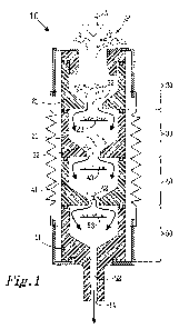
Figure 1 shows an inertial impactor 10 used to collect exhaled particles
(shown as P in
Figure 1). The impactor 10 is a container having an inlet 12 through which gas
and
exhaled particles may enter the impactor 10, and an outlet 14 through which
gas and
exhaled particles may leave the impactor 10. The impactor 10 in Figure 1 has
been
illustrated as a cylinder, with inlet 12 and outlet 14 on opposing circular
faces of the
cylinder; however, other geometries and arrangements of the inlet and outlet
12, 14 are
possible.
The impactor 10 comprises a plurality of stages 20, 30, 40, 50. Figure 1
illustrates four
stages 20, 30, 40, 50, although impactors with from 2 to 15 stages are known.
A primary
gas stream (A) comprising particles (P) enters the impactor 10 via the inlet
12 and passes
through each stage 20, 30, 40, 50 in turn before exiting the impactor 10 via
the outlet 14.
The primary gas stream (A) comprises air and particles exhaled by a subject.
The flow is
caused by a pump connected to the outlet of the impactor. Typically, according
to the
invention, the exhaled air and particles are not modified between leaving the
subject and
entering the impactor.
CA 02701352 2010-03-31
WO 2009/045163 PCT/SE2008/051110
Each stage 20, 30, 40, 50 is separated from adjacent stages by a partition 21,
31, 41, 51.
Each partition has at least one orifice 22, 32, 42, 52 (in practise, a
plurality of orifices is
present in each partition) which directs the gas stream (A) towards collection
plates 33,
5 43, 53. The major face of each collection plate 33, 43, 53 is arranged
substantially
perpendicular to the direction of flow of the gas stream (A).
The collection plates 33, 43, 53 used have a thickness of around 1 mm and are
square
with 10-12 mm side. The plates are held in place on the substrate holders by
double sided
10 tape at the exit of the air streams through the nozzles. The plates are
made of elemental
silicon since this is favourable for the ensuing analysis. The plates must be
extremely
clean since trace amounts of impurities may interfere with the ensuing
analysis of the
particles. The cleaning of the silicon plates may be done in several ways,
preferably by
ultrasonic cleaning in organic solvents followed by UV-ozone treatment, or by
immersion
in 1-10% nitric acid or hydrogen peroxide.
After cleaning, the preparation of the collection plate surfaces can be
further optimized
with respect to the collection and the ensuing chemical analysis. By varying
the
hydrophilicity or other surface chemical properties of the collection plates,
the interaction
of the particles with the surface may be controlled in a favourable way.
Preferably, the
entire surface of the collection plate is modified. A hydrophobic collection
surface will bind
hydrophobic moieties such as the hydrocarbon chains of lipids molecules more
strongly
than a hydrophilic surface. The normally hydrophilic silicon surfaces can be
made
hydrophobic by coating with a thin layer of hydrophobic substance such as
methyl silanes,
or by coating the silicon substrate with gold and then applying a monolayer of
methyl
terminated thiols onto the gold. Similarly, the collection surface can be made
to
specifically bind certain molecules. Specific proteins can be made to bind to
the collection
plate surface by coating it with antibodies for the proteins in question. By
using the proper
reagents, the binding of the analyte can induce a colour change or emission of
fluorescent
light, which can be detected in situ and in real time. In situ detection can
also be done
with an electric measurement of the current or capacitance change induced by
the binding
of the analyte to the surface of the collection plate. In this case the
collection plate also
has the necessary electrical connections that enable such a measurement. The
impactor
can comprise the necessary electrical connections which make contact with
appropriate
connections on the collection plate.
CA 02701352 2010-03-31
WO 2009/045163 PCT/SE2008/051110
11
Particles with inertia such that they are unable to follow the air stream when
it is deflected
around the first collection plate 33, will impact the collection plate 33
while particles with
less inertia will continue to the next stage 40. The inertia of a particle
depends on its mass
that, in turn, depends on its size. In this way, mass or size-segregation of
the particles is
possible.
Thus by choosing the number of orifices, their diameter and the distance from
orifice to
collection plate in each stage, it is possible to achieve mass or size
segregation of the
particles in an aerosol. Particles with high inertia, i.e. large mass/size
will be separated on
the early stages while particles with less inertia, i.e. smaller mass/size
will impact on the
later stages. By choosing the shape of the orifices, it is possible to
concentrate the
collected material in forms suitable for the ensuing chemical analysis. The
increase in
concentration of the material on the collection plates, compared with the
exhaled air or the
breath condensate, is considerable.
In the present case, a modified 3-stage Dekati PM10 was used. The
modifications
consisted of reducing the number of nozzles of the third stage by a factor of
two and
increasing the design flow rate of the impactor by a factor of 1.5.
The original impactor was a 10 liter per minute variety that was operated at a
flow of 15
liters per minute. The 20 orifices of the last stage were reduced to 10 thus
increasing the
gas velocity in each nozzle by a factor of 2. The 50% cut-off size [i.e. half
the number of
particles of that size are collected while half the number continues. This
does not show a
step-like collection characteristic, rather an "S-like" characteristic] were
7, 1.5 and 0.5 m
for the three stages, respectively.
The surface functionalization of the collector plate, as described above, can
be
miniaturized, to achieve different functionalizations at each nozzle, in order
to facilitate
optimal parallell collection different analytes. Similarly, parallell in situ
optical or electrical
detection of specific substances can be done by a collection plate chip, in
which the
appropriate surface functionalizations and/or electrical connections have been
supplied to
the collector plate at the location of the nozzle exits.
CA 02701352 2010-03-31
WO 2009/045163 PCT/SE2008/051110
12
The impactor is designed to collect the exhaled particles in an as efficient
as possible
way. This implies that virtually all particles present in the exhaled air, in
a given mass/size
interval, are collected for analysis. The particles are recovered in a
concentrated form
suitable for advanced chemical analysis.
The invention also provides a system 100 for collecting and sorting exhaled
particles, said
system comprising:
a. a reservoir 114 having first opening 112 and a second opening 113;
b. a two-way mouthpiece 110 connected to the first opening 112 of the
reservoir
114;
c. an inertial impactor 10 having an inlet 12 and an outlet 14, said impactor
10
comprising a plurality of stages 20, 30, 40, 50... arranged such that a gas
stream (A) comprising particles (P) enters the impactor 10 via the inlet 12
and
passes through each stage 20, 30, 40, 50... in turn before exiting the
impactor
10 via said outlet 14;
wherein each stage 20, 30, 40, 50... is separated from adjacent stages by a
partition 21,
31, 41, 51... having an orifice 22, 32, 42, 52... which directs the primary
gas stream (A)
towards collection plates 33, 43, 53..., the major face of each collection
plate 33, 43, 53...
being arranged substantially perpendicular to the direction of flow of the gas
stream (A);
the inlet 12 of the inertial impactor being connected to the first opening 112
of the
reservoir 114.
The collection system may be set up as is illustrated in Figure 2. The greater
part of the
system is located in a thermostatted compartment 120. The individual from whom
exhaled particles are desired inhales room air through a two-way mouthpiece
(110). Upon
inhalation, the inhaled air (A) passes a high efficiency particle filter (125)
located before
the mouthpiece.
The mouthpiece (110) is kept at a temperature such that the size distribution
of the
exhaled aerosol is not changed either by evaporation or condensation of water
vapour.
The exhaled air (A) passes the mouthpiece (110) into a system located in a
thermostatted
compartment (120), also here with the purpose of maintaining the aerosol size
distribution. In the compartment is located a reservoir (114) for the exhaled
air. Further, a
particle counter (116) is connected to the first opening (112) of the
reservoir (114) to count
CA 02701352 2010-03-31
WO 2009/045163 PCT/SE2008/051110
13
and measure particle size. An inertial impactor (10) for the collection of
particles (P) is
also connected to the reservoir first opening (112).
The flow through the impactor (10) is typically maintained by a pump (115),
located
outside the thermostatted compartment. Figure 2 also shows gas discharge (130)
and
that particle-free humidified air is added (135).
A particle counter (116), capable of measuring number-size distributions,
supplies
additional important information. The particle counter used here is a Grimm
1.108 optical
particle counter (Grimm Aerosol Technik, Ainring, Germany), capable of
counting, and
sizing particles in 15 size intervals from 0.3 to 20 micrometer. The
instrument may provide
a number size distribution of the measured aerosol or a mass distribution,
calculated from
the measured number size distribution. In the instrument, the particle-laden
air is passed
through a small, well defined, intensely illuminated volume in a manner so
that only one
particle at a time is illuminated. The illuminated particle gives rise to a
pulse of scattered
light, the intensity of which is measured. Since the intensity of scattered
light depends on
the particle size, it is possible to count and size the particles in the air
stream.
The reservoir (114) acts as a buffer where the exhaled air is stored when the
flow of
exhaled air exceeds the combined impactor (10) and particle counter (116)
flows. The
reservoir (114) supplies air to the impactor (10) and particle counter (116)
when no
exhalation is taking place. Moist, particle-free air is added at the second
opening (113) of
the reservoir (114) so that there is always a positive discharge flow. The
flow is measured
by a flow meter (119) located at the discharge end of the reservoir (114). By
displaying
the flow graphically in real time, it is possible for the subject to control
breathing frequency
and intensity according to instructions.
A sample is taken in the following way. It is assumed that the impactor is
loaded with
clean collection plates, and that the system, especially the impactor, has
attained the
desired temperature. First, the flow meter is zeroed to allow a proper
measurement of
flows, then the moist clean air flow is set at a value so that a positive flow
will be
maintained from the system during measurement. Then the impactor flow is set
at a value
lower than the clean air flow. During this procedure, no deposit will be
collected on the
plates, since the system is fed by clean particle free air. Then the optical
particle counter
is started and it is checked that no spurious particles are present, e.g.
indicating a leak
CA 02701352 2010-03-31
WO 2009/045163 PCT/SE2008/051110
14
into the system. Exhalation into the system then begins, the particle counter
continuously
draws a sample and produces a size distribution every six seconds while the
impactor
collects samples for later analysis. When a required amount of sample has been
obtained,
the collection is terminated, the time of sampling and exhaled volume
recorded. The flow
through the impactor is turned off, the impactor removed from the measurement
system
and the loaded plates are recovered.
In that two components of the system are "connected", it is to be understood
that air and
exhaled particles can flow between the components. Connection is usually made
by
tubes, with appropriate junctions, valves or seals to direct gas/particle
flow.
One possibility this system enables is a quantification of particle formation
in different
fractions at different exhalation rates. This may be a very easy way to detect
turbulent
airflow, as for example in asthma, and may be used as marker for disease.
Analysis
The collection plates 23, 33, 43, 53 and their associated particles P can be
removed from
the impactor 10 and the particles can be analyzed as to their chemical
content. The
chemical content of the particles P provides an insight into the medical
condition of a
subject (as is described below in the section entitled Medical Conditions).
In one analysis strategy, the particles are analysed while still on the
collection plates. This
is done with the following chemical analysis techniques that provide
complementary
information about specific substances present in the particles. Time-of-flight
secondary
ion mass spectrometry (TOF-SIMS) is especially useful for analysis of
substances in the
mass range up to 1000 u, in particular various types of lipids, for which the
profiles will
change during various disease conditions. Matrix assisted laser desorption
ionization
mass spectrometry (MALDI-MS) is a suitable method for analysing peptides and
larger
macromolecules (various proteins), that are associated with imflammatory
responses. The
MALDI-MS identification of proteins can be further facilitated by applying
proteolysing
enzymes, preferably trypsin, that will dissociate the proteins into segments
that can be
determined and used for conclusive protein identification by comparison with
publicly
available data bases. Analysis of specific proteins or other biomolecules
(e.g. DNA) can
also be done by applying different biochemical assays or protocols based on
labelled
antibodies, directly to the collection plates. Scanning electron microscopy
(SEM) can be
CA 02701352 2010-03-31
WO 2009/045163 PCT/SE2008/051110
used for analysing the morphology of the collected particle aggregates. Such
an analysis
can reveal particles of non-biological origin, for example, particles due do
exposure of the
subject.
5 In another analysis strategy, the collected material is removed (washed off)
from the
collection plates. The washing solution containing the collected particles can
then be
further processed for different chemical or biochemical analysis techniques.
In the
simplest analysis, the total amount of organic material in the collected
particles can be
analysed with a TOC (total organic content) analyser. Different elemental
analysers can
10 be used for obtaining the amounts of carbon, nitrogen, oxygen and sulphur
in the
collected material, which in turn reflects the relative amounts of different
classes of
biomolecules (lipids, carbohydrates, proteins). Trace amounts of inorganic
elements,
especially metals, can be determined by inductively coupled plasma mass
spectrometry
(ICP-MS). Such an analysis will provide information not only about substances
of non-
15 biological origin, but can also be used to detect metal-containing
biomolecules (proteins)
of importance in specific disease conditions, for example iron-response
protein (IRP). Cu
and Zn have also been shown to be increased in lung tumor tissue, and seem
both of
importance modulating the inflammatory response in the airways. For more
biomolecule
specific analyses, the three techniques gas-chromatography mass spectrometry
(GC-
MS), liquid chromatography mass spectrometry (LC-MS), and direct MALDI-MS,
will
provide complementary information. GC-MS will provide information about semi-
volatile
substances in the mass range up to around 500 u. LC-MS will provide
qualitative and
quantitative information about different biomolecules, such as lipids,
peptides and proteins
as well as their modifications. Direct MALDI-MS, finally, can be used for
pattern detection
of biomolecules up to several 10 000 u. allowing one detection and
identification of both
lipid and protein profiles.. The collected and washed off material can also be
subjected to
biochemical analyses, in particular labelled antibodies for specific proteins
of interest, or
quantitative PCR analysis for analysis of genetic material.
There are several techniques to facilitate the sample handling and to increase
the
sensitivity of the method. One advantage already present in the method is the
possibility
to directly analyze the collection plate taken from the impactor using surface
desorption
mass spectrometric techniques. A further advantage would be to purify the
sample and/or
modify it directly on the plate with for example the enzymes mentioned above,
so called
on-plate digestion. It is also possible to create different kinds of surfaces
on the collection
CA 02701352 2010-03-31
WO 2009/045163 PCT/SE2008/051110
16
plate which have been covalently modified with receptor molecules or enzymes
for direct
binding or modification of specific analytes in the particle sample. These
methods are well
known and can easily be applied in an organic laboratory. This will speed up
the analytical
process considerably making it more feasible for investigations of large
patient groups.
After the identification of novel biomarkers by mass spectrometric methods is
it possible to
introduce new analytical instruments such as surface plasmon resonance (SPR)
and
fluorescence spectroscopy in order to easily scale up the analysis to large
population
groups. These two methods are more easily used by non-experts which makes the
particle collection method more accessible for use at hospitals and health
care centres
and will also make studies of large patient groups more time efficient. It is
very
advantageous to be able to use the collection plate directly from the
impactor.
The different mass spectrometric (MS) techniques mentioned above have the
distinct
advantage that they provide global information about the composition of the
collected
particles. This means that by combining different MS techniques, the majority
of
biomolecules will be possible to detect in a non-predetermined way. This is in
contrast to
many other biochemical analysis techniques, which only detect pre-selected and
labelled
substances. The compatibility of the present method with MS techniques is thus
an
important advantage for identifying new specific biomarkers for different
diseases.
The analysis of the particles may be compared with a reference chemical
analysis, and
deviations and/or similarities from the reference chemical analysis can be
identified. This
can be used in determining one or more medical conditions in the subject. The
reference
chemical analysis can be from subjects having a certain medical condition (in
which case
similarities in the chemical analysis are looked for) subjects not having a
certain medical
condition (in which case deviations in the chemical analysis are looked for),
or from the
subject themselves, yet taken under different circumstances (e.g. at a later
point in time,
or after a certain course of treatment or exercise).
A particle distribution profile can be determined by sorting the particles on
each collection
plate. The particle distribution profile obtained can be used in determining
one or more
medical conditions in the subject. If diagnosis is to be made, the particle
distribution
profile of the particles exhaled by the subject is compared with a reference
particle
distribution profile. Similarities and/or deviations between the particle
distribution profile of
CA 02701352 2010-03-31
WO 2009/045163 PCT/SE2008/051110
17
the subject and the reference particle distribution profile are noted and the
deviations or
similarities between the particle distribution profile of the subject and the
reference particle
distribution profile are assigned to one or more medical conditions in the
subject.
The reference particle distribution profile may be a particle distribution
profile from a
subject not having a given medical condition. In this case, deviations may be
noted
between the particle distribution profile of the subject and the reference
particle
distribution profile, providing an indication of a medical condition.
The reference particle distribution profile may alternatively be from a
subject having a
given medical condition. Similarities can then be noted between the particle
distribution
profile of the subject and the patient, leading to the diagnosis of said given
medical
condition in the subject.
The reference particle distribution profile may also be from the subject
themselves, yet
taken under different circumstances (e.g. at a later point in time, or after a
certain course
of treatment or exercise). This would allow the monitoring of a medical
condition by the
method of the present invention.
Medical Conditions
Medical conditions which may be determined or monitored by the present
invention
include
- Asthma bronchiale
- Cystic fibrosis
- Chronic obstructive pulmonary disease (COPD)
- Lung cancer
- Interstitial lung-disease
- Sarcoidosis
- Pulmonary engagement in systemic disease such as systemic lupus
erythromatodes (SLE)
- Pulmonary infections
o pneumonia
o bacterial colonization
o viral infections
CA 02701352 2010-03-31
WO 2009/045163 PCT/SE2008/051110
18
It is plausible that also other systemic medical conditions can be monitored
such as
- Heart failure ( for example endothelin-1)
- Hypercholesterolemia (cholesterol is found in the exhaled particles)
- Diabetes (insulin is found in the particles)
- Metabolic syndrome
- Increased genetic susceptibility to disease or exposure
The particles may comprise or consist of biomarkers which are indicative of
specific
medical conditions. The method according to the invention allows the detection
of such
biomarkers.
The exhaled particles are believed to originate from the respiratory tract
lining fluid (RTLF)
covering the entire respiratory epithelium [ PediatrAllergy Immunol 15(1):4-
19] containing
large quantities of antioxidants and surfactant. One should also keep in mind
that the
constituents of the RTLF changes from the proximal to the distal airways.
One substance that is abundantly present in the RTLF is Clara cell protein 16
(CC16),
also acting as an anti-inflammatory protein, produced by the Clara cells. CC16
has until
now only been measured in BAL and blood. Other substances that so far have
gained
interest are surfactant proteins A-D, also only measured in bronchoalveolar
lavage, BAL.
Of special interest is the detection and monitoring of concentrations of anti-
oxidants in the
particles. A potential biomarker is glutathione which is in high abundance in
the
respiratory tract. Other anti-oxidants that are potential biomarkers in the
exhaled droplets
are the metal-binding proteins ceruloplasmin and transferinn which are likely
to be
detected with matrix assisted laser desorption/ionization mass spectrometry
(MALDI MS).
Additional potential antioxidants with low molecular weight, for example
ascorbate, a-
tocopherol, urate and L-cystein is also likely to be detected with mass
spectrometric
methods, these molecules are also biomarkers for oxidative stress.
Potential biomarkers that are directly involved in oxidative stress as
antioxidants are:
glutathione, ceruloplasmin, transferin, ascorbate, a-tocopherol, urate and L-
cystein.
Glutathione is especially interesting since it is highly abundant in the
airways. The
analytical methods that will be used to detect these antioxidants will be mass
spectrometry.
CA 02701352 2010-03-31
WO 2009/045163 PCT/SE2008/051110
19
a. Lipids
The profile of phospholipids in RTLF may serve as biomarkers for disease.
Alterations in
phospholipid composition (PC) have been seen in most airway diseases, such as
acute
respiratory distress syndrome (ARDS), pneumonia, cystic fibrosis and asthma.
In asthma,
PC was decreased in BAL and the relation between PC/phosphatidylglycerol (PG)
has
been shown to change after allergen challenge.
A new emerging research-area in respiratory disease is also the nitration and
oxidation of
lipids, which may alter their functions.
Surfactants, comprising phospholipids and proteins, in the RTLF are believed
to serve
important functions in the innate immune system. The phospholipids are
precursors for a
variety of cytokines active in the innate immunity such as prostaglandins,
thromboxanes.
eotaxins, lipoxins, resolvins etc. The surfactant proteins have also been
shown to play an
important role in the innate immunity, among other things acting as antigen-
presenting
cells and regulatation of cell death. The knowledge of metabolism of
surfactant is until
now very limited but believed to be important to understand pathogenesis of
respiratory
disease.
Surface analysis of the silicon collection plates with TOF-SIMS has revealed a
wide range
of phospholipids in the exhaled particles. The phospholipids detected in
particles are in
agreement with phospholipids found in RTLF in BAL studies. The relative
amounts of
phospholipids are also in agreement with BAL. The relative amounts of
phospholipids are
also in agreement with BAL. The ratio of CN-+CNO- (fragments presumably coming
mainly from proteins and peptides) to P03 was elevated among patients with
asthma and
patients with cystic fibrosis. This ratio may reflect a plasma protein leakage
into the
airspaces owing to airway disease.
b. Proteins and peptides
Proteomic analysis of bronchoalveolar lavage has revealed a multitude of
proteins present
in the sample. The analysis has been performed using 2D gels and mass
spectrometry.
Proteins involved in, among other things, imumunoinflammatory processes, cell
growth,
oxidant-antioxidant and protease-antiprotease systems as well as proteins with
unknown
functions. For example proteomic studies of BAL have been performed on
allergic
CA 02701352 2010-03-31
WO 2009/045163 PCT/SE2008/051110
asthmatic patients. In this study, 1592 proteins were identified and 160 of
these were
expressed differently in the patients compared with a control group. The most
abundant
proteins are plasma proteins that probably are derived from diffusion from the
blood-air
barrier. An increase in plasma proteins is probably due to exudation or
damage. It is very
5 likely that several of the peptides and proteins detected in BAL are also
present in the
exhaled particles.
Peptides and proteins that are biomarkers for diseases in the airways include
endothelin-
1, Interleukin-4 , Interferon-g, surfactant protein A-D and Clara cell protein
16.. These
10 molecules can be detected with ESI-MS and MALDI-MS or by immuno-assays.
There is a
high probability that more types of biomarkers will be detected in the present
invention,
since the collection of particles is more efficient than using exhaled breath
condensate
where a smaller number of particles are collected. Treating the proteins in
the collected
samples with proteolytic enzymes such as trypsin will result in several
peptide fragments
15 which will give rise to a pattern, unique for a specific protein set.
Investigation of posttranslational modifications such as phosphorylation and
glycosylation
of proteins are also potential targets for biomarkers. Wrong phosporylation
patterns are
known to be a part of several diseases.
Another important class of biomarkers in the respiratory tract is mucin
glycoproteins which
contribute to the mucociliary defense that protects the airways against
pathogens and
environmental toxins. For patients with asthma, COPD and cystic fibrosis is
there an
overproduction of mucin glycoproteins. Although there are some difficulties
with analysis
of glycoproteins due to their variable glycosylation pattern is it still
valuable to pursue this
group of compounds due to their involvement in different respiratory diseases.
An
advantage in analyzing glycoproteins is their easy purification by affinity
chromatography.
Furthermore, it is probable that variations in observed protein glycosylation
patterns will
be disease related, and therefore should be considered as a potential
biomarker.
c. Cellular material and gene expression
It is likely that the exhaled particles contain cells or cell structures
containing substances
with genetic information, in particular DNA and RNA. This cell material may be
due to
bacteria, viruses, or cells of the respiratory tract. Analysis of the genetic
expression of
such material can either provide new information about the pathology of, or be
used as a
CA 02701352 2010-03-31
WO 2009/045163 PCT/SE2008/051110
21
highly specific and sensitive means of diagnosing specific diseases. The
method could
hence be used to identify the pathogen in diseases such as pneumonia and
exacerbations of COPD, but also for early detection of for example
colonization with
Pseudomonas aeruginosa in cystic fibrosis, which often is a clinical problem.
d. Metals
It has been possible to trace exposure to metals in the EBC, such as iron,
cadmium, lead,
aluminium, copper. The metals are most probably transported to the EBC bound
to
exhaled particles. This implies that the method also has a potential to
monitor exposure to
various components of air-pollution, such as iron, zinc, cadmium or aluminium.
Exposure
to metals in ambient nano-particles have also been linked to the development
of
respiratory disease.
EXAMPLES
Exhaled particles from four healthy subjects were collected on silica wafers.
The
concentration of particles was recorded by means of an optical particle
counter (Grimm
1.108). Forced exhalations (with nose clips) were performed in order to obtain
a high
particle production. The subjects were trained to perform repeated consecutive
exhalations corresponding to 80% of their individual maximal forced expired
volume in
one second (FEV1). A deviation of 10% from the target flow was considered
acceptable.
Sampling was performed during 15 minutes in the morning of day 1 and repeated
in a
similar way day 2.
The chemical composition of exhaled particles on the silica wafers were
analyzed using
Time-of-Flight secondary ion mass spectrometry (TOF-SIMS IV IONTOF GmbH). A 25
keV Bi3+ primary ion was rastered over an area of 500 x 500 pmt centered
around the
spot with particles. Mass spectra of positive and negative secondary ions were
recorded
with the instrument optimized for maximum resolution. Spectra from the total
analysis
area or from selected regions of interest, and images for selected ions were
extracted
from the recorded raw data files using the instrument software. Assignment of
the peaks
in the spectra was done by comparison with reference spectra from pure
substances and
from published data from other mass spectrometry methods, and the assignments
were
also controlled by comparison with theoretical isotope patterns. The relative
intensities of
the identified peaks were calculated by normalization against total ion
intensities in
respective spectrum.
CA 02701352 2010-03-31
WO 2009/045163 PCT/SE2008/051110
22
Figure 3 shows positive (FIG.3A) and negative (FIG.3B) TOF-SIMS spectra of a
particle
spot from one control subject.
Figure 4 is a TOF-SIMS image of one spot with exhaled particles from one
control subject.
Figure 5 shows the concentration of exhaled particles (0.5-2.0 pm) vs. time
Figure 6 shows the ration (CN+CNO)/PO3- in a pilot study of healthy subjects
and
subjects with asthma or cystic fibrosis.
Table 1. Assignment of the m/z ratios of peaks of TOF-SIMS spectra of exhaled
particles.
Molecular species of phospholipids are named as x:a, where x is the number of
carbons
and a is the number of double bonds:
Positive ions Negative ions
Assignment m/z Assignment m/z
Phosphocholine ion 184 C 16:1 253
Cholesterol -OH 369 C 16:0 255
PC fragment 476 C 18:1 281
PC fragment 478 C 18:0 283
PC fragment 494 PA 32:1 645
PC fragment 522 PA 32:0 647
PC fragment 524 PG 28:1 663
PC fragment 650 PG 28:0 665
PC 28:0 + H 678 PG 34:2 671
PC fragment 680 PA 34:1 673
PC 30:0 + H 706 PG 32:0 721
PC 32:1 + H 732 PG 34:1 747
PC 32:0 + H 734 PG 36:2 773
PC 34:1 + H 760 PG 36:1 775
PC 34:0 + H 762 PI 34:2 833
PI 34:1 835
P1 36:2 861
PI 36:1 863
Table 2. Total exhaled volume and average concentration of exhaled particles
(0.5-2.0
pm) for the 15 minutes sampling period:
SUBSTITUTE SHEET (RULE 26)
CA 02701352 2010-03-31
WO 2009/045163 PCT/SE2008/051110
23
Subject 1 Subject 2 Subject 3 Subject 4
FEV14.1 FEV12.8 FEV13.2 FEV13.2
Day I Day 2 Day I Day 2 Day I Day 2 Day I Day 2
Volume (liters) 153 128 176 181 142 144 201 166
Particles/liter 210 123 149 55 956 343 239 180
All particle samples gave strong signals from phospholipids (Figures 2 and 4
and Table
1). Different species of phosphatidylcholine (PC) were detected as protonated
or alkali
metal cationized molecular ions in positive mode, while phosphatidylglycerol
(PG),
phosphatidylinositol (PI) and phosphatidic acid (PA) were detected as
deprotonated ions
in negative mode, Table 1. The composition of phospholipids was in agreement
with that
of earlier findings in broncho-alveolar lavage (BAL) fluid indicating that
exhaled particles
are most likely to derive from the lower airways.
Example 2. The subjects were trained to perform repeated consecutive
exhalations
corresponding to 80% of their individual maximal forced expired volume in one
second
(FEV1). Four healthy volunteers, four asthmatics and four patients with cystic
fibrosis
performed 10 forced exhalations, respectively. Exhaled particles in the size
0.5-2.0 pm
were collected on silica wafers. An optical particle counter measured the
particle
concentration in real-time. Before sampling a washout-period of 3 minutes
breathing of
particle free air was applied. Silica wafers were analyzed with Time-of-Flight
Secondary
Ion Mass Spectrometry (TOF-SIMS). Several classes of phospholipids were
detected in
the particles: phosphatidylcholine (PC), phosphatidylglycerol (PG),
phosphatidylinositol
(PI) and phosphatidic acid (PA). Some differences were observed between
groups. The
ratio of the sum of signals of PC and the sum of signals of PG tended to be
elevated
among asthmatics and patients with cystic fibrosis compared to controls. Also,
signals
known to be characteristic for proteins and peptides (CNO-) were elevated in
comparison
to phospholipids in the samples of asthmatics and patients with cystic
fibrosis compared
to controls (Figure 6).
Example 3. Subjects performed forced exhalations during 20 minutes. Exhaled
particles
in the size 0.5-2.0 pm were collected on silica wafers. Silica wafers were
stained with a
fluorescent reagent, DAPI (4,6-diamidino-2-phenylindole) that binds strongly
to DNA and
CA 02701352 2010-03-31
WO 2009/045163 PCT/SE2008/051110
24
RNA. Strong signals were obtained in the particle spots indicating that
exhaled particles
contain nucleic acids.
Example 4. Two subjects exhaled 150 L air twice; once for particle collection
and once for
breath condensate collection. Exhaled particles were desorbed from the
sicilica wafers
and breath condensate were concentrated before analysis of Surfactant protein
A by
ELIZA.. The total amount of Surfactant protein A (Sp A) were 6 times higher in
exhaled
particles than those found in exhaled breath condensate, and 4 times higher
than that in
100 pL serum. The analysis of Sp A showed high intra-individual
reproducibility when
tested (CV 5.4 on two subjects when tested at three different occations).
The developed sampling method has high potential for the detection of new
biomarkers in
exhaled air and monitoring of respiratory disease.