Note: Descriptions are shown in the official language in which they were submitted.
CA 02702489 2015-12-01
Apparatus and Method for Use in Analyzing a Patient's Bowel
David J. Vining
[01]
Field of the invention
[021 The present invention relates to an apparatus and method for use in
analyzing a
patient's bowel by indicating selected parameters, such as colonic pressure,
and
perceived patient sensations, such as pain, during a medical procedure such as
a virtual
colonoscopy examination, or for use as a diagnostic testing means.
Background of the invention
[03] Virtual colonoscopy (VC) iS an effective medical procedure for use in
identifying
polyps and cancers in the bowel. Typically, an oral laxative is administered
to a patient
to cleanse and empty the bowel of solid stool. Once the bowel is cleansed, the
colon is
insufflated with gas. After the colon is distended by the gas, the patient's
abdomen and
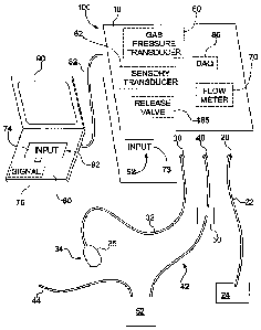
pelvis are scanned using a selected scan procedure, such as computed
tomography (CT)
or magnetic resonance imaging (MRI), while the patient lies motionless during
a breath
hold. Finally, image analysis is performed by a physician and/or computer-
aided
diagnosis to diagnose polyps, cancer or other abnormalities in the bowel.
[04] Colonic insufflation usually involves administering air or other selected
gas into a
patient's colon through a rectal catheter. A technologist monitors the flow of
gas through
the rectal catheter into the colon. Once the technologist believes that the
colon has been
sufficiently insufflated the patient's abdomen and pelvis are scanned.
CA 02702489 2010-04-12
WO 2009/052100
PCT/US2008/079826
[05] Subjective determination of colon distention during a VC procedure has
several
drawbacks. For example, false readings may be obtained if the colon is not
properly
distended throughout the CT or MRI scan. An improperly distended colon, or a
colon
affected by contractions (including normal peristalsis and spasms) may produce
misleading images and errors in diagnosis because of the potential of
collapsed bowel
segments mimicking or masking true lesions. Furthermore, patient intolerance
to colonic
distention can lead to patient motion, either respiratory or body movements,
during the
scan which can also produce image artifacts that lead to incorrect diagnoses.
[06] Accordingly, an apparatus for objectively determining distention of the
colon and
relative patient sensations, hence improving the accuracy of a medical
procedure such as
a VC scan, and for use as a diagnostic test, is desirable.
Summary of the invention
[07] In accordance with the present invention, an apparatus and method are
provided for
use in analyzing a patient's alimentary tract, and, more particularly, for use
during a scan
of a patient's colon. In accordance with the present invention, a sensory
input detector is
provided. The sensory input detector may be in the form of a pain input
detector that
communicates with the patient for detecting pain perceived by the patient
during
insufflation of the patient's colon with gas. The pain input detector may
optionally
include a pain sensor such as a squeeze bulb which may be squeezed by the
patient to
indicate the onset of pain and may be released by the patient to indicate the
cessation of
pain. The magnitude of squeeze on the squeeze bulb can also be determined
based on
pressure changes caused by squeezing and releasing of the bulb to correlate to
a
magnitude of pain perceived by the individual patient.
[08] The apparatus may also include a sensory transducer, for example, as a
part of the
sensory input detector to provide at least one parameter indicative of a
parameter such as
pain perceived by the patient. More specifically, the sensory transducer may
produce an
output parameter to indicate the onset or cessation of pain. Optionally, the
pain
2
CA 02702489 2010-04-12
WO 2009/052100
PCT/US2008/079826
transducer may be used to produce an output parameter to indicate sensations
perceived
by the patient, such as a magnitude of perceived pain. In other arrangements
the sensory
transducer may function to produce an output parameter to indicate other
sensations the
patient may perceive by any of the senses, including but not limited to,
touch, sight,
hearing, taste, or smell.
[09] A processor may be provided for communicating with the sensory transducer
to
generate an output reflecting perceived patient pain. The output may,
alternatively,
indicate other physiological parameters or sensory parameters. Sensory
parameters
include sensations perceived by the patient, whereas physiologic parameters
include
indications concerning the status of insufflation of the patient's colon. The
output may
then be used to determine validity or invalidity of a scan of a patient. For
example, if the
output is within a selected threshold, the scan may be determined to be valid.
Optionally,
if the parameter is outside of a selected threshold, the output may be used to
indicate that
a scan is not valid. A determination of validity may be conducted before the
scan is
performed. For instance, the output may be used to determine whether one or
more
parameters are within a selected threshold. If the one or more parameters have
obtained
the selected threshold, the scan may be conducted and reliable results may be
obtained.
If selected parameters are not within the selected threshold, reliable scan
results may not
necessarily be obtained. For instance, if selected parameters indicate that
the patient's
bowel is not insufflated to a desired amount, any scan that might be performed
may not
yield accurate results. Validity determinations conducted before a scan may be
referred
to as "approval." Optionally, validity may be determined during the patient
scan. During
the patient scan, if one or more parameters are within a selected threshold,
the scan
results may be accurate, thereby providing a valid scan. Alternatively, if the
parameters
are not within a selected threshold, the scan may be considered invalid and
the patient
may be scanned again. For example, a scan may be determined to be invalid if
bowel
segments are found to be collapsed during the scan. Validity determinations
conducted
during a scan may be referred to as "verification." In short, validity may be
determined
before a scan (approval) or validity may be determined during a scan
(verification).
3
CA 02702489 2010-04-12
WO 2009/052100
PCT/US2008/079826
[10] Signaling means may be provided to signal the technologist or physician
when the
output reflects the predetermined condition to validate a selected medical
procedure such
as a VC scan. Various medical procedures require the patient to be scanned.
Examples
include VC scans, CT scans, MRI scans, or any other similar procedures.
Specifically,
the term "VC scan" used herein includes both CT and MRI scans of a patient's
colon. An
indication that a VC scan is invalid may be used to prevent the commencement
of a CT
or MRI scan or may be used to indicate that an ongoing or already completed
scan may
need to be repeated.
[11] In an alternate embodiment, the apparatus may include a gas pressure
transducer
that may be used either alone or in combination with the sensory input
detector, such as a
pain input detector, to provide a physiologic parameter, such as a parameter
of gas
pressure within the patient's colon or alternatively, pain perceived by the
patient. The
gas pressure transducer communicates with the patient's colon for indicating
gas pressure
inside the lumen of the patient's colon. The processor may be utilized in
communication
with the gas pressure transducer to generate an output parameter reflecting
the pressure of
the gas in the patient's colon. A signaling means may be provided to signal
when at least
one selected output parameter reflects a predetermined condition to validate a
procedure
such as a VC scan of the patient. More specifically, the signaling means may
be used to
signal when an output from the gas pressure transducer and/or the pain
transducer is
outside of a selected threshold(s) thereby indicating an invalid scan.
Likewise, the
signaling means may be used to signal when the selected output parameter, such
as
pressure within the colon and/or pain perceived by the patient, is within a
selected
threshold(s) to indicate that a scan of the patient is valid.
[12] In accordance with another embodiment of the invention, a flow meter may
be used
to measure flow of gas being instilled into the colon of the patient. The flow
meter in a
selected embodiment may be used alone or in combination with either the gas
pressure
transducer or the sensory input detector, or both of them, to detect multiple
or additional
physiologic or sensory parameters. In such an embodiment utilizing a flow
meter, the
processor communicates with the flow meter to generate an output parameter
reflecting
4
CA 02702489 2010-04-12
WO 2009/052100
PCT/US2008/079826
the volume of gas delivered to the patient. Again, the signaling means may be
used to
signal when at least one output parameter, such as the volume of gas delivered
to the
patient, reflects a predetermined condition to validate the scan of the
patient. More
specifically, the signaling means may be used to signal when the selected
output
parameter, such as volume of gas delivered to the patient, is within a
selected threshold to
validate the scan or when the parameter is outside of a predetermined
threshold to
indicate an invalid scan. In accordance with one embodiment, the processor may
take the
form of a conventional microprocessor and assorted circuitry, a data
acquisition module
(DAQ), other hardwired circuitry, software, or much more simplified circuitry,
such as an
amplifier, converter, memory or display circuitry.
[13] In another embodiment, a method is provided for use in studying a
patient's colon
during a selected scan procedure by generating an output reflecting a selected
parameter
such as a parameter relating to perceived patient pain during the scan. The
method may
be used when a patient's colon is insufflated during a medical procedure such
as a VC
scan or it may be used as part of a diagnostic test. In a particular
application, the
patient's colon is insufflated with gas. At least one parameter indicative of
a sensation,
such as pain, perceived by the patient during or after colon insufflation is
detected. Then
a determination is made whether the at least one parameter is within a
threshold. For
example, the validity of a medical procedure, such as a VC scan, may be
determined if
the parameter is within the threshold. Alternatively, a diagnostic test for
disease may be
performed based upon a determination of whether the parameter is within or
outside a
selected threshold.
Brief description of the drawings
[14] The foregoing summary as well as the following description will be better
understood when read in conjunction with the figures in which:
[15] Fig. 1 is a schematic perspective view of a monitoring device in
accordance with the
present invention.
CA 02702489 2010-04-12
WO 2009/052100
PCT/US2008/079826
[16] Fig. 2 is schematic diagram of one configuration of a monitoring device
in
accordance with the present invention.
[17] Fig. 3 is an exemplary chart of a hypothetical representation of patient
pain, colonic
pressure, and volume of gas delivered to a patient in accordance with the
present
invention.
[18] Fig. 4 is an exemplary chart of a hypothetical representation of a
selected parameter
of perceived patient pain in accordance with the present invention.
[19] Fig. 5 is an exemplary chart of a hypothetical representation of a
selected parameter
of colonic pressure in accordance with the present invention.
[20] Fig. 6 is an exemplary chart of a hypothetical representation of a
selected parameter
of volume of gas delivered to a patient in accordance with the present
invention.
Detailed description of the invention
[21] Referring now to the Figures in general, wherein like reference numbers
refer to the
same components across the several views, there is shown an apparatus,
generally
designated 100, for use in a selected medical procedure or diagnostic test. In
general
application, the apparatus 100 may be used as a monitoring apparatus to
monitor selected
conditions of a patient during the selected medical procedure or diagnostic
test. In
specific application, the apparatus 100 may be utilized to monitor selected
physiological
and/or sensory parameters of a patient during a medical procedure, for
example, during a
scan of the patient's abdomen and colon or other type of body scan. The term
"VC scan"
as used herein encompasses both CT and MRI scans. In order to scan a patient's
colon,
the patient must be properly prepped for the scan. First, the patient's colon
must be
cleansed. Immediately prior to the VC scan, the patient's colon is insufflated
with gas to
distend the colon so that a clear image may be obtained during the scan
procedure. Gas
from an insufflator 24 is administered to the patient to distend the patient's
colon.
6
CA 02702489 2010-04-12
WO 2009/052100
PCT/US2008/079826
Although air has traditionally been used to insufflate the colon, the gas
insufflator 24 may
employ carbon dioxide, nitric oxide, xenon, krypton, oxygen, or other gas. In
one
preferred embodiment, carbon dioxide is dispensed from the insufflator 24 into
the
patient. The flow rate of gas delivered may be at a preset rate or amount. For
instance,
the rate of gas flow delivered to the patient may be approximately 3 to 5
liters per minute.
In another configuration, an insufflator which is capable of delivering a
higher flow rate
may be used. For instance, a gas insufflator capable of delivering 20 to 30
liters per
minute may be used in various procedures. In order to aid in producing a valid
scan of
the patient's colon, the apparatus 100 may be used to monitor selected
physiological and
sensory parameters of the patient, such as pressure inside the colonic lumen,
gas flow into
the colon, volume of gas delivered to the colon, and/or pain or other
sensations perceived
by the patient during the insufflation procedure. Other conditions of the
patient may also
be monitored for specific applications.
[22] As shown in Fig. 1, the apparatus 100 includes a housing 10. In order to
measure
the flow of gas supplied to the patient's colon, the apparatus 100 includes a
gas flow
meter 70. In order to supply gas to the patient's colon, a gas insufflator 24,
is connected
with a first input 20 on the apparatus 100 by a supply tube or input line 22.
A gas tank
provides gas to the insufflator 24 for insufflation to the patient. From the
input 20, the
gas flow is directed through the flow meter 70 so that the flow of gas from
the gas
insufflator can be measured in selected units such as liters per second. From
the gas flow
meter 70, the gas flow is directed out of the apparatus 100 through output 40
and is
supplied through output tube or line 42 to the patient's colon through a
rectal catheter 44
that is inserted into the patient's rectum. A waste collector 52, such as a
canister or
plastic bag, is provided between the rectal catheter 44 and the output 40 of
the apparatus
100 to collect any undesirable back flow of waste from the rectal catheter. In
order to
decrease the risk of contamination of the apparatus 100, a hydrophobic filter
50 is
positioned between the waste collector 52 and the output 40 to impede back
flow toward
the apparatus 100. In this arrangement, the gas flow meter 70 is positioned
within the
flow of gas from the gas insufflator 24 to the rectal catheter 44. The gas
flow meter 70 is
used to produce an output flow parameter reflecting the amount or rate of flow
of gas
7
CA 02702489 2010-04-12
WO 2009/052100
PCT/US2008/079826
delivered to the patient's colon. The output flow parameter may be processed
internally
of the unit 100 by processor circuitry to generate, if desired, an output
reflecting the rate
of gas delivered to the patient and/or the volume of gas delivered to the
patient. The
processor circuitry may include a conventional processor 80, a microprocessor
and
assorted circuitry, a data acquisition module (DAQ) 86, other hardwired
circuitry, or
software, or much more simplified circuitry such as an amplifier, converter,
memory or
display circuitry and/or software. The processor 80 may be positioned inside
the housing
or may be positioned external from the housing 10. The flow of gas over time
may be
measured to produce an output volume parameter providing an indication of the
volume
of gas delivered.
[23] In order to detect or measure the pressure of gas within the patient's
colon, a gas
pressure transducer 60 is provided in the apparatus 100. The gas pressure
transducer 60
communicates with the patient's colon for detecting gas pressure within the
patient's
colon. For example, the gas pressure transducer 60 may communicate at a
selected point
along the output line 42, the input line 22, or at a position within the
apparatus 100
between input 20 and output 40. A separate gas detection line could be run
with or
through the rectal catheter directly into the patient if so desired. In a
selected application,
for example, the gas pressure transducer may be connected along the flow line
at a
location before the hydrophobic filter 50. The gas pressure transducer 60
functions to
produce an output pressure parameter reflecting the gas pressure within the
patient's
colon. The output parameter from the gas pressure transducer may be processed
by the
data acquisition unit 86 and/or by the processor 80 to produce an output, if
desired,
reflecting the gas pressure within the colon.
[24] In order to measure pain sensation perceived by the patient, a sensory
transducer
62, in the form of a pain transducer, is provided on the apparatus 100. "Pain"
as used
herein includes pain and/or discomfort perceived by the patient. In this
particular
configuration, the sensory transducer 62 functions as a pain transducer to
monitor pain
perceived by a patient and to produce an output pain parameter reflective of
the perceived
pain. Otherwise, the output generated by the sensory transducer 62 may include
data
8
CA 02702489 2010-04-12
WO 2009/052100
PCT/US2008/079826
reflecting other physiological and sensory parameters. Sensory parameters
include
sensations perceived by the patient including, but not limited to, touch,
hearing, sight,
smell, and taste. For example, the patient may feel sensations such as
temperature,
visceral pain, pressure, or tenesmus. Accordingly, while the sensory
transducer 62 may
measure pain, the sensory transducer 62 may be configured to also measure
other sensory
parameters perceived by the patient. The sensory transducer 62 communicates
with an
input sensor 35, with which the patient communicates. In this particular
configuration,
the sensory transducer 62 functions as a pain transducer that communicates
with a pain
input detector 34 through input line 32 connected between the sensory input
detector 34
and an input 30 on the apparatus 100. The sensory input detector 34, when in
the form of
a pain input detector 34, communicates with the patient in order to detect
pain perceived
by the patient. For this purpose, the input sensor 35 may include a sensor in
the form of a
squeeze bulb 35 that may be actuated by the patient in response to perceived
pain or other
sensations. For example, the squeezing of the bulb 35 will serve to change the
pressure
within the bulb so as to indicate the onset of pain. Likewise, the release of
the bulb 35
will change the pressure within the bulb in the opposite direction to thereby
indicate the
cessation or reduction of pain perceived by the patient. The amount the bulb
35 is
squeezed or released can be detected to determine a change in magnitude,
either up or
down, in the amount of perceived pain. Similarly, the length of time that the
bulb 35 is
squeezed and then released can be measured to determine the duration of pain
as well as
the duration of different magnitudes of pain perceived by the patient. In such
an
arrangement, the pain transducer 62 may take the form of a pressure transducer
so as to
detect changes in pressure created at the squeeze bulb 35 by the patient in
response to
pain. In such an arrangement, the pain transducer 62 will output a pain
parameter
reflective of perceived pain. The output pain parameter may be used to
indicate absence
of pain, onset of pain, reduction of pain, cessation of pain, increase of
pain, change in
pain, magnitude of pain, as well as duration of pain and duration of selected
magnitudes
of pain. The output parameter from the pain transducer 62 may be supplied to
the DAQ
86 and/or the processor 80 in order to process the data to generate an output,
if desired,
reflecting perceived pain or a parameter of the perceived pain. Of course,
more
generally, the change in pressure may also be measured up or down, and timed
by the
9
CA 02702489 2010-04-12
WO 2009/052100
PCT/US2008/079826
sensory transducer 62 in the form of a general pressure transducer to reflect
other
sensations or parameters.
[25] The DAQ 86 may function as a type of processor in certain applications or
configurations to control and generate output data in desired format for
selected uses such
as further processing, display, analysis, or storage. The DAQ 86 may be used
to
communicate with any one or a combination of the gas pressure transducer 60,
the
sensory transducer 62 and the flow meter 70, or other selected inputs. The
data reflecting
the output parameters from the gas pressure transducer 60, the sensory
transducer 62
and/or the flow meter 70 may be communicated via the DAQ 86 to the external
processor
80 for original or further processing. A selected output reflecting at least
one of the
output parameters, including any one alone or in combination with the others,
may be
generated and displayed on a display 90. An input 92 to enable a user to input
information or data may be provided on the apparatus 100, such as input 73, or
on the
computer processor 80, such as input 74, or on both the apparatus 100 and the
computer
processor 80. The components of the apparatus 100 may also be positioned
within one
housing 10. Preset limits or other programmed information may be entered into
the input
92. If desired, the processor 80, the display 90, and the input 74 may be
incorporated
within the monitoring apparatus 100 as an integral device.
[26] Signaling means or unit 76 may also be provided to indicate at least one
parameter
reflective of a predetermined condition so as to determine the validity of a
medical
procedure such as a VC scan. Generally, the signaling means 76 may be
positioned for
use with the apparatus 100 or the external processor 80. The signaling unit 76
may be
provided to indicate that favorable conditions exist in order to start the
scan of a patient
or that favorable conditions have occurred during the performance of a scan,
hence
indicating a valid scan. A determination of validity conducted before the
start of a scan
may be referred to as "approval." A technologist or doctor may determine
whether one
or more parameters have obtained a selected threshold. If the parameters fall
within the
selected threshold, the results may be deemed valid and approval given that a
scan may
be started. If the parameters are not within a selected threshold, approval
may not be
CA 02702489 2010-04-12
WO 2009/052100
PCT/US2008/079826
given so that the results may be considered invalid and a scan may not be
conducted. An
exemplary situation where a procedure would be considered invalid could occur
if
parameters do not reach a selected threshold because the patient's colon is
not distended.
A determination of validity made during a scan may be referred to as
"verification." If
one or more parameters are within a selected threshold as the patient is being
scanned, the
technologist or physician may determine the scan is valid and verify the scan
results.
However, if the parameters are not within the selected threshold, the
technologist or
physician may not verify the parameters and instead declare the scan to be
invalid. An
example of a situation where a scan may be deemed invalid would occur if a
section of
bowel collapses during the scan. Alternatively, determination of validity may
incorporate
an automated process such as computer-aided diagnosis (CAD). For instance, the
signaling unit 76 may indicate whether an unfavorable condition occurred
during the
course of a scan, thereby indicating an invalid scan or an invalid segment of
a scan. For
this purpose, the signaling unit 76 may take a variety of different formats
and may be in
the form of an electronic signal, an alarm, a flashing light, sound alarm, or
other suitable
signaling means in desired hardware and/or software.
[27] Representations of the output parameters indicating gas pressure, gas
volume, and
pain, for example, may be displayed on the display 90. The representations may
be
displayed as functions or other wave forms on the display, or the
representation may take
the form of text or verbal messages. In selected applications, the output
parameters may
be displayed in strip chart format on the display 90. Alternatively, one or
more output
parameters could be displayed as bars or other types of graphs or charts. The
display 90
may function to display one or more parameters at the same time, for example,
superimposed on one another as shown for example in Fig. 3 or in separate
display
sections as shown for example in Figs. 4, 5, and 6. The display 90 may also be
caused by
the processor to display the representations separately. The data shown on a
display 90
may be analyzed, whether or not the data is actively displayed, by a
technologist,
physician, or CAD to determine if the pressure in the colon, the volume of gas
delivered
to the patient, and/or the amount of pain or discomfort experienced by the
patient as the
patient's colon is insufflated meets a selected threshold or criteria. Other
sensations
11
CA 02702489 2010-04-12
WO 2009/052100
PCT/US2008/079826
perceived by the patient and detected by the sensory input detector 34 may
also be shown
on the display 90. The processor 80 may analyze physiological and sensory
parameters
to diagnose certain conditions of the patient.
[28] The output generated by the processor 80 to reflect at least one output
parameter
may be used by the technologist, physician, or CAD to make decisions
concerning patient
care or to confirm (i.e., approve or verify) a clinical procedure. A
determination may be
made as to the validity of a scan of the patient. Validation is effected to
determine when
to start a scan, whether to stop a scan already in progress or whether a scan
or any part
thereof may need to be repeated under more favorable conditions. Typically,
for
relatively short scans, the scan would not be stopped but may need to be
repeated to
acquire image data under more favorable conditions. For a relatively lengthy
scan, it may
be desirable to stop the scan, for example, in order to prevent unnecessary
radiation
exposure whenever CT is being employed as the scanning method. Additionally, a
determination may be made as to efficacy of pharmacological intervention after
such
intervention has been administered. For example, the output data may be
analyzed to
determine a decrease in patient pain perception or colonic pressure spikes
when a
pharmacological agent, such as a spasmolytic agent, is administered. In one
exemplary
embodiment, for example, plotted data representing the gas pressure within the
colon
may resemble a plateau-like shape at a desired level when the has reached a
steady state
suitable for scanning. The plateau-like shape indicates that gas pressure is
steady and the
bowel is relaxed. A steady state of gas pressure within the colon at a
suitable level may
provide the greatest amount of colonic distention and the least amount of
patient
discomfort. Once a steady state has been reached, the patient may be scanned.
By
performing the scan when the bowel is relaxed, the accuracy of the scan
results increases,
thereby reducing the likelihood of the physician making erroneous diagnoses
caused by
bowel contractions that can mimic or mask cancers or other abnormalities. By
providing
more accurate determination of colon distention, the likelihood of having to
repeat a VC
scan due to poor readings is decreased, thereby reducing or eliminating
unnecessary
additional time, cost, and when CT is employed as the imaging means, radiation
exposure.
12
CA 02702489 2010-04-12
WO 2009/052100
PCT/US2008/079826
[29] During a procedure, data processed by the processor 80 may instead
indicate the
presence of contractions in the colon. Peristalsis is the normal contraction
of the bowel
that propagates contents along its lumen, whereas spasm is an exaggerated form
of
contractions that is often perceived by the patient as cramping. The term
"contraction" as
used herein refers to both peristalsis and to spasm. Contractions of any form
may cause
normal areas of the colon wall to appear on scan images as abnormally
thickened or
collapsed. Accordingly, contractions may mimic the presence of an abnormality
when in
fact none is present. Conversely, the presence of contractions or collapsed
segments of
bowel during a scan may mask the presence of cancer or polyps. If a scan is
performed
while the colon contracts or is collapsed, the scan may produce inaccurate
results.
Therefore, the detection of contractions may call into question the validity
of a scan. In
such a case, the patient may need to undergo a repeat scan. Accordingly, the
output
parameters generated may be analyzed to determine the validity of scan
results. In
general, the output parameters may be analyzed by the technologist, physician,
or CAD to
determine if any or all of the output parameters fall within a selected
threshold to
determine the validity of the scan. Conversely, if any or all of the output
parameters fall
outside a selected threshold the scan may be determined to be invalid.
[30] Considering one exemplary application, the processor 80 may process data
for three
selected parameters. Specifically, the processor 80 may process an output pain
parameter
reflecting perceived patient pain, an output volume parameter reflecting
volume of gas
delivered to the patient, and an output pressure parameter reflecting colonic
pressure.
Turning now to the pain parameter, the pain input detector 34 indicates
perceived patient
discomfort, pain, or other sensations. Information reflecting perceived
patient pain is
detected by an input sensor 35 in communication with the patient. In a
selected
configuration, the input sensor 35 may include a hand-held squeeze bulb 35,
such as a
hollow plastic or rubber ball. The bulb 35 is connected by a length of tubing
or line 32 to
input 30 and is connected with the sensory transducer 62 which may function as
a pain
transducer. The squeeze bulb detects perceived patient pain and conveys data
to the pain
transducer 62. For instance, the onset of discomfort may be detected by pain
input
13
CA 02702489 2010-04-12
WO 2009/052100
PCT/US2008/079826
detector 34 when the patient squeezes the bulb 35. The pain input detector 34
may also
detect pain relief as the patient releases the squeeze of the bulb 35.
Duration of
discomfort may be measured as the length of time between the start of a
squeeze and the
release of the squeeze. Severity of pain may also be measured by the pain
input detector
34. Severity may be indicated by how forcefully the patient squeezes or
releases the bulb
35. Specifically, a strong squeeze of the bulb 35, by the patient could
indicate a high
level of pain. Alternatively, a slight squeeze may indicate a low level of
pain or
discomfort. Likewise, a quick release may indicate a sudden drop in pain and a
slow
release may represent a slow drop in pain. Likewise, a partial release may
indicate a drop
in pain but to a lower level. As the patient squeezes and releases the bulb
the changes in
pressure produced by the bulb can be tracked both in direction and in
magnitude by the
sensory transducer 62 which functions as a pressure transducer and part of the
pain input
detector 34.
[31] In an alternate embodiment, the input detector 34 may be in the form of
some other
hand-held device with strain transducers to deliver electromechanical input to
the sensory
transducer 62, or it may take the form of a foot activated pedal or other body
activated
device, or a voice-activated or other sensory-activated sensor.
[32] Since in the present configuration the pain input detector 34 detects a
painful
sensation perceived by the patient, it is preferable for the pain sensor 35 to
reset rapidly
in preparation for another perceived pain event. For instance, if a hand-held
bulb is used,
the pain transducer 62 detects air pressure at the bulb. The hand-held bulb is
made of a
resilient material, such as rubber or plastic, so that once a patient releases
the sensor, the
sensor quickly resets to be able to register another pain indication shortly
after release.
For this purpose, the resetting of the pain input detector 34 may be
controlled by a
pressure release mechanism or other similar mechanism. Such optional
arrangements
may be used to sense or detect parameters other than pain.
[33] In operation, patient pain may be detected on a relative scale which
differs for each
patient. In one configuration, a range of 0 to 10 may be provided for a
patient, where 0
14
CA 02702489 2010-04-12
WO 2009/052100
PCT/US2008/079826
indicates no pain and 10 indicates of pain. A relative pain scale is
established by
calibrating the pain input detector 34 before starting the procedure. For
instance, if the
pain input detector 34 utilizes a squeeze bulb, the patient may be asked to
squeeze the
bulb as hard as possible in order to establish an upper limit of a selected
patient's
perceived pain. This reading may be established as a "10" on the scale.
Calibration of
the pain input detector 34 standardizes the patient pain data despite the
varying pain
threshold of each patient. Additionally, during the scan procedure the patient
may
experience a pain sensation that causes him or her to record a pain level that
exceeds the
calibrated upper limit, or the patient may just happen to squeeze harder
during the scan
procedure than during the calibration procedure.
[34] Generally, it is desired to acquire a scan as the patient lies motionless
and during a
breath hold. Excessive pain may cause the patient to breath, squirm or move
during the
scan which can produce respiratory and/or motion artifacts that can yield
inaccurate scan
results. If a patient perceives a great deal of pain during colon
insufflation, a scan should
not be conducted and some form of intervention may be required to relieve the
patient's
pain. One form of intervention consists of releasing, or venting, gas from the
patient's
colon. Venting of gas may be conducted either manually by a technologist or it
may be
automated. For example, the technologist may manually disconnect the tubing 22
from
the insufflator 24 so that the colon may be vented to atmospheric pressure.
Alternatively,
this venting mechanism could be automated and controlled via a pressure
release valve
185 mechanism incorporated into the apparatus 100. Intervention could also
take the
form of pharmacological intervention, such as administration of a spasmolytic
agent. On
the other hand, if a patient experiences a great deal of pain, but the pain
resolves quickly,
then no intervention may be required. For example, Fig. 4, number 410 shows a
pain
spike to level 8 of pain. The pain spike at level 8 may indicate that a high
magnitude of
pain is perceived by the patient. Since pain spike 410 resolves relatively
quickly,
intervention may not be required. On the other hand, a pain spike to level 10
may
require intervention regardless of how short the duration of the pain.
Alternatively, if a
patient experiences pain for a sustained length of time, for example, longer
than 5
seconds, intervention in the form of venting and/or pharmacologic intervention
may be
CA 02702489 2010-04-12
WO 2009/052100
PCT/US2008/079826
required to reduce pressure and relieve pain before continuing with the
insufflation and/or
scanning procedure. Figure 4, number 440 shows sustained pain at level 5 which
lasts
more than 20 seconds or so and may therefore be longer than a selected time
threshold.
The selected time threshold may be 5 seconds or some other suitable length of
time. The
patient may need intervention to decrease colonic pressure and pain. The
patient should
not perceive any significant pain prior to restarting the insufflation
procedure if restarting
insufflation is deemed necessary.
[35] In a selected application, a patient may experience mild pain or
discomfort but still
be scanned. If the patient experiences pain that does not reach a selected
threshold, such
as a level 2 for example, then a scan may be conducted. For example, a scan
may be
performed while the amount of pain is below a selected threshold and the
patient is
relatively pain-free or experiencing only minor discomfort, such as a bloating
sensation
or tenesmus. In Fig. 4, a valid scan may be conducted between points 420 and
430. For
example, if the pain threshold was set at level 2, and the patient perceives
pain reflecting
a value just under the threshold such as between points 420 and 430, the scan
may still be
performed. Such data may indicate that the patient's colon is sufficiently
distended so
that a scan may indicate accurate results. In one situation, such pain data
may be
combined with data indicating that at least a minimum amount of gas that has
been
delivered to the patient to indicate accurate results. A scan may be conducted
while the
patient experiences minor discomfort between points 420 and 430, but not while
the
patient experiences sudden sharp pain 410. Even though intervention may not be
necessary at point 410, the scan may still be invalid if a threshold of level
7, for example,
was selected.
[36] Colonic pressure inside the colon lumen is another variable which may be
processed
by the processor 80. Gas flow from the gas insufflator 24 may be controlled by
a preset
gas pressure limit set on the insufflator device 24. For example, the pressure
limit on the
insufflator may be set to 25 mm Hg. The insufflator generally has a built-in
pressure
monitor, and it only instills gas when the detected pressure is below the
limit. However,
the insufflator pressure limit may be changed to a lower value or threshold.
For instance,
16
CA 02702489 2010-04-12
WO 2009/052100
PCT/US2008/079826
the pressure value may be set to 20 mm Hg if a patient cannot tolerate a limit
set at 25
mm Hg.
[37] The apparatus 100 generates output data based upon the detected pressure
of gas
instilled into the patient from the insufflator 24. A scan may be conducted
when a
relatively steady colonic pressure at a suitable level is detected such as at
point 530 of
Fig. 5. If a patient's colonic pressure exceeds a preset limit or threshold as
defined on the
insufflator device, the insufflator device will stop the flow of gas. On the
other hand, if a
patient's colonic pressure exceeds a second present limit as entered in input
73, the
patient may need to be vented or some other form of intervention administered.
When a
patient is vented, gas is temporarily removed from the patient's colon to
permit the
colonic pressure to decrease to a more tolerable level. For example, a
patient's colon
typically has a resting pressure of 10 mm Hg. By venting the patient, the
pressure may
begin to return to the normal resting state. For example, if a patient is
unable to tolerate a
gas insufflator 24 setting of 25 mm Hg because of pain or discomfort, a
setting of 20 mm
Hg setting may then be used to attempt insufflation. Consequently, the second
pressure
limit prescribed in the input 73 may need to be lowered to reflect a different
colonic
pressure that would initiate intervention. A third preset (indicative of a
dangerously high
pressure condition), may also be prescribed in input 73. The third preset
limit may be a
threshold at which a patient's colonic pressure may rise to a threshold
between 60 mm
Hg and 100 mm Hg or even higher. If the third preset limit is reached, the
patient would
require immediate intervention such as rapid, automated venting.
[38] The colonic pressure is detected by the gas pressure transducer 60. The
detected
pressure may spike when a patient is turned from one side to another side,
such as from a
supine position (patient on his or her back) to a prone position (patient on
his or her
stomach). Additionally, contractions may cause the colonic pressure to spike.
[39] Turning to Fig. 5, if a patient's colonic pressure is too low for too
long, a reading
may indicate that a gas leak is present or that gas is entering the small
bowel. In Fig. 5,
number 510 depicts a situation where the patient's colonic pressure may remain
too low
17
CA 02702489 2010-04-12
WO 2009/052100
PCT/US2008/079826
for too long and consequently a scan might be considered invalid. A rapid
decrease in
pressure during insufflation, such as at point 540, may indicate that the
patient has
expelled the rectal catheter 44, and the pressure returns to atmospheric
pressure. A rapid
decrease in colonic pressure may indicate that the time to start a scan is
invalid, or if it
occurs during a scan it may indicate that a scan is invalid. If colonic
pressure readings
indicate that the patient has a relatively quiescent period of colon activity,
a scan may be
performed such as at segments 530 and 560. For instance, if colonic pressure
remains
steady for approximately 15 seconds before the start time of a scan, a scan
may be
considered valid and hence performed. Fig. 3 depicts at 310 colonic pressure
rising to
approximately 25 mm Hg after approximately a minute, which is above a
threshold of 20
mm Hg and below a threshold of 30 mm Hg 310. The colonic pressure is
maintained at a
steady level throughout insufflation at segments 320, 340, and 360 as shown in
Fig. 3.
Generally, the colonic pressure reading may range from 0 to 200 mm Hg. In one
configuration, a selected time to conduct a scan occurs when pressure in the
colon is
approximately 25 mm Hg, a minimum volume of gas has been delivered, and the
patient
is relatively pain free. For this purpose, the gas insufflator 24 may be
preset to allow the
colonic pressure to reach 25 mm Hg. In another embodiment, the colonic
pressure may
be reduced to approximately 20 mm Hg to decrease patient discomfort but still
adequately distend the patient's colon.
[40] Colonic pressure may increase due to the presence of contractions in the
patient's
colon. Pain may correlate with bowel contractions, both of which are
undesirable during
a scan. Exaggerated contractions, or spasm, can be depicted on a chart of
colonic
pressure as one or more spikes such as 520 shown in Fig. 5. If the colonic
pressure
exceeds a preset value or threshold, the insufflator 24 stops administering
gas to the
patient. Accordingly, the flow rate will drop to 0 liters/minute. Generally,
when the strip
chart shows an increased amount of colonic pressure, the patient may
experience
discomfort or pain which may be reflected in the pain recording. However, the
patient
may experience variations in colonic pressure but not experience discomfort or
pain.
Variations in colonic pressure without pain may be due to each patient's
pressure and/or
pain tolerance threshold.
18
CA 02702489 2010-04-12
WO 2009/052100
PCT/US2008/079826
[41] The processor 80 also processes data indicating the volume of gas
delivered to the
patient. Specifically, the processor 80 calculates the volume of gas
administered based
upon data reflecting rate of gas flow. The rate of gas flow is measured by the
gas flow
meter 70. Generally, the flow meter 70 may be set to "0" before an
insufflation
procedure. Data reflecting gas flow may be sent to the processor 80 from the
flow meter
70. The processor 80 (or DAQ 86) calculates the volume of gas administered by
integrating the rate of gas flow dispensed from the insufflator 24 with the
length of time
gas has been dispensed by the insufflator. By integrating the gas flow rate
and the time
elapsed, the processor 80 can calculate the volume of gas administered. Once
the
processor 80 calculates the volume of gas dispensed, the data may be plotted
and
displayed on a display 90. Generally, a scan may be started if a minimum
amount of gas
has been delivered to a patient. In one embodiment, 2 liters of gas may be too
little of an
amount of gas required to distend the colon, and hence represent a lower
threshold. In
another embodiment, 6 liters of gas may be too much gas administered to a
patient and
may for example represent an upper threshold.
[42] If too much time has passed during which the patient has received less
than a
minimum amount of gas (for example, 2 liters), then problems may be present
and the
patient should not be scanned. On the other hand, if the technologist or
physician
determines that no technical problems exist and that only an amount of gas can
be
administered that is below a defined amount, then a scan may be performed to
identify
patient-related problems, such as an obstructing mass in the colon. In Fig. 6
at number
610, too little gas has been administered after 120 seconds and a scan
conducted at this
point may be considered to be invalid. For instance, if the technologist
experiences
difficulty in administering more than 2 liters of gas to the patient before
starting a scan,
an obstructing mass may be present in the colon which limits colonic
distention.
Alternatively, an obstruction in the tubing between the insufflator and the
patient may
limit the amount of gas administered. Also, the insufflator 24 may have been
inadvertently been turned off or may be malfunctioning.
19
CA 02702489 2010-04-12
WO 2009/052100
PCT/US2008/079826
[43] On the other hand, administration of approximately more than 6 liters of
gas to the
patient before starting the scan could also signal a problem. If too much gas
is
administered to a patient as at number 630 in Fig. 6, a scan may be considered
invalid.
For instance, gas may be leaking from the insufflator 24 or the tubing 22, 42.
Gas may
also be leaking from the patient if a seal is not established between the
rectal catheter 44
and the patient. Alternatively, gas may reflux into the small intestine of the
patient. In
such an instance of gas entering the small bowel, a vasovagal response may
occur. If the
small bowel fills with gas, the patient may be at risk for experiencing a
vasovagal
response. For example, at number 620 in Fig. 6, the data shows that gas was
delivered to
a patient too quickly, perhaps resulting in small bowel distention that may
lead to a
vasovagal response. Typically when a patient experiences a vasovagal response,
the
patient may become cold, clammy and/or may lose consciousness. If any such
clinical
signs are exhibited by a patient, gas flow to the patient should be stopped
and the
patient's colon should be vented so the bowel pressure may be immediately
reduced.
Once the vasovagal symptoms subside, the insufflator 24 may be restarted. In
one
configuration, the vasovagal response may be avoided by an automated process
of
signaling the technologist that conditions are developing that might provoke a
vasovagal
response and that the patient may require manual or automated venting.
Automated
venting may include venting of the patient without signaling the technologist
and having
to wait for the technologist to make a decision (i.e., a form of computer-
aided diagnosis
and intervention).
[44] The processor 80 communicates with the pain transducer 62, the gas
pressure
transducer 60, and the flow meter 70 and generates an output reflecting at
least one or
more of the output parameters, or an integrated function or some other
mathematical
function of such parameters. Typically, each output parameter has a threshold
at which a
scan may be performed. The processor 80 and the pain transducer 62 are
calibrated to
establish a pain scale of 0 to 10, wherein each increment is indicative of a
degree of pain.
A pain threshold may be established at a selected level such as approximately
"2."
However, since each patient's pain threshold varies, the pain threshold may be
set at a
different value. The gas pressure transducer 60 and the processor 80
communicate to
CA 02702489 2010-04-12
WO 2009/052100
PCT/US2008/079826
indicate pressure within the patient's colon. A threshold may be established
so that
pressure in a patient's colon is detected at a threshold between 20 mm Hg and
30 mm Hg.
The colonic pressure may be detected outside this threshold, particularly if
the patient
experiences contractions. Other threshold limits of pressure may be set, such
as a
dangerously high threshold. The processor may also generate an output
reflecting
volume of gas delivered to the patient. Typically, 2 to 6 liters of gas may be
delivered
during a VC procedure. However, if during the course of a scan more or less
gas is
delivered (or if contractions occur as manifested by pressure spikes), this
may signal the
need for rapid inspection of the scan image data by the doctor to determine if
immediate,
repeat scanning is necessary in order to prevent the study from being declared
non-
diagnostic and/or which might require the patient having to return at a later
date for a
repeat examination. However, in other situations and applications different
amounts or
volumes of gas may be administered and different threshold limits set.
[45] Parameters indicative of colonic pressure, volume of gas delivered to a
patient's
bowel, and perceived patient pain may be detected at selected thresholds to
determine
whether a medical procedure such as a scan should be conducted. The parameters
detected and/or calculated may be used to validate or invalidate a scan of a
patient. For
an example of parameters to validate or invalidate a patient scan, see Fig. 3.
A threshold
of gas volume to be delivered may be set at 2 liters for example. The volume
of gas
delivered increases over the first 180 seconds of the insufflation procedure.
After
approximately 120 seconds, 2 liters of gas have been administered to the colon
as shown
in Fig. 3 at number 370. As the volume of gas delivered to the bowel
increases, the
colonic pressure also increases. The colonic pressure increases over the first
60 seconds
of the insufflation procedure as shown at number 310. The colonic pressure
plateaus
after approximately one minute and remains steady and quiescent throughout the
rest of
the procedure as shown at numbers 320, 340 and 360. Steady colonic pressure
may be
used to validate a scan. Perceived patient pain is also represented in the
chart. The
patient experiences a pain spike, as reflected by a squeeze on the bulb 35,
colonic
pressure has plateaued but while the volume delivered continues to increase as
shown at
number 330. A spike in pain may invalidate a scan such that a signal is
produced
21
CA 02702489 2010-04-12
WO 2009/052100
PCT/US2008/079826
indicating that undesired conditions exist, and hence the scan should not be
started, or if
the scan is already in progress, then the scan may be stopped. For instance,
the pain
threshold may be established as a "2" and the pain spike may be detected as a
"7"
magnitude. As the pain subsides at about 150 seconds, the scan may proceed
with only
minor discomfort indicated at number 350. If the pain persists or reaches a
certain level,
then intervention may be required. Episodes of minor discomfort may be
detected by the
pain transducer 60, again in response to a lighter squeeze of the bulb 35, as
a "1" on the
pain scale 350, but this is below a selected threshold of 2. When the bulb 35
is
completely released, the pain drops to "0."
[46] A patient may be prepared for a scan during the first 180 seconds as the
three
parameters stabilize. Patient scan preparation may involve some initial
insufflation while
the technologist prescribes scan parameters on the CT or MRI scanner console.
Once the
patient is prepared, a scan may be run after approximately 180 seconds. The
scan may be
started since no pressure spikes are detected for approximately 15 seconds,
the patient is
not experiencing any significant pain and more than 2 liters of gas have been
administered to the patient's bowel. The lack of pain for 15 seconds,
administration of at
least 2 liters of gas, and stable colon pressure may validate a scan of a
patient. Towards
the end of the insufflation procedure, the colonic pressure remains quiescent
and the
patient may experience only minor discomfort. The total volume of gas
delivered to the
patient may be detected by the flow meter 70 as approximately 5 to 6 liters.
However,
alternate thresholds may also be utilized when desired. Depending on the speed
of the
scanner, the study may be terminated sooner than what is depicted in Fig. 3.
For
instance, a multi-detector CT scanner is capable of scanning a patient's colon
in
approximately 10 seconds which may cause the study to end sooner than
depicted.
[47] Although Fig. 3 depicts a chart combining all three parameters, each
parameter may
be depicted on a separate chart. Additionally, each parameter may not follow
the trends
depicted in Fig. 3. Turning to Fig. 4, for example, variations in pain are
depicted. The
patient does not perceive pain at the start of the insufflation procedure. The
processor 80
may have been calibrated to detect a pain threshold of "2." The threshold is
indicated by
22
CA 02702489 2010-04-12
WO 2009/052100
PCT/US2008/079826
points 490 along the chart. After about 30 seconds, the pain input detector 34
detects a
pain spike as shown at 410. The pain spike 410 is indicative of a high
magnitude of pain
perceived by the patient which is above the level 2 threshold. Since the pain
resolves
quickly, intervention may not be required. However, the pain spike may
invalidate the
start of a scan or the scan itself if it occurs during the scan. Following the
pain spike, the
perceived pain detected again exceeds the selected threshold at about 120
seconds, but
again for only a short time. Once the pain has resolved and a steady state
below threshold
is maintained for a selected time, a scan may be run, for example, such as
between 420
and 430. Perceived pain below a selected threshold may indicate minor
discomfort due
to bloating or tenesmus caused by distention of the patient's colon. Pain
detected below
the threshold may be used to validate a scan. The scan should not be performed
when the
patient's perceived pain fluctuates above the threshold of "2" following point
430. The
patient may experience pain above a threshold for greater than a selected
length of time
and may require intervention. The patient may experience pain for longer than
5 seconds
and may need to be vented or have some form of pharmacologic agent
administered in
order to decrease gas pressure and/or decrease perceived patient pain,
respectively. Once
the pain subsides, the technologist or physician may decide to restart the
insufflation
procedure. If pain persists above a certain threshold, the patient may be
vented manually
by the technologist or the patient may undergo automated venting or
pharmacological
intervention may be administered.
[48] Variations in colonic pressure are depicted in Fig. 5. Typically a
person's bowel
maintains a resting pressure of approximately 10 mm Hg. For purposes of an
insufflation
procedure, a threshold may be established between 20 and 30 mm Hg. A lower
threshold
is indicated in Fig. 5 as points 590 along the chart. A second threshold may
also be
established. The second threshold is indicated in Fig. 5 as points 580 along
the chart. A
third threshold may also be provided to indicate a threshold such as between
60 mm Hg
and 100 mm Hg at which automated venting would occur. In another example, the
third
threshold may be provided as a threshold above 100 mm Hg. Optionally, the
apparatus
100 includes an electronically controlled release valve 185 which is
automatically
activated once the third threshold is reached. Once gas has been administered
to the
23
CA 02702489 2010-04-12
WO 2009/052100
PCT/US2008/079826
patient, the colon pressure should desirably be maintained at a steady state
within the
threshold limits and may be used to validate a scan such as at 530. The
patient may also
show minor fluctuations in pressure such as 560. If, on the other hand, the
patient's
colon does not reach a minimum threshold pressure, problems may exist and a
scan may
be invalidated, for example, as shown at 510. A low pressure reading may
indicate a gas
leak or it may indicate that the patient's small bowel in addition to the
colon is filling
with gas (if such condition is prolonged over time). If the gas pressure
transducer 60
detects a rapid decrease in pressure during insufflation, the patient may have
expelled the
rectal catheter 44 as depicted at 540, or may have developed a leak in the
tubing 42, 22.
In another situation, a pressure reading reflecting fluctuations or spikes may
indicate the
patient is experiencing bowel contractions (including normal peristalsis or
abnormal
spasm) such as 520 and 550. A scan should not be performed if the pressure
value is
displayed on a chart as spikes such as at 520 that exceed a selected upper
threshold at all
or for a selected period of time. The spike at 550 indicates a contraction and
a scan
should not be started. Also, a scan should not be performed if colonic
pressure is
detected above a selected maximum threshold. As noted above, the third
threshold may
indicate that the patient's colon is reaching a dangerously high pressure
value, such as
between 60 mm Hg and 100 mm Hg. Rapid intervention may be necessary to relieve
the
pressure in the patient's colon.
[49] Turning to Fig. 6, volume of gas delivered to the patient's colon as
derived from the
integral of flow and time is depicted. A threshold may be set so that an
acceptable level
of gas delivered is between 2 and 6 liters of gas. A medical procedure such as
a scan may
be conducted upon administration of a minimum of 2 liters of gas. In one
instance, 2
liters of gas should be supplied to the patient within the first few minutes
of insufflation.
Administration of 2 liters of gas within the first few minutes may indicate a
scan is valid.
Several situations may arise when a scan is invalidated. For instance, the
patient may
have too little gas administered within the first 2 minutes such as at 610. On
the other
hand, gas should not be delivered to the patient's colon too rapidly such as
at 620. If the
processor 80 calculates that too much gas is delivered to the patient too
quickly, this may
be an indication that gas may be leaking or filling the patient's small bowel.
When gas
24
CA 02702489 2010-04-12
WO 2009/052100
PCT/US2008/079826
fills the small bowel, the patient is at risk of experiencing a vasovagal
response. If too
much gas is delivered to a patient such as at 630, gas may be leaking from the
patient or
from the tubing connecting the patient with the insufflator 24. The delivery
of too much
gas may also indicate that gas is filling the small bowel, and the patient is
at risk for a
vasovagal response. A signal may be generated when too much gas has been
administered, however the administration to too much gas does not necessarily
invalidate
a scan. The appropriate volume of gas administered to a patient may be
determined based
on an analysis of certain patient parameters such as body mass index (BMI).
This
information may also be used to validate or invalidate a scan.
[50] In summary, a scan may be started once certain conditions are satisfied.
For
example, in Fig. 3, a scan may be started once the patient does not perceive
significant
pain for a period of time preceding the start of the scan, once the colonic
pressure has
stabilized at a sufficient level, and after a minimum amount of gas has been
administered,
as shown at number 340. The pressure of the patient's colon appears to be
steady such
that a constant pressure plateau has been reached. Colonic pressure is shown
to be steady
in Fig. 3 commencing at numbers 310 and continuing thereafter at 320, 340 and
360. If
the plateau is not reached in the span of 1 minute, an increased likelihood
exists that the
insufflator 24 should be adjusted or that the patient is not properly
receiving gas. A
steady pressure of gas at a desired level in the patient's colon reflects that
a relatively
quiet period of bowel activity has been reached. An example of a quiet period
of bowel
peristalsis might be that no pressure spikes are detected for approximately 15
seconds
preceding the start of a scan.
[51] In a selected application, the scan of the patient may take only a short
amount of
time. In the case of short scan times, generally the patient scan is completed
even if
undesirable parameters may be obtained mid-scan. Accordingly, the scan is
generally
permitted to finish instead of stopping mid-scan. Other types of scans may
take 60
seconds or more to scan a patient. However, if the technologist sees
undesirable readings
as the patient is scanned, the scan may be stopped. For example, if the output
shows that
the patient has experienced significant pain or contractions occurred during
the scan, the
CA 02702489 2010-04-12
WO 2009/052100
PCT/US2008/079826
scan may need to be repeated. The pain event or the contraction event may be
depicted
as a spike in the patient pain reading or the colonic pressure reading,
respectively, and
may signal the need for immediate review of the scan image data by the
technologist or
doctor to determine the validity or accuracy of the scan data. If contracted
or collapsed
segments of bowel are identified, then the scan may need to be repeated.
[52] It may be undesirable to conduct a scan if certain conditions are
present. For
instance, any significant pain perceived by the patient is undesirable. If a
patient
experiences pain, the patient may breath, squirm or move while the scan is
performed,
thereby leading to image artifacts and inaccurate results. During a scan, the
patient is
required to hold his or her breath while the scan is performed. Movement of
the
abdomen and diaphragm may create respiratory motion artifacts in the scan
image data
which can mimic or mask abnormalities. If a scan is performed while the
patient
experiences significant pain, the patient may also breathe inadvertently,
thereby yielding
inaccurate results. Accordingly, it is preferable to run the scan when the
patient is
relatively pain free in order to minimize or prevent motion artifacts,
including respiratory
motion artifacts.
[53] In a selected configuration, apparatus 100 may be automated. For
instance, the
processor 80 may regulate the amount and/or rate of gas delivered to the
patient. The
processor 80 can be programmed with one or more preset values for gas pressure
and
pain tolerances. For example, if the patient experiences distress, the input
sensor 35 will
signal the pain transducer 62 and then the processor 80 that the patient is
experiencing
pain, cramping or discomfort. The processor 80 may thereby signal the need for
intervention, including venting the patient and/or pharmacologic intervention.
Venting
the patient may include having the technologist manually disconnect the tubing
22 or by
having the processor 80 activate an automated pressure release valve such as
an
electronically controlled release valve 185. Automated computer analysis of
the pain,
pressure and/or flow waveforms can also be used to signal validity of
parameters within a
selected threshold so that scanning of the patient's colon may begin.
26
CA 02702489 2015-12-01
[54] The instant application may be used in other applications. For instance,
the
apparatus can be used as a diagnostic tool, with or without a concomitant
scanning
procedure, for the diagnosis of and determination of treatment efficacy for
certain
diseases such as irritable bowel syndrome (IBS). By simultaneously measuring
colonic
pressure, perceived patient pain, and/or volume of gas in the colon, diseases
such as IBS,
colonic dysmotility, and other such diseases and conditions may be studied.
The
apparatus 100 can be used as a diagnostic tool even though a patient scan is
not required.
For instance, the apparatus 100 may be used to determine physiological and
sensory
parameters of a patient regardless of whether a patient is scanned.
Accordingly, a device
which provides detailed information concerning a patient's bowel physiologic
state, such
as degree of colonic distention, coupled with perceived patient sensations,
such as pain,
would be a useful tool in diagnosing and treating such diseases.
[55] It will be recognized by those skilled in the art that changes or
modifications may be
made to the above-described embodiments without departing from the broad
inventive
concepts of the invention. The scope of the claims should not be limited by
the preferred
embodiments set forth in the examples, but should be given the broadest
interpretation
consistent with the description as a whole.
27