Note: Descriptions are shown in the official language in which they were submitted.
CA 02703060 2010-04-19
WO 2009/055364 PCT/US2008/080604
-1-
METHOD FOR IDENTIFYING A COMPOUND THAT MODULATES
TELOMERASE ACTIVITY
Introduction
This application claims benefit of priority to U.S.
Provisional Patent Application Serial No. 61/090,726, filed
August 21, 2008, and Serial No. 60/981,548, filed October
22, 2007, the contents of which are incorporated herein by
reference in their entireties.
Background of the Invention
Any organism with linear chromosomes faces a
substantial obstacle in maintaining the terminal sequence
of its DNA often referred to as the "end replication
problem" (Blackburn (1984) Annu. Rev. Biochem. 53:163-194;
Cavalier-Smith (1974) Nature 250:467-470; Cech & Lingner
(1997) Ciba Found. Symp. 211:20-34; Lingner, et al. (1995)
Science 269:1533-1534; Lundblad (1997) Nat. Med. 3:1198-
1199; Ohki, et al. (2001) Mol. Cell. Biol. 21:5753-5766).
Eukaryotic cells overcome this problem through the use of a
specialized DNA polymerase, called telomerase. Telomerase
adds tandem, G-rich, DNA repeats (telomeres) to the 3'-end
of linear chromosomes that serve to protect chromosomes
from loss of genetic information, chromosome end-to-end
fusion, genomic instability and senescence (Autexier & Lue
(2006) Annu. Rev. Biochem. 75:493-517; Blackburn & Gall
(1978) J. Mol. Biol. 120:33-53; Chatziantoniou (2001)
Pathol. Oncol. Res. 7:161-170; Collins (1996) Curr. Opin.
Cell Biol. 8:374-380; Dong, et al. (2005) Crit. Rev. Oncol.
Hematol. 54:85-93).
The core telomerase holoenzyme is an RNA-dependent DNA
polymerase (TERT) paired with an RNA molecule (TER) that
serves as a template for the addition of telomeric
CA 02703060 2010-04-19
WO 2009/055364 PCT/US2008/080604
-2-
sequences (Blackburn (2000) Nat. Struct. Biol. 7:847-850;
Lamond (1989) Trends Biochem. Sci. 14:202-204; Miller &
Collins (2002) Proc. Natl. Acad. Sci. USA 99:6585-6590;
Miller, et al. (2000) EMBO J. 19:4412-4422; Shippen-Lentz &
Blackburn (1990) Science 247:546-552). TERT is composed of
four functional domains one of which shares similarities
with the HIV reverse transcriptase (RT) in that it contains
key signature motifs that are hallmarks of this family of
proteins (Autexier & Lue (2006) supra; Bryan, et al. (1998)
Proc. Natl. Acad. Sci. USA 95:8479-8484; Lee, et al. (2003)
J. Biol. Chem. 278:52531-52536; Peng, et al. (2001) Mol.
Cell 7:1201-1211). The RT domain, which contains the active
site of telomerase is thought to be involved in loose
associations with the RNA template (Collins & Gandhi (1998)
Proc. Natl. Acad. Sci. USA 95:8485-8490; Jacobs, et al.
(2005) Protein Sci. 14:2051-2058). TERT however is unique,
when compared to other reverse transcriptases in that it
contains two domains N-terminal to the RT domain that are
essential for function. These include the far N-terminal
domain (TEN), which is the least conserved among
phylogenetic groups, but is required for appropriate human,
yeast and ciliated protozoa telomerase activity in vitro
and telomere maintenance in vivo (Friedman & Cech (1999)
Genes Dev. 13:2863-2874; Friedman, et al. (2003) Mol. Biol.
Cell 14:1-13). The TEN domain has both DNA- and RNA-binding
properties. DNA-binding facilitates loading of telomerase
to the chromosomes while RNA-binding is non-specific and
the role of this interaction is unclear (Hammond, et al.
(1997) Mol. Cell. Biol. 17:296-308; Jacobs, et al. (2006)
Nat. Struct. Mol. Biol. 13:218-225; Wyatt, et al. (2007)
Mol. Cell. Biol. 27:3226-3240). A third domain, the
telomerase RNA binding domain (TRBD), is located between
the TEN and RT domains, and unlike the TEN-domain is highly
CA 02703060 2010-04-19
WO 2009/055364 PCT/US2008/080604
-3-
conserved among phylogenetic groups and is essential for
telomerase function both in vitro and in vivo (Lai, et al.
(2001) Mol. Cell. Biol. 21:990-1000). The TRBD contains key
signature motifs (CP- and T-motifs) implicated in RNA
recognition and binding and makes extensive contacts with
stem I and the TBE of TER, both of which are located
upstream of the template (Bryan, et al. (2000) Mol. Cell
6:493-499; Cunningham & Collins (2005) Mol. Cell. Biol.
25:4442-4454; Lai, et al. (2002) Genes Dev. 16:415-420;
Lai, et al. (2001) supra; Miller, et al. (2000) supra;
O'Connor, et al. (2005) J. Biol. Chem. 280:17533-17539).
The TRBD-TER interaction is required for the proper
assembly and enzymatic activity of the holoenzyme both in
vitro and in vivo, and is thought to play an important role
(although indirect) in the faithful addition of multiple,
identical telomeric repeats at the ends of chromosomes
(Lai, et al. (2002) supra; Lai, et al. (2003) Mol. Cell
11:1673-1683; Lai, et al. (2001) supra).
Unlike TERT, TER varies considerably in size between
species. For example, in Tetrahymena thermophila TER is
only 159 nucleotides long (Greider & Blackburn (1989)
Nature 337:331-337), while yeast harbors an unusually long
TER of 1167 nucleotides (Zappulla & Cech (2004) Proc. Natl.
Acad. Sci. USA 101:0024-10029). Despite the large
differences in size and structure, the core structural
elements of TER are conserved among phylogenetic groups,
suggesting a common mechanism of telomere replication among
organisms (Chen, et al. (2000) Cell 100:503-514; Chen &
Greider (2003) Genes Dev. 17:2747-2752; Chen & Greider
(2004) Trends Biochem. Sci. 29:183-192; Ly, et al. (2003)
Mol. Cell. Biol. 23:6849-6856; Theimer & Feigon (2006)
Curr. Opin. Struct. Biol. 16:307-318) . These include the
template, which associates loosely with the RT domain, and
CA 02703060 2010-04-19
WO 2009/055364 PCT/US2008/080604
-4-
provides the code for telomere synthesis, and the TBE,
which partly regulates telomerase's repeat addition
processivity. In Tetrahymena thermophila, the TBE is formed
by stem II and the flanking single stranded regions, and is
located upstream and in close proximity to the template
(Lai, et al. (2002) supra; Lai, et al. (2003) supra; Licht
& Collins (1999) Genes Dev. 13:1116-1125). Low-affinity
TERT-binding sites are also found in helix IV and the
template recognition element (TRE) of Tetrahymena
thermophila TER.
TERT function is regulated by a number of proteins,
some of which act by direct association with the TERT/TER
complex, while others act by regulating access of
telomerase to the chromosome end through their association
with the telomeric DNA (Aisner, et al. (2002) Curr. Opin.
Genet. Dev. 12:80-85; Cong, et al. (2002) Microbiol. Mol.
Biol. Rev. 66:407-425; Dong, et al. (2005) supra; Loayza &
de Lange (2004) Cell 117:279-280; Smogorzewska & de Lange
(2004) Annu. Rev. Biochem. 73:177-208; Smogorzewska, et al.
(2000) Mol. Cell. Biol. 20:1659-1668; Witkin & Collins
(2004) Genes Dev. 18:1107-1118; Witkin, et al. (2007) Mol.
Cell. Biol. 27:2074-2083). For example, p65 in the ciliated
protozoan Tetrahymena thermophila or its homologue p43 in
Euplotes aediculatus, are integral components of the
telomerase holoenzyme (Aigner & Cech (2004) RNA 10:1108-
1118; Aigner, et al. (2003) Biochemistry 42:5736-5747;
O'Connor & Collins (2006) Mol. Cell. Biol. 26:2029-2036;
Prathapam, et al. (2005) Nat. Struct. Mol. Biol. 12:252-
257; Witkin & Collins (2004) supra; Witkin, et al. (2007)
supra) . Both p65 and p43 are thought to bind and fold TER,
a process required for the proper assembly and full
activity of the holoenzyme. In yeast, recruitment and
subsequent up regulation of telomerase activity requires
CA 02703060 2010-04-19
WO 2009/055364 PCT/US2008/080604
-5-
the telomerase-associated protein Estl (Evans & Lundblad
(2002) Genetics 162:1101-1115; Hughes, et al. (1997) Ciba
Found. Symp. 211:41-52; Lundblad (2003) Curr. Biol.
13:R439-441; Lundblad & Blackburn (1990) Cell 60:529-530;
Reichenbach, et al. (2003) Curr. Biol. 13:568-574; Snow, et
al. (2003) Curr. Biol. 13:698-704). Estl binds the RNA
component of telomerase, an interaction that facilitates
recruitment of the holoenzyme to the eukaryotic chromosome
ends via its interaction with the telomere binding protein
Cdc13 (Chandra, et al. (2001) Genes Dev. 15:404-414; Evans
& Lundblad (1999) Science 286:117-120; Lustig (2001) Nat.
Struct. Biol. 8:297-299; Pennock, et al. (2001) Cell
104:387-396).
How telomerase and associated regulatory factors
physically interact and function with each other to
maintain appropriate telomere length is under
investigation. Structural and biochemical characterization
of these factors, both in isolation and complexed with one
another, can be used to determine how the interaction of
the TRBD domain with stem I and the TBE of TER facilitate
the proper assembly and promote the repeat addition
processivity of the holenzyme.
While in vitro and in vivo screening assays have been
developed to identify agents which modulate telomerase
activity or telomere binding, focus has not been placed on
identifying agents with a degree of specificity for
particular domains or substrate pockets. See, U.S. Patent
Nos. 7,067,283; 6,906,237; 6,787,133; 6,623,930; 6,517,834;
6,368,789; 6,358,687; 6,342,358; 5,856,096; 5,804,380; and
5,645,986.
CA 02703060 2010-04-19
WO 2009/055364 PCT/US2008/080604
-6-
Summary of the Invention
The present invention features methods for identifying
a compound which modulates the activity of telomerase. The
methods of this invention involve, (a) designing or
screening for a compound which binds to at least one amino
acid residue of the TRBD domain of telomerase, at least one
amino acid residue of the "thumb" domain, at least one
amino acid residue of the "palm" domain, and/or at least
one amino acid residue of the "finger" domain; and (b)
testing the compound designed or screened for in (a) for
its ability to modulate the activity of telomerase, thereby
identifying a compound that modulates the activity of
telomerase. In one embodiment, the TRBD domain of
telomerase contains the amino acid residues set forth in
Table 1. In another embodiment, the "thumb," "palm" and/or
"finger" domain contains the amino acid residues set forth
in Table 2. In other embodiments, step (a) is carried out
in silico or in vitro. Compounds identified by this method
are also embraced by the present invention.
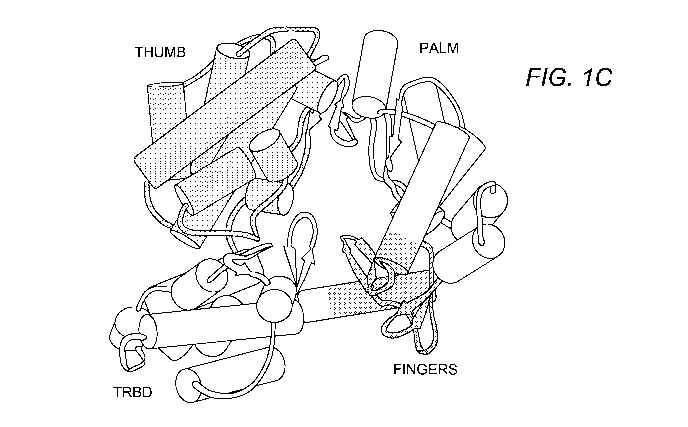
Brief Description of the Drawings
Figure 1 shows the structure of telomerase (TERT)
Figure 1A shows the primary of human, yeast and Tetrahymena
thermophila TERT showing the functional domains and
conserved motifs. Figure 1B is the primary structure and
conserved motifs of the Tribolium castaneum TERT. Figure 1C
shows TERT domain organization with the RNA-binding domain
(TRBD), the reverse transcriptase domain composed of the
"fingers" and "palm" subdomains, and the "thumb" domain
depicted.
Figures 2A and 2B show a sequence alignment and
schematic of secondary structure of Tetrahymena thermophila
TRBDs (TETTH; SEQ ID NO:1) compared with the TRBDs from
CA 02703060 2010-04-19
WO 2009/055364 PCT/US2008/080604
-7-
ciliated protozoa such as, Euplotes aediculatus (EUPAE; SEQ
ID NO:2) and Oxytricha trifallax (OXYTR; SEQ ID NO:3);
mammals such as human (SEQ ID NO:4) and mouse (SEQ ID
NO:5); fungi such as Schizosaccharomyces pombe (SCHPO; SEQ
ID NO:6) and Saccharomyces cerevisiae (YEAST; SEQ ID NO:7);
and plants such as Arabidopsis thaliana (ARATH; SEQ ID
NO:8) produced by ALSCRIPT Barton (1993) Protein Eng. 6:37-
40). Conserved residues in key signature motifs are
indicated and mutated residues that affect RNA-binding and
telomerase function are also indicated. The solid triangles
define the boundaries of the TRBD construct used in the
studies herein.
Figures 3A-3C show the sequence alignment and surface
conservation of Tribolium castaneum TERT (TRICA; SEQ ID
NO:9) compared with TERTs from various phylogenetic groups
including mammals such as mouse (SEQ ID NO:10) and human
(SEQ ID NO:11); plants such as Arabidopsis thaliana (ARATH;
SEQ ID NO:12); fungi such as Saccharomyces cerevisiae
(YEAST; SEQ ID NO:13) and Schizosaccharomyces pombe (SCHPO;
SEQ ID NO:14); and protozoa such as Tetrahymena thermophila
(TETTH; SEQ ID NO:15) and Euplotes aediculatus (EUPAE; SEQ
ID NO:16) produced by ClustalW2 (Larkin et al. (2007)
Bioinformatics 23:2947-2948). Conserved residues in key
signature motifs are indicated. K210 of helix alo and polar
residues (K406, K416, K418, N423) of the "thumb" domain
implicated in direct contacts with the backbone of the DNA
substrate are also shown.
Figure 4 is a schematic of the primary structure of
the RNA component (TER) of telomerase from Tetrahymena
thermophila. Stem I, TBE and the template are indicated.
CA 02703060 2010-04-19
WO 2009/055364 PCT/US2008/080604
-8-
Detailed Description of the Invention
Telomerase, a ribonucleoprotein complex, replicates
the linear ends of eukaryotic chromosomes, thus taking care
of the "end of replication problem". TERT contains an
essential and universally conserved domain (TRBD; Figure
1A) that makes extensive contacts with the RNA (TER)
component of the holoenzyme and this interaction
facilitates TERT/TER assembly and repeat addition
processivity. The TRBD domain is highly conserved among
phylogenetic groups and is essential for the function of
telomerase. Extensive biochemical and mutagenesis studies
have localized TRBD binding to stem I and the TEB,
interactions that are thought to be important for the
proper assembly and stabilization of the TERT/TER complex
as well as the repeat addition processivity of the
holoenzyme. The atomic structure of the TRBD domain has now
been identified, thereby providing information about
TERT/TER binding. The RNA-binding site of TRBD is an
extended groove on the surface of the protein that is
partly hydrophilic and partly hydrophobic in nature and is
formed by the previously identified T- and CP-motifs shown
to be important for telomerase function. The size,
organization and chemical nature of this groove indicates
that the TRBD domain interacts with both double- and
single-stranded nucleic acid, possibly stem I or II and the
ssRNA that connects them.
In addition to the structure of the TRBD domain, it
has now been shown that three highly conserved domains,
TRBD, the reverse transcriptase (RT) domain, and the C-
terminal extension thought to represent the putative
"thumb" domain of TERT, are organized into a ring-like
CA 02703060 2010-04-19
WO 2009/055364 PCT/US2008/080604
-9-
structure that shares common features with retroviral
reverse transcriptases, viral RNA polymerases and B-family
DNA polymerases. Domain organization places motifs
implicated in substrate binding and catalysis in the
interior of the ring, which can accommodate seven-to-eight
bases of double stranded nucleic acid. Modeling of an
RNA/DNA heteroduplex in the interior of this ring reveals a
perfect fit between the protein and the nucleic acid
substrate and positions the 3'-end of the DNA primer at the
active site of the enzyme providing evidence for the
formation of an active telomerase elongation complex.
The TRBD domain, as well as RT and "thumb" domains,
are highly conserved domains among phylogenetic groups. As
such, these domains serve as ideal candidates for
telomerase inhibitors. In this regard telomerase is an
ideal target for treating human diseases relating to
cellular proliferation and senescence, such as cancer.
Accordingly, the present invention relates to the use
of the high-resolution structure of Tetrahymena thermophila
and Tribolium castaneum telomerases for the identification
of effector molecules that modulate the activity of
telomerase. The term "effector" refers to any agonist,
antagonist, ligand or other agent that affects the activity
of telomerase. Effectors can be, but are not limited to,
peptides, carbohydrates, nucleic acids, lipids, fatty
acids, hormones, organic compounds, and inorganic
compounds. The information obtained from the crystal
structure of the present invention reveals detailed
information which is useful in the design, isolation,
screening and determination of potential compounds which
modulate the activity of telomerase. Compounds that bind
the TRBD domain and, e.g., sterically block TER binding or
block RNP assembly act as effective telomerase-specific
CA 02703060 2010-04-19
WO 2009/055364 PCT/US2008/080604
-10-
inhibitors, whereas compounds that mimic or facilitate TER
binding or RNP assembly act as effective telomerase-
specific activators. Compounds that bind and block the
active site or nucleotide binding site can also modulate
telomerase activity. Similarly, compounds that interact
with one or more amino acid residues of telomerase in
direct contact with DNA can block DNA binding and act as
effective telomerase-specific inhibitors, whereas compound
that mimic DNA act as effective telomerase-specific
activators. The effector molecules of the invention have a
wide variety of uses. For example, it is contemplated that
telomerase modulators will be effective therapeutic agents
for treatment of human diseases. Screening for agonists
provides for compositions that increase telomerase activity
in a cell (including a telomere-dependent replicative
capacity, or a partial telomerase activity). Such agonist
compositions provide for methods of immortalizing otherwise
normal untransformed cells, including cells which can
express useful proteins. Such agonists can also provide for
methods of controlling cellular senescence. Conversely,
screening for antagonist activity provides for compositions
that decrease telomere-dependent replicative capacity,
thereby mortalizing otherwise immortal cells, such as
cancer cells. Screening for antagonist activity provides
for compositions that decrease telomerase activity, thereby
preventing unlimited cell division of cells exhibiting
unregulated cell growth, such as cancer cells. In general,
the effector molecules of the invention can be used
whenever it is desired to increase or decrease a telomerase
activity in a cell or organism.
Broadly, the method of the invention involves
designing or screening for a test compound which binds to
at least one amino acid residue of an essential telomerase
CA 02703060 2010-04-19
WO 2009/055364 PCT/US2008/080604
-11-
domain disclosed herein; and testing the compound designed
or screened for its ability to modulate the activity of
telomerase. In certain embodiments, the method of the
present invention is carried out using various in silico,
in vitro and/or in vivo assays based on detecting
interactions between one or more domains or domain residues
of telomerase and a test compound.
In the context of the present invention, telomerase
refers to a family of enzymes which maintain telomere ends
by addition of the telomere repeat TTAGGG. Telomerases are
described, e.g., by Nakamura, et al. (1997) Science
277(5328) :955-9 and O'Reilly, et al. (1999) Curr. Opin.
Struct. Biol. 9(1):56-65. Exemplary telomerase enzymes of
use in accordance with the present invention are set forth
herein in SEQ ID NOs:l-16 (Figures 2A-2B and Figures 3A-3C)
and full-length sequences for telomerase enzymes are known
in the art under GENBANK Accession Nos. AAC39140
(Tetrahymena thermophila), NP197187 (Arabidopsis
thaliana), NP 937983 (Homo sapiens), CAA18391
(Schizosaccharomyces pombe), NP033380 (Mus musculus),
NP 013422 (Saccharomyces cerevisiae), AAC39163 (Oxytricha
trifallax), CAE75641 (Euplotes aediculatus) and
NP 001035796 (Tribolium castaneum). For the purposes of the
present invention, reference to telomerase refers to
allelic and synthetic variants of telomerase, as well as
fragments of telomerase. Synthetic variants include those
which have at least 80%, preferably at least 90%, homology
to a telomerase disclosed herein. More preferably, such
variants correspond to the sequence of a telomerase
provided herein, but have one or more, e.g., from 1 to 10,
such as from 1 to 5, substitutions, deletions or insertions
of amino acids. Fragments of telomerase and variants
thereof are preferably at least 20, more preferably at
CA 02703060 2010-04-19
WO 2009/055364 PCT/US2008/080604
-12-
least 50 and most preferably at least 200 amino acids in
size. An exemplary fragment includes the approximately 250
amino acid residues encompassing the TRBD domain of
telomerase. Other fragments include the "thumb" domain and
the reverse transcriptase domain and its subdomains, i.e.,
the "finger" and "palm" subdomains. As depicted in Figure
lA and Figures 2A and 2B, the TRBD domain encompasses amino
acid residues at or about 254-519 of T. thermophila
telomerase. As depicted in Figure 1B and Figures 3A-3C, the
reverse transcriptase domain encompasses amino acid
residues at or about 160-403 of T. castaneum telomerase,
and the "thumb" domain encompasses amino acid residues at
or about 404-596 of T. castaneum telomerase. Based upon the
amino acid sequence comparisons depicted in Figures 2A, 2B,
3A, and 3B, suitable domains and fragments of telomerases
from other species can be readily obtained based upon the
location of equivalent amino acid residues in a telomerase
from another species.
The nearly all-helical structure of TRBD provides a
nucleic acid binding fold suitable for TER binding. An
extended pocket on the surface of the protein, formed by
two conserved motifs (CP- and T-motifs) provides TRBD's
RNA-binding pocket. The width and the chemical nature of
this pocket indicate that it binds both single- and double-
stranded RNA, likely stem I and the template boundary
element (TBE). Essential amino acid residues involved in
RNP assembly of T thermophila telomerase and the
interaction between T thermophila telomerase TRBD and TER
are listed in Table 1. The location of these residues in
telomerases from other organisms is also listed in Table 1.
In particular embodiments, a compound of the invention
binds to one or more of the amino acid residues listed in
Table 1, thereby modulating the activity of telomerase.
CA 02703060 2010-04-19
WO 2009/055364 PCT/US2008/080604
-13-
TABLE 1
Essential Location in telomerases of other organisms
TRBD
Residues* TO Tc' At' Sp# Hs" Mm# Sc# Ot# Ea#
T-motif*
F476 F233 Y134 F265 F185 F246 F231 F186 F202 F206
Y477 Y234 Y135 Y266 Y186 Y247 Y232 Y187 Y203 Y207
T479 T236 P137 T268 T188 T249 T234 T189 T205 T209
E480 E237 I138 E269 E189 E250 E235 E190 E206 E210
Y491 Y248 I149 Y280 F200 Y261 Y246 F200 Y217 Y221
R492 R249 R150 R281 R201 R262 R247 R201 R218 R222
K493 K250 K151 K282 K202 K263 K248 H202 K219 L223
W496 W253 Y154 W285 W205 W266 W251 W205 W222 W226
CP-motif*
F323 F80 L36 L95 L55 Y90 Y90 Y58 F55 F59
L327 L84 K39 L99 Y59 L94 L94 L62 L59 L63
K328 K85 H40 D100 N60 K95 R95 N63 S60 T64
K329 K86 K41 K101 H61 T96 S96 S64 K61 K65
C331 C88 K43 C103 C63 C98 C98 C66 C63 C67
L333 L90 P45 L108 - L100 - - L65 L69
P334 P91 V46 Q109 - R101 - - P66 P70
QFP-motif*
Q375 Q132 Q47 Q158 Q83 Q145 Q130 R87 Q103 C107
I376 I133 I48 V159 V84 V146 V131 V88 I104 V108
L380 L137 L52 I163 L88 V150 L135 I92 L108 I112
I383 I140 I55 I166 I91 C153 C138 I95 F111 F115
I384 I141 I56 C167 L92 L154 L139 L96 V112 F116
C387 C144 - I170 V95 L157 V142 L99 V115 I119
V388 V145 - V171 F96 V158 V143 L100 F116 L120
P389 P146 P57 P172 P97 P159 S144 P101 P117 P121
L392 L149 Y60 L175 I100 L162 L147 M104 F120 F124
L393 L150 F61 L176 W101 W163 W148 F105 L121 L125
N397 N154 N66 Q181 I106 N168 N153 N110 N125 N129
L405 L162 V74 I189 L114 T176 L161 L118 M133 V137
F408 F165 I77 F192 F117 F179 F164 L121 F136 Y140
Y422 Y179 L91 F206 L131 L193 L178 L135 L150 L154
I423 I180 H92 L207 M132 T194 M179 L136 L151 L155
M426 M183 Y95 V210 I135 M197 M182 L139 F154 I158
W433 W190 W102 F217 W142 W204 W189 W146 W161 W165
F434 F191 L103 F218 L143 L205 L190 L147 L162 M166
Tt, Tetrahymena thermophila; At, Arabidopsis thaliana; Hs,
Homo sapiens; Sp, Schizosaccharomyces pombe; Mm, Mus
musculus; Sc, Saccharomyces cerevisiae; Tc, Tribolium
castaneum; Ot, Oxytricha trifallax and Ea, Euplotes
aediculatus.
*Location is with reference to the full-length T.
thermophila telomerase.
#Location is with reference to the telomerase sequences
depicted in Figures 2A and 2B, i.e., SEQ ID NOs:l-8.
'Location is with reference to the telomerase sequence
depicted in Figures 3A-3C.
CA 02703060 2010-04-19
WO 2009/055364 PCT/US2008/080604
-14-
As disclosed herein, the structure of T. castaneum
telomerase identified key amino acid residues of the
reverse transcriptase and "thumb" domains. In particular,
key amino acid residues of the nucleotide binding pocket
were identified as well as amino acid residues which appear
to make direct contacts with the backbone of the DNA
substrate. Accordingly, the present invention also embraces
a compound, which binds to at least one amino acid residue
of the nucleotide binding pocket of telomerase or residues
which make direct contact with DNA. These residues are
found in the "palm" and "finger" subdomains of the reverse
transcriptase domain and the "thumb" domain of T. castaneum
telomerase and are listed in Table 2. The location of these
amino acid residues in other species is also listed in
Table 2.
TABLE 2
Domain Location in telomerases of other organisms*
Residues*
Tt At Sp Hs Mm Sc Ea
"Palm"
K210 R573 K644 K547 N666 N656 E483 T558
V250 L617 A690 I589 V711 A704 F529 M602
D251 D618 V691 D590 D712 D705 D530 D603
I252 I619 D692 I591 V713 V706 V531 I604
A255 C622 A695 C594 A716 A706 C534 C607
Y256 Y623 F696 Y595 Y717 Y707 Y535 Y608
G257 D624 D697 D596 D718 D708 D536 D609
G305 G770 G801 G703 G830 G823 G629 G734
L306 I771 I802 I704 I831 I824 L630 I735
L307 P772 P803 P705 P832 P825 F631 P736
Q308 Q773 Q804 Q706 Q833 Q826 Q632 Q737
G309 G774 H805 G707 G834 G827 G633 G738
V342 T814 I859 V741 V867 V860 A669 T780
D343 D815 D860 D742 D868 D861 D670 D781
D344 D816 D861 D743 D869 D862 D671 D782
Y345 Y817 Y862 F744 F870 F863 L672 Y783
F346 L818 L863 L745 L871 L864 F673 L784
F347 F819 F864 F746 L872 L865 I674 L785
C348 I820 V865 I747 V873 V866 I675 I786
S349 S821 S866 T748 T874 T867 5676 T787
N369 N846 N891 S773 N899 N892 N701 N812
K372 K849 K894 K776 K902 K895 K704 K815
T373 I850 F895 T777 T903 T896 I705 L816
"Finger"
L184 L533 F612 1502 L621 L611 M438 L514
CA 02703060 2010-04-19
WO 2009/055364 PCT/US2008/080604
-15-
N185 R534 R613 R503 R622 R612 R439 R515
I186 I535 F614 L504 F623 F613 I440 L516
I187 I536 L615 L505 I624 I614 I441 I517
P188 P537 P616 P506 P625 P615 P442 P518
K189 K538 K617 K507 K626 K616 K443 K519
F193 F542 V621 F511 L630 L620 N447 F523
R194 R543 R622 R512 R631 R621 E448 R524
A195 P544 M623 L513 P632 P622 F449 P525
I196 I545 V624 I514 I633 I623 R450 I526
V197 M546 L625 T515 V634 V624 I451 M527
"Thumb"
K406 Q888 T937 P815 5943 S936 S729 N860
K416 T898 T947 T825 S953 S946 K739 T869
K418 N900 5949 S826 T955 T948 S741 N871
N423 K906 K955 H832 K961 K954 R746 K877
Tt, Tetrahymena thermophila; At, Arabidopsis thaliana; Hs,
Homo sapiens; Sp, Schizosaccharomyces pombe; Mm, Mus
musculus; Sc, Saccharomyces cerevisiae; Tc, Tribolium
castaneum; and Ea, Euplotes aediculatus.
*Location is with reference to the telomerase sequences
depicted in Figures 3A-3C.
In one embodiment, a compound of the invention binds
to one or more of the amino acid residues listed in Table
2, thereby modulating the activity of telomerase. In
another embodiment, a compound binds to one or more of the
amino acid residues of the nucleotide binding pocket of
telomerase (i.e., K189, R194, Y256, Q308, V342, and K372 of
T. castaneum telomerase or equivalent amino acid residues
thereof in a telomerase from another species) to modulate
nucleotide binding. In yet a further embodiment, a compound
binds to one or more amino acid residues of telomerase that
make direct contact with DNA (i.e., K210, K406, K416, K418,
or N423 of T. castaneum telomerase or equivalent amino acid
residues thereof in a telomerase from another species) to
modulate DNA binding.
Compounds designed or screened for in accordance with
the present invention can interact with at least one of the
amino acid residues of one or more domains disclosed herein
via various heterogeneous interactions including, but not
limited to van der Waals contacts, hydrogen bonding, ionic
CA 02703060 2010-04-19
WO 2009/055364 PCT/US2008/080604
-16-
interactions, polar contacts, or combinations thereof. In
general, it is desirable that the compound interacts with
2, 3, 4, 5, 6 or more of the amino acid residues of a
domain disclosed herein to enhance the specificity of the
compound for one or more telomerase proteins. In one
embodiment, the compound interacts with one or more
essential amino acids of the QFP-motif, T-motif or CP-
motif. In another embodiment, the compound interacts with
one or more essential amino acids of the T-motif and CP-
motif. In a further embodiment, the compound interacts with
one or more essential amino acids as set forth in Table 1.
In a particular embodiment, the compound interacts with one
or more essential amino acid residues set forth in Table 1,
which have not been previously identified by mutation to
affect RNA-binding and telomerase activity. In another
embodiment, the compound interacts with one or more
essential amino acids of the nucleotide binding pocket. In
a further embodiment, the compound interacts with one or
more essential amino acids of telomerase in direct contact
with DNA. In yet a further embodiment, the compound
interacts with one or more essential amino acids as set
forth in Table 2. In a particular embodiment, the compound
interacts with one or more essential amino acid residues
set forth in Table 2, which have not been previously
identified by mutation to affect nucleotide binding, DNA
binding or telomerase activity.
In accordance with the present invention, molecular
design techniques can be employed to design, identify and
synthesize chemical entities and compounds, including
inhibitory and stimulatory compounds, capable of binding to
one or more amino acids of telomerase. The structure of the
domains of telomerase can be used in conjunction with
computer modeling using a docking program such as GRAM,
CA 02703060 2010-04-19
WO 2009/055364 PCT/US2008/080604
-17-
DOCK, HOOK or AUTODOCK (Dunbrack, et al. (1997) Folding&
Design 2:27-42) to identify potential modulators of
telomerase proteins. This procedure can include computer
fitting of compounds to domains disclosed herein to, e.g.,
ascertain how well the shape and the chemical structure of
the compound will complement the TRBD domain; or to compare
the compound with the binding of TER in the TRBD; or
compare the compound with the binding of a DNA molecule to
the "thumb" domain; or compare the compound with binding of
a nucleotide substrate to the nucleotide binding pocket.
Computer programs can also be employed to estimate the
attraction, repulsion and stearic hindrance of the
telomerase protein and effector compound. Generally, the
tighter the fit, the lower the stearic hindrances, the
greater the attractive forces, and the greater the
specificity, which are important features for a specific
effector compound which is more likely to interact with the
telomerase protein rather than other classes of proteins.
In so far as the present invention has identified the amino
acid residues specifically involved in substrate binding,
the present invention offers specificity not heretofore
possible with conventional screening assays.
Alternatively, a chemical-probe approach can be
employed in the design of telomerase modulators or
effectors. For example, Goodford ((1985) J. Med. Chem.
28:849) describes several commercial software packages,
such as GRID (Molecular Discovery Ltd., Oxford, UK), which
can be used to probe the telomerase domains with different
chemical probes, e.g., water, a methyl group, an amine
nitrogen, a carboxyl oxygen, and a hydroxyl. Favored sites
for interaction between these regions or sites of the
telomerase domains and each probe are thus determined, and
from the resulting three-dimensional pattern of such
CA 02703060 2010-04-19
WO 2009/055364 PCT/US2008/080604
-18-
regions or sites a putative complementary molecule can be
generated.
The compounds of the present invention can also be
designed by visually inspecting the three-dimensional
structure of the telomerase domains to determine more
effective inhibitors or activators. This type of modeling
is generally referred to as "manual" drug design. Manual
drug design can employ visual inspection and analysis using
a graphics visualization program such as "O" (Jones, et al.
(1991) Acta Crystallographica Section A A47:110-119).
Initially effector compounds can be selected by manual
drug design. The structural analog thus designed can then
be modified by computer modeling programs to better define
the most likely effective candidates. Reduction of the
number of potential candidates is useful as it may not be
possible to synthesize and screen a countless number of
compound variations that may have some similarity to known
inhibitory molecules. Such analysis has been shown
effective in the development of HIV protease inhibitors
(Lam, et al. (1994) Science 263:380-384; Wlodawer, et al.
(1993) Ann. Rev. Biochem. 62:543-585; Appelt (1993)
Perspectives in Drug Discovery and Design 1:23-48; Erickson
(1993) Perspectives in Drug Discovery and Design 1:109-
128) . Alternatively, random screening of a small molecule
library could lead to modulators whose activity may then be
analyzed by computer modeling as described above to better
determine their effectiveness as inhibitors or activators.
Programs suitable for searching three-dimensional
databases include MACCS-3D and ISIS/3D (Molecular Design
Ltd, San Leandro, CA), ChemDBS-3D (Chemical Design Ltd.,
Oxford, UK), and Sybyl/3 DB Unity (Tripos Associates, St
Louis, MO) . Programs suitable for compound selection and
design include, e.g., DISCO (Abbott Laboratories, Abbott
CA 02703060 2010-04-19
WO 2009/055364 PCT/US2008/080604
-19-
Park, IL), Catalyst (Bio-CAD Corp., Mountain View, CA), and
ChemDBS-3D (Chemical Design Ltd., Oxford, UK).
The compounds designed using the information of the
present invention can bind to all or a portion of the TRBD
domain, nucleotide binding domain, and/or "thumb" domain of
telomerase and may be more potent, more specific, less
toxic and more effective than known inhibitors of
telomerase. The designed compounds can also be less potent
but have a longer half-life in vivo and/or in vitro and
therefore be more effective at modulating telomerase
activity in vivo and/or in vitro for prolonged periods of
time. Such designed modulators are useful to inhibit or
activate telomerase activity to, e.g., alter lifespan or
proliferative capacity of a cell.
The present invention also provides the use of
molecular design techniques to computationally screen small
molecule databases for chemical entities or compounds that
can bind to telomerase in a manner analogous to its natural
substrates. Such computational screening can identify
various groups which interact with one or more amino acid
residues of a domain disclosed herein and can be employed
to synthesize modulators of the present invention.
In vitro (i.e., in solution) screening assays are also
embraced by the present invention. For example, such assays
include combining telomerase, the telomerase TRBD domain
(e.g., as disclosed herein), or portions of the telomerase
TRBD domain with or without TER in solution and determining
whether a test compound can block or enhance telomerase
activity. Similarly, in vitro screening assays can be
carried out to monitor nucleotide or DNA binding in the
presence or absence of a test compound.
Compounds which can be screened in accordance with the
method of the present invention are generally derived from
CA 02703060 2010-04-19
WO 2009/055364 PCT/US2008/080604
-20-
libraries of agents or compounds. Such libraries can
contain either collections of pure agents or collections of
agent mixtures. Examples of pure agents include, but are
not limited to, proteins, polypeptides, peptides, nucleic
acids, oligonucleotides, carbohydrates, lipids, synthetic
or semi-synthetic chemicals, and purified natural products.
Examples of agent mixtures include, but are not limited to,
extracts of prokaryotic or eukaryotic cells and tissues, as
well as fermentation broths and cell or tissue culture
supernates. Databases of chemical structures are also
available from a number of sources including Cambridge
Crystallographic Data Centre (Cambridge, UK) and Chemical
Abstracts Service (Columbus, OH) . De novo design programs
include Ludi (Biosym Technologies Inc., San Diego, CA),
Sybyl (Tripos Associates) and Aladdin (Daylight Chemical
Information Systems, Irvine, CA).
Library screening can be performed using any
conventional method and can be performed in any format that
allows rapid preparation and processing of multiple
reactions. For in vitro screening assays, stock solutions
of the test compounds as well as assay components can be
prepared manually and all subsequent pipeting, diluting,
mixing, washing, incubating, sample readout and data
collecting carried out using commercially available robotic
pipeting equipment, automated work stations, and analytical
instruments for detecting the signal generated by the
assay. Examples of such detectors include, but are not
limited to, luminometers, spectrophotometers, and
fluorimeters, and devices that measure the decay of
radioisotopes.
After designing or screening for a compound which
binds to at least one amino acid residue of a domain
disclosed herein, the compound is subsequently tested for
CA 02703060 2010-04-19
WO 2009/055364 PCT/US2008/080604
-21-
its ability to modulate the activity of telomerase. Such
activities of telomerase include telomerase catalytic
activity (which may be either processive or non-processive
activity); telomerase processivity; conventional reverse
transcriptase activity; nucleolytic activity; primer or
substrate (telomere or synthetic telomerase substrate or
primer) binding activity; dNTP binding activity; RNA (i.e.,
TER) binding activity; and protein binding activity (e.g.,
binding to telomerase-associated proteins, telomere-binding
proteins, or to a protein-telomeric DNA complex) See,
e.g., assays disclosed in U.S. Patent No. 7,262,288.
Telomerase catalytic activity is intended to encompass
the ability of telomerase to extend a DNA primer that
functions as a telomerase substrate by adding a partial,
one, or more than one repeat of a sequence (e.g., TTAGGG)
encoded by a template nucleic acid (e.g., TER). This
activity may be processive or non-processive. Processive
activity occurs when a telomerase RNP adds multiple repeats
to a primer or telomerase before the DNA is released by the
enzyme complex. Non-processive activity occurs when
telomerase adds a partial, or only one, repeat to a primer
and is then released. In vivo, however, a non-processive
reaction could add multiple repeats by successive rounds of
association, extension, and dissociation. This can occur in
vitro as well, but it is not typically observed in standard
assays due to the vastly large molar excess of primer over
telomerase in standard assay conditions. Conventional
assays for determining telomerase catalytic activity are
disclosed, for example, in Morin (1989) Cell 59:521); Morin
(1997) Bur. J. Cancer 33:750; U.S. Patent No. 5,629,154; WO
97/15687; WO 95/13381; Krupp, et al. (1997) Nucleic Acids
Res. 25:919; Wright, et al. (1995) Nuc. Acids Res. 23:3794;
Tatematsu, et al. (1996) Oncogene 13:2265.
CA 02703060 2010-04-19
WO 2009/055364 PCT/US2008/080604
-22-
Telomerase conventional reverse transcriptase activity
is described in, e.g., Morin (1997) supra, and Spence, et
al. (1995) Science 267:988. Because telomerase contains
conserved amino acid motifs that are required for reverse
transcriptase catalytic activity, telomerase has the
ability to transcribe certain exogenous (e.g., non-TER)
RNAs. A conventional RT assay measures the ability of the
enzyme to transcribe an RNA template by extending an
annealed DNA primer. Reverse transcriptase activity can be
measured in numerous ways known in the art, for example, by
monitoring the size increase of a labeled nucleic acid
primer (e.g., RNA or DNA), or incorporation of a labeled
dNTP. See, e.g., Ausubel, et al. (1989) Current Protocols
in Molecular Biology, John Wiley & Sons, New York, NY.
Because telomerase specifically associates with TER,
it can be appreciated that the DNA primer/RNA template for
a conventional RT assay can be modified to have
characteristics related to TER and/or a telomeric DNA
primer. For example, the RNA can have the sequence
(CCCTAA)n, where n is at least 1, or at least 3, or at least
10 or more. In one embodiment, the (CCCTAA)n region is at or
near the 5' terminus of the RNA (similar to the 5'
locations of template regions in telomerase RNAs).
Similarly, the DNA primer may have a 3' terminus that
contains portions of the TTAGGG telomere sequence, for
example X,,TTAG, X,,AGGG, etc., where X is a non-telomeric
sequence and n is 6-30. In another embodiment, the DNA
primer has a 5' terminus that is non-complementary to the
RNA template, such that when the primer is annealed to the
RNA, the 5' terminus of the primer remains unbound.
Additional modifications of standard reverse transcription
assays that may be applied to the methods of the invention
are known in the art.
CA 02703060 2010-04-19
WO 2009/055364 PCT/US2008/080604
-23-
Telomerase nucleolytic activity is described in, e.g.,
Morin (1997) supra and Collins & Grieder (1993) Genes Dev.
7:1364. Telomerase preferentially removes nucleotides,
usually only one, from the 3' end of an oligonucleotide
when the 3' end of the DNA is positioned at the 5' boundary
of the DNA template sequence, in humans and Tetrahymena,
this nucleotide is the first G of the telomeric repeat
(TTAGG in humans). Telomerase preferentially removes G
residues but has nucleolytic activity against other
nucleotides. This activity can be monitored using
conventional methods known in the art.
Telomerase primer (telomere) binding activity is
described in, e.g., Morin (1997) supra; Collins, et al.
(1995) Cell 81:677; Harrington, et al. (1995) J. Biol.
Chem. 270:8893. There are several ways of assaying primer
binding activity; however, a step common to most methods is
incubation of a labeled DNA primer with telomerase or
telomerase/TER under appropriate binding conditions. Also,
most methods employ a means of separating unbound DNA from
protein-bound DNA. Such methods can include, e.g., gel-
shift assays or matrix binding assays. The DNA primer can
be any DNA with an affinity for telomerase, such as, for
example, a telomeric DNA primer like (TTAGGG),,, where n
could be 1-10 and is typically 3-5. The 3' and 5' termini
can end in any location of the repeat sequence. The primer
can also have 5' or 3' extensions of non-telomeric DNA that
could facilitate labeling or detection. The primer can also
be derivatized, e.g., to facilitate detection or isolation.
Telomerase dNTP binding activity is described in,
e.g., Morin (1997) supra and Spence, et al. (1995) supra.
Telomerase requires dNTPs to synthesize DNA. The telomerase
protein has a nucleotide binding activity and can be
assayed for dNTP binding in a manner similar to other
CA 02703060 2010-04-19
WO 2009/055364 PCT/US2008/080604
-24-
nucleotide binding proteins (Kantrowitz, et al. (1980)
Trends Biochem. Sci. 5:124). Typically, binding of a
labeled dNTP or dNTP analog can be monitored as is known in
the art for non-telomerase RT proteins.
Telomerase RNA (i.e., TER) binding activity is
described in, e.g., Morin (1997) supra; Harrington, et al.
(1997) Science 275:973; Collins, et al. (1995) Cell 81:677.
The RNA binding activity of a telomerase protein of the
invention may be assayed in a manner similar to the DNA
primer binding assay described supra, using a labeled RNA
probe. Methods for separating bound and unbound RNA and for
detecting RNA are well known in the art and can be applied
to the activity assays of the invention in a manner similar
to that described for the DNA primer binding assay. The RNA
can be full length TER, fragments of TER or other RNAs
demonstrated to have an affinity for telomerase or TRBD.
See U.S. Patent No. 5,583,016 and WO 96/40868.
To further evaluate the efficacy of a compound
identified using the method of the invention, one of skill
will appreciate that a model system of any particular
disease or disorder involving telomerase can be utilized to
evaluate the adsorption, distribution, metabolism and
excretion of a compound as well as its potential toxicity
in acute, sub-chronic and chronic studies. For example, the
effector or modulatory compound can be tested in an assay
for replicative lifespan in Saccharomyces cerevisiae
(Jarolim, et al. (2004) FEMS Yeast Res. 5(2):169-77). See
also, McChesney, et al. (2005) Zebrafish 1(4) :349-355 and
Nasir, et al. (2001) Neoplasia 3(4):351-359, which describe
marine mammal and dog tissue model systems for analyzing
telomerase activity.
Compounds which bind to at least one amino acid
residue of one or more of the telomerase domains disclosed
CA 02703060 2010-04-19
WO 2009/055364 PCT/US2008/080604
-25-
herein can be used in a method for modulating (i.e.,
blocking or inhibiting, or enhancing or activating) a
telomerase. Such a method involves contacting a telomerase
either in vitro or in vivo with an effective amount of a
compound that interacts with at least one amino acid
residue of a domain of the invention so that the activity
of telomerase is modulated. An effective amount of an
effector or modulatory compound is an amount which reduces
or increases the activity of the telomerase by at least
30%, 40%, 50%, 60%, 70%, 80%, 90% or 100% when compared to
telomerase not contacted with the compound. Such activity
can be monitored by enzymatic assays detecting activity of
the telomerase or by monitoring the expression or activity
of proteins which are known to be associated with or
regulated by telomerase.
One of skill in the art can appreciate that modulating
the activity of telomerase can be useful in selectively
analyzing telomerase signaling events in model systems as
well as in preventing or treating diseases and disorders
involving telomerase. The selection of the compound for use
in preventing or treating a particular disease or disorder
will be dependent upon the particular disease or disorder.
For example, human telomerase is involved in cancer and
therefore a compound which inhibits telomerase will be
useful in the prevention or treatment of cancer including
solid tumors (e.g., adenocarcinoma of the breast, prostate,
and colon; melanoma; non-small cell lung; glioma; as well
as bone, breast, digestive system, colorectal, liver,
pancreatic, pituitary, testicular, orbital, head and neck,
central nervous system, acoustic, pelvic, respiratory
tract, and urogenital neoplasms) and leukemias (e.g., B-
cell, mixed-cell, null-cell, T-cell, T-cell chronic,
lymphocytic acute, lymphocytic chronic, mast-cell, and
CA 02703060 2010-04-19
WO 2009/055364 PCT/US2008/080604
-26-
myeloid). Cancer cells (e.g., malignant tumor cells) that
express telomerase activity (telomerase-positive cells) can
be mortalized by decreasing or inhibiting the endogenous
telomerase activity. Moreover, because telomerase levels
correlate with disease characteristics such as metastatic
potential (e.g., U.S. Patent Nos. 5,639,613; 5,648,215;
5,489,508; Pandita, et al. (1996) Proc. Am. Ass. Cancer
Res. 37:559), any reduction in telomerase activity could
reduce the aggressive nature of a cancer to a more
manageable disease state (increasing the efficacy of
traditional interventions).
By way of illustration, Example 3 describes a cell-
based assay and animal model systems which can be used to
assess the inhibition of tumor cell growth by one or more
compounds of the invention. Another useful method for
assessing anticancer activities of compounds of the
invention involves the multiple-human cancer cell line
screening assays run by the National Cancer Institute (see,
e.g., Boyd (1989) in Cancer: Principles and Practice of
Oncology Updates, DeVita et al., eds, pp. 1-12). This
screening panel, which contains approximately 60 different
human cancer cell lines, is a useful indicator of in vivo
antitumor activity for a broad variety of tumor types
(Grever, et al. (1992) Seminars Oncol. 19:622; Monks, et
al. (1991) Natl. Cancer Inst. 83:757-766), such as
leukemia, non-small cell lung, colon, melanoma, ovarian,
renal, prostate, and breast cancers. Antitumor activities
can be expressed in terms of ED50 (or G150), where ED50 is
the molar concentration of compound effective to reduce
cell growth by 50%. Compounds with lower ED50 values tend to
have greater anticancer activities than compounds with
higher ED50 values.
CA 02703060 2010-04-19
WO 2009/055364 PCT/US2008/080604
-27-
Upon the confirmation of a compound's potential
activity in one or more in vitro assays, further evaluation
is typically conducted in vivo in laboratory animals, for
example, measuring reduction of lung nodule metastases in
mice with B16 melanoma (e.g., Schuchter, et al. (1991)
Cancer Res. 51:682-687). The efficacy of a compound of the
invention either alone or as a drug combination
chemotherapy can also be evaluated, for example, using the
human B-CLL xenograft model in mice (e.g., Mohammad, et al.
(1996) Leukemia 10:130-137). Such assays typically involve
injecting primary tumor cells or a tumor cell line into
immune compromised mice (e.g., a SCID mouse or other
suitable animal) and allowing the tumor to grow. Mice
carrying the tumors are then treated with a compound of the
invention and tumor size is measured to follow the effect
of the treatment. Alternatively, a compound of the
invention is administered prior to injection of tumor cells
to evaluate tumor prevention. Ultimately, the safety and
efficacy of compounds of the invention are evaluated in
human clinical trials.
Compounds that activate or stimulate telomerase
activity can be used in methods for treating or preventing
a disease or condition induced or exacerbated by cellular
senescence in a subject; methods for decreasing the rate of
senescence of a subject, e.g., after onset of senescence;
methods for extending the lifespan of a subject; methods
for treating or preventing a disease or condition relating
to lifespan; methods for treating or preventing a disease
or condition relating to the proliferative capacity of
cells; and methods for treating or preventing a disease or
condition resulting from cell damage or death. Certain
diseases of aging are characterized by cell senescence-
associated changes due to reduced telomere length (compared
CA 02703060 2010-04-19
WO 2009/055364 PCT/US2008/080604
-28-
to younger cells) resulting from the absence (or much
lower levels) of telomerase activity in the cell.
Telomerase activity and telomere length can be increased
by, for example, increasing the activity of telomerase in
the cell. A partial listing of conditions associated with
cellular senescence in which increased telomerase activity
can be therapeutic includes Alzheimer's disease,
Parkinson's disease, Huntington's disease, and stroke; age-
related diseases of the integument such as dermal atrophy,
elastolysis and skin wrinkling, graying of hair and hair
loss, chronic skin ulcers, and age-related impairment of
wound healing; degenerative joint disease; osteoporosis;
age-related immune system impairment (e.g., involving cells
such as B and T lymphocytes, monocytes, neutrophils,
eosinophils, basophils, NK cells and their respective
progenitors); age-related diseases of the vascular system;
diabetes; and age-related macular degeneration. Moreover,
telomerase activators can be used to increase the
proliferative capacity of a cell or in cell
immortalization, e.g., to produce new cell lines (e.g.,
most human somatic cells).
Prevention or treatment typically involves
administering to a subject in need of treatment a
pharmaceutical composition containing an effective of a
compound identified in the screening method of the
invention. In most cases this will be a human being, but
treatment of agricultural animals, e.g., livestock and
poultry, and companion animals, e.g., dogs, cats and
horses, is expressly covered herein. The selection of the
dosage or effective amount of a compound is that which has
the desired outcome of preventing, reducing or reversing at
least one sign or symptom of the disease or disorder being
treated. Methods for treating cancer and other telomerase-
CA 02703060 2010-04-19
WO 2009/055364 PCT/US2008/080604
-29-
related diseases in humans are described in U.S. Patent
Nos. 5,489,508, 5,639,613, and 5,645,986. By way of
illustration, a subject with cancer (including, e.g.,
carcinomas, melanomas, sarcomas, lymphomas and leukaemias)
can experience unexplained weight loss, fatigue, fever,
pain, skin changes, sores that do not heal, thickening or
lump in breast or other parts of the body, or a nagging
cough or hoarseness, wherein treatment with a compound of
the invention can prevent, reduce, or reverse one or more
of these symptoms.
Pharmaceutical compositions can be in the form of
pharmaceutically acceptable salts and complexes and can be
provided in a pharmaceutically acceptable carrier and at an
appropriate dose. Such pharmaceutical compositions can be
prepared by methods and contain carriers which are well-
known in the art. A generally recognized compendium of such
methods and ingredients is Remington: The Science and
Practice of Pharmacy, Alfonso R. Gennaro, editor, 20th ed.
Lippincott Williams & Wilkins: Philadelphia, PA, 2000. A
pharmaceutically-acceptable carrier, composition or
vehicle, such as a liquid or solid filler, diluent,
excipient, or solvent encapsulating material, is involved
in carrying or transporting the subject compound from one
organ, or portion of the body, to another organ, or portion
of the body. Each carrier must be acceptable in the sense
of being compatible with the other ingredients of the
formulation and not injurious to the subject being treated.
Examples of materials which can serve as
pharmaceutically acceptable carriers include sugars, such
as lactose, glucose and sucrose; starches, such as corn
starch and potato starch; cellulose, and its derivatives,
such as sodium carboxymethyl cellulose, ethyl cellulose and
cellulose acetate; powdered tragacanth; malt; gelatin;
CA 02703060 2010-04-19
WO 2009/055364 PCT/US2008/080604
-30-
talc; excipients, such as cocoa butter and suppository
waxes; oils, such as peanut oil, cottonseed oil, safflower
oil, sesame oil, olive oil, corn oil and soybean oil;
glycols, such as propylene glycol; polyols, such as
glycerin, sorbitol, mannitol and polyethylene glycol;
esters, such as ethyl oleate and ethyl laurate; agar;
buffering agents, such as magnesium hydroxide and aluminum
hydroxide; alginic acid; pyrogen-free water; isotonic
saline; Ringer's solution; ethyl alcohol; pH buffered
solutions; polyesters, polycarbonates and/or
polyanhydrides; and other non-toxic compatible substances
employed in pharmaceutical formulations. Wetting agents,
emulsifiers and lubricants, such as sodium lauryl sulfate
and magnesium stearate, as well as coloring agents, release
agents, coating agents, sweetening, flavoring and perfuming
agents, preservatives and antioxidants can also be present
in the compositions.
The compositions of the present invention can be
administered parenterally (for example, by intravenous,
intraperitoneal, subcutaneous or intramuscular injection),
topically (including buccal and sublingual), orally,
intranasally, intravaginally, or rectally according to
standard medical practices.
The selected dosage level will depend upon a variety
of factors including the activity of the particular
compound of the present invention employed, the route of
administration, the time of administration, the rate of
excretion or metabolism of the particular compound being
employed, the duration of the treatment, other drugs,
compounds and/or materials used in combination with the
particular compound employed, the age, sex, weight,
condition, general health and prior medical history of the
CA 02703060 2010-04-19
WO 2009/055364 PCT/US2008/080604
-31-
patient being treated, and like factors well known in the
medical arts.
A physician or veterinarian having ordinary skill in
the art can readily determine and prescribe the effective
amount of the pharmaceutical composition required. For
example, the physician or veterinarian could start doses of
a compound at levels lower than that required in order to
achieve the desired therapeutic effect and gradually
increase the dosage until the desired effect is achieved.
This is considered to be within the skill of the artisan
and one can review the existing literature on a specific
compound or similar compounds to determine optimal dosing.
The invention is described in greater detail by the
following non-limiting examples.
Example 1: Structure of Tetrahymena thermophila TERT
Protein Expression and Purification. The T.
thermophila TERT residues 254-519 was identified by limited
proteolysis and cloned into a modified version of the
pET28b vector containing a cleavable hexa-histidine tag at
its N-terminus. The protein was over-expressed in E. coli
BL21 (pLysS) at 20 C for 4 hours. The cells were lysed by
sonication in 50 mM Tris-HC1, 10% glycerol, 0.5 M KC1, 5 mM
(3-mercaptoethanol, and 1 mM PMSF, pH 7.5 on ice. The
protein was first purified over a Ni-NTA column followed by
TEV cleavage of the hexa-histidine tag overnight at 4 C.
The TRBD/TEV mix was diluted so that the concentration of
imidazole was at 15 mM and the protein mix was passed over
a Ni-NTA column to remove the TEV, the cleaved tag and any
tagged protein. The Ni-NTA flow through was concentrated to
1 ml and diluted to a salt concentration of 0.15 M. The
diluted TRBD sample was then passed over a POROS-HS column
(PerSeptive Biosystems, Framingham, MA). At this stage, the
CA 02703060 2010-04-19
WO 2009/055364 PCT/US2008/080604
-32-
protein was more than 99% pure. The protein was finally
passed over a SEPHADEX-S200 sizing column pre-equilibrated
with 50 mM Tris-HC1, 10% glycerol, 0.5 M KC1, and 2 mM DTT,
pH 7.5 to remove any TRBD aggregates. The pure,
monodisperse protein as indicated by SDS-page and dynamic
light scattering, respectively, was concentrated to 8 mg/ml
using an AMICON 10K cutoff (MILLIPORE, Billerica, MA) and
the protein was stored at 4 C for subsequent studies. Stock
protein was dialyzed in 5 mM Tris-HC1, 500 mM KC1, 1 mM
TCEP, pH 7.5 prior to crystallization trials.
Protein Crystallization and Data Collection. Initial
plate-like clusters of TRBD that diffracted poorly (-.4 A
resolution) were grown at 4 C using the sitting drop method
by mixing on volume of dialyzed protein with one volume of
reservoir solution containing 20% PEG 3350, 0.2 M NaNO3.
Single, well diffracting crystals were grown in microbatch
trays under paraffin oil by mixing one volume of dialyzed
protein with an equivalent volume of 50 mM HEPES (pH 7.0),
44% PEG 400, 0.4 M NaNO3 , 0.4 M NaBr and 1 mM TCEP at 40C.
Crystals were harvested into cryoprotectant solution that
contained 25 mM HEPES (pH 7.0), 25% PEG 400, 0.2 M NaNO3,
0.2 M NaBr and 1 mM TCEP and were flash frozen in liquid
nitrogen. Data were collected at the NSLS, beam line X6A
and processed with HKL-2000 (Minor (1997) Meth. Enzymol.
Macromole. Crystallogr. Part A 276:307-326) (Table 3). The
crystals belong to the monoclinic space group P21 with one
monomer in the asymmetric unit.
TABLE 3
TRBD(254-519) Native Holmium-Derivative
X Ho-2l Ho-X2
Wavelength (A) 0.9795 1.5347 1.5595
Space group P21 P21 P21
CA 02703060 2010-04-19
WO 2009/055364 PCT/US2008/080604
-33-
Cell 39.4 67.2 39.2 68.2 39.2 68.2
dimensions (A) 51.5 90.7 50.1 91.6 50.1 91.6
Resolution (A) 20-1.71 50-2.59 50-2.63
(1.77-1.71)* (2.69-2.59) (3.02-2.63)
Redundancy 3.7 (3 . 0 ) 1.7 (1.8) 1.7 (1 . 8 )
Completeness 99.3 (93.3) 92.5 (88.1) 92.9 (88.7)
RS ( o) 4.7 (48.1) 7.3 (23.8) 7.0 (21.5)
I/6 (I) 27.3 (2.6) 9 (3.4) 9.4 (3.7)
Phasing Analysis
Resolution (A) 50-2.7
Number of sites 2
Mean figure of merit (FOM) 0.43
*Values in parentheses correspond to the highest resolution
shell.
Structure Determination and Refinement. Initial phases
5 were obtained from a two-wavelength MAD holmium (Ho)
derivative that was prepared by co-crystallizing the
protein with 5 mM HoC13. Heavy atom sites were located using
SOLVE (Terwilliger (2003) Methods Enzymol. 374:22-37) and
the sites were refined and new phases calculated with
10 MLPHARE (CCP4 (1994) Acta Crystallogr. D 50:760-763) as
implemented in ELVES (Holton & Alber (2004) Proc. Natl.
Acad. Sci. USA 101:1537-1542) (Table 3). Initial maps
showed well-defined density only for the larger half of the
molecule. The density for the smaller half of the molecule
was weak mostly due to its intrinsic mobility with respect
to larger half of the molecule. The problem associated with
building the model into the density was exacerbated by the
lack of information regarding the location of specific side
chains such as selenomethionines. Key factors in building a
complete model were successive rounds of PRIME and SWITCH
in RESOLVE (Terwilliger (2002) Acta Crystallogr. D Biol.
Crystallogr. 58:1937-1940) followed by manual building in
CA 02703060 2010-04-19
WO 2009/055364 PCT/US2008/080604
-34-
COOT (Emsley & Cowtan (2004) Acta Crystallogr. D Biol.
Crystallogr. 60:2126-2132). The model was refined using
both CNS-SOLVE (Brunger, et al. (1998) Acta Crystallogr. D
Biol. Crystallogr. 54:905-921) and REFMAC5 (Murshudov, et
al. (1997) Acta Crystallogr. D Biol. Crystallogr. 53:240-
255) . The last cycles of refinement were carried out with
TLS restraints as implemented in REFMAC5 (Table 4). Figures
were prepared in PYMOL (DeLano (2002)) and electrostatic
surfaces in APBS (Baker, et al. (2001) Proc. Natl. Acad.
Sci. USA 98:10037-10041).
TABLE 4
TRBD (254-519)
Refinement Statistics
Resolution (A) 20-1.71
Rwork/Rfree (%) 20.0/23.9
RMSD bonds (A) 0.008
RMSD angles (0) 0.831
Number of atoms
Protein 2145
Bromine 7
Water 213
Average B (A2)
Protein 27.41
Bromine 42.63
Water 31.22
Ramachandran % (no res.)
Most favored 91.6
Allowed 8.4
TRBD Structure. To explore the role of the essential
RNA-binding domain of telomerase (TRBD), a construct
identified by limited proteolysis, containing residues 254-
CA 02703060 2010-04-19
WO 2009/055364 PCT/US2008/080604
-35-
519 from T. thermophila (Figure lA) was purified to
homogeneity. This protein construct was monomeric in
solution as indicated by both gel filtration and dynamic
light scattering. Crystals of this construct grew readily
and diffracted to 1.71 A resolution (Table 3). The protein
was phased to 2.7 A resolution by the multiwavelength
anomalous dispersion method (MAD) using a holmium
derivative and the phases were extended with the native
dataset to 1.71 A resolution (Table 3). In the refined
structure there was clear density for residues 257-266 and
277-519.
The structure contains twelve a-helices linked
together by several long loops and two short (3-strands. The
helices are organized so that the molecule is divided into
two asymmetric halves linked together by three extended
loops. The larger half is composed of nine a-helices, one
of which (all) runs along the middle of the domain and
spans its entire length making contacts with all other
eight helices. The smaller half of the molecule is composed
of three helices (a4, a5 and a12), all of which are
arranged at a -120 angle to the plane of the larger half
of the protein. The smaller half of the protein is somewhat
more flexible than the larger half as suggested by its high
B factors reflecting the intrinsic mobility of this region
and may result from the absence of observable contacts with
the RNA substrate. An interesting feature of the structure
is a n-hairpin formed by the 15-residues that connect
helices all and a12 of the larger and the smaller halves,
respectively. The n-hairpin protrudes from the base of the
crevice formed by the two halves of the protein and stands
at a 45 angle to the plane of the smaller half of the
molecule. The positioning and the fact that this hairpin is
CA 02703060 2010-04-19
WO 2009/055364 PCT/US2008/080604
-36-
well-defined in the density could be attributed to helix a7
and the loop that connects it to helix a8. Both of these
elements are conveniently positioned at the back of this
hairpin holding it in place. A search in the protein
structure database using the Dali server (Holm & Sander
(1996) Science 273:595-603) produced no structural
homologues, indicating that the TRBD domain of telomerase
is a novel nucleic acid binding fold. The overall
organization of the two halves of the protein has
significant implications for nucleic acid recognition and
binding.
The TRBD RNA-Binding Motifs. The ability of the TRBD
domain to interact with TER has been attributed to two
conserved motifs known as the CP-, and T-motifs, while a
third motif known as the QFP-motif is thought to be
important for RNP assembly (Figures 2A and 2B) (Bosoy, et
al. (2003) J. Biol. Chem. 278:3882-3890; Bryan, et al.
(2000) supra; Jacobs, et al. (2005) supra; Xia, et al.
(2000) Mol. Cell. Biol. 20:5196-5207) . The TRBD structure
shows that the QFP-motif is formed by several mostly
hydrophobic residues, which are located on the larger half
of the molecule and are buried within the core of the
domain making extensive hydrophobic contacts with the
surrounding residues aiding in the fold of the protein.
These residues included G1n375, Ile376, Leu380, 11e383,
Ile384, Cys387, Va1388, Pro389, Leu392, Leu393, Asn397,
Leu405, Phe408, Tyr422, I1e423, Met426, Trp433, and Phe434.
The location and the contacts of the QFP-residues indicate
that they are not directly involved in nucleic acid
binding.
The T-motif is located at the center of the molecule
where the two halves of the protein meet and it is composed
CA 02703060 2010-04-19
WO 2009/055364 PCT/US2008/080604
-37-
of residues that form both part of the n-hairpin and helix
a12. Together these structural elements form a narrow (-.10
A), well-defined pocket (T-pocket) that is lined by several
solvent exposed and highly conserved residues (Phe476,
Tyr477, Thr479, Glu480, Tyr491, Arg492, Lys493, and
Trp496). Of particular note are the side chains of the
invariant residues Tyr477 and Trp496, which are part of the
(3-hairpin and helix a12, respectively. Together these
residues form a "hydrophobic pincer" that could sandwich
the purine/pirimidine moiety of an interacting RNA
nucleotide. In this structure, the side chains of Tyr477
and Trp496 are only 4 A apart, which is not sufficient to
accommodate a nucleotide base. Insertion of a base between
the two side chains would require structural rearrangement
of the T-pocket, possibly splaying of the two halves of the
molecules apart. In addition to its hydrophobic part, the
T-pocket also contains several hydrophilic residues such as
Arg492 and Lys493 both of which are solvent exposed and are
located at the interface of the T- and CP-pocket connecting
the two together.
The CP-motif is formed by helix a3 and the following
loop. In contrast to the T-motif, which is a narrow well-
defined pocket, the CP-motif is composed a shallow, wide
(.20 A), highly positively charged cavity located adjacent
and beneath the entry of the T-pocket. Several of the
conserved residues that form the CP-motif include Phe323,
Leu327, Lys328, Lys329, Cys331, Leu333, and Pro334. These
residues are buried in the core of the larger half or the
region that connects the two halves of the molecule and are
contributing to the protein fold. Of particular interest
are residues Leu327, Cys331, Leu333 and Pro334 all of which
are buried and make direct contacts with structural
CA 02703060 2010-04-19
WO 2009/055364 PCT/US2008/080604
-38-
elements of the T-motif thus aiding in the formation of
both the CP- and the T-pockets. For example, Leu327 and
Cys331 are within Van der Waal contacts of the large
hydrophobic side chain of the invariant Phe476 and the
aliphatic part of the side chain of the conserved Arg492
both of which form part of the R-hairpin. Interestingly,
Arg492 is located at the base of helix a12 and its contact
with Leu327, Cys331, and Leu333 partially helps position
this helix at a 45 angle of the plane that runs parallel
with the larger half of the molecule thus further
facilitating the formation of the T-pocket. Moreover, the
interaction of Arg492 with Leu327, Cys331, and Leu333 helps
position the guanidine moiety, the only solvent-exposed
part of this residue, at the interface formed by the CP-
and T-pockets. The CP-pocket also contains several surface-
exposed, conserved residues that are mainly hydrophilic in
nature. These include Lys328 and Lys329 both of which are
located beneath the T-pocket and in close proximity of
Arg492 and Lys493 together forming a single large,
positively charged surface area that almost spans the
entire side of the molecule.
TRED Structure and Existing Mutants. Several mutants
of TERT that affect RNA-binding and telomerase activity
have been isolated. Several of these mutants are found in
the TRBD domain and specifically within the T- and CP-
motifs. Single- and double- as well as stretches of 4-10
amino acid alanine substitutions within these two motifs
showed moderate to severe loss (20-100%) of RNA-binding
affinity and polymerase activity when compared to the wild
type enzyme (Bryan, et al. (2000) supra; Lai, et al. (2002)
supra; Miller, et al. (2000) supra).
One set of mutants, Phe476Ala, Tyr477Ala, Thr479Ala,
CA 02703060 2010-04-19
WO 2009/055364 PCT/US2008/080604
-39-
Glu480Ala, Arg492Ala and Trp496Ala, showed severe loss (80-
100%) of RNA-binding affinity and telomerase activity
suggesting that these residues mediate direct contacts with
the RNA substrate (Bryan, et al. (2000) supra; Lai, et al.
(2002) supra) . All five residues are part of the T-motif
and, with the exception of Phe476, all of their side chains
are solvent exposed. In the structure, both Tyr477 and
Trp496 are located at the base of the T-pocket and their
side chains form a "hydrophobic pincer". Assuming that the
solvent-exposed side chains of these residues are involved
in stacking interactions with the ssRNA, mutating them to
small alanines would likely compromise substrate binding
which explains the dramatic loss of RNA-binding affinity
and telomerase function. In contrast to Tyr477 and Trp496,
Phe476 is buried and is not accessible for interactions
with the nucleic acid substrate. Instead, Phe476 is located
at the base of the R-hairpin and contributes significantly
to the formation of the T-pocket. Mutating the large
hydrophobic side chain of this residue to the small alanine
would likely lead to conformational rearrangements of this
pocket and loss of RNA-binding affinity and telomerase
activity.
A second set of alanine mutants, Leu327Ala, Lys329Ala,
Cys331Ala, and Pro334Ala, which showed moderate loss of
RNA-binding affinity and telomerase activity has also been
isolated (Bryan, et al. (2000) supra; Miller, et al. (2000)
supra). Both Leu327 and Cys331 make direct contacts with
Phe476 and the aliphatic part of the side chain of Arg492,
both of which are located at the base of the T-motif.
Mutation to the smaller alanine residue could result in the
rearrangement of the T-pocket potentially leading to loss
of interactions with the nucleic acid substrate and loss of
function. Likewise, Pro334 is located at the back of helix
CA 02703060 2010-04-19
WO 2009/055364 PCT/US2008/080604
-40-
a12 and makes direct contacts with residues of this
structural element. Helix a12 contains the invariant Trp496
and the conserved Lys493, both of which form part of the T-
pocket. Mutating Pro334 into an alanine could lead to the
displacement of helix a10 and reorganization of the T-
pocket leading to loss of function. Lys329 is also located
on helix a3 and unlike Leu327Ala, Cys331Ala, and Pro334Ala,
is solvent exposed possibly making direct contacts with the
nucleic acid substrate. Mutating it to an alanine would
lead to lose of RNA interactions and loss of RNA-binding
affinity and telomerase activity.
TRBD Domain-Mediated Formation of Stable RNP Complex
and Repeat Addition Processivity. In vivo, telomerase
exists as a stable ribonucleoprotein complex and contacts
between the protein (TERT) and the RNA components (TER) are
mediated by the TEN, TRBD and the RT domains. Extensive
biochemical and mutagenesis studies have shown that the
TRBD is involved in extensive, specific interactions with
stem I and the TBE of TER (Lai, et al. (2001) supra;
O'Connor, et al. (2005) supra) (Figure 4) . Contacts between
the TRBD and TER are thought to facilitate the proper
assembly and stabilization of the RNP complex and promote
repeat addition processivity (Lai, et al. (2003) supra). In
ciliates, in addition to the TRBD, a conserved motif (CP2)
located N-terminally to the TRBD domain is thought to be
required for TERT-TER assembly and template boundary
definition (Lai, et al. (2002) supra; Miller, et al. (2000)
supra). However, until now it has been unclear as to how
the telomerase TRBD carries out this process. The present
analysis indicates that the TRBD domain is divided into two
asymmetric halves connected by several long loops that are
shaped like a boomerang, an arrangement that has
CA 02703060 2010-04-19
WO 2009/055364 PCT/US2008/080604
-41-
significant implications for RNA recognition and binding.
The overall organization of the two lobes of the molecule
results in the formation of two well-defined cavities (CP-
and T-pockets) on the surface of the protein that consist
of several solvent-exposed, invariant/conserved residues.
The T-pocket is a narrow, deep cavity located at the
junction of the two halves of the molecule with part of it
being hydrophobic in nature while the part that is located
in proximity of the CP-pocket is positively charged.
Interestingly, the hydrophobic side chains of Tyr477 and
Trp496 are solvent-exposed and are stacked against each
other forming a narrow "hydrophobic pincer" that in this
structure could not accommodate a nucleotide base. It is,
however, worth noting that helix a12, which contains
Trp496, is somewhat flexible with respect to the R-hairpin
that contains Tyr477. The ability of helix a12 and
therefore Trp496 to move could splay the two side chains
apart thus allowing for the space required for the
accommodation of a nucleotide base between them. Another
possibility is that the polar moiety of Tyr477 and Trp496
could act together as a nucleotide base that would allow
for the formation of pseudo Watson Crick interactions with
an incoming nucleotide base. Pseudo Watson Crick
interactions have been previously observed for a number of
protein nucleic acid complexes including the Rho
transcription termination factor (Bogden, et al. (1999)
Mol. Cell 3:487-493) and the signal recognition particle
(Wild, et al. (2001) Science 294:598-601). The width and
the organization of the hydrophobic part of the T-pocket
indicate that it binds ssRNA, most likely the TBE, possibly
mediated by a network of stacking interactions.
CA 02703060 2010-04-19
WO 2009/055364 PCT/US2008/080604
-42-
In contrast to the T-pocket, the CP-pocket is a
positively charged, shallow cavity located on the side of
the molecule and forms an extension of the T-pocket.
Together the hydrophilic part of the T-pocket and the CP-
pocket are lined with several lysines and arginines the
side chains of which are solvent exposed and could be
involved in direct contacts with the backbone of double-
stranded RNA. The width and the chemical nature of this
pocket indicate that it binds double-stranded RNA, most
likely stem I or stem II (Figure 4) . The nature and the
extent of the protein/nucleic acid interactions mediated by
the TRBD binding pockets provides the stability required
for the proper assembly of a functional ribonucleoprotein
enzyme and guide TERT to a TER binding site (between stem I
and II) that has significant implications for telomerase
function.
Telomerase is unique in its ability to add multiple
short oligonucleotide repeats at the end of linear
chromosomes. The enzyme's ability to do so has been partly
attributed to the interactions of the TRBD domain with the
TBE and in ciliates both the TRBD and the CP2 motif (Lai,
et al. (2002) supra; Lai, et al. (2003) supra; Miller, et
al. (2000) supra). The TBE is composed of stem II and the
flanking ssRNA regions and is located downstream of stem I
and only a few nucleotides upstream of the RNA template
(Figure 4). The TRBD structure indicates that the T-pocket,
a narrow, hydrophobic cavity located on the surface of the
protein that can only accommodate ssRNA, may play an
important role in this process. Assuming that the T-pocket
binds the ssRNA that connects stem I and stem II, this
interaction likely forces stem II to act as a steric block,
which in turn forces the TRBD domain to stay within the
boundaries of stem I and stem II. The stem I- and II-locked
CA 02703060 2010-04-19
WO 2009/055364 PCT/US2008/080604
-43-
TRBD domain then may act as an anchor that constrains the
distance the RT domain can travel and prevents it from
moving beyond the boundaries of the RNA template thus
promoting telomerase addition processivity. In ciliates
however, the TRBD domain alone is not sufficient for
template boundary definition and it requires the action of
the CP2 motif (Lai, et al. (2002) supra; Miller, et al.
(2000) supra) . It is contemplated that CP2 binding to TER
promotes template boundary definition either via
contributing to the stabilization of the RNP complex or,
like the TRBD, it may act as an anchor that prevents
slippage of the active site of the RT domain beyond the RNA
template.
Example 2: Structure of Tribolium castaneum TERT
Protein Expression and Purification. The synthetic
gene of T. castaneum full-length TERT was cloned into a
modified version of the pET28b vector containing a
cleavable hexahistidine tag at its N-terminus. The protein
was over-expressed in E. coli BL21 (pLysS) at 30 C for 4
hours. The cells were lysed by sonication in 50 mM Tris-
HC1, 10% glycerol, 0.5 M KC1, 5 mM (3-mercaproethanol, and 1
mM PMSF, pH 7.5 on ice. The protein was first purified over
a Ni-NTA column followed by TEV cleavage of the
hexahistidine tag overnight at 4 C. The TERT/TEV mixture
was dialyzed to remove the excess imidazole and the protein
was further purified over a second Ni-NTA column that was
used to remove all his-tagged products. The Ni-NTA flow
through was then passed over a POROS-HS column (Perseptive
Biosystems) to remove any trace amounts of protein
contaminants. At this stage the protein was more than 99%
pure. The protein was finally purified over a SEPHEDEX-S200
sizing column pre-equilibrated with 50 mM Tris-HC1, 10%
CA 02703060 2010-04-19
WO 2009/055364 PCT/US2008/080604
-44-
glycerol, 0.5 M KC1, and 1 mM Tris(2-Carboxyethyl)
phosphine (TCEP), pH 7.5 to remove any TERT aggregates and
the protein was concentrated to 10 mg/ml using an AMICON
30K cutoff (MILLIPORE) and stored at 4 C for subsequent
studies. Stock protein was dialyzed in 10 mM Tris-HC1, 200
mM KC1, 1 mM TCEP, pH 7.5 prior to crystallization trials.
Protein Crystallization and Data Collection. Initial
crystal trials of the protein alone did not produce
crystals. Co-crystallization of the protein with single
stranded telomeric DNA ((TCAGG)3) produced two rod-like
crystal forms one of which belongs to the orthorhombic
space group P212121 and diffracted to 2.71 A and the other
to the hexagonal space group P61 and diffracted to 3.25 A
resolution. The protein nucleic acid mix was prepared prior
to setting crystal trials by mixing one volume of dialyzed
protein with 1.2-fold excess of the DNA substrate. Both
crystal forms where grown by the vapor diffusion, sitting
drop method by mixing on volume of the protein-DNA mix with
one volume of reservoir solution. Orthorhombic crystals
where grown in the presence of 50 mM HEPES, (pH 7.0) and
1.5 M NaNO3 while hexagonal crystals grew in the presence of
100 mM Tris (pH 8 . 0) and 2 M (NH4) 2SO4 and both at room
temperature. Orthorhombic crystals were harvested into
cryoprotectant solution that contained 50 mM HEPES (pH
7.0), 25% glycerol, 1.7 M NaNO3, 0.2 M KC1 and 1 mM TCEP and
were flash frozen in liquid nitrogen. Hexagonal crystals
were harvested into cryoprotectant solution that contained
100 mM Tris (pH 8 . 0 ) , 25% glycerol, 2 M (NH4) 2SO4, 0 . 2 M KC1
and 1 mM TCEP and were also flash frozen in liquid
nitrogen. Data were collected at the NSLS, beam line X6A
and processed with HKL-2000 (Minor (1997) Methods in
Enzymology: Macromolecular Crystallography, part A 276:307-
CA 02703060 2010-04-19
WO 2009/055364 PCT/US2008/080604
-45-
326) (Table 5) . Both crystal forms contain a dimer in the
asymmetric unit.
TABLE 5
Native 1 Native 2 Hgl Hg2
Data collection
Space group P212121 P61 P212121 P212121
Cell dimensions
a, b, c (A) 85.0420, 200.0670, 86.7165, 86.9260,
122.6570, 200.0670, 123.3500, 123.4100,
212.4060 96.4100 211.4530 211.4160
Resolution (A) 40- 40- 40- 40-
2.70(2.78- 3.25(3.32- 3.5(3.69- 3.5(3.69-
2.71)* 3.25) 3.5) 3.5)
Rsym or Rmerge 10.7 (48.1) 14.9 (42.6) 14.5(41.7) 16.1 (43.7)
-T/o1 9.3(1.7) 6.4(2.4) 7.0(3.5) 7.3(3.6)
Completeness (%) 96.97(95.84) 98.85 85.7(83.1) 93.8(94.2)
(98.1)
Redundancy 4.2(4.2) 2.8(2.5) 4.7(4.8) 5.3(5.3)
Refinement
Resolution (A) 20-2.71 20-3.25
No. reflections 56173 32773
Rwork/ Rfree 23.8/27.7 24.3/29.6
No. atoms
Protein 4982 4982
Water 358 77
B-factors
Protein 52.5 37.8
Water 41.3 26.5
R.m.s deviations
Bond lengths (A) 0.007 0.006
Bond angles ( ) 0.848 0.735
Ramachandran plot
(%)
Most favored 83.3 86.4
Allowed 15.2 11.5
Generously allowed 1.4 1.7
Disallowed 0.2 0.4
*Highest resolution shell is shown in parenthesis.
Structure Determination and Refinement. Initial phases
for the orthorhombic crystals were obtained using the
method of single isomorphous replacement with anomalous
signal (SIRAS) using two datasets collected from two
different mercury (CH3HgCl) derivatized crystals at two
different wavelengths (Hgl - 1.00850 A; Hg2 - 1.00800 A)
(Table 5). The derivatives were prepared by soaking the
crystals with 5 mM methyl mercury chloride (CH3HgCl) for 15
CA 02703060 2010-04-19
WO 2009/055364 PCT/US2008/080604
-46-
minutes. Initially, twelve heavy atom sites were located
using SOLVE (Terwilliger (2003) Methods Enzymol. 374:22-37)
and refined and new phases calculated with MLPHARE
(Collaborative Computational Project 4 (1994) Acta
Crystallogr. D 50:760-763). MLPHARE improved phases were
used to identify the remaining heavy atom sites (twenty two
in total) by calculating an anomalous difference map to 3.5
A resolution. MLPHARE phases obtained using all the heavy
atom sites where then used in DM with two-fold NCS and
phase extension using the high-resolution (2.71 A) dataset
collected, at 1.00800 A wavelength, to calculate starting
experimental maps. These maps were sufficiently good for
model building which was carried out in COOT (Emsley &
Cowtan (2004) Acta Crystallogr D Biol Crystallogr 60:2126-
32) . The electron density map revealed clear density for
all 596 residues of the protein. However, density for the
nucleic acid substrate in the structure was not observed.
The model was refined using both CNS-SOLVE (Brunger, et al.
(1998) Acta Crystallogr D Biol Crystallogr 54:905-21) and
REFMAC5 (Murshudov, et al. (1997) Acta Crystallogr D Biol
Crystallogr 53:240-55) . The last cycles of refinement were
carried out with TLS restraints as implemented in REFMAC5
(Table 5) . The P212121 refined model was used to solve the
structure of the TERT crystallized in the P61 crystal form
(data collected at 0.97980 A wavelength) by molecular
replacement with PHASER (Potterton, et al. (2003) Acta
Crystallogr D Biol Crystallogr 59:1131-7).
Architecture of the TERT Structure. The structure of
the full-length catalytic subunit of the T. castaneum
active telomerase, TERT, was determined to 2.71 A
resolution. As indicated, there was a dimer in the
asymmetric unit (AU), however the protein alone was clearly
monomeric in solution as indicated by gel filtration and
CA 02703060 2010-04-19
WO 2009/055364 PCT/US2008/080604
-47-
dynamic light scattering, indicating that the dimer
observed in the crystal was the result of crystal packing.
This was further supported by the fact that a different
crystal form (Table 5) of the same protein also contained a
dimer in the AU of different configuration. It is worth
noting that the TERT from this organism does not contain a
TEN domain, a low conservation region of telomerase (Figure
1B).
The TERT structure is composed of three distinct
domains, a TER-binding domain (TRBD), the reverse
transcriptase (RT) domain, and the C-terminal extension
thought to represent the putative "thumb" domain of TERT
(Figures 1A And 1B). As indicated herein, the TRBD is
mostly helical and contains an indentation on its surface
formed by two conserved motifs (CP and T) which bind
double- and single-stranded RNA, respectively, and has been
defined as the template boundary element of the RNA
substrate of telomerase, TER. Structural comparison of the
TRBD domain from T. castaneum with that of the structure
from T. thermophila shows that the two structures are
similar (RMSD 2.7 A), indicating a high degree of
structural conservation between these domains across
organisms of diverse phylogenetic groups.
The RT domain is a mix of a-helices and (3-strands
organized into two subdomains that are most similar to the
"fingers" and "palm" subdomains of retroviral reverse
transcriptases such as HIV reverse transcriptase (PDB code
ID 1N5Y; Sarafianos, et al. (2002) EMBO J. 21:6614-24),
viral RNA polymerases such as hepatitis C viral polymerase
(Code ID 2BRL Di Marco, et al. (2005) J Biol. Chem.
280:29765-70) and B-family DNA polymerases such as RB69
(PDB Code ID 1WAF; Wang, et al. (1997) Cell 89:1087-99),
and contain key signature motifs that are hallmarks of
CA 02703060 2010-04-19
WO 2009/055364 PCT/US2008/080604
-48-
these families of proteins (Lingner, et al. (1997) Science
276:561-7) (Figures 3A-3C). Structural comparison of TERT
with the HIV RTs, shows that the "fingers" subdomain of
TERT (i.e., motifs 1 and 2) are arranged in the open
configuration with respect to the "palm" subdomain (i.e.,
motifs A, B', C, D, and E), which is in good agreement with
the conformation adopted by HIV RTs in the absence of bound
nucleotide and nucleic acid substrates (Ding, et al. (1998)
J. Mol. Biol. 284:1095-111). One striking difference
between the putative "palm" domain of TERT and that HIV
reverse transcriptases is a long insertion between motifs A
and B' of TERT referred to as the IFD motif that is
required for telomerase processivity (Lue, et al. (2003)
Mol. Cell Biol. 23:8440-9). In the TERT structure, the IFD
insertion is composed of two anti-parallel a-helices (a13
and a14) located on the outside periphery of the ring and
at the interface of the "fingers" and the "palm"
subdomains. These two helices are almost in parallel
position with the central axis of the plane of the ring and
make extensive contacts with helices a10 and a15 and play
an important role in the structural organization of this
part of the RT domain. A similar structural arrangement is
also present in viral polymerases, and the equivalent of
helix a10 in these structures is involved in direct
contacts with the nucleic acid substrate (Ferrer-Orta, et
al. (2004) J. Biol. Chem. 279:47212-21).
In contrast to the RT domain, the C-terminal extension
is an elongated helical bundle that contains several
surface exposed, long loops. A search in the protein
structure database using the software SSM (Krissinel &
Henrick (2004) Acta Cryst. D60:2256-2268; Krissinel (2007)
Bioinformatics 23:717-723) produced no structural
CA 02703060 2010-04-19
WO 2009/055364 PCT/US2008/080604
-49-
homologues suggesting that the CTE domain of telomerase
adopts a novel fold. Structural comparison of TERT with the
HIV RT, the viral RNA polymerases and B-family DNA
polymerases places the "thumb" domain of these enzymes and
the CTE domain of TERT in the same spatial position with
respect to the "fingers" and "palm" subdomains, indicating
that the CTE domain of telomerase is the "thumb" domain of
the enzyme, a finding that is in good agreement with
previous biochemical studies (Hossain, et al. (2002) J.
Biol. Chem. 277:36174-80).
TERT domain organization brings the TRBD and "thumb"
domains, which constitute the terminal domains of the
molecule, together, an arrangement that leads to the
formation of a ring-like structure that is reminiscent of
the shape of a donut (Figure 1C). Several lines of evidence
indicate that the domain organization of the TERT structure
presented herein is biologically relevant. First, the
domains of four TERT monomers observed in two different
crystal forms (two in each asymmetric unit) are organized
the same (average RMSD = 0.76 A between all four monomers).
Second, contacts between the N- and C-terminal domains of
TERT are extensive (1677 A2) and largely hydrophobic in
nature involving amino acid residues Tyr4, Lys76, Thr79,
G1u84, Ser8l, His87, Asn142, His144, Glu145, Tyr411,
His415, Phe417, Trp420, Phe422, I1e426, Phe434, Thr487,
Ser488, Phe489, and Arg592. This observation is in
agreement with previous biochemical studies (Arai, et al.
(2002) J. Biol. Chem. 277:8538-44). Third, TERT domain
organization is similar to that of the polymerase domain
(p66 minus the RNase H domain) of its closest homologue,
HIV reverse transcriptase (Sarafianos, et al. (2002)
supra), the viral RNA polymerases (Di Marco, et al. (2005)
supra) and the B-family DNA polymerases and in particular
CA 02703060 2010-04-19
WO 2009/055364 PCT/US2008/080604
-50-
RB69 (Wang, et al. (1997) supra). The arrangement of the
TERT domains creates a hole in the interior of the particle
that is -26 A wide and -21 A deep, sufficient to
accommodate double-stranded nucleic acids approximately
seven to eight bases long, which is in good agreement with
existing biochemical data (Forstemann & Lingner (2005) EMBO
Rep. 6:361-6; Hammond & Cech (1998) Biochemistry 37:5162-
72).
The TERT Ring Binds Double-Stranded Nucleic Acid. To
understand how the TERT ring associates with RNA/DNA to
form a functional elongation complex, a double-stranded
nucleic acid was modeled into the interior using the HIV
reverse transcriptase - DNA complex (Sarafianos, et al.
(2002) supra), TERT's closest structural homologue. The
TERT-RNA/DNA model immediately showed some striking
features that supported the model of TERT-nucleic acid
associations. The hole of the TERT ring and where the
nucleic acid heteroduplex was projected to bind was lined
with several key signature motifs that are hallmarks of
this family of polymerases and have been implicated in
nucleic acid association, nucleotide binding and DNA
synthesis. Moreover, the organization of these motifs
resulted in the formation of a spiral in the interior of
the ring that resembled the geometry of the backbone of
double-stranded nucleic acid. Several of the motifs,
identified as contact points with the DNA substrate, were
formed partly by positively charged residues, the side
chains of which extended toward the center of the ring and
were poised for direct contact with the backbone of the DNA
substrate. For example, the side chain, of the highly
conserved K210 that forms part of helix a10, is within
coordinating distance of the backbone of the modeled DNA
thus providing the stability required for a functional
CA 02703060 2010-04-19
WO 2009/055364 PCT/US2008/080604
-51-
telomerase enzyme. Helix al0 lies in the upper segment of
the RT domain and faces the interior of the ring. The
location and stabilization of this helix is heavily
influenced by its extensive contacts with the IFD motif
implicated in telomerase processivity (Lue, et al. (2003)
Mol. Cell Biol. 23:8440-9) . Disruption of the IFD contacts
with helix a10 through deletion or mutations of this motif
would lead to displacement of helix a10 from its current
location, which would in turn effect DNA-binding and
telomerase function.
Structural elements of the "thumb" domain that
localized to the interior of the ring also made several
contacts with the modeled DNA substrate. In particular, the
loop ("thumb" loop) that connects the "palm" to the "thumb"
domain and constitutes an extension of the E motif also
known as the "primer grip" region of telomerase, preserves
to a remarkable degree, the geometry of the backbone of
double stranded nucleic acid. The side chains of several
lysines (e.g., Lys406, Lys416, Lys418) and asparagines
(e.g., Asn423) that formed part of this loop extended
toward the center of the TERT molecule and were within
coordinating distance of the backbone of modeled double-
stranded nucleic acid. Of particular interest was Lys406.
This lysine was located in proximity of motif E and its
side chain extended toward the nucleic acid heteroduplex
and was poised for direct contacts with the backbone of the
nucleotides located at the 3'end of the incoming DNA
primer. It is therefore possible that the side chain of
this lysine together with motif E help facilitate placement
of the 3'-end of the incoming DNA substrate at the active
site of the enzyme during telomere elongation. Sequence
alignments of the "thumb" domain of TERTs from a wide
CA 02703060 2010-04-19
WO 2009/055364 PCT/US2008/080604
-52-
spectrum of phylogenetic groups showed that the residues
predicted to contact the DNA substrate are always polar
(Figures 3A-3C). Another feature of the "thumb" domain that
supported double-stranded nucleic acid binding was helix
a19, a 310 helix ("thumb" 310 helix) that extended into the
interior of the ring and appeared to dock itself into the
minor groove of the modeled double-stranded nucleic acid
thus facilitating RNA/DNA hybrid binding and stabilization.
Deletion or mutation of the corresponding residues in both
yeast and human TERT results in sever loss of TERT
processivity clearly indicating the important role of this
motif in TERT function (Hossain, et al. (2002) J. Biol.
Chem. 277:36174-80; Huard, et al. (2003) Nucleic Acids Res.
31:4059-70; Banik, et al. Mol. Cell Biol. 22:6234-46).
The Active Site of TERT and Nucleotide Binding. The T.
castaneum TERT structure presented herein was crystallized
in the absence of nucleotide substrates and magnesium,
however, the location and organization of TERT's active
site and nucleotide binding pocket was determined on the
basis of existing biochemical data (Lingner, et al. (1997)
supra) and structural comparison with the polymerase domain
of its closest homologue, the HIV reverse transcriptase
(Das, et al. (2007) J. Mol. Biol. 365:77-89) . The TERT
active site is composed of three invariant aspartic acids
(Asp251, Asp343 and Asp344) that form part of motifs A and
C, two short loops located on the "palm" subdomain, and
adjacent to the "fingers" of TERT. Structural comparison of
TERT with HIV reverse transcriptases, as well as RNA and
DNA polymerases showed a high degree of similarity between
the active sites of these families of proteins indicating
that telomerase also employs a two-metal mechanism for
catalysis. Alanine mutants of these TERT aspartic acids
resulted in complete loss of TERT activity indicating the
CA 02703060 2010-04-19
WO 2009/055364 PCT/US2008/080604
-53-
essential role of these residues in telomerase function
(Lingner, et al. (1997) supra).
The telomerase nucleotide binding pocket is located at
the interface of the "fingers" and "palm" subdomains of
TERT and is composed of conserved residues that form motifs
1, 2, A, C, B' and D implicated in template and nucleotide
binding (Bosoy & Lue (2001) J. Biol. Chem. 276:46305-12;
Haering, et al. (2000) Proc. Natl. Acad. Sci. USA 97:6367-
72). Structural comparisons of TERT with viral HIV reverse
transcriptases bound to ATP (Das, et al. (2007) supra)
supports nucleotide substrate in this location. Two highly
conserved, surface-exposed residues Tyr256 and Va1342 of
motifs A and C, respectively, form a hydrophobic pocket
adjacent to and above the three catalytic aspartates and
could accommodate the base of the nucleotide substrate.
Binding of the nucleotide in this oily pocket places the
triphospate moiety in proximity of the active site of the
enzyme for coordination with one of the Mg2+ ions while it
positions the ribose group within coordinating distance of
an invariant glutamine (Gln308) that forms part of motif B'
thought to be an important determinant of substrate
specificity (Smith, et al. (2006) J. Virol. 80:7169-78).
Protein contacts with the triphospate moiety of the
nucleotide are mediated by motif D, a long loop, located
beneath the active site of the enzyme. In particular, the
side chain of the invariant Lys372 is within coordinating
distance of the y-phosphate of the nucleotide an
interaction that most likely helps position and stabilize
the triphosphate group during catalysis. The side chains of
the highly conserved Lys189 and Arg194 of motifs 1 and 2,
which together form a long (3-hairpin that forms part of the
"fingers" subdomain, are also within coordinating distance
of the both the sugar and triphosphate moieties of the
CA 02703060 2010-04-19
WO 2009/055364 PCT/US2008/080604
-54-
modeled nucleotide. Contacts with either or both the sugar
moiety and the triphosphate of the nucleotide substrate
would facilitate nucleotide binding and positioning for
coordination to the 3'-end of the incoming DNA primer.
TRBD Facilitates Template Positioning at the Active
Site of TERT. As with most DNA and RNA polymerases, nucleic
acid synthesis by telomerase requires pairing of the
templating region (usually seven to eight bases or more) of
TER with the incoming DNA primer (Lee & Blackburn (1993)
Mol. Cell Biol. 13:6586-99). TRBD-RT domain organization
forms a deep cavity on the surface of the protein that
spans the entire width of the wall of the molecule, forming
a gap that allows entry into the hole of the ring from its
side. The arrangement of this cavity with respect to the
central hole of the ring provides an elegant mechanism for
placement of the RNA template, upon TERT-TER assembly, in
the interior of the ring and where the enzyme's active site
is located. Of particular significance is the arrangement
of the R-hairpin that forms part of the T-motif. This
hairpin extends from the RNA-binding pocket and makes
extensive contacts with the "thumb" loop and motifs 1 and
2. Contacts between this hairpin and both the "fingers" and
the "thumb" domains place the opening of the TRBD pocket
that faces the interior of the ring in proximity to the
active site of the enzyme. It is therefore likely that this
3-hairpin acts as an allosteric effector switch that
couples RNA-binding in the interior of the ring and
placement of the RNA template at the active site of the
enzyme. Placement of the template into the interior of the
molecule would facilitate its pairing with the incoming DNA
substrate, which together would form the RNA/DNA hybrid
required for telomere elongation. RNA/DNA pairing is a
prerequisite of telomere synthesis in that it brings the
CA 02703060 2010-04-19
WO 2009/055364 PCT/US2008/080604
-55-
3'-end of the incoming DNA primer in proximity to the
active site of the enzyme for nucleotide addition while the
RNA component of the heteroduplex provides the template for
the faithful addition of identical repeats of DNA at the
ends of chromosomes. Strikingly, modeling of the RNA/DNA
heteroduplex in the interior of the TERT ring places the
5'-end of the RNA substrate at the entry of the RNA-binding
pocket and where TERT is expected to associate with TER
while it places the 3'-end of the incoming DNA primer at
the active site of TERT providing a snapshot of the
organization of a functional telomerase elongation complex.
Example 3: Efficacy of Telomerase Inhibitors
Novel telomerase inhibitors of the instant invention
can be analyzed in a variety of systems. The compounds can
be assessed in defined well-known model systems used to
assess cellular permeability, toxicity, and pharmacodynamic
effects. These assays include both cell-based and animal
based assays.
Cell-Based Assay. Cells from a P388 cell line
(CellGate, Inc., Sunnyvale, CA) or human malignant melanoma
cell line SK-MEL-2 are grown in RPMI 1640 cell medium
containing fetal calf serum (10%), L-glutamine, penicillin,
streptomycin and are split twice weekly. All compounds are
first diluted with DMSO. Later serial dilutions are done
with a phosphate-buffered saline solution. All dilutions
are done in glass vials and the final DMSO concentration is
generally below 0.5% by volume. Final two-fold dilutions
are done in a 96-well plate using cell media so that each
well contains 50 L. All compounds are assayed over
multiple concentrations. Cell concentration is measured
using a hemacytometer and the final cell concentration is
CA 02703060 2010-04-19
WO 2009/055364 PCT/US2008/080604
-56-
adjusted to about 1X10⁴ cells/mL with cell medium. The
resulting solution of cells (50 AL) is then added to each
well and the plates are incubated for 5 days in a 37 C, 5%
CO2, humidified incubator. MTT solution (3-[4,5-
dimethylthiazol-2-yl]-2,5-diphenyltetrazolium bromide, 10
AL) is then added to each well and the plates are re-
incubated under identical conditions for 2 hours. To each
well is then added acidified isopropanol (150 L of i-PrOH
solution containing 0.05 N HC1) and mixed thoroughly. The
plates are then scanned at 595 nm and the absorbances are
read (Wallac Victor 1420 Multilabel Counter) . The resulting
data is then analyzed to determine an ED50 value. Compounds
that kill cancer cells, but fail to kill normal cells, find
application in the prevention or treatment of cancer.
Mouse Ovarian Carcinoma Zenograft Model. Compounds of
the invention are evaluated in an ovarian carcinoma
xenograft model of cancer, based on that described by
Davis, et al. ((1993) Cancer Research 53:2087-2091). This
model, in brief, involves inoculating female nu/nu mice
with 1X10⁹ OVCAR3-icr cells into the peritoneal cavity. One
or more test compounds are administered, e.g., prior to or
after tumor cell injection, by the oral route as a
suspension in 1% methyl cellulose or intraperitoneally as a
suspension in phosphate-buffered saline in 0.01% TWEEN-20.
At the conclusion of the experiment (4-5 weeks) the number
of peritoneal cells are counted and any solid tumor
deposits weighed. In some experiments tumor development is
monitored by measurement of tumor specific antigens.
Rat Mammary Carcinoma Model. Compounds of the
invention are evaluated in a HOSP.1 rat mammary carcinoma
model of cancer (Eccles, et al. (1995) Cancer Res. 56:2815-
2822). This model involves the intravenous inoculation of
2X10⁴ tumor cells into the jugular vein of female CBH/cbi
CA 02703060 2010-04-19
WO 2009/055364 PCT/US2008/080604
-57-
rats. One or more test compounds are administered, e.g.,
prior to or after tumor cell injection, by the oral route
as a suspension in 1% methyl cellulose or intraperitoneally
as a suspension in phosphate-buffered saline and 0.01%
TWEEN-20. At the conclusion of the experiment (4-5 weeks)
the animals are killed, the lungs are removed and
individual tumors counted after 20 hours fixation in
Methacarn.
Mouse B16 Melanoma Model. The anti-metastatic
potential of compounds of the invention is evaluated in a
B16 melanoma model in C57BL/6. Mice are injected
intravenously with 2X10⁵ B16/F10 murine tumor cells
harvested from in vitro cultures. Inhibitors are
administered by the oral route as a suspension in 1% methyl
cellulose or intraperitoneally as a suspension in
phosphate-buffered saline pH 7.2 and 0.01% TWEEN-20. Mice
are killed 14 days after cell inoculation and the lungs
removed and weighed prior to fixing in Bouin's solution.
The number of colonies present on the surface of each set
of lungs is then counted.