Note: Descriptions are shown in the official language in which they were submitted.
CA 02703667 2013-10-02
ANTI-RSV G PROTEIN ANTIBODIES
[0001] <deleted>
Technical Field
[0002] The invention is directed to antibodies that are immunoreactive with a
functionally
important epitope contained on the G protein from respiratory syncytial virus
(RSV) that are
minimally immunogenic when administered to a human subject. These antibodies
may be used
to increase resistance of human subjects against RSV infection as well as to
diminish the level
of infection in individuals already infected or to ameliorate the symptoms
caused by RSV
infection.
Background Art
[0003] RSV infection has been a longstanding and pernicious problem globally,
including
the United States, Europe, Australia and Japan. It is particularly troublesome
in premature
infants, young children, and the elderly, and indeed for all individuals with
a weakened
immune system. It is estimated that about two thirds of children below age 1
and almost all
children between age 1 and 4 are infected at least once with RSV, with most
recovering without
any need for medical attention. However, 5-10% have prolonged severe
infection, a factor
believed to be predisposing to wheezing and asthma-like symptoms later in
childhood. RSV
has two major surface glycoproteins, F and G. The sole marketed monoclonal
antibody against
RSV is only approved for prophylactic use in premature infants to prevent
infection by RSV,
and is directed against the F protein. This antibody, palivizumab (Synagis4,
from
MedImmune) is broadly useful due to conservation of the F protein sequence
among strains.
By contrast, the G protein is quite variable except for a central -CX3C"
domain that is nearly
invariant in nearly 100 sequenced strains. This region includes a motif that
has been shown to
interact with the fractalkine receptor. That interaction is believed to
contribute to the prolonged
disease course characteristic of RSV by suppressing an effective immune
response to the virus:
Tripp, R. A, et al., Nature Immunology (2001) 2:732-738. This region has also
been shown to
be an antagonist of the Toll-like Receptor 4, which is again believed to
contribute to
suppressing an
1
CA 02703667 2010-04-22
WO 2009/055711 PCT/US2008/081175
effective immune response: Polack, et al., Proc. Natl. Acad. Sci. USA (2005)
102:8996-9001;
Shingai, et al., Intl Immunology (2008) epub July 8.
[0004] Initial attempts at prophylaxis for RSV by vaccination proved
counterproductive.
Enhanced disease and pulmonary eosinophilia were associated with vaccination
with formalin
inactivated RSV or with RSV G glycoprotein and this has been attributed to the
above noted
conserved sequence in the G protein designated CX3C region which mimics the
chemokine
fractalkine. (Haynes, L. M., et al., J. Virol. (2003) 77:9831-9844.) Passive
immunization using
antibodies directed to the G protein has generally been considered impractical
due to the lack of
conservation of the sequence of this protein among strains.
[0005] It has subsequently been confirmed by the same group that anti-G
protein antibody
responses engendered by RSV infection or vaccination are associated with
inhibition of the
binding of the G protein to the fractalkine CX3C receptor and with modulation
of RSV G-
protein-mediated leukocyte chemotaxis (Harcourt, J. L., et al., J. I D. (2004)
190:1936-1940)
and that inhibition of this binding adversely affects T cell responses
(Harcourt, J. L., et al.,
J. Immunol. (2006) 176:1600-1608). More recent vaccine efforts have avoided
the worsening
of disease associated with the formalin fixed vaccine, but the immunity
conferred by the newer
vaccines has been found to wane rapidly (weeks to months), consistent with the
poor
immunological memory to natural RSV: Yu, et al., J. Virol. (2008) 82:2350-
2357. Repeated
infection is common for this virus, unlike many others. The immunosuppressive
properties of
the G protein may be responsible for this effect.
[0006] Monoclonal antibodies directed against the G protein have been known
for over 20
years. Anderson, L. J., et al., J. Virol. (1988) 62:4232-4238 describe the
ability of mixtures of F
and G protein monoclonal antibodies (mAbs), and of the individual mAbs, to
neutralize RSV.
The mAbs relevant to binding G protein, notably 131-2G, were later studied by
Sullender, W.,
Virol. (1995) 209:70-79 in an antigenic analysis. This antibody was found to
bind both RSV
groups A and B, representing the major strains of RSV.
[0007] In addition, Mekseepralard, C., et al., J. Gen. Virol. (2006) 87:1267-
1273 summarize
earlier papers showing that passively administered antibodies both to F and G
protein were
protective against experimental infection in rodent models. These articles
include Routledge,
et al., J. Gen. Virol. (1988) 69:293-303; Stott, E. J., et al., J. Virol.
(1986) 60:607-613;
Taylor, G., et al., Immunol. (1994) 52:137-142; and Walsh, E. E., et al.,
Infect. Immun. (1984)
43:756-758. In the instant article, Mekseepralard, et al., noted that a
specific monoclonal
antibody raised against the G protein (1C2) required glycosylation in order to
neutralize the
virus in the presence of complement in vitro or when used in vivo in mice. The
authors note
2
CA 02703667 2013-10-02
that amino acids 173-186 of the G protein are conserved and that 1C2 was
directed against a
conserved region; however, the method for preparing non-immunogenic antibodies
was
relatively crude, namely chimerization of a murine Fab onto a human Fc region.
[0008] In addition, Corbeil, S., et al., Vaccine (1996) 14:521-525 demonstrate
that the
complement system is involved in the protection of mice from challenge with
RSV after
passive immunization with the murine monoclonal antibody 18A2B2, even though
this
antibody does not show neutralizing capability in vitro.
[0009] PCT publication WO 00/43040 describes the use of anti-Substance P
antibodies in
ameliorating the airway inflammation associated with infection by RSV. The
production of
Substance P, a known proinflammatory mediator, is enhanced by administration
of the G
protein of RSV and is absent in mutants of RSV that are missing the G protein
or carry a
function defeating point mutation in the central conserved region: Haynes, et
al., J Virol
(2003) 77:9831-9844.
[0010] U.S. patent publication 2006/0018925 describes and claims antibodies
and small
peptides that are able to block the interaction of CX3C region of the G
protein with its receptor.
These compositions are suggested as useful for modulating RSV infection and
inducing
immunity. Although humanization of the murine antibodies employed in the
demonstration of
the therapeutic and prophylactic value of these antibodies is suggested, no
such humanized
forms were actually produced or described.
[0011] PCT publication W02007/101441, assigned to Symphogen, is directed to
recombinant polyclonal antibodies for treatment of RSV infections. The
polyclonal
recombinant antibodies are composed of individual monoclonal antibodies that
were isolated
from human serum. Table 5 of this publication describes 12 monoclonal
antibodies that are
said to bind to a "conserved region" at amino acids 164-176 of the RSV G
protein of
subtype A. Five of these were tested for affinity to the G protein and
affinities in the range of
100-500 pM were found. Two of these antibodies were tested for neutralizing
ability using the
plaque reduction neutralization test (PANT); one showed an EC50 value of
approximately
2.5 ig/m1 and the other failed to display neutralization characteristics at
all.
3
CA 02703667 2014-10-09
Summary of Invention
[0011A] Various aspects of the present invention may provide for an isolated
monoclonal
antibody (mAb) or immunoreactive fragment thereof that: binds an epitope
within
residues 160-176 of the G protein of respiratory syncytial virus (RSV) strain
A2, is a human
antibody or fragment, has reactivity with both Ga and Gb, and has an EC50 of
10 ng/ml-
500 ng/ml in a plaque reduction neutralization test (PRNT).
[001113] Various aspects of the present invention may provide for an isolated
mAb or
immunoreactive fragment thereof wherein, the heavy chain has a CDR1 region
consisting of
SSNYYWG corresponding to positions 31-37 of SEQ ID NO: 29, a CDR2 region
consisting of
SIHDSGSIYYNPSLRS corresponding to positions 52-67 of SEQ ID NO: 29, and a CDR3
region consisting of HLVWFGELRNNWFDP corresponding to positions 100-114 of SEQ
ID
NO: 29 and the light chain has a CDR1 region consisting of RASQSVNSNLA
corresponding
to positions 24-34 of SEQ ID NO: 43, a CDR2 region consisting of GASTRAT
corresponding
to positions 50-56 of SEQ ID NO:43, and a CDR3 region consisting of QQYNNWPL
corresponding to positions 89-96 of SEQ ID NO:43 (3G12), or the heavy chain
has a CDR1
region consisting of EHAMH corresponding to positions 31-35 of SEQ ID NO: 31,
a CDR2
region consisting of GISWNSGSVGYADSVKG corresponding to positions 50-66 of SEQ
ID
NO: 31, and a CDR3 region consisting of MVATTKNDFHYYKDV corresponding to
positions 99-113 of SEQ ID NO: 31 and the light chain has a CDR1 region
consisting of
KASQSVSNHLA corresponding to positions 24-34 of SEQ ID NO: 45, a CDR2 region
consisting of ETSNRAT corresponding to positions 50-56 of SEQ ID NO: 45, and a
CDR3
region consisting of QQRNNWYT corresponding to positions 89-96 of SEQ ID NO:
45
(3D3), or the heavy chain has a CDR1 region consisting of TYPIS corresponding
to
positions 31-35 of SEQ ID NO: 33, a CDR2 region consisting of
RIIPDPPMANIAQKFQG
corresponding to positions 50-66 of SEQ ID NO: 33, and a CDR3 region
consisting of
EILQSPPFAVDV corresponding to positions 99-110 of SEQ ID NO: 33 and the light
chain has
a CDR1 region consisting of TGSSSDVGGYSHVS corresponding to positions 23-36 of
SEQ
ID NO: 47, a CDR2 region consisting of EVSNRPS corresponding to positions 52-
58 of SEQ
ID NO: 47, and a CDR3 region consisting of GSYASTNILH corresponding to
positions 91-100
3a
CA 02703667 2014-10-09
of SEQ ID NO: 47 (2B11), or the heavy chain has a CDR1 region consisting of
TYYIH
corresponding to positions 31-35 of SEQ ID NO: 37, a CDR2 region consisting of
VINPSGGSTTYAQKFQD corresponding to positions 50-66 of SEQ ID NO: 37, and a
CDR3
region consisting of VHKGRAEQWQLLHGHFDL corresponding to positions 99-116 of
SEQ
ID NO: 37 and the light chain has a CDR1 region consisting of
KSSQSVLYSSNNKTYLA
corresponding to positions 24-40 of SEQ ID NO: 51, a CDR2 region consisting of
WASTRES
corresponding to positions 56-62 of SEQ ID NO: 51, and a CDR3 region
consisting of
QQYYTTP corresponding to positions 95-101 of SEQ ID NO: 51 (1D4), or the heavy
chain
has a CDR1 region consisting of SGQYYWA corresponding to positions 31-37 of
SEQ ID NO:
38, a CDR2 region consisting of SIHYSGSTYQNPSLKS corresponding to positions 52-
67 of
SEQ ID NO: 38, and a CDR3 consisting of region QQLSLSPVENWFDP corresponding to
positions 100-113 of SEQ ID NO: 38 and the light chain has a CDR1 region
consisting of
RASRSVGSRLA corresponding to positions 24-34 of SEQ ID NO: 52, a CDR2 region
consisting of AASTRAT corresponding to positions 50-56 of SEQ ID NO: 52, and a
CDR3
region consisting of QQYKEWPL corresponding to positions 89-96 of SEQ ID NO:
52 (IG8), or the heavy chain has a CDR1 region consisting of GYAMH
corresponding to
positions 31-35 of SEQ ID NO: 40, a CDR2 region consisting of
VISFDGSNNYYADSVKG
corresponding to positions 50-66 of SEQ ID NO: 40, and a CDR3 region
consisting of
PDVIAVAGTALSNPFDL corresponding to positions 99-115 of SEQ ID NO: 40 and the
light
chain has a CDR1 region consisting of RASQSVRSNLV corresponding to positions
23-33 of
SEQ ID NO: 54, a CDR2 region consisting of GASTRAT corresponding to positions
49-55 of
SEQ ID NO: 54, and a CDR3 region consisting of QQNNNWPP corresponding to
positions 87-95 of SEQ ID NO: 54 (1006).
[0011q Various aspects of the present invention may provide for an
isolated
mAb or immunoreactive fragment thereof in which the heavy chain comprises the
amino acid
sequence of SEQ ID NO: 29 and the light chain comprises the amino acid
sequence of SEQ ID
NO: 43; or the heavy chain comprises the amino acid sequence of SEQ ID NO: 31
and the light
chain comprises the amino acid sequence of SEQ ID NO: 45; or the heavy chain
comprises the
amino acid sequence of SEQ ID NO: 33 and the light chain comprises the amino
acid sequence
of SEQ ID NO: 47; or the heavy chain comprises the amino acid sequence of SEQ
ID NO: 37
3b
CA 02703667 2014-10-09
and the light chain comprises the amino acid sequence of SEQ ID NO: 51 or the
heavy chain
comprises the amino acid sequence of SEQ ID NO: 38 and the light chain
comprises the amino
acid sequence of SEQ ID NO: 52; or the heavy chain comprises the amino acid
sequence of
SEQ ID NO: 40 and the light chain comprises the amino acid sequence of SEQ ID
NO: 54.
[0011D] Various aspects of the present invention may provide for the use of an
effective
amount of the isolated mAb or fragment as defined herein for treatment of RSV
in a subject
infected with RSV.
[0011E] Various aspects of the present invention may provide for the use of an
effective
amount of the isolated monoclonal antibody as defined herein in the
preparation of a
medicament for treatment of RSV infection in a subject.
[0011F] Various aspects of the present invention may provide for the use of an
effective
amount of the isolated monoclonal antibody as defined herein for reducing
airway
inflammation in a subject infected with RSV.
[0011G] Various aspects of the present invention may provide for the use of an
effective
amount of the isolated monoclonal antibody as defined herein in the
preparation of a
medicament for reducing airway inflammation in a subject infected with RSV.
100111-11 Various aspects of the present invention may provide for the use of
an effective
amount of the isolated monoclonal antibody as defined herein for enhancement
of resistance to
infection by RSV in a subject.
[00111]
Various aspects of the present invention may provide for the use of an
effective
amount of the isolated monoclonal antibody as defined herein in the
preparation of a
medicament for enhancement of resistance to infection by RSV in a subject.
Disclosure of the Invention
[0012] Antibodies that are specifically immunoreactive with the RSV G protein
as
compared to the F protein, including those that are immunoreactive with
strains of both
groups A and B, that have high affinity for the G protein and potent
neutralizing ability, have
been identified from human donors confirmed as having been recently infected
by RSV. In
3c
CA 02703667 2010-04-22
WO 2009/055711 PCT/US2008/081175
addition, a murine anti-G protein antibody, originally disclosed by Anderson,
et al., J Viral.
(1988) 62:4232-4238, has been modified so as to minimize the chance of
immunological
rejection when administered to human subjects. The antibodies of the invention
are useful as
therapeutic agents and also to increase resistance to RSV in human subjects.
Specifically,
antibodies to the conserved motif within positions 160-176 of the G protein of
subtype A are
therapeutically effective in clearing the virus from subjects that are already
infected and in
reducing the airway inflammation characteristic of RSV infections, as well as
for prophylactic
use.
[0013] Thus, in one aspect, the invention is directed to monoclonal antibodies
or
immunoreactive fragments thereof that bind an epitope within approximately
positions 160-176
on the G protein of the A strain of RSV and that are minimally immunogenic
when
administered to a human subject. These antibodies display neutralizing
capabilities in standard
plaque forming assays for neutralization of RSV and demonstrate EC50 in such
assays of
<500 ng/ml, preferably <200 ng/ml, more preferably <100 ng/ml. The antibodies
of the
invention also have affinities for the G protein of RSV-A2 of <1 nM,
preferably <500 pM, more
preferably <100 pM. The antibodies of the invention, in one embodiment, bind
within 30
residues of, or directly to, at least a portion of the CX3C chemokine motif
contained in the
G protein of RSV, in a region that has a high degree of amino acid identity
across multiple
strains of RSV. The CX3C chemokine motif is at approximately amino acid
positions 182-186
of strain RSV-A2 and at the corresponding positions of the G protein in other
strains. It has
been found that the relevant region, within which the antibodies of the
invention bind, is
included within residues 160-176 of the G protein of RSV-A2 and the
corresponding positions
of the G protein in other strains. This region is highly conserved within the
A strain and
contains only a few amino acid differences between the A and B strains. A
particularly highly
conserved region has the sequence HFEVFNFVPCSIC at positions 164-176 of RSV
A2.
Preferably, the antibodies of the invention bind an epitope that includes the
sequence FEVFNF
or the sequence VFNFVPCSIC. In one embodiment, the antibodies of the invention
are
immunoreactive with this region of conserved amino acid identity and, thus,
with G protein of
both group A and group B strains of this virus, and therefore with the G-
protein of most strains.
[0014] For use in the methods of the invention to treat RSV infection or to
enhance
resistance to RSV, the monoclonal antibodies or fragments of the invention may
be
immunoreactive with a multiplicity of strains in both groups A and B and a
single monoclonal
antibody may suffice to have the desired effect. Alternatively, the subject to
be treated or to be
made resistant may be administered more than a single monoclonal antibody, in
particular
4
CA 02703667 2010-04-22
WO 2009/055711 PCT/US2008/081175
where one antibody in the protocol is more highly reactive with the strains of
group A and the
other more highly reactive with the strains of group B.
[0015] The invention also includes pharmaceutical compositions useful for
prophylaxis or
treatment including ameliorating inflammation which contain as an active agent
a single
antibody or immunoreactive fragment of the invention, or no more than two
antibodies or
fragments of the invention.
[0016] Other aspects of the invention include methods of using the antibodies
to treat RSV
in human subjects or to induce resistance in these subjects.
[0017] The monoclonal antibodies of the invention may be produced
recombinantly and
therefore the invention also includes recombinant materials for such
production as well as cell
lines or immortalized cells and non-human multicellular organisms or cells
thereof, or microbial
cells, for the production of these antibodies. In one embodiment, cells
obtained from human
subjects are produced in "immortalized" form wherein they have been modified
to permit
secretion of the antibodies for a sufficient time period that they may be
characterized and the
relevant encoding sequence cloned.
Brief Description of the Drawings
[0018] Figure 1 is a plot showing the frequency in ppm of antibodies to
various RSV
antigens from human subjects. The desired strain-independent anti-G phenotype
(Gab) is quite
rare, around 10 parts per million (ppm) overall and as low as 1 ppm in certain
subjects. "Mix"
refers to antibodies binding both F and G; as F and G have no sequence
homology, the binding
is likely attributable to shared carbohydrate determinants.
[0019] Figure 2A is a diagram of the RSV G protein indicating the CX3C region
and the
location of conserved disulfide bonds. The diagrammatic version is generic to
all strains,
although the specific numbering of positions is slightly different from one
strain to the next.
[0020] Figure 2B plots serum binding from RSV exposed subjects against a panel
of
overlapping 12-mer peptides from RSV G protein, revealing poor immunogenicity
of the central
conserved region.
[0021] Figure 2C plots polymorphism frequency for a collection of over 75 RSV
strains as a
function of position in the G protein, revealing striking conservation at the
central conserved
region and at the alternative splice site that creates a soluble form of the G
protein.
[0022] Figure 3 shows the results of probing an illustrative murine monoclonal
antibody
(131-2G) against an array of peptides with overlapping sequences. This work
identifies the
CA 02703667 2013-10-02
epitope to which the mAb binds. In the instance illustrated, the epitope is
within 30 residues of the
CX3C motif.
[0023] Figures 4A-4D: Panels A and B present summary plots of blood from two
donors.
Panel A shows a donor that has a useful frequency of Ga/Gb cross-reactive
clones. Panel B shows
a donor that does not. Each point in the plot delineates the relative binding
to three probes for a
single clone's secreted antibody footprint. Panel C is the quantitative
profile of the secreted protein
footprint of a single EBV transformed B cell. Panel D shows the profiles of 4
progeny cells from a
HEK293 cell transformed with antibody genes from the cell in panel C. This
profile is identical to
that in panel C, within the precision of the assay as defined by replicates in
panel D.
[0024] Figures 5A-5B show the sequences of heavy chains (panel A) and light
chains (panel B)
for representative antibodies of the invention.
[0025] Figures 6A-6F show BiacoreTM results on determinations of affinity of
two antibodies
of the invention. As shown in panels E and F, antibody 3D3 binds the G protein
and does not
shows a barely detectable off rate. Panels A and D show binding of the
antibody to the sensor
surface. Panels B and E show the increase in sensor signal as Ga protein flows
across the surface
and is captured by the bound antibody, followed by a decline in signal as the
surface is washed with
buffer allowing the bound Ga protein to desorb from the surface. Panels C and
F similarly show
on-rates and off-rates for the Gb protein.
[0026] Figure 7 is a graph of the binding of various antibodies of the
invention as compared to
the Synagisli) F protein-binding antibody as determined in an ELISA assay
using live virus to coat
the microplate.
[0027] Figure 8 is a graph plotting affinity to G protein on the X-axis
against binding to virus
on the Y-axis. The two abilities are correlated, although 3D3 shows slightly
less affinity to live
virus than would be predicted from its affinity to G protein.
[0028] Figures 9A and 9B show a comparison of binding of several antibodies to
strains A2
and A5.
[0029] Figure 10 shows the results of neutralization assays. The results are
shown in terms of
number of plaques plotted against pig of antibody.
[0030] Figure 11 shows a comparison of antibody 3G12 of the invention with
Synagis in
neutralizing RSV strain B.
[0031] Figure 12 shows a comparison of the prophylactic activity of two
invention antibodies
with Synagis commercial antibody.
6
CA 02703667 2010-04-22
WO 2009/055711 PCT/US2008/081175
[0032] Figures 13A-13C show therapeutic efficacy of mAb 131-2G in a post-
infection
murine model of RSV (treatment at day +3 post-infection), including dose
dependent reduction
in viral load (panel A) along with other measures of reduced lung
inflammation: NK
cells and PMN cells (panel B) and interferon-gamma (IFNy) (panel C).
[0033] Figure 14 shows the time course of viral titer in a mouse model treated
with 3G12,
3D3 or Synagis antibodies at a low dose that highlights the potency advantage
of the high
affinity antibodies of the invention.
[0034] Figure 15 is a dose/response curve measuring the effect of antibodies
on RSV copy
number in the lungs of RSV-infected mice when treated at day +3 after
infection.
[0035] Figure 16 shows comparative ability of Synagise, 3D3 and 3G12 to reduce
viral load
at the end stages of infection, after treatment at day +3 after infection.
[0036] Figure 17 shows the effect of control antibody, anti-F antibody and
anti-G antibody
on BAL cells in the lungs of RSV-infected mice. Treatment was at day +3 post-
infection.
[0037] Figures 18A and 18B show that F(ab')2 immunospecific fragments of anti-
G mAb
are as effective as the intact mAbs in reducing inflammation in RSV-infected
mice when given
at day +3 post-infection, but are not effective in reducing viral load.
[0038] Figures 19A-19C show the effect of anti-G mAbs on the production of
IFNy in BAL
at various times of administration of the antibody, ranging from prophylactic
(day -1) to day +3
and day +5 post-infection.
[0039] Figure 20 shows antibody titer to the central conserved region of RSV G
protein
from elderly patients infected with RSV. The patients were selected according
to severity of
clinical signs and symptoms, severe or mild. The absence of appreciable titer
to the central
conserved region is correlated with severe disease.
Modes of Carrying Out the Invention
[0040] As used herein, the term "treat" refers to reducing the viral burden in
a subject that is
already infected with RSV or to ameliorating the symptoms of the disease in
such a subject.
Such symptoms include bronchiolitis, airway inflammation, congestion in the
lungs, and
difficulty breathing.
[0041] The term "confers resistance to" refers to a prophylactic effect
wherein viral
infection by RSV upon challenge is at least reduced in severity.
[0042] "Immortalized cells" refers to cells that can survive significantly
more passages than
unmodified primary isolated cells. As used in the context of the present
invention,
"immortalized" does not necessarily mean that the cells continue to secrete
antibodies over very
7
CA 02703667 2010-04-22
WO 2009/055711 PCT/US2008/081175
long periods of time, only that they can survive longer than primary cell
cultures. The time over
which secretion of antibody occurs need only be sufficient for its
identification and recovery of
the encoding nucleotide sequence.
[0043] The phrase "minimally immunogenic when administered to human subjects"
means
that the response to administration in humans is similar to that obtained when
human or
humanized antibodies are administered to such humans. It is known that human
or humanized
antibodies do elicit a response in 5-10% of humans treated. This is true even
of antibodies that
are isolated from humans since there is a certain level of background "noise"
in an immune
response elicited. The immune response may be humoral or cellular or both. In
particular,
elevated levels of cytokines may be found in this percentage of individuals.
[0044] The phrase "conserved region of the RSV G protein" refers to an amino
acid
sequence contained within 50 amino acids, preferably 30 amino acids, more
preferably 20
amino acids on either side of the CX3C region, which is illustrated for a
particular strain in
Figure 2A. The conserved region extends mostly at the upstream portion of the
G protein from
the CX3C-specific region. Thus, using RSV G protein of strain A2 as a model,
the conserved
region applicable to the antibodies of the invention extends from
approximately residue 160
through 188, preferably 160-176.
[0045] The antibodies of the invention have a number of desirable properties.
First, they are
immunoreactive with G protein from a multiplicity of RSV strains, and are
typically
immunoreactive with G proteins both from A type strains and B type strains.
Second, they have
quite high affinities for the G protein, some of them in the range of <2 pM.
Thus, the antibodies
of the invention have affinities of at least 10 nM, preferably 1 nM, more
preferably 500 pM,
more preferably 100 pM or 50 pM, 10 pM or 1 pM and all values between these
preferred
exemplary points. Synagis , a commercial antibody directed to the F protein,
is established to
have an affinity of about 5 nM. A higher affinity antibody against F protein,
NumaxTM
(motavizumab) is estimated to have an affinity of about 50 pM. The antibodies
of the invention
show superior ability to behave as therapeutics, and exhibit the capacity to
lower the viral count
in lungs at the peak of infection. They also exhibit this ability at a point
where typically the
infection has run its course. This is particularly useful as subjects
recovering from RSV
infection may continue to shed virus, and thus be able to infect others in a
post-clinical setting.
The antibodies and fragments thereof also treat the symptoms of infection,
including
inflammation in the lungs.
[0046] The antibodies of the invention have been obtained in two exemplary
ways. In one
approach, an existing monoclonal antibody referenced above, 131-2G, that is
known to be
8
CA 02703667 2013-10-02
immunoreactive with the conserved region of the G protein, was first sequenced
and then
humanized by fusing a human constant region with modified human variable
regions (both heavy
and light chains). The variable regions were chosen based on high homology to
the variable
regions from the 131-2G antibody, then modified to incorporate the
hypervariable amino acids from
131-2G. The methods for such humanization are generally known provided the
correct selection of
amino acid replacements can be determined. In the case of 131-2G, the original
hybridoma line
expressed more than one light chain, requiring determination of which one was
in fact responsible
for binding to the RSV conserved motif. This has been determined by the
present inventors and, in
one embodiment, the antibodies of the invention are exemplified by the
humanized form of
mAb 131-2G.
[0047] In an alternative method, the antibodies of the invention have been
recovered from RSV
exposed human donors using the proprietary CeIISpotTM method which is
described in
US 7,413,868, PCT publications WO 2005/045396 and WO 2008/008858.
[0048] In this method, 40 RSV-infected donor samples were analyzed, in a
process yielding
¨500,000 antibody-producing cells per blood sample. Thus, in total, there were
¨20,000,000
different B cells analyzed for production of antibodies which are specific to
the conserved region of
the G protein. Only ¨10% of the donors had a useful frequency of Ga/Gb
specific clones (i.e.,
strain independent), and such clones were only present at ¨1/50,000 cells even
in the highest
frequency specimens. Overall, the frequency of the desired cells was ¨0.003%,
which is low
enough to be impractical to recover by standard methods but readily accessible
using CellSpotTM.
Figure 1 shows the spectrum of reactivities to RSV antigens for 24 donors. As
shown in this figure,
even in those individuals where antibodies crossreacting with both A and B
strain-derived G protein
were found, the prevalence of these antibodies is much smaller than that of
antibodies
immunoreactive with F protein or with Ga or Gb alone. A surprisingly large
number of clones
recognized both the F and G protein (denoted "mix"), which are likely
recognizing shared
carbohydrate determinants. Affinities of such anti-carbohydrate antibodies are
typically poor and
were excluded from further consideration. The highest affinity antibody found
within this cohort of
donors, with an affinity of 1 pM, carne from one of the donors with a very low
frequency of Ga/Gb
specific clones, ¨1 ppm. That is, finding this highly favorable clone would
have been unlikely
without comprehensive screening of the full repertoire from all donors.
[0049] In order to perform this screen, B cells were immortalized with Epstein-
Barr Virus and
assessed according to the above-described methods (see Example 2 for details).
Successful
9
CA 02703667 2010-04-22
WO 2009/055711 PCT/US2008/081175
B cells were identified and the nucleotide sequences encoding the identified
monoclonal
antibodies were obtained and sequenced. These were then manipulated
recombinantly to
produce antibodies in a mammalian cell line.
[0050] An important aspect of the G protein function resides in a secreted
form of the
protein, s(G), created by an alternative splice site near residue 50.
Engineering virus to lack
s(G) resulted in reduced level of pulmonary infiltrating cells (Maher, et al.,
Microbes Infect.
(2004) 6:1049-1055). Conversely, priming mice with s(G) augments IL-5
production and lung
eosinophilia (Johnson, et al., J Virol (1998) 72:2871-2880). Accordingly,
suppressing the
activity of s(G) is important for effective treatment of RSV. Achieving that
goal requires a high
affinity antibody, as is generally known in the art (e.g., US Patent
7,083,950). Since the central
conserved region is specifically implicated in the function of s(G) as an
immuno-modulatory
agent, an effective antibody against s(G) should target this motif
[0051] Our survey of the human B cell repertoire from RSV exposed subjects was
unbiased .
in its search for antibodies that bind to the G protein from both strains A
and B (Ga/Gb cross-
reactive antibodies). Because the survey was comprehensive (40 subjects,
¨500,000 B cells
examined from each), it is a striking finding that all of the Ga/Gb cross-
reactive antibodies
binding linear epitopes suitable for mapping recognize epitopes within a few
residues of each
other, within the central conserved region. This region is known to be poorly
immunogenic, as
summarized in Figure 2B (Plotnicky-Gilquin, et al., Virology (2002) 303:130-
137), consistent
with the low frequency of high affinity clones to this region reported here.
We have further
characterized this region by examining the published sequences of G proteins
from >75 RSV
isolates. Most residues of the protein show several to many polymorphisms in
the collection.
Two regions are strikingly free of polymorphisms: the alternative splice site
that creates s(G)
and the central conserved region to which all Ga/Gb cross-reactive antibodies
bind (Figure 2C).
That is, we have discovered that a region which is highly conserved,
indicating critical
functionality, is also poorly immunogenic. A variety of mechanisms may account
for that poor
immunogenicity, for example absence of nearby proteolytic cleavage sites
suitable for
effectively presenting the region in combination with histocompatibility
antigens for display to
the rest of the immune system. Whatever the mechanism, this surprising result
is clear: those
viruses that have survived show low immunogenicity to this region. We
therefore predicted that
augmenting the immune system's activity against this region, by passive
transfer of suitable
antibodies, would be efficacious, and this has proven to be the case in our
animal models. The
alternative splice site, although equally conserved, is not unusually low in
immunogenicity
CA 02703667 2010-04-22
WO 2009/055711 PCT/US2008/081175
suggesting that its importance is only with regard to creation of s(G), thus
making it a poor
target for passive immunotherapy.
[0052] Production of the human or humanized antibody of the invention is
accomplished by
conventional recombinant techniques, such as production in Chinese hamster
ovary cells or
other eukaryotic cell lines, such as insect cells. Alternatively, techniques
are also known for
producing recombinant materials, including antibodies, in plants and in
transgenic animals, for
example in the milk of bovines, or in microbial or plant or insect derived
single cell systems.
[0053] In addition, since the nucleotide sequences encoding the antibodies are
available, the
relevant fragments which bind the same epitope, e.g., Fab, F(ab1)2 or Fv
fragments, may be
produced recombinantly (or by proteolytic treatment of the protein itself) and
the antibody may
be produced in single-chain form. A variety of techniques for manipulation of
recombinant
antibody production is known in the art.
[0054] For use in therapy, the recombinantly produced antibodies or fragments
are
formulated into pharmaceutical compositions using suitable excipients and
administered
according to standard protocols. The pharmaceutical compositions may have as
their sole active
ingredient a monoclonal antibody or fragment of the invention, especially a
monoclonal
antibody or fragment that is crossreactive with G protein of both A and B
strains. Alternatively,
two monoclonal antibodies may be the sole active ingredients wherein one more
strongly reacts
with the A strain G protein and the other more strongly with the B strain G
protein. In all of
these cases, additional therapeutic agents may be present, including one or
more antibodies that
is immunoreactive with the F protein or other therapeutic agents that are
effective against RSV
or inflammation. Thus, anti-inflammatories such as both steroidal and non-
steroidal anti-
inflammatory compounds may be included in the compositions. Also, the
compounds may
include nutritional substances such as vitamins, or any other beneficial
compound other than an
antibody.
[0055] In one embodiment, when the formulations for administration are used in
order to
increase resistance to infection, complete antibodies, including the
complement-containing Fc
region are employed. Typically, the antibodies are administered as dosage
levels of
0.01-20 mg/kg of human subjects or in amounts in the range of 0.01-5 mg/kg or
intermediate
amounts within these ranges. In one embodiment, amounts in the range of 0.1-
1.0 mg/kg are
employed. Repeated administration separated by several days or several weeks
or several
months may be beneficial. Boosters may also be offered after one or two or
five or ten years.
[0056] In another embodiment, for a therapeutic effect in order to reduce
viral load,
complete antibodies, containing the complement-containing Fc region are also
employed. The
11
CA 02703667 2013-10-02
amounts administered in such protocols are of the order of .001-50 mg/kg or
intermediate values in
this range such as 0.01, 1 or 10 mg/kg are employed. Repeated administration
may also be used.
The therapeutic treatment is administered as soon as possible after diagnosis
of infection, although
administration within a few days is also within the scope of the invention.
Repeated administration
may also be employed. In order to reduce the inflammatory response in the
lungs, only the
immunospecific fragments of the antibodies need be employed. Dosage levels are
similar to those
for whole antibodies. Administration of mixtures of immunospecific fragments
and entire
antibodies is also included within the scope of the invention.
[0057] Administration of the antibody compositions of the invention is
typically by injection,
generally intravenous injection. Thus, parenteral administration is preferred.
However, any
workable mode of administration is included.
[0058] The formulations are prepared in ways generally known in the art for
administering
antibody compositions. Suitable formulations may be found in standard
formularies, such as
Remington's Pharmaceutical Sciences, latest edition, Mack Publishing Co.,
Easton, PA. The
formulations are typically those suitable for parenteral administration
including isotonic solutions,
which include buffers, antioxidants and the like, as well as emulsions that
include delivery vehicles
such as liposomes, micelles and nanoparticles.
[0059] The desired protocols and formulations are dependent on the judgment of
the attending
practitioner as well as the specific condition of the subject. Dosage levels
will depend on the age,
general health and severity of infection, if appropriate, of the subject.
[0060] The following examples are offered to illustrate but not to limit the
invention.
Example 1
Cloning and Humanization of 131-2G
[0061] Cloning and sequencing of inAb 131-2G. Total mRNA was extracted from
131-2G
hybridoma according to the manufacturer's directions (RNeasyTM kit: Qiagen
Santa Clarita, Ca).
Seven family-specific 5' VyFR1 primers designed to target the VH1 through VH7
gene families of
Igy, and one consensus 3' Cyl primer were used to amplify and sequence the
variable region of
131-2G heavy chain. One consensus 5' Vk primer was designed to amplify each of
the Vk
families, and one reverse primer specific to the kappa constant region were
used to amplify and
sequence the kappa light chain. The VH and VL transcripts were amplified from
100 ng total
RNA using reverse transcriptase polymerase chain reaction (RT-PCR).
12
CA 02703667 2010-04-22
WO 2009/055711 PCT/US2008/081175
[0062] Two PCR reactions were run for the 131-20 hybridoma: one for light
chain
kappa (lc) and one for gamma heavy chain (y1). The QIAGEN OneStep RT-PCR kit
was used
for amplification, (Qiagen Catalog No. 210212). The extracted PCR products
were directly
sequenced using specific constant region primers. The derived sequences were
compared to
known germline DNA sequences of the Ig V- and J-regions using the V-BASE2 and
by
alignment of VH and VL genes to the mouse germ line database. Sequence
analysis: from the
nucleotide sequence information, data regarding V and J gene segment of the
heavy and light
chain of 131-2G were obtained. Based on the sequence data new primer sets
specific to the
leader sequence of the Ig VH and VK chain of 131-2G were designed. V gene
usage and
sequence analysis: Heavy chain genes of 13-12G were from the VH1 germline gene
family, the
germline gene for the D region is DSP2.2 and the J region was from the JH3
germline. Light
chain genes were from Vkappa 1(K1A5) and Jkappa4, germline gene families.
131-2G uses a V segment of the IgH-VJ558 VH1 family:
MGWS WIF LFL LSGT AGV HSE
1 ATGGGATGGA GCTGGATCTT TCTCTTCCTC CTGTCAGGAA CTGCAGGTGT CCACTCTGAG
/QLQ QSG PEL VKPG TSV KIS
61 GTCCAGCTGC AACAGTCTGG ACCTGAACTG GTGAAGCCTG GAACTTCAGT GAAGATATCC
CKAS GYS FTG FTMN WVK QSH
121 TGCAAGGCTT CTGGTTATTC ATTCACTGGC TTCACCATGA ACTGGGTGAA GCAGAGCCAT
GKNL EWF GLI NPFN GNT GYN
181 GGAAAGAACC TTGAGTGGTT TGGACTTATT AATCCTTTCA ATGGTAATAC TGGCTACAAC
QKFK GKA TLT VDKS SST AFM
241 CAGAAGTTCA AGGGCAAGGC CACATTAACT GTAGACAAGT CTTCCAGCAC AGCCTTCATG
ELLS LTS EDS AVYY CAR SGK
301 GAGCTCCTCA GTCTGACATC TGAGGACTCT GCAGTCTATT ACTGTGCAAG ATCGGGAAAA
SYDY EAW FTY WGQG TLV TVS
361 TCCTATGATT ACGAGGCCTG GTTTACTTAC TGGGGCCAAG GGACTCTGGT CACTGTCTCT
A
421 GCA
131-2G uses a V segment of the IgicV1 subgroup:
DIVM TQT TLS LPVS LGN QAS
1 GATATTGTGA TGACACAAAC TACACTCTCC CTGCCTGTCA GTCTTGGAAA TCAAGCCTCC
ISCR SSQ TIV HTNG NTY LEW
61 ATCTCTTGCA GATCTAGTCA GACCATTGTA CATACTAATG GAAACACCTA TTTAGAATGG
YLQK PGQ SPK LLIY KVS NRF
121 TACCTGCAGA AACCAGGCCA GTCTCCAAAG CTCCTGATTT ACAAAGTTTC CAACCGATTT
SGVP DRF SGS GSGT DFI LNI
181 TCTGGGGTCC CAGACAGGTT CAGTGGCAGT GGATCAGGGA CAGATTTCAT ACTCAATATC
SRVE AED LGV YYCF QGS HVP
241 AGCAGAGTGG AGGCTGAGGA TCTGGGAGTT TATTACTGCT TTCAAGGTTC ACATGTTCCA
FTFG SGT KLE IKR
301 TTCACGTTCG GCTCGGGGAC AAAGTTGGAA ATAAAACGGA
[0063] Humanization of mAb 131-2G. The binding of an Antibody (Ab) to its
cognate
Antigen (Ag) is a highly specific interaction. This specificity resides in the
structural
13
CA 02703667 2010-04-22
WO 2009/055711 PCT/US2008/081175
complementarity between the Ab-combining site and the antigenic determinant.
Ab-combining
sites are made up of residues that are primarily from the hypervariable or
complementarity
determining regions (CDRs); occasionally, residues from non-hypervariable (or
framework)
regions influence the overall domain structure and, hence, the combining site.
[0064] The mouse VH gene segment repertoire is twice the size of that in
humans and
contains more functional genes, compared with the human IgH locus. The mouse
and the
human loci bear no large-scale similarity to each other. The first two CDRs of
VH and VL
domains have a small repertoire structure of main chain conformation known as
the canonical
structures. The existence of a particular canonical structure is mainly
determined by the length
of the CDRs and the presence of key residues at particular sites in the
sequence. The same
canonical structure combinations of VH1 family (VH1 1-2) are shared between
the members of
human VH1 and mouse VH1 families. Based on the sequence analysis of heavy and
light
chains of 131-2G and the fact that 131-2G uses V segments of IgH-VJ558 VH1
family and Igicl
family, both chains were aligned and compared to the members of the human VH1
and VK1
families. Sequence homology was found to be 70% and 77% identity to the germ
line sequence
of Human VH1-8 and Vkl-18 respectively. These germ lines were picked as the
human
framework for the humanized 131-2G mAb.
[0065] Epitope mapping of mAb 1312G. Western Blot analysis using RSV lysate
and
purified Ga protein suggested that 131-2G recognizes a linear epitope. The
binding domain of
131-2G was mapped using a set of overlapping peptides derived from the RSV-GA2
protein
sequence. Figure 2A diagrams the G protein sequence, including location of the
conserved
CX3C motif. In order to obtain a fine epitope mapping, a scan was performed on
a family of
12¨mer Ga derived peptides, each shifted by one residue. An array of such
peptides was probed
with 131-2G mAb, at 1 p,g/ml. Binding of 131-2G was detected by goat anti
mouse peroxidase-
labeled antibody in combination with the super signal chemiluminescence
detection system
(Pierce, Rockford IL, USA). As summarized in Figure 3, the 131-2G antibody
reacts with 8
consecutive peptides spanning the RSV-Ga protein from residue 157 to 176. The
epitope
recognized by 131-2G is within the peptide sequences (157) SKPNNDFHFEVF (169)
and
(169) HFEVFNFVPCSI (176). Based on the common sequence from the 8 peptides,
the
131-2G binding domain was mapped to residues 164-168.
[0066] Three methods were used to characterize the affinity of 131-2G and
analogous
human mAbs. First, binding signal was measured for a fixed amount of antibody
probed against
serial dilutions of antigen in an ELISA format. The midpoint of this titration
curve is an
approximation of the affinity. In the case of 131-2G, that midpoint is 4 nM.
Second, the
14
CA 02703667 2010-04-22
WO 2009/055711 PCT/US2008/081175
affinity of 131-2G was measured by Biacore analysis at a commercial analytical
laboratory;
based on the ratio of on-rate to off-rate, the affinity was calculated as 7
nM. Third, dilution of
the Ga protein on CellSpotTM beads with serum albumin reduces the opportunity
for multiple
copies of the protein to interact with the antibody footprint. The resulting
suppression of multi-
dentate avidity effects from the raw signal allows rank ordering of a set of
clones for affinity,
relative to a known standard. This measure of affinity can be used to compare
the human
antibodies to 131-2G and efficiently select for high affinity clones. All of
these methods are
improved by availability of a consistent source of G protein antigen. In our
early studies,
antigen was extracted from virus infected cells. Due to variability in the
quality of antigen
prepared this way, we developed a recombinant expression system for producing
the G protein,
which proved to be more reliable.
Example 2
Isolation of Human B cells Secreting Antibody to RSV-Ga/Gb
[0067] Peripheral blood mononuclear cells from 40 adults with confirmed RSV
infection
were surveyed for human B cells producing anti-viral antibodies. Subjects with
the desired
antibodies against RSV attachment G protein were used for cloning of anti RSV-
G specific
mAbs. The result of the survey was that ¨10% of the subjects had a frequency
of the desired
cells greater than 1 in 100,000. Even those with a lower frequency, however,
were of interest
and in fact the highest affinity antibody identified came from a donor with a
very low frequency
of the desired B cell type, ¨1 ppm.
[0068] To accomplish the survey and recovery of rare favorable cells, we used
the
previously described Ce11SPOtTM technology. The CellSpotTM assay method
effectively shrinks
an ELISA equivalent assay down to a virtual well of near single cell
dimensions by capturing
secreted IgG from a single cell as a footprint in the vicinity of the cell. As
a result, millions of
cells can be readily analyzed. Further, by use of microscopic multiplexing
reagents
(combinatorially colored fluorescent latex microspheres, cf US 6,642,062),
each clone's
secreted antibody footprint can be characterized in detail for specificity
and/or affinity using
multiple biochemical probes. The fidelity of the quantitative assay is
sufficient to enable rescue
of extremely rare favorable cells from the survey population, with the cloned
expression cell
showing a phenotype consistent with the original identifying assay.
[0069] The screening criteria were: binding to G protein from both of the two
major strain
families, denoted Ga and Gb, and not binding to the F protein (the other major
viral coat
protein). Affinity rank ordering of clones can also be accomplished by
diluting the antigen on
CA 02703667 2010-04-22
WO 2009/055711 PCT/US2008/081175
the bead with serum albumin. This reduces the chances for multi-dentate
binding to the
secreted IgG footprint (an "avidity" effect), thus selecting for higher
intrinsic affinity. G protein
was purified from Vero cells infected with one or the other of the two RSV
strains.
[0070] Applied to human B cells, the method begins by depleting non-B cells
from PBMCs
using standard magnetic separation methods. Cells were resuspended in IMDM/20%
HI-FCS at
1e6/m1; EBV (direct pelleted from the supernatant of infected B95-8 cells) was
added at 1:100
dilution, and the cells incubated 2 hr at 37 C. Excess virus was washed away,
and cells either:
cultured at 2e6/m1 in IMDM, 20% HI-FCS, 20% Giant cell tumor conditioned
medium, 2 g/m1
CpG (0DN2006), and 10 ng/ml IL-10 for surveying only, or further selected for
surface IgG
using magnetic positive selection. Cells were cultured at 200-300 cells/well
on irradiated
human lung cells (MRC-5, 5,000 cells/well) in IMDM, 20% HI-FCS, 20% Giant cell
tumor
conditioned medium, 2 1.1.g/m1 CpG (0DN2006), and 10 ng/ml IL-10. Medium was
supplemented every 2-3 days. One half of the contents of the wells were
assayed in CellSpotTM
at day 6. The remaining cells in the small number of wells positive by the
survey assay were
then diluted to 10, 5, 1, and 0.5 cells/well with the same feeder cells and
culture conditions.
After 4-5 days these limiting dilution plates were again assayed by ELISA or
CellSpotTM.
[0071] Contents of positive wells at limiting dilution were then processed
using Reverse
Transcriptase-PCR to recover the encoding polynucleotide for the antibody
heavy and light
chains. Total time from thawing PBMCs to recovery of the encoding mRNA
sequence via RT-
PCR was 10-12 days.
[0072] Figure 4 shows illustrative data from this experiment. Examples of
CellSpotTM
profiling of favorable and unfavorable donor blood samples is illustrated in
panels A and B.
The profile of a favorable clone at initial detection is shown in panel C,
along with replicate
profiles in panel D of antibody secreted from progeny of a HEK293 cell
transformed with
cDNA cloned antibody derived from that cell. The profiles are identical within
the precision of
the assay, indicating successful recovery of the favorable clone.
[0073] As was shown in Figure 1, the majority of anti-RSV antibodies are
directed to the F
protein or to an antigenic determinant shared by F and G (most likely
carbohydrate since the
two proteins have no sequence homology). Of the G specific antibodies, most
bind only Ga or
only Gb, consistent with the known high sequence variability of the G protein.
Overall, ¨20
million individual B cells were surveyed. The 12 most promising antibodies
were recovered by
RT-PCR. Overall, then, the frequency of favorable clones is below 1 in 1
million, and over
50 million ELISA equivalent assays were needed to find those rare clones. The
CellSpotTM
technology thus enabled a more comprehensive survey of clones than would
otherwise be
16
CA 02703667 2010-04-22
WO 2009/055711 PCT/US2008/081175
practical. The quality of the resulting clones is superior to those found by
more limiting
screening, and the consensus features of that high quality set reveal
unanticipated features of the
desired antibodies.
Example 3
Cloning of Human Antibodies to RSV-Ga/Gb
[0074] Amplification of rearranged Ig Heavy and Ig Light genes from positive
ELISA wells
was accomplished using semi-nested polymerase chain reaction (PCR). For
amplification of a
priori unknown V-gene rearrangements, a collection of family-specific V-gene
primers was
constructed, which recognize nearly all V-gene segments in the human Ig Locus.
The 5'
primers were used together with primer mixes specific for the Cy, CK and Ck
gene segments.
The clonality of the limiting dilution RSV-G specific B cells was
unequivocally determined by
sequence comparison of V-gene amplificates from distinct progeny cells, and
the amplified full
length V-gene rearrangements were cloned into IgG expression vectors. This
method was also
useful to address additional issues, such as V-, D-, and J-gene usage and the
presence and
pattern of somatic mutations.
[0075] Methods. Total mRNA from the isolated human B cells was extracted using
a
commercially available RNA purification kit (RNeasyTM; Qiagen (Germany)).
Reverse
transcription-PCR was done by using total RNA preparations and
oligonucleotides as primers.
Three PCR reactions were run for each sample: one for light chain kappa (K)
one for light chain
lambda (k), and one for gamma heavy chain (y). The QIAGEN OneStep RT-PCR kit
was used
for amplification, (Qiagen Catalog No. 210212). In the coupled RT-PCR
reactions, cDNA is
synthesized with unique blend of RT enzymes (OmniscriptTM and SensiscriptTM)
using antisense
sequence specific primer corresponded to C-K, C-k or to a consensus of the CH1
regions of Cy
genes, RT is preformed at 50 C for 1 hour followed by PCR amplification of the
cDNA by
HotStarTaq DNA Polymerase for high specificity and sensitivity. Each PCR
reaction used a
mixture of 5' sense primers. Primer sequences were based on leader sequences
of VH, VK and
VL. PCR reactions were run at 95 C for 15 minutes, initial hot start followed
by 20 cycles of
95 C for 30 seconds (denaturation), 60 C for 45 seconds (annealing) and 72 C
for 1 minute
(elongation).
[0076] Nested PCR for detection and cloning of the variable Ig fragments into
expression
vectors. In the second round, an aliquot of 5 1 of the first amplification
reaction was applied.
The primers used carry the 5'BglII and 3' XbaI restriction sites. Thirty PCR
cycles were
performed. Identical conditions were used for the first and second rounds of
amplification.
17
CA 02703667 2010-04-22
WO 2009/055711 PCT/US2008/081175
Five microliters of each reaction were loaded and separated on a 1% agarose
gel and then
stained with ethidium bromide. The V-C PCR product is predicted to amplify
rearranged
fragments of VH and VL, 500 and 450 bp respectively. PCR bands with a
molecular size of
approximately 500 bp indicated a positive result. PCR products were purified
(Qiagen gel
purification kit catalog number 28704) and the extracted PCR products were
directly sequenced
using specific constant region primers. The sequences of the cloned fragments
were confirmed
by sequencing plasmids prepared for recombinant production.
[0077] Figure 5A shows the amino acid sequences of the heavy chains of the
antibodies of
the invention isolated from human subjects as well as of humanized 131-2G,
including variable
region, the D and J joining regions, the framework (FR) and complementarity
determining
(CDR) regions. All of the listed antibodies are immunoreactive with the G
protein from both
the A and B strains except for antibody 3F9, which is immunoreactive only with
G protein from
strain A. Figure 5B shows similar sequence information for the light chains of
these
antibodies. Dashes in the sequence listings represent alignment corrections in
the gene
sequences of different lengths.
[0078] The PCR fragments described above were digested and cloned into
individual
expression vectors carrying the constant region of human gamma 1, or of human
kappa or
lambda, for in vitro antibody production in mammalian cells. The expression
vectors coding for
heavy and light chains were co-transfected into the 293 (human kidney) cell
line (Invitrogen).
The expression plasmids were introduced with the use of a cationic lipid-based
transfection
reagent (293fectinTM; Invitrogen). For each transfection reaction, 20 1.1g of
purified plasmids
and 40 !IL of the 293fectinTM were mixed with 1 mL of Opti-MEM (Invitrogen)
and incubated
for 5 min at room temperature before being combined and allowed to form
complexes for 20
min at room temperature. The DNA-293fectin complexes were added to 3 x10⁶
cells seeded in
90 mm petri plates and incubated at 37 C, 8% CO2. In the final procedure, the
supernatant was
harvested 72 hrs post-transfection by centrifugation (3,000 g, 15 min at 4 C),
to recover the
secreted antibodies.
Example 4
Epitope Mapping of the Invention Antibodies and Affinity Determination
[0079] Using the technique described in Example 1 with respect to epitope
mapping of the
prior art antibody 131-2G, the epitopes corresponding to the antibodies of the
invention were
determined. The affinity of the invention antibodies was determined using the
methods
described in Example 1 with respect to mAb 131-2G.
18
CA 02703667 2010-10-21
[00801 As noted in Table 1 below, three of the antibodies bind a
conformational epitope
¨ i.e., they do not map by binding overlapping peptides. Antibodies of the
invention which map
to specific sequences are shown in the table. Also shown are the affinity
constants, determined
using standard Biacore assays with respect to recombinant Ga and Gb proteins
expressed as pM,
calculated from the measured on and off rates. The data for two of these
antibodies is shown in
Figure 6. Panels A, B and C show binding data for 3G12 and panels D, E and F
show the data
from 3D3. The top row shows loading of the biosensor chip with antibody, the
middle row
shows the signal arising from flowing Ga protein across the chip followed by
washing with
buffer, and the bottom row shows the same thing for Gb protein. The increase
in signal allows
calculation of the on-rate, while the decrease during washing allows
calculation of the off-rate.
The ratio of on to off rates is the affinity constant, Kd.
Table 1
Bin reactivity Epitope Epitope
SEQ ID NO: KD x Ga (pM) KD x Gb (pM)
1F12 Ga/Gb 166-172 EVFNFVP 10
3G12 Ga/Gb 167-176 VFNFVPCSIC 3 579 173
1A5 Ga/Gb 161-170 NDFHFEVFNF 15
3D3 Ga/Gb 164-172 HFEVFNFVP 56 1.1 3
1G1 Ga/Gb conformational
2B11 Ga/Gb 162-172 DFHFEVFNFVP 12 9 1.6
5D8 Ga/Gb 160-169 NNDFHFEVFN 13 4390 1
2D10 Ga/Gb conformational
3F9 Ga Ga only
1D4 Ga/Gb 165-171 FEVFNFV 14 230 52
1G8 Ga/Gb 161-170 NDFHFEVFNF 15 24 141
6Al2 Ga/Gb conformational
1006 Ga/Gb 164-168 *HFEVF 11 55 378
* same epitope as 131-20
Example 1
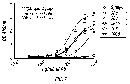
Comparison of Binding to G Protein with Binding to Virions
[0081] Figure 7 shows the results of an ELISA assay employing live virus and
assessing
the binding using a standard horseradish peroxidase assay. Viral preps from
various sources
were used to coat plates at 105 PFU/well or higher concentration in carbonate
buffer at pH 9.6
overnight at 4 C. Plates were blocked in 5% mik with PBST for an hour at room
temperature.
Serial dilutions of antibodies were added to wells in blocking buffer for one
hour at room
temperature. For detection, 1:2000 dilution of goat anti-human Fc gamma ¨ HRP
(Jackson
19
CA 02703667 2010-04-22
WO 2009/055711 PCT/US2008/081175
Immuno.) was added in blocking buffer for one hour at room temperature. Plates
were washed
extensively in PBST. Turnover of the substrate TMB was measured at 450 nm. As
shown in
Figure 7, 3D3 binds well to the live virus as do a number of other antibodies
of the invention.
The Synagis antibody, which has substantially weaker affinity, shows little
binding to live
virus even at 104 ng/ml antibody. Figure 8 shows the correlation of the
binding to recombinant
protein as compared to binding to virus particles.
[0082] Figures 9A and 9B are graphs that demonstrate comparative ability of
the antibodies
of the invention to bind to strains A2 versus A5, using the assay described
above. Figure 9A
shows that 3D3 and 3G12 bind well to strain A2 as compared to Synagis . PAB is
a
commercial polyclonal goat antibody against all RSV proteins (Chemicon,
catalog#AB1128).
[0083] Figure 9B shows that these antibodies also bind strain A5; note the
units on X-axis
are different than in Figure 9A. Similar experiments show that the antibodies
of the invention
bind to a wide variety of clinical isolates.
Example 6
Neutralization Assays
[00841 The ability of selected antibodies of the invention to neutralize virus
in vitro was
obtained by a standard plaque assay. HEp2 cells were plated in 12-well plates
at 2x10⁵ cells /
well. The following day, serial dilutions of antibodies were generated in
media. Approximately
200 PFU / well of RSV was added to the antibodies, in the presence of rabbit
complement
serum for one hour at room temperature. The antibody-virus mixture was then
added to HEp2
cells at 200 uL/well for 2 hr at room temperature to allow for infection.
Following this infection
period, media were removed and media containing 1% methyl cellulose were added
to all wells.
Plates were incubated at 35 C for 6 days, after which time, cells were fixed
and stained for
plaque number determination, as follows: Methyl cellulose is aspirated from
the cell layers, and
cells are fixed in 100% methanol for 30 min at room temperature. The plates
are then washed
3x with 5% milk in PBS. Primary antibody is added at 1:500 dilution (Goat anti-
RSV
polyclonal antibody (Chemicon Cat#AB1128)) in PBS + 5% Milk Protein for 1 hr.
Plates are
washed again 3X with 5% milk in PBS. Secondary antibody is added at 1:500
dilution in 5%
milk protein in PBS (ImmunoPure Rabbit anti-goat antibody IgG (H+L) Peroxidase
conjugated)
(Thermo Scientific, Cat#31402)) for 1 hr. Plates are washed 3X with 1X PBS.
Plaques are
visualized by adding 1-Step Chloronaphthol substrate Pierce, Cat#34012), 200
uL per well for
min. Plates are rinsed with water and allowed to air dry. Plaques are counted
in each well.
CA 02703667 2010-04-22
WO 2009/055711 PCT/US2008/081175
[0085] Figure 10 shows the results in terms of absolute numbers of plaques per
g of human
antibody and Synagis antibody is included in the results. These data show
that of the
antibodies tested, 3D3 is most potent. 3G12 has an IC50 of 15 ng/ml or an
affinity of 100 pM
according to this assay, whereas Synagis commercial antibody has an IC50 of
300 ng/ml
corresponding to an affinity of 2 nM. It was further found that Synagis and
the anti-G
antibodies of the invention were not synergistic under these conditions.
[0086] Figure 11 shows neutralization of 3G12 antibody with respect to strain
B, in
comparison with Synagis . The normalized data (% of control) are based on an
absolute plaque
number of 160-180 per experiment. The antibodies of the invention, with in
vitro affinities
from 1 pM up to 5 nM (Table 1), have EC50 values between 10-100 ng/ml.
Example 7
Anti-G Prophylaxis in Mice
[0087] The invention antibodies, Synagis , and human IgG1 were tested for
their ability to
prevent RSV infection in mice. On day -1, prior to infection, mice in the
control group were
injected i.p. with medium and PBS. In test groups, injection was of 0.15, 1.5
and 15 mg/kg of
antibodies hIgG1 (non-immune, isotype control), or 3G12 or 3D3 or Synagis .
This amounts to
approximately 3 lig, 30 lig and 300 [ig per mouse.
[0088] On day 0, the mice were inoculated with 1x10⁶ pfu RSV long-strain by
intranasal
administration. On days 0 and 5, the lungs, bronchial alveolar lavage (BAL)
and serum were
collected and body weight, lung weight, pfu in the lung lobe section, viral
load (by qPCR), lung
histology, total leukocytes, FACS, and IFNI, in BAL were all measured.
[0089] Figure 12 shows the results based on viral lung load using the plaque
assay from the
foregoing list. The data in Figure 12 show that 3G12 and 3D3 are equally
effective as Synagis
in this assay. (A typical human dose for Synagis is 15 mg/kg in humans.)
Example 8
Therapeutic Efficacy of Antibodies to RSV-Ga/Gb
[0090] Antibodies to the conserved motif on RSV-G are shown to have
therapeutic efficacy.
Mice were infected intra-nasally at day 0 with 106 pfu of RSV, then treated at
day 3 with
3 mg/kg of antibody injected i.p. and assayed at days 5 and 7 for viral load
in bronchial alveolar
lavage. In this model, the infection is more readily cleared naturally than in
humans.
Nonetheless, the antibody treatment causes acceleration in viral clearance in
a dose dependent
=
21
CA 02703667 2010-04-22
WO 2009/055711 PCT/US2008/081175
fashion as compared to a control antibody that does not bind RSV (Figure 13A).
Each treatment
group had 5 animals, and the results are statistically significant.
[0091] As described in WO 00/43040, antibodies to Substance P are beneficial
in alleviating
the lung inflammation caused by RSV, an animal model for the prolonged
morbidity that is the
clinically important feature of RSV infection. Up regulation of Substance P is
dependent on
active G protein (Haynes, L. M., et al., J. Virol. (2003) 77:9831-9844).
Reduction in measures
of lung inflammation following treatment with an antibody of the invention
have also been
observed, including reduction in inflammatory NK and total PMN cells (Figure
13B), as well as
reduction in cytokines, e.g., IFNy (Figure 13C).
[0092] In an additional test, on day 0, mice were inoculated with 106 pfu of
RSV A-type
long strain by intranasal administration.
[0093] On day 3, various groups of 4-5 mice were treated as follows:
[0094] Group 1: control group which did not receive infection on day 0 and was
treated
with PBS.
[0095] Group 2: negative control which received RSV inoculation on day 0 and
PBS
treatment on day 3.
[0096] Group 3: RSV inoculation on day 0 and Synagis antibody i.p. in saline
at 1, 10,
or 100n per mouse or 0.05, 0.5 or 5 mg/kg.
[0097] Group 4: RSV inoculated on day 0 and administered mAb 3D3 in the same
protocol
as Group 3.
[0098] Group 5: received RSV inoculation on day 0 and administered 3G12 in the
same
amounts as Groups 3 and 4.
[0099] Lungs and BAL fluid were collected on days 0, 3, 5, 7 and 10. In
addition, body
weight, lung weight, pfu in lung lobes, viral load by qPCR, lung histology,
total leukocytes,
FACS were measured as well as IFNy in BAL. The results for qPCR in the groups
administered 10 lig of mAb are shown in Figure 14.
[0100] As shown, the viral titer in Synagis -treated and untreated mice
behaves similarly at
this relatively low dose of antibody, whereas both 3D3 treated and 3G12
treated mice had
greatly lower titers at the peak of infection on day 5. This experiment
verifies that higher
affinity in vitro correlates with higher potency in vivo.
[0101] Figure 15 shows the dose response curve demonstrating that 3G12 and 3D3
were
able to lower the RSV copy number as measured by qPCR on day 7 at lower
concentrations
than Synagis . 3D3 was particularly potent, again consistent with having
higher affinity in
vitro.
22
CA 02703667 2010-04-22
WO 2009/055711 PCT/US2008/081175
[0102] Similarly, when qPCR viral counts are measured on day 10, although
viral titers are
naturally very low at this point due to natural clearance by the mouse immune
system, 3D3 is
approximately 100 times more potent than Synagis at the various dose
concentrations as shown
in Figure 16. This experiment highlights the utility of high affinity
antibodies, which continue
to be effective even when the antigen concentration drops. The human disease
course is
considerably more prolonged than in the mouse, providing a clear motivation
for use of an
antibody that continues to neutralize virus for an extended time period.
[0103] In still further experiments, mice were treated with murine anti-G mAb
or murine
anti-F mAb in groups of four, each experiment repeated three times. The mice
were immunized
on day 0 and treated with the antibodies on day 3, and various indications of
efficacy were
measured at days 3, 5 and 7.
[0104] As one index of effectiveness, inflammatory cells in the bronchial
alveolar lavage
(BAL) were measured in the three groups with the results shown in Figure 17.
BAL cells per
lung are plotted on the Y-axis from 0 to 140x103. The results show anti-F mAb
lowered the
BAL cells per lung at day 5 as compared to isotype control non-immune
antibody, whereas anti-
G mAb lowered the BAL cell count substantially more. By day 7, the infection
had run its
course.
[0105] Figures 18A and 18B show a comparison of effectiveness of anti-G mAb
(murine
131-2G) compared with anti-G F(a1302 obtained from this antibody by cleavage
with pepsin and
removal of the Fc fragments using immobilized Protein A. It has been shown
that complement
is important for the anti-viral effect of anti-G antibodies in vitro. This is
confirmed in
Figure 18A where the anti-viral effect is measured as pfu/g lung tissue.
Assays were conducted
as in Example 6. The F(a1:02 fragment of an anti-G antibody, which lacks the
Fc portion of IgG
that is needed for complement mediated activity, is little better than control
in lowering viral
load, while anti-G mAb is very effective. However, when inflammation is used
as a measure of
results, as shown in Figure 18B, the F(ab1)2 fragment of anti-G mAb is fully
as effective as the
complete antibody. This experiment establishes that neutralization of the G
protein is critical to
reducing airway inflammation. Since the virus actively secretes a soluble form
of the G protein,
and high affinity binding is important for neutralization of soluble factors,
the high affinity
antibodies of the invention are expected to have particular utility for the
anti-inflammatory
effect.
[0106] Figure 19A, B and C show the effect of anti-G mAb on the production of
IFNy in
BAL as a function of time of administration, with the cytokine serving as a
marker for airway
inflammation. Control non-immune antibody in all cases fails to reduce the
increase in IFNy
23
CA 02703667 2010-04-22
WO 2009/055711 PCT/US2008/081175
production that accompanies airway inflammation. However, whether anti-G mAb
is
administered at day -1 (panel A), at day +3 (panel B) or even at day +5 (panel
C), a dramatic
decrease in the level of IFNy at day 7 results. This experiment establishes
utility of antibodies
to the central conserved motif of the RSV G protein for treating inflammation
well past the peak
of viral load.
Example 9
Specificity of Endogenous Antibodies in Infected Subjects
[0107] Serum samples from four elderly adults with severe RSV disease and with
six
elderly adults with mild RSV disease were tested for immunoreactivity with the
synthetic
peptide:
Ac-Lys-Pro-Asn-Asn-Asp-Phe-His-Phe-Glu-Val-Phe-Asn-Phe-Val-Pro-Cyls-Ser-
Ile-qs-Ser-Asn-Asn-Pro-Thr-Clys-Trp-Ala-Ile-Cy-Lys-Arg-Ile-NH2
(disulfide bridges as shown)
which represents the conserved region of RSV G protein from strain A2. The
assay was
performed using the ELISA protocol described in Example 5. The levels of
antibodies
immunoreactive with this peptide correlate with the severity of the disease
wherein subjects
with mild forms of the disease exhibited much higher titers than subjects with
more severe
manifestations of the infection (see Figure 20). These results indicate that
antibodies
immunoreactive with this portion of the G protein are effective in
ameliorating infection.
24
CA 02703667 2011-08-25
SEQUENCE LISTING IN ELECTRONIC FORM
This description contains a sequence listing in electronic form in ASCII
text format (file no. 49324-555_ca_seglist_v1_22Apr2010.txt).
A copy of the sequence listing in electronic form is available from the
Canadian Intellectual Property Office.
The sequences in the sequence listing in electronic form are reproduced in
the following Table.
SEQUENCE TABLE
<110> TRELLIS BIOSCIENCE, INC.
<120> ANTI-RSV G PROTEIN ANTIBODIES
<130> 49324-555
<140> PCT/US2008/082175
<141> 2008-10-24
<150> US 61/000,469
<151> 2007-10-25
<150> US 61/089,401
<151> 2008-08-15
<160> 55
<170> FastSEQ for Windows Version 4.0
<210> 1
<211> 13
<212> PRT
<213> Artificial Sequence
<220>
<223> respiratory syncytial virus A2 strain
<400> 1
His Phe Glu Val Phe Asn Phe Val Pro Cys Ser Ile Cys
1 5 10
<210> 2
<211> 6
<212> PRT
<213> Artificial Sequence
<220>
<223> respiratory syncytial virus A2 strain
<400> 2
Phe Glu Val Phe Asn Phe
1 5
24a
CA 02703667 2010-04-22
<210> 3
<211> 10
<212> PRT
<213> Artificial Sequence
<220>
<223> respiratory syncytial virus A2 strain
<400> 3
Val Phe Asn Phe Val Pro Cys Ser Ile Cys
1 5 10
<210> 4
<211> 423
<212> DNA
<213> Mus musculus
<220>
<221> misc feature
<222> (1)...(423)
<223> heavy chain V segment of IgH-VJ558 family
<220>
<221> CDS
<222> (1)...(423)
<400> 4
atg gga tgg agc tgg atc ttt ctc ttc ctc ctg tca gga act gca ggt 48
Met Gly Trp Ser Trp Ile Phe Leu Phe Leu Leu Ser Gly Thr Ala Gly
1 5 10 15
gtc cac tct gag gtc cag ctg caa cag tct gga cct gaa ctg gtg aag 96
Val His Ser Glu Val Gln Leu Gln Gln Ser Gly Pro Glu Leu Val Lys
20 25 30
cct gga act tca gtg aag ata tcc tgc aag gct tct ggt tat tca ttc 144
Pro Gly Thr Ser Val Lys Ile Ser Cys Lys Ala Ser Gly Tyr Ser Phe
35 40 45
act ggc ttc acc atg aac tgg gtg aag cag agc cat gga aag aac ctt 192
Thr Gly Phe Thr Met Asn Trp Val Lys Gln Ser His Gly Lys Asn Leu
50 55 60
gag tgg ttt gga ctt att aat cct ttc aat ggt aat act ggc tac aac 240
Glu Trp Phe Gly Leu Ile Asn Pro Phe Asn Gly Asn Thr Gly Tyr Asn
65 70 75 80
cag aag ttc aag ggc aag gcc aca tta act gta gac aag tct tcc agc 288
Gln Lys Phe Lys Gly Lys Ala Thr Leu Thr Val Asp Lys Ser Ser Ser
85 90 95
aca gcc ttc atg gag ctc ctc agt ctg aca tct gag gac tct gca gtc 336
Thr Ala Phe Met Glu Leu Leu Ser Leu Thr Ser Glu Asp Ser Ala Val
100 105 110
tat tac tgt gca aga tcg gga aaa tcc tat gat tac gag gcc tgg ttt 384
Tyr Tyr Cys Ala Arg Ser Gly Lys Ser Tyr Asp Tyr Glu Ala Trp Phe
115 120 125
24b
CA 02703667 2010-04-22
act tac tgg ggc caa ggg act ctg gtc act gtc tct gca 423
Thr Tyr Trp Gly Gln Gly Thr Leu Val Thr Val Ser Ala
130 135 140
<210> 5
<211> 141
<212> PRT
<213> Mus musculus
<220>
<221> misc feature
<222> (1)...(141)
<223> heavy chain V segment of IgH-VJ558 family
<400> 5
Met Gly Trp Ser Trp Ile Phe Leu Phe Leu Leu Ser Gly Thr Ala Gly
1 5 10 15
Val His Ser Glu Val Gln Leu Gln Gln Ser Gly Pro Glu Leu Val Lys
20 25 30
Pro Gly Thr Ser Val Lys Ile Ser Cys Lys Ala Ser Gly Tyr Ser Phe
35 40 45
Thr Gly Phe Thr Met Asn Trp Val Lys Gln Ser His Gly Lys Asn Leu
50 55 60
Glu Trp Phe Gly Leu Ile Asn Pro Phe Asn Gly Asn Thr Gly Tyr Asn
65 70 75 80
Gln Lys Phe Lys Gly Lys Ala Thr Leu Thr Val Asp Lys Ser Ser Ser
85 90 95
Thr Ala Phe Met Glu Leu Leu Ser Leu Thr Ser Glu Asp Ser Ala Val
100 105 110
Tyr Tyr Cys Ala Arg Ser Gly Lys Ser Tyr Asp Tyr Glu Ala Trp Phe
115 120 125
Thr Tyr Trp Gly Gln Gly Thr Leu Val Thr Val Ser Ala
130 135 140
<210> 6
<211> 340
<212> DNA
<213> Mus musculus
<220>
<221> misc_feature
<222> (1)...(340)
<223> light chain V segment of Ig kappa V1 subgroup
<220>
<221> CDS
<222> (1)...(340)
<400> 6
gat att gtg atg aca caa act aca ctc tcc ctg cct gtc agt ctt gga 48
Asp Ile Val Met Thr Gln Thr Thr Leu Ser Leu Pro Val Ser Leu Gly
1 5 10 15
aat caa gcc tcc atc tct tgc aga tct agt cag acc att gta cat act 96
Asn Gln Ala Ser Ile Ser Cys Arg Ser Ser Gln Thr Ile Val His Thr
20 25 30
24c
CA 02703667 2010-04-22
aat gga aac acc tat tta gaa tgg tac ctg cag aaa cca ggc cag tct 144
Asn Gly Asn Thr Tyr Leu Glu Trp Tyr Leu Gln Lys Pro Gly Gln Ser
35 40 45
cca aag ctc ctg att tac aaa gtt tcc aac cga ttt tct ggg gtc cca 192
Pro Lys Leu Leu Ile Tyr Lys Val Ser Asn Arg Phe Ser Gly Val Pro
50 55 60
gac agg ttc agt ggc agt gga tca ggg aca gat ttc ata ctc aat atc 240
Asp Arg Phe Ser Gly Ser Gly Ser Gly Thr Asp Phe Ile Leu Asn Ile
65 70 75 80
agc aga gtg gag gct gag gat ctg gga gtt tat tac tgc ttt caa ggt 288
Ser Arg Val Glu Ala Glu Asp Leu Gly Val Tyr Tyr Cys Phe Gln Gly
85 90 95
tca cat gtt cca ttc acg ttc ggc tcg ggg aca aag ttg gaa ata aaa 336
Ser His Val Pro Phe Thr Phe Gly Ser Gly Thr Lys Leu Glu Ile Lys
100 105 110
cgg a 340
Arg
<210> 7
<211> 113
<212> PRT
<213> Mus musculus
<220>
<221> misc feature
<222> (1)...(113)
<223> light chain V segment of Ig kappa V1 subgroup
<400> 7
Asp Ile Val Met Thr Gln Thr Thr Leu Ser Leu Pro Val Ser Leu Gly
1 5 10 15
Asn Gln Ala Ser Ile Ser Cys Arg Ser Ser Gln Thr Ile Val His Thr
20 25 30
Asn Gly Asn Thr Tyr Leu Glu Trp Tyr Leu Gln Lys Pro Gly Gln Ser
35 40 45
Pro Lys Leu Leu Ile Tyr Lys Val Ser Asn Arg Phe Ser Gly Val Pro
50 55 60
Asp Arg Phe Ser Gly Ser Gly Ser Gly Thr Asp Phe Ile Leu Asn Ile
65 70 75 80
Ser Arg Val Glu Ala Glu Asp Leu Gly Val Tyr Tyr Cys Phe Gln Gly
85 90 95
Ser His Val Pro Phe Thr Phe Gly Ser Gly Thr Lys Leu Glu Ile Lys
100 105 110
Arg
<210> 8
<211> 12
<212> PRT
<213> Artificial Sequence
<220>
24d
CA 02703667 2010-04-22
=
<223> respiratory syncytial virus
<400> 8
Ser Lys Pro Asn Asn Asp Phe His Phe Glu Val Phe
1 5 10
<210> 9
<211> 12
<212> PRT
<213> Artificial Sequence
<220>
<223> respiratory syncytial virus
<400> 9
His Phe Glu Val Phe Asn Phe Val Pro Cys Ser Ile
1 5 10
<210> 10
<211> 7
<212> PRT
<213> Artificial Sequence
<220>
<223> respiratory syncytial virus
<400> 10
Glu Val Phe Asn Phe Val Pro
1 5
<210> 11
<211> 5
<212> PRT
<213> Artificial Sequence
<220>
<223> respiratory syncytial virus
<400> 11
His Phe Glu Val Phe
1 5
<210> 12
<211> 11
<212> PRT
<213> Artificial Sequence
<220>
<223> respiratory syncytial virus
<400> 12
Asp Phe His Phe Glu Val Phe Asn Phe Val Pro
1 5 10
<210> 13
24e
CA 02703667 2010-04-22
<211> 10
<212> PRT
<213> Artificial Sequence
<220>
<223> respiratory syncytial virus
<400> 13
Asn Asn Asp Phe His Phe Glu Val Phe Asn
1 5 10
<210> 14
<211> 7
<212> PRT
<213> Artificial Sequence
<220>
<223> respiratory syncytial virus
<400> 14
Phe Glu Val Phe Asn Phe Val
1 5
<210> 15
<211> 10
<212> PRT
<213> Artificial Sequence
<220>
<223> respiratory syncytial virus
<400> 15
Asn Asp Phe His Phe Glu Val Phe Asn Phe
1 5 10
<210> 16
<211> 32
<212> PRT
<213> Artificial Sequence
<220>
<223> respiratory syncytial virus A2 strain
<220>
<221> ACETYLATION
<222> 1
<223> acetylated lysine
<220>
<221> AMIDATION
<222> 32
<223> amide modified isoleucine
<400> 16
Lys Pro Asn Asn Asp Phe His Phe Glu Val Phe Asn Phe Val Pro Cys
1 5 10 15
24f
CA 02703667 2010-04-22
Ser Ile Cys Ser Asn Asn Pro Thr Cys Trp Ala Ile Cys Lys Arg Ile
20 25 30
<210> 17
<211> 12
<212> PRT
<213> Artificial Sequence
<220>
<223> respiratory syncytial virus A2 strain
<400> 17
Lys Pro Asn Asn Asp Phe His Phe Glu Val Phe Asn
1 5 10
<210> 18
<211> 12
<212> PRT
<213> Artificial Sequence
<220>
<223> respiratory syncytial virus A2 strain
<400> 18
Pro Asn Asn Asp Phe His Phe Glu Val Phe Asn Phe
1 5 10
<210> 19
<211> 12
<212> PRT
<213> Artificial Sequence
<220>
<223> respiratory syncytial virus A2 strain
<400> 19
Asn Asn Asp Phe His Phe Glu Val Phe Asn Phe Val
1 5 10
<210> 20
<211> 12
<212> PRT
<213> Artificial Sequence
<220>
<223> respiratory syncytial virus A2 strain
<400> 20
Asn Asp Phe His Phe Glu Val Phe Asn Phe Val Pro
1 5 10
<210> 21
<211> 12
<212> PRT
<213> Artificial Sequence
24g
CA 02703667 2010-04-22
<220>
<223> respiratory syncytial virus A2 strain
<400> 21
Asp Phe His Phe Glu Val Phe Asn Phe Val Pro Ser
1 5 10
<210> 22
<211> 12
<212> PRT
<213> Artificial Sequence
<220>
<223> respiratory syncytial virus A2 strain
<400> 22
Phe His Phe Glu Val Phe Asn Phe Val Pro Ser Ser
1 5 10
<210> 23
<211> 12
<212> PRT
<213> Artificial Sequence
<220>
<223> respiratory syncytial virus A2 strain
<400> 23
His Phe Glu Val Phe Asn Phe Val Pro Ser Ser Ile
1 5 10
<210> 24
<211> 50
<212> PRT
<213> Artificial Sequence
<220>
<223> respiratory syncytial virus A2 strain
<400> 24
Arg Gln Asn Lys Pro Pro Asn Lys Pro Asn Asn Asp Phe His Phe Glu
1 5 10 15
Val Phe Asn Phe Val Pro Cys Ser Ile Cys Ser Asn Asn Pro Thr Cys
20 25 30
Trp Ala Ile Cys Lys Arg Ile Pro Asn Lys Lys Pro Gly Lys Lys Thr
35 40 45
Thr Thr
<210> 25
<211> 50
<212> PRT
<213> Artificial Sequence
<220>
24h
CA 02703667 2010-04-22
<223> respiratory syncytial virus A2 strain
<400> 25
Arg Gln Asn Lys Pro Pro Ser Lys Pro Asn Asn Asp Phe His Phe Glu
1 5 10 15
Val Phe Asn Phe Val Pro Cys Ser Ile Cys Ser Asn Asn Pro Thr Cys
20 25 30
Trp Ala Ile Cys Lys Arg Ile Pro Asn Lys Lys Pro Gly Lys Arg Thr
35 40 45
Thr Thr
<210> 26
<211> 50
<212> PRT
<213> Artificial Sequence
<220>
<223> respiratory syncytial virus A2 strain
<400> 26
Arg Gln Asn Lys Pro Pro Ser Lys Pro Asn Asn Asp Phe His Phe Glu
1 5 10 15
Val Phe Asn Phe Val Pro Cys Ser Ile Cys Ser Asn Asn Pro Thr Cys
20 25 30
Trp Ala Ile Cys Lys Arg Ile Pro Asn Lys Lys Pro Gly Lys Lys Thr
35 40 45
Thr Thr
<210> 27
<211> 50
<212> PRT
<213> Artificial Sequence
<220>
<223> respiratory syncytial virus A2 strain
<400> 27
Arg Ser Lys Asn Pro Pro Lys Lys Pro Lys Asp Asp Tyr His Phe Glu
1 5 10 15
Val Phe Asn Phe Val Pro Cys Ser Ile Cys Gly Asn Asn Gln Leu Cys
20 25 30
Lys Ser Ile Cys Lys Thr Ile Pro Ser Asn Lys Pro Lys Lys Lys Pro
35 40 45
Thr Ile
<210> 28
<211> 127
<212> PRT
<213> Homo sapiens
<220>
<221> CHAIN
<222> (1)...(127)
<223> heavy chain of respiratory syncytial
24i
CA 02703667 2010-04-22
virus antibody
<400> 28
Gln Val Gln Leu Val Gln Ser Glu Ser Glu Val Lys Lys Pro Gly Ala
1 5 10 15
Ser Val Thr Val Ser Cys Lys Pro Ser Gly Tyr Thr Phe Asn Thr Tyr
20 25 30
Tyr Ile His Trp Val Arg Gln Ala Pro Gly Gln Gly Leu Glu Trp Met
35 40 45
Gly Val Ile Asn Pro Ser Gly Gly Ser Thr Thr Tyr Thr Gln Arg Phe
50 55 60
Gln Asp Arg Val Thr Met Thr Thr Asp Thr Ser Thr Asn Thr Ile Tyr
65 70 75 80
Met Asp Leu Thr Ser Leu Arg Ser Asp Asp Thr Ala Met Tyr Tyr Cys
85 90 95
Val Arg Gly Ser Asn Leu Leu Pro His Leu Trp Glu Trp Lys Pro Ser
100 105 110
His Phe Asp Ser Trp Gly Gln Gly Thr Leu Val Thr Val Ser Ser
115 120 125
<210> 29
<211> 126
<212> PRT
<213> Homo sapiens
<220>
<221> CHAIN
<222> (1)...(126)
<223> heavy chain of respiratory syncytial
virus antibody
<400> 29
Gln Leu Gln Leu Gln Glu Ser Gly Pro Gly Leu Val Lys Pro Ser Glu
1 5 10 15
Thr Leu Ser Leu Thr Cys Thr Val Ser Gly Gly Ser Ile Ser Ser Ser
20 25 30
Asn Tyr Tyr Trp Gly Trp Ile Arg Gln Pro Pro Gly Lys Gly Leu Glu
35 40 45
Trp Ile Ala Ser Ile His Asp Ser Gly Ser Ile Tyr Tyr Asn Pro Ser
50 55 60
Leu Arg Ser Arg Val Thr Ile Ser Val Asp Thr Ser Lys Asn Gln Phe
65 70 75 80
Ser Leu Lys Leu Ser Ser Val Thr Ala Ala Asp Thr Ala Val Tyr Tyr
85 90 95
Cys Ala Arg His Leu Val Trp Phe Gly Glu Leu Arg Asn Asn Trp Phe
100 105 110
Asp Pro Trp Gly Gln Gly Thr Leu Val Thr Val Ala Ser Ala
115 120 125
<210> 30
<211> 124
<212> PRT
<213> Homo sapiens
<220>
<221> CHAIN
<222> (1)...(124)
<223> heavy chain of respiratory syncytial
24j
CA 02703667 2010-04-22
virus antibody
<400> 30
Gln Leu Gln Leu Gln Glu Ser Gly Pro Gly Leu Val Lys Pro Ser Glu
1 5 10 15
Thr Leu Ser Leu Thr Cys Thr Val Ser Gly Asp Ser Ile Thr Ser Gly
20 25 30
Gln Tyr Tyr Trp Ala Trp Ile Arg Gln Ala Pro Gly Lys Gly Leu Glu
35 40 45
Trp Ile Gly Ser Ile His Tyr Ser Gly Ser Thr Tyr Gln Asn Pro Ser
50 55 60
Leu Lys Ser Arg Leu Thr Ile Ser Val Asp Thr Ser Arg Asp Gln Ile
65 70 75 80
Ser Met Lys Leu Ser Ser Val Thr Val Ala Glu Ser Ala Val Tyr Tyr
85 90 95
Cys Ala Arg Gln Gln Leu Ser Leu Ser Pro Val Glu Asn Trp Phe Asp
100 105 110
Pro Trp Gly Gln Gly Thr Leu Val Thr Val Ser Ser
115 120
<210> 31
<211> 124
<212> PRT
<213> Homo sapiens
<220>
<221> CHAIN
<222> (0)...(0)
<223> heavy chain of respiratory syncytial
virus antibody
<400> 31
Glu Glu Gln Leu Val Glu Ser Gly Gly Gly Leu Val Gln Pro Gly Arg
1 5 10 15
Ser Leu Arg Leu Ser Cys Val Gly Ser Gly Leu Arg Phe Glu Glu His
20 25 30
Ala Met His Trp Val Arg Gln Ala Pro Gly Arg Gly Leu Glu Trp Val
35 40 45
Ser Gly Ile Ser Trp Asn Ser Gly Ser Val Gly Tyr Ala Asp Ser Val
50 55 60
Lys Gly Arg Phe Thr Thr Ser Arg Asp Asn Ala Lys Asp Ile Leu Phe
65 70 75 80
Leu Glu Met Asn Thr Leu Arg Ser Glu Asp Thr Ala Leu Tyr Phe Cys
85 90 95
Ala Ile Met Val Ala Thr Thr Lys Asn Asp Phe His Tyr Tyr Lys Asp
100 105 110
Val Trp Gly Lys Gly Thr Thr Val Thr Val Ser Ser
115 120
<210> 32
<211> 124
<212> PRT
<213> Homo sapiens
<220>
<221> CHAIN
<222> (1)...(124)
<223> heavy chain of respiratory syncytial
24k
CA 02703667 2010-04-22
virus antibody
<400> 32
Gln Val His Leu Val Gln Ser Gly Val Glu Val Lys Lys Pro Gly Ala
1 5 10 15
Ser Val Arg Val Ser Cys Lys Ala Ser Gly Tyr Thr Phe Ala Thr Tyr
20 25 30
Gly Ile Thr Trp Val Arg Gln Ala Pro Gly Arg Gly Leu Glu Trp Val
35 40 45
Gly Trp Ile Thr Pro Tyr Asn Asp Arg Thr Ser Tyr Ala Gln Ile Phe
50 55 60
His Gly Arg Val Thr Met Thr Thr Asp Thr Ser Thr Asn Thr Ala Tyr
65 70 75 80
Met Glu Leu Arg Ser Leu Arg Ser Asp Asp Thr Ala Met Tyr Tyr Cys
85 90 95
Ala Arg Asn His Cys Asn Phe Tyr His Asp Phe Trp Ser Gly Leu Asp
100 105 110
Tyr Trp Gly Gln Gly Thr Leu Val Ser Val Ser Ser
115 120
<210> 33
<211> 121
<212> PRT
<213> Homo sapiens
<220>
<221> CHAIN
<222> (1)...(121)
<223> heavy chain of respiratory syncytial
virus antibody
<400> 33
Gln Val Gln Leu Val Gln Ser Gly Ala Glu Val Lys Lys Pro Gly Ser
1 5 10 15
Ser Val Lys Val Pro Cys Lys Ala Ser Gly Gly Thr Phe Ser Thr Tyr
20 25 30
Pro Ile Ser Trp Val Arg Gln Ala Pro Gly Gln Gly Leu Glu Trp Met
35 40 45
Gly Arg Ile Ile Pro Asp Pro Pro Met Ala Asn Ile Ala Gln Lys Phe
50 55 60
Gln Gly Arg Val Ser Phe Ser Ala Asp Lys Ser Thr Thr Ile Val Tyr
65 70 75 80
Met Glu Leu Ser Ser Leu Arg Ser Glu Asp Thr Ala Val Tyr Phe Cys
85 90 95
Ala Arg Glu Ile Leu Gln Ser Pro Pro Phe Ala Val Asp Val Trp Gly
100 105 110
Gln Gly Thr Met Val Ala Val Ser Ser
115 120
<210> 34
<211> 127
<212> PRT
<213> Homo sapiens
<220>
<221> CHAIN
<222> (1)...(127)
<223> heavy chain of respiratory syncytial
241
CA 02703667 2010-04-22
virus antibody
<400> 34
Gln Ala Gln Leu Val Glu Ser Gly Gly Gly Val Val Gln Pro Gly Arg
1 5 10 15
Ser Leu Arg Leu Ser Cys Ala Ala Ser Gly Phe Thr Phe Ser Tyr Ser
20 25 30
Gly Met His Trp Val Arg Gln Ala Pro Gly Arg Gly Leu Glu Trp Leu
35 40 45
Ala Val Ile Ser Tyr Asp Gly Asn Lys Met Tyr Tyr Ala Asp Ser Val
50 55 60
Lys Gly Arg Phe Thr Ile Ser Arg Asp Asn Ser Lys Met Thr Leu Tyr
65 70 75 80
Leu Gln Met Asn Ser Leu Arg Arg Glu Asp Thr Ala Val Tyr Tyr Cys
85 90 95
Ala Lys Asp Gly Leu Asp Tyr Gly Gly Asp Leu Val Tyr Tyr Gly Met
100 105 110
Asp Val Trp Gly Asn Gly Thr Thr Val Thr Val Ser Ser Ala Ser
115 120 125
<210> 35
<211> 126
<212> PRT
<213> Homo sapiens
<220>
<221> CHAIN
<222> (1)...(126)
<223> heavy chain of respiratory syncytial
virus antibody
<400> 35
Gln Val Gln Leu Val Gln Ser Gly Pro Glu Val Lys Lys Pro Gly Ala
1 5 10 15
Ser Val Arg Leu Ser Cys Lys Ala Ser Gly Tyr Val Phe Thr Asn Tyr
20 25 30
Gly Val Ser Trp Val Arg Gln Ala Pro Gly Gln Gly Leu Glu Trp Met
35 40 45
Gly Trp Ser Ser Pro Tyr Asn Gly Asn Thr Tyr Tyr Ala Gln Lys Leu
50 55 60
Lys Ala Arg Val Thr Met Thr Thr Asp Thr Ser Thr Asn Thr Ala Tyr
65 70 75 80
Met Glu Leu Arg Ser Leu Arg Ser Asp Asp Thr Ala Val Tyr Tyr Cys
85 90 95
Gly Arg Asp Met Leu Gly Val Val Gln Ala Val Ala Gly Pro Phe Asp
100 105 110
Ser Trp Gly Gln Gly Thr Leu Val Thr Val Ser Ser Ala Ser
115 120 125
<210> 36
<211> 126
<212> PRT
<213> Homo sapiens
<220>
<221> CHAIN
<222> (1)...(126)
<223> heavy chain of respiratory syncytial
24m
CA 02703667 2010-04-22
virus antibody
<400> 36
Gln Val Gln Leu Val Gln Ser Gly Gly Glu Val Lys Arg Pro Gly Ala
1 5 10 15
Ser Val Arg Val Ser Cys Lys Ala Ser Gly Tyr Thr Phe Ile Asn Tyr
20 25 30
Ala Ile Ser Trp Val Arg Gln Ala Pro Gly Gln Gly Leu Glu Trp Leu
35 40 45
Gly Trp Ile Ser Ala Tyr Asn Gly Asn Thr His Tyr Ala Gln Lys Val
50 55 60
Gln Asp Arg Val Thr Met Thr Thr Asp Ala Ser Thr Thr Thr Ala Tyr
65 70 75 80
Met Glu Leu Arg Gly Leu Lys Ser Asp Asp Thr Ala Val Tyr Ser Cys
85 90 95
Ala Arg Leu Pro Leu Leu Gly Tyr Ser Ser Gly Trp Tyr Ala Phe Asp
100 105 110
Met Trp Arg Gln Gly Thr Met Val Pro Val Ser Ser Ala Ser
115 120 125
<210> 37
<211> 127
<212> PRT
<213> Homo sapiens
<220>
<221> CHAIN
<222> (1)...(127)
<223> heavy chain of respiratory syncytial
virus antibody
<400> 37
Gln Val Gln Leu Leu Gln Ser Gly Ala Glu Leu Lys Lys Pro Gly Ala
1 5 10 15
Ser Val Lys Val Ser Cys Gln Ala Ser Ile Asp Thr Phe Ser Thr Tyr
20 25 30
Tyr Ile His Trp Val Arg Gln Ala Pro Gly Gln Gly Leu Glu Trp Leu
35 40 45
Gly Val Ile Asn Pro Ser Gly Gly Ser Thr Thr Tyr Ala Gln Lys Phe
50 55 60
Gln Asp Arg Ile Thr Leu Thr Thr Asp Thr Ser Thr Arg Thr Val Tyr
65 70 75 80
Met Asp Leu Ser Ser Leu Arg Ser Glu Asp Thr Ala Val Tyr Tyr Cys
85 90 95
Ala Arg Val His Lys Gly Arg Ala Glu Gln Trp Gln Leu Leu His Gly
100 105 110
His Phe Asp Leu Trp Gly Arg Gly Ser Leu Val Thr Val Ser Ser
115 120 125
<210> 38
<211> 126
<212> PRT
<213> Homo sapiens
<220>
<221> CHAIN
<222> (1)...(126)
<223> heavy chain of respiratory syncytial
24n
CA 02703667 2010-04-22
virus antibody
<400> 38
Gln Leu Gln Leu Gln Glu Ser Gly Pro Gly Leu Val Lys Pro Ser Glu
1 5 10 15
Thr Leu Ser Leu Thr Cys Thr Val Ser Gly Asp Ser Ile Thr Ser Gly
20 25 30
Gln Tyr Tyr Trp Ala Trp Ile Arg Gln Ala Pro Gly Lys Gly Leu Glu
35 40 45
Trp Ile Gly Ser Ile His Tyr Ser Gly Ser Thr Tyr Gln Asn Pro Ser
50 55 60
Leu Lys Ser Arg Leu Thr Ile Ser Val Asp Thr Ser Arg Asp Gln Ile
65 70 75 80
Ser Met Lys Leu Ser Ser Val Thr Val Ala Glu Ser Ala Val Tyr Tyr
85 90 95
Cys Ala Lys Gln Gln Leu Ser Leu Ser Pro Val Glu Asn Trp Phe Asp
100 105 110
Pro Trp Gly Gln Gly Thr Leu Val Thr Val Ser Ser Ala Ser
115 120 125
<210> 39
<211> 120
<212> PRT
<213> Homo sapiens
<220>
<221> CHAIN
<222> (1)...(120)
<223> heavy chain of respiratory syncytial
virus antibody
<400> 39
Glu Val Gln Val Val Glu Ser Gly Gly Gly Leu Val Gln Pro Gly Gly
1 5 10 15
Ser Leu Arg Leu Ser Cys Val Ala Ser Gly Phe Thr Phe Ser Ser Tyr
20 25 30
Trp Met His Trp Val Arg Gln Ala Pro Gly Lys Gly Leu Val Trp Val
35 40 45
Ser Arg Ile Tyr Ser Asp Gly Ser Ser Thr Thr Tyr Ala Asp Ser Val
50 55 60
Lys Gly Arg Phe Thr Ile Ser Arg Asp Asn Ala Lys Asn Thr Leu Tyr
65 70 75 80
Leu Gln Met Asn Ser Leu Arg Ala Glu Asp Thr Ala Val Tyr Tyr Cys
85 90 95
Val Arg Val Leu Gly Ala Ala Met Phe Asp Ile Trp Gly Gln Gly Thr
100 105 110
Val Val Thr Val Ser Ser Ala Ser
115 120
<210> 40
<211> 128
<212> PRT
<213> Homo sapiens
<220>
<221> CHAIN
<222> (1)...(128)
<223> heavy chain of respiratory syncytial
24o
CA 02703667 2010-04-22
virus antibody
<400> 40
Gln Val Gln Leu Val Glu Ser Gly Gly Gly Val Val Gln Pro Gly Arg
1 5 10 15
Ser Leu Arg Leu Ser Cys Glu Ala Ser Gly Phe Thr Phe Ser Gly Tyr
20 25 30
Ala Met His Trp Val Arg Gln Ala Pro Ala Lys Gly Leu Glu Trp Val
35 40 45
Ala Val Ile Ser Phe Asp Gly Ser Asn Asn Tyr Tyr Ala Asp Ser Val
50 55 60
Lys Gly Arg Phe Thr Ile Ser Arg Asp Asn Ser Lys Asn Met Leu Phe
65 70 75 80
Leu Gln Met Asn Ser Leu Arg Ala Glu Asp Thr Ala Val Tyr Tyr Cys
85 90 95
Val Arg Pro Asp Val Ile Ala Val Ala Gly Thr Ala Leu Ser Asn Pro
100 105 110
Phe Asp Leu Trp Gly Leu Gly Thr Met Val Thr Val Ser Ser Ala Ser
115 120 125
<210> 41
<211> 122
<212> PRT
<213> Artificial Sequence
<220>
<223> synthetic construct
<220>
<221> CHAIN
<222> (1)...(122)
<223> humanized heavy chain of respiratory
syncytial virus antibody
<400> 41
Gln Val Gln Leu Val Gln Ser Gly Ala Glu Val Lys Lys Pro Gly Ala
1 5 10 15
Ser Val Lys Val Ser Cys Lys Ala Ser Gly Tyr Ser Phe Thr Gly Phe
20 25 30
Thr Met Asn Trp Val Arg Gln Ala Thr Gly Gln Gly Leu Glu Trp Met
35 40 45
Gly Leu Ile Asn Pro Phe Asn Gly Asn Thr Gly Tyr Asn Gln Lys Phe
50 55 60
Lys Gly Arg Val Thr Met Thr Arg Asn Thr Ser Ile Ser Thr Ala Tyr
65 70 75 80
Met Glu Leu Ser Ser Leu Arg Ser Glu Asp Thr Ala Val Tyr Tyr Cys
85 90 95
Ala Arg Ser Gly Lys Ser Tyr Asp Tyr Glu Ala Trp Phe Thr Tyr Trp
100 105 110
Gly Gln Gly Thr Leu Val Thr Val Ser Ala
115 120
<210> 42
<211> 109
<212> PRT
<213> Homo sapiens
<220>
24p
CA 02703667 2010-04-22
<221> CHAIN
<222> (1)...(109)
<223> light chain of respiratory syncytial
virus antibody
<400> 42
Glu Ile Val Leu Thr Gln Ser Pro Gly Thr Leu Ser Leu Ser Pro Gly
1 5 10 15
Glu Arg Ala Thr Leu Ser Cys Arg Ala Ser Gln Ser Ile Thr Asn Asn
20 25 30
Tyr Leu Ala Trp Tyr Gln Gln Lys Pro Gly Gln Ala Pro Arg Leu Leu
35 40 45
Ile Tyr Asp Ser Phe Ser Arg Ala Thr Gly Ile Pro Glu Arg Phe Ser
50 55 60
Gly Ser Gly Ser Gly Thr Asp Phe Thr Leu Thr Ile Ser Arg Leu Glu
65 70 75 80
Pro Glu Asp Phe Ala Val Tyr Tyr Cys Gln His Tyr Val Arg Ser Pro
85 90 95
Leu Thr Phe Gly Pro Gly Thr Lys Val Glu Ile Lys Arg
100 105
<210> 43
<211> 107
<212> PRT
<213> Homo sapiens
<220>
<221> CHAIN
<222> (1)...(107)
<223> light chain of respiratory syncytial
virus antibody
<400> 43
Glu Ile Val Met Thr Gln Ser Pro Ala Thr Leu Ser Val Ser Pro Gly
1 5 10 15
Glu Arg Ala Thr Leu Ser Cys Arg Ala Ser Gln Ser Val Asn Ser Asn
20 25 30
Leu Ala Trp Tyr Gln His Lys Pro Gly Gln Ala Pro Arg Leu Leu Ile
35 40 45
Tyr Gly Ala Ser Thr Arg Ala Thr Gly Ile Pro Ala Arg Phe Ser Gly
50 55 60
Ser Gly Ser Gly Thr Asp Phe Thr Leu Thr Ile Ser Ser Leu Gln Ser
65 70 75 80
Glu Asp Phe Ala Val Tyr Tyr Cys Gln Gln Tyr Asn Asn Trp Pro Leu
85 90 95
Phe Gly Pro Gly Thr Lys Val Asp Leu Lys Arg
100 105
<210> 44
<211> 109
<212> PRT
<213> Homo sapiens
<220>
<221> CHAIN
<222> (1)...(109)
<223> light chain of respiratory syncytial
virus antibody
24q
CA 02703667 2010-04-22
<400> 44
Glu Ile Val Val Thr Gln Ser Pro Val Thr Leu Ser Val Ser Pro Gly
1 5 10 15
Glu Ser Ala Thr Leu Ser Cys Arg Ser Ala Arg Ser Val Gly Ser Arg
20 25 30
Leu Ala Trp Tyr Gln Gln Lys Pro Gly Gln Pro Pro Arg Leu Leu Ile
35 40 45
Phe Ala Ala Ser Thr Arg Ala Thr Gly Ile Pro Ala Arg Phe Ser Gly
50 55 60
Ser Gly Ser Gly Thr Asp Phe Thr Leu Ile Ile Ser Gly Leu Gln Ser
65 70 75 80
Glu Asp Tyr Ala Val Tyr Tyr Cys Gln Gln Tyr Lys Glu Trp Pro Leu
85 90 95
Phe Thr Phe Gly Pro Gly Thr Thr Val Asp Ser Lys Arg
100 105
<210> 45
<211> 106
<212> PRT
<213> Homo sapiens
<220>
<221> CHAIN
<222> (1)...(106)
<223> light chain of respiratory syncytial
virus antibody
<400> 45
Gln Ile Val Leu Thr Gln Ser Pro Ala Thr Leu Ser Leu Ser Pro Gly
1 5 10 15
Glu Arg Ala Thr Leu Ser Cys Lys Ala Ser Gln Ser Val Ser Asn His
20 25 30
Leu Ala Trp Tyr Gln Gln Lys Pro Gly Gln Ala Pro Arg Leu Leu Ile
35 40 45
Tyr Glu Thr Ser Asn Arg Ala Thr Gly Ile Pro Pro Arg Phe Ser Gly
50 55 60
Ser Gly Ser Gly Thr Asp Phe Thr Leu Thr Ile Ser Ser Leu Glu Pro
65 70 75 80
Glu Asp Phe Ala Val Tyr Tyr Cys Gln Gln Arg Asn Asn Trp Tyr Thr
85 90 95
Phe Gly Gln Gly Thr Lys Leu Glu Ile Lys
100 105
<210> 46
<211> 108
<212> PRT
<213> Homo sapiens
<220>
<221> CHAIN
<222> (1)...(108)
<223> light chain of respiratory syncytial
virus antibody
<400> 46
Ser Phe Glu Leu Thr Gln Pro Pro Ser Val Ser Val Ser Pro Gly Gln
1 5 10 15
24r
CA 02703667 2010-04-22
Thr Ala Arg Ile Thr Cys Ser Gly Asp Ala Leu Pro Lys Gln Tyr Val
20 25 30
Tyr Trp Tyr Gln Gln Lys Pro Gly Gln Ala Pro Val Leu Val Ile Tyr
35 40 45
Lys Thr Thr Glu Arg Pro Ser Gly Ile Pro Glu Arg Phe Ser Asp Ser
50 55 60
Ser Ser Gly Thr Thr Val Thr Leu Thr Ile Ser Ala Ala Gln Ala Glu
65 70 75 80
Asp Glu Ala Asp Tyr Tyr Cys Gln Ser Val Asp Ser Ser Gly Thr Tyr
85 90 95
Val Phe Gly Ile Gly Thr Lys Val Thr Val Leu Gly
100 105
<210> 47
<211> 113
<212> PRT
<213> Homo sapiens
<220>
<221> CHAIN
<222> (1)...(113)
<223> light chain of respiratory syncytial
virus antibody A2
<400> 47
Gln Ser Ala Leu Thr Gln Pro Ala Ser Val Ser Gly Ser Pro Gly Gln
1 5 10 15
Ser Ile Thr Ile Ser Cys Thr Gly Ser Ser Ser Asp Val Gly Gly Tyr
20 25 30
Ser His Val Ser Trp Tyr Gln Gln His Pro Gly Lys Val Pro Lys Leu
35 40 45
Ile Ile Ser Glu Val Ser Asn Arg Pro Ser Gly Ile Ser Asn Arg Phe
50 55 60
Ser Gly Ser Lys Ser Ala Asn Thr Ala Ser Leu Thr Ile Ser Gly Leu
65 70 75 80
Gln Pro Glu Asp Glu Ala Asp Tyr Tyr Cys Gly Ser Tyr Ala Ser Thr
85 90 95
Asn Ile Leu His Tyr Val Phe Gly Thr Gly Thr Lys Val Thr Val Leu
100 105 110
Ser
<210> 48
<211> 114
<212> PRT
<213> Homo sapiens
<220>
<221> CHAIN
<222> (1)...(114)
<223> light chain of respiratory syncytial
virus antibody
<400> 48
Ser Tyr Glu Leu Thr Gln Pro Pro Ser Val Ser Val Ser Pro Gly Gln
1 5 10 15
Thr Gly Arg Ile Thr Cys Thr Gly Ser Glu Ala Ser Gly Asp Ala Leu
20 25 30
24s
CA 02703667 2010-04-22
Ala Ser Arg Tyr Ala Tyr Trp Tyr Gln His Lys Ser Gly Gln Ala Pro
35 40 45
Val Val Leu Ile Tyr Lys Asp Thr Glu Arg Pro Ser Gly Ile Ser Glu
50 55 60
Arg Phe Ser Gly Ser Ser Ser Gly Thr Thr Val Thr Leu Ile Ile Ser
65 70 75 80
Gly Val Leu Ala Glu Asp Glu Ala Asp Tyr Tyr Cys Lys Thr Ser Val
85 90 95
Arg Asn Gly Thr Ser Trp Val Phe Gly Thr Gly Thr Met Leu Thr Val
100 105 110
Leu Arg
<210> 49
<211> 108
<212> PRT
<213> Homo sapiens
<220>
<221> CHAIN
<222> (1)...(108)
<223> light chain of respiratory syncytial
virus antibody
<400> 49
Asp Thr Pro Met Thr Gln Ser Pro Ser Ser Val Ser Ala Ser Val Gly
1 5 10 15
Asp Arg Val Thr Ile Ser Cys Arg Ala Ser Gln Gly Ile Ser Asn Ser
20 25 30
Leu Ala Trp Tyr Gln Gln Lys Leu Gly Lys Ala Pro Gln Leu Leu Ile
35 40 45
Tyr Ala Ala Ser Ser Leu Gln Ser Gly Val Pro Ser Arg Phe Ser Gly
50 55 60
Ser Gly Ser Gly Thr Asp Phe Thr Leu Thr Ile Ser Ser Leu Gln Pro
65 70 75 80
Glu Asp Phe Ala Thr Tyr Tyr Cys Gln Gln Thr Asn Thr Phe Pro Phe
85 90 95
Thr Phe Gly Pro Gly Thr Lys Val Glu Val Arg Arg
100 105
<210> 50
<211> 111
<212> PRT
<213> Homo sapiens
<220>
<221> CHAIN
<222> (1)...(111)
<223> light chain of respiratory syncytial
virus antibody
<400> 50
Gln Ser Ala Leu Thr Gln Pro Pro Ser Ala Ser Gly Ser Pro Gly Gln
1 5 10 15
Ser Val Thr Ile Ser Cys Thr Gly Thr Ser Ser Asp Val Gly Gly Tyr
20 25 30
Asn Tyr Val Ser Trp Tyr Gln Gln His Pro Gly Lys Ala Pro Lys Leu
35 40 45
24t
CA 02703667 2010-04-22
Met Ile Tyr Glu Val Asn Lys Arg Pro Ser Gly Val Pro Asp Arg Phe
50 55 60
Ser Gly Ser Lys Ser Gly Asn Thr Ala Ser Leu Thr Val Ser Gly Leu
65 70 75 80
Gln Ala Glu Asp Glu Ala Glu Tyr Tyr Cys Ser Ser Tyr Ala Gly Ser
85 90 95
Met Asn Trp Val Phe Gly Gly Gly Thr Lys Leu Thr Val Leu Gly
100 105 110
<210> 51
<211> 114
<212> PRT
<213> Homo sapiens
<220>
<221> CHAIN
<222> (1)...(114)
<223> light chain of respiratory syncytial
virus antibody
<400> 51
Asp Ile Val Met Thr Gln Ser Pro Asp Ser Leu Ala Val Ser Leu Gly
1 5 10 15
Glu Arg Ala Thr Ile Asn Cys Lys Ser Ser Gln Ser Val Leu Tyr Ser
20 25 30
Ser Asn Asn Lys Thr Tyr Leu Ala Trp Tyr Gln Gln Lys Pro Arg Gln
35 40 45
Pro Pro Glu Leu Leu Ile Tyr Trp Ala Ser Thr Arg Glu Ser Gly Val
50 55 60
Pro Asp Arg Phe Ser Gly Ser Gly Ser Gly Thr Asn Phe Thr Leu Thr
65 70 75 80
Ile Ser Ser Leu Gln Ala Glu Asp Val Ala Val Tyr Tyr Cys Gln Gln
85 90 95
Tyr Tyr Thr Thr Pro Tyr Thr Phe Gly Gln Gly Thr Lys Leu Glu Ile
100 105 110
Lys Arg
<210> 52
<211> 109
<212> PRT
<213> Homo sapiens
<220>
<221> CHAIN
<222> (1)...(109)
<223> light chain of respiratory syncytial
virus antibody
<400> 52
Glu Ile Val Val Thr Gln Ser Pro Val Thr Leu Ser Val Ser Pro Gly
1 5 10 15
Glu Ser Ala Ala Leu Ser Cys Arg Ala Ser Arg Ser Val Gly Ser Arg
20 25 30
Leu Ala Trp Tyr Gln Gln Lys Pro Gly Gln Pro Pro Arg Leu Leu Ile
35 40 45
Phe Ala Ala Ser Thr Arg Ala Thr Gly Ile Pro Ala Arg Phe Ser Gly
50 55 60
24u
CA 02703667 2010-04-22
Ser Gly Ser Gly Thr Asp Phe Thr Leu Ile Ile Ser Gly Leu Gln Ser
65 70 75 80
Glu Asp Tyr Ala Val Tyr Tyr Cys Gln Gln Tyr Lys Glu Trp Pro Leu
85 90 95
Phe Thr Phe Gly Pro Gly Thr Thr Val Asp Ser Lys Arg
100 105
<210> 53
<211> 110
<212> PRT
<213> Homo sapiens
<220>
<221> CHAIN
<222> (1)...(110)
<223> light chain of respiratory syncytial
virus antibody
<400> 53
Glu Ile Val Leu Thr Gln Ser Pro Ala Thr Leu Ser Leu Ser Pro Gly
1 5 10 15
Glu Arg Ala Thr Leu Ser Cys Arg Ala Ser Gln Ser Val Ser Ser Tyr
20 25 30
Leu Ala Trp Tyr Gln Gln Lys Pro Gly Gln Ala Pro Arg Leu Leu Ile
35 40 45
Ser Asp Ala Ser Asn Arg Ala Thr Gly Ile Pro Pro Arg Phe Ser Gly
50 55 60
Ser Gly Ser Gly Thr Asp Phe Thr Leu Thr Ile Ser Ser Leu Glu Pro
65 70 75 80
Glu Asp Phe Ala Val Tyr Tyr Cys Gln Gln Arg Ser Asn Trp Pro Pro
85 90 95
Arg Phe Thr Phe Gly Pro Gly Thr Ile Val Asp Ile Arg Arg
100 105 110
<210> 54
<211> 107
<212> PRT
<213> Homo sapiens
<220>
<221> CHAIN
<222> (1)...(107)
<223> light chain of respiratory syncytial
virus antibody
<400> 54
Glu Ile Val Met Thr Gln Ser Pro Ala Thr Leu Ser Val Ser Pro Gly
1 5 10 15
Glu Arg Ala Leu Ser Cys Arg Ala Ser Gln Ser Val Arg Ser Asn Leu
20 25 30
Val Trp Tyr Gln Gln Lys Pro Gly Gln Ala Pro Arg Leu Leu Ile Phe
35 40 45
Gly Ala Ser Thr Arg Ala Thr Gly Ile Pro Ala Arg Phe Ser Gly Ser
50 55 60
Gly Ser Gly Thr Glu Phe Thr Leu Thr Ile Ser Ser Leu Gln Ser Glu
65 70 75 80
Asp Phe Ala Leu Tyr Phe Cys Gln Gln Asn Asn Asn Trp Pro Pro Thr
85 90 95
24v
CA 02703667 2010-04-22
Phe Gly Gln Gly Thr Arg Leu Glu Ile Lys Arg
100 105
<210> 55
<211> 113
<212> PRT
<213> Artificial Sequence
<220>
<223> synthetic construct
<220>
<221> CHAIN
<222> (1)...(113)
<223> humanized light chain of respiratory
syncytial virus antibody
<400> 55
Asp Ile Val Met Thr Gln Thr Pro Leu Ser Leu Ser Val Thr Pro Gly
1 5 10 15
Gln Pro Ala Ser Ile Ser Cys Arg Ser Ser Gln Thr Ile Val His Thr
20 25 30
Asn Gly Asn Thr Tyr Leu Glu Trp Tyr Leu Gln Lys Pro Gly Gln Ser
35 40 45
Pro Gln Leu Leu Ile Tyr Lys Val Ser Asn Arg Phe Ser Gly Val Pro
50 55 60
Asp Arg Phe Ser Gly Ser Gly Ser Gly Thr Asp Phe Thr Leu Lys Ile
65 70 75 80
Ser Arg Val Glu Ala Glu Asp Val Gly Val Tyr Tyr Cys Phe Gln Gly
85 90 95
Ser His Val Pro Phe Thr Phe Gly Ser Gly Thr Lys Leu Glu Ile Lys
100 105 110
Arg
24w