Note: Descriptions are shown in the official language in which they were submitted.
CA 02704564 2010-05-03
WO 2009/061813 PCT/US2008/082472
MULTI-FREQUENCY NEURAL TREATMENTS AND ASSOCIATED
SYSTEMS AND METHODS
CROSS-REFERENCE TO RELATED APPLICATION
[0001] The present application claims priority to U.S. Provisional Application
60/985,353, filed November 5, 2007 and incorporated herein by reference.
TECHNICAL FIELD
[0002] The present disclosure relates generally to methods and apparatuses for
treating patient conditions, including chronic pain conditions via techniques
that can
include stimulating and blocking neuronal tissue associated with the spinal
cord.
BACKGROUND
A. Neural Stimulation Treatments
[0003] Existing patient treatments include applying stimulation (e.g., up-
regulating)
signals to nerves, muscles or organs for treating a wide variety of medical
disorders.
Stimulation signal parameters (e.g., pulse width, frequency, and amplitude)
are
selected to initiate neural action potentials to be propagated along the nerve
to an
organ (e.g., brain or stomach).
[0004] Down-regulating signals also can be applied to nerve fibers. Certain
signal
parameters can result in a signal that inhibits the nerve or blocks the
propagation of
action potentials along the nerve. In general, the nerve conduction block is
applied to
nerves with down-regulating signals selected to block the entire cross-section
or part of
the cross section of the nerves (e.g., afferent, efferent, myelinated, and non-
myelinated
fibers) at the site where the down-regulating signal is applied.
[0005] In some systems, down-regulating signals are used to manage motor
control over certain areas of a patient's body. For example, cryogenic nerve
blocking
of the vagus nerve to control motor activity is described in Dapoigny et at,
"Vagal
influence on colonic motor activity in conscious nonhuman primates," Am. J.
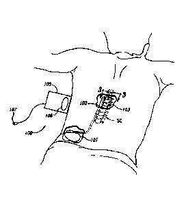
Physiol.,
262: G231 - G236 (1992). A cryogenic vagal block and the resulting effect on
gastric
CA 02704564 2010-05-03
WO 2009/061813 PCT[US2008/082472
emptying are described in Paterson CA, et al., "Determinants of Occurrence and
Volume of Transpyloric Flow During Gastric Emptying of Liquids in Dogs:
Importance of
Vagal Input," Dig Dis Sci, (2000); 45:1509-1516.
B. Application to Chronic Pain
[0006] Applying up-regulating electrical energy to the spinal cord for the
purpose
of managing pain has been actively practiced since the 1960s. While a precise
understanding of the interaction between the applied electrical energy and the
nervous
tissue is not fully appreciated, it is known that application of an electrical
field to spinal
nervous tissue can effectively mask certain types of pain transmitted from
regions of
the body associated with the stimulated tissue. Such spinal cord stimulation
(SCS) for
the treatment of chronic intractable pain was introduced by Shealy et al.
(Anesth.
Analg., 46, 489-491, 1967).
[0007] More specifically, applying up-regulating electrical pulses to the
spinal cord
associated with regions of the body (e.g., dermatomes) afflicted with chronic
pain can
induce paresthesia, or a subjective sensation of numbness or tingling, in the
afflicted
bodily regions. This paresthesia can effectively mask the non-acute pain
sensations
perceived at the brain.
[0008] Electrical energy, similar to that used to inhibit pain perception,
also may
be used to manage the symptoms of various motor disorders, for example,
tremor,
dystonia, spasticity, and the like. Motor spinal nervous tissue (e.g., nervous
tissue from
ventral nerve roots) transmits muscle/motor control signals. Sensory spinal
nervous
tissue (e.g., nervous tissue from dorsal nerve roots) transmits pain signals,
as well as
other sensory signals and proprioceptive signals.
[0009] Corresponding dorsal and ventral nerve roots depart the spinal cord
"separately." Laterally from the spinal cord, the nervous tissue of the dorsal
and
ventral nerve roots are mixed, or intertwined. Accordingly, electrical
stimulation
intended to manage and control one condition (e.g., pain) can inadvertently
interfere
with nerve transmission pathways in adjacent nervous tissue (e.g., motor
nerves).
[0010] Electrical energy is conventionally delivered through electrodes
positioned
on the dorsal column external to the dura layer surrounding a spinal cord. The
electrodes are typically carried by a percutaneous lead, although a laminotomy
lead
-2-
CA 02704564 2010-05-03
WO 2009/061813 PCT/US2008/082472
also can be used. Percutaneous leads commonly have two or more electrodes and
are
positioned within an epidural space through the use of an insertion, or Touhy-
like,
needle. An example of an eight-electrode percutaneous lead is an OCTRODE lead
manufactured by Advanced Neuromodulation Systems, Inc. of Plano, Texas.
Operationally, the insertion needle is passed through the skin, between the
desired
vertebrae, and into an epidural space located between a dural layer and the
surrounding vertebrae. The stimulation lead is fed through the bore of the
insertion
needle and into the epidural space. Laminotomy leads generally have a wider,
paddle-
like shape, and are inserted via an incision rather than through a needle. For
example,
a small incision is made in the back of a patient to access the space between
the dura
and the surrounding vertebrae.
[0011] According to the "gate-control" theory of Melzak and Wall, (Science,
150,971-978,1965), the suppression of pain sensations, accompanied by
paresthesia,
results from the activation of large cutaneous afferents (Aaf fibers). Because
these
nerve fibers are part of the dorsal root (DR) fiber that ascends in the dorsal
column
(DC), paresthetic sensations can be evoked by both DC and DR stimulation.
[0012] The potential paresthesia coverage will strongly differ, however,
depending
on whether DC fibers or DR fibers are stimulated. When stimulating the DC
fibers, the
fibers corresponding to all dermatomes from the sacral ones up to the
electrode level
may be activated, thus resulting in broad paresthesia coverage. When
stimulating DR
fibers, however, the fibers will be activated in a limited number of rootlets
close to the
cathodal contact(s), thereby resulting in a paresthesia effect confined to one
or two
dermatomes at each body side.
[0013] There are several problems with existing Spinal Cord Stimulation (SCS)
therapy techniques. One is the difficulty of obtaining a permanent optimal
position of
the lead(s), to cover the painful dermatomes with paresthesia. Another problem
is the
usually small range of stimulation amplitudes between the perception threshold
(i.e.,
the threshold at which paresthesia is effected) and the discomfort threshold
(i.e., the
threshold at which the patient experiences pain or other discomfort), often
preventing a
complete coverage of the painful area by the paresthesia needed for maximum
therapeutic effect (Holsheimer, Neurosurgery, 40, 5, 990-999, 1997).
-3-
CA 02704564 2010-05-03
WO 2009/061813 PCT/US2008/082472
SUMMARY
[0014] In some cases, low frequency signals are applied to the dorsal column
to
address chronic patient pain associated with a peripheral site. However, the
dorsal
roots also can be stimulated when low frequency stimulation is applied to the
dorsal
column to produce the paresthesia necessary to overcome the chronic pain. For
example, the dorsal roots may be stimulated if the stimulation leads are
placed too
close to the dorsal root, and/or if the amplitude of the low frequency signal
is increased
to the discomfort threshold. The discomfort threshold at the dorsal root can
be reached
before the parethesia threshold (i.e., the threshold at which paresthesia is
affected) is
reached at the dorsal column. Hence, the clinician has limited freedom to
increase the
amplitude of the signal at the dorsal column to achieve the desired
paresthesia effect,
before discomfort is felt due to the dorsal root stimulation.
[0015] Aspects of the present disclosure are directed to managing chronic pain
through the application of electrical energy to selected nervous tissue and,
in particular
embodiments, to methods and systems for treating chronic pain by applying
neuromodulation therapies to one or more regions of neuronal tissue in the
spinal
region. As the term is used herein, the "spinal region" includes the nerves of
the dorsal
column, dorsal roots, and the dorsal roots ganglion, which are located within
the dural
layer.
[0016] A method for treating patient pain in accordance with a particular
embodiment includes applying a first electrical signal to a first target
location (e.g., a
dorsal column) of the patient's spinal cord region at a frequency in a first
frequency
range of up to about 1,500 Hz. The method further includes applying a second
electrical signal to a second target location (e.g., at least one of a dorsal
root and a
dorsal root ganglion) of the patient's spinal cord region at a frequency in a
second
frequency range of from about 2,500 Hz to about 100,000 Hz. In particular
embodiments, the second frequency range can be from about 2,500 Hz to about
20,000 Hz, or about 3,000 Hz to about 10,000 Hz. Further embodiments include
inducing paresthesia by applying the first electrical signal, and at least
partially blocking
patient discomfort resulting from applying the first electrical signal by
applying the
second electrical signal.
-4-
CA 02704564 2010-05-03
WO 2009/061813 PCT/US2008/082472
[0017] A method in accordance with another embodiment includes implanting a
first electrode proximate to a dorsal column of the patient's spinal cord
region, and
implanting a second electrode proximate to at least one of a dorsal root and a
dorsal
root ganglion of the patient's spinal cord region. The method can further
include
applying a first electrical signal to the first electrode at a frequency in a
first frequency
range of up to about 1,500 Hz. If the patient experiences discomfort, a second
electrical signal is applied to the second electrode at a frequency in a
second
frequency range of from about 2,500 Hz to about 100,000 Hz in combination with
applying the first electrical signal, and without repositioning the first
electrode. In
particular embodiments, the second frequency range can be from about 2,500 Hz
to
about 20,000 Hz, or about 3,000 Hz to about 10,000 Hz.
[0018] Further embodiments are directed to systems for treating patient pain.
In a
particular embodiment, the system can include a controller having instructions
for
directing first electrical signals in a first frequency range of up to about
1,500 Hz, and
directing second electrical signals in a second frequency range of from about
2,500 Hz
to about 100,000 Hz. In particular embodiments, the second frequency range can
be
from about 2,500 Hz to about 20,000 Hz, or about 3,000 Hz to about 10,000 Hz.
A first
electrical signal delivery device can be electrically coupled to the
controller to receive
the first electrical signals, and can be configured to be positioned proximate
to a first
target location of the patient's spinal cord region (e.g., the dorsal column).
A second
electrical signal delivery device can be electrically coupled to the
controller to receive
the second electrical signals, and can be configured to be positioned
proximate to a
second target location of the patient's spinal cord region (e.g., at least one
of a dorsal
root and a dorsal root ganglion of the patient's spinal cord region).
BRIEF DESCRIPTION OF THE DRAWINGS
[0019] Figure 1 is a schematic diagram of an implantable spinal stimulator
with an
electrode array applied to the spine in accordance with an embodiment of the
present
disclosure.
[0020] Figure 2 is a schematic diagram of an implantable spinal stimulator
with
percutaneous leads and electrodes applied to the spine in accordance with
another
embodiment of the present disclosure.
-5-
CA 02704564 2010-05-03
WO 2009/061813 PCT/US2008/082472
[0021] Figure 3 is a partially schematic cross-sectional view of a spinal
column
taken along line 3-3 of Figure 1 in accordance with an embodiment of the
present
disclosure.
[0022] Figure 4 illustrates examples of biphasic, charge balanced, square wave
pulses applied to electrodes on different channels of a therapy system in
accordance
with an embodiment of the present disclosure.
[0023] Figure 5 illustrates examples of biphasic, charge balanced, sinusoidal
wave
pulses applied to electrodes on different channels of a therapy system in
accordance
with an embodiment of the present disclosure.
[0024] Figure 6 is a schematic depiction of an example blocking signal applied
to
the dorsal column in accordance with an embodiment of the present disclosure.
[0025] Figure 7 is a schematic depiction of an example high frequency (HF)
blocking signal applied to the dorsal root in accordance with an embodiment of
the
present disclosure.
[0026] Figure 8 schematically depicts the amplitude of an example low
frequency
(LF) stimulation signal likely to induce paresthesia, and the amplitude of the
LF
stimulation signal likely to induce patient discomfort at a given electrode
spacing in
accordance with an embodiment of the present disclosure.
[0027] Figure 9 is a schematic view of an HF blocking signal applied to the
dorsal
root of a patient and an LF stimulating signal applied to the dorsal column in
accordance with an embodiment of the present disclosure.
[0028] Figure 10 is a schematic diagram of an example blocking signal, which
has
an amplitude that is gradually increased to an operating amplitude over a
finite period
of time in accordance with an embodiment of the present disclosure.
[0029] Figure 11A is a schematic graph generally showing the changes in
frequency during application of a therapy in accordance with an embodiment of
the
present disclosure.
[0030] Figure 11 B is a schematic graph generally showing the changes in
amplitude during application of the therapy of Figure 11A in accordance with
an
embodiment of the present disclosure.
-6-
CA 02704564 2010-05-03
WO 2009/061813 PCT/US2008/082472
[0031] Figure 11C is a schematic graph generally showing the changes in
charge/phase during application of the therapy of Figure 1 1A in accordance
with an
embodiment of the present disclosure.
[0032] Figure 12 is a schematic depiction of an example blocking signal
initially
having a high frequency (e.g., about 30-50 KHz) and a high amplitude (e.g.,
about 15-
20 mA) in accordance with an embodiment of the present disclosure.
[0033] Figure 13 shows the blocking signal of Figure 12 with an initial ramp-
up
period in accordance with an embodiment of the present disclosure.
[0034] Figure 14 is a schematic depiction of an example LF signal and an
example
HF signal indicating a representative timing strategy for applying the LF and
HF signals
in accordance with an embodiment of the present disclosure.
[0035] Figures 15-18 are schematic block diagrams of representative electrode
arrays including four electrodes implanted at the spinal cord of a patient in
accordance
with an embodiment of the present disclosure.
[0036] Figure 19A is a schematic block diagram of a lead configuration in
which
first and second percutaneous leads are implanted within the patient together
in
accordance with an embodiment of the present disclosure.
[0037] Figure 19B is a schematic block diagram of a lead configuration in
which a
first percutaneous lead is implanted within the patient adjacent the dorsal
column and a
second percutaneous lead is implanted within the patient adjacent the dorsal
root in
accordance with an embodiment of the present disclosure.
[0038] Figure 19C is a partially schematic illustration of percutaneous leads
positioned at lumbar locations in accordance with embodiments of the
disclosure.
[0039] Figure 20 is a schematic block diagram of a multi-channel, percutaneous
lead arrangement having first and second leads configured to deliver multiple
therapy
signals to a dorsal column of a patient in accordance with an embodiment of
the
present disclosure.
[0040] Figure 21 is a schematic block diagram of a multi-channel, percutaneous
lead arrangement having first and second leads configured to deliver multiple
therapy
-7-
CA 02704564 2010-05-03
WO 2009/061813 PCT/US2008/082472
signals to a dorsal root of a patient in accordance with an embodiment of the
present
disclosure.
[0041] Figure 22 illustrates a first treatment signal being applied to nerves
of a
dorsal column of a patient in accordance with an embodiment of the present
disclosure.
DETAILED DESCRIPTION
[0042] Figure 1 schematically illustrates a representative therapy system 100
for
providing relief from chronic pain, arranged relative to the general anatomy
of a spinal
cord SC of a patient. The therapy system 100 can include a controller (e.g., a
pulse
generator 101) implanted subcutaneously within the patient. The pulse
generator 101
is attached via a lead body 102 to an electrode array 103 or other signal
delivery
device, which is implanted in close proximity to the spinal cord SC. The
electrode array
103 can include multiple electrodes or electrode contacts carried by a support
substrate. The pulse generator 101 or other controller transmits instructions
and power
to the electrode array 103 via the lead body 102 to apply therapy signals
(e.g.,
electrical impulses) to the nerve fibers of the patient to up-regulate (e.g.,
stimulate)
and/or down-regulate (e.g., block or partially block) the nerves. Accordingly,
the pulse
generator 101 can include a computer-readable medium containing the
instructions.
The pulse generator 101 and/or other elements of the system 100 can include
one or
more processors, memories and/or input/output devices. The pulse generator 101
can
include multiple portions, e.g., for directing signals in accordance with
multiple signal
delivery parameters, housed in a single housing (as shown in Figure 1) or in
multiple
housings.
[0043] In some embodiments, the pulse generator 101 can obtain power to
generate the therapy signals from an external power source 105. The external
power
source 105, which is arranged external to the patient, can transmit power to
the
implanted pulse generator 101 using electromagnetic induction (e.g., RF
signals). For
example, the external power source 105 can include an external coil 106 that
communicates with a corresponding coil (not shown) within the implantable
pulse
generator 101. The external power source 105 can be portable for ease of use.
-8-
CA 02704564 2010-05-03
WO 2009/061813 PCT/US2008/082472
[0044] In another embodiment, the pulse generator 101 can obtain the power to
generate therapy signals from an internal power source. For example, the
implanted
pulse generator 101 can include a non-rechargeable battery or a rechargeable
battery
to provide the power. When the internal power source includes a rechargeable
battery,
the external power source 105 can be used to recharge the battery. The
external
power source 105 in turn can be recharged from a suitable power source e.g.,
via a
standard power plug 107.
[0045] In still further embodiments, an external programmer (not shown) can
communicate with the implantable pulse generator 101 via electromagnetic
induction.
Accordingly, a practitioner can update the therapy instructions provided by
the pulse
generator 101. Optionally, the patient may also have control over at least
some
therapy functions, e.g., starting and/or stopping the pulse generator 101.
[0046] Figure 2 illustrates another therapy system 200 in which the
implantable
pulse generator 101 is connected to percutaneous lead bodies 108 and 109,
which are
in turn connected to electrodes 110. The leads 108, 109 and electrodes 110 are
shown in a bipolar configuration with two electrodes 110 carried by each lead
108, 109.
In other embodiments, however, the leads 108, 109 can each contain more
electrodes
110 (e.g., three, four, five, eight, or more) for applying therapy signals. In
any of the
foregoing embodiments, the electrodes (e.g., the electrode array 103 or the
electrodes
110 of the percutaneous leads 108,109) can be arranged adjacent different
nerve
fibers within the patient to enable the application of different types of
therapy, as is
discussed further below.
[0047] Figure 3 is a cross-sectional illustration of a spinal region SR that
includes
the spinal cord SC and an adjacent vertebra VT (based generally on information
from
Crossman and Neary, "Neuroanatomy," 1995 (publ. by Churchill Livingstone)),
along
with selected representative locations for representative leads 108 (shown as
leads
108a-108d) in accordance with several embodiments of the disclosure. The
spinal
cord SC is situated between a ventrally located vertebral body WB and a
dorsally
located vertebral body DVB that includes a transverse process 198 and spinous
process 197. Arrows V and D identify ventral and dorsal directions,
respectively. In
particular embodiments, the vertebra VT and leads can be at T10 or T11 (e.g.,
for axial
low back pain or leg pain) and in other embodiments, the leads can be placed
at other
-9-
CA 02704564 2010-05-03
WO 2009/061813 PCT/US2008/082472
locations. The spinal cord SC itself is located within the dura mater DM,
which also
surrounds portions of the nerves exiting the spinal cord SC, including the
dorsal roots
DR, dorsal root ganglia G and ventral roots VR. The spinal cord SC is
illustrated as
having identifiable areas of afferent and efferent fibers including ascending
pathway
areas AP and descending pathway areas DP.
[0048] The leads are generally positioned to stimulate tactile fibers and to
avoid
stimulating fibers associated with nociceptive pain transmission. In a
particular
embodiment, a lead 108a (e.g., a first lead) can be positioned centrally in a
lateral
direction (e.g., aligned with the spinal cord midline ML) to provide signals
directly to the
dorsal column DC of spinal cord SC. In other embodiments, the first lead can
be
located laterally from the midline ML. For example, single or paired leads can
be
positioned just off the spinal cord midline ML (as indicated by leads 108b) to
provide
signals to the dorsal column DC. One or more other leads (e.g., second leads)
can be
positioned proximate to the dorsal root DR or dorsal root entry zone DREZ
(e.g., 1-4
mm from the spinal cord midline ML, as indicated generally by lead 108c),
and/or
proximate to the dorsal root ganglion G (as indicated by lead 108d). Other
suitable
locations for the second lead include the "gutter," also located laterally
from the midline
ML. In still further embodiments, the leads 108 may have other locations
proximate to
the spinal cord SC and/or proximate to other target neural populations e.g.,
laterally
from the midline ML and medially from the dorsal root ganglion 194. For
example, the
leads can be located subdurally rather epidurally, as shown in dashed lines
for midline
lead 108a and off-midline leads 108b. The practitioner may select any of a
variety of
combinations of the foregoing locations, depending on the particular patient's
needs
and condition. In at least some embodiments, the practitioner can place two
leads,
each positioned to direct signals to a different target location (e.g., neural
population) of
the patient's spinal cord SC. In other embodiments, a single lead may have
electrodes
positioned at two or more target locations. In either case, individual
electrodes can
deliver signals with different characteristics to different neural populations
to achieve a
beneficial effect for the patient.
A. Therapy Options
[0049] In general, different types of therapy signals can be applied to the
nerve
fibers of a patient to different effect. For example, applying a low-frequency
(LF)
-10-
CA 02704564 2010-05-03
WO 2009/061813 PCT/US2008/082472
therapy signal to the nerve fibers of a patient can stimulate the nerve fibers
to create an
effect known in the art as "paresthesia," which creates a sensation of
numbness in the
patient. This paresthesia effect can mask chronic pain, providing relief to
the patient.
Such an application of therapy signals is generally known as Spinal Cord
Stimulation
(SCS) therapy. In a particular embodiment of the present disclosure, the LF
signal can
have a frequency in the range of up to about 1,500 Hz, and a pulse width equal
to or
less than half of the period of the signal. In a particular embodiment, the LF
signal can
have a frequency in the range of from about 40 Hz to about 500 Hz.
[0050] Applying a high-frequency (HF) therapy signal to the nerves can produce
a
block or partial block on the nerves. Accordingly, as used herein, the term
"block"
refers generally to an at least partial block (e.g., a partial or complete
block), and the
term "blocking signal" refers generally to a signal that creates an at least
partial block.
In addition, while it is believed that the block inhibits or prevents the
transmission of
neural signals, a desired effect on the patient (e.g., pain reduction) is not
necessarily
limited to such a mechanism, and in at least some embodiments, pain reduction
may
be achieved by one or more other mechanisms. This block inhibits and/or
prevents
excitatory responses from reaching the brain of the patient. Typically, the HF
therapy
signal includes a biphasic signal. In a particular embodiment, the HF therapy
signal is
a biphasic (alternating current) signal having a 50% duty cycle and a
frequency in the
range of from about 2,500 Hz to about 100,000 Hz. In particular embodiments,
the HF
signal can have a frequency in the range of from about 2,500 Hz to about
20,000 Hz,
and in further particular embodiments, about 3,000 Hz to about 10,000 Hz.
[0051] Representative examples of HF signal waveforms that can be applied to
the dorsal column DC (Figure 3) are shown in Figures 4 and 5. The signal
waveforms
shown in Figure 4 include biphasic, charge balanced, square wave pulses. In
the
example shown, a first waveform 400 is applied to a first signal channel C1
and a
second waveform 450 is applied to a second signal channel C2. In a particular
embodiment, the waveform on the first signal channel C1 is interlaced with the
waveform on the second signal channel C2 to minimize interaction between the
signals
400, 450. This option is generally available when the HF signal is applied at
a duty
cycle of less than 50%, using one or more contacts that are shared between the
first
channel C1 and the second channel C2. When the HF signal has a 50% duty cycle,
-11-
CA 02704564 2010-05-03
WO 2009/061813 PCT/US2008/082472
separate dedicated contacts can be used for each of the first and second
channels C1,
C2 to avoid interference between signals on the two channels. In still further
embodiments, signal waveforms other than those shown in Figure 4 can be used.
For
example, Figure 5 illustrates biphasic, charge balanced, sinusoidal pulses
500, 550
which can be applied via the first and second signal channels C1, C2,
respectively.
[0052] Detailed treatment processes for administering therapy signals for
chronic
pain management are described below. In certain embodiments, a physician or
other
practitioner can choose to combine two or more of the treatment processes
described
below for administering therapy for chronic pain management. The combination
of the
different types of therapy can provide pain relief on multiple fronts,
providing extended
coverage to the patient. For example, in one embodiment, multiple treatment
processes can be applied to a patient simultaneously. In other embodiments,
the
therapies can be combined, but chronologically spaced, or offset, which can
also have
advantages. For example, as noted in further detail later, one therapy signal
can be
used to facilitate the initialization and/or the maintenance of another
therapy signal.
1. Blocking at the Dorsal Column
[0053] A representative first treatment process for administering therapy for
chronic pain management includes applying an HF blocking signal directly to
the dorsal
column DC of the patient. For example, Figure 6 is a schematic depiction of a
representative HF blocking signal 600 applied to the dorsal column DC. This HF
blocking signal can be applied to the dorsal column DC in place of an LF
stimulation
signal to replace the pain relief provided by the paresthesia.
[0054] In general, the HF stimulation blocking signal 600 is applied to the
dorsal
column DC to establish a partial or total neuronal block at the dorsal column
DC
sufficient to block the chronic pain felt by the patient. The HF therapy
signal can be
applied to one or more select regions (e.g., vertebral levels) of the dorsal
column DC to
block transmission of pain signals from lower dermatomes. The HF blocking
signal can
inhibit or prevent the sensation of pain (e.g., to effect anesthesia) in the
dermatomes
corresponding to the selected regions.
-12-
CA 02704564 2010-05-03
WO 2009/061813 PCT/US2008/082472
2. Blocking at the Dorsal Root and/or the Dorsal Root Ganglion
[0055] In a representative second treatment process for administering therapy
for
chronic pain management, an HF blocking signal is applied to one or more
dorsal roots
DR and/or dorsal root ganglion(s) G of a patient, instead of directly to the
dorsal
column DC. Figure 7 is a schematic depiction of an example HF blocking signal
700
applied to the dorsal root DR. Blocking at the dorsal root DR and/or the
dorsal root
ganglion G facilitates blocking sensation signals associated with one or more
select
regions of the body. In contrast, blocking at the dorsal column DC generally
blocks
only tactile and proprioceptive signals, generally at all dermatomes
associated with
sections of the dorsal column DC located below the blocking electrodes.
[0056] Arranging the electrodes (e.g., the electrodes carried by the array 103
shown in Figure 1 or the electrodes 110 shown in Figure 2) at the dorsal root
DR and/or
dorsal root ganglion G can enhance the range and effectiveness of the therapy
signals.
At such locations, the CSF fluid layer is not as thick as it is at the dorsal
column DC,
which can allow more current to flow to the spinal region. The CSF fluid layer
is thicker
closer to the dorsal column DC, which can shunt much of the current before the
current
reaches the dorsal column DC. By positioning the electrodes away from the
dorsal
column DC, it is expected that an electrical block of the nerve fibers may be
established with less power.
[0057] In addition, sensory nerve responses typically proceed through the
dorsal
roots DR to the dorsal column DC, whereas motor nerve responses proceed
through
the ventral roots VR (see Figure 3) to the spinal cord SC. Applying therapy
signals to
the dorsal root DR, therefore, can facilitate blocking of sensory responses
(e.g., pain)
without decreasing or eliminating the transmission of motor control impulses.
3. Blocking at Peripheral Nerves
[0058] In a third treatment process for administering therapy for chronic pain
management, an HF blocking signal can be applied to the peripheral nerves of
the
patient (e.g., the nerves distal of the spinal cord SC). For example, an HF
blocking
signal can be applied to the somatic nerves of the patient. In another
embodiment, the
HF blocking signal can be applied to the autonomic nerves of the patient.
Applying the
HF block to the peripheral nerves can enable placement of the electrodes away
from
-13-
CA 02704564 2010-05-03
WO 2009/061813 PCT/US20081082472
the spinal cord SC and the spinal fluid, and can therefore reduce the
likelihood for
interference with spinal function.
4. Combining Blocking with Stimulation Therapy
[0059] Other treatment processes for administering therapy for chronic pain
management combine the application of an HF blocking signal with the process
of
applying an LF stimulating signal to the dorsal column DC of the patient to
induce
paresthesia. In general, the HF blocking signal can facilitate the inducement
of
paresthesia by alleviating patient discomfort resulting from the application
of the LF
stimulation signal.
[0060] The application of an LF stimulation signal to the dorsal column DC can
induce paresthesia and/or induce patient discomfort, depending on the distance
between the electrode(s) and the spinal cord (e.g., the thickness of the
intermediate
cerebral spinal fluid layer). As used herein, the term "discomfort" refers
generally to an
unpleasant, undesirable, uncomfortable and/or unwanted sensation or other
response.
The term includes, but is not limited to, pain. Typically, in conventional SCS
treatment,
patient discomfort results from the inadvertent application of the electric
field produced
by the electrode(s) to an adjacent dorsal root DR. In general, the greater the
distance
between the electrode and the spinal cord, the greater the likelihood that the
electric
field will interact with the dorsal root DR to stimulate pain sensations on
the dorsal root
OR, thus causing discomfort and/or pain as the signal amplitude is increased.
[0061] Figure 8 schematically depicts the amplitude of an LF stimulation
signal
likely to induce paresthesia (represented by threshold curve Tp) and the
amplitude of
the LF stimulation signal likely to induce patient discomfort (represented by
threshold
curve TO as a function of spacing between the electrodes and the spinal cord.
Figure
8 is not intended as an exact plot of amplitude as a function of the spacing,
but rather
is intended to illustrate the general relationship amongst the paresthesia
threshold Tp,
the patient discomfort threshold TD, and the spacing.
[0062] As shown in Figure 8, when the electrodes are spaced relatively close
to
the spinal cord (e.g., when the spacing is less than about distance X), the
electric field
created by the electrode(s) induces paresthesia before causing discomfort.
However,
when the electrodes are spaced farther from the spinal cord (e.g., when the
spacing is
greater than about distance X), the LF stimulation signal can stimulate the
dorsal root
-14-
CA 02704564 2010-05-03
WO 2009/061813 PCT/US2008/082472
DR fibers, thereby potentially causing discomfort, before stimulating the
dorsal column
fibers at a level sufficient to induce paresthesia. The paresthesia threshold
Tp and the
patient discomfort threshold TO cross at the electrode spacing distance X,
which is
approximately 2 mm in at least some embodiments, and can vary depending on
factors
that include signal delivery parameters. Further details regarding the
relationship
amongst electrode spacing, paresthesia, and pain can be found, e.g., in
Effectiveness
of Spinal Cord Stimulation in the Management of Chronic Pain: Analysis of
Technical
Drawbacks and Solutions by Jan Holsheimer (Neurosurgery, Vol. 40, No. 5, May
1997),
the disclosure of which is hereby incorporated herein by reference in its
entirety.
(0063] Some combination treatment processes in accordance with embodiments
of the disclosure for administering therapy for chronic pain management use an
HF
blocking signal to inhibit the discomfort sensation produced when the LF
signal
amplitude reaches the discomfort threshold TD, thereby enabling the amplitude
of the
LF signal to be increased further to the paresthesia threshold Tp. This in
turn can allow
the LF signal to be effective, even if it is provided by an electrode that
would otherwise
be too far away from the target nerve region (e.g., the dorsal column) to
produce
paresthesia without also producing discomfort. Other combination treatment
processes
augment the pain relief provided by paresthesia with the pain relief provided
by
blocking different sections of the spinal region, as will be discussed later.
a. Blocking at Dorsal Root
[0064] A representative fourth treatment process for administering therapy for
chronic pain management applies an HF blocking signal to the dorsal root DR
(and/or
dorsal root ganglion G) while applying the LF stimulating signal at the dorsal
column
DC. As used herein, the term "dorsal root" can include the dorsal root itself,
the dorsal
root entry region, and the conus. Figure 9 is a schematic illustration of an
HF blocking
signal 900 applied to the dorsal root DR of a patient, and an LF stimulating
signal 950
applied to the dorsal column DC. The HF signal can establish a block on the
dorsal
root DR that inhibits the transmission to the brain of pain sensations induced
by the
electric field of the LF stimulation signal.
[0065] In some embodiments, the HF blocking signal 900 is applied to the
dorsal
root DR prior to application of the LF stimulating signal 950 to the dorsal
column DC.
In other embodiments, however, the HF blocking signal 900 can be applied at
generally
-15-
CA 02704564 2010-05-03
WO 2009/061813 PCT/US2008/082472
the same time as or after the LF stimulating signal 950 is applied to the
dorsal column
DC. In one embodiment, the LF stimulation signal 950 can be initiated with a
low-level
amplitude that is subsequently ramped up to a suitable operating amplitude.
(0066] In other embodiments, the HF blocking signal applied to the dorsal root
DR
augments the pain relief provided by the paresthesia. For example, blocking
the dorsal
root DR is expected to block peripheral pain (e.g., any peripheral pain) from
being
transmitted through the dorsal root DR. This can include not only discomfort
caused by
the LF signal, but also the pain that the LF signal is expected to address.
b. Blocking at Dorsal Column
[0067] A representative fifth treatment process for administering therapy for
chronic pain management applies an HF blocking signal at a first section of
the dorsal
column DC while applying the LF stimulating signal at a second section the
dorsal
column DC. The LF stimulating signal is expected to induce a sensation of
paresthesia
in dermatomes (e.g., all dermatomes) associated with the second section of the
dorsal
column DC and lower sections (e.g., all lower sections). The HF blocking
signal is
expected to block excitatory responses produced at the first section and lower
sections
from reaching the brain.
[0068] In some embodiments, the HF blocking signal is applied to the dorsal
column DC prior to application of the LF stimulating signal to the dorsal
column DC. In
other embodiments, however, the HF blocking signal can be applied at
substantially the
same time as or after the LF stimulating signal is applied. In one embodiment,
the LF
stimulation signal can be initiated with a low-level amplitude that is
subsequently
ramped up to a suitable operating amplitude.
[0069] In other embodiments, the HF blocking signal applied to the dorsal
column
DC augments the pain relief provided by the paresthesia. For example, the LF
stimulating signal can boost nerve responses that inhibit the sensation of
pain and the
HF blocking signal can inhibit nerve responses that transmit pain signals to
the brain.
[0070] In general, the HF signal can be applied to the dorsal column DC above
(superior) or below (inferior) the site at which the LF signal is applied.
Signals applied
to the dorsal column DC will tend to induce action potentials in both
directions along
the target sensory signal route, e.g., toward the brain (orthodromic) and away
from the
-16-
CA 02704564 2010-05-03
WO 2009/061813 PCT/US2008/082472
brain (antidromic). If the orthodromic LF signal creates a pleasant (or at
least non-
objectionable) sensation, such as tingling, that masks the target pain, then
there may
be no need for an HF signal applied to the dorsal column DC. However, if the
LF
signal creates an unpleasant sensation (an orthodromic signal), and the
corresponding
antidromic signal acts to mitigate the target pain, then an HF signal may be
applied
superior to the LF stimulation site to suppress the unpleasant sensation
caused by the
orthodromic signal, while having no effect on the beneficial antidromic
signal.
Accordingly, the patient can be outfitted with a device that includes an LF
signal
generator coupled to electrical contacts at the dorsal column, and an HF
signal
generator coupled to electrical contacts located superiorly on the dorsal
column DC. In
particular embodiments, the HF signal generator is activated if (a) the
paresthesia
created by the LF signal is objectionable to the patient, and (b) the
antidromic action
potentials created by the LF signal reduce the target pain.
[0071] In another embodiment, the HF signals can be applied to the dorsal
column
DC at a location inferior to where the LF signals are applied. In this case,
it is assumed
that the antidromic signals produced by the LF signals do not contribute (or
do not
contribute significantly) to reducing the target pain. Accordingly, applying
HF signals at
an inferior location, which is expected to block such antidromic signals, is
not expected
to impact the effectiveness of the LF signals, e.g., the orthodromic
paresthesia effect.
It is further assumed, based on recent evidence, that dorsal column DC fibers
transmit
pain, in contrast to more traditional models which posit that pain travels
through the
spinothalamic tract. Based on this assumption, blocking orthodromic pain
signals
passing along the dorsal column is expected to reduce the target pain.
B. Treatment Parameters
[0072] In general, the therapy systems 100, 200 (Figures 1 and 2) can be
utilized
to provide chronic pain management to patients using one of the above
described
therapy options, or one or more combinations thereof. The following treatment
parameters are representative of treatment parameters in accordance with
particular
embodiments.
1. Signal Parameters
[0073] In general, HF blocking signals can have a frequency ranging between
about 2,500 Hz and about 100,000 Hz. In a particular embodiment, the HF
blocking
-17-
CA 02704564 2010-05-03
WO 2009/061813 PCT/US2008/082472
signal has a frequency ranging between about 2,500 Hz and about 20,000 Hz and
in
another particular embodiment, between about 3,000 Hz and about 10,000 Hz. In
other particular embodiments, the HF signal has a frequency of greater than
10,000
Hz. Frequencies above 10,000 Hz may result in shorter transition times, e.g.,
shorter
times required to establish a block. The current of the HF blocking signals
generally
can range from about 2 mA to about 20 mA. In a particular embodiment, the
current of
a representative HF blocking signal is about 5-10 mA.
2. Modulating Signal Amplitude After Initialization
[0074] After an HF blocking signal has been initialized, the amplitude of the
blocking signal can be reduced from a first operating level to a second, lower
operating
level without affecting the sensory experience of the patient. For example, in
particular
embodiments, the amplitude of the HF blocking signal can be reduced by about
10-
30% after initialization without affecting the established block. Such a
result can
advantageously decrease the amount of power required to operate the therapy
system
100, 200 (Figures 1 and 2). For example, decreasing the operating power can
increase
the battery life of the pulse generator 101 or otherwise decrease the drain on
the power
source.
3. Modulation of On/Off Time
[0075] In certain embodiments, therapy can be applied in a discontinuous
fashion
so as to include periods when the therapy is applied, and periods when the
therapy is
terminated according to a duty cycle. In different embodiments, therapy
application
periods can range from a few seconds to a few hours. In other embodiments, the
duty
cycle of a therapy signal can extend over a few milliseconds.
C. Initializing Blocking Signals
[0076] When HF blocking signals are initially applied to nerve fibers, the
patient
can experience an onset response before the block takes effect. An onset
response is
induced by a brief activation of the nerve fibers resulting in sudden pain
and/or
involuntary muscle contractions. Such an onset response can occur regardless
of
whether the therapy signals are applied to the dorsal column DC, the dorsal
root DR,
the dorsal root ganglions G, or to the peripheral nerves of the patient.
-18-
CA 02704564 2010-05-03
WO 2009/061813 PCT/US2008/082472
[0077] In order to alleviate these symptoms, various initialization procedures
can
be used as described below. For example, the nerve activation caused by
initializing
the blocking signal can be mitigated by adjusting the signal parameters (e.g.,
amplitude
and/or frequency) of the blocking signal. Alternatively, patient discomfort
caused by
the onset response can be masked by applying additional pain management
therapy.
1. Mitigating an Onset Response
[0078] As the term is used herein, mitigation of an onset response refers
generally
to a decrease in the otherwise resulting activation of the nerve to which the
blocking
signal is being applied.
a. Amplitude Ramp-Up
[0079] A first initialization procedure for mitigating patient onset response
includes
gradually ramping up the amplitude of the blocking signal being applied to the
nerve.
As the term is used herein, the amplitude of the blocking signal can refer to
the current
amplitude and/or the voltage amplitude of the signal since a direct
relationship exists
between the current and the voltage of the blocking signal.
[0080] By starting the signal at a lower amplitude, fewer nerve fibers are
affected
and stimulated initially. As the amplitude is increased, additional nerve
fibers are
stimulated as the block is established at the previous nerve fibers. The total
number of
nerve fibers activated at any one time, therefore, is decreased when compared
with an
un-ramped initialization. Patient discomfort that may be caused by the
stimulated
fibers is likewise expected to be mitigated.
[0081] For example, in Figure 10, the amplitude and/or frequency of
representative blocking signal 1000 is gradually increased to an operating
amplitude
OA over a finite period of time. In one embodiment, the amplitude of the
waveform
1000 is increased over a period of a few seconds. In other embodiments,
however, the
amplitude and/or frequency can be increased over a greater or lesser period of
a time
(e.g., a few minutes or a few milliseconds). In still further embodiments, the
amplitude
and/or frequency can be decreased over time, as is discussed further below
with
reference to Figures 11A-11C.
-19-
CA 02704564 2010-05-03
WO 2009/061813 PCT[US2008/082472
b. Amplitude and Frequency Modulation
[0082] Referring to Figures 11A-11 C, a second initialization procedure for
reducing
the onset response to treatment can include at least two phases, one in which
the
applied frequency and/or amplitude are above general operating levels, and one
in
which the frequency and/or amplitude are reduced to operating levels. These
phases,
as well as additional (and in some cases, optional) phases are described
below.
[0083] In some embodiments, the second initialization procedure can include an
optional onset phase PO during which the frequency of the blocking signal is
maintained at a constant level F1 (see Figure 11A) and the amplitude of the
blocking
signal is ramped up from a low amplitude Al to a high amplitude A2 (see Figure
11 B).
[0084] In a first phase P1, a blocking signal having a frequency F1 and
amplitude
A2 greater than the general operating frequency FO1 and operating amplitude
AO1 is
applied to a nerve. For example, a blocking signal having a frequency in the
range of
about 2,500 Hz to above 20 KHz and an amplitude up to about 20 mA can be
applied
during the first phase P1.
[0085] In some embodiments, the application of the blocking signal having a
very
high frequency F1 and a high amplitude A2 rapidly results in a block on the
nerve. In
other embodiments, however, the second initialization procedure can include an
optional transition phase P2 during which a block is established (i.e., during
which the
signal increases in strength above the threshold T1). Even when the transition
phase
P2 is utilized, however, the blocking signal establishes a block on the nerve
more
rapidly than would a signal that simply has the operating frequency and
operating
amplitude.
[0086] During the transition phase P2, the frequency of the blocking signal is
decreased from the very high frequency F1 to a frequency F2 (see Figure 11A).
Frequency F2 is lower than frequency F1, but still significantly higher than
the operating
frequency FO. Decreasing the frequency increases the charge per phase and
hence
the strength of the blocking signal (see Figure 11 C). The frequency is
lowered until the
signal strength crosses the blocking threshold T1. In one embodiment, the
amplitude
may be further increased as well during the transition phase P2.
-20-
CA 02704564 2010-05-03
WO 2009/061813 PCT/US2008/082472
[0087] In a subsequent phase P3, the frequency and amplitude of the blocking
signal can be reduced from a level at which the block is established to first
operating
levels (e.g., FO1, AO1 shown in Figure 1113). In one embodiment, a block is
established when the charge per phase of the blocking signal passes above a
blocking
threshold T1 (see Figure 11C). Decreasing the amplitude of the blocking signal
lessens the drain on the power source. Decreasing the frequency increases the
charge
per phase (e.g., the stimulation applied to the nerve fibers) to compensate
for the
reduction in amplitude. In one embodiment, a practitioner begins ramping down
the
frequency and the amplitude concurrently. In other embodiments, however, the
amplitude and frequency can be ramped down at different times.
[0088] In some embodiments, an optional phase P4 includes decreasing the
amplitude of the signal from the first operating level AO1 to a different
operating level
A02 after the block is established (see Figure 11 B). Decreasing the amplitude
lowers
the charge per phase (see Figure 11C). The block can be maintained, even if
the
charge per phase drops below the first threshold T1, as long as the charge per
phase
does not drop below a second threshold T2 (see Figure 11 C). Typically,
threshold T2
is 10-30% less than the threshold T1.
[0089] Figure 12 is a schematic depiction of an example blocking signal 1200
initially having a high frequency F1 (e.g., about 30-50 KHz) and a high
amplitude A2
(e.g., about 15-20 mA). In the example shown, the blocking signal 1200 is a
biphasic,
charge balanced, square waveform. In other embodiments, however, the blocking
signal 1200 can include any desired waveform. When the block on the nerve is
established, the amplitude of the blocking signal 1200 is ramped down to an
appropriate operating level AO (e.g., about 5-10 mA). As further shown in
Figure 12,
the frequency of the blocking signal 1200 also can be decreased to an
appropriate
operating level FO (e.g. about 3-10 KHz).
[0090] Figure 13 shows the blocking signal 1200 having an initial ramp-up
period
shown at 1200a, during which the signal amplitude is increased to a maximum
amplitude MA. Ramping up the amplitude of the signal can allow the signal to
be
initiated safely with reduced or non-existent patient discomfort. In other
embodiments,
however, the onset phase PO can be skipped and the very high amplitude A2 of
the
blocking signal can be applied from the beginning.
-21-
CA 02704564 2010-05-03
WO 2009/061813 PCT/US2008/082472
2. Masking Onset Response
[0091] As the term is used herein, masking of an onset response refers
generally
to a decrease in the discomfort of the patient otherwise resulting from an
onset
response, without affecting activation of the nerve to which the blocking
signal is being
applied.
a. Inducing Paresthesia
[0092] Referring to Figure 14, paresthesia induced by an LF stimulating signal
applied to the dorsal column DC can mitigate the onset response of an HF
blocking
signal applied to the dorsal root DR. The low-level paresthesia, while not
strong
enough to control the chronic pain of the patient, can alleviate some or all
of the
discomfort experienced by the patient as a result of the initialization of the
HF blocking
signal. Examples of the relative timing for the therapy signals are shown in
Figure 14.
[0093] As shown in Figure 14, an LF stimulating signal 1450 having a low
amplitude and a low frequency (e.g., in the range of about 40 Hz to about 250
Hz) is
applied to the dorsal column DC of a patient to induce paresthesia. Next, an
HF
blocking signal 1400 having a high frequency (e.g., ranging from about 2,500
Hz to
about 100,000 Hz, and in a particular embodiment, from about 2,500 Hz to about
20,000 Hz, and in a further particular embodiment, about 2,500 Hz to about
10,000 Hz)
is applied to the dorsal root DR of the patient. The paresthesia induced by
stimulating
the dorsal column DC can enhance patient comfort while the partial or complete
HF
block is established at the dorsal root DR. In a representative example, an LF
signal is
applied to the dorsal column DC for a period of several seconds before
applying the HF
signal, at least up to an amplitude below that which causes discomfort and/or
pain. In
particular embodiments (e.g., in cases for which the HF blocking signal by
itself has a
sufficient therapeutic effect), the LF signal can be halted once the HF signal
is
established and the period for experiencing an onset response has passed. In a
representative embodiment, this time period can be from about 5 seconds to
about 5
minutes. The LF signal can then be re-established for a short period the next
time an
HF signal is initiated to again reduce or eliminate the onset response. In
this manner,
the onset response can be controlled without requiring a continuous (and
therefore
power consuming) LF signal. This arrangement can be used when the LF signal is
applied at a location superior to the HF signal location, e.g., when both the
LF and HF
-22-
CA 02704564 2010-05-03
WO 2009/061813 PCT/US2008/082472
signals are applied to the dorsal column DC, or when the LF signal is applied
to the
dorsal column DC above a dorsal root DR location at which the HF signal is
applied.
b. Pharmacological Anesthetic
[0094] One or more pharmaceutical drugs affecting the pain neural transmission
synapse or neuromuscular junction also can be given to the patient prior to
initiating a
therapy signal, such as an HF blocking signal. For example, bupivacaine and/or
other
suitable local anesthetics may be used in this regard, when injected
epidurally. The
various classes of analgesics used for epidural and spinal block include local
anesthetics, opioids, adrenergic agonists, and cholinergic agonists. Local
anesthetics
inhibit neural conduction by reversibly blocking conductance in axonal sodium
channels. Opioids exert their effect by reversibly binding to opioid receptors
in the
dorsal horn of the spinal cord. Alpha-2 adrenergic agents interact with alpha-
2
adrenergic receptors in the spinal cord, and cholinergic agonists produce
analgesia by
increasing the concentration of acetylcholine proximate to muscarinic and
nicotinic
receptors in the superficial layers of the dorsal horn of the spinal cord. The
pharmacological agent can be delivered via the same device that supplies the
electrical
signals, or the agent can be delivered via a separate device. In a particular
embodiment, PLGA or another suitable polymer can be used to exude the agent.
D. Electrode Configurations
[0095] Figures 15-18 illustrate different design variations that include an
electrode
array having four electrodes. In other embodiments, arrays can include a
greater or
lesser number of electrodes arranged in the same or other patterns. In a
particular
embodiment, an array can contain two electrodes. In another embodiment, an
array
can contain three electrodes. In yet another embodiment, an array can contain
up to
sixteen or more electrodes. Increasing the number of electrodes increases the
number
of channel vectors which can be utilized during therapy, thereby broadening
the types
of therapy applied and/or the regions over which the therapy is applied.
[0096] Figure 15 illustrates an example electrode array 119 including four
electrodes 115, 116, 117, 118 implanted at the spinal cord SC. In the
embodiment
shown in Figure 15, a first therapy signal (e.g., for affecting paresthesia at
the dorsal
column DC) is applied via a first output channel C1 (shown schematically) of
the array
119 that extends along the dorsal column DC and can include a first pair of
electrodes
-23-
CA 02704564 2010-05-03
WO 2009/061813 PCT/US2008/082472
116, 117. A second therapy signal (e.g., for blocking pain in the dorsal root
DR) is
transmitted via a second output channel C2 (shown schematically) of the array
119 that
extends at an angle (e.g., 10 , 300, 60 , 90 , 1200, etc.) to the first output
channel C1
and can include a second pair of electrodes 115, 116.
[0097] In such a configuration, the vector of the electrical stimulation
applied via
the first channel C1 between electrode 116 and electrode 117 is angled
relative to the
vector of the electrical stimulation applied through the second channel C2
between
electrode 116 and electrode 115. By arranging the electrodes to provide angled
(e.g.,
orthogonal) signal channels C1, C2, electric field interaction between the
channels C1,
C2 can be reduced or minimized. Furthermore, the first channel C1 can be
oriented to
align with the dorsal column DC and the second channel C2 can be oriented to
align
with the dorsal root DR. For example, the second channel C2 can be arranged
generally orthogonal adjacent the thoracic region of the spine, and more
acutely angled
closer to the lumbar region.
[0098] The remaining electrode 118 can be used to create other channels for
applying therapy signals. For example, if the dorsal root crosses the
electrode array
119 above the second pair of electrodes 115, 116, then the second therapy
signal can
be applied along a third channel (not shown) between electrodes 117, 118 to
block the
dorsal root DR. In other embodiments, the remaining electrode 118 can provide
other
stimulation vectors for the dorsal column DC to further optimize the therapy.
[0099] The foregoing arrangement, in which one of the first electrodes (e.g.,
first
electrode 116) forms part of both the first channel C1 and the second channel
C2 can
be suitable when the signals applied to both channels C1, C2 are interlaced.
For
example, this arrangement can be suitable when an HF signal applied to the
second
channel C2 has a duty cycle of less than 50%, and an LF signal applied to the
first
channel C1 is interlaced with the HF signal. In another arrangement (shown in
dashed
lines in Figure 15), an additional first electrode 116a is used in combination
with the
electrode 117 for the first channel Cl, and electrodes 115, 116 form a
separate second
channel C2. This arrangement can be used when the duty cycle applied to one or
both
channels C1, C2 is 50%. Though not shown for purposes of clarity, a similar
arrangement can be applied to the embodiments shown in other Figures as well,
e.g.,
Figures 16 and 18.
-24-
CA 02704564 2010-05-03
WO 2009/061813 PCT/US2008/082472
a. Lateral Spacing
[00100] Figure 16 shows an electrode array 120, which is a variant of the
electrode
array 119 shown in Figure 15. The electrode array 120 includes an electrode
123 that
is laterally offset from the corresponding electrode 115 shown in Figure 14
and
accordingly forms a second output channel C2a having an increased length. The
increased length of the channel C2a produces an electric field having a wider
coverage. In specific patient anatomies, an increased field can be
advantageous, for
example, when it is desirable to block an increased number of fibers. In
general, the
larger the electric field, the greater number of nerve fibers affected by the
therapy
signal. When applied along the dorsal column DC, a large electric field
penetrates
deeper and more laterally into the dorsal column DC, thereby inhibiting pain
over a
large region of the body (e.g., by covering multiple dermatomes).
[00101] However, as noted above, it is not always desirable to affect large
regions
of nerve fiber. For example, a larger electric field applied to the dorsal
column DC may
be more likely to "leak" to adjacent fibers on the dorsal root DR or ventral
root. In
addition, a larger electric field can stimulate or block fibers carrying motor
control
impulses (e.g., ventral roots). Large electric fields can be more likely to
affect these
motor nerve fibers and cause undesirable side effects to the treatment.
Accordingly, in
at least some such instances, the array 119 shown in Figure 15 may be more
appropriate.
b. Axial Spacing
[00102] Electrodes within an electrode array also can be axially spaced to
increase
the penetration along the dorsal column DC. For example, in an arrangement
shown in
Figure 17, an electrode array 121 can include an electrode 124 axially aligned
with
electrodes 116, 117, but arranged in an axially inferior position relative to
the electrode
116.
[00103] In some embodiments, channels can be formed between non-adjacent
electrodes to increase the length of the channels. For example, in the
embodiment
shown in Figure 17, the electrode 124 can form a first channel C1a with the
electrode
117. In other embodiments, however, channel length is increased by increasing
the
spacing between adjacent electrodes.
-25-
CA 02704564 2010-05-03
WO 2009/061813 PCT/US2008/082472
c. Non-Orthogonal Orientation
[001041 In certain embodiments, electrode arrays can be configured to provide
vectors for electrical stimulation that reflect the anatomy of the patient.
For example,
an electrode array 122 shown in Figure 18 includes electrodes 115, 116, 117
that are
generally similar to the corresponding electrodes discussed above with
reference to the
array 119. In addition, the electrode array 122 includes an electrode 125
spaced
axially from electrode 115. In the example shown, the electrode 125 is spaced
at an
axially inferior position relative to electrode 115. Electrode 125 can be
included in
place of electrode 118 of array 119.
[00105] Electrode array 122 can advantageously provide channel vectors (e.g.,
channel C2b) oriented in directions generally followed by dorsal roots DR
leaving the
dorsal column DC at the intervertebral foramen of the spinal cord SC. Proximal
the
brain, the dorsal root DR branches from the dorsal column DC at a generally
orthogonal orientation relative to the dorsal column DC. Distal of the brain,
however,
the dorsal roots DR branch from the dorsal column DC at increasingly downward
angles. Accordingly, an array of the type shown in Figure 18 may be
particularly
suitable for applications distal of the brain.
3. Percutaneous Lead Configurations
[00106] Various details of array electrode configurations are described above.
It
will be appreciated that many of the same electrode configurations can be
achieved by
the use of bipolar or multi-polar, percutaneous leads as described in
connection with
Figures 19A-21. Typically, percutaneous leads require less invasive surgery
and,
therefore, are more convenient to implant than electrode arrays.
a. Bipolar Leads
[00107] A lead configuration 140, shown schematically in Figure 19A, includes
a
first percutaneous lead 126 that is implanted within the patient together with
a second
percutaneous lead 130. The first percutaneous lead 126 has first and second
electrodes 127, 129, respectively, and the second percutaneous lead 130 has
first and
second electrodes 131, 133, respectively. The electrodes 127, 129, 131, 133
are
generally aligned along the spinal cord SC. Typically, the electrodes 127, 129
of the
-26-
CA 02704564 2010-05-03
WO 2009/061813 PCT/US2008/082472
first lead 126 are aligned parallel, but laterally displaced from the
electrodes 131, 133
of the second lead 130.
[00108] Therapy signals can be generated using one or both leads 126, 130. To
apply a therapy signal to the dorsal column DC, the therapy signal is
typically
generated by electrodes arranged along a single lead (e.g., the first lead
126). To
apply a therapy signal to the dorsal root DR, the therapy signal is typically
generated by
electrodes on two or more different leads (e.g., a first electrode 129 on the
first lead
126, and a second electrode 133 on the second lead 130). In the example shown,
an
LF stimulation signal can be applied to the dorsal column DC via the first
lead 126 and
an HF blocking signal can be applied to the dorsal root DR via electrodes 129,
133 on
the first and second leads 126, 130, respectively.
[00109] In other embodiments, other types of therapy signals can be applied
via the
first and second leads 126, 130. For example, an HF blocking signal can be
applied to
the dorsal column DC via the electrodes 131, 133 of the second lead 130.
[00110] Figure 19B illustrates another embodiment in which a second lead 130a
is
positioned along the dorsal root DR and a first lead 126a is positioned along
the dorsal
column DC (see Figure 19B). In one aspect of this embodiment, an up-regulating
(e.g.,
paresthesia-inducing) signal can be applied to the first lead 126a at the
dorsal column
DC and a down-regulating (e.g., blocking) signal can be applied to the second
lead
130a at the dorsal root DR.
[00111] Figure 19C illustrates the inferior portion of the spine, including
the lower
lumbar and sacral vertebrae, and associated nerve roots. Signals (e.g., HF
signals)
can be applied to these roots alone or in conjunction with signals applied
superiorly to
the dorsal column. In particular arrangements, leads or pairs of leads can be
positioned between adjacent roots to provide signals to a number of roots that
is
greater than the number of leads. For example, a first pair of leads 152a,
154b, each
having electrodes or electrode contacts 160, can be positioned along opposite
sides of
the S3 root to provide signals to at least the S2, S3 and S4 roots. In another
representative example, a second pair of leads 152b, 154b can be placed
alongside
the L5 root to provide signals to the L5 root, the S1 root and optionally the
L4 root. In
other embodiments, leads having similar (or other) structures can be placed
along
-27-
CA 02704564 2010-05-03
WO 2009/061813 PCT/US2008/082472
other roots. An advantage of the foregoing arrangement is that a reduced
number of
leads can be used to apply signals to a greater number of roots.
b. Multi-Channel Lead Arrangement
[00112] Figures 20 and 21 illustrate a multi-channel, percutaneous lead
arrangement 150 having first and second leads 152, 154 configured to deliver
multiple
therapy signals to a patient. Figure 20 illustrates how the lead arrangement
150 can be
used generally to apply therapy signals to the dorsal column DC. Figure 21
illustrates
how the lead arrangement 150 can be used generally to apply therapy signals to
the
dorsal root DR. In different embodiments, the leads 152, 154 can cooperate to
provide
multiple types of therapy signals to the dorsal column DC and/or dorsal root
DR of a
patient.
[00113] Each lead 152, 154 of the lead arrangement 150 includes a first
arrangement 155 of electrodes, a second arrangement 157 of electrodes, and a
third
arrangement 159 of electrodes. In the example shown, the first and third
arrangements
155, 159 include bipolar electrodes. The second arrangement 157 includes a
tripolar
electrode arrangement (e.g., a central cathode with anodes on either side). In
such an
embodiment, current can be controlled independently to adjust therapy for
variations in
electrode-to-nerve positioning. In other embodiments, however, the leads 152,
154
can include other arrangements of electrodes. In the example shown, each lead
152,
154 of the lead arrangement 150 includes seven electrodes. In other
embodiments,
however, a lead can include one, two, three, four, five, or more electrodes.
[00114] In general, the first arrangement 155 of electrodes on one or both
leads
152, 154 can apply an LF stimulation signal to the dorsal column DC to induce
a
sensation of paresthesia. Typically, the electric field of the stimulating
signal can be
generated by electrodes on a single lead so that the electric field is
oriented along the
length of the dorsal column DC. For example, in Figure 20, the electrodes of
the first
arrangement 155 of the first lead 152 create an electric field at the dorsal
column DC to
induce a sensation of paresthesia.
[00115] In one embodiment, the electrodes of the second arrangement 157 of one
of the leads 152, 154 can generate an electric field of an HF blocking signal
at the
dorsal column DC to establish a block on the dorsal column DC. For example,
the
electrodes of the second arrangement 157 can form a tripolar configuration to
produce
-28-
CA 02704564 2010-05-03
WO 2009/061813 PCT/US2008/082472
an HF blocking signal as shown in Figure 20. In other configurations, the HF
blocking
signal can be generated using a lesser or greater number of electrodes of the
second
arrangement 157.
[00116] In another embodiment, the HF blocking signal can be applied to a
dorsal
root DR along at least some of the electrodes of the second arrangement 157 on
both
leads 152, 154. For example, in Figure 21, the middle electrodes of the second
arrangement 157 on both leads 152, 154 cooperate to form an electric field.
This
electric field is oriented generally orthogonal to the electric field form
from the tripolar
electrode arrangement of Figure 20.
[00117] In other embodiments, additional electrodes from the second
arrangement
157 on one of both leads 152, 154 can cooperate to form the electric field.
For
example, Figure 21 also shows a therapy signal channel between a first
electrode 157a
and a second electrode 157b. The therapy channel is angled with respect to the
leads
152, 154. Such an angle may facilitate applying the therapy signal along the
length of
a dorsal root DR as the root branches from the dorsal column DC.
[00118] In the above paragraphs, a number of therapy combinations have been
described which include dorsal column low frequency stimulation and/or high
frequency
blocking, dorsal root high frequency blocking, and peripheral nerve high
frequency
blocking. Procedures to avoid patient discomfort in the onset and subsequent
therapy
phases also have been discussed. In other embodiments, therapy can be
performed in
accordance with other permutations and combinations of the aforementioned
parameters, time variations, and therapeutic phases.
[00119] To aid in understanding the above described treatment options, the
following example applications are provided. Figure 22 illustrates a first
treatment
signal 2610 being applied to nerves of a dorsal column DC of a patient. The
first
treatment signal 2610 is an LF signal configured to up-regulate the nerves of
the dorsal
column DC to induce a sensation of paresthesia, and can be provided by a first
portion
of the pulse generator 101 described above with reference to Figure 1.
[00120] A second treatment signal 2620 is applied to a dorsal root DR of the
patient
subsequent to the initialization of the first treatment signal 2610. The
second treatment
signal 2620 is an HF signal configured to down-regulate the nerves of the
dorsal root
DR to establish a block on the nerves, and can be provided by a second portion
of the
-29-
CA 02704564 2010-05-03
WO 2009/061813 PCT/US2008/082472
pulse generator 101 described above with reference to Figure 1. The
paresthesia
induced by the first treatment signal 2610 at least partially masks the onset
response
experienced by the patient when the second treatment signal 2620 is initiated.
[00121] As shown, a third treatment signal 2630 is applied to the dorsal
column DC
after the second treatment signal 2620 is initiated. In a particular
embodiment, the
third treatment signal 2630 is applied to the dorsal column DC after the
second
treatment signal 2620 establishes a block on the dorsal root DR. The third
treatment
signal 2630 is configured to establish a block on the dorsal column DC.
[00122] In another representative example, a practitioner can implant multiple
electrodes at the patient's spinal region, with at least one of the electrodes
positioned
to provide spinal cord stimulation, and at least one of the electrodes
positioned to apply
signals to the dorsal root or the dorsal root ganglion. The practitioner can
then apply
an LF signal to the first electrode to induce paresthesia and address pain
suffered by
the patient. In at least some cases, the paresthesia may be sufficient to
address the
patients pain symptoms, and accordingly, an HF signal need not be applied to
the
second electrode. In other instances, however, an initial LF signal applied to
the first
electrode may not adequately address the patient's pain. In such instances,
the
amplitude of the signal supplied to the first electrode may be increased to
produce
paresthesia. The increase may be required because the position of the first
electrode
is not optimal, and/or because of patient-specific physiological effects. In
any of these
embodiments, increasing the amplitude of the signal applied to the first
electrode may,
at the same time it causes paresthesia, separately cause patient discomfort.
Accordingly, the practitioner can apply HF signals to the second electrode to
block the
patient discomfort, without the need for repositioning the first electrode.
This
arrangement can accordingly reduce the invasiveness of the implantation
procedure.
[00123] In another example, the patient may suffer from lower back pain. The
lower back pain may be transmitted along afferent nerve fibers that enter the
spinal
column channel at the L5 vertebrae, which is below the end of the spinal cord.
Accordingly, the practitioner may apply LF spinal cord stimulation at a higher
spinal
elevation, for example, at the T10 vertebrae. In at least some instances, the
paresthesia resulting from such LF signals may reduce pain somewhat, but not
completely. Accordingly, the practitioner may additionally apply HF signals at
the L5
-30-
CA 02704564 2010-05-03
WO 2009/061813 PCTIUJS2008/082472
location to block lower back pain sensations. In this instance, the HF signal
is applied
at a different spinal elevation than the low frequency signal.
[00124] In still another example, the patient may suffer from pain transmitted
along
several neural pathways that enter the spinal column at L1 (e.g., at the
conus). The
practitioner may apply HF signals at the conus, in combination with LF signals
at a
higher spinal elevation (e.g., T8, T9 or T10). This is unlike several existing
stimulation
techniques, which deliberately avoid the conus as an implantation/stimulation
site.
[00125] From the foregoing, it will be appreciated that specific embodiments
of the
disclosure have been described herein for purposes of illustration, but that
various
modifications may be made without deviating from the disclosure. For example,
the LF
signals may be provided on a generally continuous basis in some embodiments,
and
may be turned off and on automatically in other embodiments, or in response to
a
patient request in still further embodiments. In some embodiments, directions
and/or
instructions were described in the context of a pulse generator, and in other
embodiments, such directions and/or instructions may be handled by other
controller
components. Certain aspects of the disclosure described in the context of
particular
embodiments may be combined or eliminated in other embodiments. For example,
while HF and LF signals were discussed in the context of lower back pain and
applied
to different spinal elevations, in other embodiments, such signals may be
applied at
different spinal elevations to address other patient pain symptoms. Further,
while
advantages associated with certain embodiments have been described in the
context
of those embodiments, other embodiments may also exhibit such advantages. Not
all
embodiments need necessarily exhibit such advantages to fall within the scope
of the
disclosure. Accordingly, the disclosure can include other embodiments not
shown or
described above.
-31-