Note: Descriptions are shown in the official language in which they were submitted.
CA 02706476 2016-07-08
CA 2706476
14-3-3 ETA ANTIBODIES AND USES THEREOF FOR THE
DIAGNOSIS AND TREATMENT OF ARTHRITIS
FIELD
10011 This disclosure pertains to antibodies that specifically bind to the
eta isoform of 14-3-3
protein and are capable of discriminating between the eta isoform and other 14-
3-3 protein isoforms.
BACKGROUND
1002] 14-3-3 proteins are a family of conserved intracellular regulatory
molecules that are
ubiquitously expressed in eukaryotes. 14-3-3 proteins have the ability to bind
a multitude of
functionally diverse signaling proteins, including kinases, phosphatases, and
transmembrane
receptors. Indeed, more than 100 signaling proteins have been reported as 14-3-
3 ligands. 14-3-3
proteins may be considered evolved members of the Tetratrico Peptide Repeat
superfamily. They
generally have 9 or 10 alpha helices, and usually form homo- and/or hetero-
dimer interactions along
their amino-termini helices. These proteins contain a number of known domains,
including regions
for divalent cation interaction, phosphorylation & acetylation, and
proteolytic cleavage, among
others. There are seven distinct genetically encoded isoforms of the 14-3-3
proteins that are known
to be expressed in mammals, with each isoform comprising between 242-255 amino
acids. The
seven 14-3-3 protein isoforms are designated as 14-3-3 a/13 (alpha/beta), 14-3-
315/ (delta/zeta), 14-
3-3 E (epsilon), 14-3-3 y (gamma), 14-3-3 0 (eta), 14-3-3 T/0 (tau/theta), and
14-3-3
(sigma/stratifin).
10031 14-3-3 proteins have a high degree of sequence similarity.
Consequently, anti-14-3-3
antibodies typically recognize more than one 14-3-3 protein isoform. Several
anti-14-3-3 antibody
preparations that have been characterized are commercially available. For
example, rabbit
polyclonal antibodies that recognize 14-3-3 protein are available from Biomol,
Santa Cruz
Biotechnology, Upstate Biotechnology, and Assay Designs. These polyclonal
antibody preparations
recognize 14-3-3 eta in some form; however none are selective for the eta
isoform over other 14-3-3
protein isoforms. See also Martin, H. et al., (1993) Antibodies against the
major brain isoforms of
14-3-3 protein. FEBS 331:296-303. See also WO 2007/128132 filed 9 May 2007. In
addition to
lacking isoform selectivity, few 14-3-3 antibodies have been shown to
recognize 14-3-3 protein in its
native configuration.
10041 14-3-3 proteins have been implicated in a variety of conditions.
However, the ubiquity and
functional diversity of 14-3-3 proteins largely precludes therapeutic
application of antibodies that
bind to multiple 14-3-3 protein isoforms ("pan 14-3-3 antibodies") and/or are
incapable of
recognizing 14-3-3 protein in its native configuration. Moreover, particular
14-3-3 isoforms are
implicated in particular conditions, which pan 14-3-3 antibodies may not
confidently detect in
diagnostic assays and which may not be treatable in a targeted manner by such
pan 14-3-3
antibodies. For example, 14-3-3 eta and 14-3-3 gamma have been implicated in
arthritis. See WO
1
CA 02706476 2016-07-08
CA 2706476
2007/128132 filed 9 May 2007. See also Kilani et al. (2007, J. Rheum. 34: 1650-
1657; WO
2007/128132) who have reported that two members of the 14-3-3 protein family,
particularly 14-3-3
eta and 14-3-3 gamma, are present within the synovial fluid and serum of
patients with arthritis, and
these isoforms are directly correlated with the levels of MMP-1 and MMP-3 in
the synovial fluid and
serum.
SUMMARY
10051 The presently disclosed subject matter and the claimed invention stem in
part from the
surprising finding that antibodies selective for the eta isoform of 14-3-3
protein in its native
configuration may be made using select epitopes of 14-3-3 eta, despite the
high degree of sequence
identity between 14-3-3 isoforms. The present disclosure provides anti-14-3-3
protein antibodies
that (i) bind specifically to the 14-3-3 eta protein in its native
configuration, as evidenced by, for
example, immunoprecipitation, and (ii) bind selectively to 14-3-3 eta protein
over other 14-3-3
protein isoforms. This combination of qualities distinguishes such antibodies
from the prior art and
provides for the use of selective anti-14-3-3 eta antibodies in diagnostic and
therapeutic methods
directed to conditions in which 14-3-3 eta is implicated.
10061 Accordingly, in one aspect, this disclosure provides anti-14-3-3 eta
antibodies. The anti-14-
3-3 antibodies are capable of (i) binding specifically to human 14-3-3 eta
protein in its native
configuration, as evidenced by, for example, immunoprecipitation of native 14-
3-3 eta, and (ii)
binding selectively to human 14-3-3 eta protein over other human 14-3-3
protein isoforms.
10071 In a preferred embodiment, an anti-14-3-3 eta antibody is capable of
binding to 14-3-3 eta
protein that is aberrantly localized in the extracellular synovial space in
arthritis.
10081 In a preferred embodiment, an anti-14-3-3 eta antibody does not bind to
an epitope located
at the N-terminus of the human 14-3-3 eta protein.
10091 In a preferred embodiment, an anti-14-3-3 eta antibody is capable of
binding to an epitope
comprising a peptide selected from the group consisting of 14-3-3 eta loop
peptides, 14-3-3 eta helix
peptides, and 14-3-3 eta non-helix peptides, with eta loop peptides being
especially preferred.
100101 In a preferred embodiment, the 14-3-3 eta loop peptide comprises an
amino acid sequence
selected from the group consisting of SEQ ID NOs:11-16. In another embodiment,
an anti-14-3-3
eta antibody binds to a region of 14-3-3 eta that overlaps with an amino acid
sequence
corresponding to a sequence selected from the group consisting of SEQ ID
NOs:11-16.
100111 In a preferred embodiment, the 14-3-3 eta helix peptide comprises an
amino acid sequence
selected from the group consisting of SEQ ID NOs:1-10. In another embodiment,
an anti-14-3-3 eta
antibody binds to a region of 14-3-3 eta that overlaps with an amino acid
sequence corresponding to
a sequence selected from the group consisting of SEQ ID NOs:1-10.
100121 In a preferred embodiment, the 14-3-3 eta non-helix peptide comprises
an amino acid
sequence selected from the group consisting of SEQ ID NOs:17-32. In another
embodiment, an
2
CA 02706476 2016-07-08
CA 2706476
anti-14-3-3 eta antibody binds to a region of 14-3-3 eta that overlaps with an
amino acid sequence
corresponding to a sequence selected from the group consisting of SEQ ID
NOs:17-32.
100131 In an especially preferred embodiment, an anti-14-3-3 eta antibody
binds to an amino acid
sequence selected from the group consisting of LDKFLIKNSNDF(SEQ ID NO:30),
KKLEKVKAYR
(SEQ ID NO:31), and KNSWEASEAAYKEA (SEQ ID NO:32).
100141 Exemplary 14-3-3 eta loop, helix, and non-helix peptides are disclosed
in Table 1 herein.
Notably, SEQ ID NO:30 varies from corresponding 14-3-3 eta sequence in that a
cysteine occurring
in 14-3-3 eta sequence has been replaced by serine to avoid disulfide bond
formation. In one
embodiment, this disclosure provides antibodies that also bind to the natural
14-3-3 sequence
correlate of SEQ ID NO:30 comprising a cysteine. In one embodiment, this
disclosure provides
antibodies capable of binding to peptide sequences that vary from those listed
in Table 1 by
substitution of serine for cysteine.
100151 In one embodiment, an anti-14-3-3 eta antibody is capable of inhibiting
the induction of
MMP by 14-3-3 eta. Preferably, the MMP is selected from the group consisting
of MMP-1, 3, 8, 9,
10, 11 and 13, with MMP-1 and MMP-3 being especially preferred.
100161 In one aspect, this disclosure provides methods for diagnosing diseases
and conditions that
involve 14-3-3 eta. The methods comprise using an anti-14-3-3 eta antibody to
detect an alteration
in 14-3-3 eta protein, e.g., a change in expression, localization, function,
etc. In one embodiment,
detection involves immunoprecipitation with an anti-14-3-3 eta antibody. In
one embodiment,
detection involves the use of ELISA employing an anti-14-3-3 eta antibody. In
one embodiment,
detection involves Western blotting using an anti-14-3-3 eta antibody. In one
embodiment, detection
involves the use of an anti-14-3-3 eta antibody in immunohistochemistry. In
one embodiment,
detection involves the use of an anti-14-3-3 eta antibody in
immunofluorescence. In one
embodiment, detection involves the use of an anti-14-3-3 eta antibody in FACS
analysis. In one
embodiment, detection involves the use of an anti-14-3-3 eta antibody in
radioimmunoassay. In one
embodiment, detection involves the use of an anti-14-3-3 eta antibody in a
strip test. In one
embodiment, detection involves the use of an anti-14-3-3 eta antibody in a
point of care test. In one
embodiment, detection of 14-3-3 eta is combined with detection of another
marker of the condition
(e.g., MMP for arthritis).
100171 In one embodiment, this disclosure provides methods for diagnosing
inflammatory
conditions. In a preferred embodiment, methods for diagnosing arthritis are
provided. Included are
methods for diagnosing a disease selected from the group consisting of
ankylosing spondylitis,
Behget's Disease, diffuse idiopathic skeletal hyperostosis (DISH), Ehlers-
Danlos Syndrome (EDS),
Felty's Syndrome, fibromyalgia, gout, infectious arthritis, juvenile
arthritis, lupus, mixed connective
tissue disease (MCTD), osteoarthritis, Paget's Disease, polymyalgia
rheumatica, polymyositis and
dermatomyositis, pseudogout, psoriatic arthritis, Raynaud's Phenomenon,
reactive arthritis,
rheumatoid arthritis, scleroderma, SjOgren's Syndrome, Still's Disease, and
Wegener's
granulomatosis.
3
CA 02706476 2016-07-08
CA 2706476
100181 In one embodiment, the methods involve detecting 14-3-3 eta protein in
the synovial fluid,
plasma, or serum of a patient. In one embodiment, detection is done by
immunoprecipitation of 14-
3-3 eta protein from synovial fluid, plasma, or serum using an anti-14-3-3 eta
antibody. In one
embodiment, detection involves the use of ELISA employing an anti-14-3-3 eta
antibody. In one
embodiment, detection involves Western blotting of a sample comprising
synovial fluid, plasma, or
serum from a patient using an anti-14-3-3 eta antibody. In one embodiment,
detection involves the
use of radioimmunoassay. In one embodiment, detection involves the use of a
strip test. In one
embodiment, detection involves the use of a point of care test. In one
embodiment, detection of 14-
3-3 eta is combined with detection of another marker of arthritis (e.g., MMP,
anti-CCP, anti-RF
and/or CRP).
100191 In one embodiment, this disclosure provides methods for diagnosing
neurological
conditions. In a preferred embodiment, methods for diagnosing a disease
selected from the group
consisting of bacterial meningitis and Creutzfeldt Jakob disease are provided.
100201 In one aspect, this disclosure provides methods for treating diseases
that involve 14-3-3 eta.
The methods comprise administering a therapeutically effective amount of an
anti-14-3-3 eta
antibody to a patient. In some embodiments, the methods comprise combination
treatments.
100211 In one embodiment, this disclosure provides methods of treating an
inflammatory condition.
In a preferred embodiment, methods for treating arthritis are provided.
Included are methods of
treating a disease selected from the group consisting of ankylosing
spondylitis, Behget's Disease,
diffuse idiopathic skeletal hyperostosis (DISH), Ehlers-Danlos Syndrome (EDS),
Felty's Syndrome,
fibromyalgia, gout, infectious arthritis, juvenile arthritis, lupus, mixed
connective tissue disease
(MCTD), osteoarthritis, Paget's Disease, polymyalgia rheumatica, polymyositis
and
dermatomyositis, pseudogout, psoriatic arthritis, Raynaud's Phenomenon,
reactive arthritis,
rheumatoid arthritis, scleroderma, SjOgren's Syndrome, Still's Disease, and
Wegener's
gran ulomatosis.
100221 In one embodiment, the method involves a combination treatment, wherein
at least one
other therapeutic agent is administered in addition to one or more anti-14-3-3
eta antibodies. In a
preferred embodiment, the therapeutic agent is selected from the group
consisting of disease-
modifying antirheumatic drugs (DMARDs), disease modifying osteoarthritis drugs
(DMOADs; for
example, see Loeser, Reumatologia, 21:104-106, 2005), anti-TNFa antibody, anti-
IL-1 antibody,
anti-CD4 antibody, anti-CTLA4 antibody, anti-CD20 antibody, anti-IL-6
antibody, leflunomide,
sulfasalazine, and methotrexate.
100231 In one aspect, this disclosure provides prophylactic methods for
preventing the development
of conditions involving 14-3-3 eta.
100241 In one embodiment, this disclosure provides prophylactic methods for
preventing the
development of an inflammatory condition in a subject at risk of developing an
inflammatory
condition. In a preferred embodiment, prophylactic methods for preventing
arthritis in a subject at
4
CA 02706476 2016-07-08
CA 2706476
risk of developing arthritis are provided. Included are prophylactic methods
for preventing a disease
selected from the group consisting of ankylosing spondylitis, Behget's
Disease, diffuse idiopathic
skeletal hyperostosis (DISH), Ehlers-Danlos Syndrome (EDS), Felty's Syndrome,
fibromyalgia, gout,
infectious arthritis, juvenile arthritis, lupus, mixed connective tissue
disease (MCTD), osteoarthritis,
Paget's Disease, polymyalgia rheumatica, polymyositis and dermatomyositis,
pseudogout, psoriatic
arthritis, Raynaud's Phenomenon, reactive arthritis, rheumatoid arthritis,
scleroderma, SjOgren's
Syndrome, Still's Disease, and Wegener's granulomatosis. The methods comprise
administering to
the subject an anti-14-3-3 eta antibody. In one embodiment the anti-14-3-3 eta
antibody is
administered as a component of a combination therapy described herein.
100251 In one aspect, this disclosure provides methods for monitoring
treatment of a disease
involving 14-3-3 eta. The methods involve determining the level of 14-3-3 eta
in patient samples
using an anti-14-3-3 eta antibody and monitoring the level of 14-3-3 eta in a
patient undergoing
treatment.
100261 In one embodiment, this disclosure provides methods for monitoring
treatment of an
inflammatory condition. In a preferred embodiment, methods for monitoring the
treatment of arthritis
are provided. Included are methods for monitoring the treatment of a disease
selected from the
group consisting of ankylosing spondylitis, Behget's Disease, diffuse
idiopathic skeletal hyperostosis
(DISH), Ehlers-Danlos Syndrome (EDS), Felty's Syndrome, fibromyalgia, gout,
infectious arthritis,
juvenile arthritis, lupus, mixed connective tissue disease (MCTD),
osteoarthritis, Paget's Disease,
polymyalgia rheumatica, polymyositis and dermatomyositis, pseudogout,
psoriatic arthritis,
Raynaud's Phenomenon, reactive arthritis, rheumatoid arthritis, scleroderma,
SjOgren's Syndrome,
Still's Disease, and Wegener's granulomatosis.
100271 In one aspect, this disclosure provides methods for determining the
response potential of a
patient to treatment directed at a disease involving 14-3-3 eta. In one
embodiment, the methods
involve determining the level of 14-3-3 eta in a patient sample using an anti-
14-3-3 eta antibody. In
a preferred embodiment, the level of 14-3-3 eta in the patient sample is
compared to that of samples
from subjects whose ability to respond to treatment is known.
100281 In one embodiment, this disclosure provides methods for determining the
response potential
of a patient to treatment directed at an inflammatory condition. In a
preferred embodiment, methods
for determining the response potential of a patient to treatment directed at
arthritis are provided.
Included are methods for determining the response potential to treatment of a
disease selected from
the group consisting of ankylosing spondylitis, Behget's Disease, diffuse
idiopathic skeletal
hyperostosis (DISH), Ehlers-Danlos Syndrome (EDS), Felty's Syndrome,
fibromyalgia, gout,
infectious arthritis, juvenile arthritis, lupus, mixed connective tissue
disease (MCTD), osteoarthritis,
Paget's Disease, polymyalgia rheumatica, polymyositis and dermatomyositis,
pseudogout, psoriatic
arthritis, Raynaud's Phenomenon, reactive arthritis, rheumatoid arthritis,
scleroderma, SjOgren's
Syndrome, Still's Disease, and Wegener's granulomatosis.
CA 02706476 2016-07-08
CA 2706476
100291 In one aspect, this disclosure provides methods for distinguishing
between subtypes of
diseases involving 14-3-3 eta.
100301 In one embodiment, methods for distinguishing between subtypes of
inflammatory
disorders. In a preferred embodiment methods for distinguishing between
subtypes of arthritis are
provided. Included are methods for differentiating between groups consisting
of ankylosing
spondylitis, Behget's Disease, diffuse idiopathic skeletal hyperostosis
(DISH), Ehlers-Danlos
Syndrome (EDS), Felty's Syndrome, fibromyalgia, gout, infectious arthritis,
juvenile arthritis, lupus,
mixed connective tissue disease (MCTD), osteoarthritis, Paget's Disease,
polymyalgia rheumatica,
polymyositis and dermatomyositis, pseudogout, psoriatic arthritis, Raynaud's
Phenomenon, reactive
arthritis, rheumatoid arthritis, scleroderma, SjOgren's Syndrome, Still's
Disease, and Wegener's
granulomatosis. In one embodiment, the methods involve determining the level
of 14-3-3 eta in a
patient sample using an anti-14-3-3 eta antibody. In a preferred embodiment,
the level of 14-3-3 eta
in the patient is compared to that of samples from subjects whose subtype of
inflammatory disorder
or prognosis is known.
100311 In one aspect, this disclosure provides methods for reducing the damage
to a joint injured
by trauma. The methods comprise administering an anti-14-3-3 eta antibody to a
subject having a
joint injured by trauma. In one embodiment the anti-14-3-3 eta antibody is
administered as a
component of a combination therapy described herein.
100321 In one aspect, this disclosure provides methods of decreasing MMP
expression. In one
embodiment, the MMP expression to be decreased is in the synovium. The methods
comprise
delivering an anti-14-3-3 eta antibody to a tissue or compartment in which MMP
producing cells are
present, wherein the MMP producing cells are responsive to 14-3-3 eta protein.
Delivery may be
direct to the affected tissue or compartment, or indirect. In a preferred
embodiment, the responsive
cells are fibroblasts or FLS cells.
100331 In a preferred embodiment, the MMP expression that is to be decreased
is MMP expression
that is associated with arthritis.
100341 In a preferred embodiment, the MMP expression that is to be decreased
is that of an MMP
selected from the group consisting of MMP-1, 3, 8, 9, 10, 11 and 13. In an
especially preferred
embodiment, the MMP expression that is to be decreased is that of MMP-1 or MMP-
3.
100351 In one aspect, this disclosure provides methods of inhibiting MMP
induction by 14-3-3 eta
protein. Inhibition may be partial or complete. The methods comprise
delivering an anti-14-3-3 eta
antibody to a tissue or compartment in which MMP producing cells are present,
wherein the MMP
producing cells are responsive to 14-3-3 eta protein. Delivery may be direct
to the affected tissue or
compartment, or indirect. In a preferred embodiment, the anti-14-3-3 eta
antibody is administered to
the synovium. In a preferred embodiment, the responsive cells are fibroblasts
or FLS cells.
100361 In a preferred embodiment, the MMP induction that is to be inhibited is
that of an MMP
which is upregulated in arthritis.
6
CA2706476
=
[00371 In a preferred embodiment, the MMP induction that is to be inhibited is
that of an MMP
selected from the group consisting of MMP-1, 3, 8, 9, 10, 11 and 13. In an
especially preferred
embodiment, the MMP induction that is to be inhibited is that of MMP-1 or MMP-
3.
[00381 In one aspect, this disclosure provides methods of decreasing joint
swelling in a subject.
The methods comprise administering an anti-14-3-3 eta antibody to an affected
subject.
100391 In one aspect, this disclosure provides methods of decreasing cartilage
degradation in a
subject. The methods comprise administering an anti-14-3-3 eta antibody to an
affected subject.
100401 In one aspect, this disclosure provides methods of decreasing bone
degradation in a
subject. The methods comprise administering an anti-14-3-3 eta antibody to an
affected subject.
100411 In one aspect, this disclosure provides methods of decreasing pro-
inflammatory cytokine
accumulation in synovial fluid. The methods comprise administering an anti-14-
3-3 eta antibody to
an affected subject.
100421 For methods involving administration of an anti-14-3-3 eta antibody to
an affected subject, in
a preferred embodiment, intracapsular delivery is used. In another embodiment,
systemic delivery is
used. The therapeutic compositions are formulated and administration is such
that the anti-14-3-3
eta antibody so delivered is available to engage extracellularly localized 14-
3-3 eta protein.
100431 In one aspect, this disclosure provides kits useful for diagnosing a
condition involving 14-3-3
eta or determining the prognosis of a patient affected by a condition
involving 14-3-3 eta.
100441 In one aspect, this disclosure provides pharmaceutical compositions
useful for the treatment
of diseases involving 14-3-3 eta. The pharmaceutical compositions comprise an
anti-14-3-3 eta
antibody. In a preferred embodiment, pharmaceutical compositions useful for
the treatment of
arthritis are provided.
100451 In one aspect, this disclosure provides methods for producing a
medicament useful for the
treatment of a condition involving 14-3-3 eta.
100461 The invention disclosed and claim herein pertains to an anti-14-3-3 eta
antibody, wherein
said antibody specifically binds to a human 14-3-3 eta protein in its natural
configuration, exhibits
selectivity for said human 14-3-3 eta protein over other human 14-3-3 protein
isoforms, and binds an
epitope comprising an amino acid sequence selected from the group consisting
of SEQ ID Nos: 1-
32. Also claimed is an anti-14-3-3 eta antibody that competes with a claimed
antibody for such
binding and which exhibits such selectivity. Also claimed is a method of
making an anti-14-3-3 eta
antibody as defined in claim 1 or 2, comprising immunizing a mammal with a
peptide selected from
the group consisting of 14-3-3 eta loop peptides, 14-3-3 eta helix peptides,
and 14-3-3 eta non-helix
peptides,_wherein said anti-14-3-3 eta antibody binds an epitope comprising an
amino acid
sequence selected from the group consisting of SEQ ID Nos: 1-32. Also claimed
is use of such an
antibody to detect 14-3-3 eta protein. The detection may be in a sample
comprising synovial fluid,
plasma or serum and such detection may be employed in diagnosis of arthritis.
Also claimed is a kit
7
CA 2706476 2018-09-17
CA2706476
for use in diagnosis of arthritis comprising such an antibody and instructions
for use of the
antibody to detect 14-3-3 eta protein in a sample comprising synovial fluid,
plasma or serum
from a patient.
[0047] The invention disclosed and claimed herein also pertains to a method
for
determining the presence of 14-3-3 eta protein in a sample, the method
comprising: (a)
contacting a sample with a first antibody, wherein the first antibody is an
anti-14-3-3 eta
antibody immobilized on a solid support, and wherein said antibody
specifically binds to a
human 14-3-3 eta protein in its natural configuration, exhibits selectivity
for said human 14-
3-3 eta protein over human 14-3-3 alpha, beta, delta, epsilon, gamma, sigma,
tau, and zeta
proteins, and binds to an epitope comprising an amino acid sequence selected
from the
group consisting of SEQ ID NOs: 1-32, (b) incubating under conditions such
that 14-3-3 eta
protein within the sample is allowed to bind to the immobilized first
antibody, (c) washing
to remove unbound sample from the immobilized protein-antibody complexes, (d)
contacting the sample bound to the immobilized first antibody with a second
antibody for
detecting the bound 14-3-3 eta protein, (e) incubating under conditions such
that the second
antibody is allowed to bind to the 14-3-3 eta proteins bound to the
immobilized first
antibody, wherein said second antibody is labeled with a detection reagent, (0
washing to
remove unbound second antibody, and (g) determining the amount of detection
reagent that
remains bound to the solid support. Also claimed is an antibody immobilized on
a solid
support, wherein the antibody is an anti-14-3-3 eta antibody that specifically
binds to a
human 14-3-3 eta protein in its natural configuration, exhibits selectivity
for said human 14-
3-3 eta protein over human 14-3-3 alpha, beta, delta, epsilon, gamma, sigma,
tau, and zeta
proteins, and binds an epitope comprising an amino acid sequence selected from
the group
consisting of SEQ ID NOs: 1-32. Also claimed is a kit comprising the antibody
immobilized on a support as described herein and a second antibody that
detects 14-3-3 eta
protein when bound to the antibody immobilized on the support.
8
Date Recue/Date Received 2020-07-06
CA 02 706476 2016-07-08
CA 2706476
BRIEF DESCRIPTION OF THE DRAWINGS
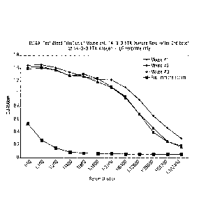
100481 Figure 1. ELISA: Test Bleed Titration of Mouse Anti-AUG1-CLDK Immune
Serum (after
2nd boost) on AUG1-CLDK-BSA Antigen (IgG response only).
100491 Figure 2. ELISA: Test Bleed Titration of Mouse Anti- AUG2-KKLE Immune
Serum (after
2nd boost) on AUG2-KKLE-BSA antigen (IgG response only).
100501 Figure 3. ELISA: Test Bleed Titration of Mouse Anti-AUG3-CKNS Immune
Serum (after 2nd
boost) on AUG3-CKNS-BSA Antigen (IgG response only).
100511 Figure 4. Sequence alignment for various 14-3-3 protein isoforms.
100521 Figure 5. Western Blot showing cross reactivity of a commercially
available 14-3-3 eta
polyclonal antibody against the seven isoforms of 14-3-3 proteins.
100531 Figure 6. Western Blot showing cell lysate-derived 14-3-3 eta protein
and human
recombinant 14-3-3 eta immunoprecipated by monoclonal antibody raised against
full length human
recombinant 14-3-3 eta.
100541 Figure 7. Western Blot showing cell lysate-derived 14-3-3 eta protein
and human
recombinant 14-3-3 eta immunoprecipated by monoclonal antibody raised against
a human 14-3-3
eta peptide fragment 142-158 SEQ ID NO:24 from a non-helical region of the
protein.
100551 Figure 8. ELISA: Test Bleed Titration of Mouse anti-14-3-3 eta Immune
Sera (after 2nd
boost) on 14-3-3 eta Antigen (IgG response only)
DETAILED DESCRIPTION
100561 An antibody, or antigen-binding fragment thereof, is said to
"specifically bind,"
"immunologically bind," and/or is "immunologically reactive" if it reacts at a
detectable level (within,
for example, an ELISA assay) with ligand, and does not react detectably with
unrelated ligands
under similar conditions.
100571 Immunological binding, as used in this context, generally refers to the
non-covalent
interactions of the type which occur between an immunoglobulin molecule and an
antigen for which
the immunoglobulin is specific. The strength, or affinity of immunological
binding interactions can be
expressed in terms of the dissociation constant (Kd) of the interaction,
wherein a smaller Kd
represents a greater affinity. Immunological binding properties can be
quantified using methods well
known in the art. For example, see Davies et al. (1990) Annual Rev. Biochem.
59:439-473.
8a
CA 02706476 2010-05-20
WO 2009/067811
PCT/CA2008/002094
10058] "Antibody" refers to a composition comprising a protein that binds
specifically to a
corresponding antigen and has a common, general structure of immunoglobulins.
The term
antibody specifically covers polyclonal antibodies, monoclonal antibodies,
dimers, multimers,
multispecific antibodies (e.g., bispecific antibodies), and antibody
fragments, so long as they
exhibit the desired biological activity. Antibodies may be murine, human,
humanized, chimeric, or
derived from other species. Typically, an antibody will comprise at least two
heavy chains and two
light chains interconnected by disulfide bonds, which when combined form a
binding domain that
interacts with an antigen. Each heavy chain is comprised of a heavy chain
variable region (VH)
and a heavy chain constant region (CH). The heavy chain constant region is
comprised of three
domains, CH1, CH2 and CH3, and may be of the mu, delta, gamma, alpha or
epsilon isotype.
Similarly, the light chain is comprised of a light chain variable region (VL)
and a light chain
constant region (CL). The light chain constant region is comprised of one
domain, CL, which may
be of the kappa or lambda isotype. The VH and VL regions can be further
subdivided into regions
of hypervariability, termed complementarity determining regions (CDR),
interspersed with regions
that are more conserved, termed framework regions (FR). Each VH and VL is
composed of three
CDRs and four FRs, arranged from amino-terminus to carboxy-terminus in the
following order:
FR1, CDR1, FR2, CDR2, FR3, CDR3, FR4. The variable regions of the heavy and
light chains
contain a binding domain that interacts with an antigen. The constant regions
of the antibodies
may mediate the binding of the immunoglobulin to host tissues or factors,
including various cells of
the immune system (e.g., effector cells) and the first component (Clq) of the
classical complement
system. The heavy chain constant region mediates binding of the immunoglobulin
to host tissue or
host factors, particularly through cellular receptors such as the Fc receptors
(e.g., FcyRI, FcyRII,
FcyRIII, etc.). As used herein, antibody also includes an antigen binding
portion of an
immunoglobulin that retains the ability to bind antigen. These include, as
examples, F(ab), a
monovalent fragment of VL CL and VH CH antibody domains; and F(ab')2 fragment,
a bivalent
fragment comprising two Fab fragments linked by a disulfide bridge at the
hinge region. The term
antibody also refers to recombinant single chain Fv fragments (scFv) and
bispecific molecules
such as, e.g., diabodies, triabodies, and tetrabodies (see, e.g., U.S. Patent
No. 5,844,094).
[0059] Antibodies may be produced and used in many forms, including antibody
complexes. As
used herein, the term "antibody complex" refers to a complex of one or more
antibodies with
another antibody or with an antibody fragment or fragments, or a complex of
two or more antibody
fragments. Antibody complexes include multimeric forms of anti-14-3-3
antibodies such as
homoconjugates and heteroconjugates as well as other cross-linked antibodies
as described
herein.
[0060] "Antigen" is to be construed broadly and refers to any molecule,
composition, or particle
that can bind specifically to an antibody. An antigen has one or more epitopes
that interact with
the antibody, although it does not necessarily induce production of that
antibody.
100611 The terms "cross-linked", "cross-linking" and grammatical equivalents
thereof, refer to the
attachment of two or more antibodies to form antibody complexes, and may also
be referred to as
9
CA 02706476 2010-05-20
WO 2009/067811
PCT/CA2008/002094
multimerizatIon. Cross-linking or multimerization includes the attachment of
two or more of the
same antibodies (e.g. homodimerization), as well as the attachment of two or
more different
antibodies (e.g. heterodimerization). Those of skill in the art will also
recognize that cross-linking
or multimerization is also referred to as forming antibody homoconjugates and
antibody
heteroconjugates. Such conjugates may involve the attachment of two or more
monoclonal
antibodies of the same clonal origin (homoconjugates) or the attachment of two
or more antibodies
of different clonal origin (also referred to as heteroconjugates or
bispecific). Antibodies may be
crosslinked by non-covalent or covalent attachment. Numerous techniques
suitable for cross-
linking will be appreciated by those of skill in the art. Non-covalent
attachment may be achieved
through the use of a secondary antibody that is specific to the primary
antibody species. For
example, a goat anti-mouse (GAM) secondary antibody may be used to cross-link
a mouse
monoclonal antibody. Covalent attachment may be achieved through the use of
chemical cross-
linkers.
[0062] "Epitope" refers to a determinant capable of specific binding to an
antibody. Epitopes are
chemical features generally present on surfaces of molecules and accessible to
interaction with an
antibody. Typical chemical features are amino acids and sugar moieties, having
three-dimensional
structural characteristics as well as chemical properties including charge,
hydrophilicity, and
lipophilicity. Conformational epitopes are distinguished from non-
conformational epitopes by loss
of reactivity with an antibody following a change in the spatial elements of
the molecule without
any change in the underlying chemical structure.
100631 "Humanized antibody" refers to an immunoglobulin molecule containing a
minimal
sequence derived from non-human immunoglobulin. Humanized antibodies include
human
immunoglobulins (recipient antibody) in which residues from a complementary
determining region
(CDR) of the recipient are replaced by residues from a CDR of a non-human
species (donor
antibody) such as mouse, rat or rabbit having the desired specificity,
affinity and capacity. In some
instances, Fv framework residues of the human immunoglobulin are replaced by
corresponding
non-human residues. Humanized antibodies may also comprise residues which are
found neither
in the recipient antibody nor in the imported CDR or framework sequences. In
general, a
humanized antibody will comprise substantially all of at least one, and
typically two, variable
domains, in which all or substantially all of the CDR regions correspond to
those of a non-human
immunoglobulin and all or substantially all of the framework (FR) regions are
those of a human
immunoglobulin consensus sequence. A humanized antibody will also encompass
immunoglobulins comprising at least a portion of an immunoglobulin constant
region (Fc),
generally that of a human immunoglobulin (Jones et al., Nature 321:522-525
(1986); Reichmann et
al, Nature 332:323-329 (1988)).
100641 "Immunogen" refers to a substance, compound, or composition which
stimulates the
production of an immune response.
100651 The term "immunoglobulin locus" refers to a genetic element or set of
linked genetic
elements that comprise information that can be used by a B cell or B cell
precursor to express an
CA 02706476 2010-05-20
WO 2009/067811
PCT/CA2008/002094
immunoglobulin polypeptide. This polypeptide can be a heavy chain polypeptide,
a light chain
polypeptide, or the fusion of a heavy and a light chain polypeptide. In the
case of an unrearranged
locus, the genetic elements are assembled by a B cell precursor to form the
gene encoding an
immunoglobulin polypeptide. In the case of a rearranged locus, a gene
encoding an
immunoglobulin polypeptide is contained within the locus.
[0066] "Isotype" refers to an antibody class defined by its heavy chain
constant region. Heavy
chains are generally classified as gamma, mu, alpha, delta, epsilon and
designated as IgG, IgM,
IgA, IgD, and IgE. Variations within each isotype are categorized into
subtypes, for example
subtypes of IgG are divided into IgG1, IgG2, IgG3, and IgG4, while IgA is
divided into IgA1 and
IgA2. The IgY isotype is specific to birds.
100671 "Monoclonal antibody" or "monoclonal antibody composition" refers to a
preparation of
antibody molecules of single molecular composition. A monoclonal antibody
composition displays
a single binding specificity and affinity for a particular epitope.
100681 The term "human monoclonal antibody" includes antibodies displaying a
single binding
specificity which have variable and/or constant regions (if present) derived
from human
immunoglobulin sequences. In one embodiment, the human monoclonal antibodies
are produced
by a hybridoma which includes a B cell obtained from a transgenic non-human
animal, e.g., a
transgenic mouse, having a geriume comprising a human heavy chain transgene
and a light chain
transgene, fused to an immortalized cell.
100691 "Single chain Fv" or "scFv" refers to an antibody comprising the VH and
VL regions of an
antibody, wherein these domains are present in a single polypeptide chain.
Generally, an scFv
further comprises a polypeptide linker between the VH and VL domains which
enables the scFv to
form the desired structure for antigen binding.
100701 "Subject" and "patient" are used interchangeably and refer to, except
where indicated,
mammals such as humans and non-human primates, as well as rabbits, rats, mice,
goats, pigs,
and other mammalian species.
100711 "Recombinant antibody" refers to all antibodies produced by recombinant
techniques.
These include antibodies obtained from an animal that is transgenic for the
immunoglobulin locus,
antibodies expressed from a recombinant expression vector, and antibodies
created, prepared,
and expressed by splicing of any immunoglobulin gene sequence to any other
nucleic acid
sequence.
100721 Anti-14-3-3 Antibodies
[0073] In one aspect, the invention provides anti-14-3-3 eta antibodies. The
anti-14-3-3 eta
antibodies of the invention are capable of (i) binding specifically to human
14-3-3 eta protein in its
native configuration, as evidenced by, for example, immunoprecipitation, and
(ii) binding
selectively to human 14-3-3 eta protein over other human 14-3-3 protein
isoforms.
11
CA 02706476 2010-05-20
WO 2009/067811
PCT/CA2008/002094
100741 By specifically binding to a human 14-3-3 eta protein in its "natural
configuration" is meant
an ability to bind to 14-3-3 protein as encountered in vivo. This may be
evidenced, for example, by
the ability of antibody to immunoprecipitate 14-3-3 eta protein from a
biological sample.
100751 By 'selectivity for said human 14-3-3 eta protein over other human 14-3-
3 protein
isoforms" is meant an ability to bind specifically to human 14-3-3 eta protein
and to bind
preferentially to 14-3-3 eta as compared to other human 14-3-3 protein
isoforms under the same
conditions. Selectivity may be evidenced, for example, using an ELISA assay,
which may be done
using, for example, supernatant from hybridoma clones. A
control (e.g., pre-immune serum) is
preferably used. A "selective" antibody is capable of recognizing 14-3-3 eta
and generating a
higher signal against 14-3-3 eta as compared to other 14-3-3 isoforms,
preferably at least a 1.5
fold, more preferably at least a 2 fold higher signal as compared to other
isoforms. In a preferred
embodiment, a selective antibody has an ability to selectively
immunoprecipitate 14-3-3 eta as
compared to other 14-3-3 isoforms.
100761 In a preferred embodiment, the anti-14-3-3 eta antibody exhibits
selectivity for said human
14-3-3 eta protein over human 14-3-3 alpha, beta, delta, epsilon, gamma, tau,
and zeta proteins.
This may be evidenced, for example, by ELISA.
10077] In a preferred embodiment, an anti-14-3-3 eta antibody of the invention
is capable of
binding to 14-3-3 eta protein that is aberrantly localized in the
extracellular synovial space in
arthritis. This may be evidenced, for example, by lmmunoprecipitation of 14-3-
3 eta protein
present in a synovial fluid sample from a patient having arthritis.
100781 In a preferred embodiment, an anti-14-3-3 eta antibody is capable of
inhibiting the
induction of MMP by 14-3-3 eta. Preferably, the MMP is selected from the group
consisting of
MMP-1, 3, 8, 9, 10, 11 and 13, with MMP-1 and MMP-3 being especially
preferred. Such
capability may be determined by in vitro assay or in vivo assay. As will be
appreciated by one of
skill in the art, the assays will be designed such that in the absence of anti-
14-3-3 eta antibody, the
presence of 14-3-3 eta will result in the induction of MMP. An ability to
reduce this induction of
MMP by 14-3-3 eta can evidence such a function-inhibiting capability for an
anti-14-3-3 antibody.
100791 14-3-3 Eta Epitopes
100801 In a preferred embodiment, an anti-14-3-3 eta antibody of the invention
does not bind to
an epitope at the N-terminus of 14-3-3 eta. By 14-3-3 eta "N-terminus" is
meant amino acids 1-12
(i.e., DREQLLQRARLA (SEQ ID NO:33).
100811 In a preferred embodiment, an anti-14-3-3 eta antibody of the invention
is capable of
binding to an epitope comprising a peptide selected from the group consisting
of 14-3-3 eta loop
= peptides, 14-3-3 eta helix peptides, and 14-3-3 eta non-helix peptides,
with eta loop peptides being
especially preferred. See Table I herein. Exemplary 14-3-3 eta loop, helix,
and non-helix peptides
are disclosed in Table 1 herein. Notably, SEQ ID NO:30 varies from
corresponding 14-3-3 eta
sequence in that a cysteine occurring in 14-3-3 eta sequence has been replaced
by serine to avoid
disulfide bond formation. In one embodiment, the invention provides antibodies
that also bind to
12
CA 02706476 2010-05-20
WO 2009/067811
PCT/CA2008/002094
the natural 14-3-3 sequence correlate of SEQ ID NO:30 comprising a cysteine.
In one
embodiment, the invention provides antibodies capable of binding to peptide
sequences that vary
from those listed in Table 1 by substitution of serine for cysteine.
10082] (i) Loop peptides
100831 In a preferred embodiment, the 14-3-3 eta loop peptide comprises an
amino acid
sequence selected from the group consisting of SEQ ID NOs:11-16. In another
embodiment, an
anti-14-3-3 eta antibody binds to a region of 14-3-3 eta that overlaps with an
amino acid sequence
corresponding to a sequence selected from the group consisting of SEQ ID
NOs:11-16.
100841 In an especially preferred embodiment, an anti-14-3-3 eta antibody of
the invention binds
to an amino acid sequence selected from the group consisting of
LDKFLIKNSNDF(SEQ ID
NO:30), KKLEKVKAYR (SEQ ID NO:31), and KNSVVEASEAAYKEA (SEQ ID NO:32).
100851 (ii) Helix peptides
100861 In a preferred embodiment, the 14-3-3 eta helix peptide comprises an
amino acid
sequence selected from the group consisting of SEQ ID NOs:1-10. In another
embodiment, an
anti-14-3-3 eta antibody binds to a region of 14-3-3 eta that overlaps with an
amino acid sequence
corresponding to a sequence selected from the group consisting of SEQ ID NOs:1-
10.
10087] (iii) Non-helix peptides
100881 In a preferred embodiment, the 14-3-3 eta non-helix peptide comprises
an amino acid
sequence selected from the group consisting of SEQ ID NOs:17-32. In another
embodiment, an
anti-14-3-3 eta antibody binds to a region of 14-3-3 eta that overlaps with an
amino acid sequence
corresponding to a sequence selected from the group consisting of SEQ ID
NOs:17-32.
100891 Monoclonal Antibodies, Hybridomas, and Methods of Making the Same
100901 In one embodiment, the present invention provides anti-14-3-3 eta
antibodies that are
monoclonal anti-14-3-3 eta antibodies. Also provided are hybridoma cell lines
capable of
producing such antibodies. Also provided are methods for producing such
hybridomas and
methods for producing such antibodies.
100911 The monoclonal anti-14-3-3 eta antibodies provided include antibodies
that bind to 14-3-3
eta loop, helix, and non-helix peptides described herein.
100921 In one aspect, the invention provides hybridomas produced by fusion of
a spleen cell
derived from a mouse immunized with an immunogen comprising a 14-3-3 eta loop,
helix, or non-
helix peptide. Also provided are monoclonal antibodies produced by such
hybridomas.
100931 The present invention further provides methods of producing such
monoclonal antibodies,
or derivatives thereof, comprising cultivating a hybridoma of the invention
under suitable
conditions, whereby a monoclonal antibody is produced, and obtaining the
antibody and/or
derivative thereof from the cell and/or from the cell culture medium.
13
CA 02706476 2010-05-20
WO 2009/067811
PCT/CA2008/002094
[0094] Antibodies can be produced readily by one skilled in the art. The
general methodology for
making monoclonal antibodies by hybridonnas is now well known to the art. See,
e.g., M. Schreier
et at., Hybridoma Techniques (Cold Spring Harbor Laboratory) 1980; Hammerling
et at.,
Monoclonal Antibodies and T-Cell Hybridomas (Elsevier Biomedical Press) 1981.
.. [0095] In some embodiments, these methods comprise cultivating a hybridoma
cell under
suitable conditions wherein the antibody is produced, and obtaining the
antibody and/or derivative
thereof from the cell and/or from the cell culture medium.
[0096] The present invention also contemplates the use of phage libraries to
pan for antibodies
capable of binding to the 14-3-3 peptides of interest described herein. For
example, see Konthur
et al., Targets, 1: 30-36, 2002.
[0097] The antibodies produced by any means can be purified by methods known
to the skilled
artisan. Purification methods include, among others, selective
precipitation, liquid
chromatography, HPLC, electrophoresis, chromatofocusing, and various affinity
techniques.
Selective precipitation may use ammonium sulfate, ethanol (Cohn
precipitation), polyethylene
glycol, or other agents available in the art. Liquid chromatography mediums,
include, among
others, ion exchange medium DEAE, polyaspartate, hydroxylapatite, size
exclusion (e.g., those
based on crosslinked agarose, acrylamide, dextran, etc.), hydrophobic matrices
(e.g., Blue
Sepharose). Affinity techniques typically rely on proteins that interact with
the immunoglobulin Fc
domain. Protein A from Staphylococcus aureas can be used to purify antibodies
that are based on
human y1, y2, or y4 heavy chains (Lindmark et al., J. Immunol. Meth. 62:1-13
(1983)). Protein G
from C and G streptococci is useful for all mouse isotypes and for human .y3
(Guss et al., EMBO
J. 5:15671575 (1986)). Protein L, a Peptostreptococcus magnus cell-wall
protein that binds
immunoglobulins (Ig) through k light-chain interactions (BD
Bioscience/ClonTech. Palo Alto, CA),
is useful for affinity purification of Ig subclasses IgM, IgA, IgD, IgG, IgE
and IgY. Recombinant
forms of these proteins are also commercially available. If the antibody
contains metal binding
residues, such as phage display antibodies constructed to contain histidine
tags, metal affinity
chromatography may be used. When sufficient amounts of specific cell
populations are available,
antigen affinity matrices may be made with the cells to provide an affinity
method for purifying the
antibodies.
[00981 In a preferred embodiment, isolation involves affinity chromatography
using 14-3-3 eta or
fragment thereof.
100991 The present invention provides the antibodies described herein, as well
as corresponding
antibody fragments and antigen-binding portions. All are encompassed by the
term anti-14-3-3 eta
antibody. The terms "antibody fragment" or "antigen-binding portion" of an
antibody (or simply
.. "antibody portion") of the present invention, as used herein, refers to one
or more fragments of an
antibody that retain the ability to specifically bind to an antigen. It has
been shown that the
antigen-binding function of an antibody can be performed by fragments of a
full-length antibody.
Examples of binding fragments encompassed within the term "antibody fragment"
or "antigen-
binding portion" of an antibody include (i) a Fab fragment, a monovalent
fragment consisting of the
14
CA 02706476 2010-05-20
WO 2009/067811
PCT/CA2008/002094
VL, VH, CL and CH1 domains; (ii) a F(ab')2 fragment, a bivalent fragment
comprising two Fab
fragments linked by a disulfide bridge at the hinge region; (iii) a Fd
fragment consisting of the VH
and CH1 domains; (iv) a Fv fragment consisting of the VL and VH domains of a
single arm of an
antibody, (v) a dAb fragment (e.g., Ward et al., (1989) Nature 341:544-546),
which consists of a
VH domain; and (vi) an isolated complementarity determining region (CDR), and
(vii) bispecific
single chain Fv dimers (e.g., PCT/US92/09965). Furthermore, although the two
domains of the Fv
fragment, VL and VH, are coded for by separate genes, they can be joined,
using recombinant
methods, by a synthetic linker that enables them to be made as a single
protein chain in which the
VL and VH regions pair to form monovalent molecules (known as single chain Fv
(scFv); see e.g.,
Bird et al. (1988) Science 242:423-426; and Huston et al. (1988) Proc. Natl.
Acad. Sci. USA
85:5879-5883). Such single chain antibodies are also intended to be
encompassed within the
term "antigen-binding portion" of an antibody. These antibody fragments are
obtained using
conventional techniques known to those with skill in the art, and the
fragments are screened for
utility in the same manner as are intact antibodies. The antibody fragments
may be modified. For
example, the molecules may be stabilized by the incorporation of disulphide
bridges linking the VH
and VL domains (Reiter et al., 1996, Nature Biotech. 14:1239-1245).
1001001 lmmunoglobulin molecules can be cleaved into fragments. The antigen
binding region of
the molecule can be divided into either F(ab')2 or Fab fragments. The F(ab')2
fragment is divalent
and is useful when the Fc region is either undesirable or not a required
feature. The Fab fragment
is univalent and is useful when an antibody has a very high avidity for its
antigen. Eliminating the
Fc region from the antibody decreases non-specific binding between the Fc
region and Fc receptor
bearing cells. To generate Fab or F(ab')2 fragments, the antibodies are
digested with an enzyme.
Proteases that cleave at the hinge region of an immunoglobulin molecule
preserve the disulfide
bond(s) linking the Fab domains such that they remain together following
cleavage. A suitable
protease for this purpose is pepsin. For producing Fab fragments, proteases
are chosen such that
cleavage occurs above the hinge region containing the disulfide bonds that
join the heavy chains
but which leaves intact the disulfide bond linking the heavy and light chain.
A suitable protease for
making Fab fragments is papain. The fragments are purified by the methods
described above,
with the exception of affinity techniques requiring the intact Fc region
(e.g., Protein A affinity
chromatography).
1001011 Antibody fragments can be produced by limited proteolysis of
antibodies and are called
proteolytic antibody fragments. These include, but are not limited to, the
following: F(ab')2
fragments, Fab' fragments, Fab'-SH fragments, and Fab fragments. ''F(ab')2
fragments" are
released from an antibody by limited exposure of the antibody to a proteolytic
enzyme, e.g., pepsin
or ficin. An F(ab')2 fragment comprises two "arms," each of which comprises a
variable region that
is directed to and specifically binds a common antigen. The two Fab' molecules
are joined by
interchain disulfide bonds in the hinge regions of the heavy chains; the Fab'
molecules may be
directed toward the same (bivalent) or different (bispecific) epitopes. "Fab'
fragments" contain a
single antigen-binding domain comprising an Fab and an additional portion of
the heavy chain
through the hinge region. "Fab'-SH fragments" are typically produced from
F(ab')2 fragments,
CA 02706476 2010-05-20
WO 2009/067811
PCT/CA2008/002094
which are held together by disulfide bond(s) between the H chains in an
F(ab')2 fragment.
Treatment with a mild reducing agent such as, by way of non-limiting example,
beta-
mercaptoethylamine, breaks the disulfide bond(s), and two Fab' fragments are
released from one
F(ab.)2 fragment. Fab-SH fragments are monovalent and monospecific. "Fab
fragments" (i.e., an
antibody fragment that contains the antigen-binding domain and comprises a
light chain and part
of a heavy chain bridged by a disulfide bond) may be produced by papain
digestion of intact
antibodies. A convenient method is to use papain immobilized on a resin so
that the enzyme can
be easily removed and the digestion terminated. Fab fragments do not have the
disulfide bond(s)
between the H chains present in an F(ab')2 fragment.
[00102] "Single-chain antibodies" are one type of antibody fragment. The term
single chain
antibody is often abbreviated as "scFv" or "sFv." These antibody fragments are
produced using
recombinant DNA technology. A single-chain antibody consists of a polypeptide
chain that
comprises both a VH and a VL domains which interact to form an antigen-binding
site. The VH and
VL domains are usually linked by a peptide of 10 to 25 amino acid residues.
[00103] The term "single-chain antibody" further includes but is not limited
to a disulfide-linked Fv
(dsFv) in which two single-chain antibodies (each of which may be directed to
a different epitope)
are linked together by a disulfide bond; a bispecific sFv in which two
discrete scFvs of different
specificity are connected with a peptide linker; a diabody (a dinnerized sFv
formed when the
domain of a first sFv assembles with the VL domain of a second sFv and the VL
domain of the first
sFv assembles with the VH domain of the second sFv; the two antigen-binding
regions of the
diabody may be directed towards the same or different epitopes); and a
triabody (a trimerized sFv,
formed in a manner similar to a diabody, but in which three antigen-binding
domains are created in
a single complex; the three antigen binding domains may be directed towards
the same or different
epitopes).
[00104] "Complementary determining region peptides" or "CDR peptides" are
another form of an
antibody fragment. In one embodiment, the invention provides such CDR
peptides. In a preferred
embodiment, such CDR peptides function as 14-3-3 eta antagonists. A CDR
peptide (also known
as "minimal recognition unit") is a peptide corresponding to a single
complementarity-determining
region (CDR), and can be prepared by constructing genes encoding the CDR of an
antibody of
interest. Such genes are prepared, for example, by using the polymerase chain
reaction to
synthesize the variable region from RNA of antibody-producing cells. See, for
example, Larrick et
al., Methods: A Companion to Methods in Enzymology 2:106, 1991.
[00105] In "cysteine-modified antibodies," a cysteine amino acid is inserted
or substituted on the
surface of antibody by genetic manipulation and used to conjugate the antibody
to another
molecule via, e.g., a disulfide bridge. Cysteine substitutions or insertions
for antibodies have been
described (see U.S. Pat. No. 5,219,996). Methods for introducing Cys residues
into the constant
region of the IgG antibodies for use in site-specific conjugation of
antibodies are described by
Stimmel et al. (J. Biol. Chem 275.330445-30450, 2000).
16
CA 02706476 2010-05-20
WO 2009/067811
PCT/CA2008/002094
1001061 The present disclosure further provides humanized and non-humanized
antibodies.
Humanized forms of non-human (e.g., mouse) antibodies are chimeric antibodies
that contain
minimal sequence derived from non-human immunoglobulin. Generally, humanized
antibodies are
non-human antibodies that have had the variable-domain framework regions
swapped for
sequences found in human antibodies. The humanized antibodies may be human
immunoglobulins (recipient antibody) in which residues from a hypervarable
region of the recipient
are replaced by residues from a hypervariable region of a non-human species
(donor antibody)
such as mouse, rat, rabbit or nonhuman primate having the desired specificity,
affinity, and
capacity. In some instances, framework region (FR) residues of the human
immunoglobulin are
replaced by corresponding non-human residues. Furthermore, humanized
antibodies may
comprise residues that are not found in the recipient antibody or in the donor
antibody. These
modifications are made to further refine antibody performance. In general, the
humanized
antibody will comprise substantially all of at least one, and typically two,
variable domains, in which
all or substantially all of the hypervariable loops correspond to those of a
non-human
immunoglobulin and all or substantially all of the FRs are those of a human
immunoglobulin
sequence. The humanized antibody optionally also will comprise at least a
portion of an
immunoglobulin constant region (Fc), typically that of a human immunoglobulin.
1001071 Generally, in a humanized antibody, the entire antibody, except the
CDRs, is encoded by
a polynucleotide of human origin or is identical to such an antibody except
within its CDRs. The
CDRs, some or all of which are encoded by nucleic acids originating in a non-
human organism,
are grafted into the beta-sheet framework of a human antibody variable region
to create an
antibody, the specificity of which is determined by the engrafted CDRs. The
creation of such
antibodies is described in, e.g., WO 92/11018, Jones, 1986, Nature 321:522-
525, Verhoeyen et
al., 1988, Science 239:1534-1536. Humanized antibodies can also be generated
using mice with
a genetically engineered immune system. e.g., Roque et al., 2004, Biotechnol.
Frog. 20:639-654.
1091081 It can be desirable to modify the antibodies of the invention with
respect to effector
function. For example, cysteine residue(s) can be introduced into the Fc
region, thereby allowing
interchain disulfide bond formation in this region. Homodimeric antibodies can
also be prepared
using heterobifunctional cross-linkers, e.g., Wolff et at. Cancer Research,
53:2560-2565 (1993).
Alternatively, an antibody can be engineered that has dual Fc regions. See for
example
Stevenson et al., Anti-Cancer Drug Design, 3:219-230 (1989).
1001091 Modified Antibodies
1001101 In one embodiment, the invention provides anti-14-3-3 eta antibodies
that are modified
antibodies. Modified antibodies include recombinant antibodies as described
herein.
1001111 Numerous types of modified or recombinant antibodies will be
appreciated by those of skill
in the art. Suitable types of modified or recombinant antibodies include,
without limitation,
engineered monoclonal antibodies (e.g. chimeric monoclonal antibodies,
humanized monoclonal
antibodies), domain antibodies (e.g. Fab, Fv, VH, scFV, and dsFy fragments),
multivalent or
17
CA 02706476 2010-05-20
WO 2009/067811
PCT/CA2008/002094
multispecific antibodies (e.g. diabodies, minibodies, miniantibodies, (scFV)2,
tribodies, and
tetrabodies), and antibody conjugates as described herein.
1001121 In one aspect, the present invention provides anti-14-3-3 eta
antibodies which are domain
antibodies. 'Domain antibodies" are functional binding domains of antibodies,
corresponding to
the variable regions of either the heavy (VH) or light (VL) chains of human
antibodies. Domain
antibodies may have a molecular weight of approximately 13 kDa, or less than
one-tenth the size
of a full antibody. They are well expressed in a variety of hosts including
bacterial, yeast, and
mammalian cell systems. In addition, domain antibodies are highly stable and
retain activity even
after being subjected to harsh conditions, such as freeze-drying or heat
denaturation. See, for
example, US Patent 6,291,158; 6,582,915; 6,593,081; 6,172,197; US Serial No.
2004/0110941;
European Patent 0368684; US Patent 6,696,245, W004/058821, W004/003019 and
W003/002609. In one embodiment, the domain antibody of the present invention
is a single
domain. Single domain antibodies may be prepared, for example, as described in
U.S. Patent Na.
6,248,516.
1001131 In another aspect, the present invention includes multi-specific
antibodies. Multi-specific
antibodies include bispecific, trispecific, etc. antibodies. Bispecific
antibodies can be produced via
recombinant means, for example by using leucine zipper moieties (i.e., from
the Fos and Jun
proteins, which preferentially form heterodimers; e.g., Kostelny et al., 1992,
J. lmmnol. 148:1547)
or other lock and key interactive domain structures, for example as described
in U.S. Pat. No.
5,582,996. Additional useful techniques include those described in U.S. Pat.
No. 5,959,083; and
U.S. Pat. No. 5,807,706.
[00114] Bispecific antibodies are also sometimes referred to as "diabodies."
These are antibodies
that bind to two (or more) different antigens. Also known in the art are
triabodies (a trimerized sFy,
formed in a manner similar to a diabody, but in which three antigen-binding
domains are created in
a single complex; the three antigen binding domains may be directed towards
the same or different
epitopes) or tetrabodies (four antigen-binding domains created in a single
complex where the four
antigen binding domains may be directed towards the same or different
epitopes). Dia-, tria- and
tetrabodies can be manufactured in a variety of ways known in the art (e.g.,
Holliger and Winter,
1993, Current Opinion Biotechnol. 4:446-449), e.g., prepared chemically or
from hybrid
hybridomas. In addition, such antibodies and fragments thereof may be
constructed by gene
fusion (e.g., Tomlinson et. al., 2000, Methods Enzymol. 326:461-479;
W094/13804; Holliger et al.,
1993, Proc. Natl. Acad. Sci. U.S.A. 90:6444-6448).
1001151 In another embodiment, the present invention provides minibodies,
which are minimized
antibody-like proteins that include a scFV joined to a CH3 domain, that are
derived from an anti-
14-3-3 eta antibody. Minibodies can be made as described in the art (e.g., Hu
et al., 1996, Cancer
Res. 56:3055-3061).
1001161 In another embodiment, the present invention provides 14-3-3 eta
binding domain-
immunoglobulin fusion proteins. In one embodiment, the fusion protein may
include a 14-3-3 eta
binding domain polypeptide fused to an immunoglobulin hinge region
polypeptide, which is fused
18
CA 02706476 2010-05-20
WO 2009/067811
PCT/CA2008/002094
to an immunoglobulin heavy chain CH2 constant region polypeptide fused to an
immunoglobulin
heavy chain CH3 constant region polypeptide. Under the present invention, 14-3-
3 antibody fusion
proteins can be made by methods appreciated by those of skill in the art (See
for example
published U.S. Patent Application Nos. 20050238646, 20050202534, 20050202028,
2005020023,
2005020212, 200501866216, 20050180970, and 20050175614).
[001171 In another embodiment, the present invention provides a heavy-chain
protein derived from
a an anti-14-3-3 eta antibody.
Naturally-occurring heavy chain antibodies (e.g. camelidae
antibodies having no light chains) have been utilized to develop antibody-
derived therapeutic
proteins that typically retain the structure and functional properties of
naturally-occurring heavy-
chain antibodies. They are known in the art as Nanobodies. Heavy chain
proteins derived from an
anti-14-3-3 eta heavy chain antibody may be made by methods appreciated by
those of skill in the
art (See for example published U.S. Patent Application Nos. 20060246477,
20060211088,
20060149041, 20060115470, and 20050214857). Further, regarding the production
of heavy
chain-only antibodies in light chain-deficient mice, see for example Zou et
al., JEM, 204:3271-
3283, 2007.
1001181 In one embodiment, the invention provides modified anti-14-3-3 eta
antibodies that are
human antibodies. In one embodiment, fully human 14-3-3 antibodies are
provided. "Fully human
antibody" or "complete human antibody" refers to a human antibody having only
the gene
sequence of an antibody derived from a human chromosome. The anti-14-3-3
complete human
antibody can be obtained by a method using a human antibody-producing mouse
having a human
chromosome fragment containing the genes for a heavy chain and light chain of
a human antibody
[see for example Tom izuka, K. et al., Nature Genetics, 16, p.133-143, 1997;
Kuroiwa, Y. et al.,
Nuc. Acids Res., 26, p.3447-3448, 1998; Yoshida, H. et al., Animal Cell
Technology: Basic and
Applied Aspects vol. 10, p.69-73 (Kitagawa, Y., Matuda, T. and lijima, S.
eds.), Kluwer Academic
Publishers, 1999; Tomizuka, K. et al., Proc. Natl. Acad. Sci. USA, 97, 722-
727, 2000] or obtained
by a method for obtaining a human antibody derived from a phage display
selected from a human
antibody library (see for example Wormstone, I. M. et al., Investigative
Ophthalmology & Visual
Science. 43(7), p.2301-8, 2002; Carmen, S. et at., Briefings in Functional
Genomics and
Proteomics, 1 (2), p.189-203, 2002; Siriwardena, D. et al., Ophthalmology,
109(3), p.427-431,
2002).
1001191 In one aspect, the present invention provides a 14-3-3 antibody that
is an antibody analog,
sometimes referred to as "synthetic antibodies." For example, alternative
protein scaffolds or
artificial scaffolds with grafted CDRs may be used. Such scaffolds include,
but are not limited to,
synthetic scaffolds consisting, for example, of biocompatible polymers. See,
for example,
Korndorfer et al., 2003, Proteins: Structure, Function, and Bioinformatics,
Volume 53, Issue 1:121-
129. Roque et al., 2004, Biotechnol. Prog. 20:639-654. In addition, peptide
antibody mimetics
("PAMs") can be used, as well as antibody mimetics utilizing fibronectin
components as a scaffold.
1001201 In one embodiment, the present invention provides cross-linked
antibodies that include
two or more antibodies described herein attached to each other to form
antibody complexes.
19
CA 02706476 2010-05-20
WO 2009/067811
PCT/CA2008/002094
Cross-linked antibodies are also referred to as antibody multimers,
homoconjugates, and
heteroconjugates.
[00121] In some embodiments, the antibody complexes provided herein include
multimeric forms
of anti-14-3-3 antibodies. For example, antibody complexes of the invention
may take the form of
antibody dimers, trimers, or higher-order multimers of monomeric
immunoglobulin molecules.
Crosslinking of antibodies can be done through various methods know in the
art. For example,
crosslinking of antibodies may be accomplished through natural aggregation of
antibodies, through
chemical or recombinant linking techniques or other methods known in the art.
For example,
purified antibody preparations can spontaneously form protein aggregates
containing antibody
homodimers, and other higher-order antibody multimers.
[00122] In one embodiment, the present invention provides homodimerized
antibodies that
specifically bind to 14-3-3 eta.
[00123] Antibodies can be cross-linked or dimerized through linkage techniques
known in the art.
Non-covalent methods of attachment may be utilized. In a specific embodiment,
crosslinking of
antibodies can be achieved through the use of a secondary crosslinker
antibody. The crosslinker
antibody can be derived from a different animal compared to the antibody of
interest. For
example, a goat anti-mouse antibody (Fab specific) may be added to a mouse
monoclonal
antibody to form a heterodimer. This bivalent crosslinker antibody recognizes
the Fab or Fe region
of the two antibodies of interest forming a homodimer.
[00124] In one embodiment of the present invention, an antibody that
specifically binds to 14-3-3
antigen is cross-linked using a goat anti-mouse antibody (GAM). In another
embodiment, the
GAM crosslinker recognizes the Fab or Fc region of two antibodies, each of
which specifically
binds 14-3-3 eta.
1001251 Methods for covalent or chemical attachment of antibodies may also be
utilized. Chemical
crosslinkers can be homo or heterobifunctional and will covalently bind with
two antibodies forming
a homodimer. Cross-linking agents are well known in the art; for example, homo-
or hetero-
bifunctional linkers as are well known (see the 2006 Pierce Chemical Company
Crosslinking
Reagents Technical Handbook; Hermanson, G.T., Bioconjugate Techniques,
Academic Press,
San Diego, CA (1996); Aslam M. and Dent AH., Bioconjugation: protein coupling
techniques for
the biomedical sciences, Houndsmills, England: Macmillan Publishers (1999);
Pierce: Applications
Handbook & Catalog, Perbio Science, Ermbodegem, Belgium (2003-2004);
Haughland, R.P.,
Handbook of Fluorescent Probes and Research Chemicals Eugene, 9th Ed.,
Molecular Probes,
OR (2003); and U.S. Patent No. 5,747,641) Those of skill in the art will
appreciate the suitability of
various functional groups on the amino acid(s) of an antibody for
modification, including cross-
linking. Suitable examples of chemical crosslinkers used for antibody
crosslinking include, but not
limited to, SMCC [succinimidyl 4-(maleimidomethyl)cyclohexane-1-carboxylate],
SATA [N-
succinimidyl S-acethylthio-acetate], hemi-succinate esters of N-
hydroxysuccinimide; sulfo-N-
hydroxy-succinim ide; hydroxybenzotriazole, and p-nitrophenol;
dicyclohexylcarbodiimide (DCC), 1-
(3-dimethylaminopropy1)-3-ethylcarbodiimide (ECD), and
1-(3-d im ethylaminopropyI)-3-
CA 02706476 ,2016-07-08
CA 2706476
ethylcarbodiimide methiodide (EDCI) (see, e.g., U.S. Patent No. 4,526,714).
Other linking reagents
include glutathione, 3-(diethoxyphosphoryloxy)-1,2,3- benzotriazin-4(3H)-one
(DEPBT), oniunn salt-
based coupling reagents, polyoxyethylene-based heterobifunctional cross-
linking reagents, and
other reagents (Haitao, et at., Organ Lett 1:91-94 (1999); Albericio et at., J
Organic Chemistry
63:9678-9683 (1998); Arpicco et at., Bioconjugate Chem. 8:327-337 (1997);
Frisch et at.,
Bioconjugate Chem. 7:180-186 (1996); Deguchi et al., Bioconjugate Chem. 10:32-
37 (1998); Beyer
et at., J. Med. Chem. 41:2701-2708 (1998); Drouillat etal., J. Pharm. Sci.
87:25-30 (1998); Trimble
et at., Bioconjugate Chem. 8:416-423 (1997)). An exemplary protocols for the
formation of antibody
homodimers is given in U.S. Patent Publication 20060062786. Techniques for
conjugating
therapeutic compounds to antibodies are also described in Arnon et at.,
"Monoclonal Antibodies for
Immunotargeting of Drugs in Cancers Therapy," in Monoclonal Antibodies and
Cancer Therapy,
Reisfeld et at., ed., pp243-256, Alan R. Liss, Inc. (1985); Thorpe, et at.
"The Preparation and
Cytotoxic Properties of Antibody Toxin Conjugates," lmmunol. Rev. 62:119-58
(1982); and Pietersz,
G.A., "The linkage of cytotoxic drugs to monoclonal antibodies for the
treatment of cancer,"
Bioconjugate Chemistry 1(2):89-95 (1990).
1001261 In addition, the antibody-antibody conjugates of this invention can be
covalently bound to
each other by techniques known in the art such as the use of the
heterobifunctional cross-linking
reagents, GMBS (maleimidobutryloxy succinimide), and SPDP (N-succinimidyl 3-(2-
pyridyldithio)propionate) [see, e.g., Hardy, "Purification And Coupling Of
Fluorescent Proteins For
Use In Flow Cytometry", Handbook Of Experimental Immunology, Volume 1,
Immunochemistry,
Weir et al. (eds.), pp. 31.4-31.12 4th Ed., (1986), and Ledbetter et al. U.S.
Patent No. 6,010,902].
1001271 In addition, antibodies may be linked via a thioether cross-link as
described in U.S. Patent
Publication 20060216284, U.S. Patent No. 6,368,596. As will be appreciated by
those skilled in the
art, antibodies can be crosslinked at the Fab region. In some embodiments, it
is desirable that the
chemical crosslinker not interact with the antigen-binding region of the
antibody as this may affect
antibody function.
1001281 Conjugated Antibodies
The anti-14-3-3 eta antibodies disclosed herein include antibodies conjugated
to inorganic or
organic compounds, including, by way of example and not limitation, other
proteins, nucleic acids,
carbohydrates, steroids, and lipids (see for example Green, et at., Cancer
Treatment Reviews,
26:269-286 (2000). The compound may be bioactive. Bioactive refers to a
compound having a
physiological effect on the cell as compared to a cell not exposed to the
compound. A physiological
effect is a change in a biological process, including, by way of example and
not limitation, DNA
replication and repair, recombination, transcription, translation, secretion,
membrane turnover, cell
adhesion, signal transduction, cell death, and the like. A
bioactive compound includes
pharmaceutical compounds. In one
embodiment, an anti-14-3-3 eta antibody
21
CA 02706476 2010-05-20
WO 2009/067811
PCT/CA2008/002094
is conjugated to a 14-3-3 antagonist peptide, preferably R-18, preferably via
a linker. Regarding
R18, see, for example, (Wang et al. 1999¨ REF 35)
1001301 Pharmaceutical Compositions, Administration, and Dosages
[00131] The anti-14-3-3 eta antibodies of the invention can be incorporated
into pharmaceutical
compositions suitable for administration to a subject. Typically, the
pharmaceutical composition
comprises an anti-14-3-3 eta antibody of the invention and a pharmaceutically
acceptable carrier.
As used herein, "pharmaceutically acceptable carrier" includes any and all
solvents, dispersion
media, coatings, antibacterial and antifungal agents, isotonic and absorption
delaying agents, and
the like that are physiologically compatible. Examples of pharmaceutically
acceptable carriers
include one or more of water, saline, phosphate buffered saline, dextrose,
glycerol, ethanol and
the like, as well as combinations thereof. In many cases, it will be
preferable to include isotonic
agents, for example, sugars, polyalcohols such as mannitol, sorbitol, or
sodium chloride in the
composition. Pharmaceutically acceptable substances such as wetting or minor
amounts of
auxiliary substances such as wetting or emulsifying agents, preservatives or
buffers, which
enhance the shelf life or effectiveness of the anti-14-3-3 eta antibody.
1001321 In a preferred embodiment, the anti-14-3-3 eta antibodies are targeted
to 14-3-3 eta
protein that is localized extracellularly. Accordingly, such therapeutic
compositions are formulated
and administration is such that the anti-14-3-3 eta antibody so delivered is
available to engage
extracellular 14-3-3 eta protein.
100133] The compositions of this invention may be in a variety of forms. These
include, for
example, liquid, semi-solid and solid dosage forms, such as liquid solutions
(e.g., injectable and
infusible solutions), dispersions or suspensions, tablets, pills, powders,
liposomes and
suppositories. The preferred form depends on the intended mode of
administration and
therapeutic application. Typical preferred compositions are in the form of
injectable or infusible
solutions, such as compositions similar to those used for passive immunization
of humans with
other antibodies. The preferred mode of administration is parenteral (e.g.,
intravenous,
subcutaneous, intraperitoneal, intramuscular, with intracapsular being
especially preferred). In one
embodiment, the anti-14-3-3 eta antibody is administered by intravenous
infusion or injection. In
another preferred embodiment, the anti-14-3-3 eta antibody is administered by
intramuscular or
subcutaneous injection. In a preferred embodiment, direct injection into the
synovium is done.
1001341 Therapeutic compositions typically must be sterile and stable under
the conditions of
manufacture and storage. The composition can be formulated as a solution,
microemulsion,
dispersion, liposome, or other ordered structure suitable to high drug
concentration. Sterile
injectable solutions can be prepared by incorporating the active compound in
the required amount
.. in an appropriate solvent with one or a combination of ingredients
enumerated above, as required,
followed by filtered sterilization. Generally, dispersions are prepared by
incorporating the active
compound into a sterile vehicle that contains a basic dispersion medium and
the required other
ingredients from those enumerated above. In the case of sterile powders for
the preparation of
sterile injectable solutions, the preferred methods of preparation are vacuum
drying and freeze-
22
CA 02706476 2010-05-20
WO 2009/067811
PCT/CA2008/002094
drying that yields a powder of the active ingredient plus any additional
desired ingredient from a
previously sterile-filtered solution thereof. The proper fluidity of a
solution can be maintained, for
example, by the use of a coating such as lecithin, by the maintenance of the
required particle size
in the case of dispersion and by the use of surfactants. Prolonged absorption
of injectable
compositions can be brought about by including in the composition an agent
that delays
absorption, for example, monostearate salts and gelatin.
1001351 The anti-14-3-3 eta antibodies of the present invention can be
administered by a variety of
methods known in the art, including intravenous injection or infusion. Direct
administration to the
synovium is one preferred route of administration. As will be appreciated by
the skilled artisan, the
route and/or mode of administration will vary depending upon the desired
results. In certain
embodiments, the active compound may be prepared with a carrier that will
protect the compound
against rapid release, such as a controlled release formulation, including
implants, transdermal
patches, and microencapsulated delivery systems. Biodegradable, biocompatible
polymers can be
used, such as ethylene vinyl acetate, polyanhydrides, polyglycolic acid,
collagen, polyorthoesters,
and polylactic acid. Many methods for the preparation of such formulations are
patented or
generally known to those skilled in the art. See, e.g., Sustained and
Controlled Release Drug
Delivery Systems, J. R. Robinson, ed., Marcel Dekker, Inc., New York, 1978.
Representative
formulation technology is taught in, inter alia, Remington: The Science and
Practice of Pharmacy,
19th Ed., Mack Publishing Co., Easton, PA (1995) and Handbook of
Pharmaceutical Excipients,
3rd Ed, Kibbe, A.H. ed., Washington DC, American Pharmaceutical Association
(2000)
1001361 In certain embodiments, an anti-14-3-3 eta antibody of the invention
may be orally
administered, for example, with an inert diluent or an assimilable edible
carrier. The compound
(and other ingredients, if desired) may also be enclosed in a hard or soft
shell gelatin capsule,
compressed into tablets, or incorporated directly into the subject's diet. For
oral therapeutic
administration, the compounds may be incorporated with excipients and used in
the form of
ingestible tablets, buccal tablets, troches, capsules, elixirs, suspensions,
syrups, wafers, and the
like. To administer a compound of the invention by other than parenteral
administration, it may be
necessary to coat the compound with, or co-administer the compound with, a
material to prevent
its inactivation.
1001371 Supplementary active compounds can also be incorporated into the
compositions. In
certain embodiments, an anti-14-3-3 eta antibody of the invention is
coformulated with and/or
coadministered with one or more additional therapeutic agents. For example, a
DMARD or
DMOAD or another antibody. Such combination therapies may advantageously
utilize lower
dosages of the administered therapeutic agents, thus avoiding possible
toxicities or complications
associated with the various monotherapies.
1001381 The pharmaceutical compositions of the invention may include a
"therapeutically effective
amount" or a "prophylactically effective amount" of an antibody of the
invention. A "therapeutically
effective amount" refers to an amount effective, at dosages and for periods of
time necessary, to
achieve the desired therapeutic result. A therapeutically effective amount of
the anti-14-3-3 eta
23
CA 02706476 2010-05-20
WO 2009/067811
PCT/CA2008/002094
antibody may vary according to factors such as the disease state, age, sex,
and weight of the
individual, and the ability of the anti-14-3-3 eta antibody to elicit a
desired response in the
individual. A therapeutically effective amount is also one in which any toxic
or detrimental effects
of the antibody are outweighed by the therapeutically beneficial effects. A
"prophylactically
effective amount" refers to an amount effective, at dosages and for periods of
time necessary, to
achieve the desired prophylactic result. Typically, since a prophylactic dose
is used in subjects
prior to or at an earlier stage of disease, the prophylactically effective
amount will be less than the
therapeutically effective amount.
[00139] Dosage regimens may be adjusted to provide the optimum desired
response (e.g., a
therapeutic or prophylactic response). For example, a single bolus may be
administered, several
divided doses may be administered over time or the dose may be proportionally
reduced or
increased as indicated by the exigencies of the therapeutic situation. It is
especially advantageous
to formulate parenteral compositions in dosage unit form for ease of
administration and uniformity
of dosage. Dosage unit form as used herein refers to physically discrete units
suited as unitary
dosages for the mammalian subjects to be treated; each unit containing a
predetermined quantity
of active compound calculated to produce the desired therapeutic effect in
association with the
required pharmaceutical carrier. The specification for the dosage unit forms
of the invention are
dictated by and directly dependent on (a) the unique characteristics of the
active compound and
the particular therapeutic or prophylactic effect to be achieved, and (b) the
limitations inherent in
the art of compounding such an active compound for the treatment of
sensitivity in individuals.
[00140] An exemplary, non-limiting range for a therapeutically or
prophylactically effective amount
of an antibody of the invention is 0.1-20 mg/kg, more preferably 1-10 mg/kg.
It is to be noted that
dosage values may vary with the type and severity of the condition to be
alleviated. It is to be
further understood that for any particular subject, specific dosage regimens
should be adjusted
over time according to the individual need and the professional judgment of
the person
administering or supervising the administration of the compositions, and that
dosage ranges set
forth herein are exemplary only and are not intended to limit the scope or
practice of the claimed
corn position.
[00141] The pharmaceutical compositions described herein may be presented in
unit-dose or
multi-dose containers, such as sealed ampoules or vials. Such containers are
typically sealed in
such a way to preserve the sterility and stability of the formulation until
use. In general,
formulations may be stored as suspensions, solutions or emulsions in oily or
aqueous vehicles, as
indicated above. Alternatively, a pharmaceutical composition may be stored in
a freeze-dried
condition requiring only the addition of a sterile liquid carrier immediately
prior to use.
.. 1001421 Therapeutic Use of Anti-14-3-3 Eta Antibodies
[00143] By "treatment" herein is meant therapeutic or prophylactic treatment,
or a suppressive
measure for the disease, disorder or undesirable condition.
Treatment encompasses
administration of the subject anti-14-3-3 eta antibodies in an appropriate
form prior to the onset of
disease symptoms and/or after clinical manifestations, or other
manifestations, of the disease to
24
CA 02706476 2010-05-20
WO 2009/067811
PCT/CA2008/002094
reduce disease severity, halt disease progression, or eliminate the disease.
Prevention of the
disease includes prolonging or delaying the onset of symptoms of the disorder
or disease,
preferably in a subject with increased susceptibility to the disease.
1001441 In one aspect, the invention provides methods of treating diseases
that involve 14-3-3 eta.
The methods comprise administering a therapeutically effective amount of an
anti-14-3-3 eta
antibody of the invention to a patient. In some embodiments, the methods
comprise combination
treatments.
1001451 In one embodiment, the invention provides methods of treating
arthritis, including methods
of treating ankylosing spondylitis, Behcet's Disease, diffuse idiopathic
skeletal hyperostosis
(DISH), Ehlers-Danlos Syndrome (EDS), Felty's Syndrome, fibromyalgia, gout,
infectious arthritis,
juvenile arthritis, lupus, mixed connective tissue disease (MCTD),
osteoarthritis, Paget's Disease,
polymyalgia rheumatica, polymyositis and dermatornyositis, pseudogout,
psoriatic arthritis,
Raynaud's Phenomenon, reactive arthritis, rheumatoid arthritis, scleroderma,
SjOgren's Syndrome,
Still's Disease, and Wegener's granulomatosis.
1001461 In one embodiment, the method involves a combination treatment,
wherein at least one
other therapeutic agent is administered in addition to one or more anti-14-3-3
eta antibodies of the
invention. In a preferred embodiment, the therapeutic agent is selected from
the group consisting
of disease-modifying antirheurnatic drugs (DMARDs), disease modifying
osteoarthritis drugs
(DMOADs; for example, see Loeser, Reumatologia, 21:104-106, 2005), anti-TNFa
antibody, anti-
IL-1 antibody, anti-CD4 antibody, anti-CTLA4 antibody, anti-CD20 antibody,
anti-IL-6 antibody,
leflunomide, sulfasalazine, and methotrexate.
[00147] Diagnostic, Prognostic and Theragnostic Methods, and Treatment
Monitoring
100148] In one aspect, the invention provides methods for diagnosing diseases
and conditions that
involve 14-3-3 eta. The methods comprise using an anti-14-3-3 eta antibody of
the invention to
detect an alteration in 14-3-3 eta protein, e.g., a change in expression,
localization, function, etc.
In one embodiment, detection involves immunoprecipitation with an anti-14-3-3
eta antibody of the
invention. In one embodiment, detection involves the use of ELISA employing an
anti-14-3-3 eta
antibody of the invention. In one embodiment, detection involves Western
blotting using an anti-
14-3-3 eta antibody of the invention. In one embodiment, detection involves
the use of an anti-14-
3-3 eta antibody of the invention in immunohistochemistry. In one embodiment,
detection involves
the use of an anti-14-3-3 eta antibody of the invention in immunofluorescence.
In one
embodiment, detection involves the use of an anti-14-3-3 eta antibody of the
invention in FACS
analysis. In one embodiment, detection involves the use of an anti-14-3-3 eta
antibody of the
invention in radioimmunoassay. In one embodiment, detection involves the use
of an anti-14-3-3
eta antibody of the invention in a strip test. In one embodiment, detection
involves the use of an
anti-14-3-3 eta antibody of the invention in a point of care test. In one
embodiment, detection of
14-3-3 eta is combined with detection of another marker of the condition
(e.g., MMP, anti-COP,
anti-RF and / or CRP for arthritis).
CA 02706476 2010-05-20
WO 2009/067811
PCT/CA2008/002094
[00149] In one embodiment, the invention provides methods for diagnosing
inflammatory
conditions. In a preferred embodiment, methods for diagnosing arthritis are
provided. Included
are methods for diagnosing a disease selected from the group consisting of
ankylosing spondylitis,
Behcet's Disease, diffuse idiopathic skeletal hyperostosis (DISH), Ehlers-
Danlos Syndrome (EDS),
Felty's Syndrome, fibromyalgia, gout, infectious arthritis, juvenile
arthritis, lupus, mixed connective
tissue disease (MCTD), osteoarthritis, Paget's Disease, polymyalgia
rheumatica, polymyositis and
dermatomyositis, pseudogout, psoriatic arthritis, Raynaud's Phenomenon,
reactive arthritis,
rheumatoid arthritis, scleroderma, Sjogren's Syndrome, Still's Disease, and
Wegener's
granulomatosis.
[001501 In general, arthritis may be detected in a patient based on the
presence 14-3-3 eta in the
synovial fluid, plasma, or serum of a patient. In other words, extracellular
14-3-3 eta protein may
be used as a marker to indicate arthritis.
100151] In addition, the presence of 14-3-3 eta, or the relative levels of
isoforms of 14-3-3 proteins
including 14-3-3 eta, as determined through the use of an anti-14-3-3 eta
antibody of the invention
and other anti-14-3-3 antibodies may be a prognostic indicator of early-stage
arthritis, before it
progresses to a debilitating form. An advantage of early prognosis or
diagnosis is earlier
implementation of a treatment regimen.
[00152] The presence or relative levels of 14-3-3 eta may correlate with the
presence or relative
levels of other proteins in the patient sample, for example matrix
metalloproteinases (MMPs), such
as MMP-1 or MMP-3. At least 25 different MMPs have been identified. Detection
of 14-3-3 eta in
combination with at least one MMP in a patient sample may be used to diagnose
arthritis.
Additionally, the presence or relative levels of 14-3-3 eta in combination
with at least one MMP in a
patient sample may be used as a prognostic indicator of early-stage arthritis,
before the arthritis
progresses to a debilitating form.
[00153] In one embodiment, the methods involve detecting 14-3-3 eta protein in
the synovial fluid,
plasma, or serum of a patient. In one embodiment, detection is done by
immunoprecipitation of
14-3-3 eta protein from synovial fluid, plasma, or serum using an anti-14-3-3
eta antibody of the
invention. In one embodiment, detection involves the use of ELISA employing an
anti-14-3-3 eta
antibody of the invention. In one embodiment, detection involves Western
blotting of a sample
.. comprising synovial fluid, plasma, or serum from a patient using an anti-14-
3-3 eta antibody of the
invention. In one embodiment, detection involves the use of radioimmunoassay.
In one
embodiment, detection involves the use of a strip test. In one embodiment,
detection involves the
use of a point of care test. In one embodiment, detection of 14-3-3 eta is
combined with detection
of another marker of arthritis (e.g., MMP, anti-COP, anti-RF and /or CRP).
1001541 In one embodiment the invention provides methods for diagnosing
neurological conditions.
In a preferred embodiment, methods for diagnosing a disease selected from the
group consisting
of bacterial meningitis and Creutzfeldt Jakob disease are provided. In one
embodiment, the
presence of 14-3-3 eta in cerebrospinal fluid is detected.
26
CA 02706476 2010-05-20
WO 2009/067811
PCT/CA2008/002094
[00155] In one aspect, the invention provides methods for determining the
response potential of a
patient to treatment directed at an inflammatory condition. In a preferred
embodiment, methods for
determining the response potential of a patient to treatment directed at
arthritis are provided.
Included are methods for determining the response potential to treatment of a
disease selected
from the group consisting of ankylosing spondylitis, Behget's Disease, diffuse
idiopathic skeletal
hyperostosis (DISH), Ehlers-Danlos Syndrome (EDS), Felty's Syndrome,
fibromyalgia, gout,
infectious arthritis, juvenile arthritis, lupus, mixed connective tissue
disease (MCTD), osteoarthritis,
Paget's Disease, polymyalgia rheumatica, polynnyositis and dermatomyositis,
pseudogout,
psoriatic arthritis, Raynaud's Phenomenon, reactive arthritis, rheumatoid
arthritis, scleroderma,
Sjogren's Syndrome, Still's Disease, and Wegener's granulomatosis.
1001561 In one embodiment, the methods involve determining the level of 14-3-3
eta in a patient
sample using an anti-14-3-3 eta antibody of the invention. In a preferred
embodiment, the level of
14-3-3 eta in the patient sample is compared to that of samples from subjects
whose ability to
respond to treatment is known. A relatively high level of 14-3-3 eta in a
first patient sample as
compared to a sample from a non-inflammatory subject and/or a sample from
another
inflammatory patient may indicate the first patient is a preferred candidate
for treatment with anti-
14-3-3 eta antibody or an alternate DMARD therapy such as anti-TNF.
Conversely, a relatively
low level of 14-3-3 eta in a first patient sample as compared to a sample from
another
inflammatory patient may indicate the first patient is not a preferred
candidate for treatment with
anti-14-3-3 eta antibody or an alternate DMARD therapy such as anti-TNF,
especially if the level is
closer to that of a sample from a non-inflammatory subject.
1001571 In one aspect, the invention provides methods for distinguishing
between subtypes of
inflammatory disorders. In a preferred embodiment methods for distinguishing
between subtypes
of arthritis are provided. In one embodiment, the methods involve determining
the level of 14-3-3
eta in a patient sample using an anti-14-3-3 eta antibody of the invention. In
a preferred
embodiment, the level of 14-3-3 eta in the patient is compared to that of
samples from subjects
whose subtype of inflammatory disorder or prognosis is known.
[00158] In one aspect, the invention provides prophylactic methods for
preventing the development
of conditions involving 14-3-3 eta.
[00159] In one embodiment, the invention provides prophylactic methods for
preventing the
development of an inflammatory condition in a subject at risk of developing an
inflammatory
condition. In a preferred embodiment, prophylactic methods for preventing
arthritis in a subject at
risk of developing arthritis are provided. Included are prophylactic methods
for preventing a
disease selected from the group consisting of ankylosing spondylitis, Behget's
Disease, diffuse
idiopathic skeletal hyperostosis (DISH), Ehlers-Danlos Syndrome (EDS), Felty's
Syndrome,
fibromyalgia, gout, infectious arthritis, juvenile arthritis, lupus, mixed
connective tissue disease
(MCTD), osteoarthritis, Paget's Disease, polymyalgia rheumatica, polymyositis
and
dermatomyositis, pseudogout, psoriatic arthritis, Raynaud's Phenomenon,
reactive arthritis,
rheumatoid arthritis, scleroderma, Sjogren's Syndrome, Still's Disease, and
Wegener's
27
CA 02706476 2010-05-20
WO 2009/067811
PCT/CA2008/002094
granulomatosis. The methods comprise administering to the subject an anti-14-3-
3 eta antibody of
the invention. In one embodiment the anti-14-3-3 eta antibody is administered
as a component of
a combination therapy described herein.
[001601 In one aspect, the invention provides methods for monitoring treatment
of an inflammatory
condition. In a preferred embodiment, methods for monitoring the treatment of
arthritis are
provided. Included are methods for monitoring the treatment of a disease
selected from the group
consisting of ankylosing spondylitis, Behget's Disease, diffuse idiopathic
skeletal hyperostosis
(DISH), Ehlers-Danlos Syndrome (EDS), Felty's Syndrome, fibromyalgia, gout,
infectious arthritis,
juvenile arthritis, lupus, mixed connective tissue disease (MCTD),
osteoarthritis, Paget's Disease,
polymyalgia rheumatica, polymyositis and dermatomyositis, pseudogout,
psoriatic arthritis,
Raynaud's Phenomenon, reactive arthritis, rheumatoid arthritis, scleroderma,
Sjogren's Syndrome,
Still's Disease, and Wegener's granulomatosis.
[00161] In one embodiment, the methods involve determining the level of 14-3-3
eta in patient
samples using an anti-14-3-3 eta antibody of the invention and monitoring the
level of 14-3-3 eta in
a patient undergoing treatment.
[00162] In one aspect the invention provides kits for detecting the presence
of 14-3-3 eta and
optionally other markers, e.g., MMPs, in a patient sample, the kit being
useful for providing a
diagnostic or prognostic result suitable for diagnosing or differentiating
various types of diseases
involving 14-3-3 eta. A kit comprises an anti-14-3-3 eta antibody of the
invention. Such a kit may
further include detection reagents specific for particular MMPs that are
markers of arthritis. The kit
may further include other reagents necessary for the detection of 14-3-3 eta
immunologically, such
as labeled secondary antibodies, chromogenic or fluorogenic reagents,
polymerization agents
and/or instructions for using the kit for diagnostic or prognostic purposes.
[00163] Regarding diagnostic methods, see also WO 2007/128132 filed 9 May
2007.
1001641 There are a variety of assay formats known to those of ordinary skill
in the art for using an
antibody to detect protein markers in a sample. See, e.g., Harlow and Lane,
Antibodies: A
Laboratory Manual, Cold Spring Harbor Laboratory, 1988. In general, the
presence or absence of
arthritis or other condition involving 14-3-3 eta, or patient prognosis, may
be determined by (a)
contacting a biological sample obtained from a patient with an anti-14-3-3 eta
antibody of the
invention; (b) detecting in the sample a level of 14-3-3 eta that binds to the
antibody; and (c)
comparing the level of polypeptide with a predetermined cut-off value (i.e.,
control).
1001651 In a preferred embodiment, the assay involves the use of an anti-14-3-
3 eta antibody of
the invention immobilized on a solid support to bind to and remove the 14-3-3
eta protein from the
remainder of the sample. The bound 14-3-3 eta protein may then be detected
using a detection
reagent that contains a reporter group and specifically binds to the
antibody/protein complex.
Such detection reagents may comprise, for example, a binding agent that
specifically binds to the
14-3-3 protein. Alternatively, a competitive assay may be utilized, in which a
14-3-3 eta protein is
labeled with a reporter group and allowed to bind to the immobilized antibody
after incubation of
28
CA 02706476 2010-05-20
WO 2009/067811
PCT/CA2008/002094
the antibody with the sample. The extent to which components of the sample
inhibit the binding of
the labeled 14-3-3 eta protein to the antibody is indicative of the reactivity
of the sample with the
immobilized antibody. Suitable proteins for use within such assays include
full length 14-3-3 eta
proteins and polypeptide portions thereof to which the antibody binds.
100166] The solid support may be any material known to those of ordinary skill
in the art. For
example, the solid support may be a test well in a microtiter plate or a
nitrocellulose or other
suitable membrane. Alternatively, the support may be a bead or disc, such as
glass, fiberglass,
latex or a plastic material such as polystyrene or polyvinylchloride. The
support may also be a
magnetic particle or a fiber optic sensor, such as those disclosed, for
example, in U.S. Pat. No.
5,359,681. The antibody may be immobilized on the solid support using a
variety of techniques
known to those of skill in the art, which are amply described in the patent
and scientific literature.
In the context of the present invention, the term "immobilization" refers to
both noncovalent
association, such as adsorption, and covalent attachment (which may be a
direct linkage between
the antibody and functional groups on the support or may be a linkage by way
of a cross-linking
agent). Immobilization by adsorption to a well in a microtiter plate or to a
membrane is preferred.
In such cases, adsorption may be achieved by contacting the antibody, in a
suitable buffer, with
the solid support for a suitable amount of time. The contact time varies with
temperature, but is
typically between about 1 hour and about 1 day. In one embodiment, a
microtitre plate coated with
streptavidin is used in conjunction with a biotinylated antibody.
[00167] Covalent attachment of antibody to a solid support may generally be
achieved by first
reacting the support with a bifunctional reagent that will react with both the
support and the
antibody.
[00168] In certain embodiments, the assay is a two-antibody sandwich assay.
This assay may be
performed by first contacting an antibody that has been immobilized on a solid
support, commonly
the well of a microtiter plate, with the sample, such that 14-3-3 eta proteins
within the sample are
allowed to bind to the immobilized antibody. Unbound sample is then removed
from the
immobilized protein-antibody complexes and a detection reagent (preferably a
second antibody
capable of binding to a different site on the polypeptide) containing a
reporter group is added. The
amount of detection reagent that remains bound to the solid support is then
determined using a
method appropriate for the specific reporter group.
[00169] The immobilized and detection antibodies are preferably different.
In a preferred
embodiment, the immobilized antibody is an anti-14-3-3 eta antibody of the
invention, and the
detection antibody is another anti-14-3-3 eta antibody of the invention or
another anti-14-3-3
antibody capable of binding to 14-3-3 eta. In one embodiment, the detection
antibody is a pan 14-
3-3 antibody.
1001701 In another embodiment, the detection antibody is an anti-14-3-3 eta
antibody of the
invention, and the immobilized antibody is another anti-14-3-3 eta antibody of
the invention or
another anti-14-3-3 antibody capable of binding to 14-3-3 eta. In one
embodiment, the
immobilized antibody is a pan 14-3-3 antibody.
29
CA 02706476 2010-05-20
WO 2009/067811
PCT/CA2008/002094
1001711 The methods comprise use of an anti-14-3-3 eta antibody of the
invention. As an
alternative to the second antibody, another ligand that binds to 14-3-3 eta
may be used in
conjunction with the anti-14-3-3 eta antibody of the invention. An example of
such a ligand is R18.
(Wang et al. 1999 ¨ REF 35)
1001721 Once the antibody is immobilized on the support as described above,
the remaining
protein binding sites on the support are typically blocked. Any suitable
blocking agent known to
those of ordinary skill in the art, such as bovine serum albumin or skim milk
powder. The
immobilized antibody is then incubated with the sample, and 14-3-3 eta protein
is allowed to bind
to the antibody. The sample may be diluted with a suitable diluent, such as
phosphate-buffered
saline (PBS) prior to incubation. In general, an appropriate contact time
(i.e., incubation time) is a
period of time that is sufficient to detect the presence of 14-3-3 eta protein
within a sample
obtained from an individual with arthritis or other condition involving 14-3-3
eta. Preferably, the
contact time is sufficient to achieve a level of binding that is at least
about 95% of that achieved at
equilibrium between bound and unbound 14-3-3 eta protein. Those of ordinary
skill in the art will
recognize that the time necessary to achieve equilibrium may be readily
determined by assaying
the level of binding that occurs over a period of time. At room temperature,
an incubation time of
about 30 minutes is generally sufficient.
[00173] Unbound sample may then he removed by washing the solid support with
an appropriate
buffer, such as PBS containing 0.1% Tween 20 Tm. The second antibody, which
contains a reporter
group, may then be added to the solid support. Reporter groups appropriate to
the present
methods are well known in the art.
[00174] The detection reagent is then incubated with the immobilized antibody-
protein complex for
an amount of time sufficient to detect the bound 14-3-3 eta protein. An
appropriate amount of time
may generally be determined by assaying the level of binding that occurs over
a period of time.
Unbound detection reagent is then removed and bound detection reagent is
detected using the
reporter group. The method employed for detecting the reporter group depends
upon the nature of
the reporter group. For radioactive groups, scintillation counting or
autoradiographic methods are
generally appropriate. Spectroscopic methods may be used to detect dyes,
luminescent groups
and fluorescent groups. Biotin may be detected using avidin, coupled to a
different reporter group
(commonly a radioactive or fluorescent group or an enzyme). Enzyme reporter
groups may
generally be detected by the addition of substrate (generally for a specific
period of time), followed
by spectroscopic or other analysis of the reaction products.
1001751 To determine the presence or absence of arthritis, or other condition
involving 14-3-3 eta,
the signal detected from the reporter group that remains bound to the solid
support is generally
compared to a signal that corresponds to a predetermined cut-off value
(control). In one preferred
embodiment, the cut-off value is the average mean signal obtained when the
immobilized antibody
is incubated with samples from patients without arthritis, or other condition
involving 14-3-3 eta. In
general, a sample generating a signal that is three standard deviations above
the predetermined
cut-off value is considered positive for arthritis, or other condition
involving 14-3-3 eta. In an
CA 02706476 2010-05-20
WO 2009/067811
PCT/CA2008/002094
alternate preferred embodiment, the cut-off value may be determined using a
Receiver Operator
Curve, for example see the method of Sackett et al., Clinical Epidemiology: A
Basic Science for
Clinical Medicine, Little Brown and Co., 1985, p. 106-7. Briefly, in this
embodiment, the cut-off
value may be determined from a plot of pairs of true positive rates (i.e.,
sensitivity) and false
positive rates (100%-specificity) that correspond to each possible cut-off
value for the diagnostic
test result. The cut-off value on the plot that is the closest to the upper
left-hand corner (i.e., the
value that encloses the largest area) is the most accurate cut-off value, and
a sample generating a
signal that is higher than the cut-off value determined by this method may be
considered positive.
Alternatively, the cut-off value may be shifted to the left along the plot, to
minimize the false
positive rate, or to the right, to minimize the false negative rate. In
general, a sample generating a
signal that is higher than the cut-off value determined by this method is
considered positive for
arthritis, or other condition involving 14-3-3 eta.
[001761 In one embodiment, the assay is provided as a point of care assay. For
example, in a
related embodiment, the assay is performed in a flow-through or strip test
format, wherein the
antibody is immobilized on a membrane, such as nitrocellulose. In the flow-
through test, 14-3-3
proteins within the sample bind to the immobilized antibody as the sample
contacts the membrane.
A second, labeled binding agent then binds to the binding agent-polypeptide
complex as a solution
containing the second binding agent contacts the membrane. The detection of
bound second
binding agent may then be performed as described above. In the strip test
format, one end of the
membrane to which antibody is bound is immersed in a solution containing the
sample. The
sample migrates along the membrane through a region containing second 14-3-3
eta binding
agent and to the area of immobilized antibody. Concentration of second binding
agent at the area
of immobilized antibody indicates the presence of arthritis, or other
condition involving 14-3-3 eta,
or patient prognosis, etc. Typically, the concentration of second binding
agent at that site
generates a pattern, such as a line, that can be read visually. The absence of
such a pattern
indicates a negative result. In general, the amount of binding agent
immobilized on the membrane
is selected to generate a visually discernible pattern when the biological
sample contains a level of
polypeptide that would be sufficient to generate a positive signal in the two-
antibody sandwich
assay, in the format discussed above. Preferred binding agents for use in such
assays are
antibodies and antigen-binding fragments thereof. Such tests can typically be
performed with a
very small amount of biological sample and at the point of care.
[001771 In a preferred embodiment, the immobilized antibody is an anti-14-3-3
eta antibody of the
invention. The second binding agent is another 14-3-3 eta ligand that may or
may not bind
selectively to 14-3-3 eta protein.
[001781 In another embodiment, the second binding agent is an anti-14-3-3 eta
antibody of the
invention, most preferably an antigen-binding fragment thereof, and the
immobilized antibody is an
anti-14-3-3 antibody capable of binding to 14-3-3- eta protein. The antibody
may or may not bind
selectively to 14-3-3 eta protein.
31
CA 02706476 2010-05-20
WO 2009/067811
PCT/CA2008/002094
[001791 Of course, numerous other assay protocols exist that are suitable for
use with the anti-14-
3-3 antibodies of the present invention. The above descriptions are intended
to be exemplary
only.
1001801 To improve sensitivity, multiple markers may be assayed within a given
sample. In
particular, one or more other markers of arthritis, or other condition
involving 14-3-3 eta, or
prognostic indicators, etc., may be assayed in combination with 14-3-3
protein. These other
markers may be proteins or nucleic acids. In a preferred embodiment, wherein
the disease is
arthritis, one or more of the other markers are MMP proteins or nucleic acids
or other factors which
are commonly used as indicators for arthritis, e.g., anti-CCP, anti-RF, CRP,
etc. Methods for
isolating and assaying nucleic acids based on reference sequences are well
known in the art.
[00181] Combination assays may be done concurrently or sequentially. The
selection of markers
may be based on routine experiments to determine combinations that results in
optimal sensitivity.
1001821 The present invention further provides kits for use within any of the
above diagnostic
methods. Such kits typically comprise two or more components necessary for
performing a
diagnostic assay. Components may be compounds, reagents, containers and/or
equipment. For
example, one container within a kit may contain a monoclonal anti-14-3-3 eta
antibody of the
invention. Such antibodies may be provided attached to a support material, as
described above.
One or more additional containers may enclose elements, such as reagents or
buffers, to be used
in the assay. Such kits may also contain a detection reagent as described
above that contains a
reporter group suitable for direct or indirect detection of antibody binding.
1001831 A kit may also include reagents for detecting additional markers of
arthritis, including
particular mRNAs encoding particular MMPs.
EXPERIMENTAL
1001841 Table 1: 14-3-3 Eta epitopes
SEQ ID NO:1 93-107 helix LETVCNDVLSLLDKF
SEQ ID NO:2 191-199 helix EQACLLAKQ
SEQ ID NO:3 144-155 helix NSVVEASEAAYK
SEQ ID NO:4 144-152 helix NSVVEASEA
SEQ ID NO:5 147-155 helix VEASEAAYK
SEQ ID NO:6 163-170 helix EQMQPTHP
SEQ ID NO:7 168-177 helix THPIRLGLAL
SEQ ID NO:8 82-92 helix VKAYTEKIEKE
SEQ ID NO:9 68-79 helix QKTMADGNEKKL
SEQ ID NO:10 138-146 helix ASGEKKNSV
32
CA 02706476 2010-05-20
WO 2009/067811
PCT/CA2008/002094
SEQ ID NO:11 69-77 loop KTMADGNEK
SEQ ID NO:12 32-40 loop ELNEPLSNE
SEQ ID NO:13 103-117 loop LLDKFLIKNCNDFQY
SEQ ID NO:14 130-143 loop YYRYLAEVASGEKK
SEQ ID NO:15 184-194 loop YEIQNAPEQAC
SEQ ID NO:16 206-218 loop AELDTLNEDSYKD
SEQ ID NO:17 44-57 non-helix LLSVAYKNVVGARR
SEQ ID NO:18 15-23 non-helix EQAERYDDM
SEQ ID NO:19 130-138 non-helix YYRYLAEVA
SEQ ID NO:20 118-125 non-helix ESKVFYLK
SEQ ID NO:21 210-218 non-helix TLNEDSYKD
SEQ ID NO:22 77-84 non-helix KKLEKVKA
SEQ ID NO:23 76-86 non-helix EKKLRKVKAYR
SEQ ID NO:24 142-158 non-helix KKNSVVEASEAAYKEAF
SEQ ID NO:25 105-120 non-helix DKFLIKNCNDFQYESK
SEQ ID NO:26 237-246 non-helix QQDEEAGEGN
SEQ ID NO:27 75-82 non-helix NEKKLEKVK
SEQ ID NO28 104-116 non-helix LDKFLIKNCNDFQ
SEQ ID NO:29 141-146 non-helix EKKNSV
SEQ ID NO:30 104-115 non-helix LDKFLIKNS*NDF
SEQ ID NO:31 , 77-86 non-helix KKLEKVKAYR
SEQ ID NO:32 143-157 non-helix KNSVVEASEAAYKEA
SEQ ID NO:33 1-12 non-helix DREQLLQRARLA
*The internal cysteine amino acid was replaced by the amino acid serine to
prevent formation of
disulfide bonds.
1001851 Table 2. Protein Sequence of recombinant human 14-3-3 eta (SEQ ID
NO:63)
SEQ ID NO:63 MGDREQLLQR ARLAEQAERY DDMASAMKAV TELNEPLSNE 40
DRNLLSVAYK NVVGARRSSW RVISSIEQKT MADGNEKKLE 80
KVKAYREKIE KELETVCNDV LSLLDKFLIK NCNDFQYESK 120
VFYLKMKGDY YRYLAEVASG EKKNSVVEAS EAAYKEAFEI 160
SKEQMQPTHP IRLGLALNFS VFYYEIQNAP EQACLLAKQA 200
33
CA 02706476 2010-05-20
WO 2009/067811
PCT/CA2008/002094
FDDAIAELDT LNEDSYKDST LIMQLLRDNL TLWTSDQQDE 240
EAGE GN
[001861 In sequences comprising a cysteine residue, in one embodiment, the
cysteine residue is
replaced by a serine residue to avoid the formation of disulfide bonds. The
cysteine may be an
internal cysteine residue or a terminal cysteine residue.
1001871 Peptide epitopes may be modified for various purposes, including
conjugation to an
additional moiety, e.g., conjugation to a moiety to produce an immunogen
comprising the epitope.
As will be appreciated, cysteine may be placed appropriately for conjugation
to carrier and to
provide for exposure of the area that is desired to be exposed for purposes of
making antibody. In
case of KKLE the cysteine was added on to the C-terminal end in order to
expose the other side.
The carrier used may be quite large and may mask the first few amino acids.
100188] Example 1: 14-3-3 eta lmmunogen Sequences and anti-14-3-3 eta
antibodies
1001891 To prepare monospecific anti-14-3-3 eta antibodies, various peptides,
8 to 15 amino acids
in length, were selected based on our own criteria. These peptides, as well as
full-length
recombinant native (untagged) 14-3-3 eta were used as immunogens in the
production of
monoclonal antibodies. A protein sequence alignment for the 7 isoforms of 14-3-
3 is shown in
Figure 4.
1001901 Immunogen #1: C-LDKFLIKNSNDF (Amino Acid Sequence 104¨ 115; "AUG1-
CLDK"). A
peptide corresponding to a segment of human 14-3-3 eta residues 104-115 was
modified by
addition of an N-terminal cysteine moiety for conjugation to carrier, and
replacement of internal
cysteine-112 moiety to avoid formation of internal disulphide bonds.
1001911 lmmunogen #2: KKLEKVKAYR-C (Amino Acid Sequence 77 ¨ 86; "AUG2-KKLE").
A
peptide corresponding to a segment of human 14-3-3 eta residues 77-86 was
modified by addition
of a C-terminal cysteine moiety for conjugation to carrier.
100192] lmmunogen #3: C-KNSVVEASEAAYKEA (Amino Acid Sequence 143 ¨ 157; "AUG3-
CKNS"). A peptide corresponding to a segment of human 14-3-3 eta residues 143-
157 was
modified by addition of an N-terminal cysteine moiety for conjugation to
carrier.
100193] lmmunogen #4: Full length human recombinant 14-3-3 eta (SEQ ID NO:
63), Protein
Accession #: NP 003396.
1001941 Immunization
1001951 Groups of 4 female BALB/c mice were initially immunized by
intraperitoneal injections
using 50 ug of antigen (Immunogen #1, #2, #3 or #4) per mouse in Complete
Freund's Adjuvant.
Four subsequent boosts were administered as above, spaced at 3 week intervals,
with antigen in
Incomplete Freund's Adjuvant. When the serum titre had risen more than 10-fold
from the pre-
immune serum sample, as determined by ELISA, the 2 highest responders in each
group were
each boosted intravenously with 10 ug of antigen in 100 ul of sterile PBS pH
7.4. The titrations of
34
CA 02706476 2016-07-08
CA 2706476
serum samples from the immunized mice taken after the second boost are shown
in Figure 1
(Immunogen #1; CLDK), Figure 2 (Immunogen #2; KKLE) Figure 3 (Immunogen #3;
CKNS), and
Figure 8 (Immunogen #4).
1001961 Fusion Method
1001971 Three days after the final boost, the donor mice were sacrificed and
the spleen cells were
harvested and pooled. Fusion of the splenocytes with SP2/0 BALB/c parental
myeloma cells was
performed as previously described (Kohler et al., infra), except that one-step
selection and cloning of
the hybridomas was performed. Clones were picked 11 days post fusion and
resuspended in wells
of 96-well tissue culture plates in: 200 pl of D-MEM medium containing 1%
hypoxanthine/thymidine,
20% fetal bovine serum, 2 mM GlutaMax ITM, 1 mM Sodium Pyruvate, 50 pg/ml
Gentamycin, 1%
OPI and 0.6 ng/ml IL-6. After 4 days, the supernatants were screened by ELISA
for antibody activity
on plates coated with 1 ug/well of purified antigen.
1001981 Procedure for Revival of Slow Growing Hybridoma Clones
1001991 Hybridoma cell lines that were growing slowly or looked unhealthy
could usually be rescued
by the addition of a rich growth media containing: D-MEM medium with 1%
hypoxanthine/thymidine,
20% fetal bovine serum, 2 mM GlutaMax I, TM 1 MM Sodium Pyruvate, 50 pg/ml
Gentamycin, 1%
OPI, 20% conditioned EL-4 tissue culture supernatant and 0.6 ng/ml IL-6. EL-4
is a murine
thymoma cell line, which when stimulated with phorbal 12-myristate 12- acetate
(PMA, from Sigma,
cat # P-8139) causes the cells to secrete interleukin 2 (IL-2), a B cell
differentiating factor (EL-
BCDF-nak), and two B cell growth factors (BSF-pl and EL-BCGF-swa) and other
additional
lymphokines, which greatly enhance lymphocyte growth and differentiation. See
G. Kohler, and C.
Milstein, Preparation of monoclonal antibodies, Nature 25 (1975) 256-259; Ma,
M., S. Wu, M.
Howard and A. Borkovec. 1984. Enhanced production of mouse hybridomas to
picomoles of antigen
using EL-4 conditioned media with an in vitro immunization protocol. In Vitro
20:739.
1002001 After 30 days of stability testing, a total of 100 viable clones were
obtained that secreted IgG
capable of recognizing recombinant 14-3-3 eta. For the purposes of identifying
lead clones to
pursue, the 100 viable clones were screened using a series of methods
including: immunoblotting
(dot blot), a trapping assay and a custom capture (sandwich) ELISA. All 100
clones were also
tested for cross-reactivity using the custom capture (sandwich) ELISA with the
other six 14-3-3
isoforms.
1002011 Example 2: Testing the cross-reactivity of tissue culture (TC)
supernatants from hybridoma
clones using biotinylated 14-3-3 isoforms as bait in a capture ELISA
1002021 We have utilized a custom capture ELISA using the seven 14-3-3
isoforms as "bait" to
determine whether any of the hybridoma clones that we have produced cross-
react or recognize any
of the six isoforms other than 14-3-3 Eta (1). As is evidenced by the
representative data presented
in Table 4, four of the selected hybridoma clones (AUG3-CKNS-2D5, AUG3-CKNS-
7F8, AUG3-
CKNS-7H8, AUG4-ETA-8F10) bind to and recognize 14-3-3 Eta at two serial
dilutions, but do not
CA 02706476 2016-07-08
CA 2706476
bind with or cross-react with any of the other 14-3-3 isoforms, even at the
lower dilution tested. This
data clearly demonstrates that these clones are highly specific for 14-3-3 Eta
(ii). By contrast, one
clone, AUG3-CKNS-4F10, binds with or cross-reacts with three other 14-3-3
isoforms, mainly 14-3-3
gamma, beta and zeta respectively. Taken together, these data indicate that
our capture ELISA
represents an effective method for identifying hybridoma clones which are
highly specific for the 14-
3-3 Eta (ll) isoform.
1002031 The Custom Capture ELISA experiment in Table 4 was carried out as
follows. ELISA plates
were coated with neat overgrown TC supernatant at 1004/well and incubated
overnight at 4 C.
Biotin-labeled 14-3-3 (corresponding to all seven isoforms) was titrated from
1/500 to 1/16000
overtop and incubated for 1 hour at room temperature. Plates were then blocked
with 3% skim milk
powder in PBS (pH 7.4) at 1004/well and incubated for 1 hour at room
temperature. 1/8000
Streptavidin-HRPO was diluted in PBS-Tween, added at 1004/well and incubated
for 1 hour at
37 C with shaking. TMB buffer was added at 504 per well and incubated in the
dark at room
temperature. Reactions were stopped with 504 1M HCl per well after 10 minutes
and read at
OD450nm.
36
1002041 Table 4a: Testing Cross-reactivity by ELISA
Testing the cross-reactivity of tissue culture (TO) supernatants from
hybridoma clones using biotinylated 14-3-3 isoforms as bait
in a capture ELISA (measured at OD450nm)
14-3-3 Isoform: Gamma (y) Beta (13) Sigma (a) Theta/Tau (0)
Zeta () Epsilon (E) Eta (ii)
dilution 1
1
1:1500 1:3000 1:1500 1 1:3000 1:1500 [1:3000 1:1500 1 1:3000 1:1500 1 1:3000
1:1500 1 1:3000 1:1500 1:3000
TC supernatant
AUG3-CKNS- 0.076 0.073 0.085 0.075 0.084 0.076 0.101 0.082 0.097 0.076 0.074
0.064 0.351 0.263 n
2D5
o
*AUG3-CKNS-
Iv
0.084 0.076 0.096 0.085 0.125 0.102 0.185 0.153 0.167 0.122 0.139 0.101 0.114
0.09
4F10
--.1
0
01
AUG3-CKNS- . .
A.
0.072 0.067 0.078 0.076 0.083 0.076 0116 0.104 0093 0.084 0.089 0.076 0.946
0.741 ...1
7F8
a)
AUG3-CKNS-
- Iv
0.07 0.066 0.072 0.063 0.087 0.078 0.098 0.083 0.089 0.08 0.074 0.064 0.774
0.608 o
oi 7H8
1-,
-4
o,
AUG4-ETA-
I
0.072 0.069 0.073 0.069 0.092 0.084 0.109 0.097 0.099 0.082 0.099 0.09 0.169
0.131 o
8F10
I
o
pre-immune
co
0.097 0.074 0.093 0.081 0.136 0.113 0.193 0.158 0.152 0.119 0.144 0.115 0.152
0.11
serum (1:250)
*control antibody
[002051 Table 4b: Testing Cross-reactivity by ELISA (background (pre-immune
serum) values subtracted out)
Testing the cross-reactivity of tissue culture (IC) supernatants from
hybridoma clones using biotinylated 14-3-3 isoforms as bait in
a capture ELISA (measured at OD450nm)
14-3-3 lsoform: Gamma (y) Beta (p) Sigma (a) Theta/Tau (8)
Zeta () Epsilon (c) Eta (n)
dilution
1:1500 1:3000 1:1500 1:3000 1:1500 1:3000 1:1500 1:3000 1:1500 1:3000 1:1500
1:3000 1:1500 1:3000
TC supernatant
0
AUG3-CKNS-
-0.021 -0.001 -0.008 -0.006 -0.052 -0.037 -0.092 -0.076 -0.055 -0.043 -0.070 -
0.051 0.199 0.153
2D5
0
01
*AUG3-CKNS-
-0.013 0.002 0.003 0.004 -0.011 -0.011 -0.008 -0.005 0.015 0.003 -0.005 -0.014
-0.038 -0.020
0.) 4F10
co
'
AUG3-CKNS-
-0.025 -0.007 -0.015 -0.005 -0.053 -0.037 -0.077 -0.054 -0.059 -0.035 -0.055 -
0.039 0.794 0.631
7F8
AUG3-CKNS-
0
-0.027 -0.008 -0.021 -0.018 -0.049 -0.035 -0.095 -0.075 -0.063 -0.039 -0.070 -
0.051 0.622 0.498
7H8
0
03
AUG4-ETA-
-0.025 -0.005 -0.020 -0.012 -0.044 -0.029 -0.084 -0.061 -0.053 -0.037 -0.045 -
0.025 0.017 0.021
8F10
*control antibody
CA 027.06476 ,2016-07-08
CA 2706476
1002061 Example 3: Cross reactivity of commercially available anti-14-3-3 eta
polyclonal antibody
1002071 Commercially available anti-14-3-3 rabbit polyclonal antibody raised
against a 12 amino acid
peptide (Ac-DREOLLORARLA-NH2) epitope from the N-terminus of 14-3-3 eta
(Biomol International
LP, Cat. SA476-0100 was used to evaluate the specificity of the antibody.
Briefly, 1pg human
recombinant 14-3-3 eta, gamma, sigma, apha/beta, epsilon, theta or zeta were
resolved by SDS-
PAGE and probed with the anti-14-3-3 eta antibody.
1002081 In marked contrast to the results obtained with the antibodies of the
present invention, the
results in Figure 5 show that this commercially available antibody against 14-
3-3 eta cross reacted
with other 14-3-3 isoforms, primarily gamma. Lane 1: Molecular Weight
Standards; Lane 2:
recombinant 14-3-3 eta; Lane 3: recombinant 143-3 gamma; Lane 4: recombinant
14-3-3 sigma;
Lane 5: recombinant 14-3-3 alpha/beta; Lane 6: recombinant 14-3-3 epsilon;
Lane 7: recombinant
14-3-3 theta; Lane 8: recombinant 14-3-3 zeta.
1002091 Example 4: lmmunoprecipitation of human recombinant 14-3-3 eta and
endogenous 14-3-3
eta from HeLa cells
1002101 Monoclonal anti-14-3-3 antibodies from Example 1 were tested for their
ability to
immunoprecipitate or "capture" both recombinant and endogenous cellular 14-3-3
eta. For the
therapeutic methods of the invention described herein, it is preferable to use
antibodies that have
the ability to immunoprecipitate or recognize 14-3-3 eta in its native 3-D
configuration. Culture
supernatants from anti 14-3-3 eta hybridoma clones were incubated at 4 C for 2
hours with either
buffer containing 100 ng human recombinant 14-3-3 eta, or buffer containing
supernatant (200 jig
protein) from lysed HeLa cells. lnnmunoprecipitates were collected with
Protein A/G agarose using
standard methodology. Immunoprecipitates were analysed by SDS-PAGE and Western
Blotting.
Figure 6 shows a Western Blot obtained using Hybridoma clone 7611, which was
made using
Immunogen #4 (full length recombinant 14-3-3 eta. Lane 1: Protein A/G agarose
beads alone; Lane
2: Protein A/G agarose beads were mixed with cell lysate; Lane 3: Protein A/G
agarose beads were
mixed with recombinant human 14-3-3 eta; Lane 4: Protein A/G agarose beads
were mixed with
hybridoma supernatant; Lane 5: Protein A/G agarose beads were mixed with
hybridoma supernatant
and cell lysate; Lane 6: Protein A/G agarose beads were mixed with hybridoma
supernatant and
recombinant 14-3-3 eta. The data show that clone 7611 immunoprecipitated both
HeLa cell-derived
14-3-3 eta (Lane 5) and human recombinant 14-3-3 eta (Lane 6).
1002111 Figure 7 shows a Western Blot obtained by using hybridoma clone 2D5
made against
Immunogen #3 (CKNS). Lane 1: Protein A/G agarose beads alone; Lane 2: Protein
A/G agarose
beads were mixed with cell lysate; Lane 3: Protein A/G agarose beads were
mixed with recombinant
human 14-3-3 eta; Lane 4: Protein A/G agarose beads were mixed with hybridoma
supernatant;
Lane 5: Protein PJG agarose beads were mixed with hybridoma supernatant and
cell lysate; Lane 6:
Protein A/G agarose beads were mixed with hybridoma supernatant and
recombinant 14-3-3 eta.
The data show that clone 2D5 immunoprecipitated both HeLa cell lysate-derived
14-3-3 eta (Lane 5)
and human recombinant 14-3-3 eta (Lane 6).
39
CA 02706476 2016-07-08
CA 2706476
1002121 Similar analyses were performed for several other hybridoma clones
(data not shown).
These experiments demonstrate that the monoclonal antibodies produced in
Example 1 are capable
of binding to and immunoprecipitating or "capturing" 14-3-3 eta in its native
configuration, as
evidenced by the immunoprecipitation of the protein from HeLa cell lysates.
1002131 Example 5: 14-3-3 expression in synovial fluid and serum of RA
affected patients
1002141 The levels of the different isoforms of 14-3-3 proteins - 13, y, E, q,
T o and -in pooled patient
synovial fluid (SF) and serum (PS) samples were analyzed by western analysis
using keratinocyte
cell lysate (K) as a positive control. Only the n and y isoforms were detected
in SF samples, and
stained with greater intensity compared to PS. Articular joint synovial fluid
samples from 17 RA
patients who presented with active synovitis, but had not yet received anti-
TNF therapies also
exhibited consistent expression of the n isoform of 14-3-3 (data not shown).
All patients had a
disease activity score (DAS) greater than 6Ø
1002151 Example 6: MMP expression in patient synovial fluid serum
1002161 To determine if these variations were correlated to those of MMP-1 and
MMP-3 in the same
synovial samples, a total of 12 RA synovial fluid samples and their matched
serum samples were
simultaneously evaluated for 14-3-3 n and y as well as for MMP-1 and MMP-3
proteins. 14-3-3 n
I was detected in all samples. MMP-1 was detected in all samples, both SF and
PS, while MMP-3
was more variable in the levels detected. The 14-3-3 y isoform was also
detected in patient synovial
fluid and serum samples (data not shown).
1002171 The expression of MMP-1 and MMP-3 demonstrate significant correlation
with the
expression of the 14-3-3 n and y isoforms in both synovial fluid and serum
(Table 5).
1002181 Table 5. Correlation of MMP and 14-3-3 protein levels in serum and
synovial fluid.
14-3-3 n 14-3-3 q 14-3-3 y 14-3-3 y
serum Synovium serum synovium
MMP-1 r=0.62; p=0.02 r=0.83; p=0.03 1=0.77; p=0.02
r=0.65; p=0.03
MMP-3 r=0.68; p=0.01 r=0.77; p=0.003 r=0.80; p=0.03
r=0.76; p=0.04
1002191 Example 7: Sensitivity of western blot detection of 14-3-3 protein in
patient serum and
synovial fluid samples.
1002201 To determine the detection level of 14-3-3 n in synovial fluid and
serum samples, samples
from 12 RA-affected or normal patients were pooled, and limiting dilutions of
the pooled samples
were analyzed by western blot. 14-3-3 n was detectable over a range of
dilutions - as low as 0.1 pl
effective volume of synovial fluid and 1.0 pl effective volume of serum (data
not shown).
1002211 2 pl of pooled normal serum (NS) or patient serum (PS) was run
alongside known
concentrations of recombinant 14-3-3 n, ranging from 0.05 -2.0 pg. The 2 pl
volume of NS and PS
samples was estimated to have approximately 1-1.5 and 15-20 pg of 14- 3-3 q,
respectively (data
not shown). This suggests that the level of 14-3-3 q occurs in about a 10-fold
excess in the serum
of RA affected patients, compared to normal patients.
CA 02706476 2016-07-08
CA 2706476
1002221 For more details, and results, see Kilani et al., J. Rheumatology,
34:1650-1657, 2007.
(002231 Example 8: Anti-14-4-3 antibody reduces MMP expression in mouse RA
model
1002241 Collagen-induced arthritis is induced in Male DBA mice by injection of
100 IA of purified
type II collagen emulsified in Freund's complete adjuvant at the base of the
tail as described in
Williams et al., PNAS, 89:9784-9788, 1992. Mice are inspected daily thereafter
and mice that
exhibit erythema and/or swelling in one of more limbs are assigned randomly to
a treatment regimen
with anti-14-3-3 eta antibody described herein or a placebo treatment.
Alternatively, a treatment
regimen is begun on the day prior to immunization with type II collagen.
Various treatment regimens
are implemented, using groups of 10 mice, as follows:
1002251 1) selected anti-14-3-3 eta antibodies obtained and purified from the
hybridoma
supernatants of Example 1 are administered at various dosages ranging from
0.10 to 20 mg/kg (a)
intraperitoneally or (b) into the synovium, twice weekly.
1002261 (2) Placebo treatment
1002271 The arthritis is monitored over a 20-day treatment period, and the
following disease indices
are evaluated.
1002281 Clinical score. Mouse limbs are assessed for swelling, erythema, joint
rigidity, and paw
swelling. The clinical indicia of arthritis is reduced in animals in which the
treatment regimen has
been efficacious, as compared to placebo controls.
1002291 14-3-3 eta, MMP-1 and/or MMP-3 expression in the synovium. Synovial
samples are taken
at various time points, and the 14-3-3 eta, MMP-1 and /or MMP-3 levels are
determined. The levels
of MMP-1 and MMP-3 are reduced in animals in which the treatment regimen has
been efficacious,
as compared to placebo controls.
1002301 Histopathological assessment. Arthritic paws are fixed, embedded in
paraffin, sectioned
and stained with hematoxylin and eosin for microscopic evaluation. The
severity of arthritis in each
joint is graded according to the following criteria: mild = minimal synovitis,
cartilage loss, and bone
erosions limited to discrete foci; moderate = synovitis and erosions present
but normal joint
architecture intact; severe = synovitis, extensive erosions, and joint
architecture disrupted. The
severity of arthritis detected by histopathology is reduced in animals in
which the treatment regimen
has been efficacious, as compared to placebo controls.
1002311 Example 9: Anti-14-4-3 antibody reduces MMP expression in rabbit RA
model induced by
implantation of cells secreting IL-1
1002321 The 14-3-3 eta antibodies of the invention are evaluated in a rabbit
model in which arthritis
is induced by the implantation of 5X 10⁵ IL-1 producing cells into the knee
joints of New Zealand
white rabbits as described in Yao et al., Arthritis Research & Therapy 2006,
8:R16, available online
at http://arthritis-research.com/content/8/1/R16. Testing and evaluation is
done essentially as
described in Example 8.
41
CA 02706476.2016-07-08
CA 2706476
1002331 Example 10: Anti-14-4-3 antibody reduces MMP expression in RA model
1002341 Experimental arthritis is induced in Brown Norway rats or in New
Zealand white rabbits by
the injection of recombinant 14-3-3 eta protein into the synovium of leg
joints. Testing and
evaluation is done essentially as described in Example 8.
1002351 Other models of rheumatoid arthritis (collagen-induced arthritis,
"CIA") and experimental
designs useful for the methods of the invention can be found for example, in
the following
references: Williams, Methods Mol Med. 2004;98:207-16. Collagen-induced
arthritis as a model for
rheumatoid arthritis; Brand, Corn. Med., 55:114-122, 2005; Vierboom et al.,
Drug Discovery Today,
12:327-335, 2007; Sakaguchi et al., Curr. Opin. Immunol., 17:589-594, 2005.
1002361 Prior to commencing an initial therapeutic regimen in a particular
animal model, it is
preferable to first validate the model as an inflammatory disorder model
involving 14-3-3 eta.
Preferably, the levels of 14-3-3 eta and MMP, preferably MMP-1 and/or MMP-3,
are determined to
show elevation following the induction of experimental arthritis in the model.
(002371 General Methods
1002381 Western blotting
1002391 Samples (synovial fluid or serum (2 pl of each), recombinant human 14-
3-3 eta, cell lysates
or cell-lysate immunoprecipitates) were subjected to SDS-PAGE analysis with 12-
15% (wt/vol)
acrylamide gel, and electrotransferred onto PVDF membranes. Non-specific
proteins on membranes
were blocked in 5% skim milk powder in PBS-0.1% Tween-20 overnight.
Immunoblotting for
Example 3 was performed using 2 pg/ml of 7 isoforms specific rabbit anti-human
14-3-3 polyclonal
antibodies (Martin H, Patel Y, Jones D, Howell S, Robinson K and Aitken A
1993. Antibodies against
the major brain isoforms of 14-3-3 protein. An antibody specific for the N-
acetylated amino-terminus
of a protein. FEBS Letters. 331:296-303). In some experiments, mainly Example
7, the antibodies
from the hybridoma clones in Example 1 were used for the immunoprecipitation
or 'capture'
experiments. The innmunprecipitates were resolved by SOS-PAGE and the
membranes were
blocked in skim milk and then incubated with primary 14-3-3 eta (1:1000,
BioMol International SE-
486) and then the appropriate secondary horseradish peroxidise conjugated anti-
rabbit IgG or anti-
mouse IgG antibodies (1:2500 dilution). Immunoreactive proteins were then
visualized using the
ECL plus western blotting detection system. Keratinocyte cell lysate (K),
recombinant protein and /or
HeLa cell lysate was used as a positive control. SF: synovial fluid; PS:
patient serum.
1002401 Patient samples
1002411 Synovial fluid was obtained from the knee joints of patients with
active synovitis prior to the
institution of anti-TNF therapeutics. All patients had a DAS score >6Ø
Matched blood samples
were obtained by standard venipuncture procedures. The clot was removed by
centrifugation.
42
CA 02706476 2016-07-08
CA 2706476
1002421 Recombinant 14-3-3 eta
1002431 cDNA for keratinocyte-derived 14-3-3 eta was prepared from total RNA
extracted from
human keratinocytes, cloned and expressed in E. coli, and affinity purified,
following the methods
described in Ghahary et al 2004 J Invest Dermatol 122:1188-1197 (REF 36,
infra). Primers used
for PCR amplification of the 14-3-3 eta cDNA
were
(GCGAATTCCTGCAGCGGGCGCGGCTGGCCGA) and
(GCTCGAGCCTGAAGGATCTTCAGTTGCCTTC).
1002441 Untagged Recombinant 14-3-3 proteins
1002451 cDNA was derived from a human source, cloned and expressed in E.coli,
and affinity
purified. Primers used for the PCR amplification of the 14-3-3 eta cDNA were:
(agaattcagttgccttctcctgctt) and (acatatgggggaccggga); for 14-3-3 gamma
(agaattcttaattgttgccttcgccg)
and (acatatggtggaccgcgagc); for 14-3-3 beta (acatatgacaatggataaaagtgagctg) and
(agaattcttagttctctccctccccagc); for 14-3-3
epsilon (acatatggatgatcgagaggatctg) and
(agaattctcactgattttcgtcttccac); for 14-3-3 sigma
(acatatggagagagccagtctgatcc) and
(agaattcagctctggggctcctg); for 14-3-3 theta
(acatatggagaagactgagctgatcc) and
(agaattcttagttttcagccccttctgc); for 14-3-3 zeta
(acatatggataaaaatgagctggttc) and
(agaattcttaattttcccctccttctcct).
1002461 ELISA assay conditions
1002471 For screening and testing: For screening and testing, 1.0 pg/well of
anti-AUG1-CLDK, anti-
AUG2-KKLE, anti-AUG3-CKNS or anti-14-3-3 eta antigen was coated onto ELISA
plates in dH20 at
504 /well and dried down overnight at 37 C. Testing on 14-3-3 ETA antigen
0.25ug/well was
coated in carbonate coating buffer and incubated at 4 C overnight.
1002481 For testing by antibody trapping assay: 1/10000 Goat anti-mouse
IgG/IgM trapping antibody
(Pierce cat# 31132) was coated onto ELISA plate in carbonate coating buffer
(pH 9.6) at 1004/well
incubated overnight at 4 C.
1002491 For testing on negative control antigen: 0.5ug/well HT (human
transferrin) antigen was
coated onto ELISA plate in dH20 at 50pL/well and dried down overnight at 37 C.
1002501 For testing by Capture ELISA: ELISA plate was coated with neat
overgrown TC sup at
1004/well incubated overnight at 4 C. Biotin labeled 14-3-3- ETA (or one of
the six other 14-3-3
family members) was titrated from 1/500 to 1/16000 overtop and incubated for 1
hour at room
temperature.
1002511 Blocking: Plates were blocked with 3% skim milk powder in PBS (pH 7.4)
at 1004/well and
incubated for 1 hour at room temperature.
1002521 10 antibody: Mouse anti-AUG1-CLDK, anti-AUG2-KKLE, anti-AUG3-CKNS or
anti-14-3-3
eta hybridoma tissue culture supernatant and mouse monoclonal controls were
added at 1004 neat
per well for screening and testing. Mouse anti- AUG1-CLDK, anti-AUG2-KKLE,
anti-AUG3-CKNS or
43
CA 02706476 2016-07-08
CA 2706476
anti-14-3-3 eta immune serum and mouse pre-immune serum were diluted 1/500 in
SP2/0 tissue
culture supernatant added at 1004/well for screening and testing. Incubated
for 1 hour at 37 C
with shaking for both the screening and testing.
1002531 2 antibody used for screening and testing: 1/25000 Goat anti-mouse
IgG Fc HRP
conjugated (Jackson cat#115-035-164) was used in screening and testing.
Secondary antibody
diluted in PBS-Tween added at 1004/well and incubated for 1 hour at 37 C with
shaking.
1002541 Streptavidin used for Capture ELISA: Added 100u1/well of Streptavidin
HRPO (1:8000,
CedarLane cat#CLCSA1007) and incubated for 1 hour at room temperature with
shaking.
1002551 Substrate: TMB buffer (BioFx cat# TMBW-1000-01) was added at 504 per
well and
incubated in the dark at room temperature. Reactions for screening and testing
were stopped with
504 1M HCI per well after 10 minutes and read at OD450nm.
1002561 Dot Blot Conditions:
1002571 For Screening: Millipore, lmmobilonTM Transfer Membrane cat#IPVH304F0
was used. 14-3-
3 ETA antigen was boiled in sample buffer 5 minutes and allowed to cool.
Antigen was dotted on for
a total of 6pg dot amounts with a pipettor. After allowing antigen to dry for
15 minutes blots were
washed with several changes of PBS-Tween pH7.4. Blots were kept in separate
petri dishes for
entire screening process.
1002581 Blocking: The PVDF membrane was blocked with 5% milk powder in PBS (pH
7.4) for 1
hour at room temperature. Blot was washed after blocking for 15 minutes with
several changes of
PBS-Tween pH7.4. Blots were allowed to dry on paper towels face up for 10
minutes prior to
primary antibody application.
1002591 1 antibody: Mouse AUG1-CLDK, anti-AUG2-KKLE, anti-AUG3-CKNS or anti-
14-3-3 eta
hybridoma tissue culture supernatant and mouse monoclonal controls were
incubated with blots in
separate petri dishes. Mouse anti-AUG1-CLDK, anti-AUG2-KKLE, anti-AUG3-CKNS or
anti-14-3-3
eta immune and mouse pre-immune sera were diluted 1/500 in SP2/0 tissue
culture supernatant
used as controls. Blots were incubated with shaking for 1 hour at room temp.
Blots were washed
after primary antibody incubation for 30 minutes with 5 changes of PBS-Tween
pH7.4.
1002601 2 antibody: 1/5000 Goat anti-mouse IgG/IgM, (H+L), Alkaline
Phosphatase Conjugated
(Rockland 610-4502) diluted in PBS-Tween pH 7.4 was added to the blots and
incubated with
shaking in Petri dishes for one hour at room temperature. Blots were washed
after secondary
antibody incubation for 30 minutes with 5 changes of PBS-Tvveen pH7.4. Blots
were equilibrated in
Iris 0.1M pH 9 buffer for 10 minutes at room temp and then dripped dried
before addition of
substrate.
1002611 Substrate: BCIP/NBT developer 1 component AP membrane substrate (BioFX
product #
BCID-1000-01) was dripped onto blot neat at room temp. The reaction was
stopped after 5 minutes
44
CA 02706476 , 2016-07-08
CA 2706476
with cold tap water and results were determined quantitatively by eye and
given a score of strong
positive +++, moderate positive ++, weak positive +, slight positive +/-,
negative -.
1002621 References
1002631 1. Harris ED Jr., History and Epidemiology of Rheumatoid Arthritis:
How long has it affected
us, and who is at risk? In: Rheumatoid Arthritis. Philadelphia: W.B. Saunders
Company, 1997: 21-
27.
1002641 2. Harris ED Jr., Introduction. In: Rheumatoid Arthritis.
Philadelphia: W.B. Saunders
Company, 1997: xix-xxiii.
1002651 3. Harris ED Jr., Rheumatoid Synoviunn: Complex, and More Than the Sum
of its Parts. In:
Rheumatoid Arthritis. Philadelphia: W.B. Saunders Company, 1997: 127-149.
1002661 5. Firestein GS. (1997). Rheumatoid synovitis and pannus. In: J.H.
Klippel and P.A. Dieppe,
Editors, Rheumatology, Mosby, London , pp. 5113.1-5/13.5, 1997.
1002671 6. Pap T, Shigeyama Y, Kuchen S. (2000). Arthritis Rheum. 43: 1226-
1232.
1002681 7. Tolboom TCA, Pieterman E, van der Laan WE. Ann. Rheum. Dis. 61: 975-
980, 2002.
1002691 8. Sorsa T, Konttinen YT, Lindy 0. Arthritis Rheum. 22: 44-53, 1992.
1002701 9. Lindy 0, Konttinen YT, Sorsa T. Arthritis Rheum. 40:1391-1399,
1997.
1002711 10. Ahrens D, Koch AE, Pope RM. Arthritis Rheum. 39:1576-1587, 1996.
1002721 11. Smeets TJM, Dayer JM, Karan MC. Arthritis Rheum. 43:270-274, 2000.
[002731 12. Poole AR; Cartilage in health and disease. In: Koopman WJ. Ed.
Arthritis and Allied
conditions. A textbook of rheumatology. 14th ed. Baltimore: Williams and
Wilikins, 2001: 226-284.
1002741 13. Konttinen YT, Ceponis A, Takagi M, Ainola M, Sorsa T, Sutinen M,
et al. Matrix Biol.
17:585-601, 1998.
1002751 14. Katrib.A, McNeil HP, Youssef PP: Inflamm. Res. 51: 170-175, 2002.
1002761 15. Harris ED Jr., Cytokines, Lymphokines, Growth Factors, and
Chemokines. In:
Rheumatoid Arthritis. Philadelphia: W.B. Saunders Company, 1997: 105-125,
1002771 16. Jasser, M.Z., Mitchell P.G. and Cheung, H.S.: induction of
stomelysin-1 and
collagenases synthesis in fibrochondrocytes by TNF-alpha. Matrix Biology 14:
241, 1994.
1002781 17. Burger D, Rezzonico R, Li JM, Modoux C, Pierce RA, Welgus HG,
Dayer JM. Arthritis
Rheum. 41(10):1748-59, 1998
1002791 18. Y. Yamamura, R. Gupta, Y. Mont, X. He, R. Pai, J. Endres, A.
Freiberg, K. Chung and
D.A. Fox. J. Immunol. 166 (2001), pp. 2270-2275
1002801 19. Miranda-Carus ME, Balsa A, Benito-Miguel M, Perez de Ayala C,
Martin-Mola E. J.
lmmunol. 173:1463-1476, 2004
CA 02706476, 2016-07-08
CA 2706476
1002811 20. Cho ML, Yoon CH, Hwang CY, Arthritis Rheum. 50:776-784, 2004
1002821 21. Bombara MP, Webb DL, Conrad P. J. Leukocyte Biol. 54: 399-406,
1993.
1002831 22. McInnes IB, Leung BP, Liew FY. Arthritis Res. 2(5):374-8.34, 2000.
1002841 23. FU H, Subramanian RR, Masters SC:Annu Rev Pharmacol Toxicol 40:617-
647, 2000.
1002851 24. Hsich G, Kenney K, Gibbs CJ, Lee KH, Harrington MG: N Engl J Med
335:924-30, 1996
1002861 25. Wilker E, Yaffe MB: J Mol Cell Cardiol 37: 633-642, 2004.
1002871 26. Moore et al. 1967.
1002881 27. Ichimura T, Isobe T, Okuyama T, Yamauchi T, Fujisawa H (1987) FEBS
Lett. 219:79-82.
1002891 28. Ichimura T, Isobe T, Okuyama T, Takahashi N, Araki K, Kuwano R,
Akahashi Y (1988).
Proc Natl Acad Sci USA, 85:7084-8.
1002901 29. Toker A, Ellis CA, Sellers LA, Atiken A 1990. Eur J Biochem
191:421-429.
1002911 30 Craparo A, Freund R, Gustafson 1(1997). J Biol Chem 272:11663-69.
1002921 31. Yaffe MB (2002). FEBS Lett 513(1):53-57.
1002931 32. Hermeking H, Lengauer C, Polyak K, He TC, Zhang L, Thiagalingam S,
Kinzler KW,
Volgelstein B. Mol Cell 1:3-11, 1997.
1002941 33. Chan TA, Hermeking H, Lengauer C, Kinzler KW, Volgelstein B.
Nature 401:616-620,
1999.
1002951 34. Laronga C, Yang HY, Neal C, Lee MH (2000). J. Biol. Chem.
275:23106-23112.
1002961 35, Wang B, Yang H, Liu Y, Jelinek T, Zhang L, Ruoslahti E, Fu H
(1999) Biochemistry 38:
12499-12504.
1002971 36. Ghahary A, Karimi-Busheri F, Marcoux Y, Li Y, Tredget EE, Kilani
RT, Li L, Zheng J,
Karami A, Keller B, Weinfeld M. J. Invest. Dermatol. 122(5): 1188-1197, 2004.
SEQUENCE LISTING
1002981 This description contains a sequence listing in electronic form in
ASCII text format. A copy
of the sequence listing is available from the Canadian Intellectual Property
Office.
46