Note: Descriptions are shown in the official language in which they were submitted.
CA 02707296 2010-06-11
RMA-003
METHOD FOR RELATIVE QUANTITATION OF CHROMOSOMAL DNA COPY
NUMBER IN SINGLE OR FEW CELLS
BACKGROUND OF THE INVENTION
[0001] This application claims the benefit of the filing
date of United States Provisional Patent Application No.
61/268,483 filed June 12, 2009, the disclosure of which is
hereby incorporated herein by reference.
[0002] For several decades many couples have been treated
for infertility using the technique of in vitro fertilization
(IVF). This procedure involves the in vitro incubation of
sperm and an egg in culture media in which fertilization takes
place. The fertilized egg is then cultured in special media
for several days before the embryo is transferred into the
female patient.
[0003] Typically, embryos are cultured for 3 days prior to
transfer. It is also clinically possible to culture IVF
embryos for several more days during which time the embryo
develops into a blastocyst. Delaying embryo transfer until
day 5 is thought to result in a greater chance of
implantation, thus clinicians need not transfer as many
embryos as might be typically transferred on day 3, thus
reducing the possibility of a high risk multiple pregnancy. In
some cases, embryos may be transferred on day 6, as some
blastocysts may develop more slowly than others, but are still
reproductively competent.
[0004] Preimplantation genetic diagnosis (PGD) may be used
to screen IVF embryos for genetic defects, or otherwise grade
the embryo's viability, prior to embryo transfer. Of the
possible genetic defects, aneuploidy is the most prevalent
genetic abnormality in human embryos derived through in vitro
fertilization. By identifying embryos with chromosomal
abnormalities such as aneuploidy, PGD can be used to avoid
transferring embryos which may fail to implant or which may
eventually end in a miscarried pregnancy. Using PGD to
CA 02707296 2010-06-11
determine the presence of chromosomal abnormalities in an IVF
embryo prior to transfer can also ease the minds of
individuals with a family history of genetic disease and who
fear passing on a genetic abnormality to their child.
[0005] PGD involves the analysis of nucleic acid derived
from cells removed from an IVF embryo during the
preimplantation stage of development. While biopsy of first
polar bodies prior to fertilization or second polar bodies
after fertilization on day 1 is possible, typically, PGD is
performed using nucleic acid isolated from a single cell from
a day 3 embryo. At least one healthy embryo identified by
genetic analysis can then be transferred. If the embryo is to
be transferred before day 5 (or day 6, in some cases), the
embryos need not be frozen.
[0006] US 2008/0243398 and related application,
2007/0184467 (Rabinowitz et al.) describe a mathematical
protocol for cleansing noisy genetic data and determining
chromosome copy number. The techniques disclosed in these
references involve assay of the genotype of one or more
fertilized embryos as well as of the parents or other related
individuals. Through sophisticated mathematical filtering,
the genomes are compared in order to reconstruct the
incomplete genetic data obtained from the embryo with the data
obtained from the parents or related individuals to permit
analysis of chromosome copy number in the embryo or to make
phenotypic predictions. However, this technique involves
whole genome analysis of the embryo, parents and/or other
related individuals, the creation of data which may contain
significant amplification errors, as well as the mathematical
manipulation of a considerable volume of data. (See also,
Johnson, D.S. et al., Fertility and Sterility, Vol. 90, Suppl
1, September 2008, pp. S309-S310; Rabinowitz, M. et al.,
Fertility and Sterility, Vol. 90, Suppl 1, September 2008,
2
CA 02707296 2010-06-11
p. S23; and Johnson, D.M. et al., Fertility and Sterility,
Vol. 89, Issue 4, p. S5).
[0007] US 7,442,506 and US 7,332,277 disclose methods for
screening a fetus at multiple loci of interest associated with
a trait or disease state to detect genetic disorders in a
fetus.
[0008] As understood by one of skill in the art, the real
time polymerase chain reaction (RT-PCR) is a conventional tool
of molecular biology which is used to amplify and quantify a
target DNA molecule in a sample. The amount of DNA may be
determined as an absolute copy number or as a relative amount.
Specifically, the use of RT-PCR to quantify gene expression
using the comparative CT method is familiar to one of skill in
the art. (See, e.g., Schmittgen, T. and Livak, K. Nature
Protocols, Vol. 3, No. 6, pp 1101-1108, (2008)).
[0009] In general, the threshold cycle (CT) for a given
genetic locus may be determined by arbitrarily setting a
signal intensity threshold that falls within the linear range
of amplification of real time PCR data. Previous application
of this calculation has been used, for example, to normalize
an assay for a target gene to an assay of an "endogenous
control" gene and then to normalize the data to a calibrator
sample such as an untreated reference sample to see if the
treatment causes differential expression of the target gene.
The equation has been typically applied to mRNA
characterization and genomic DNA.
BRIEF SUMMARY OF THE INVENTION
[0010] In one aspect, the present invention is directed to
a method for preimplantation genetic diagnosis and fresh
transfer of a day 3, day 4, day 5 or day 6 IVF embryo
comprising (a) performing real-time PCR and 2- CT analyses to
determine normalized copy number of at least one invariant
locus in the embryo on at least one chromosome of the IVF
embryo; (b) determining the presence or absence of a genetic
3
CA 02707296 2010-06-11
defect in the embryo based on the normalized copy number of
the invariant loci in the embryo; and (c) transferring at
least one embryo if determined to be without genetic defect
within about 24 hours of performing step (a).
[0011] In one embodiment, the genetic defect is aneuploidy,
e.g., nullisomy, monosomy, disomy, trisomy, and tetrasomy.
[0012] In another embodiment, the IVF embryo is also
screened for a genetic defect that is not aneuploidy, e.g., a
genetic defect selected from the group consisting of those
provided in Table 2.
[0013] In yet another aspect, the invention is directed to
a method for preimplantation genetic diagnosis of a day 3, day
4, day 5 or day 6 embyro, the diagnosis occurring within 24
hours prior to transfer of an embryo determined to be without
genetic defect, the method comprising (a) performing real-time
PCR and 2- cT analyses to determine normalized copy number of
at least one invariant locus in the embryo on at least one
chromosome of the IVF embryo; and (b) determining the presence
or absence of a genetic defect in the embryo based on the
normalized copy number of the invariant loci in the embryo.
[0014] In one embodiment, the genetic defect is aneuploidy,
e.g., nullisomy, monosomy, disomy, trisomy, and tetrasomy.
[0015] In another embodiment, the IVF embryo is also
screened for a genetic defect that is not aneuploidy, e.g., a
genetic defect selected from the group consisting of those
provided in Table 2.
[0016] While the present invention permits PGD and the
advantage of fresh transfer of an IVF embryo, it is also
contemplated herein that steps (a) and (b) of the above
methods may be performed and subsequently followed by freezing
the embryo, including any embryo determined to be without
genetic defect, e.g., if embryo transfer at a later date is
more convenient or medically appropriate for the patient.
4
CA 02707296 2010-06-11
It is also contemplated herein that steps (a) and (b) may be
performed without a subsequent transfer step at all.
[0017] Thus, in a further aspect the present invention is
directed to a method for preimplantation genetic diagnosis of
a day 3, day 4, day 5 or day 6 IVF embryo comprising (a)
performing real-time PCR and 2-81\CT analyses to determine
normalized copy number of at least one invariant locus in the
embryo on at least one chromosome of the IVF embryo; (b)
determining the presence or absence of a genetic defect in the
embryo based on the normalized copy number of the invariant
loci in the embryo; and (c) freezing said embryo.
[0018] In one embodiment, the genetic defect is aneuploidy,
e.g., nullisomy, monosomy, disomy, trisomy, and tetrasomy.
[0019] In another embodiment, the IVF embryo is also
screened for a genetic defect that is not aneuploidy, e.g., a
genetic defect selected from the group consisting of those
provided in Table 2.
[0020] In a further aspect, the invention is directed to a
method for transferring an IVF embryo comprising (a)
performing real-time PCR and 2- CT analyses to determine the
presence or absence of a genetic defect in the embryo based on
normalized copy number of at least one invariant locus on at
least one chromosome collected from at least one cell of the
embryo; and (b) transferring the embryo if determined to be
without genetic defect within about 154 hours of
fertilization.
[0021] In one embodiment, the embryo is transferred between
about 48 and about 144 hours of fertilization.
[0022] In further embodiments, the performing and
transferring steps are accomplished within a period of about
48 hours, about 24 hours, about 16 hours, about 12 hours,
about 8 hours or about 5 hours.
[0023] In one embodiment, the genetic defect is aneuploidy,
e.g., nullisomy, monosomy, disomy, trisomy, and tetrasomy.
CA 02707296 2010-06-11
[0024] In another embodiment, the IVF embryo is also
screened for a genetic defect that is not aneuploidy, e.g., a
genetic defect selected from the group consisting of those
provided in Table 2.
[0025] In another aspect, the invention relates to a method
for determining the presence or absence of a genetic defect in
an IVF embryo prior to transfer comprising:(a) performing
real-time PCR and 2- cT analyses to determine normalized copy
number of at least one invariant locus on at least one
chromosome collected from at least one cell of the embryo and
(b). selecting a candidate IVF embryo determined to be without
genetic defect for transfer.
[0026] In one embodiment, the genetic defect is aneuploidy,
e.g., nullisomy, monosomy, disomy, trisomy, and tetrasomy.
[0027] In another embodiment, the IVF embryo is also
screened for a genetic defect that is not aneuploidy, e.g., a
genetic defect selected from the group consisting of those
provided in Table 2.
[0028] In another embodiment, determining the presence or
absence of a genetic defect in the embryo comprises copy
number analysis of at least one invariant locus on all of the
chromosomes of the embryo.
[0029] In various additional embodiments, the IVF embryo is
a human embryo, and may be a day 3, day 4, day 5 or day 6
embryo.
[0030] In a further embodiment, selecting a candidate IVF
embryo determined to be without genetic defect is performed
within 3-6 days of in vitro fertilization of said embryo.
[0031] In a further embodiment, the invariant loci are
located on chromosomes selected from the group consisting of
chromosomes 1, 2, 3, 4, 5, 6, 7, 8, 9, 10, 11, 12, 13, 14, 15,
16, 17, 18, 19, 20, 21 22, X and Y. In a particular
embodiment, the chromosomes are chromosomes 13, 18, and 21.
6
CA 02707296 2010-06-11
[0032] In another embodiment, the method further comprises
transferring the selected candidate IVF embryo on the same day
as the steps of performing and selecting.
[0033] In an additional embodiment, the performing,
selecting and transferring of the IVF embryo are accomplished
within about 12 hours or less, within about 8 hours or less,
or within about 5 hours or less.
[0034] In a particular embodiment, the IVF embryo is a
blastocyst. In a further embodiment, the cells are biopsied
from trophoectoderm.
[0035] In yet an additional embodiment, the performing,
selecting and transferring of the blastocyst are accomplished
within about 24 hours or less, within about 12 hours or less,
within about 8 hours or less or within about 5 hours or less.
[0036] In yet additional embodiments, three or less IVF
embryos are transferred, two or less IVF embryos are
transferred, or one IVF embryo is transferred.
[0037] In an additional embodiment, determining the
presence or absence of a genetic defect in the embryo is based
on the copy number of about 100 or less invariant loci per
chromosome, about 50 or less invariant loci per chromosome,
about 40 or less invariant loci per chromosome, about 20 or
less invariant loci per chromosome.
[0038] In another embodiment, determining the presence or
absence of a genetic defect in the embryo is based on the copy
number of at least two invariant loci, at least three
invariant loci, at least five invariant loci, or at least ten
invariant loci.
[0039] In a further aspect, the invention is directed to
arrays comprising a plurality of nucleic acid probes
comprising nucleic acid for at least one invariant locus from
at least one human chromosome. In a particular embodiment,
the probes are immobilized on a solid support. In an
additional embodiment, the nucleic acid in the array comprises
7
CA 02707296 2010-06-11
at least two invariant loci from at least one of human
chromosomes 1-22, X and Y.
[0040] In another aspect, the invention relates to a method
for making an array for preimplantation genetic diagnosis of
an IVF embryo comprising (a) identifying at least one
invariant loci for preimplantation genetic diagnosis, (b)
selecting at least one invariant loci for at least one
chromosome, and (c) affixing nucleic acid probes for the
invariant loci on a solid support. In a particular
embodiment, from about one to about 100 invariant loci for at
least one chromosome are selected.
[0041] In a further embodiment, the invention is directed
to kits comprising an array of nucleic acid probes immobilized
on a solid support, the array comprising nucleic acid probes
for at least one invariant locus from at least one human
chromosome wherein the invariant loci are useful for
determining the presence or absence of a genetic defect in an
IVF embryo prior to transfer according to the methods of the
invention.
BRIEF DESCRIPTION OF THE DRAWINGS
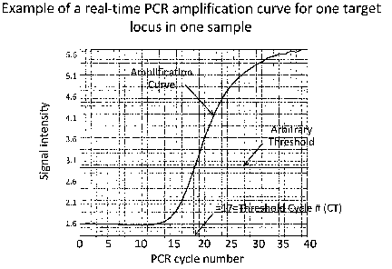
[0042] Figure 1 is a graph which provides an example of a
real-time PCR amplification curve for a target locus (the
human 18S ribosomal RNA gene, chromosome 22p12), in a normal
female lymphocyte, (ID GM00321; Coriell Cell Repository,
Camden, NJ). Data for the curve was obtained by following the
manufacturer's recommended protocol for real-time PCR (TAQMAN
Gene Expression Assays Protocol Revision G and the TAQMAN Gene
Expression Assay ID #Hs99999901 sl; Applied Biosystems (ABI),
Foster City, California) . Data represents the cycle number at
which a specific target sequence is amplified enough to reach
an arbitrary threshold.
[0043] Figure 2 is a plot illustrating the results of an
analysis of chromosome 21 copy number using 5-cell lysates
from 3 cell lines known to possess 1, 2 or 3 copies of
8
CA 02707296 2010-06-11
chromosome 21 (chr21). DNA was collected from 8 samples (n=8)
of 5 cells each from cell lines with the following karyotypes:
45, XY-21 (1 copy of chr2l, Coriell ID GM01201) ; 46,XX (2
copies of chr2l, Coriell ID GM00321); and 48,XY,+16,+21 (3
copies of chr2l, Coriell ID GM04435). Target invariant loci
included those indicated in Table 7 (FAM assays for chr2l)
from Applied Biosystems (ABI).
[0044] Figure 3 is a plot illustrating the results of an
analysis of chromosome X copy number using a 5 cell lysate
from 4 cell lines known to possess 1, 2, 3 or 4 copies of
chrX. DNA was collected from 8 samples (n=8) of 5 cells each
from cell lines with the following karyotypes: 46,XY (1 copy
of chrX, Coriell ID 00323); 46,XX (2 copies of chrX, Coriell
ID GM00321); 47,XXX (3 copies of chrX, Coriell ID GM04626);
and 49,XXXXY (4 copies chrX, Coriell ID GM00326). Target
invariant loci included those indicated in Table 7 (FAM assays
for chrX) from ABI.
[0045] Figure 4 is a graph depicting the determination of
chromosomal copy number in a trisomy 21 female (47,XX, +21,
Coriell ID AG16777) according to the methods of the present
invention. Four loci per chromosome (96 FAM assays found in
Table 7) were evaluated according to the methods described in
the present invention.
DETAILED DESCRIPTION
[0046] While the specification concludes with the claims
particularly pointing out and distinctly claiming the
invention, it is believed that the present invention will be
better understood from the following description.
[0047] All percentages and ratios used herein are by weight
of the total composition and all measurements made are at 25 C
and normal pressure unless otherwise designated. All
temperatures are in Degrees Celsius unless specified
otherwise. The present invention can comprise (open ended) or
consist essentially of the components of the present invention
9
CA 02707296 2010-06-11
as well as other ingredients or elements described herein. As
used herein, "comprising" means the elements recited, or their
equivalent in structure or function, plus any other element or
elements which are not recited. The terms "having" and
"including" are also to be construed as open ended unless the
context suggests otherwise.
[0048] All ranges recited herein include the endpoints,
including those that recite a range "between" two values.
Terms such as "about," "generally," "substantially," and the
like are to be construed as modifying a term or value such
that it is not an absolute, but does not read on the prior
art. Such terms will be defined by the circumstances and the
terms that they modify as those terms are understood by those
of skill in the art. This includes, at very least, the degree
of expected experimental error, technique error and instrument
error for a given technique used to measure a value. Unless
otherwise indicated, as used herein, "a" and "an" include the
plural, such that, e.g., "a cell" can mean more than one cell.
[0049] As contemplated herein, Applicant's present
invention is directed to methods for preimplantation genetic
diagnosis of IVF embryos which uses RT-PCR methods to perform
relative quantitation of chromosomal DNA copy number of
invariant loci in single or few cells collected from an IVF
embryo. Determination of chromosomal copy number in this way
allows for the detection of aneuploidy in an IVF embryo.
[0050] Advantages associated with performing PGD according
to the methods described herein include the ability to
simultaneously characterize aneuploidy of all 24 chromosomes,
single gene disorders, and other chromosomal abnormalities
such as inheritance of reciprocal translocation derivatives.
Significantly, the present method allows for an unprecedented
ability to characterize the embryo for these abnormalities
within 6 hours or less. This is a huge advance for the field
of PGD because it provides the opportunity to perform analysis
CA 02707296 2010-06-11
of a trophectoderm (TE) biopsy and select the embryo(s) for
transfer in the same day. This avoids cryopreservation, may
be less invasive than a blastomere biopsy since TE is
extraembryonic, may give more accurate results than a day 3
blastomere biopsy since a TE biopsy yields more than a single
cell, and may provide fewer sampling errors derived from
mosaicism.
[0051] In addition, the short turn-around time also
provides an opportunity to evaluate samples from around the
world at one reference lab since delivery time can be
accommodated and allow for fresh embryo transfer on day 5
after day 3 biopsy.
[0052] Furthermore, the present invention does not rely
upon polymorphism characterization thereby allowing the same
method to be applied to any IVF patients' embryos and without
the need for parental DNA haplotype analysis. The method is
also much more practical to perform since it is considerably
less expensive than polymorphism microarray based methods
previously described.
[0053] As contemplated herein, in order to determine
chromosomal copy number of all chromosomes in a cell, for each
chromosome, the threshold cycle (CT) for at least one, but
typically multiple, invariant loci found on the chromosome is
determined. The average CT for each chromosome is calculated
and normalized by subtracting the average CT determined for
the other chromosomes (calculated by computing the combined
average of all the CT values obtained for all other
chromosomes) from the average CT determined for the chromosome.
(See, Table 1, Equation 1). The process is repeated to
determine the ACT for each chromosome.
[0054] Multiple such CT determinations are also performed on
the corresponding chromosomes in a control, or reference,
sample to obtain a ACT for the chromosomes in the reference
sample. As contemplated herein, reference samples are euploid
11
CA 02707296 2010-06-11
cells (cells containing a normal number of chromosomes) and
appropriate reference samples for comparison are familiar to
one of skill in the art. For example, for assaying human IVF
embryos, one could use 46,XY cells from a well characterized
euploid cell line, e.g., cells from a repository such as
Coriell that have a 46,XX (ID GM00323) or 46,XY (ID GM00321)
karyotype.
[0055] Once ACT values are obtained for a chromosome and its
corresponding reference chromosome, subtraction of the ACT of
the reference chromosome from the ACT of the corresponding
chromosome is performed to generate a AACT for each chromosome.
(See, Table 1, Equation 2).
[0056] As understood by those of skill in the art, a 1 cycle
difference is theoretically a 2 fold difference in quantity of
the target DNA due to the exponential nature of PCR. Thus,
the fold change of each chromosome relative to its
corresponding reference sample may then be determined by
applying Equation 3 in Table 1. In this calculation, AACT
determined for each chromosome serves as the negative exponent
of 2 in order to calculate fold change for each chromosome
relative to its corresponding reference sample.
[0057] Copy number of each chromosome may then be
determined by multiplying the fold change by 2. (See, Table 1,
Equation 4) As understood herein, a chromosomal specific
fold change of 1 (i.e., a copy number of 2) indicates normal
copy number of that chromosome in the IVF embryo. In contrast,
a chromosome specific fold change of 0.5 or 1.5 would be
considered abnormal, and indicate an IVF embryo with monosomy
or trisomy of that chromosome, respectively. It is noted
herein that this step is not performed for the Y chromosome,
as normal males have one copy number.
12
CA 02707296 2010-06-11
TABLE 1: Determination of Chromosome Copy Number
Equation 1: Using CT data collected for each
invariant loci, normalized value (ACT) is
determined for each chromosome and a
corresponding normal reference chromosome
for at least one chromosome 1-22, X, and Y
(chromosome number is referred to
collectively herein as "N").
ACT chN= (average CT for chromosome N assays)-
(average CT for all other chromosome assays)
Note: Only autosomes are used to normalize the
data for each individual chromosome N. where N
is 1-22, X and Y.
Equation 2: The ACT for the reference sample
chromosome is subtracted from the test sample
ACT of the same chromosome to generate a AACT
for that chromosome.
AACT ChN = (test sample chromosome ACT chN) -
(reference ACTChN)
Equation 3: The fold change of each chromosome
relative to the normal reference sample is
then calculated.
Fold ChangechN=2- CTChN
Equation 4: Fold change is multiplied by 2 in
order to obtain a copy number for each
chromosome (not performed for Y chromosome and
not performed for X if reference sample is
male).
Copy Number = 2 x Fold ChangechN
[0058] As contemplated herein, copy number may be
determined for any or all chromosomes in a cell according to
the methods described herein. Specifically, it is understood
that the methods may be employed to determine the copy number
of all chromosomes, only a subset of chromosomes, or of a
single chromosome of particular interest.
13
CA 02707296 2010-06-11
[0059] As contemplated herein, test sample data for
chromosomes is normalized using only the average CT determined
for autosomes which ensures that the data is normalized using
data for chromosomes with the best potential for a normal copy
number of 2. Accordingly, it is understood that one would
typically exclude data for the sex chromosomes from the
calculation when subtracting the average CT of "all other
chromosomes" in Equation 1. In contrast to autosomes, which
are present in pairs in euploid cells, as understood by one of
skill in the art, the copy number of sex chromosomes can be
normal and be either 0 (chrY in a normal female), 1 (chrX and
Y in a normal male), or 2 copies (chrX in a normal female).
As such, errors might be introduced if the data for a given
chromosome was normalized with data including the sex
chromosomes, since statistically approximately half the time
the sample will be male (1 X and 1 Y) and half the time the
sample will be female (2 X and 0 Y). It is also contemplated
herein, however, that as the methods of the present invention
can indicate the sex of an IVF embryo, it is possible that
where the methods indicate that the X chromosome condition is
disomy, X chromosome data could be included to normalize the
chromosome data.
[0060] The methods described herein and detailed in Table 1
can be used to determine the copy number of sex chromosomes as
well as autosomes. As contemplated herein, however, data for
the sex chromosomes would be evaluated in a manner consistent
with the gender of the reference sample used. For chrX,
either a normal female or normal male could be used as the
reference sample. If the reference sample is female, then the
copy number can be calculated in a manner identical to that
described for chrl-22 above. If the reference sample is male,
than the fold change value wouldn't be multiplied by two and
instead would already be equal to the copy number of chrX.
For chrY, a normal male would need to be used as the reference
14
CA 02707296 2010-06-11
sample and the fold change would be equal to the copy number
and wouldn't require multiplication by 2.
[0061] In addition to monosomy or trisomy, other forms of
aneuploidy, e.g., nullisomy, disomy (of the Y chromosome), and
tetrasomy may also be detected according to the methods of the
present invention. Chromosomal nullisomy may be diagnosed, for
example, by detecting the absence of an amplification signal
for a chromosome. Similarly, detecting 2 copies would
indicate chromosomal disomy, and detecting 4 copies would
reflect tetrasomy.
[0062] Data manipulation and calculations necessary to
carry out the methods of the present invention may be
performed according to any suitable means familiar to one of
skill in the art. These include, but are not limited to
commercially available computer software programs, e.g.,
Microsoft Excel, SDS (Applied Biosystems), or other similar
computer software packages.
[0063] As contemplated herein, the methods of the present
invention are useful for detecting the copy number of whole
chromosomes. As used herein, a "whole" chromosome means that
the intended resolution is a complete, intact chromosome, and
not, e.g., a chromosomal fragment or microdeletion.
Typically, if a microdeletion, balanced translocation or other
similar chromosomal aberration occurs de novo in an embryo
(i.e., such mutation isn't present in either parent and thus
was not inherited by the embryo), it is unlikely to be
diagnosed by the methods of the present invention. As such, it
is contemplated herein that additional assays designed to
detect such chromosomal defects in an IVF embryo may be
performed in conjunction with the methods of the present
invention.
[0064] In addition, where genetic disorders other than
aneuploidy are known or suspected, it is contemplated herein
that genetic assays to identify such abnormalities in the IVF
CA 02707296 2010-06-11
embryo may be performed in concert with the methods of the
present invention. Additional assays useful to detect such
genetic disorders unrelated to copy number and applicable to
PGD are familiar to one of skill in the art and include, for
example, using sequencing primers capable of determining
single gene disorder mutation sequences, or using primers
designed for specific cytobands of a chromosome known to be on
either side of known breakpoints in a patient with a
reciprocal balanced translocation.
[0065] As used herein, the terms "locus" or "loci" (plural)
refer to the position on a chromosome of a particular gene.
The human genome is comprised of both "invariant" and
"variable" loci. Variable loci are those loci for which
alternative alleles exist and include, e.g., those alleles
with single nucleotide polymorphisms (SNPS).
[0066] In contrast, as understood herein, "invariant loci"
are positions in the genome of an organism for which no
evidence exists of any polymorphism or variation in any
population evaluated to date. As understood by one of skill in
the art, invariant loci may typically be found in highly
conserved regions encoding conserved functions in the human
genome. These include, for example, the active sites of
enzymes and the binding sites of protein receptors. Invariant
loci are more likely found in exons; in contrast, one would
expect to find a greater number of variable loci in introns or
"junk DNA".
[0067] According to the methods of the present invention,
invariant loci are assayed to determine chromosomal copy
number since analyzing loci possessing polymorphisms such as a
SNP or a copy number variant (which may be found in normal
"euploid" individuals) could indicate that the locus is
present at a different copy number in different individuals
with different polymorphisms. This could result in a
misdiagnosis, e.g., of monosomy or trisomy, rather than
16
CA 02707296 2010-06-11
indicating the true euploid nature of a cell. In addition,
loci of variant nucleotide sequences can have different
amplification efficiencies. For example, the efficiency of
amplification of a SNP-containing locus may be less in one
individual that is homozygous for an adenosine nucleotide (AA)
than in another individual who is homozygous for a cytosine
nucleotide (CC). As a result, the lower efficiency would make
it appear as though the AA individual had fewer copies than
the CC individual despite both individuals having two copies
of the SNP-containing locus.
[0068] One of skill in the art may identify suitable
invariant loci for use with the methods of the present
invention by reviewing databases of genetic information. As
used herein, the term "database of genetic information"
includes databases which contain data characterizing the
frequency of genetic loci for various populations and
includes, for example, the Entrez SNP database available from
NCBI, as well as databases available from NCI, WICGR, HGBASE
or the International HapMap Project (see, e.g., International
HapMap Consortium, Nature 449, 18 October 2007, 851-862).
Extensive proprietary SNP databases are also available through
commercial vendors, and include, for example, Applied
Biosystems' SNP database which supports their commercial
TAQMAN SNP Genotyping Assays. Analyzing SNP databases would
be useful with respect to the present invention, e.g., with
regard to the identification of variant loci (i.e., loci with
known polymorphisms) which could thus be avoided in the
selection of loci for use as disclosed herein.
[0069] Other databases useful with respect to the methods
of the present invention include databases which contain
information regarding copy number variation. These databases
are familiar to one of skill in the art and include, for
example, Chromosome Abnormality Database ("CAD"; a collection
of both constitutional and acquired abnormal karyotypes
17
CA 02707296 2010-06-11
reported by UK regional cytogenetics centers) ; Database of
Genomic Variants (a curated catalogue of large-scale variation
in the human genome); DECIPHER (Database of Chromosomal
Imbalance and Phenotype in Humans using Ensembl Resources,
Decipher Consortium); ECARUCA (European Cytogeneticists
Association Register of Unbalanced Chromosome Aberrations);
Human Genomic Structural Variation Database (a catalogue of
human genomic polymorphisms ascertained by experimental and
computational analyses, Eichler laboratory, University of
Washington, Seattle, WA); The Chromosome Anomaly Collection
(contains examples of unbalanced chromosome abnormalities
(UBCAs) without phenotypic effect; compiled by J. Barber,
National Genetics Reference Laboratory (Wessex), Salisbury
NHS Foundation Trust); CNV Project (The Copy Number Variation
(CNV) Project Data Index; Sanger Institute); Structural Genome
Variation.
[0070] Loci suitable for use in the methods of the present
invention may also be identified by reviewing other publicly
or commercially available genetic databases to identify loci
that occur in a population at a frequency such that the loci
may be deemed "invariant" and may be used to produce
statistically useful data for PGD as contemplated herein.
These databases include the Allele Frequency Database
("ALFRED") supported by the US National Science Foundation.
Based on the allele frequency data in this database, one could
compile a list of loci to avoid using in the methods of the
present invention; e.g., loci reported to exhibit different
allele frequencies in different human populations. In
addition, while not necessary to perform the methods of the
present invention, based on the data provided in the ALFRED
database, one could also identify and select suitable
invariant loci for PGD in view of the parentage (e.g.,
ethnicity) of an IVF embryo to be analyzed.
18
CA 02707296 2010-06-11
[0071] Along with the review of genetic databases discussed
above, factors for consideration in the selection of possible
invariant loci for use with the methods of the invention
include whether the locus sequence is specific (i.e.
homologues or pseudogenes aren't present in the genome),
whether primers and a probe can be designed to perform under
common PCR conditions so that an assay for the locus can be
performed under the same conditions as assays for other loci,
and whether it is located in a region of the chromosome that
is distant enough from other loci used so that, for example,
both arms of each chromosome might be evaluated.
[0072] While it is contemplated herein that the loci deemed
invariant in the Examples and Tables described herein may be
used to screen for aneuploidy in any IVF embryo, it is
possible that for any given invariant locus, there could exist
a rare, previously undetected polymorphism in the human
population. If an IVF embryo did possess such a rare,
previously undetected polymorphism at a given "invariant"
locus, chromosomal copy number could still be detected in that
embryo where more than one locus per chromosome is employed in
the methods provided herein.
[0073] Invariant loci may also be confirmed for use with a
particular IVF embryo by using conventional methods to detect
the presence of the invariant loci in the parental DNA using
conventional methods. Analysis of parental DNA could be
performed at any time, including prior to creation of the
embryo.
[0074] Once candidate invariant loci are identified in
silico, one may then select any number for further analyses.
Criteria for selection of various loci for additional testing
include: whether an assay for the specific locus is readily
available from a commercial supplier, whether an assay for the
specific locus follows an expected sigmoidal pattern of
amplification under standardized PCR conditions (i.e., see
19
CA 02707296 2010-06-11
Figure 1), and whether an assay for the locus performs well
when evaluated on cells with known abnormalities. For example,
suspected invariant loci may be utilized to screen euploid and
aneuploid cells of known karyotype according to the methods of
the present invention. In addition to using aneuploid and
euploid cell lines, in some cases one may also biopsy frozen
embryos, including previously diagnosed aneuploid embryos, to
identify useful invariant loci to employ in the methods of the
present invention. IVF embryos with karyotypes such as
described in Table 4 are not uncommon; one of skill in the
art, e.g., clinicians who routinely perform in vitro
fertilization and PGD, typically have access to similar
aneuploid embryos and have been given consent to use biopsied
material from such embryos for research purposes.
[0075] By evaluating the ability of a particular locus to
correctly identify copy number of a characterized sample, one
of skill in the art can arrive at a set of invariant loci
which can then be used to accurately predict the correct copy
number for any given chromosome for PGD of an IVF embryo. As
described in detail in the Examples and Tables provided herein
below, such assays are easily performed and can be used to
identify numerous suitable invariant loci for use in the
practice of the present invention such as listed in detail in
Table 7.
[0076] While any number of invariant loci may be used
according to the methods of the present invention, suitable
invariant loci may be identified and further characterized
into sets and subsets of invariant loci for use as described
herein. For example, in addition to displaying a lack of
associated polymorphisms, and the ability to predict copy
number of samples with known karyotype, subsets may be
characterized based on various additional criteria including,
but not limited to, the robustness of the chemistry and other
technical factors associated with assaying a particular loci,
CA 02707296 2010-06-11
e.g., availability of primers or other factors associated with
amplifying particular loci, costs associated with use of
particular loci and other practical aspects associated with
performing the methods described herein that would be familiar
to one of skill in the art.
[0077] Sets and/or subsets of invariant loci that may be
used in the methods of the present invention may comprise
hundreds of thousands of loci, but as contemplated herein, the
methods of the present invention do not require the evaluation
of a volume of data such as might be generated from a whole
genome analysis. Rather, a set of invariant loci for use
according to the methods of the present invention typically
comprises less data than the entire genome of an individual,
e.g., less than about 100,000, less than about 50,000, less
than about 20,000, less than about 10,000, less than about
5,000, or less than about 1,000 invariant loci.
[0078] Loci may be evaluated for ability to predict
chromosome copy number as provided herein and particular loci
that perform well for each chromosome may be easily
identified. As described above, invariant loci are more likely
found in highly conserved regions in the genome, e.g., exons
that code for proteins with key biochemical functions. Based
on review of a publicly available human genome database, one
of skill in the art can design primers that fall within a
single exon and optimize such primers for performance in PCR
and prediction of chromosome copy number as detailed herein.
In addition, sets of assays that are designed to detect a
single exon are commercially available. These "assays" or
"sets of assays" include commercially available nucleic acid
assays with fluorescently labeled probes typically used for
gene expression analysis, e.g., ABI TAQMAN Gene Expression
Assays. As contemplated herein, suitable commercial assays for
use herein include those that are also capable of detecting
genomic DNA.
21
CA 02707296 2010-06-11
[0079] As contemplated herein, any number of loci may be
routinely selected for assay, however, since locus drop out
from single cell amplification is likely, it is contemplated
herein that typically at least more than one invariant locus
would be evaluated per chromosome. For example, using 96 well
plates and conventional high throughput methodologies, 192
loci (e.g., 96 samples in duplicate) may be easily assayed and
provide accurate results according to the methods of the
present invention.
[0080] By focusing on only invariant loci, a high quality
set of loci may be obtained and used (in whole or in part in
the form of subset(s) thereof) to analyze an embryo. Thus, by
employing a robust set of invariant loci in combination with
high throughput analysis using the comparative CT method, it is
contemplated herein that PGD may be performed more accurately,
more efficiently and more rapidly than currently available
modes of PGD analyses. For example, by employing the methods
of the present invention, one may avoid the less efficient and
error prone method of whole genome amplification based
embryonic analysis which necessitates supplementary analysis
of parental DNA to confirm embryonic data and/or to identify
additional informative loci for embryonic genotyping.
[0081] As contemplated herein, the methods of the present
invention permit assaying the embryo, determining the presence
or absence of genetic defect, and selecting and transferring
an embryo to be performed within a period of about 24 hours or
less, e.g., within about 16, about 12, about 8 or about
hours or less. As such, same day cell biopsy and fresh
transfer of an IVF embryo is possible. It is further
contemplated that the method steps may be performed such than
an embryo is determined to be without genetic defect and
transferred within about 154 hours of fertilization,
particularly between about 48 and about 144 hours of
fertilization.
22
CA 02707296 2010-06-11
[0082] Although the methods of the present invention
provide the advantage of PGD and fresh transfer, it is
envisioned that there may be situations in which fresh
transfer of an IVF embryo assayed and selected for transfer
according to the methods of the present invention is not
desired. Such situations may include, e.g., when fresh
transfer is not convenient, or not medically appropriate. In
such instances, it is contemplated herein that the embryo may
be preserved, e.g., frozen or otherwise cryopreserved and
maintained for possible transfer at a later date according to
conventional methods. It is also possible that no embryo may
be transferred.
[0083] As contemplated herein, nucleic acid probes for
invariant loci can be provided in the form of an array for PGD
according to the methods of the present invention. Such arrays
are familiar to one of skill in the art and may be in various
forms, including but not limited to, a solid support such as a
chip or glass slide, e.g., in the form of a microarray or
preloaded assay plate, to which the nucleic acid may be
affixed or loaded according to methods familiar to one of
skill in the art. Custom made assay chips with nucleic acid
for loci of interest affixed or preloaded thereto may be
obtained from commercial vendors, e.g., Applied Biosystems
Inc. (Foster City, CA), Affymetrix Inc. (Santa Clara, CA), or
Illumina Inc. (San Diego, CA).
[0084] Further contemplated herein are kits that comprise
arrays for use with the methods of the present invention. For
example, a kit might comprise an array of nucleic acid probes
immobilized on a solid support or preloaded on an assay plate,
the array comprising nucleic acid probes for at least one
invariant locus from at least one human chromosome wherein the
loci are informative for determining the presence or absence
of a genetic defect in an IVF embryo prior to transfer
according to the methods discussed herein. Additional
23
CA 02707296 2010-06-11
components of such kits may comprise instructions as well as
reagents, primers, probes or other tools of molecular biology
familiar to one of skill in the art that might be of use in
conducting PGD of an IVF embryo.
[0085] It is contemplated herein that PGD according to the
methods of the present invention may be performed quite
effectively utilizing the invariant loci provided herein.
(See, e.g., Table 7). It is further contemplated, however,
that additional invariant loci may be identified. As discussed
above, such invariant loci may be identified in silico by
reference to known databases of human genetic information, and
may be further selected in view of data for a particular
ethnic or geographic group. Selection of appropriate invariant
loci may be confirmed by performing the methods of the present
invention using control or reference samples of known
karyotype and evaluating ability of the loci to correctly
predict known copy number.
[0086] As envisioned, in order to maximize efficiency where
possible and practical, data necessary to perform the methods
of the present invention may be on hand when an IVF embryo
becomes available for PGD. Thus, in order to facilitate the
PGD and fresh transfer of an IVF embryo according to the
methods of the present invention in the time frame described,
invariant loci may be identified prior to the actual creation
of an IVF embryo.
[0087] One of skill in the art will appreciate that the
steps of the methods of the present invention may take place
in different locations. For example, biological material may
be obtained from the prospective parents of an IVF embryo and
used to create an IVF embryo at the same clinic, or the
materials may be transported according to conventional methods
to a second location at which the IVF embryo may be created
and maintained in vitro. Similarly, cells may be obtained from
an IVF embryo and screened for PGD according to the methods of
24
CA 02707296 2010-06-11
the present invention in the same laboratory, or the cells may
be delivered to a second location for PGD. If PGD is
performed at a different location than where the embryo is
maintained, PGD results may be transmitted back to the
laboratory maintaining the embryo where transfer of suitable
embryos into the recipient may then be performed. As would be
apparent to one of skill in the art, the steps of the method
are ideally performed in locations such that the entire
process takes place in the most efficient manner possible; for
example, in one embodiment, the IVF embryo is maintained in
the same facility in which the genetic screening is performed
and in which embryo transfer takes place. In this way, loss
of time associated with shipping the cells to a second
laboratory for genetic analysis is avoided. This would be
especially advantageous where the time available for
performing PGD and embryo transfer is extremely limited, for
example, with regard to the same day biopsy, PGD and fresh
transfer of a day 5 or day 6 blastocyst.
[0088] As contemplated herein, genetic analysis of an
"embryo" includes assay of nucleic acid from cells from an IVF
embryo (an embryo fertilized not less than about 40 hours
before analysis), cells from a blastocyst (typically an embryo
at day 4, day 5 or day 6 after fertilization) as well as cells
biopsied from an embryo but of extraembryonic origin, e.g.,
trophectoderm, or polar bodies. The plural form of this term
is included, such that, the term "an embryo" as used herein
contemplates that more than one embryo or blastocyst may be
concurrently assayed or transferred according to the methods
of the present invention.
[0089] It is further contemplated herein that more than one
cell of an embryo may be biopsied as conditions permit, for
example, at least one cell of trophectoderm may be biopsied
and assayed according to the methods of the present invention.
Assaying more than one cell in this way can be used to detect
CA 02707296 2010-06-11
mosaicism in an embryo (a condition in which cells in an
embryo may differ genetically from other cells in the embryo)
which cannot be detected if only a single cell is assayed.
Thus, as contemplated herein, the methods of the present
invention can be used to biopsy a day 5 or day 6 embryo,
screen the embryo for mosaicism, and still permit fresh
transfer of the embryo on the same day.
[0090] "Transferring" an IVF embryo refers to the process
of placing an IVF embryo into a female patient with the
objective that the embryo will implant and result in a viable
pregnancy.
[0091] The term "fresh transfer" refers to the transfer of
an embryo which has not been subjected to cryogenic
preservation.
[0092] As used herein, a "plurality of chromosomes" refers
to more than one chromosome.
[0093] "Candidate IVF embryos" are those embryos determined
to be without genetic defect according to the methods of the
present invention. These embryos may be deemed suitable for
transfer, however, it is understood that other criteria
familiar to one of skill in the art may also be taken into
consideration in the selection of particular embryos for
transfer.
[0094] The terms "chromosomal abnormality" and "genetic
defect" are used interchangeably herein and refer to a
deviation between the structure or copy number of the subject
chromosome and a normal chromosome. The term "normal" refers
to the predominate karyotype or banding pattern found in
healthy individuals of a particular species. A chromosomal
abnormality or genetic defect can be numerical or structural,
and includes but is not limited to, single gene defects, sex-
linked disorders, or chromosomal disorders, e.g., aneuploidy,
polyploidy, inversion, a trisomy, a monosomy, duplication,
deletion or additions of entire chromosomes or parts thereof,
26
CA 02707296 2010-06-11
insertions, rearrangements, and translocations. As provided
herein, the methods of the present invention may be used to
detect aneuploidy in an IVF embryo, however, it is further
contemplated herein that an IVF embryo may be screened for
additional chromosomal abnormalities other than aneuploidy.
Additional genetic screening may be performed using methods
familiar to one of skill in the art and concurrently with the
methods of the present invention.
[0095] Chromosomal abnormalities or genetic defects can be
correlated with presence of a pathological condition or with a
predisposition to develop a pathological condition. Numerous
examples of pathological conditions associated with genetic
defects on particular chromosomes and/or linked to a
particular gene are known to those of skill in the art and
literature references and electronic databases containing
extensive and detailed information describing genetic defects
are widely available. With such knowledge, one may employ the
methods of the present invention alone or in combination with
additional diagnostic methods to determine copy number and/or
the presence of other chromosomal abnormalities in order to
elucidate whether an IVF embryo may possess or be prone to a
particular pathological condition. Such conditions include,
for example, the genetic diseases listed herein in Table 2 and
as described in US Patent 7,439,346.
Table 2: Examples of Genetic Disease
Chromosome Genetic Disease
Number
13 Breast and ovarian
cancers, deafness,
Wilson's Disease. Patau's
Syndrome
15 Marfan Syndrome,
Tay-Sach's Disease
27
CA 02707296 2010-06-11
16 Polycystic kidney disease,
Alpha thalassemia
17 Charcot-Marie-Tooth
Disease
18 Niemann-Pick Disease,
pancreatic cancer,
Edward's Syndrome
21 Down's Syndrome
X Duchenne muscular
dystrophy (DMD), Turner's
Syndrome, Fragile X
Syndrome, Klinefelter's
Syndrome
X-linked diseases:
hemophilia,
adrenoleukodystrophy, and
Hunter's disease
Y Acute myeloid leukemia
[0096] Specific examples of pathological conditions
associated with aneuploidy which may be detected in an IVF
embryo according to the methods of the present invention
include: Turner's Syndrome (a single X chromosome, e.g., 45, X
or 45, XO); Klinefelter's Syndrome (an extra X chromosome,
e.g., 47, XXY); Edward's Syndrome (three copies of chromosome
18); Down's Syndrome (three copies of chromosome 21); Patau's
Syndrome (three copies of chromosome 13), trisomy 8, trisomy 9
and trisomy 16.
[0097] As discussed above, in addition to screening an IVF
embryo for aneuploidy according to the methods of the present
invention, it is further contemplated herein that an IVF
28
CA 02707296 2010-06-11
embryo may also be screened for other genetic disorders using
additional methods familiar to one of skill in the art. For
example, an embryo may be concurrently screened for
chromosomal microdeletions, translocations, or rearrangements
using targeted preamplification strategies performed in
parallel with the methods of the present invention. As used
herein, a "targeted preamplification strategy" could include
creation of custom probes for specific patients in order to
successfully screen for inheritable disorders. Such
additional screening methods can include for example, single
nucleotide polymorphism real-time PCR, sequencing, restriction
fragment polymorphism analysis, or short tandem repeat
fragment size analyses. Such methods are familiar to one of
skill in the art.
[0098] In addition, as mentioned herein above an IVF embryo
may be screened for genetic defects according to the methods
of the present invention while also being screened for at
least one single gene genetic disorder. Such disorders may be
detected based on the presence or absence of a SNP allele
identified according to conventional methods or as described
in copending US patent application USSN 61/205,522. For
example, if a single gene disorder were identified in the
parents of an IVF embryo, that disorder could be evaluated in
conjunction with the methods of the present invention by co-
amplifying the disease causing target sequence or linked DNA
sequences with the (aneuploidy informative) invariant loci.
Analysis of the aneuploidy state of each chromosome and the
presence or absence of the single gene disorder could then be
evaluated in each resulting embryo.
[0099] As used herein, an IVF embryo "determined to be
without genetic defect" refers to an embryo that is determined
to be free of a particular genetic defect for which it was
screened. It is understood that while the methods of the
present invention are accurate, they may not be able to detect
29
CA 02707296 2010-06-11
100% of the genetic abnormalities in an IVF embryo. In some
cases, data may be interpreted as meaning that there is a
greatly reduced chance of the IVF embryo having a particular
genetic defect, e.g., as would be the case for diagnosing
mosaicism in an embryo, given that only a few cells, at best,
may be assayed.
[0100] Methods for PGD described herein involve nucleic
acid analysis; standard techniques for nucleic acid isolation
and purification are known and are described in, for example,
in Miller (ed.) 1972 Experiments in Molecular Genetics, Cold
Spring Harbor Laboratory, Cold Spring Harbor, New York; Old
and Primrose, 1994 Principles of Gene Manipulation, 5th ed.,
University of California Press, Berkeley; Schleif and Wensink,
1982 Practical Methods in Molecular Biology; Glover (Ed.) 1985
DNA Cloning: Vols. I AND II, IRL Press, Oxford, UK; Harnes and
Higgins (Eds.) 1985 Nucleic Acid Hybridization, IRL Press,
Oxford, UK; and Setlow and Hollaender 1979 Genetic
Engineering: Principles and Methods, Vols. 1-4, Plenum Press,
New York City.
[0101] The sequence of a nucleic acid may be determined as
necessary using conventional methods. These methods include,
for example, PCR, gel electrophoresis, ELISA, mass
spectrometry, MALDI-TOF mass spectrometry hybridization,
primer extension, fluorescence detection, fluorescence
resonance energy transfer (FRET), fluorescence polarization,
DNA sequencing, Sanger dideoxy sequencing, DNA sequencing
gels, capillary electrophoresis on an automated DNA sequencing
machine, microchannel electrophoresis, microarray, southern
blot, slot blot, dot blot, single primer linear nucleic acid
amplification, as described in U.S. Pat. No. 6,251,639, SNP-
IT, GeneChips , HuSNP , BeadArray, TAQMAN assay, Invader
assay, MassEXTEND , or MassCLEAVETM (hMC) method.
[0102] Nucleic acid amplification methods are also known
and may be used in PGD as contemplated herein, including the
CA 02707296 2010-06-11
polymerase chain reaction (PCR) (PCR Protocols, A Guide to
Methods and Applications, ed. Innis, Academic Press, N.Y.
1990; PCR: A Practical Approach, M. J. McPherson, et al., IRL
Press (1991)) and particularly real-time, quantitative PCR
discussed hereinabove (Schmittgen, T. and Livak, K., Nature
Protocols 2008; Vol 3, No.6:1101-1108; Livak, K. and
Schmittgen, T., Methods 2001; 25:402-408). Other known
amplification methods include ligase chain reaction (LCR)
(Landegren, Science 1988; 241:1077); transcription
amplification (Kwoh, Proc. Natl. Acad. Sci. USA 1989;
86:1173); self-sustained sequence replication (Guatelli, Proc.
Natl. Acad. Sci. USA 1990; 87:1874); Q Beta replicase
amplification (Smith, J., Clin. Microbial. 1997; 35:1477-
1491), and other RNA polymerase mediated techniques such as
nucleic acid sequence based amplification, NASBA (U.S. Pat.
Nos. 4,683,195 and 4,683,202); 3SR (self-sustained sequence
reaction); RACE-PCR (rapid amplification of cDNA ends); PLCR
(a combination of polymerase chain reaction and ligase chain
reaction); SDA (strand displacement amplification); and SOE-
PCR (splice overlap extension PCR).
[0103] Errors associated with amplification include
allelic, or locus dropout (LDO) and such errors are familiar
to one of skill in the art. LDO rate estimates can differ
based on the method by which they are measured. For example,
microarrays often underestimate LDO rates. There are analysis
settings that can be adjusted that will lead to different
estimates, e.g., stringent genotype calls are associated with
higher LDO rate estimates. Real time PCR is the most
stringent method for evaluating LDO so estimates based on the
use of real time PCR may be higher than microarray measures.
[0104] In order to avoid misdiagnoses due to amplification
errors such as allelic or locus drop out, it is understood
herein that sequencing an allele of interest may include
sequencing nucleic acid around the allele to ensure
31
CA 02707296 2010-06-11
amplification accuracy. For example, the disease-causative
allele may be physically linked (close together) with a non-
causitive allele nearby in the DNA sequence. These two sites
in the DNA are very likely to be inherited together, barring
any meiotic recombination between the sites. Sites nearer
each other are less likely to undergo recombination. As a
result, the non-causative allele can be used as a confirmatory
marker of the disease causing allele in order to avoid
misdiagnosis from disease allele PCR dropout. Such techniques
are familiar to one of skill in the art.
[0105] It is contemplated herein that conventional methods
to analyze nucleic acid include methods that permit the
analysis of nucleic acid from a small number of cells. Such
methods may include performing a "preamplification" of DNA
prior to real-time PCR using invariant locus specific primers.
Such methods are a modification of methods familiar to one of
skill in the art, and kits to perform such preamplification
are commercially available, for example, TAQMAN PreAmp Cells-
to-CtTM Kit from Applied Biosystems. While these kits are
designed to preamplify cDNA derived from RNA, they can also be
used successfully on genomic DNA.
[0106] As contemplated herein, in a particular embodiment
the methods of the present invention are performed using means
which permit multiple, parallel real-time PCR reactions,
including, but not limited to, high throughput genotyping
using one or more assay platforms. By evaluating multiple loci
of interest in this way, preimplantation genetic diagnosis may
be performed quickly and efficiently such that embryo
diagnosis and transfer may occur without the need for
cryopreservation of the embryo; ideally, such steps occur in
the same day. For example, it is contemplated herein that
candidate embryo selection and embryo transfer may be
performed within about 5 hours after biopsy of the embryo for
diagnosis. It is further contemplated herein that the methods
32
CA 02707296 2010-06-11
of the present invention may permit the genetic screening of
all chromosomes of an IVF embryo followed by fresh transfer of
the embryo on the same day.
[0107] As discussed above, materials and methods for high
throughput real-time PCR, including the basic concept of
analyzing real-time PCR data by the comparative CT method, are
familiar to those of skill in the art. These include, but are
not limited to, commercial real-time PCR assay plates designed
for such purposes. For example, high throughput methodologies
which may be employed to practice the methods of the present
invention include commercially available microarray plates
that use nanoliter fluidics, for example, Applied Biosystems'
TAQMAN OpenArrayTM Genotyping Plates, which may be customized
to contain nucleic acid for invariant loci of interest.
Additional systems include the Fluidigm Inc. BioMark System
for genetic analysis, or the Roche Applied Science LightCycler
1536 Real-Time PCR System.
[0108] Suitable primers for use in the methods of the
present invention may be obtained from commercial vendors.
They may also be designed by one of skill in the art according
to conventional methods and published sequence information for
any loci of interest.
[0109] Each loci-specific PCR reaction may include probes
with unique fluorescent properties and reaction products will
result in a different wavelength of quantifiable fluorescence.
For example, gene expression assays which include a probe with
a FAM label are commercially available (e.g., TAQMAN gene
expression assays, Applied Biosystems) . Assays can also be
designed to include a VIC label on the probe. The loci of
interest can be analyzed using conventional gel
electrophoresis followed by fluorescence detection or read
using a commercially available fluorescent plate reader or
scanner. Commercially available computer imaging systems
designed for high throughput include, e.g., 7900HT (Applied
33
CA 02707296 2010-06-11
Biosystems Inc.), BioMark (Fluidigm, Inc.), or the
LightCycler 480 (Roche) . The resulting fluorescence data can
be evaluated with statistical protocols and computer programs
such as SDS (Applied Biosystems), or Microsoft Excel
(Microsoft Inc.). (See, e.g., Livak, K.L. and Todd, J.A.,
Nature Genetics Vol. 9, April 1995, 341-342).
[0110] The methods of the present invention may be used to
screen at least one candidate IVF embryo concurrently such
that more than one IVF embryo without genetic defect may be
identified and transferred. The number of such embryos that
may be appropriate to transfer may be determined by one of
skill in the art according to conventional methods.
[0111] All publications cited in the specification are
indicative of the level of skill of those skilled in the art
to which this invention pertains. All these publications are
herein incorporated by reference in their entirety to the same
extent as if each individual publication were specifically and
individually indicated to be incorporated by reference.
[0112] Although the invention herein has been described
with reference to particular embodiments, it is to be
understood that these embodiments are merely illustrative of
the principles and applications of the present invention. It
is therefore to be understood that numerous modifications may
be made to the illustrative embodiments and that other
arrangements may be devised without departing from the spirit
and scope of the present invention as defined by the appended
claims.
EXAMPLES
Example 1: Identification of Invariant Loci and Analysis of
Chromosome 13 Copy Number in Cell Lines and Embryonic Tissue
using Real-time PCR and 2-8 CT Analyses
[0113] Genetic loci suitable for predicting chromosomal
copy number according to the methods of the present invention
("invariant loci") were identified by assaying the ability of
34
CA 02707296 2010-06-11
loci to accurately predict the correct chromosomal copy number
of cells of known karyotype (Coriell Cell Repository, Camden,
NJ) (Table 3) and cells from frozen aneuploid embryos
(Table 4).
[0114] In this example we established a set of loci for
chromosome 13 that perform well. To do this, we obtained
cells from cell lines and embryos that were previously
determined to be either 46,XX (GM00321, Table 3), 47,XY,+13
(GM02948, Table 3), or 45,XY,-13 (embryo 8, Table 4).
[0115] For the analysis of copy number of chromosome 13 in
the given samples, 8 invariant loci (as FAM labeled assays)
were selected based on commercial availability and design
specific for exon detection (sl assay designation, ABI) and
are listed in Table 5. Targeting loci within an exon will
provide the most opportunity for avoiding areas of the genome
with polymorphisms (genetic variation in normal euploid
cells). This is due to the fact that exon sequences are more
critical to protein function than are other regions such as
introns or "junk DNA." For that reason, exon sequences are
more highly evolutionarily conserved and less variable, making
them the ideal target for this method. In addition, exon
sequences are well characterized for possible polymorphisms
that can be evaluated in silico by referring to the
aforementioned genetic databases.
[0116] In this experiment, embryo biopsy and cell line
lysates were preamplified with 16 cycles. Preamplified DNA was
then loaded in quadruplicate into a Fluidigm BioMark RealTime
PCR 96.96 Gene Expression Array according to the
manufacturer's instructions (Fludigm Inc.) and using Gene
Expression Master Mix (ABI). CT values were obtained for each
assay for each replicate for each sample.
[0117] In this case, instead of computing the average CT for
all chromosome 13 loci, each individual locus for chromosome
13 was first computed separately to generate a copy number
CA 02707296 2010-06-11
prediction. The copy number prediction of each locus was then
evaluated for accuracy against the reference data for these
samples. The 4 loci with the fewest errors (indicated in bold
in Table 5) were identified. These loci provided the best
accuracy of copy number assignments considering what to expect
based on previously determining the karyotypes of the samples
used. Once identified, the 4 best performing loci were then
reanalyzed as an average CT to determine the copy number for
chromosome 13 (Table 6). This provided confirmation that
these 4 loci were sufficient to provide an accurate diagnosis
(i.e., loci associated with chromosome 13 that can reliably
identify the correct copy number). Moreover, these data
indicate that these 4 loci are acceptable to use for
aneuploidy diagnosis of chromosome 13 in PGD of any IVF
embryo.
[0118] This process was repeated in order to obtain
reference data for all 24 chromosomes to establish 192 loci
(Table 7) which may be reliably used to indicate correct
chromosomal copy number and using combinations of cell lines
and embryos described in Table 3 and 4. To further illustrate
the criteria for identification of useful loci, examples of
the separation observed for chromosome X and 21 are shown in
Figures 2 and 3 and further described below.
36
CA 02707296 2010-06-11
Table 3: Cell Lines
Coriell Cell Repository ID Karyotype
G M00321 46,XX
GM09286 47,XY,+9
GM02948 47,XY,+13
GM01359 47,XY,+18
GM04610 47,XX,+8
GM03184 47,XY,+15
G M04435 48,XY,,+16,+21
GM04626 47,XXX
GM01201 45,XX,-21
GM00326 49,XXXXY
GM02067 47,XY,+21
GM00323 46,XY
GM07106 47, XY, +22s+
GM00875 45,XO
GM00325 47,XXY
GM09326 47,XYY
GM02521 48,XXYY
GM11420 49,XYYYY
GM11534 56,XY,t(11;22)(g24;g12),+5,+7,+8,+15,
+18,+20,+21,+der(22)t(11;22)(g24;g12) +mars
GM10401 47,XX,+2
GM01454 47,XY,+12
GM07408 47,XX,+20
GM00980 45,XX,+der(11),-22
37
CA 02707296 2010-06-11
Table 4: Embryos
Embryo Number Karyotype
1 46,X0,+3,+16; 1.9
2 44, XY, -11, -13
3 46,XX,+8,-2 2
4 44,XX, 10, 16
46,XY,+der(5)t(3,5)(g26.2;p15.1), 7,+11,-15r 16,+17 18,+22
6 46,XX,der(5)t(5:17)(g13;g2L3),+9r 22
7 45,XX,-19
8 45,XY,-13
9 48,XXX,+12
45,XY,-8
11 45,XY,-4
12 46,XX,-15,+17
13 47,XY,+16
14 44, XX, -1, -4,+16,-22
44,XY,-9,-14
16 45,XY,-6
17 48,XY,+1,+12
18 47,XX,+14
19 47,XY,+15
48,XY,+17,+22
21 47,XX,+22
22 45,XX,-22
23 48,XX,+13,+15,-21,+22
24 47, XY, +22
45,XY,-22
26 45,XX,-12
27 45,)00,-13
28 47, XXY, -7, +14
29 45,XY,-4
44,XY,-.20,-21
31 47,XY,+6
32 45, XY, -17
33 43, XY, -7, -13, -14
34 47,)00,+18
45,XX,-8
36 45,XY, 22
37 45,XX~-15
38 48,XY,+5,+16
39 47,XX,+der(2)t(2:20)(g21;p12.2),+17a 20
47,XX,+7
41 47,XY,+9,+19,-21
42 45,XX,-17
43 48,XXX,+2,+13,-19
44 45,XX,-2
44, XY, -5, -11
38
CA 02707296 2010-06-11
Table 5: Chromosome 13 assays tested
Assay ID Gene Symbol Gene Name Chromosome
Hs00937168 sl CYSLTR2 cysteinyl leukotriene receptor 2 13 q 14.2
Hs01028557 sl SUTRKI SLIT and NTRK-like family, member 1 13 q 31.1
Hs01037385 sl HMGB1 high-mobility group box 1 13 q 12.3
Hs01072517 sl SOX21 SRY (sex determining region Y)-box 21 13 q 32.1
Hs01635854 sl SIAH3 seven in absentia homolog 3 (Drosophila) 13 q 14.12
Hs01650625_sl KBTBD6 ketch repeat and BTB (POZ) domain containing 6 13 q 14.11
Hs01879077 s1 UTP14C UTP14, U3 small nucleolar ribonucleoprotein, homolog C
(yeast) 13 q 14.3
Hs01921463 sl GPR18 G protein-coupled receptor 18 13 q 32.3
Assays indicated in bold performed well on cells with known
abnormality for chrl3.
Table 6: Copy number results on chromosome 13 using the best
performing assays from Table 5.
Sample Type Embryo 8 GM00321 GM00321 GM00321 GM00321 GM02948 GM02948 GM02948
GM02948
Karyotype 45,XY,-13 46,XX 46,XX 46,XX 46,XX 47,XY,+13 47,XY,+13 47,XY,+13
47,XY,+13
Copy Number 1.0 1.9 2.1 2.1 1.9 2.8 3.4 3.7 3.4
39
CA 02707296 2010-06-11
[0119] For chromosome 21 (chr2l), DNA was collected from
eight 5-cell samples from cell lines with the following
karyotypes: either 45, XY-21 (1 copy of chr2l, Coriell ID
GM01201); 46,XX (2 copies of chr2l, Coriell ID GM00321); or
48,XY,+16,+21 (3 copies of chr2l, Coriell ID GM04435).
[0120] The results provided in Figure 2 show box and mean
plots and summary statistics for the samples using the best
performing loci indicated in Table 7. The best performing loci
were determined using methods similar to those described above
for chromosome 13. Statistics include the number of
observations analyzed, and the mean, median, standard
deviation, standard error, minimum, maximum and interquartile
range (IQR) . Confidence intervals are calculated for the mean
and median. The interval shows, for the 95% level of
certainty, the range of the true underlying population mean.
Results indicate that copy number of chromosome 21 was
accurately detected by assaying the 4 loci (indicated in Table
7) according to the methods of the present invention.
[0121] Importantly, as seen in Figure 2, the results
indicate that the distributions of observed copy number values
for abnormal cells with 1 or 3 copies of chr2l do not overlap
with the distribution of data for the euploid cells with 2
copies of chr2l. Thus, a "threshold" copy number value may be
established based on these data. In this context, a
"threshold" refers to a copy number value such that different
copy number values above and below the threshold may be
assigned with confidence. For example, a monosomy copy
number threshold could be applied to this data so that cells
with monosomy (1 copy) are always below and cells with disomy
(2 copies) are always above the threshold. Similarly, a
trisomy copy number threshold could be applied to this data so
that cells with trisomy are always above and cells with disomy
are always below a given threshold. Possible thresholds
meeting this description are indicated with a black bar in
CA 02707296 2010-06-11
Figure 2. Previously defined thresholds for chromosome 21,
provide the opportunity to test an embryo and assign a copy
number with a high degree of sensitivity and specificity.
[0122] For chromosome X (chrX), similar predictive copy
number values and aneuploidy thresholds for analysis were
obtained. Specifically, chromosome X copy number was analyzed
according to the methods of the present invention using eight
5-cell lysates from each of 4 cell lines known to possess 1,
2, 3 or 4 copies of chrX. Specifically, DNA was collected from
cell lines with the following karyotypes: 46,XY (1 chrX copy,
Coriell ID 00323); 46,XX (2 chrX copies, Coriell ID GM00321);
47,XXX (3 chrX copies, Coriell ID GM04626) ; and 49,XXXXY (4
chrX copies, Coriell ID GM00326) . The best performing chrX
loci (ChrX FAM assays) were determined as described for
chromosome 13 and are provided in Table 7. The data for these
best performing loci are represented in the box plot in Figure
3 with possible thresholds indicated with black bars. Results
indicate that, using the indicated loci, the methods of the
present invention were able to accurately predict the copy
number of chrX in the tested reference samples.
Table 7: Examples of useful assays defined by analysis of cell
lines and embryos.
Chromosom Chromosom
VIC Assay ID Gene Symbol a FAM Assay ID Gene Symbol e
Hs00920816_sl GJB4 1 4s00329637_sl KIAA1026 1
Hs00921604_sl OR6K3 1 Hs00539900_sl Clorf116 1
Hs00947222_sl LOC148696 1 Hs00S41426_sl Clorf65 1
H500954595_sl SPRRIA 1 Hs01104142_sl Clorf68 1
Hs00S40269_sl RHBDDI 2 Hs00703942s1 C2orfS3 2
Hs00983296_sl LOC1720 2 Hs00706886_sl C2orf83 2
Hs01372895_sl 0R6133 2 Hs00707637_sl C2orf16 2
Hs01374521_sl UGT1A5 2 4s00745096s1 C2orf19 2
Hs00928897_sl CCR1 3 Hs00261464_s1 C3orf42 3
Hs01053049_sl SOX2 3 Hs00748167_sl C3orf48 3
Hs01053201_sl ZBTB38 3 Hs01907876_sl CCBP2 3
4501868189_sl TRIM59 3 H01930174_sl OR5AC2 3
4s00944192_sl ATOH1 4 Hs00257530_sl C4orf23 4
Hs01043024_sl PDHA2 4 Hs00259260_sl C4orf18 4
Hs01872448_s1 TLR2 4 Hs00S39499_sl C4orf3O 4
Hs01877256_sl RHOH 4 Hs00758583_sl C4orf39 4
Hs00746872_sl NBPF22P 5 Hs00535083_sl CSorf4 5
Hs00972208_sl GPR151 5 Hs00924759_s1 PCDHB9 5
Hs01585827_sl PCDHB10 5 Hs00950829_sl 0R2V2 5
Hs01594572_sl PCDHA7 5 Hs01588662_sl C5orf39 5
Hs0074S107_sl HLA-DQB2 6 4s00256056_51 C6orf106 6
Hs01038S22_s1 CNR1 6 4s00832315s1 C6orf66 6
Hs0106S322 sl PAQRS 6 4s00918806 s1 ORSV1 6
41
CA 02707296 2010-06-11
Hs01894157_s1 08291 6 Hs00937283s1 OR2B3 6
Hs00541664_sl DKFZP58611420 7 Hs00290888_sl C7orf4l 7
Hs01073352_sl 0R2A2 7 Hs00760712_sl MKRN1 7
Hs01114065_sl OR2A5 7 Hs00976831_sl CLDN4 7
Hs01114862_sl 0R2A12 7 Hs01010356_sl OR9A2 7
11s00535362_sl ASAP1 8 Hs00252427_sl C8orfl7 8
9s00976551_sl NPBWRl 8 Hs00535539_sl C8orf4 8
Hs01084964_sl BHLHE22 B Hs00703902_si C8orf77 8
Hs01854954_sl NAT2 8 Hs00708650_sl C8orf1S 8
Hs00762234_sl SSNA1 9 Hs00275297_sl C9orf53 9
Hs00950222_s1 ORSL1 9 Hs00537371_sl C9orf66 9
Hs00979063_sl OR5C1 9 9s00273991_51 C9orf38 9
Hs00979082_sl 0R112 9 Hs02379724_sl C9orf167 9
Hs00946166s1 RPP38 10 Hs00328927_sl C100r171 10
Hs01074992_sl FU40536 10 8500740771_sl C10orf26 10
Hs01102141_sl OR13A1 10 Hs00744574_sl ClOorfill 10
9501119480_sl 10C100128295 10 9s00800009_sl Cloorf58 10
Hs00937357_sl KCNA4 11 9s00535489_sl Cllorf7l 11
Hs00939787_sl OR1OA3 11 9500743006_sl Cllorf34 11
Hs00942596_sl OR8G5 11 Hs00829922_sl CllorfSl 11
Hs00943966_sl ORSAP2 11 Hs01876789_s1 Cllorf46 11
Hs01081979_sl CMKLRI 12 Hsi0541466_sl C12orfl2 12
Hs01653110_s1 KRT18 12 9s00703760_s5 C12orf27 12
Hs01656228_sl HNRNPAI 12 Hs01000430_sl ORIOAD1 12
Hs01675517_sl OR10P1 12 Hs01853597_sl C12orf47 12
Hs00251199_sl CENPJ 13 Hs00937168_sl CYSLTR2 13
Hs00703252_s1 HS6ST3 13 9s01072517_s1 SOX21 13
Hs00705554_sl DLEU1 13 6501635854_sl 51AH3 13
Hs01057642_sl SOXI 13 8501879077_51 UTP14C 13
Hs00745797_s1 HSPA2 14 Hs00544515_sl C14orf139 14
1-1500746721_sl GLRXS 14 Hs00740834_sl C14orf169 14
Hs01040441_s1 AKAP5 14 Hs00762454_s1 C14orf166 14
Hs01083178_sl ADAM20 14 9500952438_sl C14orfll3 14
9500534885_sl EID1 15 9500257547_sl C15orf28 15
9s01045722_sl OR4F6 15 Hs00258453_sl C15orfS 15
Hs01098626_sl 0R4F15 15 H500611754_sl C15orf45 15
Hs01921558_si ISLR 15 Hs00752513_sl C15orf21 15
1-1500990407_sl SSTR5 16 9s00536809_sl MGC16385 16
9501874446_sl CHST4 16 Hs00743683_sl ORA13 16
9s01934174_sl PDP2 16 H500752754_sl C16orf54 16
9s02515558_sl CHST6 16 Hs00990408_s1 SSTR5 16
9s00928342_sl FAM18B2 17 Hs00362804_s1 C17orf88 17
Hs00962379_sl OR4D2 17 Hs00536384_si C17orf9l 17
Hs00990499_sl 09361 17 Hs00753167_sl C17orfSS 17
Hs01004392_sl 09405 17 Hs00760708_s1 C17orf79 17
Hs00252683_sl CHMPIB 18 H500260650_sl C18or112 18
9500255543_si TCEB3B 18 Hs00537168_sl C18orfl5 18
9s00535127_sl MEX3C 18 Hs00743508_sl C18orf32 18
Hs00799523_sl ZNF271 18 Hs00751979_sl C18orf25 18
1-4s01044607_sl VN1R1 19 H500253883_sl C19orf28 19
H01089409_sl OR7D2 19 Hs00928195_sl 51PR5 19
Hs01102536_sl GPR32 19 Hs00980194_s1 LOC100128439 19
Hs01125256_sl OR1M1 19 9s01587883_sl C19orf42 19
1400744406_sl RPS21 20 Hs00274662_sl C20o0117 20
H00921443_sl MOCS3 20 Hs00329245_sl C200rf117 20
9501920617_sl PRNP 20 8500708855_sl LOC100128496 20
H01933675_sl PCMTD2 20 Hs00708917_sl C20o09 20
Hs00270822_sl KCNE2 21 Hs00257856_sl C21orf96 21
Hs00273282_sl CLDN8 21 1s00897512_s1 C21orf104 21
Hs00937184_sl LRRC3 21 Hs00964422_sl C21orf62 21
Hs00942766_sl NRIP1 21 9501022032_sl C21orf2 21
Hs01934782_sl RTN4R 22 Hs00535829_sl C22orf29 22
Hs01939563_sl SLC35E4 22 9s00703724s1 C22orf37 22
H02379589_sl MGAT3 22 Hs00704377_sl C22orf15 22
8502512069_sl 05111 22 Hs00739973_sl C22orf13 22
Hs00918411_sl NGFRAP1 X Hs00542771_sl CXorf52 X
Hs00937238_sl OTUD6A X Hs00702966_sl CXorf50 X
Hs00961353_si RPA4 X 9s00996628_sl CXorf52 X
Hs01651394_sl USP51 X 9501589213_sl CXorf27 X
HsG0243216_sl SRY Y Hs00295373_sl CYorf15B Y
Hs00371558_sl CDYI Y Hs00245052_si XKRY Y
Hs00976796_sl SRY Y Hs00536782_sl TGIF2LX Y
Hs03034378 sl NLGN4Y Y Hs00758603 sl XKRY Y
42
CA 02707296 2010-06-11
Example 2: Copy Number Assignment for 24 Chromosomes in a
Trisomy 21 Female (47, XX +21):
[0123] Employing the methods of the present invention using
the 96 loci indicated in Table 7 (FAM assays), accurate copy
number was able to be detected for all 24 chromosomes in an
aneuploid cell line obtained from a trisomy 21 female (47, XX
+21) (Coriell ID AG16777). In this experiment, 5 cells were
lysed in alkaline lysis buffer, preamplified with 96 target
loci assays (Table 7 FAM assays), and preamplified DNA was
then loaded in quadruplicate into a Fluidigm BioMark RealTime
PCR 96.96 Gene Expression Array according to the
manufacturer's instructions (Fludigm Inc.) and using Gene
Expression Master Mix (ABI). Normal female samples were run
in parallel and the chromosome copy numbers were calculated as
described here. Data are depicted herein in Figure 4.
Example 3: PGD of an IVF Embryo may be Performed Using
Invariant Loci and Real-time PCR and 2- 8cT Analyses
[0124] It is contemplated that the invariant loci described
in the Examples and Table 7 provided herein, as well as
additional invariant loci that may be identified as disclosed,
may be used to detect chromosome copy number in an IVF embryo
using real-time PCR and 2- CT analyses as outlined below:
[0125] As contemplated herein, embryos for PGD could
undergo trophectoderm (TE) biopsy in the morning of day 5 or
day 6 post-fertilization. A TE biopsy may be performed by
conventional methods involving placing individual blastocysts
into HTF-Hepes media (InVitro Care, Inc., Frederick, MD) and
opening a 5- 10 pm hole in the zona pellucida, e.g., with a
series of 1-3 single pulses from an infrared 1.48pm diode
laser utilizing a 1 millisecond single pulse duration at 100%
power (Hamilton-Thorne Research, Beverly, MA). Herniating TE
cells would then be aspirated into a trophectoderm biopsy
pipette (Humagen, Charlottesville, VA) and detached from the
blastocyst by firing several pulses at the constricted area of
43
CA 02707296 2010-06-11
TE cells at the end of the pipet. The biopsied piece of
trophectoderm tissue would then be placed intact into a
microcentrifuge tube after several washes through hypotonic
solution. For example, for PGD of 10 embryos, this process
would result in 10 PCR tubes, 1 for each embryo, and an extra
tube in which an aliquot of the wash buffer would be loaded to
serve as a negative control for possible contamination.
[0126] Each sample would then undergo lysis using standard
procedures, e.g., using alkaline lysis methods familiar to one
of skill in the art. For example, using this method, for a
given sample, the sample volume would be brought to 8 pl with
nuclease free water. One pl of potassium hydroxide lysis
solution would then be added, and the sample would be mixed
and incubated at 65 C for 10 minutes. One pl of potassium
chloride/Tris-HC1 neutralization buffer would then be added
and the sample mixed. (See, e.g., Cui et al., Proc. Natl.
Acad. Sci USA Vol 86, pp 9389-9393, 1989 for detailed lysis
and neutralization buffer recipes).
[0127] Referring to a set of invariant loci such as
provided herein (e.g., as listed in Table 7 or otherwise
previously determined as indicated herein), the lysate would
then undergo preamplification of these targeted invariant loci
using a pool of primer sets for each of those loci. Primer
sets may be easily designed by one of skill in the art and/or
obtained from commercial vendors, e.g., from Applied
Biosystems Inc. The preamplification step would involve mixing
the lysate with the primer pool, a DNA polymerase, and an
appropriate reaction buffer (for example, PreAmp Cells-to-CT
Master Mix (ABI)), and incubating the sample in a PCR
thermalcycler, e.g., for 14, 16, or 18 cycles (or according to
the recommendations of the reagent supplier).
[0128] Upon completion of the preamplification step, real-
time PCR would then be performed on the samples. For example,
each sample reaction could be aliquoted to real-time PCR
44
CA 02707296 2010-06-11
reaction plate wells where real-time PCR reactions (e.g., 96
duplex wells) are set up in quadruplicate. Each duplex
reaction could consist of two primer/probe sets, with one
primer set and labeled probe (e.g., VIC labeled probe) that
will produce fluorescence upon amplification that is
distinguishable from fluorescence derived from the second
primer set and labeled probe (e.g., FAM labeled probe).
Results for 96 duplex real-time PCR reactions in quadruplicate
would be 768 real-time PCR curves (4 replicates X 96 reactions
X 2 primer sets, 1 VIC and 1 FAM, per reaction) with signal
intensities at each of 40 cycles. Data analysis software
(such as ABI SDS or BioMark Real-Time PCR) could then be used
to generate CT values. The CT value represents the cycle
number for which a specific target sequence is amplified
enough to reach an arbitrary threshold. (See, for example,
Figure 1). Premade primer sets that include premixed ready-
to-use probes can be purchased from commercial providers such
as Applied Biosystems TAQMAN Gene Expression Assays or Roche
Applied Science Universal ProbeLibrary Reference Gene Assays.
Alternatively, custom primers and probes can be developed
manually through the use of primer design software familiar to
one of skill in the art.
[0129] The resulting CT values can then be exported to a
data analysis program, e.g., Microsoft Excel or similar
program which can be used to evaluate the data for each sample
chromosome according to the calculations described above.
(See, e.g. Table 1).
[0130] By way of example, calculations for the analysis of
all 24 chromosomes, beginning with the analysis of
chromosome 1, could be performed in an IVF embryo as follows:
[0131] First, the average of the CTS determined for target
chromosome 1 is calculated ("target chromosome average CT").
(The actual number of total CT values will depend on the number
of loci assayed per chromosome.) Then the average of the CTS
CA 02707296 2010-06-11
for the remaining autosomes is determined ("endogenous control
average CT"). This is computed by adding all CT values for all
remaining autosomes and dividing by the number of remaining
autosomal loci assays. Then the endogenous control average CT
is subtracted from the target chromosome average CT. This
value may be termed "the test sample ACT for target
chromosome 1". The next step involves creating a ACT from
euploid reference samples previously evaluated in a manner
similar to the procedure described above for the test sample
ACT for chromosome 1. Instead of only averaging CTS from one
reference sample, however, many reference samples can be used
to improve accuracy. Thus, the reference sample ACT for
chromosome 1 could be the average of all CTS for chromosome 1
assays for as many reference samples as possible or available
at the time of embryo test sample analysis. Once the
reference sample ACT for chromosome 1 has been obtained it
would then be subtracted from the test sample ACT for
chromosome 1. This value will be termed the "AOCT for the test
sample for chromosome 1". The AACT for the test sample for
chromosome 1 is then used as the negative exponent of 2 in
order to compute fold change of chromosome 1 in the test
sample relative to the reference sample(s). The chromosome 1
fold change is then multiplied by 2 in order to obtain the
copy number of chromosome 1 in the test sample. In the case
of analysis of all 24 chromosomes, this process is then
repeated for chromosomes 2-22.
[0132] With regard to the sex chromosomes, the
determination of the copy number for chromosome X or Y is
performed slightly differently than the determination of copy
number of autosomes. While the ACT for chrX and Y is
calculated in a manner identical to that described above for
chrl-22, calculation of the AACTfor chrX and Y requires paying
special attention to the type of reference sample used. For
chrX, if the normal reference sample set is female, then the
46
CA 02707296 2010-06-11
same protocol used for chrl described above is adequate.
However, if the normal reference set is male (1 copy of X)
then the fold change of chrX is equal to the copy number of
chrX and it does not need to be multiplied by 2. For chrY,
the normal reference set has to be male and the fold change is
equal to the copy number and doesn't need to be multiplied
by 2.
[0133] The result of this series of analyses is a copy
number for each of the 24 chromosomes of the test sample.
This process would then be repeated as necessary so that each
of the embryos to be analyzed for PGD would have copy number
assignment for each chromosome. Copy number assignments would
be evaluated in order to establish a diagnosis, e.g., of
euploidy or monosomy, disomy, trisomy, for each chromosome in
the IVF embryo and thus the embryo can be evaluated for the
presence of a genetic defect.
[0134] As contemplated herein this entire process could be
accomplished within 5 hours or less, thus, this procedure
allows PGD to be performed on numerous embryos, followed by
the same-day transfer of one or more embryos determined to be
without genetic defect.
47