Note: Descriptions are shown in the official language in which they were submitted.
CA 02708074 2010-06-03
WO 2009/079793 PCT/CA2008/002273
NON-AGGREGATING HUMAN VH DOMAINS
Field of the Invention
The present invention relates to antibody heavy chain variable domains. In
particular, the
invention relates to non-aggregating human VH domains and methods of preparing
and using
same.
Background of the Invention
Antibodies play an important role in diagnostic and clinical applications for
identifying and
neutralizing pathogens. An antibody is constructed from paired heavy and light
polypeptide
chains. When an antibody is correctly folded, each chain folds into a number
of distinct
globular domains joined by more linear polypeptide sequences. For example, the
light chain
folds into a variable NO and a constant (CL) domain. Interaction of the heavy
and light chain
variable domains (VH and VL) results in the formation of an antigen binding
region (Fv).
Generally, both VH and VL are required for optimal antigen. binding, although
heavy chain
dimers and amino-terminal fragments have been shown to retain activity in the
absence of light
chain.
The light and heavy chain variable regions are responsible for binding the
target antigen and
can therefore show significant sequence diversity between antibodies. The
constant regions
show less sequence diversity, and are responsible for binding a number of
natural proteins to
elicit important biochemical events. The variable region of an antibody
contains the antigen
binding determinants of the molecule, and thus determines the specificity of
an antibody for its
target antigen. The majority of sequence variability occurs in the
complementarity-determining
regions (CDRs). There are six CDRs total, three each per variable heavy and
light chain,
designated VH CDR1, VH CDR2, VH CDR3, VL CDR1, VL CDR2, and VL CDR3. The
region
outside of the CDRs is referred to as the framework region (FR). This
characteristic structure
of antibodies provides a stable scaffold upon which substantial antigen-
binding diversity can be
explored by the immune system to obtain specificity for a broad array of
antigens.
The immune repertoire of camelids (camels, dromedaries and llamas) is unique
in that it
possesses unusual types of antibodies referred to as heavy-chain antibodies
(Hamers et al,
1993). These antibodies lack light chains and thus their combining sites
consist of one
domain, termed VHH. Single domain antibodies (sdAbs) have also been observed
in shark and
are termed VNARs.
1
CA 02708074 2010-06-03
WO 2009/079793 PCT/CA2008/002273
sdAbs provide several advantages over single-chain Fv (scFv) fragments derived
from
conventional four-chain antibodies. Single domain antibodies are comparable to
their scFv
counterparts in terms of affinity, but outperform scFvs in terms of
solubility, stability, resistance
to aggregation, refoldability, expression yield, and ease of DNA manipulation,
library
construction and 3-D structural determinations. Many of the aforementioned
properties of
sdAbs are desired in applications involving antibodies. However, the non-human
nature of
naturally-occurring sdAbs (camelid VHHs and shark VNARs) limits their use in
humans due to
immunogenicity. In this respect, human VH domains ("VHs") are ideal candidates
for
immunotherapy in humans. While naturally-occurring single domain antibodies
can be isolated
from libraries (for example, phage display libraries) by panning based solely
upon binding
property as the selection criterion (Arbabi-Ghahroudi et al., 1997; Lauwereys
et al., 1998), this
is not true in the case of human VHS, as they are prone to forming high
molecular weight
aggregates in solution.
Attempts have been made to isolate non-aggregating VHS (Davies et al., 1994;
Tanha et al.,
2001; Tanha et al., 2006; Jespers et al., 2004a; To et al., 2005). One prior
art method involves
phage display libraries and sequential steps of subjecting the library to heat
to denature
phage-displayed VHS; to cooling; and to target antigens in the binding stage
of the panning
(Jespers et al., 2004a). VHS with reversible unfolding characteristic regain
their binding during
the cooling step and are subsequently selected during the binding step,
however the ones with
irreversible denaturation characteristic, which include insoluble VHS, are
lost to aggregation
and are eliminated. The method is conducted with phage vector-based phage
display libraries.
However, this approach requires multivalent display of VHS on the phage
surface; it has been
demonstrated that this method was effective with phage vector-based display
libraries, but not
in a monovalent display format bestowed with phagemid vector-based systems
(for phage
display systems and their characteristics, see Winter et al., 1994; Bradbury
and Marks, 2004).
It is desirable to isolate VHS that are antigen-specific, soluble and
structurally stable for use in
clinical and diagnostic applications. Thus, there is a need in the art for non-
aggregating
human VH domains and methods of producing non-aggregating human VH domains
that
mitigate the disadvantages of the prior art.
Summary of the Invention
In one aspect, the present invention comprises antibody heavy chain variable
(VH) domains. In
view of the problems associated with known VH domains and methods of isolating
same, novel
human VH domains have been engineered that display beneficial properties for
clinical and
diagnostic applications.
2
CA 02708074 2010-06-03
WO 2009/079793 PCT/CA2008/002273
Accordingly, in one aspect, the present invention comprises a non-aggregating
human VH
domain or libraries thereof comprising at least one disulfide linkage-forming
cysteine in at least
one complementarity determining region, and having an acidic isoelectric
point.
The VH domain may be soluble, capable of reversible thermal unfolding, or
capable of binding
to protein A. The VH domain may have at least one cysteine in CDR1, and/or it
may have at
least three cysteines in CDR3. The VH may form non-canonical disulfide
linkages within one
CDR, e.g., intra-CDR, or between CDRs, e.g., inter-CDR. These intra- or inter-
CDR disulfide
linkages may form extended loops. The VH may be an enzyme inhibitor, and the
inhibition may
be through the extended loops (or CDR) formed by the disulfide linkages.
In a further embodiment, the VHs of the present invention may be characterized
by the
presence of an acidic residue (aspartate or glutamate) at position 32 in CDR1.
The VH
domain may also have an acidic isoelectric point of below 6.
In another aspect of the present invention, the non-aggregating VH domain or
libraries thereof
comprise human framework sequences and at least one CDR from a different
species; for
example, the VH domain may comprise human framework sequences, and camelid CDR
sequences. Alternatively, and in a further non-limiting example, the VH domain
may comprise
human framework sequences, human CDR1/HI, human CDR2/H2, and camelid CDR3/H3.
The non-aggregating VH domain or libraries thereof may also comprise mixed
randomized
sequences or libraries.
In another embodiment, the non-aggregating VH domain or libraries thereof
comprise a
sequence selected from any one of SEQ ID NOs: 24-90, SEQ ID NOs:101-131, SEQ
ID NOs:
132-162, and combinations thereof.
In a further aspect, the invention may comprise non-aggregating VH domain or
libraries thereof
may be based on human VH germline sequences, for example 1-f VH segment, 1-24
VH
segment and 3-43 VH segment. Alternatively, the VHs and the libraries thereof
of the present
invention may be based on camelid VH cDNAs or camelid germline VH segments
with acidic
pls. In another alternative, the VHs and the libraries thereof may be based on
camelid VHH
cDNAs or camelid germline VHH segments with acidic pls.
In another embodiment, the VH domain comprises one of huVHAm302 (SEQ ID
NO:15),
huVHAm309 (SEQ ID NO:17), huVHAm316 (SEQ ID NO:19), huVHAm303 (SEQ ID NO:164),
huVHAm304 (SEQ ID NO:16), huVHAm305 (SEQ ID NO:15165 huVHAm307 (SEQ ID
NO:166), huVHAm311 (SEQ ID NO:167), huVHAm315 (SEQ ID NO:18), huVHAm301 (SEQ
3
CA 02708074 2010-06-03
WO 2009/079793 PCT/CA2008/002273
ID NO:163), huVHAm312 (SEQ ID NO:168), huVHAm320 (SEQ ID NO:171), huVHAm317
(SEQ ID NO:170), huVHAm313 (SEQ ID NO:169), huVHAm431 (SEQ ID NO:23),
huVHAm427 (SEQ ID NO:21), huVHAm416 (SEQ ID NO:20), huVHAm424 (SEQ ID NO:175),
huVHAm428 (SEQ ID NO:22), huVHAm430 (SEQ ID NO:176), huVHAm406 (SEQ ID
NO:172), huVHAm412 (SEQ ID NO:173) or huVHAm420 (SEQ ID NO:174).
In one embodiment, the VH domain is isolated from a phagemid-based phage
display library.
The isolation of the VH domain may include a selection step that either
enhances the power or
efficiency of selection for non-aggregating VH domains.
In a specific embodiment, the present invention provides a VH domain or
library thereof,
wherein a) the VH domain is based on HVHP430 (SEQ ID NO:1); b) the Cys at
positions 99
and 100d of CDR3 are maintained; c) the remaining 14 amino acid residues of
CDR3 are
randomized; d) amino acid residue 94 is randomized; and e) the 8 amino acid
residues of
CDR1/H1 are randomized.
In another aspect, the invention comprises a VH domain library, wherein a) the
VH domain is
based on HVHP430 (SEQ ID NO:1); b) the amino acid residues at 93-102 (93/94-
CDR3)
positions are derived from llama VHHs; c) the 8 amino acid residues of CDR1/H1
are
randomized.
In yet another embodiment, the present invention encompasses a VH domain or
library thereof,
wherein a) the VH domain is based on HVHP430 (SEQ ID NO:1); b) the CDR3
comprises a
sequence selected from SEQ ID NOs:24-90 and SEQ ID NOs:33-63; c) the 8 amino
acid
residues of CDR1/H1 are randomized.
In another aspect, the present invention also provides a method of increasing
the power or
efficiency of selection of non-aggregating VH domains by:
a) providing a phagemid-based VH domain phage-display library, wherein the
library is
produced by multivalent display of VH domains on the surface of phage; and
b) panning, using the phage- VH domain library and a binding target,
where the method comprises a step of selecting non-aggregating phage-VH
domains. The
selection step may be a step of subjecting the phage-VH domain library to a
heat
denaturation/re-naturation, which would occur prior to the step of panning
(step b)).
Alternatively, the selection step may be a step of sequencing individual
clones to identify the
VH with acidic pis occurring following panning (step b)). In yet another
alternative, the
4
CA 02708074 2010-06-03
WO 2009/079793 PCT/CA2008/002273
selection step may comprise both heat denaturation/re-naturation and
sequencing of individual
clones to identify the VH with acidic pis.
In one embodiment, the method may further comprise a step of isolating
specific VH domains
from the phagemid-based VH domain phage-display library.
In an alternative embodiment, the method may comprise the steps of:
c) providing a phage vector-based VH domain phage-display library, wherein the
library is
produced based on a VH domain scaffold having an acidic pl;
d) panning, using the phage- VH domain library and a target; and
e) sequencing individual clones to identify VH domains having an acidic pl.
The VH domain scaffolds for the described method may be based on human
germline
sequences with acidic pl, camelid VH cDNAs, camelid germline VH segments with
acidic pis,
camelid VHH cDNAs, or camelid germline VHH segments with acidic pls. Specific
VH domains
from the phage vector-based VH domain phage-display library may be isolated.
The method
as described above may further comprise a step of isolating specific VH
domains from the
phage vector-based VH domain phage-display library.
In another aspect, the present invention comprises nucleic acids encoding the
VHS of the
present invention, vector comprising the nucleic acid, and a host cell
comprising the nucleic
acid or the vector. In another aspect, VHS may be expressed in a host
including, but not limited
to any yeast strains.
In yet another aspect, the invention comprises a pharmaceutical composition
comprising one
or more VH domains in an effective amount for binding thereof to an antigen,
and a
pharmaceutically-acceptable excipient.
In another aspect, the invention comprises a use of a VH domain in the
preparation of a
medicament for treating or preventing a medical condition by binding to an
antigen.
The invention also provides a method of treating a patient, comprising
administering a
pharmaceutical composition comprising one or more VH domains to a patient in
need of
treatment.
5
CA 02708074 2010-06-03
WO 2009/079793 PCT/CA2008/002273
In still another aspect, the invention provides a kit comprising one or more
VH domains and one
or more reagents for detection and determination of binding of the one or more
VH domains to
a particular antigen in a biological sample.
The VHS of the present invention may also be used in a high-throughput
screening assay, such
as microarray technology, in which the use of the VH domain is advantageous to
conventional
IgG due to its size and stability.
Embodiments of the present invention utilize a heat denaturation panning
approach to a
phagemid-based VH phage display library. Phagemid vector-based phage display
systems
offer many advantages over phage vector-based systems, including ease-of-use,
suitability for
isolation of high affinity binders, and rapid antibody expression and
analysis. In addition, the
use of helper phages result in multivalent display (Rondot et al.,2001; Baek,
et al., 2002;
Soltes et al.,2003), and therefore in a high yield of binders, fewer rounds of
panning and more
efficient enrichment. Moreover, with a phagemid vector system, switching
between monovalent
and multivalent formats can be accomplished at will, by using the appropriate
type of helper
phage (Rondot et al., 2001; O'Connell et al., 2002; Kirsch et al., 2005).
VHS of the present invention are characterized by non-aggregation and
reversible thermal
unfolding properties. The methods of the present invention combines selection
for the
biophysical properties mentioned above offered by phage vector-based display
libraries
(Jespers, et al., 2004) and the convenience of constructing large-size
libraries with phagemid
vectors, resulting in a more efficient selection for non-aggregating binders
by tapping into
larger sequence space. The present approach can also be used to simultaneously
select for (i)
non-aggregation and (ii) high affinity by alternating between panning in a
multivalent display
format with heat denaturation and in a monovalent display format. The
presently described
selection method can be applied to phagemid libraries with the aforementioned
attribute to
improve the enrichment not only for non-aggregating binders, but also for
those with reversible
thermal unfolding properties.
The present invention shows successful extension of the heat denaturation
approach (Jespers
et al., 2004) to selection of non-aggregating VHS from a large synthetic human
VH library in a
phagemid vector format. When panned in a multivalent display format, through
phage rescue
with hyperphage (M13KO7ApIII helper phage), and with a heat denaturation step,
the library
yielded non-aggregating VHS that demonstrated reversible thermal unfolding.
Selection was
characterized by enrichment for VHS with acidic pls and/or inter-CDR1-CDR3
disulfide
linkages. The library design included a feature to increase the frequency of
enzyme-inhibiting
VHS in the library.
6
CA 02708074 2010-06-03
WO 2009/079793 PCT/CA2008/002273
Additional aspects and advantages of the present invention will be apparent in
view of the
following description. The detailed description and examples, while indicating
preferred
embodiments of the invention, are given by way of illustration only, as
various changes and
modifications within the scope of the invention will become apparent to those
skilled in the art
in light of the teachings of this invention.
Brief Description of the Drawings
These and other features of the invention will now be described by way of
example, with
reference to the appended drawings, wherein:
FIGURE 1 illustrates (i) molecular mass profiles obtained by mass spectrometry
of
unreduced/alkylated (unred/alk) and reduced/alkylated (red/alk) HVHP430 VH and
(ii) the
results of alkylation reaction/mass spectrometry experiments for HVHP430 and
four anti-a-
amylase VHS. The theoretical values for the number of disulfide linkages are
calculated based
on the assumption that all the CDR Cys residues would be involved in disulfide
linkage
formation. The "Total" number of disulfide linkages is the sum of the intra-
/inter-CDR disulfide
linkages and the canonical disulfide linkage between Cys 22 and Cys 92.
FIGURE 2A shows the amino acid sequence of HVHP430 (SEQ ID NO:1), with the
randomized residues underlined. H1 (hypervariable loop 1) spans residues 26-32
(GFTFSNY;
SEQ ID NO:177) (Chothia, et al., 1992). CDR1 (complementarity-determining
region 1)
overlaps with H1 and spans residues 31-35 (NYAMS; SEQ ID NO:178). CDR and
framework
region (FR) designations and numbering are according to Kabat et al (1991).
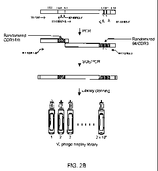
FIGURE 2B shows schematic steps in the construction of the human VH phage
display library.
FIGURE 3 shows a map of pMED1 phagemid vector, with the nucleotide sequence of
the
multiple cloning site and its immediate surroundings shown in (ii). RBS,
ribosome binding site;
L, left; R, right; HA, heaemagglutinin; fd, filamentous bacteriophage, fd.
FIGURE 4 shows size exclusion chromatograms of the VHS isolated by panning the
VH library
against a-amylase in a monovalent display format (A) or a multivalent display
format with a
heat denaturation step (B). (A) huVHAm455 (dotted line) precipitated highly
and thus gave low
absorbance signals. (B) huVHAm304: dotted-dashed line; huVHAm309: dotted line;
huVHAm428: solid line; huVHAm416: dashed line. (C) Expansion of Figure 4B to
show an
improved resolution of the peaks.
FIGURE 5 shows graphs illustrating the aggregation tendencies of VHS in terms
of the
percentage of their monomeric contents.
7
CA 02708074 2010-06-03
WO 2009/079793 PCT/CA2008/002273
FIGURE 6 shows steps in the determination of the identity of the amino acid
coded by the
amber codon at position 32 of huVHAm302. (A) Sequence of huVHAm302 as
determined by
mass spectrometry. Spaces define the boundaries between FRs and CDRs (see
Figure 2A).
The determined peptide sequences from the analysis of the tryptic digest of
huVHAm302 using
nanoRPLC-MS/MS are boldfaced (see also Figure 6B). The amber codon at position
32 was
found to code for an E (underlined). The N-terminus of huVHAm302 was
determined as
pyroglutamine (pyroQ). The N-terminal tryptic peptide sequence,
pyroQVQLVESGGGLIKPGGSLR (SEQ ID NO:179), was obtained from the MS/MS spectrum
of a prominent doubly protonated ion at m/z 939.50 (2+) (data not shown).
Moreover, the N-
terminal fragment ions from the CID of the protonated protein ion at m/z
1413.71(11+) showed
the N-terminus of huVHAm302 as pyroQ as well (data not shown). The determined
molecular
weight of the protein (15,541.2 Da) also indicated that the N-terminus of the
protein is
pyroglutamine. The C-terminal tryptic peptide ion at m/z 585.91 (3+) from
LSEEDLNHHHHHH
(SEQ ID NO: 180) was prominent in the survey scan of the DDA experiment.
Peptides having
amino acids attached after the C-terminal histidine were not observed. In
addition, collision
induced dissociation (CID) of the protein ion [M + 11H] 11+ at m/z 1413.71
(11+) was
performed and the C-terminal tryptic peptide sequence VTVSSGSEQKLSEEDLNHHHHHH
(SEQ ID NO:181) was obtained from the C-terminal fragment ions of the protein
(MS/MS data
not shown). (B) MS/MS spectrum of the doubly protonated ion at m/z 1036.47
(2+) for the
tryptic peptide LSCamAASGDTVSDESMTWVR (SEQ ID NO:13; residues 20-38 of
huVHAm302). The amber-coded amino acid, E, at position 32 is underlined. The
mass
spectrometry experiments also showed that the CDR3 Cys residues formed a
disulfide linkage.
FIGURE 7 shows SDS-PAGE analysis of VHS (huVHAm431, huVHAm416) isolated by
panning
the VH library against a-amylase by the heat denaturation method (arrow
denotes the disulfide-
mediated dimeric VH. R: reduced; NR: not reduced).
FIGURE 8 shows sensorgram overlays showing the binding of native (thick lines)
and refolded
(thin lines) huVHAm309 (A) and huVHAm416 (B) to immobilized protein A at 0.1,
0.2, 0.3, 0.4,
0.5, 1 and 2 pM (huVHAm309) and 0.1, 0.2, 0.3, 0.4, 0.5, 1, 2 and 4 pM
(huVHAm416).
FIGURE 9 shows binding analyses by ELISA of VHS identified by the heat
denaturation
panning approach against a-amylase, with (A) binding of VHS against
immobilized a-amylase
(dotted columns) and bovine serum albumin, BSA (checkered columns) and (B)
binding of
horseradish peroxidase-protein A conjugate to immobilized VHS and BSA control.
In both A
and B, binding to BSA is at a background level.
8
CA 02708074 2010-06-03
WO 2009/079793 PCT/CA2008/002273
FIGURE 10 shows aspects of determining enzyme inhibition activity of anti-a-
amylase VHS. (A)
a-amylase activity, measured as AA405 nm, as a function of time. A clear
inhibition can be
seen with the amylase binder huVHAm302 (filled square) and not with the
control VH HVHP430
(Filled triangle). (B) Residual activity of a-amylase in the presence of
various concentrations of
anti-a-amylase VHS. Only huVHAm302 acts as an enzyme inhibitor at all the VH
concentrations
tested. Filled square: huVHAm302; open square: huVHAm428; filled circle:
huVHAm304; open
circle: huVHAm416.
FIGURES 11A-F are graphs illustrating theoretical pl distribution for L. glama
cDNA VHHs of
subfamilies VHH1, VHH2 and VHH3, C. dromedarius cDNA VHHs, germline VHH
segments and
germline VH segments, human germline VH segments and the HVHP430 library VHS.
FIGURE 12A shows a sample of CDR3 sequences from the llama VHH CDR3 plasmid
library
with the CDR3 sequences derived from VHH2 subfamily marked by asterisks;
cysteine
residues are underlined. The numbering system is that described by Kabat et
al. (1991).
FIGURE 12B shows the length distribution of a sample of CDR3 sequences from
the llama
VHH CDR3 plasmid library; the horizontal line denotes both the mean CDR3
length as well as
the median (M).
FIGURE 13 shows a CDR3 length distribution of a sample of VHS from HVHP430LGH3
VH
phage display library, from which thirty-one VHS were analyzed; the horizontal
line denotes
both the mean CDR3 length as well as the median (M).
FIGURE 14 shows sequences for acidic human germline VH segments.
Detailed Description of the Invention
The present invention relates to antibody heavy chain variable domains. In
particular, the
invention relates to non-aggregating human VH domains and methods of isolating
same.
The present invention comprises non-aggregating human VH domains and libraries
thereof,
having at least one disulfide linkage-forming cysteine in at least one
complementarity-
determining region and having an acidic isoelectric point. The VH domain as
just described
may also be soluble, capable of reversible thermal unfolding, and/or capable
of binding to
protein A. The VH domain may comprise at least one cysteine in CDR1. The VH
domain as
described may comprise at least three cysteines in CDR3.
The VHS may display high solubility and/or reversible thermal unfolding. They
may also be
capable of binding to protein A. In a specific, non-limiting embodiment, the
human VH domain
has an isoelectric point of below 6. The VH domains and libraries thereof of
the present
9
CA 02708074 2010-06-03
WO 2009/079793 PCT/CA2008/002273
invention may further comprise an Asp or Glu at position 32 of H1/CDR1 or
other positions in
H1/CDR1 or in H1/CDR1, H2/CDR2 or H3/CDR3.
As used herein, "VH domain" or "VH" refers to an antibody heavy chain variable
domain. The
term includes naturally-occurring VH domains and VH domains that have been
altered through
selection or engineering to change their characteristics including, for
example, stability or
solubility. The term includes homologues, derivatives, or fragments that are
capable of
functioning as a VH domain.
As is known to one of skill in the art, a VH domain comprises three
"complementarity
determining regions" or "CDRs"; generally, each CDR is a region within the
variable heavy
chain that combines with the other CDR to form the antigen-binding site. It is
well-known in the
art that the CDRs contribute to binding and recognition of an antigenic
determinant. However,
not all CDRs may be required for binding the antigen. For example, but without
wishing to be
limiting, one, two, or three of the CDRs may contribute to binding and
recognition of the
antigen by the VH domains of the present invention. The CDRs of the VH domain
are referred
to herein as CDR1, CDR2, and CDR3.
The numbering of the amino acids in the VH domains of the present invention is
done
according to the Kabat numbering system, which refers to the numbering system
used for
heavy chain variable domains or light chain variable domains from the
compilation of
antibodies in Kabat et al., Sequences of Proteins of Immunological Interest,
5th Ed. Public
Health Service, National Institutes of Health, Bethesda, Md. (1991). This
system is well-known
to one of skill in the art, and may be determined for a given antibody by
alignment at regions of
homology of the sequence of the antibody with a "standard" Kabat numbered
sequence. The
positions of the CDRs in VHs, according to Kabat numbering are as follows:
CDR1 - residues
31-35B; CDR2 - residues 50-65; and CDR3 - residues 95-102.
VH domains are also characterized by hypervariable regions, labelled H1, H2
and H3, which
overlap the CDRs. H1 is defined as residues 26-32, H2 is defined as 52-56, and
H3 is defined
as residues 95-102 (http://www.bioinf.org.uk/abs/). The hypervariable regions
are directly
involved in antigen binding.
The VH domains and libraries thereof of the present invention comprise at
least one disulfide
linkage-forming cysteine in at least one CDR. By the term "disulfide linkage-
forming cysteine"
it is meant a cysteine that forms a disulfide bridge (also referred to as
"disulfide bond" or
"disulfide linkage") with another cysteine through oxidation of their thiol
groups. Without
wishing to be bound by theory, disulfide bridges help proteins and enzymes
maintain their
CA 02708074 2010-06-03
WO 2009/079793 PCT/CA2008/002273
structural configuration. In particular, VH domains comprise a canonical
(i.e., highly conserved)
disulfide bond between Cys 22 and Cys 92. In addition to this canonical
disulfide bond, the
VHS of the present invention comprise at least one non-canonical disulfide
bond. The latter
may be at any non-canonical position in the VH structure; for example, the non-
canonical
disulfide bond may be in the framework region, in a CDR, in the hypervariable
loop, or any
combination thereof.
In one embodiment, there is an even number of disulfide linkage-forming Cys.
For example,
and not wishing to be limiting in any manner, there may be at least one
disulfide linkage-
forming Cys in CDR1; in another non-limiting example, there may be at least
one Cys in
CDR3; in yet another non-limiting example, there may be at least three Cys in
CDR3. The
disulfide linkage-forming Cys of the VH domains may form intra-CDR disulfide
bonds or inter-
CDR disulfide bonds. For examples, and without wishing to be limiting, the Cys
residues in
CDR3 of VHS form intra-CDR disulfide linkages; in another non-limiting
example, the Cys
residues in CDR1 and CDR3 of VHS form inter-CDR disulfide linkages.
Furthermore, and without wishing to be bound by theory, the non-canonical
disulfide linkages
in the CDR of the VH of the present invention may be useful in producing
enzyme inhibitors;
specifically, the disulfide linkage(s) may form protruding CDR loops, and
particularly CDR3
loops, for accessing cryptic epitopes or enzyme active sites. Non-canonical
disulfide linkages
have also been shown to be important in single domain antibody stability
(Nguyen et al., 2000;
Harmsen et al., 2000; Muyldermans et al., 1994; Vu et al., 1997; Diaz et al.,
2002), as well as
in shaping the combining site for novel topologies and increased repertoire
diversity.
The generation of antibody-based inhibitors to enzymes and proteases that are
involved in the
pathobiology of a number of disease states is of particular interest from a
pharmaceutical
standpoint. Human VH domains are superior for therapeutic applications due to
their expected
lower immunogenicity, small size, and stability.
However, human VH domains tend to form high molecular weight aggregates in
solution.
These include structures that are not soluble as monomers and show non-
specific interactions
to other molecules or surfaces, sometimes refers to as "stickiness". A VH
domain can form
dimeric, or multimeric or high molecular weight aggregates, none of these are
desirable or
useful. The term "non-aggregating" refers to the reduced tendency or inability
of the VH
domain to form such aggregates. The VH domains of the present invention are
non-
aggregating. This is verified by elution on a gel filtration column, for
example but no limited to
SuperdexTM 75 column, where the VH domain is essentially monomeric. By
"essentially
monomeric", it is mean that 95%, 96%, 97%, 98%, 99%, or 100% of the VH domains
elute as
11
CA 02708074 2010-06-03
WO 2009/079793 PCT/CA2008/002273
monomers. Preferably, the non-aggregating VH domains of the present invention
are stable
and do not precipitate over time.
The VH domains and libraries thereof of the present invention also have acidic
pl. The term
"pl" or "isoelectric point" means the pH at which the VH domain carries no net
electrical charge.
Generally, solubility is at a minimum when the pH is at the pl. An acidic
isoelectric point may
be below 7; for example, the acidic pl may be below 7, 6, 5, 4, 3, 2, or 1, or
any value
therebetween, or within a range described by these values; in a non-limiting
example, the pl of
the VH domains of the present invention is below 6. A neutral pl is 7, and a
basic pl is above 7.
Without wishing to be limiting, the acidic pl of the VH domains of the present
invention
originates primarily from non-randomized regions, including, for example, the
framework
regions.
The "solubility" of the VH of the present invention refers to its ability to
dissolve in a solvent, as
measured in terms of the maximum amount of solute dissolved in a solvent at
equilibrium. The
VH of the present invention is soluble in monomeric form, with no stickiness.
The VH domains
as presently described are soluble in an aqueous buffer, for example, but not
limited to Tris
buffers, PBS buffers, HEPES buffers, carbonate buffers, or water.
The VH of the present invention may also exhibit "reversible thermal
unfolding". Thermal
unfolding refers to the temperature-induced unfolding of a molecule from its
native, folded
conformation to a secondary, unfolded conformation. Thermal unfolding is
reversible if the
molecule can be restored from the secondary, unfolded conformation to its
native, folded
conformation. Reversible thermal unfolding is measured by the thermal
refolding efficiency
(TRE) of a molecule. The non-aggregating VH domains as described above may
show higher
THE than aggregating VH domains and refold to their native state more
efficiently. The
temperature at which the present VHS unfold will vary depending on the nature
of the VH and on
its melting temperature. In general, most VH will be unfolded at temperatures
above 60 C,
above 85 C, or above 90 C. In a non-limiting example, the VHS of the present
invention may
be able to regain antigen specificity following prolonged incubations at
temperatures above
80 C, or even above 90 C.
The VHS may also bind to protein A, a molecule well-known to those of skill in
the art. Protein
A is often coupled to other molecules without affecting the antibody binding
site; for example,
and without wishing to be limiting, protein A may be coupled to fluorescent
dyes, enzymes,
biotin, colloidal gold, radioactive iodine, and magnetic, latex, and agarose
beads. Protein A
can also be immobilized onto a solid support and used as a reliable method for
purifying
immunoglobulin from mixtures - for example from serum, ascites fluid, or
bacterial extract - or
12
CA 02708074 2010-06-03
WO 2009/079793 PCT/CA2008/002273
coupled with one of the above molecules to detect the presence of antibodies.
The ability of
VHS of the present invention to bind to protein A may be exploited for VH
purification and
detection in diagnostic tests, immunoblotting and immunocytochemistry.
Libraries of VH domains are also encompassed by the present invention. The VH
domain
libraries may include a variety of display formats, including phage display,
ribosome display,
microbial cell display, yeast display, retroviral display, or microbead
display formats or any
other suitable format.
Analysis of the VHS of the present invention and naturally occurring camelid
VHH and shark
VNAR single-domain antibodies show analogies in displaying high solubility and
reversible
thermal unfolding. It is presently found, through analysis of pl (see Example
8), that camelid
VHH pools have an abundance of clones with acidic pl (53% acidic versus 43%
basic). In
germline clones (C. dromedaries), the VH pool is predominantly comprised of VH
segments of
basic pl, while the opposite is true of the VHH pool, which is predominately
populated with VHH
segments of acidic pl. It is also presently observed that an overwhelming
majority of VH
segments (92%) in the human germline VH pool are basic. Thus, a clear
correlation has been
presently identified between VH solubility and acidic pl; while not all the
non-aggregating VHS
are acidic, the acidic VHS are non-aggregating. Therefore, the proportion of
non-aggregating
VHS in a library can be increased by using an acidic scaffold for library
construction and/or
biasing randomization towards acidic residues and/or against basic ones.
The VH domains and libraries thereof of the present invention may further
comprise an acidic
amino acid in CDR1, CDR2, and/or CDR3. For example, and without wishing to be
limiting, VH
domains and libraries thereof may comprise Asp or Glu at position 32 of
H1/CDR1, or at other
positions in H1/CDR1 or in H1/CDR1, H2/CDR2 or H3/CDR3.
The VH domain and libraries thereof of the present invention may be based on
any appropriate
VH sequence known in the art. By the term "based on", it is meant that the VH
domain is
obtained by the methods of the present invention using a "scaffold" as the
initial VH domain. A
person of skill in the art would readily understand that, while a VH domain
library may be based
on a single scaffold, or a number of scaffolds, the CDR/hypervariable loops
may be
randomized. As such, a large number of VH domains with sequences varying in
the
randomized regions may be obtained; this is known in the art as a "pool" or
"library" of VH
domains. The VH domains in the pool VH domains may each recognize the same or
different
epitopes. Additionally, the scaffolds upon which the VH domains of the present
invention are
based may possess one or more of the characteristics of non-aggregating VH
domains, as
described above.
13
CA 02708074 2010-06-03
WO 2009/079793 PCT/CA2008/002273
In a particular non-limiting example, the VH domains of the present invention
are based on VH
sequences having an acidic pl. The VH domains of the present invention may be
based on
any human germline sequences with acidic pl, in particle those from the VH3
family, and more
particularly those with protein A binding activity; for example, but not to be
considered limiting,
the VH domain may be based on human germline sequence 1-f VH segment, 1-24 VH
segment
and 3-43 VH segment (see Figure 14; SEQ ID NOs: 182-184). Alternatively, the
VH domain
may be based on camelid VH cDNAs or camelid germline VH segments with acidic
pls. The
acidic camelid germline VH segments used as library scaffold can be any of
those known in the
art; in a specific, non-limiting example, the VH segments may be those
described in Nguyen et
al., 2000. In yet another alternative, the VHS and the libraries thereof
presently described may
be based on camelid VHH cDNAs or camelid germline VHH segments with acidic
pls. The
acidic camelid VH or VHH cDNA or germline sequences used as library scaffold
can any of
those known in the art; for example, but not limited to those described in
Harmsen et al.
(2000), Tanha et al. (2002), those in the pool of VHHs with NCBI Accession
numbers
AB091838-ABO92333, in Nguyen et al. (2000), or those in the VBASE database of
human
sequences (Medical Research Council, Centre for Protein Engineering).
The VH domain and libraries thereof of the present invention may also be based
on a scaffold
further comprising an acidic amino acid in CDR1, CDR2, and/or CDR3. In a non-
limiting
example, the scaffold may comprise Asp or Glu at position 32 of H1/CDR1, or at
other
positions in H1/CDR1 or in H1/CDR1, H2/CDR2 or H3/CDR3.
The VH domains and libraries thereof of the present invention may further be
based on
chimeric scaffolds; for example, and without wishing to be limiting, the
chimeric scaffolds may
comprise one or more camelid or shark CDR/hypervariable loop sequences on
human
framework sequences. In a specific, non-limiting example, the chimeric
scaffold comprises a
camelid CDR3/H3 loop on a human VH framework (human CDR1/H1 and CDR2/H2).
Chimeric
antibody domains are well-known in the art, as are the methods for obtaining
them.
In a specific non-limiting example, the present invention provides a VH domain
or library
thereof, wherein a) the VH domain is based on HVHP430 (SEQ ID NO:1); b) the
Cys at
positions 99 and 100d of CDR3 are maintained; c) the remaining 14 amino acid
residues of
CDR3 are randomized; d) amino acid residue 94 is randomized; and e) the 8
amino acid
residues of CDR1/H1 are randomized. In a further non-limiting example, there
is provided a VH
domain library, wherein a) the VH domain is based on HVHP430 (SEQ ID NO:1); b)
the amino
acid residues at 93-102 (93/94-CDR3) positions are derived from llama VHHs; c)
the 8 amino
acid residues of CDR1/H1 are randomized. In yet another non-limiting example,
a VH domain
or library thereof is provided, wherein a) the VH domain is based on HVHP430
(SEQ ID NO:1);
14
CA 02708074 2010-06-03
WO 2009/079793 PCT/CA2008/002273
b) the CDR3 comprises a sequence selected from SEQ ID NOs:24-90 and SEQ ID
NOs:33-63;
c) the 8 amino acid residues of CDR1/H1 are randomized.
The proportion of non-aggregating VHS in the libraries of the present
invention, as described
above, may be greater than in conventional libraries.
In yet another aspect, the VH domains and libraries thereof of the present
invention may be
mixed randomized libraries. In this type of library, the CDRs are produced in
vitro by using
randomized oligonucleotides and methods known in the art.
Using a method of the present invention, non-aggregating, refoldable VHS were
isolated in one
example. Among these, three had acidic pl and two had a CDR1 Cys residue that
formed inter
CDR1-CDR3 disulfide linkages. In addition, three VHS with a pair of Cys in
their CDR3 (as well
as the parent scaffold, HVHP430) formed intra-CDR3 disulfide linkages.
However, in one
embodiment, the VHS of the present invention comprising non-canonical
disulfide linkage
spanning CDR1 to CDR3 refold to their native structure more efficiently than
those with intra-
CDR3 disulfide linkages or only the canonical disulfide bond between Cys22 and
at Cys92
during the refolding step of the panning. Therefore, these VHS may be
favorably selected
during the binding step of the panning. Additionally, most non-aggregating,
refoldable VHS
have theoretical pls below 6, possibly due to the fact that above pl 6 (and
especially closer to
pl 7) VHS become aggregation-prone, as their net charge approaches zero. Among
the nine
VHS isolated by the heat-denaturation method, three of the four VHS with
lowest solubility had a
pl around 7.0 (6.4-7.3).
The VH of the present invention may be any VH that exhibits the desired
characteristics, as
described herein. In a specific, non-limiting example, the human VH domain may
comprise one
of huVHAm302 (SEQ ID NO:15), huVHAm309 (SEQ ID NO:17), huVHAm316 (SEQ ID
NO:19), huVHAm303 (SEQ ID NO:164), huVHAm304 (SEQ ID NO:16), huVHAm305 (SEQ ID
NO:15165 huVHAm307 (SEQ ID NO:166), huVHAm311 (SEQ ID NO:167), huVHAm315 (SEQ
ID NO:18), huVHAm301 (SEQ ID NO:163), huVHAm312 (SEQ ID NO:168), huVHAm320
(SEQ ID NO:171), huVHAm317 (SEQ ID NO:170), huVHAm313 (SEQ ID NO:169),
huVHAm431 (SEQ ID NO:23), huVHAm427 (SEQ ID NO:21), huVHAm416 (SEQ ID NO:20),
huVHAm424 (SEQ ID NO:175), huVHAm428 (SEQ ID NO:22), huVHAm430 (SEQ ID
NO:176), huVHAm406 (SEQ ID NO:172), huVHAm412 (SEQ ID NO:173) or huVHAm420
(SEQ ID NO:174). In another non-limiting example, the human VH domain or
libraries thereof
comprises a sequence selected from any of SEQ ID NOS: 101 to 131, or 132-162,
or a
sequence selected from any of those shown in Figure 12A (SEQ ID NOs:24-90), or
combinations thereof.
CA 02708074 2010-06-03
WO 2009/079793 PCT/CA2008/002273
The VH domain as described herein may be obtained by the novel methods
described below.
In a non-limiting example, the VH domain may be isolated from a phagemid-based
phage-
display library. The use of a fully-synthetic designed phagemid-based phage
display library,
followed by selection characterized by enrichment for human VHS with the
desired properties
mentioned herein, is an approach that has not been previously used for human
VHS.
In one embodiment, the present invention provides a method of increasing the
power or
efficiency of selection of non-aggregating VH domains by:
a) providing a phagemid-based VH domain phage-display library, wherein the
library is
produced by multivalent display of VH domains on the surface of phage; and
b) panning, using the phage-VH domain library and a binding target,
wherein the method comprises a step of selection of non-aggregating phage-VH
domains. In
one example, the selection step may occur prior to the step of panning and may
comprise
subjecting the phage-VH domain library to a heat denaturation/re-naturation
step. Alternatively,
the selection step may occur following panning and may comprise sequencing
individual
clones to identify the VH with acidic pls. In another alternative, both the
heat denaturation/re-
naturation step and the sequencing step are performed.
For example, and without wishing to be limiting, the method of increasing the
power or
efficiency of selection of non-aggregating VH domains may comprise:
a) providing a phagemid-based VH domain phage-display library, wherein the
library is
produced by multivalent display of VH domains on the surface of phage;
b) subjecting the phagemid-based VH domain phage-display library to a heat
denaturation/re-naturation step; and
c) panning, using the phage- VH domain library and a target.
In another non-limiting example, the method of increasing the power or
efficiency of selection
of non-aggregating VH domains may comprise:
a) providing a phagemid-based VH domain phage-display library, wherein the
library is
produced by multivalent display of VH domains on the surface of phage;
b) panning, using the phage- VH domain library and a target; and
c) sequencing individual clones to identify VH domains having an acidic pl,
16
CA 02708074 2010-06-03
WO 2009/079793 PCT/CA2008/002273
The method as described above may comprise subsequent rounds of panning; for
example, 2,
3, 4, 5, 6, 7, 8, 9, or 10 rounds of panning may be performed. The method as
described may
also comprise isolation of specific VH domains by amplifying the nucleic acid
sequences coding
for the VH domains; cloning the amplified nucleic acid sequences into an
expression vector;
transforming host cells with the expression vector under conditions allowing
expression of
nucleic acids coding for VH domains; and recovering the VH domains having the
desired
specificity.
The phagemid-based VH domain phage-display library may be prepared by any
method known
in the art. For example, and without wishing to be limiting, the library may
be prepared by
inserting phagemids, each comprising a nucleic acid encoding a VH domain, into
a bacterial
species; contacting the bacterial species with a hyperphage and subjecting the
bacterial
species to conditions for infection; and, subjecting the phagemid-inserted and
hyperphage-
infected bacterial species to conditions for production of a phage- VH domain
library.
The "phagemid" used in the method of the present invention is a vector derived
by modification
of a plasmid, containing an origin of replication for a bacteriophage as well
as the plasmid
origin of replication. The phagemids comprise the filamentous bacteriophage
gill or a fragment
thereof; in this example, the nucleic acid encoding the VH domain is expressed
in fusion with
the full or truncated gill product (pill) and displayed through the pill on
the phage particle. The
phagemids also comprise a nucleic acid encoding a VH domain; each phagemid may
comprise
a nucleic acid encoding various members of a pool of VH domains. The insertion
of the
phagemids into the bacterial species may be done by any method know in the
art.
The VH domain encoded in the phagemids may be based on any appropriate VH
sequence.
The VH domain scaffold may be any suitable scaffold known in the art. In a
particular non-
limiting example, the VH domains of the present invention are based on VH
sequences having
an acidic pl. The VH domains of the present invention may be based on any
known human
germline sequences with acidic pl, in particle those from the VH3 family, and
more particularly
those with protein A binding activity; for example, but not to be considered
limiting, the VH
domain may be based on human germline sequence 1-f VH segment, 1-24 VH segment
and 3-
43 VH segment (see Figure 14). Alternatively, the VH domain may be based on
camelid VH
cDNAs or camelid germline VH segments with acidic pls. The acidic camelid
germline VH
segments used as library scaffold can be any of those known in the art; in a
specific, non-
limiting example, the VH segments may be those described in Nguyen et al.,
2000. In yet
another alternative, the VHs and the libraries thereof presently described may
be based on
camelid VHH cDNAs or camelid germline VHH segments with acidic pls. The acidic
camelid
VHH cDNA used as library scaffold can any of those known in the art; for
example, but not
17
CA 02708074 2010-06-03
WO 2009/079793 PCT/CA2008/002273
limited to the VH segments may be those described in Harmsen et al. (2000),
Tanha et al.
(2002), those in the pool of VHHs with NCBI Accession numbers AB091838-
AB092333, or in
Nguyen et al. (2000). Various other scaffolds on which the VH domains can be
based are
described above. As would be recognized by those of skill in the art, while
the VH domains in
the library may be based on a scaffold, a large number of different VH domains
are present in
the library due to randomization of selected regions. The proportion of non-
aggregating VHS in
the library of the present invention may be greater than in conventional
libraries.
The phagemid may be inserted into any suitable bacterial species and strain; a
person of skill
in the art would be familiar with such bacterial species and strains. Without
wishing to be
limiting, the bacterial species may be, for example, E. coli; in another non-
limiting example, the
E. coli strain may be TG1, XL1-blue, SURE, TOP10F', XL1-Blue MRF', or ABLE K.
Methods
for inserting the phagemid into the bacterial species are well known to those
in the art.
In the method of the present invention as just described above, the library
used is produced by
multivalent display of VH domains on the surface of phage. This may be
accomplished by
contacting the bacterial species, into which the phagemid has been inserted,
with a
hyperphage and subjecting the bacterial species to conditions for infection.
"Hyperphage" are a type of helper that have a wild-type pill phenotype and are
therefore able
to infect F(+) Escherichia coli cells with high efficiency; however, their
lack of a functional pIll
gene means that the phagemid-encoded pIll-antibody fusion is the sole source
of pIll in phage
assembly. This results in a considerable increase in the fraction of phage
particles carrying an
antibody fragment on their surface and leads to phage particles displaying
antibody fragments
multivalently. In one non-limiting example, the hyperphage may be M13KO7Aplll.
However,
other suitable homologues can be used in the method of the present invention;
for example,
and without wishing to be limited in any manner, Ex-phage (Baek et al, 2002)
or Phaberge
(Soltes et al, 2003).
The conditions under which the bacterial species are infected by hyperphage
are well known in
the art; for example, and without wishing to be limiting in any manner, the
conditions may be
those described in Arbabi-Ghahroudi, et al. (2008) or Rondot et al. (2001), or
any other
conditions suitable for infection of the bacteria by the hyperphage.
The infected bacterial species is then submitted to conditions for production
of a phage-VH
domain library. Such conditions are well known in the art; for example, and
without wishing to
be limiting, suitable conditions are described in (Arbabi-Ghahroudi, et al.,
2008; Harrison, et
al., 1996).
18
CA 02708074 2010-06-03
WO 2009/079793 PCT/CA2008/002273
In the method of the present invention, panning is performed using the phage-
VH domain
library and a target. As is known to a person of skill in the art, "panning"
refers to a process in
which a pool of filamentous phage-displayed antibody libraries (for example,
the phage- VH
domain library of the present invention) is exposed to the target (or
"antigen") of interest. The
target may be either fixed or available, or may be on a solid surface, in
solution, on the cell
surface, or any other suitable format. The non-binding phage-antibodies may be
removed by
various methods, including washing extensively with buffer containing
detergents such as
Tween 20; alternatively, phage bound to a biotinylated target may be captured
out by
streptavidin magnetic beads. The bound phage-antibodies may then be eluted
from the target
by methods well-known in the art. The eluted phage-antibodies may then be
amplified
(propagated) in F+ bacterial host. The process of selection and amplification
may be
performed in one or more than one round of panning; for example, 2, 3, 4, 5,
6, 7, 8, 9, or 10
rounds of panning may be performed. This results in specific enrichment of
antibody-phage
binders to the target and leads to the isolation of mono-specific antibody
(for instance VH
domains). Conditions for panning are well-known to those of skill in the art;
for example, the
conditions may be those described in Marks et al (1991), Griffiths et al
(1994), or Sidhu et al
(2004), Hoogenboom (2002), Bradbury (2004) or any other suitable conditions.
The "target" used in the panning step may be any appropriate selected target.
For example,
the target may be a substantially purified antigen, antigen conjugated to
molecules such as
biotin or similar molecules, a partially-purified antigen, a cell, a tissue;
the target may also be
may be either fixed or available, or may be on a solid surface, in solution,
on the cell surface,
or any other suitable format (see Hoogenboom, 2005). The conjugation of
antigen with, for
example biotin, make the selection step straightforward and more efficient and
required much
lower amount of purified antigen. The target may also be selected based on the
desired
specificity of the resulting phagemid-based VH domain phage-display library or
of the VH
domains. The target may be any type of molecule of interest; for example, the
target may be
an enzyme, a cell-surface antigen, TNF, interleukins, molecules in the ICAM
family etc. A
person of skill in the art would readily understand that the VH domain
libraries obtained by the
methods described herein can be directed toward any target of interest or of
therapeutic
importance. For example, and without wishing to be limiting in any manner, the
enzyme may
be a-amylases, carbonic anhydrases, or lysozymes.
A method of the present invention may further comprises a step of selection of
non-
aggregating phage-VH domains.
In one embodiment, the selection step may occur prior to the step of panning
and may
comprise subjecting the phage-VH domain library to a heat denaturation/re-
naturation step.
19
CA 02708074 2010-06-03
WO 2009/079793 PCT/CA2008/002273
This step involves thermal unfolding of the VH domains, with subsequent
refolding to their
native conformation, and is undertaken by any method know in the art; see for
example
Jespers et al (2004). For example, and without wishing to be limiting, the
phage- VH domain
library may be subjected to denaturation at a temperature in the range of
about 55 C to about
90 C; the temperature may be 55, 60, 65, 70, 75, 80, 85, or 90 C, or any
temperature
therebetween. In one embodiment, the phage- VH domain library is maintained at
this elevated
temperature for a time in the range of about 1 minute to about 30 minutes; for
example and
without wishing to be limiting, the temperature may be maintained for 1, 2, 3,
4, 5, 6, 7, 8, 9,
10, 11, 12, 13, 14, 15, 16, 17, 18, 19, 20, 21, 22, 23, 24, 25, 26, 27, 28, 29
or 30 minutes, or
any time therebetween. Subsequently, phage- VH domain library is subjected to
renaturation
by returning the temperature to a lower temperature, for example room
temperature or lower, 4
or 5 C, for an amount of time similar to that used for denaturation. A person
of skill in the art
will recognize that the temperature at which the VHS in the phage- VH domain
library denature
will depend on the nature of the VH domain(s) and their melting temperature.
Furthermore, the
skilled person will understand that, in some embodiments, higher denaturation
temperatures
may be combined with shorter exposure times; similarly, in other embodiments,
lower
denaturation temperatures may be combined with longer exposure times. The
denaturation/renaturation step may be performed in any appropriate aqueous
buffer know in
the art; for example, and without wishing to be limiting in any manner, the
buffer may be a Tris
buffer, PBS buffer, HEPES buffer, carbonate buffer, or water.
In another embodiment, the method may comprise the step of sequencing
individual clones to
identify VHS with acidic pls. This screening step of non-aggregating VH
domains is based on
theoretical pl values, which may be determined by any method known in the art.
For example,
and without wishing to be limiting, the theoretical pis may be determined by
commercially
available software packages. As described previously, the present invention
has shown that
VH having an acidic pl may be soluble and non-aggregating. Screening non-
aggregating VH
domains from among the aggregating VHS based on pl values obtained simply by
DNA
sequencing avoids the need for subcloning, expression, purification and
biophysical
characterization of a large number of VHS.
In a further embodiment, both the heat denaturation/re-naturation step and the
sequencing
step are performed.
The method as described herein may also comprise isolation of specific VH
domains by
amplifying the nucleic acid sequences coding for the VH domains in the
recovered phage-VH
domains; cloning the amplified nucleic acid sequences into an expression
vector; transforming
host cells with the expression vector under conditions allowing expression of
nucleic acids
CA 02708074 2010-06-03
WO 2009/079793 PCT/CA2008/002273
coding for VH domains; and recovering the VH domains having the desired
specificity. Methods
and specific conditions for performing these steps are well-known to a person
of skill in the art.
The method as described above is a novel combination of using a phagemid-
vector based
phage-display produced by the use of hyperphage and a selection step based on
heat
denaturation or analysis of theoretical pis. This novel method can increase
the efficiency for
selection of non-aggregating human VHS. In a non-limiting example, the present
method may
select VH domains comprising non-canonical disulfide bonds, as described
above; without
wishing to be limiting, the non-canonical disulfide bonds may occur in CDR1
and/or CDR3. In
another example, the method as described above may select VH domains with
acidic pis.
Compared to phage vector-based systems, the phagemid vector-based phage
display system
of the present method provides many advantages including: ease of constructing
large libraries
which is desirable in the case of non-immune libraries; suitability for
isolating high affinity
binders from immune or affinity maturation libraries; ease of manipulation for
improving affinity
or biophysical properties; and facile switching from antibody-pIll fusion to
un-fused antibody
fragments for rapid antibody expression and analysis. In addition, use of
helper phages that
result in a multivalent display (Rondot et al., 2001; Baek, H. et al., 2002;
Soltes, G. et al.,
2003), e.g., hyperphage (M13KO7ApIII) in the method of the present invention
provides the
advantages afforded by the phage vector-based display systems (due to the
avidity effect),
including: high yield of binders and fewer rounds of panning (O'Connell et
al., 2002); a more
efficient enrichment of antibodies for cell-surface antigens; and suitability
for selecting
antibodies to cell surface receptors that require self-cross linking (Becerril
et al., 1999; Huie et
al., 2001). Moreover, with the phagemid vector system, switching between
monovalent and
multivalent formats can be readily made by using the appropriate type of
helper phage (Rondot
et al., 2001; O'Connell et al., 2002; Kirsch et al., 2005). In order to
further leverage the
advantages phagemid-based libraries offer in terms selecting for non-
aggregating VHS, we
decided to employ hyperphage technology (Rondot et al.,2001) to adapt the heat
denaturation
strategy described above (Jespers et al., 2004) to phagemid-based libraries.
In yet another embodiment, the present invention provides a method of
increasing the power
or efficiency of selection of non-aggregating VH domains by, comprising:
a) providing a phage vector-based VH domain phage-display library, wherein the
library is
produced based on a VH domain scaffold having an acidic pl;
b) panning, using the phage- VH domain library and a target; and
c) sequencing individual clones to identify VH domains having an acidic pl
21
CA 02708074 2010-06-03
WO 2009/079793 PCT/CA2008/002273
The phage vector-based VH domain phage-display library may be prepared by any
method
known in the art. For example, and without wishing to be limiting, the library
may be prepared
by inserting phage vectors, each comprising a nucleic acid encoding a VH
domain, into a
bacterial species; and, subjecting the phage vector-inserted bacterial species
to conditions for
production of a phage-VH domain library.
A "phage vector" refers to a vector derived by modification of a phage genome,
containing an
origin of replication for a bacteriophage, but not one for a plasmid; the
phage vector may or
may not have an antibiotic resistance marker.
The methods for producing a phage vector-based phage-display library are well-
established in
the art, and would be well-known to the skilled person.
The method as described herein may also comprise isolation of specific VH
domains by
amplifying the nucleic acid sequences coding for the VH domains in the
recovered phage-VH
domains; cloning the amplified nucleic acid sequences into an expression
vector; transforming
host cells with the expression vector under conditions allowing expression of
nucleic acids
coding for VH domains; and recovering the VH domains having the desired
specificity. Methods
and specific conditions for performing these steps are well-known to a person
of skill in the art.
The present invention is also directed to VHS of the present invention that
are fused to a cargo
molecule. As used herein, a "cargo molecule" refers to any molecule for the
purposes of
targeting, increasing avidity, providing a second function, or otherwise
providing a beneficial
effect. The cargo molecule(s) may have the same or different specificities as
the VHS of the
invention. For example, and without wishing to be limiting, the cargo molecule
may be: a toxin,
an Fc region of an antibody, a whole antibody, or enzyme as in the context of
antibody-
directed enzyme pro-drug therapy (ADEPT) (Bagshawe, 1987: 2006); one or more
than one
single domain such as VH, VL, VHH, VNAR, etc with the same or different
specificities; a
liposome for targeted drug delivery; a therapeutic molecule, a radioisotope;
or any other
molecule providing a desired effect. Methods of coupling or attaching a cargo
molecule to a
VH domain of the present invention are well-known to those skilled in the art.
The methods and VH domain libraries of the present invention need not be
limited to phage-
display technologies, but may also be extended to other formats. For example,
and without
wishing to be limiting, the methods and VH domain libraries of the present
invention may be
ribosome and mRNA display, microbial cell display, retroviral display,
microbead display, etc.
(see Hoogenboom, 2005). Conditions for performing these types of display
methods are well-
known in the art.
22
CA 02708074 2010-06-03
WO 2009/079793 PCT/CA2008/002273
The VHS of the present invention may also be recombinantly produced in
multimeric form; in a
non-limiting example, the VHS may be produced, as dimers, trimers, pentamers,
etc.
Presentation of the VHS of the present invention in multimeric form(s) may
increase avidity of
the VHS. The monomeric units presented in the multimeric form may have the
same or
different specificities.
The present invention further encompasses nucleic acids encoding the VHS of
the present
invention. As used herein, a "nucleic acid" or "polynucleotide" includes a
nucleic acid, an
oligonucleotide, a nucleotide, a polynucleotide, and any fragment, variant, or
derivative thereof.
The nucleic acid or polynucleotide may be double-stranded, single-stranded, or
triple-stranded
DNA or RNA (including cDNA), or a DNA-RNA hybrid of genetic or synthetic
origin, wherein
the nucleic acid contains any combination of deoxyribonucleotides and
ribonucleotides and
any combination of bases, including, but not limited to, adenine, thymine,
cytosine, guanine,
uracil, inosine, xanthine and hypoxanthine. The nucleic acid or polynucleotide
may be
combined with a carbohydrate, a lipid, a protein, or other materials. A
nucleic acid sequence
of interest may be chemically synthesized using one of a variety of techniques
known to those
skilled in the art, including, without limitation, automated synthesis of
oligonucleotides having
sequences which correspond to a partial sequence of the nucleotide sequence of
interest, or a
variation sequence thereof, using commercially-available oligonucleotide
synthesizers, such as
the Applied Biosystems Model 392 DNA/RNA synthesizer.
The nucleic acids of the VHS of the present invention may be comprised in a
vector. Any
appropriate vector may be used, and those of skill in the art would be well-
versed on the
subject.
The present invention also provides host cells comprising the nucleic acid or
vector as
described above. The host cell may be any suitable host cell, for example, but
not limited to E.
coli, or yeast cells. Non-limiting specific examples of suitable E. coli
strains are: TG1,
BL21(DE3), and BL21(DE3)pLysS.
The VH domains of the present invention may possess properties that are
desirable for clinical
and diagnostic applications. In one embodiment, the VHS may be labelled with a
detectable
marker or label. Labelling of an antibody may be accomplished using one of a
variety of
labelling techniques, including peroxidase, chemiluminescent labels known in
the art, and
radioactive labels known in the art. The detectable marker or label of the
present invention
may be, for example, a non-radioactive or fluorescent marker, such as biotin,
fluorescein
(FITC), acridine, cholesterol, or carboxy-X-rhodamine, which can be detected
using
fluorescence and other imaging techniques readily known in the art.
Alternatively, the
23
CA 02708074 2010-06-03
WO 2009/079793 PCT/CA2008/002273
detectable marker or label may be a radioactive marker, including, for
example, a radioisotope.
The radioisotope may be any isotope that emits detectable radiation.
Radioactivity emitted by
the radioisotope can be detected by techniques well known in the art. For
example, gamma
emission from the radioisotope may be detected using gamma imaging techniques,
particularly
scintigraphic imaging. In addition, detection can also be made by fusion to a
green fluorescent
protein (GFP), RFP, YFP, etc.
The VHS of the present invention may also be used in a high-throughput
screening assay, such
as microarray technology, in which the use of the VH domain is advantageous or
provides a
useful alternative compared to conventional IgG.
In another aspect, the invention provides a pharmaceutical composition
comprising one or
more than one VHS in an effective amount for binding thereof to an antigen,
and a
pharmaceutically-acceptable excipient. Appropriate pharmaceutical excipients
are well-known
to those of skill in the art.
In a further embodiment, the invention provides a method of treating a
patient, comprising
administering a pharmaceutical composition comprising one or more VHS to a
patient in need
of treatment. For those in the art, it is apparent that libraries such as
those disclosed herein
may be a source of binders to targets. Therefore, they can be used for
therapy, diagnosis and
detection. Indications that can be targeted by VH domains of the present
invention are cancer
(for detection of tumor markers and/or treatment of any cancer), inflammatory
diseases (which
include killing the target cells, blocking molecular interactions, modulating
target molecules by
antibodies), autoimmune diseases (for example, lupus, rheumatoid arthritis
etc.),
neurodegenerative diseases (for example Parkinson's disease, Alzheimer's
disease, etc)
infectious disease caused by prion, viral, bacterial and fungi agents or, in
general, any
infectious disease resulted from infection by any known or unknown
microorganism or agent.
Targets may include any molecules that are specific to a given disease state.
For example,
and not wishing to be limiting in any manner, the targets may include: cell-
surface antigens,
enzymes, TNF, interleukins, molecules in the ICAM family etc. The libraries
obtained in
accordance to the present invention may also be used to obtain VH domains for
detecting
pathogens. Pathogens can include human, animal or plant pathogens such as
bacteria,
eubacteria, archaebacteria, eukaryotic microorganisms (e.g., protozoa, fungi,
yeasts, and
moulds), prions, viruses, and biological toxins (e.g., bacterial or fungal
toxins or plant lectins).,
A person of skill in the art would readily understand that the VH domain
libraries obtained by
the methods described herein can be directed toward any target of interest. In
a non-limiting
example, the target may be an enzyme; in a further example, and without
wishing to be
limiting, the enzyme may be lysozymes, a-amylases or carbonic anyhdrases.
24
CA 02708074 2010-06-03
WO 2009/079793 PCT/CA2008/002273
In yet another aspect, the invention contemplates the provision of a kit
useful for the detection
and determination of binding of one or more than one VH to a particular
antigen in a biological
sample. The kit comprises one or more than one VH and one or more reagents.
The one or
more than one VH domain may be labelled. Additionally, the kit may also
comprise a positive
control reagent. Instructions for use of the kit may also be included.
The VH domains of the present invention may also be used in antibody
microarray technology.
This technology is an alternative to traditional immunoassays, and many
thousands of assays
can be run in parallel. Antibody VH domains are favoured over whole IgG in
this type of assay
since they are small, stable and highly specific reagents. Methods for
antibody microarrays
are well-known in the art.
The present invention will be further illustrated in the following examples.
However, it is to be
understood that these examples are for illustrative purposes only and should
not be used to
limit the scope of the present invention in any manner.
Examples
Unless indicated otherwise, molecular biology work was done using standard
cloning
techniques (Sambrook et al., 1989). Phagemid pHEN4 (Arbabi-Ghahroudi et al.,
1997) was
modified by introducing a second non-compatible Sfi I site and six His codons.
The new vector
designated pMED1 was used for phage display library construction. pSJF2H
plasmid was
used for soluble expression of single domain antibodies in E. coli. pSJF2H is
identical to
pSJF2 (Tanha et al., 2003), except that it expresses proteins in fusion with
His6 instead of Hiss.
Example 1: HVHP430 VH Library Construction
A fully-synthetic, phagemid-based human VH phage display library was
constructed.
In constructing the VH library on the HVHP430 scaffold (Fig 2A, SEQ ID NO:1),
the two CDR3
Cys were maintained to promote the formation of intra-CDR disulfide linkage
and thus, to
increase the frequency of enzyme-inhibiting VHS in the library. The remaining
14 CDR3
positions, position 94 as well as eight H1/CDR1 positions were randomized
(Figure 2A).
CDR2 was left untouched as it has been shown to be involved in protein A
binding (Randen et
al., 1993; Bond et al., 2003). Besides, CDR2-lacking VNARs (Stanfield et al.,
2004) or camelid
VHHs utilizing their CDR1 and CDR3 (Decanniere et al., 1999) or just CDR3
(Desmyter et al.,
2001) for antigen recognition demonstrate nanomolar affinities. The library
was constructed
with a phagemid vector (Figure 3) according to the scheme shown in Figure 2B.
CA 02708074 2010-06-03
WO 2009/079793 PCT/CA2008/002273
The human VH HVHP430 (To et al., 2005), which has two Cys residues in its CDR3
at position
99 and 100d, was used as the framework to construct a library by randomizing
residues in
CDRI and CDR3 and position 94. Using a plasmid containing the HVHP430 gene as
template
and the primer pairs HVHBRI-R/HVHFR2-F and HVHBR3-R/HVHFR5-F (see Table 1 for
listing of primers used), two overlapping fragments with randomized H1/CDR1
and 94/CDR3
codons, respectively, were constructed by standard polymerase chain reactions
(PCRs).
Table 1. List of the primers used for VH clonings.
Designation Sequence
HVHBR1-R 5'-
(SEQ ID NO:4) CATGTGTAGACTCGCGGCCCAGCCGGCCATGGCCCAGGTGCAGCT
GGTGGAGTC-3'
HVHFR2-F 5'-GAGCCTGGCGGACCCAGSYCATANHSTNAKNGNTAANSNTAWM
(SEQ ID NO:5) TCCAGAGGCTGCACAGGAG-3'
HVHBR3-R 5'-TGGGTCCGCCAGGCTCCAGGGAAG-3'
(SEQ ID NO:6)
HVHFR5-F 5'-TGAAGAGACGGTGACCATTGTCCCTTGGCCCCAADASBNMNNM
(SEQ ID NO:7) NNMNNMNNGCAMNNMNNMNNMNNACAMNNMNNMNNMNNWSY
CACACAGTAATACACAGCCGT-3'
HVHFR4-F 5'-CATGTGTAGATTCCTGGCCGGCCTGGCCTGAAGAGACGGTGACC
(SEQ ID NO:8) ATTGTCC-3'
HVHP430Bam 5'-TTGTTCGGATCCTGAAGAGACGGTGACCAT-3'
(SEQ ID NO:9)
HVHP430Bbs 5'-TATGAAGACACCAGGCCCAGGTGCAGCTGGTGGAGTCT-3'
(SEQ ID NO:10)
M13 RP 5'-CAGGAAACAGCTATGAC-3'
(SEQ ID NO:11
The PCR products were run on a 1% agarose gel and the sub-fragments were gel-
purified
using the QlAquick Gel ExtractionTM kit (QIAGEN Inc., Mississauga, ON,
Canada). The sub-
fragments were spliced and subsequently amplified by splice overlap extension-
PCR (Aiyar et
al., 1996), using HVHBRI-R and HVHFR4-F primers. The constructed VH products
were
purified using the QlAquick PCR PurificationTM kit (QIAGEN Inc.) and digested
with Sfi I
restriction endonuclease. In parallel, pMED1 phagemid vector was cut with Sfi
I restriction
endonuclease, and then with Pst I and Xho I and the linearized vector was
purified using
QlAquick PCR PurificationTM kit. Ligation and transformations were performed
(Arbabi-
Ghahroudi et al., 2009). Ligation was performed in a total volume of 1 mL with
a 1:1.5 molar
ratio of vector to insert using a total of 84 pg vector and 11 pg of VH insert
and the ligated
mixture was desalted prior to transformation using QlAquick PCR PurificationTM
kit. A total of
105 transformations were performed by mixing 50 pL of TG1 cells with 1 pL of
the ligated
product. The library was amplified and stored frozen. The functional size of
the library was
26
CA 02708074 2010-06-03
WO 2009/079793 PCT/CA2008/002273
determined. Library phage production was performed according to Arbabi-
Ghahroudi et al.
(2009) except that 5 x 10¹⁰ library cells were used to inoculate a 500 mL
2xYT/Amp/1%
glucose medium and the overnight phage amplification was performed in 500 ml-
instead of
300 mL of the recommended medium. The VHs are in frame with PeIB leader
peptide on their
N-termini and His6-tag, HA-tag, amber stop codon and fd gene III on their C-
termini. A
monovalent display of VH on the surface of phage is based upon using the
helper phage
M13KO7 for superinfection.
DNA sequencing of a library sample (n = 36) revealed unique clones with
mutations at the
intended positions and no deleted VH varieties.
Example 2: Panning and Phage ELISA
A. Panning in a Monovalent Display Format
In the first panning attempt, the helper phage M13KO7 was used for super-
infection, resulting
in a monovalent display of VHs on the surface of the phage (O'Connell et al.,
2002). The initial
aim was to explore the feasibility of the library in yielding enzyme
inhibitors. Four rounds of
panning were performed against a-amylase, lysozyme and carbonic anhydrase, as
described
below.
A total of 50 pg antigen (lysozyme, (x-amylase and carbonic anhydrase) in 100
pL PBS was
used to coat MaxisorpTM wells (Nunc, Roskilde, Denmark) overnight at 4 C. The
solutions
were then removed and the wells were blocked by adding 200 pL of 3% BSA in PBS
and
incubating the wells for 2 h at 37 C. The blocking reagent was removed and
1012 library phage
(input) was added to each well, and the wells were incubated for 2 h at 37 C.
The
supernatants were removed and wells were washed 7 times with 0.1 %PBST (0.1 %
v/v Tween
20 in PBS). To elute the bound phage, 100 pL triethylamine (100 mM in H2O,
made fresh
daily) was added to each well followed by incubation at room temperature for
10 min. To
neutralize the phage solution, the eluted phages were transferred to a tube
containing 100 pL
1 M Tris-HCI buffer pH 7.5 and mixed. The phages were used to infect 2 mL of
exponentially
growing TG1 bacterial cells in LB medium for 15 min at 37 C (Arbabi-Ghahroudi
et al., 2009).
Two pL of the infected cells was removed to determine the titer of the eluted
phage (output)
and to the remainder, 6 mL of 2xTY was added. Ampicillin was added at a final
concentration
of 50 pg/mL and the culture was incubated at 37 C for 1 h at 220 rpm. The
cells were
superinfected by adding M13KO7 helper phage or hyperphage at a 20:1 phage-to-
cell ratio
and incubating the mixture at 37 C for 30 min without shaking then for 1.5 h
with shaking. The
cells were transferred to a flask containing 92 mL of 2xTY medium. Ampicillin
and kanamycin
were added to a final concentration of 100 and 50 pg/mL, respectively, and the
culture was
27
CA 02708074 2010-06-03
WO 2009/079793 PCT/CA2008/002273
incubated at 37 C overnight at 250 rpm. Phage was purified and titered (Arbabi-
Ghahroudi et
al., 2009) and used as input for the second round of panning. For the second,
third and fourth
rounds of panning, a total of 40, 30 and 20 pg of antigen, respectively, were
used. The input
phage was the same for all rounds but the number of washes was increased to 9x
for the
second round, 12x for the third round and 15x for the fourth round.
Sequencing of 80 clones from various rounds showed a predominant enrichment
for Gly at
positions 35, most likely due to the favorable biophysical properties G1y35
confers to VHS
(Jespers et al., 2004a). However, over 40% of the VHS had amber stop codon
(TAG), almost
exclusively at CDR1 position 32. The amber stop codon is read as Glu in the
phage host, E.
coli TG1 (see below).
Following panning, 10-20 round 4 clones were tested for binding to their
target antigens by
phage ELISA (Arbabi-Ghahroudi et al., 2009); 6/20, 10/10 and 19/20 were
positive for binding
to lysozyme, a-amylase and carbonic anhydrase, respectively. Twelve VHS (three
a-amylase
binders, four lysozyme binders and five carbonic anyhdrase binders) were sub-
cloned into a
vector, expressed in 1 L cultures and subjected to SuperdexTM 75 gel
filtration chromatography
for examining their aggregation states. None of the VHS were completely
monomeric, ranging
from as low as 12% and 16% monomeric to 85% at best (median: 78%) (Figures 4A
and 5).
Additionally, several VHS precipitated at 4 C, not long after their
purification.
B. Panning in a Multivalent Display Format with Heat Denaturation
The results of panning with the VH phage display library demonstrated that a
selection based
solely on binding was not efficient in yielding functional binders. A heat
denaturation
approach, previously shown to efficiently yield non-aggregating binders from
VH phage display
libraries (Jespers et al., 2004a), was used. The method was shown to work with
a phage
vector-based library because of its multivalent presentation but not with a
phagemid-based
library with a monovalent display format. Thus, to have the phagemid-based
phage display
library in a multivalent display format, hyperphage, rather than helper phage,
was used for
superinfection (Rondot et al., 2001).
For selection by heat denaturation, input phage in a multivalent display
format was used (i.e.,
the phages were produced by using hyperphage for superinfection). The input
phage was
heated at 80 C for 10 min, cooled at 4 C for 20 min, centrifuged at maximum
speed for 2 min
in a microfuge and the supernatant was added to antigen-coated wells for
binding. Three
rounds of panning against a-amylase by the heat denaturation method were
performed. A
non-treatment panning was also carried out in parallel as control. Following
three rounds of
panning, for each condition twenty clones were tested by phage ELISA and all
were found to
28
CA 02708074 2010-06-03
WO 2009/079793 PCT/CA2008/002273
bind to a-amylase (data not shown). Monoclonal Phage ELISA on single colonies
was
performed (Arbabi-Ghahroudi et al., 2009). ELISA-positive clones were
subjected to DNA
sequencing to identify their VHS (Arbabi-Ghahroudi et al., 2009). Isoelectric
points, pis, of the
VHS were determined using the software Laser gene v6.0 (DNASTAR, Inc.,
Madison, WI).
There is minor variance between pl values obtained here (higher by 2%) and
those reported
elsewhere (Jespers et al., 2004a).
All forty clones were subjected to DNA sequencing, revealing no sequence
overlaps between
the treatment and non-treatment VHS. Except for two VHS, which were among the
non-heat-
treatment clones, the remaining VHS had non-Ser residues, predominantly Gly at
position 35.
Additionally, all VHS had amber stop codons in their CDR1 (position 32) and/or
CDR3 and as
before, amber codons were predominantly at position 32. In contrast to the non-
treatment
panning, which similar to the one in the monovalent display format did not
yield repeating
clones, the panning under heat denaturation yielded VHS which occurred more
than once
(Table 2, huVHAm302, huVHAm309, huVHAm316), suggesting a non-randomness
character
of the selection under heat denaturation. Panning was continued only for the
one under heat
denaturation. Twenty-seven ELISA-positive clones from rounds 4 were sequenced
and out of
the nine newly identified VHS, eight had amber stop codons (Table 2).
Interestingly, for all the
round 3 and four clones position 32 if not occupied by an amber codon
contained either Asp
(D) or Glu (E), suggesting the importance of acidic residues at position 32
for VH stability and
non-aggregation. Biased enrichments for binders (scFvs) with amber codons have
been
observed with other synthetic libraries (Marcus, W. D. et al., 2006a) (Marcus,
W. D. et al.,
2006b) (Yan, J. P. et al., 2004). A reduced expression of the VHS with amber
codons in E. coli
TG1 compared to those without should give the phages displaying such VHS
growth
advantage, leading to their preferential selection.
Selection was characterized by enrichment for VHS with (i) disulfide forming
cysteine (Cys) in
their CDRs and (ii) acidic isoelectric points (pl). After the third round of
panning, the library
was enriched for VHS which had acidic pis and/or an even number of Cys
residues in their
CDRs where one CDR1 Cys was matched with one or three CDR3 Cys residues (Table
2).
Furthermore, either one Cys is missing from or added to the two fixed CDR3 Cys
residues to,
together with the CDR1 Cys, keep the total number of Cys two or four,
respectively. Very
frequently camelid and shark single domains have non-canonical Cys residues
which almost
invariantly come in pairs to form disulfide linkages, many between CDR1 and
CDR3. Strong
selection for the above two properties is further underlined by the fact that
none of the 36 VHS
sequenced from the unpanned library had acidic pl or paired Cys residues in
their CDR1 and
CDR3.
29
CA 02708074 2010-06-03
WO 2009/079793 PCT/CA2008/002273
Legend for Table 2:
Asterisks in CDR1 and 3 denote the amber stop codon which is read as Glu (E)
in the phage
host, E. coli TG1.
#Mutations in FRs were observed.
tTheoretical pl.
$Thermal refolding efficiency.
Nd, not determined
CA 02708074 2010-06-03
WO 2009/079793 PCT/CA2008/002273
D
0
2
N N to
a) :L
E Lo rn 00 rn 00
0
a) W
CL Lq d, rn O rn
F C rn l C
O
N
C
>
. L '= 'C r
N y 3 7 7 7 7 7
r N
'a 7 X 7 DC 7 7 9C >C DC
a õa
r-
r'C 777 7 777 7
m o 'a. c
L X X - X X X 7 7
_T
M N Ln LU M (b N N O OJ N O O L(1 M d~ co co W Ln Ln N O
N _
0) N co 00 co L(1 00 co N N 00 co co co co 0o to Ln dl Ln lD CO o Ln
CB
Q >
2
> rn 0 0 0 0 0 0 0 0 0 0 0 0 0 \o N H H H H H H
O
6
LO M
O LL -- N N H H ,-{ H c-I rl H H r-I I,-AIC) IC, 0 0 0 0 0 0 0
L
Cn U) r-4 rZ4 En Cn >+ CO U) Cn Cn U) U) i-a l Cn
pt~ E-i PY >4 M a X a a U) Ull UII Q
CB Ei H a, a > W LL UI f1 3'4 >4 s C7
O E-4 Cn Ei a* axa Ei a Oxw a
W M U) C7 W H a U) w W P 0 0- v) Q Q a
Ei a x w W x a x a Ei K 0 a, as
U U U U U U U UI U U U Ull UII > 3 U
2 p a s 19C a Q x Z O Z Cn x O w Q a Z
> U U) W a y Q a -x x 04 0 0 x 04 9 -x w C7
E-{ x Ei U) O Z Ei En M W x 04 Q w O w x C
UU)) Qs O EA * Ei Z Ei a E-+ Ei U) C7 x KC rY. UI ~4 U a
CU = U U U U U U U UI U U U C7 O UII Z UII UII C004 UI M , (U
o Cn g pCY 4WEi wOa C7Z -x Owl z Cn
0 Ei x 0404>E-i -x E-4 114 xx C7 Ei a0E Ow 04 awa za&a0Q UIIQ ~Ij a~ ox E-1
N Q a a a W m a >+
a Ei EA < Ei rz Ei H a FC < FC4 Ei KC F:C Cz W fc~ 4 Ei Ei 0 <
C
m E-1
- x 0 U 1 0 0 H 0 0 0 0 Z 0 0 0
O r U) 0 v) C7 Q UI 0 6 m H Q UII UII UII UI UII C UI Ul Q
N x W * W x W * W W Q -x Q 9x -x -x
O p Q Z >i >1 Q z Q 044 M x Q Q Q En a Q M Q a s ~C >+
' U) M Ei Cn M Z U) U1 U) U) U) H U] Cn Ei H U) U) Ei U) U)
T > w w w w w> r~4 H 0 > H w> Q w>,./ a w w
2 Q Q w w >4 w w Q w > w / > f P4 Q w Q Q
cv
N Ol to c,) dl L(1 N H L(1 H N O N M H N d1 GD O ~-o N O
() 0 0 r-1 0 0 0 0 r1 1-1 O r-1 N 1-i H M N -4 N N M O 1-1 N
M M M M M M M M M n M M n m 41 dv
s y y y y
H > . E: Jz:
31
CA 02708074 2010-06-03
WO 2009/079793 PCT/CA2008/002273
The enriched pool of VHS contained a proportion of aggregating VHS. This is
not unexpected
since CDRs can affect VH solubility (Jespers et al., 2004a; Jespers et al.,
2004b; Martin et at.,
1997; Desmyter et at., 1996; Decanniere et al., 1999; Vranken et al., 2002). A
stable scaffold
for library construction was used since it tolerates destabilizing mutations,
thus accepting a
wider range of beneficial mutations without losing its native fold (Bloom et
al., 2006). It has
been shown that a library constructed from a stable version of cytochrome P450
BM3 yielded
three times more mutants with new or improved enzymatic activity compared to
those built on
a marginally stable version (Bloom et al., 2006). Similarly, a library
constructed with a VH
scaffold with improved solubility and stability yielded functional binders
against several
antigens whereas a library of identical size built on the aggregation-prone
wild type version
yielded only nonfunctional, truncated VHS (Tanha et at., 2006). In both
studies, the library
members based on more stable scaffolds were more likely to fold than the ones
based on less
stable ones. Differences in scaffold stability may account for the fact that
the prior art has
isolated only one functional VH against human serum albumin from a VH phage
display library
(Jespers et at., 2004a) compared to several isolated using the method of the
present invention
from a VH library with 10-fold less diversity. The comparative yield becomes
even more
significant considering that a subset of the library was tapped into, that
with amber-containing
VHS, for obtaining binders.
The present library failed to yield soluble binders when panned in a
monovalent display format.
However, when panned in a multivalent display format by using hyperphage for
superinfection
and heat denaturation, the library surprisingly yielded non-aggregating VHS.
Use of
hyperphage is contrary to the prior art, which typically teaches the use of
helper phages such
as VCS leading to monovalent-display libraries (Vieira et al., 1987; Vaughan
et al., 1996; Baca
et at., 1997; Hoogenboom et al., 1991).
Example 3: Analysis of Clones
Nine VHS, huVHAm302, huVHAm304, huVHAm309, huVHAm315, huVHAm316, huVHAm416,
huVHAm427, huVHAm428 and huVHAm431, were identified for subcloning and further
analysis. However, all except one (huVHAm431) had amber stop codon which would
impede
their expression even in an amber suppressing strain such as TG1 which was to
be used as
the expression host (in TG1 cells, the amber stop codon is read as an amino
acid but mostly
as a stop codon). Thus, the amber codons were replaced with a non-stop codon
that would
code for the same amino acid and re-express the resultant VHS.
However, in selecting the appropriate replacement codon, inconsistent
information was found
with regards to the nature of the amino acids being coded by the amber codon
in E. coli TG1
32
CA 02708074 2010-06-03
WO 2009/079793 PCT/CA2008/002273
cells. Some have reported Glu as the overwriting amino acids (Hoogenboom, H.
R. et al.,
1991), (Baek, H. et al., 2002) while others GIn (Soltes, G. et al., 2003)
(Marcus, W. D. et al.,
2006a). As an exact amino acid designation was essential in terms of avoiding
possible
disruption of antigen-antibody interactions and not reaching erroneous
conclusions in the VH pl
analysis (see Example 8), the nature of the amino acid(s) being coded by the
amber codon
was determined.
To this end, the eight VHS which had amber codons were subcloned in TG1 cells
for
subsequent amino acid determination by mass spectrometry. However, only one
VH,
huVHAm302, was produced in sufficient quantity for mass spectrometry analysis.
A 60 pL solution of huVHAm302 at 50 ng/pL in 50 mM ammonium bicarbonate was
reduced
with 100 pL of 2 mM DTT at 37 C for 1 h and alkylated with 40 pL of 50 mM
iodoacetamide at
37 C for 30 min. The reagents used for reduction and alkylation were removed
by centrifugal
ultrafiltration (3,000 MWCO). The protein solution (0.25 mL in 50 mM ammonium
bicarbonate)
was incubated at 37 C for 16 h after addition of 1 pL of trypsin solution
(0.33 pg/pL). An aliquot
of the tryptic digest of huVHAm302 was re-suspended in 10 pL of 0.2% formic
acid (aq) and
analyzed by nano-reversed-phase HPLC mass spectrometry (nanoRPLC-MS) using a
CapLCTM capillary liquid chromatography system coupled to a Q-TOF UltimaTM
hybrid
quadrupole/time of flight mass spectrometer (Waters, Millford, MA) with DDA.
The peptides
were first loaded onto a 300 pm W. x 5 mm C18 PepMap100TM trap (LC Packings,
San
Francisco, CA), then eluted off to a PicofritTM column (New Objective, Woburn,
MA) using a
linear gradient from 5% to 42% solvent B (acetonitrile, 0.2% formic acid) in
23 min, 42% - 95%
solvent B in 3 min. Solvent A was 0.2% formic acid in water. The peptide MS/MS
spectra were
searched against the protein sequence using the MascotTM database searching
algorithm
(Matrix Science, London, UK).
The identification coverage of huVHAm302 from the analysis of the tryptic
protein digest using
nanoRPLC-MS/MS with data dependent analysis (DDA) was 86% (Figure 6A; SEQ ID
NO:12).
A prominent doubly protonated ion at m/z 1036.47 (2+) was sequenced as
LSCamAASGDTVSDESMTWVR (SEQ ID NO:13; Cam is carboxyamidomethylated cysteine,
whose residue mass is 160.03 Da) for residues 20-38 of huVHAm302 (Figure 6B).
Peptide
ions from LSCamAASGDTVSDQSMTWVR (SEQ ID NO:14) were not detected at all
indicating
100% occupancy of glutamic acid (underlined) at position 32 of huVHAm302. The
remaining
amino acid sequence was identical to that expected for huVHAm302. The
possibility that the
amber codon was read as Q during translation but later deaminated to E is
excluded, as all the
other Qs (see tryptic fragments in Figure 6A) were indeed Q. Immediately
following its Hiss tag,
huVHAm302 had another amber codon preceding a TAA translation stop codon. To
provide a
33
CA 02708074 2010-06-03
WO 2009/079793 PCT/CA2008/002273
second example, the identity of the amino acid coded by this second amber
codon was
determined. However, the mass spectrometry results showed that in this case
the amber
codon was completely read as stop codon. The amber codons are known to be
inefficiently
suppressed in suppressor strains, e.g., TG1 E. coli, when they are followed by
a T or C (Miller,
J. H. et al., 1983) (Bossi, L., 1983). The determined molecular weight of the
protein (15,541.2
Da) also confirmed that huVHAm302 had the His6 tag as its last residues.
Therefore, all the
VHS were recloned, substituting the amber codon with a Glu codon.
Example 4: Production of Soluble Human VHS
The nine VHS, huVHAm302 (SEQ ID NO:15), huVHAm304 (SEQ ID NO:16), huVHAm309
(SEQ ID NO:17), huVHAm315 (SEQ ID NO:18), huVHAm316 (SEQ ID NO:19), huVHAm416
(SEQ ID NO:20), huVHAm427 (SEQ ID NO:21), huVHAm428 (SEQ ID NO:22) and
huVHAm431 (SEQ ID NO:23), were subcloned, substituting the amber codon with a
Glu
codon.
VH genes were sub-cloned into pSJF2H vector for soluble expression in E. coli
strain TG1
(Arbabi-Ghahroudi et al., 2009) using the primers HVHP430Bam and HVHP430Bbs.
The VH
silent mutants with their amber codon at position 32 replaced with Glu codon
were constructed
by SOE and PCR using pSJF2H vectors containing VH genes as templates (Ho et
al., 1989)
(Yau et at., 2005). In each case, specific mutagenic primers were included to
amplify two
fragments which had the aforementioned mutation in CDR1 gene. The two
fragments were
then spliced together by SOE, amplified again by PCR and cloned for
expression. Expression
and purification were carried out (Arbabi-Ghahroudi et al., 2009). Size
exclusion
chromatography of the purified VHS was performed with a SuperdexTM 75 column
(GE
Healthcare, Baie d'Urfe, QC, Canada).
Size exclusion chromatography of the VHS showed a significant improvement in
the solubility of
VHS. Figure 5 shows graphs illustrating the aggregation tendencies of VHS in
terms of the
percentage of their monomeric contents. Percent monomer was obtained by
integrating the
area under the monomeric and multimeric peaks from size exclusion
chromatograms. "Mono"
denotes VHS identified by panning in monovalent phage display format. All the
VHS had basic
pl (9.1 0.3, mean SD). "Multi/Ht" denotes VHS identified by panning in
multivalent display
with a heat denaturation step. The median values, shown by horizontal bars are
78% for
"Mono VHS " and 90% for "Multi/Ht VHS." The inset shows the aggregation states
of Multi/Ht
VHS as a function of their pls.
Compared to the VHS isolated by panning in a monovalent display format
(median: 78%), the
VHS isolated by heat denaturation in a multivalent display format show a
higher proportion of
34
CA 02708074 2010-06-03
WO 2009/079793 PCT/CA2008/002273
monomer contents (median: 90%) with four (huVHAm304, huVHAm309, huVHAm416,
huVHAm428) being completely monomeric (Figures 4B and 5). Interestingly, of
these four
VHS, three are acidic (huVHAm304, pl 5.3; huVHAm416, pl 5.8; huVHAm428, pl
5.8), whereas
only one was basic (huVHAm309, pl 8.2) (Figure 5 inset; Table 2). The
remaining five VHS
were basic or almost neutral (Table 2). Of the four VHS with the least
monomeric contents
three (huVHAm315, huVHAm427, huVHAm302) had pls around the neutral pH (7.3,
7.0, 6.4).
Previously it was observed that a VH obtained with the heat denaturation
approach had an
acidic pl (5.7) and showed a reversible folding upon heat denaturation;
however, other VHS
obtained without the heat step had higher pls (7.4 1.2, mean SD) and did
not show
reversible heat denaturation (Jespers et al., 2004a). Also of the six
aggregation-resistant
protein A binding VHS, four had acidic pl (4.3-4.7) whereas two, C85 and C36,
had neutral (7.0)
and basic (8.0) pls, respectively. Also, a highly refoldable and non-
aggregating lysozyme-
specific VH, HEL4 (Jespers et al., 2004b), was also shown to have a very
acidic pl, 4.7. All the
aggregating VHS isolated by the method of the present invention with the
monovalent display
format had basic pls (9.1 0.3, mean SD) (Figure 5). Interestingly, the
analysis described in
Example 8 (see below) show that all the non-aggregating, acidic VHS (Jespers
et al., 2004b;
Jespers et al., 2004a) have pls less than 6.
Example 5: Alkylation Reactions and Molecular Mass Determinations by Mass
Spectrometry
SDS-PAGE analyses of the five aggregating VHS (huVHAm302, huVHAm315,
huVHAm316,
huVHAm427 and huVHAm431) revealed dimer species on non-reducing gels but not
on
reducing gels for four of the VHS, indicating the existence of inter-domain
disulfide linkages in
these VHS (Figure 7; Table 1). Thus, for these VHS the non-canonical Cys
residues contribute
to their aggregation. The four non-aggregating VHS were further tested for the
presence of
intra- and inter-CDR disulfide linkages by alkylation reaction/mass
spectrometry experiments.
Alkylation reactions/mass spectrometry was conducted according to Tanha et al.
(2001) with
iodoacetamide as the alkylating reagent. Briefly, Cold acetone (5x vol) was
added to 30 pg of
VH solution and the contents were mixed and centrifuged in a microfuge at
maximum speed at
4 C for 10 min. The pellet was exposed to air for 5 min, dissolved in 250 pL
of 6 M guanidine
hydrochloride and 27.5 pL of 1 M Tris buffer, pH 8.0, was added. 20x DTT in
molar excess of
Cys residues was added and the mixture was incubated at room temperature for
30 min. A 5
molar excess, relative to DTT, of freshly-made iodoacetamide was added and the
reaction was
incubated at room temperature for 1 h in the dark. The alkylated product was
dialyzed in 3.5 L
of ddH2O at 4 C using a Slide-A-LyzerTM with 10 kDa MWCO (Pierce, Rockford,
IL). The
reaction solutions were reduced to 15 pL with a SpeedVac and were subsequently
subjected
CA 02708074 2010-06-03
WO 2009/079793 PCT/CA2008/002273
to MALDI mass spectrometry for molecular mass determination of VHs. Control
experiments
were identical except that DTT was replaced with ddH2O.
Figure 1 illustrates (i) molecular mass profiles obtained by mass spectrometry
of
unreduced/alkylated (unred/alk) and reduced/alkylated (red/alk) HVHP430 VH.
Figure 1(ii)
presents the results of alkylation reaction/mass spectrometry experiments for
HVHP430 and
four anti-a-amylase VHS identified in this study. All the VHS have c-Myc-
His5(6) tags. The mass
spectrometry profiles of the HVHP430 VHS are combined to provide a better
visual comparison.
The unreduced, iodoacetamide-treated VH has a mass of 15,517.25 Da, a mass
expected for
an unalkylated VH (15,524.39 Da). In contrast, the reduced, iodoacetamide-
treated VH shows a
mass increase of 232.32 Da with respect to the unreduced VH, indicating
alkylation at all four
Cys residues (4 x 58.08 Da = 232. 32 Da). The observation that VH alkylation
occurs only after
reducing the Cys sulfhydride groups demonstrates that the two CDR3 Cys
residues are
engaged in an intra-CDR3 disulfide linkage.
As shown in Figure 1(ii) all the CDR cysteines are engaged in disulfide
linkages. Thus,
huVHAm304 and huVHAm309 have intra-CDR3 disulfide linkages, huVHAm428 has a
CDR1-
CDR3 disulfide linkage and huVHAm416 has both intra- and inter-CDR disulfide
linkages.
Example 6: Thermal Refolding Efficiency Experiments
The four non-aggregating VHS (huVHAm304, huVHAm309, huVHAm416 and huVHAm428)
were examined for their reversible thermal unfolding status by comparing the
KDs for the
binding of the native (KDn) and heat-treated/cooled (KDref) VHS to protein A
(To et al., 2005).
Thermal refolding efficiency of VHS at concentrations of 0.5 and 5 pM was
determined by
measuring the binding of native and heat denatured/cooled VHS to protein A
from surface
plasmon resonance (SPR) data collected with BIACORE 3000 biosensor system
(Biacore Inc.,
Piscataway, NJ). 600 resonance units (RUs) of protein A (Sigma) or ovalbumin
(Sigma) as a
reference protein were immobilized on research grade CM5-sensorchip (Biacore
Inc.).
Immobilizations were carried out at a protein concentration of 50 pg/mL in 10
mM acetate
buffer pH 4.5 using amine coupling kit supplied by the manufacturer. All VHS
were passed
though SuperdexTM 75 column (GE Healthcare) and the monomeric species were
collected for
refolding efficiency experiments. To obtain refolding efficiency values, VHS
were incubated at
85 C for 20 min at the concentration of 0.5 and 5 pM and were cooled to room
temperature for
30 min. The VHS were centrifuged at 16,000g in a microfuge for 5 min at 22 C
to pellet and
remove any possible aggregates. Binding analyses of native and heat
denatured/cooled VHS
against protein A were carried out at 25 C in 10 mM HEPES, pH 7.4 containing
150 mM NaCI,
3 mM EDTA and 0.005% surfactant P20 at a flow rate of 40 pL/min. The surfaces
were
36
CA 02708074 2010-06-03
WO 2009/079793 PCT/CA2008/002273
washed thoroughly with the running buffer for regeneration. Refolding
efficiencies were
calculated from the amounts bound at steady state. Data were analyzed with
BlAevaluation
4.1 software.
Figure 8 shows sensorgram overlays showing the binding of native (thick lines)
and refolded
(thin lines) huVHAm309 (A) and huVHAm416 (B) to immobilized protein A at 0.1,
0.2, 0.3, 0.4,
0.5, 1 and 2 pM (huVHAm309) and 0.1, 0.2, 0.3, 0.4, 0.5, 1, 2 and 4 pM
(huVHAm416). KDn
and KDref were calculated from respective sensorgrams and used to determine
TREs. Data
are for thermal unfolding of VHS at 5 pM concentrations (KDn, KD of the native
VH; KDref, KD
of the refolded (heat denatured/cooled) VH).
The ratio of KDn to KDref defined as thermal refolding efficiency (TRE) gives
a measure of the
degree the VHS refold to their native state following thermal denaturation.
The denaturation
and measurement of TREs were performed at two different VH concentrations: 0.5
and 5 pM
(Figure 8; Table 2). Only huVHAm304 showed a concentration-dependent TRE where
its TRE
decreased from 99% at 0.5 pM to 87% at 5 NM. Aggregation formation which is
accelerated at
higher protein concentrations is most likely the cause of this decrease.
However, for all VHS,
the TRE values were still very high at 5 pM ranging from 86% to 97%. The
highest TRE is
demonstrated by huVHAm416 which has one more non-canonical disulfide linkage
than the
other three (see above), underlining the importance of non-canonical disulfide
linkages in
single domain stability.
Example 7. a-amylase Binding and Inhibition Assays
The four monomeric VHS, huVHAm304, huVHAm309, huVHAm416 and huVHAm428 were
chosen for further binding analysis against a-amylase
a-amylase inhibition assays were performed essentially as described
(Lauwereys, M. et al.,
1998). Briefly, the enzyme at a final concentration of 1.5 pg/mL in 0.1 %
casein, 150 mM NaCl,
2 mM CaCl2, 50 mM Tris-HCI pH 7.4 was preincubated with various concentrations
of purified
monomeric anti-a-amylase VHS at room temperature for 1 h (total volume: 50
pL). The mixture
was split in two ELISA wells and to each well 75 pL of substrate solution (0.2
mM 2-chloro-4-
nitrophenyl maltotrioside, 150 mM NaCl, 2 mM CaC12, 50 mM Tris-HCI pH 7.4) was
added.
Controls reactions included ones with no VH and ones with HVHP430 VH at all VH
concentrations tested. The progress of reactions was monitored continuously at
25 C by
measuring the change in absorbance of reaction solutions at 405 nm (DA405 nm)
using a
PowerWave 340 microplate spectrophotometer (BioTek Instruments, Inc. Winooski,
VT).
Enzyme's residual activity was calculated relative to its activity in the
presence of the non-
37
CA 02708074 2010-06-03
WO 2009/079793 PCT/CA2008/002273
binder, library scaffold, HVHP430 VH. Equilibrium dissociation constants by
Biacore could not
be determined, because a-amylase lost its activity upon immobilization on
Biacore chips. The
VHs was thus analyzed by ELISA.
To assess binding of VHS to a-amylase by ELISA, MaxisorpTM microtiter plates
(Nunc) were
coated with 100 pL of 10 pg/mL porcine pancreatic a-amylase (Sigma, Oakville,
ON, Canada)
in PBS overnight at 4 C. After blocking with 3% bovine serum albumin (300 pL)
for 2 h at 37 C
and subsequent removal of blocking agent, 100 pL His6-tagged VHS at
concentrations of a few
pM were added, followed by incubation for 2 h at 37 C. Wells were washed 5x
with PBST and
100 pL rabbit anti-His-IgG/horse radish peroxidase (HRP) conjugate (Bethyl
Laboratories, Inc.,
Montgomery, TX) was added at a dilution of 1:5000. The wells were then
incubated for 1 h at
37 C. After washing the wells with PBST, 100 pL ABTS substrate (KPL,
Gaithersburg, MD)
was added and the reaction, seen as color development, was stopped after 5 min
by adding
100 pL of 1 M phosphoric acid. Absorbance values were measured at a wavelength
of 450 nm
using a microtiter plate reader. The protein A binding activity of the VHS was
assessed as
above where a protein A/ HRP conjugate (Upstate, Lake Placid, NY) was added as
the
detection reagent to the wells coated with VHS. Assays were performed in
duplicates.
As shown in Figure 9A, all four clones bound to a-amylase. They, as well as
the other five VHS
(huVHAm302, huVHAm315, huVHAm316, huVHAm427 and huVHAm431), bound to protein A
(Table 1, Figure 9B). Moreover, of the four VHS tested in enzyme inhibition
assays, one
(huVHAm302) which also formed intra-CDR3 disulfide linkage (see Table 1)
inhibited a-
amylase (Figure 10).
Example 8: Analysis of the Isoelectric Points of VH and VHH Domains
A theoretical pl distribution analysis was conducted of Lama glama cDNA VHHs
(Harmsen et
al., 2000; Tanha et al., 2002), Camelus dromedarius cDNA VHHs (NCBI, Accession
Nos.
AB091838-ABO92333), C. dromedarius germline VHH and VH segments (Nguyen et
al., 2000)
and human germline VH segments (V BASE; http://vbase.mrc-cpe.cam.ac.uk/) using
Laser
gene V6.0 software. Figures 11A-F show graphs illustrating theoretical pl
distribution (A-F) for
L. glama cDNA VHHs of subfamilies VHH1, VHH2 and VHH3, C. dromedarius cDNA
VHHs,
germline VHH segments and germline VH segments, human germline VH segments and
the
HVHP430 library VHS. The dotted line denotes pl 7Ø In F, for each of the
seven VH / VHH
group (A-D), percentage of the clones with neutral pl (white bars), basic pl
(black bars) and
acidic pl (grey bars) are shown. A+B shows the composite profile obtained by
pooling the L.
glama and C. dromedaries cDNA VHHs together.
38
CA 02708074 2010-06-03
WO 2009/079793 PCT/CA2008/002273
Regarding L. glama cDNA VHHs from VHH1 subfamily (68 clones), 22% of the VHHs
are acidic
compared to 72% basic. The figures for VHH2 subfamily members (49 clones) are
comparable: 23% acidic versus 71% basic. Conversely, for VHH3 subfamily (34
clones), 68%
of the VHHs have acidic pl, versus 29% with basic pl. However, many of the
sequence entries
do not have the first few FR1 amino acids, which often have acidic amino acids
at position 1.
With an acidic residue included in FR1, the proportion of the acidic VHHs
could be as high as
34% (VHH1), 37% (VHH2) and 79% (VHH3). C. dromedaries VHH pool (495 clones
[NCBI,
Accession Nos. AB091838-ABO92333]) shows a similar pattern to the L. glama one
of VHH3
subfamily, consisting mostly of acidic VHHs (56% acidic versus 41% basic).
Interestingly, of
the three L. glama VHH subfamilies, VHH3 subfamily is also the one with which
C. dromedarius
VHHs shares structural features the most. The composite figure, taking into
consideration all
646 camelid VHHs, for acidic VHHs is 50% which can be as high as 53% with the
inclusion of
the acidic residue at position 1 (versus 43% for basic VHHs). A comparison of
C. dromedarius
germline VH segments versus VHH segments reveals that while for VHS, the pl
distribution
pattern is 64% basic versus 36% acidic, for VHHs the pattern is reverse: 69%
acidic versus
29% basic. In the instance of human germline VH segments, the overwhelming
majority of VHS
have basic pl: 92% basic versus 6% acidic (1-f VH segment, pl 4.4; 1-24 VH
segment, pl 4.7; 3-
43 VH segment, pl 5.1). Of the 36 library clones analyzed, none had acidic pl
(8.7 0.7, mean
SD) (Figure 11 E). Thus, based on the biophysical and statistical date
accumulated so far on
human and camelid VHS / VHHs in this study and previously (Jespers, L. et al.,
2004b; Jespers,
L. et al., 2004a) it is possible that the high abundance of acidic VHHs in
camelid sdAb
repertoire is not a random occurrence, rather the result of nature arriving at
a solution to
generate soluble and stable sdAbs by in vivo evolution. Protein acidification
may be another
approach to creating functional single domains.
Example 9: Cloning Llama VHH CDR3 Repertoire
A plasmid library of llama VHH CDR3s was constructed in E. coli. Two hundred
and sixty
nanogram of RNA, purified from 110 pL of a llama (Lama glama) blood by QlAamp
RNA Blood
MiniTM kit (QIAGEN Inc.), was used as template to synthesize cDNA using the
First-Strand
cDNA SynthesisTM kit (GE Healthcare) and pd(T)18 provided by the manufacturer.
The entire
cDNA prep was amplified by PCR using the primer pairs VHHFR3Bgl-R/CH2B3-F,
VHBACKA6/CH2B3-F, VHHFR3Bgl-R/CH2FORTA4 and VHBACKA6/CH2FORTA4 (see Table
3 for a list of primers and subsection 'HVHP430LGH3 VH Library Construction').
The amplified
products were run on agarose gels and the bands derived from heavy-chain
antibodies were
gel-purified using QlAquick Gel ExtractionTM kit (QIAGEN Inc.). A total of 730
ng of purified
DNA was subjected to a second round of PCR using the primer pair VHHFR3BgI-
R/VHHFR4Bgi-F. The amplified products were digested with BgI II and purified
by QlAquick
39
CA 02708074 2010-06-03
WO 2009/079793 PCT/CA2008/002273
PCR PurificationTM kit (QIAGEN Inc.). Examination of the 174 VHH sequences had
shown that
only two VHHs had internal Bgl II restriction sites in their CDR3). The
cloning vector, pSJF2,
(Tanha et al., 2003) was digested with Bgl II and gel-purified. Ligation
reaction was performed
at 16 C overnight in a total volume of 200 pL and contained 1.25 pg of total
digested DNA at
2:1 insert:vector molar ratio and 4 pL 400 units/pL DNA ligase (NEB,
Pickering, ON, Canada)
in the buffer provided by the manufacturer. The ligation product was desalted
using the PCR
purification kit and eluted with 90 pL deionized water. To transform, 50 pL of
E. coli TG1 cells
were mixed with 5 pL of the ligation product and electroporated (Tanha et at.,
2001). Following
transformation, cells were transferred immediately to 1 mL SOC medium
(Sambrook et al.,
1989). A total of 18 electroporations were performed. The electroporated cells
were pooled
(total volume = 18 mL) and incubated at 37 C for 1 h at 220 rpm. Small
aliquots were
removed, and serial dilutions of the cells in LB medium were made and spread
on LB plates
containing 100 pg/mL ampicillin and the titer plates were incubated at 32 C
overnight. To the
remaining cells in SOC, ampicillin was added to a final concentration of 100
pg/mL followed by
incubation at 37 C for 2.5 h at 220 rpm. The culture was transferred to a
flask containing 1 L
of LB plus 100 pg/mL ampicillin and incubated at 37 C overnight at 220 rpm.
100 mL was
used to obtain a stock of purified library plasmid using Plasmid MaxiTM kit
(QIAGEN Inc.), the
remainder was centrifuged and the pelleted cells were resuspended in 15%
glycerol in LB and
stored frozen in small aliquots at -80 C. The number of colonies on the titer
plates was used to
calculate the size of the library. CDR3 sequences were amplified by colony PCR
of single
colonies from the titer plates, purified (QlAquick PCR PurificationTM kit) and
sequenced.
The plasmid library of llama VHH CDR3s had 9.3 x 10⁸ independent
transformants. Ninety one
VHH clones were selected from the library titer plates and sequenced. All had
legitimate CDR3
sequences ranging in length from 5 to 31 amino acids with a mean/median value
of 15 amino
acids (Figure 12). Fifteen CDR3 sequences were present more than once (2-5
times) (Figure
12A; SEQ ID NOs:24-90). The inventors encountered such repetition of VHH
clones with
identical CDR3 in previous studies (data not shown). Also, others, in a sample
of about 170
rearranged L. glama VHHs, found several VHHs with at least 80% sequence
identity in CDR3
(Harmsen et al., 2000). Eighteen clones (13 different sequences) had Cys
residues in CDR3,
predominantly the ones with longer CDR3 as observed before (Harmsen et al.,
2000). Four
clones had two Cys residues (Harmsen et at., 2000). Ten CDR3 sequences could
be traced
back to their VHH2 subfamily origin since they had Asn or His at position 93
(Harmsen et at.,
2000). As for the origin of the remaining CDR3, a definitive conclusion cannot
be drawn but it
is very likely that at least some of the CDR3s with Cys are derived from VHHs
from VHH3
subfamily. Additionally, it is possible that many of the shorter CDR3s are of
VHH1 and VHH2
subfamily origin, while the longer ones are derived from VHH3 family.
CA 02708074 2010-06-03
WO 2009/079793 PCT/CA2008/002273
Example 10: HVHP430LGH3 VH Library Construction
A VH synthetic phage display library based on HVHP430 VH scaffold was
constructed. The
diversity of the library was generated by surmounting the CDR3 sequences from
the VHH
CDR3 plasmid library and the H1/CDR1 sequences from the HVHP430 phage display
library
as described herein. Generating diversity by in vitro CDR randomization may
also result in VH
species in the library that are insoluble. VHH CDR3s, however, are known to
solubilize VHHs
(Desmyter et al., 2002) (Vranken et al., 2002) (Tanha et al., 2002 and
references therein) and
may have been evolutionarily selected for this purpose. It was for their
solubilization property
that llama VHH CDR3 was incorporated into the inventors' library to minimize
the proportion of
insoluble VHS, while at the same time creating diversity.
Primers used for library construction are listed in Table 3 below; the first
two primers are
already in Table 2. The FR3- and FR4-specific primers, VHHFR3Bgl-R and
VHHFR4Bgi-F,
were designed based on alignment of nucleotide sequences of 174 L. glama VHHs
belonging
to subfamilies VHH1, VHH2 and VHH3 (Harmsen et al., 2000;Tanha et al., 2002).
Table 3. Primers used to construct HVHP430LGH3 VH phage display library
designation sequence
HVHBRI-R 5'-
(SEQ ID NO:91) CATGTGTAGACTCGCGGCCCAGCCGGCCATGGCCCAGGTGCAGC
TGGTGGAGTC-3'
HVHFR4-F 5'-
(SEQ ID NO:92) CATGTGTAGATTCCTGGCCGGCCTGGCCTGAAGAGACGGTGACC
ATTGTCC-3'
VHHFR3Bgl-R 5'-ACTGACAGATCTGAGGACACGGCCGTTTATTACTGT-3'
(SEQ ID NO:93)
VHHFR4Bgi-F 5'-ACTGACAGATCTTGAGGAGACGGTGACCTG-3'
(SEQ ID NO:94)
VHBACKA6* 5'-GATGTGCAGCTGCAGGCGTCTGGRGGAGG-3'
(SEQ ID NO:95)
CH2B3-F 5'-GGGGTACCTGTCATCCACGGACCAGCTGA-3'
(SEQ ID NO:96)
CH2FORTA4* 5'-CGCCATCAAGGTACCAGTTGA-3'
(SEQ ID NO:97)
P430FR3-R 5'-CTGAGGACACGGCTGTGTATTACTGT-3'
(SEQ ID NO:98)
P430FR3-F 5'-ACAGTAATACACAGCCGTGTCCTCAG-3'
(SEQ ID NO:99)
P430FR4Mod-F 5'-TGAGGAGACGGTGACCATGGTCCCCTGGCCCCA -3'
(SEQ ID NO:100)
To construct the library, two overlapping fragments were generated by standard
PCRs. The
first, upstream fragment containing a randomized H1/CDR1 was generated using
the
41
CA 02708074 2010-06-03
WO 2009/079793 PCT/CA2008/002273
HVHP430 library phagemids as the template and primers HVHBR1-R and P430FR3-F.
The
second, downstream fragment containing the llama VHH CDR3 repertoire was
generated using
the VHH CDR3 repertoire plasmids as the template and primers P430FR3-R and
P430FR4Mod-F. The two fragments were gel-purified (Qiagen Inc.), mixed in
equimolar
amount and spliced/amplified by splice overlap extension/PCR to construct full
length VH
genes. SOE/PCR was carried out using Expand high fidelity DNA polymerase
system
(Hoffmann-La Roche Limited, Mississauga, ON, Canada) and framework 1- and 4-
specific
primers HVHBRI-R and HVHFR4-F, respectively, which were tailed with non-
compatible Sfi I
restriction enzymes sites. The VH fragments were purified with QlAquick PCR
Purification TM kit
(Qiagen Inc.), digested along with the phagemid vector (pMED1) overnight with
Sfi I enzyme.
To minimize vector self ligation during ligation reactions, pMED1 was further
digested for 3 h
with Pst I and Xho I which have recognition sites between the two Sfi I sites.
The digested
vector and VH preparations were subsequently purified by the PCR purification
kit and were
ligated in a 1:2 molar ratio, respectively, using LigaFastTM Rapid DNA
Ligation System
(Promega , Madison, WI). A total of 112.5 pg vector and 20 pg VH were
combined, ligation
buffer and T4 DNA ligase were added and the contents were mixed and incubated
for 2 h at
room temperature. The ligated materials were subsequently purified by the PCR
purification
kit and concentrated to approximately 1 pg/pL. Transformations were performed
by a standard
electroporation using a mixture of 50 pL of electrocompetent TG1 cells
(Stratagene, La Jolla,
CA) and 2 pL of ligated material per electroporation cuvette. A total of 50
electroporations
were performed. After each electroporation, the electroporated bacterial cells
were diluted in 1
mL SOC medium and incubated in a shaker incubator for 1 h at 37 C and 200 rpm.
Following
incubation, an aliquot was removed for library size determination purposes,
and the remaining
cell library was amplified in 200 mL of 2xYT containing 100 pg/mL ampicillin
and 2% glucose
(2xYT/Amp/2%Glu) overnight at 37 C and 200 rpm. The cells were pelleted by
centrifugation,
resuspended in a final volume of 20 mL of 35% glycerol in YT/Amp/1%Glu and
stored in one-
mL aliquots at -80 C.
The size of the HVHP430LGH3 phage display library was 4.5 x 10⁸. Thirty one
clones from the
library were selected at random and their VHs were sequenced as set out in
Table 4.
Table 4: Sequence of CDR3 for 31 clones from the HVHP430LGH3 VH phage display
library.
Clone H1/CDR1 SEQ ID NO. 93-102 (93/94/CDR3) SEQ ID NO.
HLIib25 FMFSN*IMS 101 AVDEGLLYNDNYYFTLHPSAYDY 132
HLIibM6 DSVTHECMT 102 GQGQGLYNSVADYYTGRADFDS 133
HLIib12 VRFIDEVMG 103 ITVQLNPWFGAGWIIDYNY 134
HLIibM14 FNFIAETMT 104 AAATRPSIAFPISVGAYET 135
HLIibM5 VMLNHECMT 105 VTLYDAVCATYVPEGLRDY 136
42
CA 02708074 2010-06-03
WO 2009/079793 PCT/CA2008/002273
HLIibM3 YILTAESMT 106 VTNTNYLSF*RASIVRSF 137
HLIib18 FIFSYEGMG 107 AANQGGHSRFAQRYDY 138
HLIibM16 TIIIPECMT 108 TLTQAC*TACRIGPPS 139
HLIibM18 FNFSAEIMT 109 PNWSRLTHQCSPNMSY 140
HLIibl6 VSFSA*FMA 110 GARIGWYTCRYDYDY 141
HLIibM12 DNFTPEFMS 111 GARIGWYTCRYDYDY 142
HLIibO5 VMFTP*DMG 112 YLQLFRSTTRSYDTY 143
HLIibM9 FTSIAEVMG 113 AADIRSPSRFSISGY 144
HLIibM7 VKFTSKSMT 114 VGITMSWG*LCARY 145
HLIibM15 TNLTHETMA 115 AAGPTLSTDAYEYRY 146
HLIibO2 FNISTYFMG 116 NADYFRGNSYRTMT 147
HLIibl0 YMVIS*AMA 117 NARQWKNTDWVDY 148
HLIib13 YMFSYEVMG 118 NARQWKNTDWVDY 149
HLIibM1 YSVTTETMS 119 NARQWKNTDWVDY 150
HLIibM5 FMFTPETMA 120 NARQWKNTDWVDY 151
HLIibl4 FIVNDESMT 121 AAKKIDGPRYDY 152
HLIib23 YTLSYEIMA 122 NARTGSGLREY 153
HLlib2l FMLSSYAMT 123 NAMKRLYCMTT 154
HLIib28 VRFSDEFMG 124 YARSVRSPDDY 155
HLlibM17 DIFIAESMG 125 VTTMNPVPAPS 156
HLIibM8 DMFSHESMG 126 NAESSAVPYDY 157
HLIibM9 DSLSYENMT 127 TVRGPYGSSRY 158
HLIib01 FMFSS*CMA 128 TTSPFGTPNY 159
HLIibl5 FKFSYECMG 129 AADLLSGRL 160
HLIibl9 FTLNTEFMA 130 NAQNW 161
HLIib1 YSFNSESMG 131 VAWF 162
*coded by amber stop codon, overwritten as Glu
The VHS were different with respect to H1/CDR1, but six showed sequence
overlap with
respect to CDR3 (HLIibl6 and HLIibM12; HLIibl0, HLIibl3, HLIibM1 and HLIibM5).
In fact, the
latter four clones had the same CDR3 as clone CH2-16A from the plasmid CDR3
library.
Interestingly, 28 out of the 31 clones had the acidic residue E at position
32. The lengths of
CDR3s ranged from 2-21 amino acids with a mean/median value of 12 (Table 4 and
Figure
13).
Example 11: Production of Library Phages
A 1-ml- frozen aliquot of the library (- 5 x 10¹⁰ cells) was thawed on ice,
mixed with 200 mL
2xYT/Amp/1 %Glu and grown at 37 C and 220 rpm to an OD600 of 0.5. The culture
was
infected with helper phage at 20:1 ratio of phage to bacterial cells and
incubated for 15 min
without shaking followed by 1 h incubation at 37 C with shaking at 200 rpm.
Bacterial cells
were then pelleted by centrifuging at 3,000 g for 10 min and resuspended in
200 mL of
2xYT/Amp containing 50 pg/mL kanamycin. The culture was incubated in a shaker
incubator
overnight at 37 C and 220 rpm. Phages were purified in a final volume of 2 mL
sterile PBS,
43
CA 02708074 2010-06-03
WO 2009/079793 PCT/CA2008/002273
aliquoted and stored frozen at -20 C. Phage titrations were performed as
described (Arbabi et
al., 2009).
Panning is performed as previously described.
The embodiments and examples described herein are illustrative and are not
meant to limit the
scope of the invention as claimed. Variations of the foregoing embodiments,
including
alternatives, modifications and equivalents, are intended by the inventors to
be encompassed
by the claims. Furthermore, the discussed combination of features might not be
necessary for
the inventive solution.
References
All patents, patent applications and publications referred to herein are
hereby incorporated by
reference.
Aiyar, A., Xiang, Y., and Leis, J. (1996). Site-directed mutagenesis using
overlap extension
PCR. Methods Mol.Biol. 57: 177-191.
Arbabi-Ghahroudi, M., Desmyter, A., Wyns, L., Harriers, R., and Muyldermans,
S. (1997).
Selection and identification of single domain antibody fragments from camel
heavy-chain
antibodies. FEBS Lett 414: 521-526.
Arbabi-Ghahroudi, M., Tanha, J., and MacKenzie, R. (2009) Isolation of
monoclonal antibody
fragments from phage display libraries. Methods Mol. Biol. 502: 341-364
Arbabi-Ghahroudi, M., To, R., Gaudette, N., Hirama, T., Ding, W., MacKenzie,
R., and Tanha,
J. (2008). Aggregation-resistant VHs selected by in vitro evolution tend to
have disulfide-
bonded loops and acidic isoelectric points. Protein Eng Des Sel. Electronic
Baca, M., Presta, L. G., O'Connor, S. J., and Wells, J. A. (1997). Antibody
humanization using
monovalent phage display. J.Biol.Chem. 272: 10678-10684.
Baek, H., Suk, K. H., Kim, Y. H., and Cha, S. (2002). An improved helper phage
system for
efficient isolation of specific antibody molecules in phage display. Nucleic
Acids Res. 30: el 8.
Bagshawe, K. D, (1987), Br. J. Cancer 56, 531-532.
Bagshawe, K. D. (2006). Antibody-directed enzyme prodrug therapy (ADEPT) for
Cancer.
Expert Review of Anticancer Therapy.Vol. 6, 10, Pages 1421-1431
44
CA 02708074 2010-06-03
WO 2009/079793 PCT/CA2008/002273
Becerril, B., Poul, M. A., and Marks, J. D. (1999). Toward selection of
internalizing antibodies
from phage libraries. Biochem.Biophys.Res. Comm un. 255: 386-393.
Bloom, J. D., Labthavikul, S. T., Otey, C. R., and Arnold, F. H. (2006).
Protein stability
promotes evolvability. Proc.Natl.Acad.Sci.U.S.A 103: 5869-5874.
Bond, C. J., Marsters, J. C., and Sidhu, S. S. (2003). Contributions of CDR3
to VHH domain
stability and the design of monobody scaffolds for naive antibody libraries.
J.Mol.Biol. 332:
643-655.
Bradbury, A.R. & Marks, J.D. (2004) Antibodies from phage antibody librairies.
J. Immunol
Methods 290, 29-49.
Christ, D., Famm, K., and Winter, G. (2006) Nucleic Acids Res. 34, el08
Christ, D., Famm, K., and Winter, G. (2007) Protein Eng. Des. Sel. 20, 413-416
Conrath, K. E., Lauwereys, M., Galleni, M., Matagne, A., Frere, J. M., Kinne,
J., Wyns, L., and
Muyldermans, S. (2001). Beta-lactamase inhibitors derived from single-domain
antibody
fragments elicited in the camelidae. Antimicrob.Agents Chemother. 45: 2807-
2812.
Davies, J. and Riechmann, L. (1994). 'Camelising' human antibody fragments:
NMR studies on
VH domains. FEBS Left 339: 285-290.
Davies, J. and Riechmann, L. (1995). Antibody VH domains as small recognition
units.
Biotechnology 13, 475-479.
Davies, J. and Riechmann, L. (1996). Single antibody domains as small
recognition units:
design and in vitro antigen selection of camelized, human VH domains with
improved protein
stability. Protein Eng 9: 531-537.
De, G. E., Silence, K., Decanniere, K., Conrath, K., Loris, R., Kinne, J.,
Muyldermans, S., and
Wyns, L. (2006). Molecular basis for the preferential cleft recognition by
dromedary heavy-
chain antibodies. Proc.Natl.Acad.Sci.U.S.A 103: 4586-4591.
Decanniere, K., Desmyter, A., Lauwereys, M., Arbabi-Ghahroudi, M.,
Muyldermans, S., and
Wyns, L. (1999). A single-domain antibody fragment in complex with RNase A:
non-canonical
loop structures and nanomolar affinity using two CDR loops. Structure. 7: 361-
370.
CA 02708074 2010-06-03
WO 2009/079793 PCT/CA2008/002273
Desmyter, A., Decanniere, K., Muyldermans, S., and Wyns, L. (2001). Antigen
specificity and
high affinity binding provided by one single loop of a camel single-domain
antibody.
J.Biol.Chem. 276: 26285-26290.
Desmyter, A., Spinelli, S., Payan, F., Lauwereys, M., Wyns, L., Muyldermans,
S., and
Cambillau, C. (2002). Three camelid VHH domains in complex with porcine
pancreatic alpha-
amylase. Inhibition and versatility of binding topology. J.Biol.Chem. 277:
23645-23650.
Desmyter, A., Transue, T. R., Arbabi-Ghahroudi, M., Thi, M. H., Poortmans, F.,
Harriers, R.,
Muyldermans, S., and Wyns, L. (1996). Crystal structure of a camel single-
domain VH
antibody fragment in complex with lysozyme. Nat.Struct.Biol. 3: 803-811.
Diaz, M., Stanfield, R. L., Greenberg, A. S., and Flajnik, M. F. (2002).
Structural analysis,
selection, and ontogeny of the shark new antigen receptor (IgNAR):
identification of a new
locus preferentially expressed in early development. Immunogenetics 54: 501-
512.
Dooley, H., Flajnik, M. F., and Porter, A. J. (2003). Selection and
characterization of naturally
occurring single-domain (IgNAR) antibody fragments from immunized sharks by
phage display.
Mol.lmmunol. 40: 25-33.
Goldman, E. R., Anderson, G. P., Liu, J. L., Delehanty, J. B., Sherwood, L.
J., Osborn, L. E.,
Cummins, L. B., and Hayhurst, A. (2006). Facile generation of heat-stable
antiviral and
antitoxin single domain antibodies from a semisynthetic llama library.
Anal.Chem. 78: 8245-
8255.
Griffiths, A. D., Williams, S. C., Hartley, 0., Tomlinson, I. M., Waterhouse,
P., Crosby, W. L.,
Kontermann, R. E., Jones, P. T., Low, N. M., Allison, T. J., and et al (1994)
Isolation of high
affinity human antibodies directly from large synthetic repertoires. EMBO J
13, 3245-3260.
Harmsen, M. M., Ruuls, R. C., Nijman, I. J., Niewold, T. A., Frenken, L. G.
J., and de Geus, B.
(2000). Llama heavy-chain V regions consist of at least four distinct
subfamilies revealing
novel sequence features. Mol.Immunol 37: 579-590.
Harrison, J. L., Williams, S. C., Winter, G., and Nissim, A. (1996). Screening
of phage antibody
libraries. Methods Enzymol. 267: 83-109.
Ho, S. N., Hunt, H. D., Horton, R. M., Pullen, J. K., and Pease, L. R. (1989).
Site-directed
mutagenesis by overlap extension using the polymerase chain reaction. Gene 77:
51-59.
46
CA 02708074 2010-06-03
WO 2009/079793 PCT/CA2008/002273
Holliger, P. and Hudson, P. J. (2005). Engineered antibody fragments and the
rise of single
domains. Nat.Biotechnol. 23: 1126-1136.
Holt, L. J., Herring, C., Jespers, L. S., Woolven, B. P., and Tomlinson, I. M.
(2003). Domain
antibodies: proteins for therapy. Trends Biotechnol. 21: 484-490.
Hoogenboom, H. R., Griffiths, A. D., Johnson, K. S., Chiswell, D. J., Hudson,
P., and Winter,
G. (1991). Multi-subunit proteins on the surface of filamentous phage:
methodologies for
displaying antibody (Fab) heavy and light chains. Nucleic Acids Res. 19: 4133-
4137.
Hoogenboom, H.R. (2002) Overview of antibody phage-display technology and its
applications. Methods Mol Biol 178, 1-37
Hoogenboom, H.R. (2005) Selecting and screening recombinant antibody
libraries. Nature
Biotechnol 23, 1105-1116.
Huie, M. A., Cheung, M. C., Muench, M. 0., Becerril, B., Kan, Y. W., and
Marks, J. D. (2001).
Antibodies to human fetal erythroid cells from a nonimmune phage antibody
library.
Proc.Natl.Acad.Sci.U.S.A 98: 2682-2687.
Jespers, L., Schon, 0., Famm, K., and Winter, G. (2004a). Aggregation-
resistant domain
antibodies selected on phage by heat denaturation. Nat.Biotechnol. 22: 1161-
1165.
Jespers, L., Schon, 0., James, L. C., Veprintsev, D., and Winter, G. (2004b).
Crystal Structure
of HEL4, a Soluble, Refoldable Human V(H) Single Domain with a Germ-line
Scaffold.
J. Mol. Biol. 337: 893-903.
Kabat, E. A., Wu, T. T., Perry, H. M., Gottesman, K. S. and Foeller, C.
(1991). Sequences of
Proteins of Immunological Interest. US Department of Health and Human
Services, US Public
Health Service, Bethesda, MD.
Kirsch, M., Zaman, M., Meier, D., Dubel, S., and Hust, M. (2005). Parameters
affecting the
display of antibodies on phage. J Immunol Methods 301: 173-185.
Ladenson, R. C., Crimmins, D. L., Landt, Y., and Ladenson, J. H. (2006).
Isolation and
characterization of a thermally stable recombinant anti-caffeine heavy-chain
antibody
fragment. Anal.Chem. 78: 4501-4508.
47
CA 02708074 2010-06-03
WO 2009/079793 PCT/CA2008/002273
Laune D, Molina F, Ferrieres G, Mani JC, Cohen P, Simon D, Bernardi T,
Piechaczyk M, Pau
B, Granier C. (1997) Systematic exploration of the antigen binding activity of
synthetic peptides
isolated from the variable regions of immunoglobulins. J Biol Chem.
272(49):30937-44
Lauwereys, M., Arbabi-Ghahroudi, M., Desmyter, A., Kinne, J., Holzer, W., De
Genst, E.,
Wyns, L., and Muyldermans, S. (1998). Potent enzyme inhibitors derived from
dromedary
heavy-chain antibodies. EMBO J. 17: 3512-3520.
Liu, J. L., Anderson, G. P., Delehanty, J. B., Baumann, R., Hayhurst, A., and
Goldman, E. R.
(2007). Selection of cholera toxin specific IgNAR single-domain antibodies
from a naive shark
library. Mol.lmmunol. 44: 1775-1783.
Marks, J. D., Hoogenboom, H. R., Bonnert, T. P., McCafferty, J., Griffiths, A.
D., and Winter, G.
(1991) By-passing immunization. Human antibodies from V-gene libraries
displayed on phage.
J Mol Biol 222, 581-597.
Martin, F., Volpari, C., Steinkuhler, C., Dimasi, N., Brunetti, M., Biasiol,
G., Altamura, S.,
Cortese, R., De Francesco, R., and Sollazzo, M. (1997). Affinity selection of
a camelized V(H)
domain antibody inhibitor of hepatitis C virus NS3 protease. Protein Eng 10:
607-614.
Muyldermans S. Single domain camel antibodies: current status. (2001) J
Biotechnol.
74(4):277-302.
Muyldermans, S., Atarhouch, T., Saldanha, J., Barbosa, J. A., and Hamers, R.
(1994).
Sequence and structure of VH domain from naturally occurring camel heavy chain
immunoglobulins lacking light chains. Protein Eng 7: 1129-1135.
Neurath H. (1989) Proteolytic processing and physiological regulation.Trends
Biochem Sci.
14(7):268-71.
Nguyen, V. K., Hamers, R., Wyns, L., and Muyldermans, S. (2000). Camel heavy-
chain
antibodies: diverse germline VHH and specific mechanisms enlarge the antigen-
binding
repertoire. EMBO J 19: 921-930.
O'Connell, D., Becerril, B., Roy-Burman, A., Daws, M., and Marks, J. D.
(2002). Phage versus
phagemid libraries for generation of human monoclonal antibodies. J.Mol.Biol.
321: 49-56.
Paz K, Brennan LA, lacolina M, Doody J, Hadari YR, Zhu Z (2005)Human single-
domain
neutralizing intrabodies directed against Etk kinase: a novel approach to
impair cellular
transformationMol Cancer Ther. 4(11):1801-9.
48
CA 02708074 2010-06-03
WO 2009/079793 PCT/CA2008/002273
Perez, J. M. J., Renisio, J. G., Prompers, J. J., van Platerink, C. J.,
Cambillau, C., Darbon, H.,
and Frenken, L. G. J. (2001). Thermal unfolding of a llama antibody fragment:
A two-state
reversible process. Biochemistry 40: 74-83.
Randen, I., Potter, K. N., Li, Y., Thompson, K. M., Pascual, V., Forre, 0.,
Natvig, J. B., and
Capra, J. D. (1993). Complementarity-determining region 2 is implicated in the
binding of
staphylococcal protein A to human immunoglobulin VHIII variable regions.
Eur.J.lmmunol. 23:
2682-2686.
Riechmann L, Muyldermans SSingle domain antibodies: comparison of camel VH and
camelised human VH domains.J Immunol Methods. 1999 Dec 10;231(1-2):25-38.
Rondot, S., Koch, J., Breitling, F., and Dubel, S. (2001). A helper phage to
improve single-
chain antibody presentation in phage display. Nat.Biotechnol. 19: 75-78.
Saerens, D., Kinne, J., Bosmans, E., Wernery, U., Muyldermans, S., and
Conrath, K. (2004).
Single domain antibodies derived from dromedary lymph node and peripheral
blood
lymphocytes sensing conformational variants of prostate-specific antigen.
J.Biol.Chem. 279:
51965-51972.
Sambrook, J., Fritsch, E. F., and Maniatis, T. (1989). "Molecular Cloning: A
laboratory Manual,
2nd Ed.", Cold Spring Harbor Laboratory, Cold Spring Harbor, NY.
Shusta, E. V., Raines, R. T., Pluckthun, A., and Wittrup, K. D. (1998).
Increasing the secretory
capacity of Saccharomyces cerevisiae for production of single-chain antibody
fragments.
Nat.Biotechnol. 16: 773-777.
Sidhu, S. S., Li, B., Chen, Y., Fellouse, F. A., Eigenbrot, C., and Fuh, G.
(2004) Phage-
displayed antibody libraries of synthetic heavy chain complementarity
determining regions. J
Mol. Biol. 338, 299-310.
Soltes, G., Barker, H., Marmai, K., Pun, E., Yuen, A., Wiersma, E. J. (2003) J
Immunol
Methods 274, 233-244.
Stanfield, R. L., Dooley, H., Flajnik, M. F., and Wilson, I. A. (2004).
Crystal structure of a shark
single-domain antibody V region in complex with lysozyme. Science 305: 1770-
1773.
Stanfield, R. L., Dooley, H., Verdino, P., Flajnik, M. F., and Wilson, I. A.
(2007). Maturation of
shark single-domain (IgNAR) antibodies: evidence for induced-fit binding.
J.Mol.Biol. 367: 358-
372.
49
CA 02708074 2010-06-03
WO 2009/079793 PCT/CA2008/002273
Streltsov, V. A., Carmichael, J. A., and Nuttall, S. D. (2005). Structure of a
shark IgNAR
antibody variable domain and modeling of an early-developmental isotype.
Protein Sci. 14:
2901-2909.
Tanha, J., Dubuc, G., Hirama, T., Narang, S. A., and MacKenzie, C. R. (2002).
Selection by
phage display of llama conventional V(H) fragments with heavy chain antibody
V(H)H
properties. J.Immunol. Methods 263: 97-109.
Tanha, J., Muruganandam, A., and Stanimirovic, D. (2003). Phage display
technology for
identifying specific antigens on brain endothelial cells. Methods Mol.Med. 89:
435-449.
Tanha, J., Nguyen, T. D., Ng, A., Ryan, S., Ni, F., and MacKenzie, R. (2006).
Improving
solubility and refolding efficiency of human V(H)s by a novel mutational
approach. Protein Eng
Des Sel 19: 503-509.
Tanha, J., Xu, P., Chen, Z. G., Ni, F., Kaplan, H., Narang, S. A., and
MacKenzie, C. R. (2001).
Optimal design features of camelized human single-domain antibody libraries.
J.Biol.Chem
276: 24774-24780.
Thomassen, Y. E., Meijer, W., Sierkstra, L., and Verrips, T. (2002). Large-
scale production of
VHH antibody fragments by Saccharomyces cerevisiae . Enzyme and Microbial
Technology
30: 273-278.
To, R., Hirama, T., Arbabi-Ghahroudi, M., MacKenzie, R., Wang, P., Xu, P., Ni,
F., and Tanha,
J. (2005). Isolation of monomeric human V(H)s by a phage selection. J
Biol.Chem 280: 41395-
41403.
Transue, T. R., De Genst, E., Ghahroudi, M. A., Wyns, L., and Muyldermans, S.
(1998). Camel
single-domain antibody inhibits enzyme by mimicking carbohydrate substrate.
Proteins 32:
515-522.
van den Beucken T, van Neer N, Sablon E, Desmet J, Celis L, Hoogenboom HR,
Hufton SE.
Building novel binding ligands to B7.1 and B7.2 based on human antibody single
variable light
chain domains J Mol Biol. 2001 Jul 13;310(3):591-601
van der Linden, R. H., Frenken, L. G., de Geus, B., Harmsen, M. M., Ruuls, R.
C., Stok, W., de
Ron, L., Wilson, S., Davis, P., and Verrips, C. T. (1999). Comparison of
physical chemical
properties of llama VHH antibody fragments and mouse monoclonal antibodies.
Biochim.Biophys.Acta 1431: 37-46.
CA 02708074 2010-06-03
WO 2009/079793 PCT/CA2008/002273
Vaughan, T. J., Williams, A. J., Pritchard, K., Osbourn, J. K., Pope, A. R.,
Earnshaw, J. C.,
McCafferty, J., Hodits, R. A., Wilton, J., and Johnson, K. S. (1996). Human
antibodies with
sub-nanomolar affinities isolated from a large non-immunized phage display
library.
Nat.Biotechnol. 14: 309-314.
Vieira, J. and Messing, J. (1987). Production of single-stranded plasmid DNA.
Methods
Enzymol. 153: 3-11.
Vranken, W., Tolkatchev, D., Xu, P., Tanha, J., Chen, Z., Narang, S., and Ni,
F. (2002).
Solution structure of a llama single-domain antibody with hydrophobic residues
typical of the
VHNL interface. Biochemistry 41: 8570-8579.
Vu, K. B., Arbabi-Ghahroudi, M., Wyns, L., and Muyldermans, S. (1997).
Comparison of llama
VH sequences from conventional and heavy chain antibodies. Mol.lmmunol 34:
1121-1131.
Winter, G., Griffiths, A. D., Hawkins, R. E., and Hoogenboom, H. R. (1994).
Making antibodies
by phage display technology. Annu.Rev Immunol 12: 433-455.
Yau, K. Y., Dubuc, G., Li, S., Hirama, T., MacKenzie, C. R., Jermutus, L.,
Hall, J. C., and
Tanha, J. (2005). Affinity maturation of a V(H)H by mutational hotspot
randomization. J
Immunol Methods 297: 213-224.
Zhao B, Helms LR, DesJarlais RL, Abdel-Meguid SS, Wetzel R. (1995) A paradigm
for drug
discovery using a conformation from the crystal structure of a presentation
scaffold. Nat Struct
Biol. 2(12):1131-7.
51
CA 02708074 2010-06-03
WO 2009/079793 PCT/CA2008/002273
APPENDIX - Sequences
HVHP430: SEQ ID NO 1 (Fig 2a)
QVQLVESGGGLIKPGGSLRLSCAASGFTFSNYAMSWVRQAPGKGLEWVSAISSSGGSTYYA
DSVKGRFTISRDNSKNTVYLQMNSLRAEDTAVYYCVREEYRCSGTSCPGAFDIWGQGTMVT
VSS
SEQ ID NOs:2-3 Fig 3
SEQ ID NOs:4-11 Table 1
SEQ ID NO 12 Fig 6A
SEQ ID NOs: 13-14 Example 3, 5th para.
huVHAm302: SEQ ID NO 15
QVQLVESGGGLIKPGGSLRLSCAASGDTVSDESMTWVRQAPGKGLEWVSAISSSGGSTYYA
DSVKGRFTISRDNSKNTVYLQMNSLRAEDTAVYYCVTDNRSCQTSLCTSTTRSWGQGTMVT
VSS
huVHAm304: SEQ ID NO 16
VQLVESGGGLIEPGGSLRLSCAASGFSFSDEGMAWVRQAPGKGLEWVSAISSSGGSTYYAD
SVKGRFTISRDNSKNTVYLQMNSLRAEDTAVYYCVRLPKQCTSPDCETEVSSWGQRTMVTV
SS
huVHAm309: SEQ ID NO 17
QVQLVESGGGLIKPGGSLRLSCAASGVNFSNEGMAWVRQAPGKGLEWVSAISSSGGSTYYA
DSVKGRFTISRDNSKNTVYLQMNSLRAEDTAVYYCVTAQRACANSPCPGSITSWGQETMVTV
SS
huVHAm315: SEQ ID NO 18
QVQLVESGGGLI KPGGSLRLSCAASGDM FSSEGMAWVRQAPGKGLEWVSAI SSSGGSTYYA
DSVKGRFTISRDNSKNTVYLQMNSLRAEDTAVYYCVAAPTTCTSHNCAEPFRSWGQETMVT
VSS
52
CA 02708074 2010-06-03
WO 2009/079793 PCT/CA2008/002273
huVHAm316: SEQ ID NO 19
QVQLVESGGGLIKPGGSLRLSCAASGDRFTYESMGWVRQAPGKGLEWVSAISSSGGSTYYA
DSVKGRFTISRDNSKNTVYLQMNSLRAEDTAVYYCVALETACTRPACAHTPRFWGQGTMVT
VSS
huVHAm416: SEQ ID NO 20
QVQLVESGGGLIKPGGSLRLSCAASGVSFTDDCMAWVRQAPGKGLEWVSAISSSGGSTYYA
DSVKGRFTISRDNSKNTVYLQMNSLRAEDTAVYYCVADHTQCRQPECESQLCSWGQGTMVT
VSS
huVHAm427: SEQ ID NO 21
QVQLVESGGGLIKPGGSLRLSCAASGVTLSPECMAWVRQAPGKGLEWVSAISSSGGSTYYA
DSVKGRFTISRDNSKNTVYLQMNSLRAEDTAVYYCVSCEGENAFWGQGTMVTASS
huVHAm428: SEQ ID NO 22
QVQLVESGGGLIKPGGSLRLSCAASGFSLSDDCMGWVRQAPGKGLEWVSAISSSGGSTYYA
DSVKGRFTISRDNSKNTVYLQMNSLRAEDTAVYYCVTGNQACKHEPWPDEALLLGPRDNVTV
SS
huVHAm431: SEQ ID NO 23
QVQLVESGGGLI KPGGSLRLSCAASGYTVSSECMGWVRQAPGKGLEWVSAISSSGGSTYYA
DSVKGRFTISRDNSKNTVYLQMNSLRAEDTAVYYCVRDSKNCHDKDCTRPYCSWGQGTMVT
VSS
SEQ ID NOs: 24-90 Fig 12A
SEQ ID NOs: 91-100 Table 3
SEQ ID NOs: 101-162 Table 4
huVHAm301: SEQ ID NO 163
QVQLVESGGGLIKPGGSLRLPCAASGFRISHEGMGWVRQAPGKGLEWVSAISSSGGSTYYA
DSVKGRFTISRDNSKNTAYLQMNSLRAEDTAVYYCVAYNEECTKPSCHTKARSWGQGTMVT
VSS
huVHAm303: SEQ ID NO 164
53
CA 02708074 2010-06-03
WO 2009/079793 PCT/CA2008/002273
QVQLVESGGGLI KPGGSLRLSCAASGFRFSYEVMGWVRQAPGKGLEWVSAISSSGGSTYYA
DSVKGRFTISRDNSKNTVYLQMNSLRAEDTAVYYCVTPKVDCETHPCRERPYFWGQGTMVT
VSS
huVHAm305: SEQ ID NO 165
QVQLVESGGGLIKPGGSLRLSCAASGYRFNNEVMGWVRQAPGKGLEWVSAISSSGGSTYYA
DSVKGRFTISRDNSKNTVYLQMNSLRAEDTAVYYCVTSTPACNQDKCERWRPSWGQGTMV
TASS
huVHAm307: SEQ ID NO 166
QVQLVESGGGLIKPGGSLRLSCAASGFSVSDEDMGWVRQAPGKGLEWVSAISSSGGSTYYA
DSVKGRFTISRDNSKNTVYLQMNSLRAEDTAVYYCVTPLPKCTNPNCKSPPKYWGQETMVTV
SS
huVHAm311: SEQ ID NO 167
QVQLVESGGGLIKPGGSLRLSCAASGFRVTPECMTWVRQAPGKGLEWVSAISSSGGSTYYA
DSVKGRFTISRDNSKNTVYLQMNSLRAEDTAVYYCVRHEVECPTEQCPFHCPSWGQGTMVT
VSS
huVHAm3l2: SEQ ID NO 168
QVQLVESGGGLIKPGGSLRLSCAASGVMGWVRQAPGKGLEWVSAISSSGGSTYYADSVKGR
FTISRDNSKNTVYLQMNSLRAEDTAVYYCVAPETQCSEGRCLGTASSWGQGTMVTVSS
huVHAm3l3: SEQ ID NO 169
QVQLVESGGGLIKPGGSLRLSCAASGFRFIDEDMGWVRQAPGKGLEWVSAISSSGGSTYYA
DSVKGRFTISRDNSKNTVYLQMNSLRAEDTAVYYCVAGAKGQWSPSLQAQAGQ
huVHAm3l7: SEQ ID NO 170
QVQLVESGGGLIKPGGSLRLSCAASGYMISDEIMAWVRQAPGKGLEWVSAISSSGGSTYYAD
SVKGRFTISRDNSKNTVYLQMNSLRAEDTAVYYCVAPNRAKGQWSTVSS
huVHAm320: SEQ ID NO 171
QVQLVESGGGLIKPGGSLRLSCAASGYSVSDESMGWVRQAPGKGLEWVSAISSSGGSTYYA
DSVKGRFTISRDNSKNTVYLQMNSLRAEDTAVYYCVTTDPLGAKGQWSPSSSGQAGQ
54
CA 02708074 2010-06-03
WO 2009/079793 PCT/CA2008/002273
huVHAm406: SEQ ID NO 172
QVQLVESGGGLI KPGGSLRLSCAASGFSFTPECMGWVRQAPGKGLEWVSAISSSGGSTYYA
DSVKGRFTISRDNSKNTVYLQMNSLRAEDTAVYYCVGHKNNCPGQGTMVTVSS
huVHAm412: SEQ ID NO 173
QVQLVESGGGLIKPGGSLRLSCAASGDMLSAECMGWVRQAPGKGLEWVSAISSSGGSTYYA
DSVKGRFTISRDNSKNTVYLQMNSLRAEDTAVYYCVAKPYHCAVQGTMVTVSS
huVHAm420: SEQ ID NO 174
QVQLVESGGGLI KPGGSLRLSCAASGDRFSYEDMAWVPQAPGKGLEWVSAISSSGGSTYYA
DSVKGRFTISRDNSKNTVYLQMNSLRAEDTAVYYCVATEESCPEGNCPPPRRSWGQETMVT
VSS
huVHAm424: SEQ ID NO 175
QVQLVESGGGLI KPGGSLRLSCAASGDRVISECMGWVSAISSSGGSTYYADSVKGRFTISRD
NSKNTVYLQMNSLRAEDTAVYYCVALPPEVCEADVPDRGDLLGPRTMVTVSS
huVHAm430: SEQ ID NO 176
QVQLVESGGGLIKPGGSLRLSCAASGDRVSPEDMAWVRQAPGKGLEWVSAISSSGGSTYYA
DSVKGRFTISRDNSKNTVYLQMNSLRAEDTAVYYCVTSGVPSGSFWGQETMVTVSS
SEQ ID NOs:177-178 Figure legend of fig 2A
SEQ ID NOs:179-181 Figure legend of Fig 6
SEQ ID NOs: 182-184 Fig 14