Note: Descriptions are shown in the official language in which they were submitted.
CA 02711430 2010-07-05
WO 2009/107121 PCT/IL2008/001316
MEDICAL APPARATUS AND METHOD FOR ATTACHING A SUTURE
TO A BONE
FIELD AND BACKGROUND OF THE INVENTION
The present invention relates to medical apparatus, particularly to a medical
implement and a kit including such an implement, and also to a method, for
attaching a
suture to a bone. The invention is especially useful in an arthroscopic
surgical
procedure for attaching a tendon of a rotator cuff muscle to the humerus bone
for
repairing a damaged shoulder joint, and is therefore described below with
respect to
such a procedure.
While the shoulder joint has a great range of motion, it is not very stable.
Four
rotator cuff muscles (supraspinatus, infraspinatus, subscapularis and teres
minor)
surround the shoulder joint and provide the power to lift and rotate the arm
while
keeping the head of the upper arm bone (humerus) in approximation to the
socket in the
shoulder blade (glenoid) for stability. Each of these muscles is attached by a
tendon to
the humerus bone. The supraspinatus muscle is attached by the supraspinatus
tendon to
the superior aspect of the greater tubercle. The infraspinatus muscle is
attached by the
infraspinatus tendon to the posterolateral aspect of the greater tubercle. The
teres minor
muscle is attached by the teres minor tendon to the lower aspect of the
greater tubercle.
The subscapularis muscle is attached by the subscapularis tendon to the lesser
tubercle.
As one ages, these muscles and tendons become thinner and prone to rupture. A
rotator
cuff tear may develop gradually or may result suddenly from a single traumatic
event.
In a younger patient, rupture is usually associated with significant trauma.
Rotator cuff
tears are tears of one or more of the four tendons of the rotator cuff muscles
listed
above. Tears of the surpraspinatus tendon are the most common, most often
involving
CA 02711430 2010-07-05
WO 2009/107121 2 PCT/IL2008/001316
detachment of the tendon from the bone. The tear of the supraspinatus tendon
usually
occurs at its point of insertion onto the humeral head at the greater
tubercle. Since this
tear is the most common, the following description will refer to the
supraspinatus tear.
However, it is submitted that the invention described below is applicable to
any of the
rotator cuff tears and in fact to any tear of a tendon from a bone.
When surgical intervention is indicated to repair a rotator cuff tear, the
procedure can be performed as an open surgical procedure, or as a minimally
invasive
(arthroscopic) surgical procedure. Both procedures aim to re-attach the tendon
to the
bone over an area extending from the anatomical neck to the lateral surface of
the
tubercle. The relatively large area of attachment is desirable for
strengthening purposes
and for assisting recovery and healing. This procedure of osseointegration of
the tendon
to the bone causes bony tissue to be formed around the tendon and anchors it
in place.
In open surgery, after the joint has been exposed, the tubercle is accessed
laterally, and a row of holes are drilled aiming to exit in the area of the
anatomical neck.
Sutures are led through these holes; the tendon is stretched to lie over the
planned area
of attachment; and the suture coming from the exit point is passed through the
tendon.
When the sutures leading from the inlet and exit points are knotted, one
strand overlies
the tendon, thus achieving attachment over the surface of the tubercle from
the lateral
inlets to the anatomical neck.
In contrast, the arthroscopic procedures use bone anchors. Two rows of anchors
are implanted, one in the neck area and one on the lateral surface of the
tubercle.
Sutures leading from the anchors are passed through the tendon and are knotted
over it.
Both procedures have shortcomings. With the open method, the bone tunnels
for the sutures can be drilled only in one direction, from the lateral
upwardly to the
CA 02711430 2010-07-05
WO 2009/107121 3 PCT/IL2008/001316
anatomical neck. Access for drilling from the anatomical neck at an angle to
reach the
side of the tubercle is obstructed by the patient's neck and head. It is
difficult to achieve
exactly the desired exit points for the drill. Exiting on the spherical
humeral head must
be avoided. Drilling at a more acute angle for safety may result in being too
close to the
surface of the tubercle.
A minimally invasive (arthroscopic) method is desirable when not contra-
indicated from medical considerations. However, the conventional arthroscopic
procedure uses anchors resulting in points or lines of attachment, rather than
in
attachments over a significant surface.
OBJECTS AND BRIEF SUMMARY OF THE PRESENT INVENTION
Objects of the present invention are to provide a medical implement, a kit
including such an implement, and a method, for attaching a suture to a bone
having
advantages in one or more of the above respects and particularly useful in an
arthroscopic surgical procedure.
There is thus provided in accordance with an exemplary embodiment of the
invention a method of forming a channel in a bone, the method comprising:
providing a first bore in the bone; an
forming, a second bore in the bone at a predefined angle from said first bore,
using said first bore as a reference point for defining the location of the
second bore in
the bone, wherein the first and second bores intersect in the bone.
In an exemplary embodiment defining the location further comprises defining
the depth of the second bore such that the second bore intersects with the
first bore but
will not exit the bone except at a single point.
CA 02711430 2010-07-05
WO 2009/107121 4 PCT/IL2008/001316
Optionally, using said first bore as a reference point comprises inserting a
hook
in said first bore. Optionally, said hook and a drill for forming said second
bore are
linked such that the second bore is at a predetermined angle from the first
bore.
Optionally, forming said second bore comprises forming such that the second
bore extends past the intersection of the first and second bore in the bone.
Optionally,
forming said second bore comprises forming such that the second bore does not
cross
the bone.
In an exemplary embodiment said bone is a humerus bone.
There is further provided in accordance with an exemplary embodiment of the
invention, a method of forming a bore in a bone, the method comprising:
providing a first bore in the bone;
providing an implement comprising a hook having an end portion for insertion
in the first bore and a passageway for receiving tools, the passageway being
at a non-
zero angle to the portion;
inserting the hook in the first bore;
clamping the implement to the bone using the hook as an arm of the clamp;
forming a second bore using a drill inserted through said passageway in the
implement, such that the first and second bore intersect in the bone.
In an exemplary embodiment, said implement further comprises a locking
mechanism for clamping the implement to the bone. Optionally, forming a second
bore
comprises forming a second bore extending past the intersection of the first
and second
bore in the bone. Optionally, forming a second bore comprises forming a second
bore
which does not exit the bone at more than one point.
In an exemplary embodiment, said bone is a humerus bone.
CA 02711430 2010-07-05
WO 2009/107121 5 PCT/IL2008/001316
There is further provided in accordance with an exemplary embodiment of the
invention, a method of attaching a suture to a bone, the method comprising:
providing a first and second bore in a bone, the first and second bores
intersecting in the bone;
providing a suture having a first and second end;
threading the first end of a suture through the second bore, while leaving the
second end outside the bone;
capturing the first end of the suture from the first bore at the intersection
of the
bores in the bone; and
threading the first end of the suture through the first bore.
Optionally, said suture is also threaded through a tendon. Optionally, the
method
further comprises knotting the first and second ends of the suture.
In an exemplary embodiment, said bone is a humerus bone.
There is further provided in accordance with an exemplary embodiment of the
invention, a medical implement for forming a bore in a bone, comprising:
a hook for inserting in a first bore in a bone;
a passageway for receiving tools;
wherein said passageway is adapted for receiving a drill for forming a second
bore in a bone when said hook is inserted in the first bore, and wherein the
second bore
is oriented with respect to the hook such that said second bore intersects
with the first
bore in the bone.
Preferably, the second bore.is oriented with respect to the hook such that the
first
bore and second bore define a predefined angle.
CA 02711430 2010-07-05
WO 2009/107121 6 PCT/IL2008/001316
Optionally, said drill comprises a stop adapted to define the depth of the
second
bore to be formed. Optionally, said implement further comprises a locking
mechanism
for clamping the implement to the bone.
In an exemplary embodiment, said locking mechanism consists of a first and a
second element which clamp the bone between them and wherein said first
element is
the hook inserted into the first bore. Preferably, said predefined angle is
700.
Optionally, said predefined angle is between 65 and 75 .
There is further provided in accordance with an exemplary embodiment of the
invention, a medical implement for forming a bore in a bone, comprising:
a hook for inserting in a first bore in a bone; and
a passageway for placement at an entrance to a second bore in the bone,
wherein said hook comprises a loop extending thereform, said loop being
adapted to grasp an end of a suture inserted through the passageway and
through the
second bore.
Optionally, said hook is further adapted to extract the suture through the
first
bore. Optionally, said implement further comprises a locking mechanism for
clamping
the passageway against the bone when the hook is inserted into the first hole.
There is further provided in accordance with an exemplary embodiment of the
invention, a medical implement for forming a bore in a bone, the implement
comprising:
a hook for inserting in a first bore in a bone;
a passageway for placement at an entrance to a second bore in the bone; and
a locking mechanism for clamping the passageway against the bone,
CA 02711430 2010-07-05
WO 2009/107121 7 PCT/IL2008/001316
wherein said locking mechanism consists of a first and a second element which
clamp the bone between them and wherein said first element is the hook
inserted into
the first bore.
There is further provided in accordance with an exemplary embodiment of the
invention a medical kit of instruments for forming a bore in a bone,
comprising:
a first drill for drilling a first bore in a bone;
a second drill for drilling a second bore in the bone;
a suture for threading through said first and second bores in the bone; and
a medical implement according to any of claims 40-51.
Optionally, said kit further comprises: a drill guide for receiving said first
drill
and forming said first bore.
In an exemplary embodiment said second drill is thinner then said first drill.
Optionally, said first and second drills comprise a stop such that said first
bore
formed with said drill does not pass through the bone.
Optionally, said kit further comprises a suture loader for threading said
suture
through said second bore.
According to one aspect of the invention, there is provided a medical
implement
for attaching suture to a bone, particularly useful in arthroscopic surgical
procedures,
comprising a handle having a proximal end for manual grasping and a distal end
for
engagement with a bone to which a suture is to be attached, the bone being pre-
formed
with a first bore for receiving one end of the suture; a hook carried at the
distal end of
the handle, spaced from an outer surface at the distal end of the handle, and
configured
for reception in the first bore of the bone; the hook or distal end of the
handle being in
the form of a movable member movable to an extended position with respect to
the
CA 02711430 2010-07-05
WO 2009/107121 8 PCT/IL2008/001316
other to facilitate reception of the hook into the first bore, and to a
retracted position
with respect to the distal end of the handle for clamping the handle to the
bone at a
predetermined angle with respect to the first bore; and a manually
manipulatable
member carried by the proximal end of the handle and coupled to the movable
member
for moving the movable member to the extended and retracted positions; the
handle
being formed with a passageway extending longitudinally therethrough from its
proximal end to its distal end, the passageway being configured for receiving
a drill,
after the handle has been clamped to the bone, for drilling a second bore
through the
bone at the predetermined angle with respect to the first bore and, after the
drill has
been removed from the passageway, for passing through the passageway the one
end of
the suture to traverse, and to extend past, the first bore such that the one
end of the
suture is accessible through the first bore for being pulled out therefrom,
while the
opposite end of the suture is accessible through the second bore for pulling
out
therefrom in order to attach the suture to the bone.
Two embodiments of the invention are described below for purposes of
example. In one described embodiment, the movable member is the hook, and is
movable to its extended and retracted positions with respect to the distal end
of the
handle. In a second described embodiment, the movable member is the distal end
of the
handle, which is movable to its extended and retracted positions with respect
to the
hook.
In both described preferred embodiments, the hook includes a loop movable to
an extended position with respect to the hook for facilitating entry of the
first end of the
suture through the loop into the first bore of the bone, and to a retracted
position
towards the hook for clamping the first end of the suture to enable pulling-
out the first
CA 02711430 2010-07-05
WO 2009/107121 9 PCT/IL2008/001316
end of the suture through the first bore of the bone. In addition, the handle
includes a
second manually manipulatable member carried by the proximal end of the handle
and
coupled to the handle for moving the loop to its extended and retracted
positions.
In another described embodiment, the first end of the suture is accessed
through
the first bore in the bone, and pulled out, by a suture hook.
According to another aspect of the present invention, there is provided a
medical
kit useful for attaching a suture to a bone, the kit including a medical
implement as
described above, and a drill receivable within the passageway of the handle
for drilling
the second bore in the bone; the drill including a stop engageable with the
proximal end
of the handle to fix the length of the second bore to extend for a slight
distance past the
first bore, to thereby permit the first end of the suture to traverse and
extend past the
first bore in order to facilitate its access and extraction via the first
bore.
According to a still further aspect of the present invention, there is
provided a
method for attaching a suture to a bone particularly in an arthroscopic
procedure,
comprising: forming a first bore in the bone; forming a second bore in the
bone
intersecting the first bore and extending at a predetermined angle thereto;
introducing
into the second bore a first end of a suture to extend through the second bore
past the
intersection of the second bore with the first bore, with the opposite end of
the suture
extending outwardly of the second bore; accessing the first end of the suture
via the first
bore; and extracting the first end of the suture via the first bore, to enable
attaching the
suture to the bone via the end of the suture.
As will be described more particularly below, the invention enables the
surgeon
to perform bone tunnels in the bone in exactly the required locations to
receive sutures,
and thereby to achieve reattachment of the torn tendon without the use of bone
anchors,
CA 02711430 2010-07-05
WO 2009/107121 PCT/IL2008/001316
in a manner similar to that of the open surgical procedure. The invention is
therefore
particularly useful in an arthroscopic surgical procedure, but may also be
used in an
open surgical procedure.
Further features and advantages of the invention will be apparent from the
5 description below.
BRIEF DESCRIPTION OF THE DRAWINGS
The invention is herein described, by way of example only, with reference to
the
accompanying drawings, wherein:
FIG's. 1A, I B and 1C illustrate the construction of the shoulder joint to be
10 repaired according to an embodiment of the present invention, including the
construction of the upper part of the humerus bone (FIG. 1A), a schematic
section
through the shoulder joint (FIG. 1B), and a damaged shoulder joint (FIG. 1C)
to be
repaired;
FIG. 2 is a perspective view of one preferred embodiment of a medical bone-
tunneling implement constructed according to the present invention for use in
repairing
a damaged shoulder joint;
FIG, 3 is an enlarged detailed view of the distal end of the medical implement
of
FIG. 2, and particularly the loop projecting from the hook at the distal end;
FIG. 4 is a top view of the implement of FIG. 2;
FIG. 5 is a sectional view along line V - V of FIG. 4;
FIG's. 6A and 6B are enlarged fragmentary views of the encircled portions of
FIG. 5;
FIG. 7 is a side view of the medical implement of FIG. 2;
CA 02711430 2010-07-05
WO 2009/107121 11 PCT/IL2008/001316
FIG. 8 is a sectional view along line VIII - - VIII of FIG. 7;
FIG. 9 is an enlarged detail view of the encircled portion of FIG. 8;
FIG's. 1 OA-1 OD are enlarged views of the hook at the distal end of the
medical
implement of FIG. 2;
FIG. 11 illustrates the contents of a medical kit including the implement of
FIG. 2, together with other implements, particularly useful for performing an
arthroscopic surgical procedure according a preferred embodiment of the
invention
described herein;
FIG. 12 more particularly illustrates the construction of the suture loading
needle
in the medical kit of FIG. 11;
FIG's. 13A and 13B are enlarged fragmentary views of the encircled portions of
FIG. 12;
FIG. 14 illustrates a suture hook which may be included in the medical kit of
FIG. 11 for use in performing the arthroscopic surgical procedure according to
another
embodiment of the invention;
FIG. 15 is an enlarged fragmentary view of the encircled portion of FIG. 14;
FIG. 16 is a perspective view of a second preferred embodiment of a medical
bone-tunneling implement constructed according to the present invention for
use in
repairing a damaged shoulder joint;
FIG's. 17 and 18 are top and side views, respectively, of the medical
implement
of FIG. 16;
FIG. 19 is a longitudinal sectional view along section line A - - A of FIG.
17;
FIG. 20 is an enlarged fragmentary view of encircled portion B of FIG. 19;
FIG. 21 is an enlarged fragmentary view of the encircled portion C of FIG. 19;
CA 02711430 2010-07-05
WO 2009/107121 12 PCT/IL2008/001316
FIG. 22 is a flow diagram of a method of attaching a suture to a bone in
accordance with an exemplary embodiment of the invention;
FIG's. 23A-J are schematic illustrations of phases of the method of FIG. 22;
and
FIG. 24 is a schematic illustration of an implement used for repairing a
damaged
shoulder joint in accordance with another exemplary embodiment of the
invention.
It is to be understood that the foregoing drawings, and the description below,
are
provided primarily for purposes of facilitating understanding the conceptual
aspects of
the invention and possible embodiments thereof, including what is presently
considered
to be a preferred embodiment. In the interest of clarity and brevity, no
attempt is made
to provide more details than necessary to enable one skilled in the art, using
routine skill
and design, to understand and practice the described invention. It is to be
further
understood that the embodiments described are for purposes of example only,
and that
the invention is capable of being embodied in other forms and applications
than
described herein.
DESCRIPTION OF PREFERRED EMBODIMENTS OF THE INVENTION
An aspect of some embodiments of the invention relates to attaching a tendon
to
a bone by threading a suture through a channel in the bone and through the
tendon. In an
exemplary embodiment of the invention, the channel comprises of a first and a
second
bore intersecting in the bone. In an exemplary embodiment of the invention,
the
intersection of the bores define a predetermined angle between them, suitable
for
attaching a tendon to the bone by threading a suture through the bores and the
tendon.
Preferably, the first and second bores are formed in an arthroscopic
procedure.
In an exemplary embodiment, the first bore is formed first and is then used to
assist forming the second bore. Optionally, the first bore is used as a
reference point for
CA 02711430 2010-07-05
WO 2009/107121 13 PCT/IL2008/001316
determining the location and/or alignment of the second bore in the bone.
Alternatively
or additionally, the reference point is used for determining the depth of the
second bore
such that the first and second bores intersect in the bone. Optionally, at
least one of the
first and second bore extends past the intersection of the bores in the bone
such that a
hook or loop may be inserted in one of the bores, crossing the intersection of
the bores
in the bone, to conveniently capture a suture inserted via the other bore.
In an exemplary embodiment of the invention, a hook inserted into the first
bore
is used for clamping an implement, with which the second bore is formed, to
the bone,
thereby stabilizing the implement when drilling the second bore. Optionally,
the hook
extending from the implement is inserted into the first bore and is used as an
arm for the
clamp. The implement is then locked in a position clamped to the bone, with
the first
bore as both a reference and a holding point for the implement. The second
bore is then
formed by a tool preferably guided by a passageway in the implement.
Optionally, the
hook has a tip in the form of a rod, inserted in the first bore.
An aspect of some embodiments of the invention relates to threading a suture
though the second bore and extracting the suture through the first bore. In an
exemplary
embodiment of the invention an end of a suture is threaded through the second
bore and
grasped at the intersection of the bores in the bone by a loop extending from
the hook
inserted through the first bore. The loop with the suture is then extracted
through the
first bore. Optionally, said loop is replaced with any other suitable
mechanism known
in the art for grasping a suture, for example, a hook.
In an exemplary embodiment of the invention there is provided an implement
comprising a hook for insertion into a first bore. In an exemplary embodiment,
the hook
comprises a mechanism configured to grasp an end of a suture threaded through
the
CA 02711430 2010-07-05
WO 2009/107121 14 PCT/IL2008/001316
second bore at the intersection of the bores in the bone. Optionally, the
mechanism is
further configured to thread the end of the suture through the first bore when
extracting
the hook from the bore. Optionally, said mechanism comprises a loop.
Optionally, the
loop is movable between an extracted position in which it is removed from the
bore and
an extended position in which it is extended into the intersection of the
bores to grasp
the end of the suture.
In an exemplary embodiment of the invention, the implement comprising a hook
further comprises a passageway for receiving tools for forming of or inserting
into a
second bore. Optionally, the hook and the passageway are located such that
when the
hook is inserted into the first bore, a drill inserted through the passageway
is configured
to drill a second bore at a predefined angle from the first bore. Optionally,
the
predefined angle is 70 . Alternatively, the predefined angle is between 65
and 75 .
Alternatively, the predefined angle is between 30 and 120 .
Optionally. The drill is further configured to aid in determining the depth of
the
second bore to be drilled, by reference to the position of the hook which is
inserted in
the first bore. In an exemplary embodiment of the invention, the drill is
configured such
that the depth of the second bore to be drilled is suitable for different
bones of a
plurality of subjects and does not require adjustment for specific bones.
Optionally, the implement further comprises a locking mechanism for clamping
the implement to the bone when the hook is inserted into the first bore.
In an exemplary embodiment, the first and second bores are formed with a same
implement comprising of two channels for receiving tools such as a drill and a
suture.
In this embodiment the two bores are formed at the same side of the bone and
the angle
formed between the bores is preferably less than 45 .
CA 02711430 2010-07-05
WO 2009/107121 15 PCT/IL2008/001316
In the following description reference will be made to attaching a
supraspinatus
tendon to a humerus bone. However, it is understood that the invention may be
adapted
to attach any tendon or other soft tissue to any bone of a body.
The Shoulder Joint and its Repair (FIG's. 1 A-1 C)
FIG. 1 A schematically illustrates the structure of the upper part of a
humerus
bone 2. It includes a spherical head 3 for reception in the glenoid socket 4,
and an
anatomical neck 5 joined to a greater tubercle 6 and a lesser tubercle 7. The
juncture of
the latter with the humerus bone 2 is a surgical neck 8.
FIG. lB is a schematic coronal section through the shoulder joint, showing: a
supraspinatus tendon 10 of a supraspinatus muscle 11, the greater tubercle 6,
and a
shoulder blade (scapula) 12.
FIG. 1 C schematically shows the separation of the tendon 10 from the greater
tubercle 6 in a damaged shoulder joint which is to be repaired by the
reattachment of the
tendon.
As will be described below, this is accomplished, according to some
embodiments of the present invention, by forming two bores BI and B2 (FIG.
1C). The
tendon is captured and drawn toward the left in the figure, so that it covers
bore B2, as
described below and a suture or sutures are passed through the two bores and
through
the tendon 10; and knotting the two ends of the suture or sutures, to thereby
firmly
attach the tendon to the bone. As will also be described below, the invention
enables
the two bores B1, B2 to be conveniently made in their required locations and
at a
predetermined angle to each other to achieve the reattachment of the torn
tendon by
sutures, rather than by bone anchors, in a manner similar to the open surgical
CA 02711430 2010-07-05
WO 2009/107121 16 PCT/IL2008/001316
procedures, thereby enabling an arthroscopic surgical procedure to be used for
repairing
a damaged shoulder joint.
The Medical Implement of FIG's. 2-10D
The medical implement illustrated in FIG's.2-10D is a bone-tunneling
implement according to an embodiment of the invention, designed for use in a
medical
procedure, particularly an arthroscopic surgical procedure for attaching a
suture to a
bone, utilizing the two-bore technique of FIG. 1 C for receiving the suture
through the
two bores B1, B2, as briefly described above, and as to be described more
particularly
below.
FIG. 2 illustrates the medical implement used for producing bore B2 (FIG. 1C)
after bore B1 has been formed, so that the two bores are located to intersect
each other at
a preselected angle for receiving the suture. The implement illustrated in
FIG. 2
includes a handle 20 having a proximal end 21 for manual grasping, and a
distal end 22
for engagement with the bone to which the suture is to be attached after bore
B1 has
been formed in the bone. As shown in FIG. 2, the distal end 22 of the
illustrated
implement is optionally formed with a ribbed outer surface 23 to enable secure
engagement with the bone.
Handle 20 is further preferably formed with a passageway 20a therethrough
extending from the proximal end toward its distal end (FIG's. 2 and 5). As
will be
described more particularly below, this passageway is located and configured
first to
receive a drill for making bore B2 (FIG. 1 C), and then to receive one end of
a suture to
be attached to the bone.
CA 02711430 2010-07-05
WO 2009/107121 17 PCT/IL2008/001316
The proximal end 21 of the implement illustrated in FIG. 2 is optionally of an
octagonal shape. It is further optionally formed with an elongated slot 24
longitudinally
of its upper face terminating in a transversely extending slot formed through
its two
opposed side faces.
The proximal end 21 of handle 20 further carries an optional manually
manipulatable member 26 optionally including two opposed finger pieces 26a,
26b
connected by a central stem 26c (FIG. 9) located within slots 24 and 25 and
movable
therealong preferably to the ends of the two slots 24 and 25. The distal end
21 of handle
20 further carries an optional lock nut 27 having internal threads threadedly
received on
threads 27a formed in the side walls in the proximal end 21 of handle 20. As
will be
described more particularly below, manipulatable member 26 is slidable within
slot 25,
and lock nut 27 is effective to lock member 26 in its moved position.
The distal end 21 of handle 20 further includes another manipulatable member
28 having a stem 28a (FIG. 4) extending through the upper slot 24, and an
optional knob
28b engageable by the user for moving member 28 to its forward position
illustrated in
FIG. 2, or to its rearward position at the end of slot 24.
The distal end 22 of handle 20 is formed with a slot 29 (FIG. 2) for the
reception
of a hook, generally designated 30, coupled to manipulatable member 26 so as
to be
movable from an extended position, illustrated in FIG. 2, to a retracted
position towards
the ribbed distal outer surface 23 of the handle. Manipulatable member 26 is
used for
extending hook 30 to its illustrated extended position to facilitate reception
of the hook
into the first bore B 1 (FIG. I C) of the bone, and to its retracted position
for clamping the
handle to the bone such that the passageway 20a, which is used for making the
second
bore B2 (FIG. 1 C), is at a predetermined angle (in this case 90 ) with
respect to, and in
CA 02711430 2010-07-05
WO 2009/107121 18 PCT/IL2008/001316
alignment with, the first bore B1. Optionally, the angle is 70 .
Alternatively, the angle
is between 65 and 75 . alternatively, the angle is between 30 and 120 .
Thus, as shown
in FIG's. 2 and 10B, hook 30 includes a first section 31 received within slot
29 of the
handle, an upwardly-extending section 32 joined to a horizontal section 33,
and a
downwardly-extending end section 34 to be received within bore B1, as shown in
FIG.
23.
A loop 35 is optionally received within the downwardly-extending end section
34 of hook 30. It includes a pair of parallel legs 35a, 35b (FIG's. 3 and 10D)
joined by a
bridging section 35c. As will be described more particularly below, hook 35 is
coupled
to manipulatable member 28. Manipulatable member moves the hook to an extended
position, as illustrated in FIG's. 2 and 3 or to a retracted position towards
the tip of the
downwardly-extending section 34 of hook 30. The position of the hook in the
extended
position enables a suture to pass between the two legs 35a, 35b. Optionally,
the hook is
then moved to a retracted position for extraction from bore BI (FIG. 1C) of
the bone to
which the suture is to be attached.
The manner in which the hook 30 is coupled to manipulatable member 26, in the
preferred embodiment, for moving the hook to its illustrated extended position
or to its
retracted position, is more particularly seen in FIG's. 8 and 9. As shown
particularly in
FIG. 8, the central pin 26c of manipulatable member 26 is formed with a bore
26d into
which is threaded a screw 26e for securing manipulatable member 26 to an
elongated
sleeve 40 (FIG. 9) integrally formed with or fixed to the hook 30.
As shown in FIG. 5, and more particularly in FIG's. 2, 3 and 1OA-10D, loop 35
is slidably received within hook 30. The two legs, 35a, 35b of the loop
normally extend
outwardly of the distal end of the hook and are joined at juncture 35d to a
wire 35e
CA 02711430 2010-07-05
WO 2009/107121 19 PCT/IL2008/001316
passing through sleeve 40 of the hook. The proximal end of wire 35e is fixed
to a
connector 35f receiving the stem 28a of manipulatable member 28 (FIG's. 6A and
6B).
In an exemplary embodiment of the invention, the arrangement is such that hook
30 and loop 35 are normally in their extended positions as illustrated in FIG.
2, wherein
the leg 34 of loop 30 is spaced away from the distal end 22 of handle 20, and
loop 35 is
extended outwardly of the hook leg 34. In this condition, in accordance with
this
embodiment, the hook 30 is inserted within bore B 1 (FIG. 1C) of the bone to
which the
suture is to be attached, with the loop 35 straddling bore B1 and spaced away
from the
distal tip of leg 34 of the hook in order to receive one end of the suture to
be attached to
the bone. This arrangement enables the loop to be positioned for receiving the
suture
from bore B2 and not to collide with residues from drilling bore B2. In this
embodiment, bore B2 is narrower than bore B 1 thereby enabling the drill
forming bore
B2 to pass through the loop which is inserted through bore B 1.
In another embodiment of the invention, hook 30 and loop 35 are in their
retracted position, as shown in FIG. 5, when inserted into bore B 1. The
retracted
position of hook 30 and loop 35 easies the insertion of the hook into the bore
since the
end of the hook is more rigid in its retracted position. Optionally, loop 35
is moved to
its retracted position before forming bore B2 such that the drill forming bore
B2 will
pass through loop 35.
After the hook has thus been inserted into bore B 1 of the bone, in accordance
with the above described embodiments, manipulatable member 26 is moved
rearwardly
and is locked in position for example by optional locking nut 27 to firmly
clamp the
hook to the bone, with the passageway 20a within handle 20 in alignment with
bore B1
and at a predetermined angle therewith (in this case 90 ). A drill is then
passed through
CA 02711430 2010-07-05
WO 2009/107121 20 PCT/IL2008/001316
passageway 20a of handle 20 to drill bore B2 in the bone intersecting bore B1
and
extending slightly past that bore. After the drill is removed, the loop is
moved to its
extracted position if necessary. One end of the suture to be attached to the
bone is then
passed through passageway 20a, and between legs 35a and 35b of loop 35 to the
end of
bore B2.
Knob 28 is then moved rearwardly to move loop 35 to its retracted position,
i.e,
towards the end hook leg 34, to firmly grasp the suture. The arrangement is
such that
the two bores B 1, B2 are located for attaching the suture to the bone, one
end of the
suture may be extracted via bore B1, and the other end of the suture may be
extracted
via bore B2.
The Medical Kit of FIG. 11
FIG. 11 illustrates the contents of a medical kit which may be supplied for
use in
performing the arthroscopic surgical procedure described above, and to include
the
medical implement as described above with respect to FIG's. 2-10D.
Such a kit would include one or more of the following additional tools: a
drill
guide 51, an obturator 52, and a drill bit 53, for use in forming bore B1
(FIG. 1C)
through the bone to which the suture is to be attached; a drill 54 for forming
bore B2 via
passageway 20a of implement 50; and a suture loader 55 for forcing the suture,
via
passageway 20a, into bore B2.
As shown particularly in FIG. 11, drill 54 used for forming bore B2 includes a
stop 54a at its proximal end to define the depth of bore B2 to be produced
thereby. The
depth of bore B2 should be coordinated with the length of bore B 1 and with
the distance
between distal end 22 of implement 50 and hook 30 after the implement is
extended and
clamped to the bone. This distance is varies for different bodies depending on
the bone
CA 02711430 2010-07-05
WO 2009/107121 21 PCT/IL2008/001316
structure of the treated body. Optionally, stop 54a is not fixed and can be
moved to vary
the depth of bore B2 for different bones. In some embodiments, markings are
provided
on the extended portion of implement 50 to indicate the distance of extension.
Drill 54
also includes markings which coordinate with the markings on implement 50 and
allows
positioning of stop 54a according to the distance between distal end 22 and
hook 30 of
implement 50. Alternatively, stop 54a is fixed and such that the depth of bore
B2 will
suit any bone in a body.
In an exemplary embodiment, drill 53 used. for forming bore B 1 also includes
a
stop 53a. In this embodiment, the position of stop 53a and stop 54a are
coordinated so
that the depth of bores B 1 and B2 are defined and the bores intersect in the
bone.
In an exemplary embodiment, drill 54 is narrower than drill 53 such that drill
54
will pass through a loop inserted in bore B 1 which was formed by drill 53.
Alternatively, drill 54 is used for forming both bore B 1 and bore B2.
In an exemplary embodiment, suture loader 55, as more particularly illustrated
in FIG's. 12 and 13A-13B, includes a shank 55a formed with a notch 55b at its
distal
end and handle 55c at its proximal end, which shank is formed with a pair of
opposed
flat surfaces 55d parallel to notch 55b, to facilitate aligning the suture
received within
notch 55b to bore Bt formed in the bone. Other designs of suture manipulators
can also
be used.
In an exemplary embodiment, cannulated drill guide 51 is inserted into
passageway 20a of implement 50 and is adapted to receive both obturator 52 and
drill
53. The obturator 52 is inserted within drill guide 51 in order to locate the
exact location
of bore B 1 and to clear a path through the soft tissue surrounding the bone
for the drill to
drill bore B1 in the bone. The obturator is then removed to enable drill 53 to
drill bore
CA 02711430 2010-07-05
WO 2009/107121 PCT/IL2008/001316
22
B1 via guide 51. After bore B1 has been drilled, medical implement 50 is then
used to
drill bore B2 by inserting hook 30 into bore B1 to locate passageway 20a of
implement
50 in precise alignment with bore B1 and at a predetermined optimum angle
thereto. In
some situations an obturator may not be required.
Drill 54 is then passed through passageway 20a of implement 50 to drill bore
B2.
Drill 54 is then removed and knob 28 is moved to extend loop 35 from hook 30,
if it
was not already extended during the drilling of bore B2. The suture to be
attached to the
bone is then fed through passageway 20a by suture loader 55 traversing bore B1
between the two legs 35a, 35b of loop 35 to the end of bore B2. Optionally,
drill 54 is
cannulated and the suture is threaded through cannulated drill 54. In this
option, the drill
is removed after the thread is already in place or together with the entire
implement.
Loop 35 is then retracted within the end of hook 30 and to firmly clamp the
suture to the
end of hook 30. Hook 30 is removed from bore B1 to expose the clamped end of
the
suture, and handle 20 of implement 50 is also removed to expose the other end
of the
suture. Accordingly, with the suture passing through the two bores B1 and B2,
and the
two ends of the suture exposed outwardly of these bores, the sutures may now
be used
for attaching the bone tendon to the bone, or for any other purpose, instead
of the anchor
pins as previously used.
It will thus be seen that the implement and kit described are particularly
useful
for arthroscopic medical procedures to attach a tendon to a bone, such as to
repair a
damaged shoulder joint. However, the invention could also be used in open
surgical
procedures, or for other procedures requiring the attachment of a suture to a
bone
instead of anchor pins heretofore used for such purposes.
CA 02711430 2010-07-05
WO 2009/107121 23 PCT/IL2008/001316
Variation in Use of Medical Kit
FIG's. 14 and 15 illustrate a variation wherein one end of the suture is
accessed
and extracted via bore B1, not by the loop 35 as described above, but rather
by a suture
hook, generally designated 56 in FIG'S. 14 and 15. Such a suture hook includes
a shank
56a formed with a crochet notch 56b at its distal end, and an optional handle
56c at its
proximal end. Such a suture hook enables the end of the suture received within
bore B2
to be accessed and extracted via bore B1, thereby obviating the need for the
loop 35 in
implement 50 as described abode with respect to FIG's.2-10D. Suture hook 56
illustrated in FIG's. 14 and 15 may thus also be included in the tool kit
illustrated in
FIG. 11 to be used instead of the loop 35 of implement 50.
The Medical Implement of FIG's. 16-21
FIG's. 16-21 illustrate another bone-tunneling implement constructed in
accordance with an embodiment of the present invention, generally
corresponding to the
implement described above with respect to FIG's. 2-10D, but incorporating a
number of
modifications.
In the bone-tunneling implement illustrated in FIG's. 2-10D, the hook 30 is
the
movable element movable with respect to the distal end of the handle 20 in
order to
facilitate the reception of the hook into the first bore B1, and for clamping
the handle to
the bone at a predetermined angle with respect to the first bore. In the
medical
implement of FIG's. 16-21 this arrangement in reversed; that is, in the
medical
implement of FIG's. 16-21, the movable member is the distal end of the handle,
which
is movable towards and away from the hook. Such an arrangement has the
advantage of
simplifying the mechanism for moving the movable element.
CA 02711430 2010-07-05
WO 2009/107121 24 PCT/IL2008/001316
Another difference in the structure of the medical implement of FIG's. 16-21,
over that of FIG's. 2-10D, is that the manipulatable member or knob 28 in
FIG's. 2-10D
is not slidable along an axis parallel to the longitudinal axis of the handle,
but rather is
pivotal along a pivot axis perpendicular to the longitudinal axis of the
handle. In
addition, the lock nut 27 used in the medical implement of FIG's. 2-10D to
lock the
manually-manipulatable member 26, is omitted. Such features not only simplify
the
structure, but also facilitate its use by the surgeon.
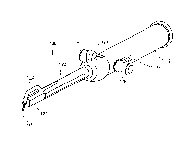
The medical implement illustrated in FIG's. 16-21 is generally designated 100.
It includes a handle 120 having a proximal end 121 for manual grasping, and a
distal
end 122 for engagement with a bone in which a suture is to be attached. As in
the
previously-described embodiment, the bone would be pre-formed with a first
bore B1
(FIG. lQ. The medical implement of FIG's. 16-21 also includes a hook 130 at
the
distal end of the handle, and a loop 135 movable to an extended position with
respect to
the hook for facilitating entry of one end of the suture through the loop, and
to a
retracted position for clamping the respective end of the suture, as described
above with
respect to FIG'S. 2-10D.
In the implement of FIG's. 2-10D, the hook (30) is the movable element with
respect to the distal end (22) of the handle; the construction of FIG's. 16-21
reverses
these parts. That is, in the construction of FIG's. 16-21, the hook 130 is
fixed, and the
distal end 122 of the handle is movable towards and away from the hook in
order to
facilitate the entry and removal of the hook into the first bore B I.
This feature can be best seen in FIG. 19, wherein it will be seen that the
handle
120 is hollow, and slidably receives a core 123 having a distal end 122
movable towards
and away from the hook 130.
CA 02711430 2010-07-05
WO 2009/107121 25 PCT/IL2008/001316
As clearly seen in FIG. 19, core 123 is urged by a spring 124 in the direction
of
bringing the distal end 122 into engagement with the hook 130, but may be
manually
moved rearwardly by means of manipulatable member 126 having a stem passing
through longitudinal slots 127 (FIG. 18) in handle 120 so as to permit the
distal end 122
to be retracted rearwardly of hook 130 against the bias of spring 124. As
further seen
particularly in FIG. 19, the core 123 is formed with passageway 120a
(corresponding to
passageway 20a in FIG's. 2-10D) used for making a second bore B2 shown in FIG.
1 C.
At the distal end 122, passageway 120a is a slot in core 123.
Loop 135 received within hook 130 is basically of the same construction as
described above with respect to loop 35 in FIG's. 2-10D, in that it is coupled
to knob
128 to move the hook to its extended and retracted positions in order to grasp
a suture
passed between the two legs of the hook for extraction from bore B1, as
described above
with respect to FIG's. 2-10D. The coupling of the loop 135 to knob 128 is also
effected
by means a wire 135e passing through the distal end of handle 120.
In the implement of FIG's. 16-21, however, manipulatable knob 128 for loop
135 is pivotal about an axis 128a extending perpendicularly to the
longitudinal axis of
the handle 120 and located slightly above that longitudinal axis, as can be
seen
particularly in FIG. 19. Thus, as shown in FIG. 19, loop 135 would normally be
urged,
by spring 124 to its retracted position within hook 130 (FIG. 19), but is
conveniently
movable to its extended position, shown in FIG. 18, by merely pivoting knob
128
forwardly, whenever it is desired to extend the loop for receiving the end of
the suture.
Releasing the knob will then effect the retraction of the loop to firmly grasp
the suture,
as described above with respect to the implement of FIG's. 2-10D.
CA 02711430 2010-07-05
WO 2009/107121 26 PCT/IL2008/001316
In addition, whereas hook 30 shown in FIG. 2 formed an angle of about 90
between horizontal section 33 and end section 34, in the embodiment shown in
FIG's.
16-21, hook 130 forms a larger angle of about 110 -115 between horizontal
section 33
and end section 34. Accordingly, with this embodiment, the angle between bores
B1 and
B2 formed using implement 100 is about 65 -70 .
In substantially all other respects, the bone-tunneling implement illustrated
in
FIG's. 16-21 is constructed, and operates, in the same manner as described
above.
It is to be understood that, where applicable, implements according to various
embodiments of the invention can include features taken from both described
embodiments of the bone-tunneling implements. Furthermore, it should be clear
that
other methodologies to provide the functions performed by the two above
embodiments
can also be used.
Method of attaching a suture to a bone (FIG's. 22 and 23A-G)
FIG. 22 is a block diagram of a method 220 of attaching a suture to a bone in
accordance with an exemplary embodiment of the invention. FIG's. 23A-G are
illustrations of stages of method 220. the illustrations and description below
refers to
implement 100 shown in FIG's. 16-21. It is noted that method 220 with
appropriate
changes may be applied with implement 50 shown in FIG's. 2-1OD or other
variations of
the implement.
A first bore BI is formed in the humerus bone at 222. Preferably, bore B l is
formed near the greater tubercle 6 shown in FIG. 23A and FIG's. IA-IC and is
not long
enough to exit the bone. Optionally, as shown in FIG. 23A, bore B1 is drilled
with a
drill bit 53 inserted through drill guide 51. Optionally, an obturator is
first inserted
CA 02711430 2010-07-05
WO 2009/107121 27 PCT/IL2008/001316
through drill guide 51 in order to clear a path through the soft tissue
surrounding the
bone. Alternatively, bore B I is formed by any other method known in the art.
Hook 130 is then inserted into bore B 1 at 224. Optionally, the hook is
inserted
through drill guide 51, as indicated in FIG. 23B, in order to ease locating
bore Bl and
the drill guide is removed after insertion.
Optionally, the insertion of the hook into bore B1 is used as a reference
point for
forming bore B2, such that bores BI and B2 intersect at a predetermined angle.
Alternatively or additionally, hook 130 is used as an arm clamping implement
100 to
the bone. Before inserting hook 130 to bore B1, manipulatable member 126 is
moved
rearwardly so as permit distal end 122 of the core to be retracted rearwardly
of hook
130. After insertion of hook 130 in bore B1, manipulatable member 126 is
released and
distal end 122 is clamped to the bone by bias of spring 124 as shown in FIG.
19, or
otherwise.
FIG. 23C depicts the hook inserted into the first hole and distal end 122
clamped
to the bone. As further shown in FIG. 23C, manipulatable knob 128 is moved to
shift
loop 135 to its extended position.
At 226 a second bore B2 is formed through the bone (FIG. 23D) at a
predetermined angle from bore Bl. Bore B2 is drilled to a depth such that bore
B2
meets bore B 1 in the humerus bone thereby enabling a suture to be threaded
through the
two bores. Preferably, bores B 1 and B2 extend past the intersection of the
bores in the
bone such that drill 54 passes through loop 135 in bore B1. Preferably, bore
B2 is not
long enough to exit the bone. Optionally, bores B 1 and B2 define a 70 angle
in the
bone. Preferably, bores B 1 and B2 define an angle of between 65 and 75 in
the bone.
CA 02711430 2010-07-05
WO 2009/107121 28 PCT/IL2008/001316
Optionally, before drilling bore B2, an obturator, such as the obturator 52
shown
in FIG. 11, is used to clear the path and locate the exact location of bore
B2. Bore B2 is
then drilled at the location indicated by the obturator, using a drill such as
drill 54
shown in FIG. 11.
After forming bore B2 a first end of a suture 57 is threaded through bore B2
at
228. As shown in FIG. 23E, a suture loader such as suture loader 55 depicted
in FIG. 12
is optionally used for threading suture 57 through bore B2 and through loop
135
extending from hook 130. Suture loader 55 is then removed, leaving suture 57
in the
bore.
At 230 the first end of the suture is caught at the intersection of bores B2
and B I
by loop 135. Manipulatable knob 128 is moved to shift loop 135 to its
retracted position
inside hook 130. Suture 57 which was threaded through loop 135 is now caught
inside
hook 130.
At 232 manipulatable member 126 is moved to release distal end 122 from being
clamped to the bone. Hook 130, is removed from bore 131 and suture 57 which is
clamped in hook 130 is thereby threaded through bore B 1. The end of the
suture is then
extracted from the bone by threading through bore B1 at 232. As shown in the
magnified section of FIG. 23G, suture 57 is now threaded through a channel in
the bne
consisting of bores B1 and B2.
In an alternative embodiment, the suture is first threaded into bore B 1 and
caught through bore B2.
At 233 the suture is threaded through the tendon according to any procedure
known in the art. The tendon is then pulled into place by means known in the
art. At 234
the two ends of the suture, the first end extending from bore B 1 and the
second end
CA 02711430 2010-07-05
WO 2009/107121 29 PCT/IL2008/001316
extending from bore B2 and tendon 10 may be knotted together thereby attaching
the
tendon to the humerus bone. FIG. 23H is a coronal section view of a humerus
bone
illustrating a suture knotted through the bone and tendon. The suture,
threaded through a
into bore B 1 is exited bore B2 at b, retrieved through tendon 10 at c and is
knotted at d.
FIG. 231 is a lateral view of a row of sutures tied over tendon 10 in
accordance with an
exemplary embodiment of the invention. A first suture is passed through the
bone at al
to b 1 and retrieved through the tendon at c l and then knotted at dl. A
second suture is
passed through a channel in the bone from a2 to b2 and passed through the
tendon at c2
to be knotted at d2, etc.
FIG. 23J illustrates a lateral view of another embodiment of the invention
where
two sutures are passed through a single bone channel and are tied over
different
locations through the tendon. For example, a first and a second suture are
threaded
through a bone channel from al to b l . The first suture is retieved through
the tendon at
c 11 and knotted at d 11. The second suture is retrieved through the tendon at
c 12 and
knotted at d 12.
It is noted that a plurality of ways of tying the sutures are known in the
art.
FIG's. 231-J are provided an example and other methods such as for example
crossing
the sutures over the tissue, to increase the area of the tendon held against
the bone, are
also covered by the present invention.
The medical implement of FIG. 24
In another embodiment of the invention, bores BI and B2 are formed with a
same implement 240 as shown in FIG. 24.
Implement 240 includes two channels 242 and 244 for receiving tools such as
drills, holders, etc. Instrument 240 is brought close to the bone at the
greater tubercle 6.
CA 02711430 2010-07-05
WO 2009/107121 30 PCT/IL2008/001316
A drill, such as drill 53 shown in FIG. 11, is inserted into channel 242 for
forming a first
bore B1. Optionally, an obturator, such as obturator 52 shown in FIG. 11, is
first
inserted into channel 242 for clearing the path through the soft tissue and
indicating the
location of bore B 1.
A pin or hook is then inserted into channel 242 and bore BI for positioning
implement 240 to the bone in order to stabilize the implement when forming a
second
bore B2. optionally, the drill used for forming bore B1 is kept in the bore
for
stabilization of implement 240 and a second drill is used for forming bore B2.
A drill is then inserted through channel 244 and second bore B2 is formed.
Channels 242 and 244 are located in implement 240 such that bores B 1 and B2
formed
with drills inserted through the channels, intersect in the bone. Preferably,
bores B 1 and
B2 are not long enough to exit the bone. Optionally, a stop on the drill
forming bores BI
and B2 causes the bores to be formed to a certain depth such that the bores
will intersect
in the bone and will not cross the bone.
In the embodiment shown in FIG. 24, the angles formed between the bores is
preferable less than 90 so as to allow the two bores to be formed from the
same side of
the bone. Optionally, the bores define an angle of less than 45 in the bone.
Optionally,
the bores define an angle of less than 30 in the bone.
Implement 240 further comprises two channels 246 and 248 for receiving a
suture. Channels 246 and 248 join with channels 242 and 248 at intersection
points 250
and 252 respectively. After bores 131 and B2 are formed, a suture is inserted
through
channel 246 and bore 131 and is retrieved through bore B2 and channel 248 in a
manner
similar to that described with respect to the embodiments shown in FIG's. 2
and 16
above.
CA 02711430 2010-07-05
WO 2009/107121 31 PCT/IL2008/001316
It will be appreciated that other variations, modifications and applications
of the
invention may be made. For example, other means may be used for extracting the
end
of the suture via bore B1 than those described above. In addition, other
constructions of
the slidable manipulatable members 26 and 28 may be used for removing the hook
and/or the loop. The couplings of manual manipulatable member 26 may include a
slip
or yielding coupling in order to prevent excessive force from being applied by
hook 30
to the bone, and thereby reduce the possibility of breakage of the bone.
Many other variations, modifications and applications of the invention will be
apparent.