Language selection

Search

Patent 2714632 Summary

Third-party information liability

Some of the information on this Web page has been provided by external sources. The Government of Canada is not responsible for the accuracy, reliability or currency of the information supplied by external sources. Users wishing to rely upon this information should consult directly with the source of the information. Content provided by external sources is not subject to official languages, privacy and accessibility requirements.

Claims and Abstract availability

Any discrepancies in the text and image of the Claims and Abstract are due to differing posting times. Text of the Claims and Abstract are posted:

  • At the time the application is open to public inspection;
  • At the time of issue of the patent (grant).
(12) Patent Application: (11) CA 2714632
(54) English Title: SPECTROMETRIC METHODS AND APPARATUS
(54) French Title: PROCEDES ET APPAREIL DE SPECTROMETRIE
Status: Deemed Abandoned and Beyond the Period of Reinstatement - Pending Response to Notice of Disregarded Communication
Bibliographic Data
(51) International Patent Classification (IPC):
  • G01N 21/01 (2006.01)
  • A61M 5/14 (2006.01)
  • A61M 5/31 (2006.01)
  • G01N 21/90 (2006.01)
(72) Inventors :
  • FLANK, WILLIAM H. (United States of America)
  • FLANK, SANDRA GLASSMAN (United States of America)
  • FLANK, SHARON (United States of America)
  • CIURCZAK, EMIL WALTER (United States of America)
  • OPPEDAHL, CARL (United States of America)
(73) Owners :
  • VERRANA, LLC
(71) Applicants :
  • VERRANA, LLC (United States of America)
(74) Agent: CASSAN MACLEAN
(74) Associate agent:
(45) Issued:
(86) PCT Filing Date: 2009-03-04
(87) Open to Public Inspection: 2009-09-11
Examination requested: 2014-02-12
Availability of licence: N/A
Dedicated to the Public: N/A
(25) Language of filing: English

Patent Cooperation Treaty (PCT): Yes
(86) PCT Filing Number: PCT/US2009/036056
(87) International Publication Number: WO 2009111579
(85) National Entry: 2010-08-09

(30) Application Priority Data:
Application No. Country/Territory Date
61/038,229 (United States of America) 2008-03-20
61/059,033 (United States of America) 2008-06-05
61/067,974 (United States of America) 2008-03-04

Abstracts

English Abstract


A box 14 having a body 13 is used with a transparent flexible
bag containing a liquid such as an IV bag. First and second faces 12 are
positioned relative to each other. The faces each have therewithin an end or
fiber optic port 11 of a respective light path. A light source is optically
coupled
with the light path of the first face and a spectrometer is optically coupled
with the light path of the second face. The light paths are coaxial and
are disposed so that the transparent flexible bag is positionable
therebetween.
The spectrometer is disposed to detect an anomaly in the liquid
within the transparent flexible bag, and to annunciate the anomaly to a human
user. The box defines a reproducible light path length through the liquid.
A caliper 29 having a body 22 may be used in spectrometric analysis of
a transparent tube containing a liquid such as a syringe or an IV line. The
caliper has finger pads 27 which permit opening the spring-loaded caliper
as needed. Rivets 25 provide a pivoting action relative to a pivot structure
21 which can also serve as a distance gauge. Compression spring 24 urges
the caliper jaws together at lens locations 26. Lens locations 26 are
optically
coupled with internal fiber optic lines 28, and thence to external fiber optic
connectors 23. A light source is optically coupled with one of the connectors
23 and a spectrometer is optically coupled with the other of the connectors
23.


French Abstract

Une boîte 14 comportant un corps 13 est utilisée avec un sac souple transparent contenant un liquide tel un sac IV. Les première et seconde faces 12 sont positionnées l'une par rapport à l'autre. Les faces comportent chacune une extrémité ou port de fibre optique 11 d'un trajet de lumière respectif. Une source de lumière est couplée optiquement au trajet de lumière de la première face et un spectromètre est couplé optiquement au trajet de lumière de la seconde face. Les trajets de lumière sont coaxiaux et sont disposés de façon à pouvoir positionner le sac souple transparent entre eux. Le spectromètre est agencé de façon à détecter une anomalie dans le liquide à l'intérieur du sac souple transparent, et pour avertir un utilisateur humain de lanomalie. La boîte définit une longueur de trajet de lumière reproductible à travers le liquide. Un calibre 29 comportant un corps 22 peut être utilisé dans l'analyse spectrométrique d'un tube transparent contenant un liquide tel qu'une seringue ou une voie IV. Le calibre comporte des doigtiers 27 permettant d'ouvrir le calibre à ressort si nécessaire. Des rivets 25 fournissent une action pivotante par rapport à une structure de pivot 21 qui peut également servir comme jauge à distance. Le ressort de compression 24 presse ensemble les mâchoires du calibre sur des emplacements de lentilles 26. Les emplacements de lentilles 26 sont couplés optiquement avec des lignes de fibres optiques internes 28, et de là, à des connecteurs de fibres optiques externes 23. Une source de lumière est couplée optiquement à l'un des connecteurs 23 et un spectromètre est couplé optiquement à l'autre connecteur 23.

Claims

Note: Claims are shown in the official language in which they were submitted.


Claims
-1. A method for use with a transparent intravenous drip line, and for use
with a clip having
opposed first and second jaws urged toward each other, each jaw having a
respective groove, the
grooves opposing each other, each groove having therewithin an end of a
respective light path, the
method comprising the steps of:
clipping the clip onto the transparent intravenous drip line with the
transparent intravenous drip line
seized within the groove of the first jaw and the groove of the second jaw;
passing a liquid through the transparent intravenous drip line and into a vein
of a human being;
after the clipping, passing light through the light path of the groove of the
first jaw, and through the
transparent intravenous drip line and through the liquid, and through the
light path of the groove of
the second jaw, and to a spectrometer, and carrying out a spectrometric
analysis upon the light
passing to the spectrometer; and
removing the clip from the transparent intravenous drip line.
-2. The method of claim 1 wherein the transparent intravenous drip line is
cylindrical in cross
section and the light paths are disposed relative to the grooves so that each
light path impinges upon
the drip line normal thereto, and so that the light paths are diametrically
opposed across the
transparent intravenous drip line.
-3. The method of claim 1 wherein the spectrometric analysis comprises
detecting an anomaly in
the liquid passing through the transparent intravenous drip line, and
annunciating the anomaly to a
human user.
-4. The method of claim 2 wherein the clip further comprises a sensor sensing
the relative positions
of the first and second jaws, wherein the sensor communicates to the
spectrometer information
indicative of a diameter of the transparent intravenous drip line, and wherein
the spectrometric
analysis is carried out making use of the information indicative of the
diameter of the transparent
intravenous drip line.
-9-

-5. A method for use with a transparent syringe containing a liquid, and for
use with a clip having
opposed first and second jaws urged toward each other, each jaw having a
respective groove, the
grooves opposing each other, each groove having therewithin an end of a
respective light path, the
method comprising the steps of:
clipping the clip onto the transparent syringe with the transparent syringe
seized within the groove
of the first jaw and the groove of the second jaw;
after the clipping, passing light through the light path of the groove of the
first jaw, and through the
transparent syringe and through the liquid, and through the light path of the
groove of the second
jaw, and to a spectrometer, and carrying out a spectrometric analysis upon the
light passing to the
spectrometer; and
removing the clip from the transparent syringe.
-6. The method of claim 5 wherein the transparent syringe is cylindrical in
cross section and the
light paths are disposed relative to the grooves so that each light path
impinges upon the drip line
normal thereto, and so that the light paths are diametrically opposed across
the transparent syringe.
-7. The method of claim 5 wherein the spectrometric analysis comprises
detecting an anomaly in
the liquid within the transparent syringe, and annunciating the anomaly to a
human user.
-8. The method of claim 6 wherein the clip further comprises a sensor sensing
the relative positions
of the first and second jaws, wherein the sensor communicates to the
spectrometer information
indicative of a diameter of the transparent syringe, and wherein the
spectrometric analysis is carried
out making use of the information indicative of the diameter of the
transparent syringe.
-9. A method for use with a transparent flexible bag containing a liquid, and
for use with a box
having opposed first and second faces positioned relative to each other, each
face having
therewithin an end of a respective light path, the method comprising the steps
of:
placing the transparent flexible bag into the box and in contact with the
opposed first and second
faces;
-10-

after the placing, passing light through the light path of the first face, and
through the transparent
flexible bag and through the liquid, and through the light path of the second
face, and to a
spectrometer, and carrying out a spectrometric analysis upon the light passing
to the spectrometer;
and
removing the transparent flexible bag from the box.
-10. The method of claim 9 wherein the light paths are coaxial across the
transparent flexible bag.
-11. The method of claim 9 wherein the spectrometric analysis comprises
detecting an anomaly in
the liquid within the transparent flexible bag, and annunciating the anomaly
to a human user.
-12. The method of claim 9 wherein the box defines a reproducible light path
length through the
liquid.
-13. The method of claim 9 further comprising the step, performed after the
removing step, of
employing the transparent flexible bag in an intravenous drip, and passing
some of the liquid into a
vein of a human patient.
-14. Apparatus for use in spectrometric analysis of a transparent flexible bag
containing a liquid,
the apparatus comprising:
a box having opposed first and second faces positioned relative to each other,
each face having
therewithin an end of a respective light path,
a light source optically coupled with the light path of the first face, and
a spectrometer optically coupled with the light path of the second face.
-15. The apparatus of claim 14 wherein the light paths are coaxial and are
disposed so that the
transparent flexible bag is positionable therebetween.
-16. The apparatus of claim 14 wherein the spectrometer is disposed to detect
an anomaly in the
liquid within the transparent flexible bag, and to annunciate the anomaly to a
human user.
-11-

-17. The apparatus of claim 14 wherein the box defines a reproducible light
path length through the
liquid.
-18. Apparatus for use in spectrometric analysis of a transparent tube
containing a liquid, the
apparatus comprising:
a clip having opposed first and second jaws urged toward each other, each jaw
having a respective
groove, the grooves opposing each other, each groove having therewithin an end
of a respective
light path;
a light source optically coupled with the light path of the groove of the
first jaw
a spectrometer optically coupled with the light path of second jaw.
-19. The apparatus of claim 18 wherein the light paths are disposed relative
to the grooves so that
each light path impinges upon the transparent tube normal thereto, and so that
the light paths are
diametrically opposed across the transparent tube.
-20. The apparatus of claim 18 wherein the spectrometer is disposed to detect
an anomaly in the
liquid within the transparent tube, and to annunciate the anomaly to a human
user.
-21. The apparatus of claim 18 wherein the clip further comprises a sensor
sensing the relative
positions of the first and second jaws, wherein the sensor communicates to the
spectrometer
information indicative of a diameter of the transparent tube, and wherein the
spectrometric analysis
is carried out making use of the information indicative of the diameter of the
transparent tube.
-22. Apparatus for use in spectrometric analysis of a transparent flexible bag
containing a liquid,
the apparatus comprising:
a box having opposed first and second faces positioned relative to each other,
each face having
therewithin an end of a respective light path,
a connection point for a light source, the connection point optically coupled
with the light path of
-12-

the first face, and
a connection point for a spectrometer, the connection point optically coupled
with the light path of
the second face.
-23. The apparatus of claim 22 wherein the light paths are coaxial and are
disposed so that the
transparent flexible bag is positionable therebetween.
-24. The apparatus of claim 22 wherein the box defines a reproducible light
path length through the
liquid.
-25. Apparatus for use in spectrometric analysis of a transparent tube
containing a liquid, the
apparatus comprising:
a clip having opposed first and second jaws urged toward each other, each jaw
having a respective
groove, the grooves opposing each other, each groove having therewithin an end
of a respective
light path;
a connection point for a light source, the connection point optically coupled
with the light path of
the groove of the first jaw
a connection point for a spectrometer, the connection point optically coupled
with the light path of
second jaw.
-26. The apparatus of claim 25 wherein the light paths are disposed relative
to the grooves so that
each light path impinges upon the transparent tube normal thereto, and so that
the light paths are
diametrically opposed across the transparent tube.
-27. The apparatus of claim 25 wherein the clip further comprises a sensor
sensing the relative
positions of the first and second jaws, wherein the sensor communicates
external to the clip
information indicative of a diameter of the transparent tube.
-13-

Description

Note: Descriptions are shown in the official language in which they were submitted.


CA 02714632 2010-08-09
WO 2009/111579 PCT/US2009/036056
SPECTROMETRIC METHODS AND APPARATUS
Cross-reference to related applications
This patent application claims the benefit of US application number 61/038,229
filed March 20,
2008, US application number 61/059,033 filed June 5, 2008, and US application
number
61/067,974 filed March 4, 2008, each of which is hereby incorporated herein by
reference for all
purposes.
Background
Counterfeiting and errors threaten patient safety. There are 1.25 million
adverse reactions and 7,000
patient deaths annually in the United States as a result of drug errors.
Existing verification relies
largely on tagging and checking drug packaging. Of course, in a hospital
setting, packaging and
product are often separate.
Formulated medications created in the pharmacy, including but not limited to
intravenous
medication delivered in IV bags, pose a special challenge. Once the medicine
or bag is made up,
how do you tell whether it contains the proper medication, at the right
concentration, and that the
drug is both current and genuine? Opening the drug to sample it risks
contaminating it.
For operating rooms, the identity of leftover waste drug is of concern. A
hospital employee may try
to steal leftover drugs and sell them, and substitute a substance such as
saline or dextrose in the
original container.
Immune globulin is often counterfeited.
Chemotherapy is expensive, as are antibiotics. Mistakes are even more
expensive: an average
lawsuit may cost nearly half a million dollars.
Patients may receive the wrong drug or the wrong dose, or an infused drug may
spill or not be
delivered correctly because of a blockage.
There is thus a great need for approaches to verify products in a hospital or
long-term care
-1-

CA 02714632 2010-08-09
WO 2009/111579 PCT/US2009/036056
environment. Such approaches need to be reliable, simple to use, and accurate.
None of the prior-
art approaches known to the applicants are completely satisfactory.
Summary of the invention
The present invention describes a verification system that works in the
hospital or longterm care
environment. The current invention verifies the product itself, checking for
the correct medication,
dosage, quality and purity, including a check for whether the drug is
counterfeit.
The current invention, in its preferred embodiment, uses near-infrared
spectroscopy (NIR) to look
into the bag, including through the plastic, and tell in an instant whether it
is right. The NIR shines a
light on the substance and compares its optical components to a chemical
library, a gold standard.
The present invention makes it possible to verify that the waste in a
container is in fact the drug, not
a substitute, without having to send it to a lab for analysis. The current
invention includes hardware
and software for a portable detection system that can tell in seconds whether
the substance matches
what it is expected to be. An advantage of the current invention in its
portable embodiment is that it
can be used by a nurse or technician on site, and does not need to be in a
lab.
The present technique also checks for quality and purity, even allowing in-
syringe verification.
The present invention makes it possible to check drugs as they leave the
hospital pharmacy, and at
later points as necessary.
Analytical chemical techniques such as near-infrared spectroscopy make it
possible to monitor the
drug once it has been compounded, through a syringe or IV bag, or even once it
has entered the
patient's body under the skin. Monitoring flow rate, as in this invention,
offers new opportunities to
keep patients safe.
A box 14 having a body 13 is used with a transparent flexible bag containing a
liquid such as an IV
bag. First and second faces 12 are positioned relative to each other. The
faces each have
therewithin an end or fiber optic port 11 of a respective light path. A light
source is optically
coupled with the light path of the first face and a spectrometer is optically
coupled with the light
path of the second face. The light paths are coaxial and are disposed so that
the transparent flexible
-2-

CA 02714632 2010-08-09
WO 2009/111579 PCT/US2009/036056
bag is positionable therebetween. The spectrometer is disposed to detect an
anomaly in the liquid
within the transparent flexible bag, and to annunciate the anomaly to a human
user. The box
defines a reproducible light path length through the liquid. A caliper 29
having a body 22 may be
used in spectrometric analysis of a transparent tube containing a liquid such
as a syringe or an IV
line. The caliper has finger pads 27 which permit opening the spring-loaded
caliper as needed.
Rivets 25 provide a pivoting action relative to a pivot structure 21 which can
also serve as a
distance gauge. Compression spring 24 urges the caliper jaws together at lens
locations 26. Lens
locations 26 are optically coupled with internal fiber optic lines 28, and
thence to external fiber
optic connectors 23. A light source is optically coupled with one of the
connectors 23 and a
spectrometer is optically coupled with the other of the connectors 23.
Description of the drawing
The invention will be described with respect to a drawing in several figures,
of which:
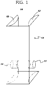
Fig. 1 is a perspective view of a box according to the invention.
Fig. 2 is a top view of the box of Fig. 1.
Fig. 3 is a side view of the box of Fig. 1.
Fig. 4 shows a caliper according to the invention.
Fig. 5 shows a clip according to the invention.
Fig. 6 shows a detail of a position or angle sensor according to the
invention.
Where possible, like elements have been depicted with like reference
designations among the
figures.
Detailed description
A system and process is described in which a spectroscopic or similar
instrumented technique, such
as NIR, Raman IR, UV-VIS, x-ray, etc., suitably supported with identification,
quantitation,
-3-

CA 02714632 2010-08-09
WO 2009/111579 PCT/US2009/036056
diagnostic and control software, is used in operations where a substance or
substances are being
transported from one location to another, to identify the transported
material, the rate at which the
transfer is taking place, the amount of transfer accomplished during any given
time interval, the
recognition in some form of the transfer of a predetermined amount or amounts,
the recognition in
some form of a significant defined deviation from the expected integrated rate
or rates of delivery,
and similar such functions or operations as may be needed to more fully
identify, quantify, monitor,
control and communicate information relative to the said transfer operation. A
further embodiment
of this invention relates to the detection and monitoring of end-of-process
and end-of-transfer
progress, wherein failure and/or progress and/or completion of the process is
confirmed and
communicated. A further embodiment of this invention relates to processes and
transformations
whose component identities, properties, progress with respect to time and/or
completion, etc., are
monitored, communicated and controlled.
An example of an embodiment of this invention relates to the administration
under monitored
and/or controlled conditions of fluids and/or medications via gravity-driven
or pumped intravenous
infusion.
A further example relates to the detection and monitoring of fluids or
medication delivery in vivo to
allow positive confirmation at the end of the process of identity, rate and
amount of substance
transferred, e.g., at the tip of a needle as the substances enter the
bloodstream.
A further example relates to the automated formulation or compounding of a
mixture of materials,
or material processing, whereby said process is under the control of a system
capable of achieving
said identification, monitoring, quantitation, control and information
communication functions,
wherein said system has had entered into it the requisite data necessary for
such identification,
monitoring, quantitation, control and information communication functions.
A further example relates to the monitoring and control of systems for
sorting, dispensing or
packaging of substances, wherein identity, properties, process rate and amount
can be determined
and communicated as needed, and records recorded thereof.
Additional developments include using a pair of fiber optic fibers inside the
needle, which could
send a beam and receive a spectral signal of what is coming out of the needle
tip as it enters the
bloodstream in the vein. This could allow an independent verification of flow,
to supplement the
-4-

CA 02714632 2010-08-09
WO 2009/111579 PCT/US2009/036056
empirical relating of concentration and flow with the signal obtained from
under the skin, which
may be attenuated to some extent from what comes down from the bag or the
pump. The
methodology would mimic an oximeter, where two wavelengths are used to monitor
the oxygenated
hemoglobin, with one of the wavelengths used to compensate for arterial
pulsations.
The fibers may turn out to be all that is needed, and that might simplify the
equipment involved.
Micro-devices like cameras are used in arterial catheterization, for example,
so there is precedent.
Indeed, plain fibers would be simpler, and cheaper, and safer. The device
could be frozen at a
specific angle and actually be above the skin. This is similar to
"interactance" measurements, used
by USDA for meat. The skin acts as a natural scatterer of light, so there is a
combination of
reflection and absorption taking place.
If the fibers can spectroscopically look for the iron-porphyrin complex in
hemoglobin as well as
monitor the infusate, then with an audible signal one might also determine if
the needle tip is
approaching and entering a blood vessel. Vein-finding might thus become an
integral part of the
system. The hemoglobin has a red-NIR component, so either may be used (both
are used in
oximeters).
Micropressure sensing is an option, since suitable equipment might exist or be
developed to monitor
flow (or cessation of flow). Differential pressure measurement is an
established method for
measuring flow rates. This may be harder to use; awake patients normally have
increased blood
pressure due to an inherent dislike of needles and tension of receiving a
drug.
The system may be employed in a hospital, long term care facility, or other
health care environment
to verify medication, checking for errors in dosage, concentration,
medication, purity, and/or
quality. It may be applied to formulated or compounded drugs. It may be
applied to intravenous
medication. It may be applied to medication in syringes. It may be applied to
operating room waste
or leftover medication.
Steps may include identification and/or confirmation of various infusate
species, measurement of
their concentrations and rates of flow, detection of leakages or blockages or
changes in flow, and
which may be coupled with a method for finding veins, where spectroscopic
techniques, which can
be of several kinds, including but not limited to ultraviolet, visible, near
infrared, infrared, far
infrared, Raman and other electromagnetic spectrum wavelengths in absorbance
or reflectance
-5-

CA 02714632 2010-08-09
WO 2009/111579 PCT/US2009/036056
mode, and can use double-beam methodologies, provide signals which can be
processed to provide
a variety of data outputs, including but not limited to instantaneous and
integrated graphical
displays, digital records of various kinds, visual and audible signals, etc.
A gold standard may be created, followed by checking other preparations
against that gold standard.
One sequence of steps can be to test all the medications in a group and to
identify outliers as
potentially problematic.
It is also possible to use one or more optical fibers within the shaft of a
needle used for infusion
purposes, so that monitoring of one or more species occurs as the infusate
leaves the tip of the
needle inserted into a subject's vein or body. It is possible to monitor the
changing signal intensity
in a spectroscopic measurement to detect the changing proximity of hemoglobin
in blood as a
means of locating a vein or artery to be used for a particular purpose,
including but not limited to
infusion.
It is also possible to use a needle within a needle, or two needles alongside
each other, such that one
tube contains infusate and one or more optical fibers, and the other space is
used to facilitate
differential pressure measurements that will allow independent flow rate
determinations to be made.
This differential method is not limited to needles.
It is also possible to employ wireless signal transmission from one or more
measurement units to a
central console where continual signal sampling and processing will produce a
variety of desired
outputs.
Particular detailed embodiments of the invention will now be described.
A box 14 (Figs. 1, 2, 3) having a body 13 is used with a transparent flexible
bag containing a liquid
such as an IV bag omitted for clarity in Fig. 1. First and second faces 12 are
positioned relative to
each other. The faces each have therewithin an end or fiber optic port 11 of a
respective light path.
A light source (omitted for clarity in Fig. 1) is optically coupled with the
light path of the first face
and a spectrometer (omitted for clarity in Fig. 1) is optically coupled with
the light path of the
second face. Typically the light paths are coaxial and are disposed so that
the transparent flexible
bag is positionable therebetween. Typically the spectrometer is disposed to
detect an anomaly in
-6-

CA 02714632 2010-08-09
WO 2009/111579 PCT/US2009/036056
the liquid within the transparent flexible bag, and to annunciate the anomaly
to a human user.
Typically the box defines a reproducible light path length through the liquid.
The box 14 may have a removable front face to allow for easier insertion and
removal of the bag, as
compared with stuffing the bag in from the top.
A caliper 29 (Fig. 4) having a body 22 may be used in spectrometric analysis
of a transparent tube
containing a liquid such as a syringe or an IV line. The caliper has finger
pads 27 which permit
opening the spring-loaded caliper as needed. Rivets 25 provide a pivoting
action relative to a pivot
structure 21 which can also serve as a distance gauge. Compression spring 24
urges the caliper
jaws together at lens locations 26. Lens locations 26 are optically coupled
with internal fiber optic
lines 28, and thence to external fiber optic connectors 23. A light source is
optically coupled with
one of the connectors 23 and a spectrometer is optically coupled with the
other of the connectors 23.
Fig. 5 is a face-on view of the lens location 26 disposed within body 22. Fig.
6 is a cross-sectional
view of the lens location 26 disposed within body 22, showing the internal
fiber optic line 28. As
may be seen the surface at 26 which engages the syringe or IV line has some
concavity and thus can
capture the syringe or IV line and keep it in place.
The distance gauge 21 comprises a sensor sensing the relative positions of the
first and second jaws,
and the sensor communicates to the spectrometer information indicative of a
diameter of the
transparent tube. The spectrometric analysis is carried out making use of the
information indicative
of the diameter of the transparent tube.
Another embodiment is shown in Figs. 7 and 8 with clip 33. Jaws 36 are V-
shaped and are
dimensioned so as to provide reproducible positioning relative to cylinders or
transparent tubes of
some range of diameters. Light paths 37, 38 are positioned so that each light
path impinges upon
the transparent tube normal thereto, and so that the light paths are
diametrically opposed across the
transparent tube. Spring 34 urges the jaws 26 together relative to a hinge or
pivot 35. Handles 31,
32 may be squeezed by a human operator to open the jaws 36. A sensor 39 is
shown in more detail
in Fig. 8. A movable piece 40 (attached to one of the jaws 26) moves relative
to LED-
phototransistor sensors 41, 42, offering perhaps three different discrete
sensed signals depending on
whether the jaws 36 are separated by a first distance, a second distance, or a
third distance. In this
way, if the cylinders are of any of three different standardized diameters,
they may be
-7-

CA 02714632 2010-08-09
WO 2009/111579 PCT/US2009/036056
disambiguated.
It will be appreciated that it is not crucial to use the particular sensing
mechanism portrayed here.
The methods to be carried out may include the following.
The clip is clipped onto a transparent intravenous drip line with the
transparent intravenous drip line
seized within the groove of the first jaw and the groove of the second jaw. A
liquid is passed
through the transparent intravenous drip line and into a vein of a human
being. After the clipping,
light is passed through the light path of the groove of the first jaw, and
through the transparent
intravenous drip line and through the liquid, and through the light path of
the groove of the second
jaw, and to a spectrometer. A spectrometric analysis is carried out upon the
light passing to the
spectrometer. Later the clip is removed from the transparent intravenous drip
line.
Alternatively the clip may be clipped onto a transparent syringe containing a
liquid.
An IV bag may be placed into the box 14 and in contact with the opposed first
and second faces 12.
Light is passed through the light path of the first face, and through the
transparent flexible bag and
through the liquid, and through the light path of the second face, and to a
spectrometer. A
spectrometric analysis is carried out upon the light passing to the
spectrometer. Later the the bag
may be removed from the box. Still later the bag may be put to use in an
intravenous drip, and
some of the liquid may be passed into a vein of a human patient.
Those skilled in the art will have no difficulty devising myriad obvious
improvements and variants
without deviating in any way from the invention, all of which are intended to
be encompassed
within the claims which follow.
-8-
Representative Drawing
A single figure which represents the drawing illustrating the invention.
Administrative Status

2024-08-01:As part of the Next Generation Patents (NGP) transition, the Canadian Patents Database (CPD) now contains a more detailed Event History, which replicates the Event Log of our new back-office solution.

Please note that "Inactive:" events refers to events no longer in use in our new back-office solution.

For a clearer understanding of the status of the application/patent presented on this page, the site Disclaimer , as well as the definitions for Patent , Event History , Maintenance Fee  and Payment History  should be consulted.

Event History

Description Date
Application Not Reinstated by Deadline 2016-03-04
Time Limit for Reversal Expired 2016-03-04
Deemed Abandoned - Failure to Respond to Maintenance Fee Notice 2015-03-04
Letter Sent 2014-02-19
All Requirements for Examination Determined Compliant 2014-02-12
Request for Examination Requirements Determined Compliant 2014-02-12
Inactive: Correspondence - Prosecution 2014-02-12
Request for Examination Received 2014-02-12
Inactive: IPC assigned 2011-02-21
Inactive: First IPC assigned 2011-02-21
Inactive: IPC removed 2011-02-21
Inactive: Cover page published 2010-11-10
Inactive: Notice - National entry - No RFE 2010-10-07
Letter Sent 2010-10-07
Inactive: First IPC assigned 2010-10-01
Inactive: IPC assigned 2010-10-01
Application Received - PCT 2010-10-01
Inactive: IPC assigned 2010-10-01
National Entry Requirements Determined Compliant 2010-08-09
Application Published (Open to Public Inspection) 2009-09-11

Abandonment History

Abandonment Date Reason Reinstatement Date
2015-03-04

Maintenance Fee

The last payment was received on 2014-02-03

Note : If the full payment has not been received on or before the date indicated, a further fee may be required which may be one of the following

  • the reinstatement fee;
  • the late payment fee; or
  • additional fee to reverse deemed expiry.

Please refer to the CIPO Patent Fees web page to see all current fee amounts.

Fee History

Fee Type Anniversary Year Due Date Paid Date
Registration of a document 2010-08-09
Basic national fee - standard 2010-08-09
MF (application, 2nd anniv.) - standard 02 2011-03-04 2011-03-04
MF (application, 3rd anniv.) - standard 03 2012-03-05 2012-02-24
MF (application, 4th anniv.) - standard 04 2013-03-04 2013-01-23
MF (application, 5th anniv.) - standard 05 2014-03-04 2014-02-03
Request for examination - standard 2014-02-12
Owners on Record

Note: Records showing the ownership history in alphabetical order.

Current Owners on Record
VERRANA, LLC
Past Owners on Record
CARL OPPEDAHL
EMIL WALTER CIURCZAK
SANDRA GLASSMAN FLANK
SHARON FLANK
WILLIAM H. FLANK
Past Owners that do not appear in the "Owners on Record" listing will appear in other documentation within the application.
Documents

To view selected files, please enter reCAPTCHA code :


To view images, click a link in the Document Description column. To download the documents, select one or more checkboxes in the first column and then click the "Download Selected in PDF format (Zip Archive)" or the "Download Selected as Single PDF" button.

List of published and non-published patent-specific documents on the CPD .

If you have any difficulty accessing content, you can call the Client Service Centre at 1-866-997-1936 or send them an e-mail at CIPO Client Service Centre.


Document
Description 
Date
(yyyy-mm-dd) 
Number of pages   Size of Image (KB) 
Description 2010-08-09 8 403
Claims 2010-08-09 5 209
Abstract 2010-08-09 2 87
Drawings 2010-08-09 4 43
Representative drawing 2010-10-04 1 4
Cover Page 2010-11-10 1 51
Notice of National Entry 2010-10-07 1 195
Courtesy - Certificate of registration (related document(s)) 2010-10-07 1 102
Reminder of maintenance fee due 2010-11-08 1 113
Reminder - Request for Examination 2013-11-05 1 117
Acknowledgement of Request for Examination 2014-02-19 1 177
Courtesy - Abandonment Letter (Maintenance Fee) 2015-04-29 1 171
PCT 2010-08-09 2 82