Note: Descriptions are shown in the official language in which they were submitted.
CA 02717352 2010-09-01
WO 2009/111512 PCT/US2009/035935
Low Profile Patient Specific Cutting Blocks for a Knee Joint
Cross-Reference to Related Applications
[0001] This application claims the benefit of U.S. Provisional Application
No.61/033,419, filed March 3, 2008 and further claims the benefit of U.S.
Provisional
Application No. 61/089,373, filed August 15, 2008. The disclosure of each
application is
incorporated by reference in its entirety.
Field of the Invention
[0002] This invention relates generally to cutting blocks for bone resections
at a knee
joint and, more particularly, for cutting blocks designed for a patient's
specific bone and
cartilage and further configured to a surgeon's preferences.
Summary of the Invention
[0003] It is in view of problems related to the field above that the present
invention
was developed.
[0004] In one aspect of the invention, a low profile patient specific cutting
block for
a knee comprises a plurality of bone interfacing portions and a cutting slot.
The plurality of
bone interfacing portions are configured to overlie portions of an end of a
bone. The bone
interfacing portions each have a surface generally a negative of the portion
of the bone the
bone interfacing portion overlies. The bone interfacing portions are angularly
offset from
each other such that a first of the bone interfacing portions overlies an
anterior portion of the
bone and a second of the bone interfacing portions overlies a portion of bone
generally
perpendicular to the anterior portion of bone. The cutting slot is oriented in
a fixed position
relative to the bone interfacing portions such that the cutting slot directs a
cutting tool at a
fixed angle and at a fixed depth from the bone interfacing portions.
[0005] In another embodiment of the invention the low profile patient specific
cutting
CA 02717352 2010-09-01
WO 2009/111512 PCT/US2009/035935
block is a femoral cutting block. The block further comprises bosses having a
thickness and
an aperture extending through the bosses. The aperture has a diameter. The
bosses are
configured to direct a pin through the boss. The thickness of the boss is
greater than the
diameter of the aperture.
[0006] In yet another embodiment, the bone interfacing portions are generally
oriented in the middle of the low profile patient specific cutting block in an
anterior portion of
the low profile patient specific cutting block and are oriented medially and
laterally at a
posterior portion of the low profile patient specific cutting block.
[0007] Alternatively, the low profile patient specific cutting block is a
tibial block
and the cutting slot is offset and medialized relative to the tibial bone.
[0008] Another embodiment includes a paddle extending posteriolaterally from a
middle portion of the patient specific cutting block. The paddle has a raised
portion anteriorly
oriented on the low profile patient specific cutting block such that the
paddle does not touch
the tibia on an anterior proximal surface and does touch the tibia on a
posterior proximal
surface.
[0009] Further features, aspects, and advantages of the present invention, as
well as
the structure and operation of various embodiments of the present invention,
are described in
detail below with reference to the accompanying drawings.
Brief Description of the Drawings
2 0 [0010] The accompanying drawings, which are incorporated in and form a
part of the
specification, illustrate embodiments of the present invention and together
with the
description, serve to explain the principles of the invention. In the
drawings:
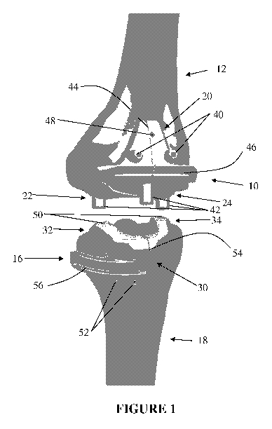
[0011] Figure 1 is a view of a knee joint with a femoral patient specific
cutting block
and a tibial patient specific block;
-2-
CA 02717352 2010-09-01
WO 2009/111512 PCT/US2009/035935
[0012] Figure 2 is an anterior view of a femoral patient specific cutting
block
according to an aspect of the invention;
[0013] Figure 3 is a proximal posterior view of a femoral patient cutting
specific
block according to an aspect of the invention;
[0014] Figure 4 is an anterior view of a tibial patient specific cutting block
according
to an aspect of the invention; and
[0015] Figure 5 is a distal posterior view of a tibial patient specific
cutting block
according to an aspect of the invention.
Detailed Description of the Embodiments
[0016] Referring to the accompanying drawings in which like reference numbers
indicate like elements, Figure 1 illustrates a view of a knee joint with a
femoral patient specific
cutting block 10 and a tibial patient specific block 14. The femoral patient
specific cutting
block 10 is attached to a femur 12. The tibial patient specific cutting block
14 is attached to a
tibia 16. The patient specific cutting blocks 10 and 14 are configured to
engage portions of
bone and cartilage on the femur 12 and tibia 16 to align cutting surfaces
within the patient
specific cutting blocks so that a distal cut (on the femur) and a proximal cut
(on the tibia) may
be made without using either intramedullary or extramedullary guides.
[0017] The femoral patient specific cutting block 10 includes an anterior
femoral
portion 20, a medial femoral paddle 22 and a lateral femoral paddle 24. These
portions
overlie portions of the anterior face, medial condyle and lateral condyle of
the femur 12,
respectively. Because the portions 20, 22, and 24 only overlie portions of the
femur 12 instead
of an entire conforming overlay of the end of the femur 12, the patient
specific cutting block
10 may have a lower profile, both in the medio-lateral and anterior-posterior
direction.
-3-
CA 02717352 2010-09-01
WO 2009/111512 PCT/US2009/035935
[0018] Pin holes 40 and 48, guide bosses 42, a mechanical axis index 44, and a
femoral cutting slot 46 are oriented on the exterior surface of the femoral
patient specific
cutting block 10. The pin holes 40 and 48 are oriented to pin the cutting
block 10 to the femur
12. Guide bosses 42 are oriented in order to set pins for the other box cuts
necessary to
prepare the femur 12 for an implant.
[0019] The pin holes 40 and 48 are oriented on the anterior face of the
patient specific
cutting block 10. The pin holes 40 and 48 may have bosses (as shown with
reference to pin
holes 40) or may be flush with the surface (as shown with pin hole 48). The
bosses may be
used to direct the pins, for example, away from the edges of the bone. The low
profile of the
patient specific cutting block 10 would allow a pin hole without a boss to
allow a pin to extend
in a wide variety of angular directions. By extending bosses a thickness
greater than the
diameter of the aperture through the boss, the bosses may orient the pins by
adding a guide
through the patient specific cutting block 10 so that the pins are directed as
they are impacted
or drilled into the bone.
[0020] The mechanical axis index 44 is oriented along the mechanical axis of
the
femur 12. A cutting slot 46, oriented relative to anatomical structures and
defined by the
surgeon, directs the distal cut for an implant. As will be described below, MR
and X-rays of
the patient are used to align the mechanical axis index 44 to the patient
specific cutting block
10.
[0021] In addition to the MR and X-ray information, surgeon preferences are
used to
place the cutting slot 46 on the patient specific cutting block 10. The
cutting slot 46 may be
oriented relative to the mechanical axis in a varus or valgus orientation
(according to surgeon
preference based upon the X-ray data). The flexion gap may be adjusted by
adjusting the angle
of the cutting slot 46 relative to the patient specific cutting block 10. The
depth of the
-4-
CA 02717352 2010-09-01
WO 2009/111512 PCT/US2009/035935
resection cut is also determined by the placement of the cutting slot 46 and
is determined from
the distal point on the condyles.
[0022] The guide bosses 42 are also placed on the patient specific cutting
block 10
according to MR data, X-ray data and surgeon preference. The guide bosses 42
may set the
rotation of the implant by adjusting the posterior bosses 42 relative to one
another. The
relative placement of the bosses 42 allows for pins to be placed so that the
pins guide a further
cutting guide over the distal cut of the femur in order to make the anterior
and posterior cuts
and any chamfer cuts required by the bone interfacing surfaces of the implant.
Internal/
external rotation is directed by moving the depth of one of the posterior
bosses relative to the
other posterior boss. A-P placement of the implant is adjusted by moving both
of the posterior
bosses 42 together in the A-P direction.
[0023] The tibial patient specific cutting block 14 includes an anterior
femoral
portion 300, a medial femoral paddle 32 and a lateral femoral paddle 34. These
portions
overlie portions of the anterior face, medial plateau and lateral plateau of
the tibia 16,
respectively. Because the portions 20, 22, and 24 only overlie portions of the
tibia 16 instead
of an entire conforming overlay of the end of the tibia 16, the patient
specific cutting block 14
may have a lower profile, both in the medio-lateral and anterior-posterior
direction.
[0024] Pin holes 50 and 52, an M-L index 54 and a cutting slot 56 are oriented
on the
outer surface of the tibial patient specific cutting block 14. The pin holes
50 and 52 may fix
the patient specific cutting block 14 to the bone and may additionally align
the pins relative to
one another for further orientation, if necessary, in tibial preparation.
[0025] The tibial cutting slot 56 is medialized relative to the anterior
surface of the
tibia 16 (i.e., the tibial cutting slot 56 is oriented on the medial half of
the anterior side of the
patient specific cutting block 14). The lateral paddle 34 may extend around
the front of the
tibial eminence extending posterior-laterally toward the lateral plateau.
These features may
-5-
CA 02717352 2010-09-01
WO 2009/111512 PCT/US2009/035935
allow the guide to be used in a MIS procedure, where lateral clearance is
minimized by cutting
the tibia from the medial half of the anterior face of the tibia while
minimizing the medial
approach to the tibia, which would involve additional soft tissue issues.
Thus, the medialized
and rotated cutting slot 56 is oriented for clearance and accessibility even
for an MIS approach.
[0026] Turing now to Figures 2 and 3, Figure 2 is an anterior view of a
femoral patient
specific cutting block according to an aspect of the invention. Figure 3 is a
proximal posterior
view of a femoral patient cutting specific block according to an aspect of the
invention. In
addition to the features described above, the patient specific cutting block
10 may also include
an epicondylar index 68. The epicondylar index 68 may be used as a visual
"feel good" for the
rotation of the holes and the A-P placement of the holes, similar to the
purpose of the
mechanical axis index described above.
[0027] In Figure 3, the bone interfacing surfaces 80, 82, 84 and 86 are shown.
The
anterior bone interfacing portion overlies a portion of the anterior surface
of cartilage and
bone. The medial bone interfacing portion 82, the lateral bone interfacing
portion 84 and the
intracondylar bone interfacing portion 86 overlie the medial, lateral and
intracondylar notch
portions of the condyles, respectively. The bone interfacing portions 80, 82
and 84 align to the
anterior and distal faces of the femur while the intracondylar bone
interfacing portion 86
orients the block medio-laterally. By using relatively small portions of the
surfaces, the profile
of the patient specific cutting block 10 may be lowered. Additionally, the fit
may be better as
smaller portions may result in fewer osteotomes on the bone surface (which may
cause poor fit
of the patient specific cutting block to the bone. The paddles 22 and 24 may
also be relatively
thin posteriorly. This further minimizes the profile of the patient specific
cutting block.
[0028] The cutting slot 46 may be formed through the bone interfacing portions
of the
patient specific cutting block or may be recessed from the surface. The
thickness of the cutting
slot helps to direct the orientation of the cutting tool as the cutting tool
advances through the
-6-
CA 02717352 2010-09-01
WO 2009/111512 PCT/US2009/035935
cutting slot 46. As previously discussed, the relative angle of the cutting
slot 46 to the patient
specific cutting block 10 (and particularly to the bone interfacing portions)
orients the flexion
gap while the translation of the cutting slot 46 relative to the patient
specific cutting block 10
sets the resection depth.
[0029] Turing now to Figures 4 and 5, Figure 4 is an anterior view of a tibial
patient
specific cutting block according to an aspect of the invention. Figure 5 is a
distal posterior
view of a tibial patient specific cutting block according to an aspect of the
invention. In
addition to the features described above, the tibial patient specific cutting
block also may
include a posterior chamfer 96 and a planar proximal surface 100. The
posterior chamfer 96
allows for the tibial patient specific cutting block to be positioned
posteriorly without
distracting the soft tissue around the knee more than necessary. Similar to
other features, this
feature helps the overall profile of the implant.
[0030] The planar proximal surface 100 may match the distal femur resection
plane
from the femoral patient specific cutting block. This feature may allow
intraoperative
flexion/extension testing when the tibial patient specific cutting block is
secured to the tibia.
[0031] Bone interfacing surfaces 90, 92, and 94 are shown in Figure 5. The
anterior
bone interfacing portion 90 overlies a portion of the anterior surface of
cartilage and bone.
The medial bone interfacing portion 92 and the lateral bone interfacing
portion 84 overlie the
medial and lateral portions of the tibia, respectively. The bone interfacing
portions 92 and 94
align to the proximal faces of the tibial plateaus (thus orienting the patient
specific cutting
block proximally) while the anterior bone interfacing portion 90 orients the
block medio-
laterally and in the AP direction. By using relatively small portions of the
surfaces, the profile
of the patient specific cutting block may be lowered. Additionally, the fit
may be better as
smaller portions may result in fewer osteotomes on the bone surface (which may
cause poor fit
of the patient specific cutting block to the bone. The paddles 32 and 34 may
also be relatively
-7-
CA 02717352 2010-09-01
WO 2009/111512 PCT/US2009/035935
thin posteriorly and may be elevated from the tibial plateau surface
anteriorly to avoid poor
placement. This further minimizes the profile of the patient specific cutting
block.
[0032] The cutting slot 56 may be formed through the bone interfacing portions
of the
patient specific cutting block or may be recessed from the surface. If the
cutting slot 56 is
recessed from the surface, then impingement of the block on bone may be
minimized, again
increasing the fit of the patient specific cutting block to the bone. The
thickness of the cutting
slot helps to direct the orientation of the cutting tool as the cutting tool
advances through the
cutting slot 56. As previously discussed, the relative angle of the cutting
slot 56 to the patient
specific cutting block 10 (and particularly to the bone interfacing portions)
orients the flexion
gap while the translation of the cutting slot 46 relative to the patient
specific cutting block 10
sets the resection depth.
[0033] The MR data and X-ray data may be taken by known means. As an example,
the following protocols may be used. Different MR protocols may be executed on
different
patients. To minimize scan time, a fast spin echo imaging technique may be
used for any
protocol, essentially producing a proton density (PD) weighted image. One
protocol may
use the spoiled gradient echo technique with a low repetition time (TR) and
low echo time
(TE) and a flip angle of 30 degrees combined with a fat saturation technique.
A second
protocol and third protocol may use a high TR and a low TE combined with a fat
saturation
technique. The only difference between the second protocol and third protocol
is that the
second protocol has lower TE than the third protocol, which in turn offers
more Ti and less
PD properties. The increased Ti relaxation time may help to increase the image
contrast
within the different tissues in the MR image.
[0034] Bone models of the femur and tibia may be extracted from the MR images
and appropriate anatomic reference landmarks may be identified. Full leg x-
rays may be
used to determine the mechanical axis alignment. Femoral and tibial cutting
blocks may
-8-
CA 02717352 2010-09-01
WO 2009/111512 PCT/US2009/035935
then be designed through computer aided design (CAD) modeling such that they
conform to
the bone models on one side for proper seating and have cutting slots at the
appropriate
resection depth and angle specific to the patient. The cutting blocks may be
made from
medical grade Nylon 12 using the EOSint P system. Since the surface geometries
of these
blocks are based on the patient's MR data set, clean data (properly
differentiating between
bone and cartilage and soft tissue) should be used to ensure the fit and
functionality of the
blocks.
[0035] In view of the foregoing, it will be seen that the several advantages
of the
invention are achieved and attained.
[0036] The embodiments were chosen and described in order to best explain the
principles of the invention and its practical application to thereby enable
others skilled in the
art to best utilize the invention in various embodiments and with various
modifications as are
suited to the particular use contemplated.
[0037] As various modifications could be made in the constructions and methods
herein described and illustrated without departing from the scope of the
invention, it is
intended that all matter contained in the foregoing description or shown in
the accompanying
drawings shall be interpreted as illustrative rather than limiting. Thus, the
breadth and scope
of the present invention should not be limited by any of the above-described
exemplary
embodiments, but should be defined only in accordance with the following
claims appended
hereto and their equivalents.
-9-