Note: Descriptions are shown in the official language in which they were submitted.
CA 02718107 2010-09-09
WO 2009/117462 PCT/US2009/037469
MYCOBACTERIAL CULTURE SCREENING TEST FOR
MYCOBACTERIUM AVIUM COMPLEX BACTERIA
CROSS-REFERENCE TO RELATED APPLICATIONS
[0001] This invention claims priority to U.S. Provisional Patent Applications
Serial Nos. 61/037,665, filed March 18, 2008, and 61/038,288, filed March 20,
2008, both of which are herein incorporated by reference.
FIELD OF THE INVENTION
[0002] This invention relates to the field of diagnostic assays for detecting
the presence of mycobacteria.
BACKGROUND
[0003] Members of the Mycobacterium avium complex (MAC) are a family
of intracellular bacterial pathogens causing significant disease in both
animals
and humans. The complex contains four subspecies of M. avium: M. avium
subsp. avium (MAA), M. avium subsp. paratuberculosis (MAP), M. avium
subsp. hominissuis (MAH), and M. avium subsp. silvaticum (MAS).
Mycobacterium intracellulare is also a member of the complex.
[0004] The most common method for diagnosis of mycobacterial infections
in humans and animals begins with growth (isolation) of the causative
organism in liquid culture media. While direct detection technology (such as
PCR) can be done on clinical samples, most studies find trying to find the
bacteria's DNA less sensitive than culture-based methods. Also, culture
yields the pathogens needed for subsequent testing such as antibiotic
susceptibility or molecular epidemiology. The success of culture-based
methods for mycobacteria depends heavily on specimen processing
techniques that eliminate non-mycobacterial microflora from the sample. The
tubes of inoculated liquid media are monitored by instruments that incubate
CA 02718107 2010-09-09
WO 2009/117462 PCT/US2009/037469
2
and "examine" the culture for evidence of microbial growth using indicators
such as oxygen consumption or gas pressure inside the sealed culture vessel.
Examples of these diagnostic systems are the BACTEC MGIT 960 system
(BD Diagnostic Systems, Franklin Lakes, NJ), and Trek ESP II system (Trek
Diagnostic Systems, Cleveland, OH). These instruments generally handle
300-1,000 cultures at a time and cost in excess of $50,000. The culture
media associated with each instrument retails for $5 to $10 per sample tube
depending on instrument lease agreements and volume discounts.
[0005] After an instrument signals that microbial growth is occurring in the
culture tube, a variety of assays must be performed to identify the micro-
organism(s) in the tube that triggered the signal. Such assays vary in
sensitivity, specificity, and cost. Possible outcomes of mycobacteria
identification assays on isolates from liquid cultures include: 1) false-
signal by
the instrument, i.e. no microorganisms were detected, 2) identification of a
mycobacterial pathogen of limited clinical significance, e.g. a non-pathogenic
environmental Mycobacterium sp., or 3) identification of a mycobacterial
pathogen of importance.
[0006] The vast majority of clinically important mycobacterial pathogens
generally fall into two groups: Mycobacterium tuberculosis Complex (MTBC)
and the Mycobacterium avium complex (MAC). Mycobacterium leprae, the
cause of leprosy, is also an important mycobacterial pathogen but it is far
less
common and is presently not cultivable in vitro. In developing countries
MTBC is the predominant mycobacterial pathogen of concern. In developed
countries MAC pathogens are more common, particularly in
immunocompromised patients such as people with HIV or under treatment for
cancer.
[0007] Typically, definitive identification of mycobacterial pathogens
isolated from culture is primarily based on PCR. The PCR assays differ in
design from those used for direct pathogen detection in clinical samples and
are marketed specifically for mycobacterial identification from cultures. An
example of one of the leading products in this market is AccuProbe (Gen-
CA 02718107 2010-09-09
WO 2009/117462 PCT/US2009/037469
3
Probe, San Diego, CA). Most such assays are target pathogen DNA-specific
and rule in/out a specific pathogen group, e.g. MTBC or MAC. If the assay is
positive, the result is reported as, for example, MAC complex positive. The
specific species of mycobacteria within the complex is usually not determined.
If the PCR assay is negative, other PCRs may be required to arrive at a
diagnosis. The PCR assays may be costly and laborious.
[0008] A positive signal appears in automated liquid culture systems used
for mycobacteria isolation when growth of a microorganism triggers the
system's sensor. Rapid, low-cost methods are therefore desired to weed out
diagnostically irrelevant cultures that can be signal-positive due to non-
mycobacterial organisms. The present invention provides compositions and
methods for achieving these and related objectives.
BRIEF SUMMARY
[0009] Provided are compositions and methods for the detection of
mycobacteria growing in liquid samples. Also provided are novel antigen and
antibody preparations, kits, systems, and methods that can be used in assays
for detecting the presence of Mycobacterium avium complex bacteria.
[0010] Methods of detecting the presence of mycobacteria in liquid cultures
are provided. The methods include the steps of: providing capture antibodies
obtained from a subject immunized with mycobacteria-secreted antigens;
contacting the liquid culture with the capture antibodies; providing detection
antibodies obtained from a subject immunized with the mycobacteria-secreted
antigens; and detecting the presence of antigen-bound detection antibodies in
the liquid culture to indicate the presence of the mycobacteria in the liquid
culture. The detected mycobacteria may include Mycobacterium avium
complex.
[0011] The methods may include absorbing nonspecific antibodies using
heterologous antigens, where absorbtion is performed prior to contacting the
capture antibody and the detection antibody with the liquid culture. In the
CA 02718107 2010-09-09
WO 2009/117462 PCT/US2009/037469
4
practice of the methods, both antibodies may be specific for one or more
Mycobacterium avium complex secreted antigens. In some embodiments, the
heterologous antigens may be from M. phlei, other mycobacteria, or E. coli. In
the practice of the methods, detection of the presence of antigen-bound
detection antibodies in the liquid culture may include using an enzyme-linked
immunosorbent assay (ELISA). The capture antibodies may be affixed to
solid support. In some embodiments, the capture antibody may be produced
in chickens, and the detection antibodies may be produced in rabbits.
[0012] In the practice of the methods, the detection antibodies may be
labeled. The detection antibodies may be conjugated to enzymes. Preferably
a conjugate such as anti-rabbit Ig conjugated to an enzyme may be used to
determine that the detection antibody has bound to Mycobacterium avium
complex antigens captured in the assay. The methods may employ any
conjugate capable of reacting with bound Mycobacterium avium complex
antigen detection antibody.
[0013] Systems for the detection of the presence of mycobacterial
pathogens in liquid cultures are provided. The systems include: capture
antibodies obtained from subjects immunized with a Mycobacterium-secreted
antigens; and detection antibodies obtained from a subject immunized with
the Mycobacterium-secreted antigens. The mycobacterial pathogens may be
Mycobacterium avium complex. In the systems, the capture antibodies may
be chicken anti-Mycobacterium avium complex antibodies, and the detection
antibodies may be rabbit anti-Mycobacterium avium complex antibodies.
[0014] The capture antibodies may be affixed to solid support. The
detection antibodies may be labeled. The detection antibodies may be
conjugated to enzymes. The systems may further include an absorbing
antigen preparation. The absorbing antigen preparation may include
heterologous antigens. The absorbing antigen preparation may include
heterologous antigens from M. phlei or E. coll.
CA 02718107 2010-09-09
WO 2009/117462 PCT/US2009/037469
BRIEF DESCRIPTION OF THE DRAWINGS
[0015] Figure 1 is a schematic outline of one embodiment of the present
invention.
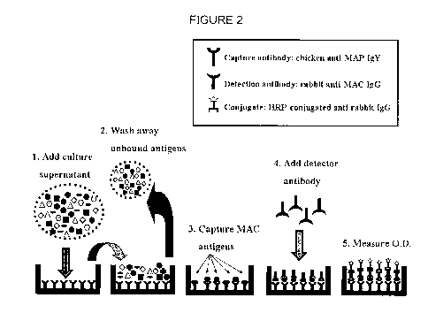
[0016] Figure 2 is a schematic diagram of one embodiment of the MAC-
ELISA procedure according to the present invention.
[0017] Figure 3 is a graph showing the analytical sensitivity of the methods
according to the present invention, depicting enhanced specificity and
sensitivity of the MAC-ELISA by absorption of chicken anti-MAP IGY capture
antibody and rabbit anti-MAC IgG detector antibody.
[0018] Figure 4 is a graph showing the analytical detection limit of the
MAC-ELISA using purified MAP culture filtrate.
[0019] Figure 5 is a graph showing the analytical detection limit of the
MAC-ELISA using purified MAA culture filtrate.
[0020] Figure 6 is a graph showing assessment of the MAC-ELISA on
1,275 well-defined clinical cultures, as a scatter plot of MAC-ELISA OD values
for MAC and mycobacteria other than MAC.
[0021] Figure 7 is a graph showing ROC analysis of the scatter plot data
depicted in Figure 6.
[0022] Figure 8 illustrates clinical application of the MAC-ELISA in
conjunction with the MGIT ParaTB mediumTM and MGIT 960 instrument.
[0023] Figure 9 shows representative data of multiplex PCR. M: Marker, 1:
M. paratuberculosis ATCC1 9698, 2: M. paratuberculosis human isolate UCF-
5, 3: M. avium ATCC35712, 4: M. avium human isolate 104, 5: M.
intracellulare ATCC1 3950, 6: MAC clinical isolate, 7: M. terrae ATCC1 5755,
M. asiaticum ATCC25274, 9: M. scrofulaceum ATCC19981, 10: E. coli DH5a,
11: Negative control.
[0024] Figure 10 is a graph depicting comparison of single antibody cross-
reactivity pre- and post-absorption, using chicken anti-MAC IgY.
[0025] Figure 11 is a graph depicting comparison of single antibody cross-
reactivity pre- and post-absorption, using rabbit anti-MAC IgG.
CA 02718107 2010-09-09
WO 2009/117462 PCT/US2009/037469
6
DETAILED DESCRIPTION OF THE PRESENTLY PREFERRED
EMBODIMENTS
[0026] "Mycobacterium avium complex" (MAC) is a group of genetically-
related bacteria belonging to the genus Mycobacterium. It includes
Mycobacterium avium subspecies avium (MAA), Mycobacterium avium
subspecies hominissuis (MAH), and Mycobacterium avium subspecies
paratuberculosis (MAP). Historically, MAC has also included Mycobacterium
intracellulare (MI) - a distinct species of bacteria. The present invention
specifically contemplates MI as being included in MAC.
[0027] "Antigen" is a substance that evokes an immune response in a
subject, especially the production of antibodies. Antigens are usually
proteins
or polysaccharides foreign to the subject, but may also be any type of
molecule, including small molecules (haptens) coupled to a carrier-protein.
For example, a M. paratuberculosis antigen is a substance that evokes an
anti-M. paratuberculosis response in a subject, when the subject is immunized
with that antigen.
[0028] "Antigenic preparation" is a preparation that includes antigens.
[0029] "Antibody" is used in the broadest sense and specifically covers
paratuberculosis-specific monoclonal antibodies (including agonist,
antagonist, and neutralizing antibodies), paratuberculosis-specific antibody
compositions with polyepitopic specificity, single chain paratuberculosis-
specific antibodies, and fragments of paratuberculosis-specific antibodies.
The antibodies may be anti-M. avium monoclonal or polyclonal antibodies per
se, immunologically effective fragments thereof (e.g., Fab, Fab', or F ab')2),
or a
single chain version of the antibodies, usually designated as Fõ regions.
Methods of producing polyclonal and monoclonal antibodies, including binding
fragments and single chain versions, are well known in the art.
[0030] "Immunoglobulin" refers to a glycoprotein that functions as an
antibody. The terms antibody and immunoglobulin may be used
CA 02718107 2010-09-09
WO 2009/117462 PCT/US2009/037469
7
interchangeably. Immunoglobulins are found in the blood and tissue fluids, as
well as many other body secretions; they take part in an immune response of
an organism to bacteria or foreign substances.
[0031] MAC capture antibody (or simply capture antibody) may include a
variety of antibodies, and is preferably chicken anti-MAC SA IgY. The capture
antibody may include polyclonal immunoglobulin "Y" (IgY) produced by
immunization of chickens with Mycobacterium avium complex (MAC) secreted
antigens (SA) and rendered highly specific for MAC SA by absorption with
heterologous bacteria (mycobacteria and non-mycobacteria) or antigens from
these bacteria. The IgY is harvested from eggs laid by the immunized
chickens, purified by standard methods, and absorbed with heterologous
bacterial cells or antigens prior to use in the assay as anti-MAC SA IgY.
[0032] MAC detection antibody (or simply detection antibody or detector
antibody) may include a variety of antibodies, and is preferably rabbit anti-
MAC SA IgG. The detection antibody may include polyclonal immunoglobulin
"G" (IgG) produced by immunization of rabbits with Mycobacterium avium
complex (MAC) secreted antigens (SA) and rendered highly specific for MAC
SA by absorption with heterologous bacteria (mycobacteria and non-
mycobacteria) or antigens from these bacteria. The IgG is harvested from
rabbit serum, purified by standard methods, and absorbed with heterologous
bacterial cells or antigens prior to use in the assay as anti-MAC SA IgG. It
is
contemplated that the IgG for the detection antibody could be raised in many
mammalian animal species.
[0033] "Conjugate" when used herein refers to a detector molecule, such as
goat anti-rabbit immunoglobulin-specific antibody, that has been chemically
coupled to an indicator system, also called a "label". Other such systems
capable of detecting immunoglobulins, such as enzyme-conjugated protein G,
can also be used for practicing the present invention. It is also contemplated
that the term "conjugate" specifically includes a conjugated antibody against
detector (i.e. detection) antibody (see Figures 1 and 2).
CA 02718107 2010-09-09
WO 2009/117462 PCT/US2009/037469
8
[0034] "Label" when used herein refers to a detectable compound or
composition which is conjugated directly or indirectly to an antibody so as to
generate a "labeled" antibody (conjugate). The label may be detectable by
itself (for example radioisotope label or fluorescent label) or, in the case
of an
enzymatic label, may catalyze chemical alteration of a substrate compound or
composition the product of which is then detectable.
[0035] "Subject" refers to any organism classified as a mammal, including
humans, domestic and farm animals, and zoo, sports, or pet animals, such as
dogs, horses, cats, cattle, pigs, sheep, etc. Preferably, the mammal is
human. More preferably, the mammal is bovine.
[0036] "Specificity" as used in this application refers to analytical
specificity,
meaning the ability of the assay to distinguish among similar but not
identical
analytes as in the secreted antigens of M. avium versus secreted antigens of
M. phlei
[0037] "Sensitivity" as used in this application refers to analytical
sensitivity,
the minimum concentration of analytes detectable by an assay; for example,
in the preferred construction the MAC-ELISA can detect 20 ng/mL MAC
secreted antigens in a culture.
[0038] Detection of antigens specific for MAC bacteria secreted during
growth in liquid culture media using antigen-capture ELISA technology is
provided. This assay is also referred to as "MAC-ELISA". The assay takes
advantage of the abundant cell products made by the pathogen during culture,
not solely on the DNA of the organism.
[0039] Novel compositions are provided, which include a capture antibody
and a detection antibody. The capture antibody may be affixed to solid
support, such as a microtiter plate. The capture antibody may be polyclonal
or monoclonal. For example, the capture antibody may be polyclonal chicken
anti-MAC secreted antigens. The detection antibody may also be polyclonal
or monoclonal. For example, the detection antibody may be polyclonal rabbit
anti-MAC secreted antigens. Both antibodies can be prepared by
immunization of the respective animals with MAC secreted antigens.
CA 02718107 2010-09-09
WO 2009/117462 PCT/US2009/037469
9
Preferably, this is followed by removal of non-MAC-specific antibodies by
absorption, for example with other bacteria or their antigens.
[0040] In one embodiment of the invention, the step-wise MAC-ELISA
procedure is conducted as follows: coat a microtiter plate with capture
antibody, add sample (liquid culture medium), incubate, wash away unbound
antigens, add the detection antibody and wash away unbound antibody, add a
conjugate, e.g. enzyme-conjugated goat anti-rabbit IgG, wash away unbound
material, add substrate, and measure the resulting color using an ELISA
reader. After plate coating (an overnight procedure) the assay typically
requires less than 2 hours to complete.
[0041] Figures 1 and 2 are schematic outlines of the systems and methods
according to the present invention. In one example of the assay, shown in
Figures 1 and 2, there are 4 layers. They are described from the bottom
surface (e.g. plastic surface) up. Layer #1 is a capture antibody. This
capture
antibody can be chicken anti-MAC secreted antigen antibody (IgY) that, prior
to use, has been preferably absorbed with heterologous antigens rendering it
highly specific for MAC antigens. Layer #2 is MAC secreted antigens from a
liquid culture, which antigens bind to the capture antibody. Layer #3 is a
detector antibody. In this example, the detector antibody is rabbit anti-MAC
secreted antigen antibody (IgG) that, prior to use, has been absorbed with
heterologous antigens rendering it highly specific for MAC antigens. In this
example, the detector antibody is not labeled, although in some embodiments
the detector antibody may be labeled. Layer #4 is a signal system, such as a
conjugate (conjugated antibody against detector antibody). The purpose of
the signal system is to recognize that the detector antibody has bound in the
reaction and trigger a measureable signal. Examples of commercially
available suitable conjugates include, for example HRP-conjugated goat anti-
rabbit IgG. This is a common way to detect antibody binding, but there are
others, some of which are non-antibody based, e.g. they are based on the use
of protein G or A.
CA 02718107 2010-09-09
WO 2009/117462 PCT/US2009/037469
[0042] Some of the advantages of this invention, called MAC-ELISA,
include that it is simpler and it saves both time and money relative to
current
technologies. It also is a novel approach to identification of mycobacteria in
culture and could be evaluated for M. tuberculosis complex pathogens as well.
It eliminates the need for expensive instrument, e.g. BACTEC MGIT 960
(-$50,000 each), and allows cultures to be screened for presence of MAC
mycobacteria for relatively small per assay costs. Assay specificity is
sufficiently high that no further testing, e.g. PCR, is necessary in many
situations when diagnosis of a MAC species is sufficiently precise. In one
aspect, the MAC-ELISA triages cultures allowing PCR resources and
technician time to be focused only on those cultures with a high likelihood of
containing MAC. If desired, follow-up PCR assays can simply confirm the
MAC-ELISA diagnosis or determine which member of the MAC is growing in
the culture tube, e.g. M. avium subspecies avium, M. avium subspecies
paratuberculosis, or M. avium subspecies hominissuis. The MAC-ELISA thus
saves laboratory time by triaging culture-positive samples and eliminating the
need to perform PCRs on mycobacterial isolates that are not pathogenic (i.e.
non-MAC).
[0043] In one aspect of the present invention, MAC culture detection based
on secreted antigens (SA) common to all MAC is provided. Antigens typically
exceed cells by 1,000-fold making better detection targets. Novel antigen-
capture antibody is provided. Use of chickens to produce anti-MAC SA IgY is
provided. The antigens are used to immunize chickens. Absorption of IgY to
make it MAC-specific may be performed. Novel and different detector
antibodies are also provided. For example, the use of rabbits to produce anti-
MAC SA IgG is contemplated. Antigens may be used to immunize rabbits.
Absorption of IgG to make it MAC-specific may be performed. Thus, a
relatively low-cost technology for detection of mycobacterial pathogens is
provided.
[0044] The methods of the present invention can be practiced with a variety
of bacterial strains, preferably mycobacterial strains, including but not
limited
CA 02718107 2010-09-09
WO 2009/117462 PCT/US2009/037469
11
to the Mycobacterium avium complex (Mycobacterium avium subspecies
avium, Mycobacterium avium subspecies hominissuis, Mycobacterium avium
subspecies paratuberculosis, and Mycobacterium avium subspecies
sylvaticum), the Mycobacterium tuberculosis complex (MTBC),
Mycobacterium simiae, etc.
[0045] Liquid cultures useful for practicing the invention may be obtained
from other mycobacterial strains. Such liquid cultures may be used as
antigenic preparations; liquid cultures from two or more bacterial strains may
also be combined. Preferably, liquid cultures should be obtained from clinical
M. avium strains, rather than from laboratory-maintained M. avium strains.
[0046] To practice this invention, a skilled artisan will know to use other
media compositions, broths, etc., suitable for growth of mycobacteria, in
order
to obtain secreted antigens in liquid cultures. These media may be modified,
supplemented with various compounds, acidified, etc. One skilled in the art
will know how to optimize the assays by defining the optimal culture
incubation time window, and by developing new media to induce antigen
secretion. Addition of glycerol enhances bacterial growth and yield of
antigens. Preferably, the media should be glycerol-based. Existing
commercial media may be modified; for example, 7H9 broth (Becton
Dickinson, Cockeysville, MD) may be modified by replacing the glucose with
glycerol. This substitution enhances bacterial growth and results in improved
yield of antigens. The pH of the media should preferably be kept at 5.5 to
6.5.
More preferably, the pH of the media should be kept at about 6Ø In a
preferred embodiment, the media for bacterial growth is modified Watson-
Reid (WR) broth (formulation described in Sung and Collins, 2003, App!.
Environ. Microbiol. 69: 6833-6840) with a pH of about 6Ø
[0047] In one example, the culture of M. paratuberculosis in an early-log
phase is centrifuged to remove (pellet) the bacteria. The remaining aqueous
liquid culture is then concentrated using a size-exclusion filter, preferably
a
5,000 molecular weight size-exclusion filter. The liquid culture may also be
dialyzed, for example using 10 mM PBS, pH 6.8.
CA 02718107 2010-09-09
WO 2009/117462 PCT/US2009/037469
12
[0048] The entire aqueous phase that is obtained from a bacterial culture
should be considered a cellular filtrate, synonymous with secreted antigens.
For example, if centrifugation of 3,000 x g for 10 minutes was used to
separate (pellet) the bacteria, then the entire supernatant should be
considered liquid culture.
[0049] The liquid culture may include a variety of antigenic compounds,
such as various mycobacterial proteins, carbohydrates, lipids, metabolites,
growth factors, etc. Some of these proteins may, for example, be further
modified by phosphorylation, glycosylation, and/or acetylation. The
compounds in the liquid culture may be extracellular, secreted, excreted,
byproducts of bacterial metabolism, etc. In general, it is only required that
the
liquid culture includes compounds that act as antigens and that are capable of
eliciting the immune response.
[0050] In one embodiment, the antibodies are mixed with absorbing
antigens prior to contacting them with liquid cultures containing M. avium
antigens. The absorbing antigens are mixed to absorb the nonspecific
antibodies in the sample. The absorbing antigens may be added in the form
of an absorbing antigen preparation. These absorbing antigens may be from
one type of mycobacteria. Alternatively, they may be from multiple different
mycobacteria. Preferably, the absorbing antigens are from Mycobacterium
terrae, Mycobacterium phlei, E. coli, or any combinations thereof. The
antigens may include cellular extracts of these organisms and in some
examples are used at a final concentration of about 250 micrograms per
milliliter of diluted sample, the preferred sample dilution being 1:50.
[0051] In another embodiment, the invention provides an antibody that
specifically binds to any of the above or below described antigens. This is a
M. avium complex-specific antibody. Optionally, the antibody is a monoclonal
antibody, humanized antibody, antibody fragment, or single-chain antibody.
The M. avium -specific antibody is capable of binding to the antigen, creating
an antigen-antibody complex. Examples of antibodies are the capture
antibody and the detection antibody.
CA 02718107 2010-09-09
WO 2009/117462 PCT/US2009/037469
13
[0052] The antigen-capture antibody complex may be attached to solid
support. Once an antigen-capture antibody complex is formed, a second
detection antibody or comparable detection molecule is used to detect the
presence of the antigen. The detection antibody binds to the antigen. The
second antibody is thus used to detect the presence of the MAC antigens
bound to the first antibody. The conjugate then detects the presence of the
detector antibody and creates a measureable signal such as color reaction
measured by an ELISA reader.
[0053] As an alternative to the conjugate, the detector antibody itself may
be labeled with a label, for example, a bead, a radioisotope, a ligand, a
chemiluminescent molecule, a dye, a fluorescent molecule, or an enzyme.
Labeled antibodies and reagents useful in immunoassays are disclosed in
U.S. Patent No. 4,490,473.
[0054] Radioactive labels such as iodine-125 (1251) or other radioactive
elements may be applied by known procedures. Techniques for labeling
antibodies with 1251 or other radioactive labels are described in Greenwood et
a/., 1963, Biochem. J. 89: 114-123; Harlow and Lane, 2006, Labeling
Antibodies with Iodine, Cold Spring Harbor Protocols, 2006: pdb.prot4287.
[0055] Fluorescent labels and procedures for coupling them to antibodies
are described in U.S. Patents Nos. 4,256,834 and 4,261,968. Labeled
secondary antibody conjugates are known, and may include labeled biotin-
binding proteins for detection of biotinylated targets, fluorophore-labeled
Protein A and G conjugates, gold conjugates, and the zenon antibody labeling
technology (Invitrogen, Carlsbad, CA).
[0056] A wide variety of enzymatic labels may be applied, and these are
selected in conjunction with the substrate to be used in the analysis by
procedures well-known in the art. For example, enzymes such as alkaline
phosphatase, horseradish peroxidase, catalase, peroxidase,
betaglucuronidase, glucose-6-phosphate dehydrogenase, urease,
phosphatase, and glucose oxidase are conveniently linked to antibodies by art
CA 02718107 2010-09-09
WO 2009/117462 PCT/US2009/037469
14
recognized techniques such as those described in U.S. Patents Nos.
3,875,011, 3,791,932, and 3,879,262.
[0057] Alternatively, the binding of detection antibody may be inferred by
the adherence of the complex to a solid surface to which this second antibody
is adherent, or by the ability of the complex to activate the complement
components in sera, or by other means known in the art.
[0058] The assay format may be Western blot, radioimmunoprecipitation,
radioimmunoassay (RIA), latex particle agglutination, or an enzyme-linked
immunosorbent assay (ELISA), including a sandwich ELISA or lateral-flow
ELISA.
[0059] The method may employ a solid support such as a column, a
dipstick, a filter or a microtiter dish. The ELISA may include detection of an
antibody that binds to a M. avium-specific antigen using a labeled anti-Ig
antibody. The antigen used in the practice of the method may be obtained
from M. avium complex. Preferably, the antigen may be obtained from liquid
culture of M. avium complex. The ELISA also may be a competitive assay.
The assay may involve quantification. The assay may also be automated,
and may, for example, be run on standard ELISA automated plate readers.
[0060] In one embodiment, systems for the detection of M. avium complex
are provided. The systems include antibodies that bind immunologically to M.
avium complex -specific antigens from a provided sample. The systems may
include an antigen obtained from M. avium complex liquid culture as a positive
control.
[0061] The detection antibody may be directly labeled, for example, with a
bead, a radioisotope, a ligand, a chemiluminescent molecule, a fluorescent
molecule, an enzyme, or with another detectable conjugate. Alternatively,
presence of the antigen-bound detection antibody in the assay can be
determined by a conjugate such as an antibody-conjugate. The determination
of conjugate binding can be quantitative.
[0062] In one embodiment, the present invention relates to an ELISA
diagnostic kit for the assay of M. avium complex antigens in a sample
CA 02718107 2010-09-09
WO 2009/117462 PCT/US2009/037469
obtained from a liquid culture (MAC-ELISA). A skilled artisan may further
improve the signal-to-noise ratio of the diagnostic test by amplifying the
signal
coming from the label such as using the biotin-avidin labeling method for
antibody conjugate signal amplification.
[0063] In one embodiment, the kit can contain the already absorbed
chicken anti-MAC antibody. Alternatively, the end user can do the absorption
step.
[0064] Some embodiments of the present invention are described in Shin et
a/., 2009, Clinical and Vaccine Immunology (in press), which is herein
incorporated by reference.
EXAMPLES
[0065] It is to be understood that this invention is not limited to the
particular methodology, protocols, subjects, or reagents described, and as
such may vary. It is also to be understood that the terminology used herein is
for the purpose of describing particular embodiments only, and is not intended
to limit the scope of the present invention, which is limited only by the
claims.
The following examples are offered to illustrate, but not to limit the claimed
invention.
[0066] Bacterial strains, cultures, and preparations of antigens
[0067] Bacterial strains used in the experiments included: Mycobacterium
avium subsp. paratuberculosis (MAP): JTC303, DT114, K-10, ATCC19698,
other mycobacteria: 62 species including M. avium and M. phlei; non-
mycobacterial spp.: 17 species including E. coli and unknown fungus spp.
[0068] To develop a MAC-antigen capture ELISA with anti-MAC antibody
as the solid phase, a number of organism cultures were prepared.
Mycobacterium avium complex (MAC) strains were selected to encompass
the most clinically important M. avium subspecies using both type strains and
clinical strains. Antibodies were produced by immunization of rabbits (IgG)
and chickens (IgY) with MAP and MAC culture filtrate (CF) antigens. The
CA 02718107 2010-09-09
WO 2009/117462 PCT/US2009/037469
16
bacterial strains used for antibody production and tested in this study are
listed in Table 1. Briefly, MAP ATCC19698, MAP JTC114, and MAP JTC303
were cultivated in modified Watson-Reid (mWR; pH 6.0) broth media
supplemented with 2 pg/ml of Mycobactin J (Allied Monitor, Fayette, MO).
The static cultivation was performed by inoculating 100pL of 109 CFU/mL
seedlot culture into the cell culture flask (75 cm2 canned neck, Corning INC.,
NY) containing 50 ml of mWR broth medium for 10 weeks at 37 C in 5% CO2
humidified conditions.
[0069] MAP CF antigens were harvested and pooled as previously
described (Shin et al., 2008, Clin. Vaccine Immunol. 15: 1277-1281). Strains
MAA (ATCC35712) and MAH 104 were cultured in mWR for 6 weeks at 37 C
to obtain and pool MAC antigens.
[0070] Cellular extracts (CEAs) were used to remove by absorption cross-
reactive antibodies from the rabbit anti-MAC IgG and chicken anti-MAC IgY as
previously described (Shin et al., 2008, Clin. Vaccine Immunol. 15: 1277-
1281). To prepare cellular extract antigens, Mycobacterium intracellularae
ATCC 13950, M. intracellularae ATCC 25122, and Mycobacterium
scrofulaceum ATCC 19981 were cultivated in mWR broth for 4 weeks 37 C.
Mycobacterium phlei ATCC 11758 and Mycobacterium terrae ATCC 15755
strains were cultivated in mWR for 2 weeks at 37 C.
[0071] To evaluate antibody specificity, other non-MAC mycobacterial
strains were cultured in 7H9 broth supplemented with 10% OADC (Becton
Dickinson, Sparks, MD) for 2 to 4 weeks at 37 C (Table 1). Non-
mycobacterial strains were grown in Luria-Bertani (LB) broth.
[0072] For preparation of cellular antigen extracts from each bacterium
grown in mWR, 7H9 or LB broth were prepared as previously described. The
antigens included: concentrated liquid culture antigens (CFA); and cellular
extract antigens (CEA). The concentration of proteins in each CFA and CEA
preparation was determined by BCA protein assay kit (Pierce, Rockford, IL).
CA 02718107 2010-09-09
WO 2009/117462 PCT/US2009/037469
17
[0073] Antibody production
[0074] MAC (two strains) and MAP (three strains) antigen pools were made
to immunize rabbits and chickens. Briefly, 250 pL of culture filtrate antigens
from each strain were pooled, mixed, adjusted to a final concentration of 1000
pg/mL and stored as 1 mL aliquots at -20 C until use. A total of four chickens
and four rabbits were used for production of antibody, two each for anti-MAP
and anti-MAC.
[0075] At each immunization, laying chickens were inoculated with 500 pl
of CF antigens mixed with an equal volume of Freund's incomplete adjuvant
(FIA). The first immunization was given subcutaneously. Subsequent
immunizations were given intramuscularly, the first 2 weeks later, and the
remaining four at 1 week intervals. Eggs from each hen were collected daily
after the second immunization, labeled, and stored at 4 C until use.
[0076] The IgY was precipitated from egg yolk by adding 1 volume of 40%
PEG 8000 (Sigma, Gaithersburg, MD) in PBS to 3 volumes of egg yolk then
centrifuged at 13,000 x g for 20 min. The purified IgY was then dialyzed four
times with 1 L 10 mM PBS.
[0077] Immunization of rabbits for production of rabbit anti-MAP and anti-
MAC antibody followed essentially the same protocol as used for chickens
with slight modification. Briefly, each rabbit was intradermally inoculated
with
500 pg/mL CF antigen pool in an equal volume of FIA. The subsequent three
immunizations were done by subcutaneous inoculation of 250 pg/mL CF
antigen pool in an equal volume of FIA at 2 week intervals. After the first
and
third immunizations, the serum antibody levels to each antigen were
measured by ELISA. After the fourth immunization serum was harvested from
each rabbit. Rabbit IgG purification was then performed using ImmunoPure
(G) IgG Purification Kit (Pierce, Rockford, IL) following manufacturer's
instructions.
[0078] Both chicken IgY and rabbit IgG were pure as evidenced by a single
band by SDS-PAGE comparable to the commercial antibody controls. The
CA 02718107 2010-09-09
WO 2009/117462 PCT/US2009/037469
18
yield was 4-5 mg/mL of IgY from a single egg and 10 mL of 2-3 mg/mL rabbit
IgG by BCA assay.
[0079] Enhancement of antibody specificity
[0080] The specificity of rabbit anti-MAP and anti-MAC IgG was enhanced
by absorption by both M. phlei and E. coli antigens; chicken anti-MAP and
anti-MAC IgY was enhanced by absorption with M. phlei antigens. Briefly,
100 pg of purified IgY was mixed with 107 CFU/mL of M. phlei ATCC 11758
and incubated at 4 C overnight. The mixture was then filtered using a 0.2 pm
syringe filter (Nalgene, Rochester, NY). The filtered antibody was dialyzed in
mM PBS three times and the final concentration of absorbed anti-MAP and
anti-MAC IgY was determined using the BCA protein assay. As intact
mycobacterial cells alone were not sufficient for removal of the cross-
reactivity
of rabbit anti-MAP and anti-MAC IgG with other bacteria, CE antigens of both
M. phlei ATCC 11758 (500 pg/mL) and E. coli DH5a (200 pg/mL) were used
to absorb cross-reactive rabbit antibodies. Only absorbed chicken IgY and
rabbit IgG were employed in the final assay (referred to as "chicken anti-MAP
IgY/ anti-MAC IgY" and "rabbit anti-MAP IgG" and "rabbit anti-MAC IgG").
[0081] Figure 10 shows a comparison of single antibody cross-reactivity
pre- and post-absorption, using a chicken anti-MAC IgY. The vertical bars
indicate: 1: Mycobacterium avium subsp. paratuberculosis ATCC1 9968, 2:
Mycobacterium avium subsp. avium ATCC35712, 3: Mycobacterium
intracellulare ATCC25122, 4: Mycobacterium phlei ATCC11758, 5:
Mycobacterium terrae ATCC15755, 6: Mycobacterium scrofulaceum
ATCC19981, 7: Corynebacterium pseudotuberculosis clinical isolate, 8:
Escherichia co/iATCC25922, 9: A mixture of environmental bacteria including
Aeromonas hydrophila, Enterobacter aerogenes, Enterococcus faeca/is,
Klebsiella pneumonia, Pseudomonas aeruginosa, and Proteus vulgaris.
[0082] Figure 11 shows a comparison of single antibody cross-reactivity
pre- and post-absorption, using a rabbit anti-MAC IgG. The vertical bars
CA 02718107 2010-09-09
WO 2009/117462 PCT/US2009/037469
19
indicate: 1: Mycobacterium avium subsp. paratuberculosis ATCC19968, 2:
Mycobacterium avium subsp. avium ATCC35712, 3: Mycobacterium
intracellulare ATCC25122, 4: Mycobacterium phlei ATCC11758, 5:
Mycobacterium terrae ATCC15755, 6: Mycobacterium scrofulaceum
ATCC1 9981, 7: Corynebacterium pseudotuberculosis clinical isolate, 8:
Escherichia co/iATCC25922, 9: A mixture of environmental bacteria including
Aeromonas hydrophila, Enterobacter aerogenes, Enterococcus faecalis,
Klebsiella pneumonia, Pseudomonas aeruginosa, and Proteus vulgaris.
[0083] Specificity of anti-MAC antibody preparations before and after
absorption
[0084] Reactivity of chicken anti-MAC IgY and rabbit anti-MAC IgG was
tested by ELISA both before and after absorption using multiple mycobacterial
CFA and CE antigens. Briefly, 2 g/mL of test antigen was coated on wells of
a 96 well plate (Maxisorp , Nalge Nunc International, Rochester, NY) by
overnight incubation at 4 C. After washing three times with wash buffer (KPL,
Gaithersburg, MD), the wells were blocked with 10% normal goat serum
(Sigma, St. Louis, MO) at room temperature for 2 hrs. Either (a) 100 pL of 2
pg/mL absorbed or non-absorbed anti-MAC IgY or (b) 100 pL of 1:4,000
diluted absorbed or non-absorbed rabbit anti-MAC IgG were added to each
well then incubated at RT for 30 min while shaking at 60 rpm. After washing
wells five times with wash buffer, HRP-conjugated rabbit anti-IgY (Gentel
Biosciences, Madison, WI) at a dilution of 1:4,000 or HRP-conjugated sheep
anti-rabbit IgG (Vector, Burlingame, CA) at a dilution of 1:5,000 was added to
each well and incubated for 30 min at RT. Plates were washed five times with
wash buffer (KPL, Gaithersburg, IL), after which 100 pL of TMB substrate
(TMBE-500, Moss Inc., Pasadena, MD) was added to each well followed by
incubation for 1 min at RT. The reaction was then stopped by addition of 100
pL stop solution (KPL, Gaithersburg, IL). The optical density (OD) of final
CA 02718107 2010-09-09
WO 2009/117462 PCT/US2009/037469
reaction in each well was measured at 450 nm using an ELISA reader
( Quant, Bio-Tek instruments Inc., Winooski, VT).
[0085] Development of MAC-ELISA protocol
[0086] Important reagents in the MAC-ELISA are: 1) the solid phase
capture antibody: chicken anti-MAP IgY, 2) the test substance: mycobacterial
broth culture fluid potentially containing secreted MAC antigens, 3) the
detector antibody: rabbit anti-MAC IgG, and 4) the conjugate: HRP-conjugated
goat anti-rabbit IgG (Vector, Burlingame, CA) (see, e.g., Figures 1 and 2).
The concentrations and volumes of the important components were optimized
for analytical sensitivity and specificity by reagent titration individually
and in
various combinations with culture fluid from pure cultures of MAP, MAA, MAH,
M. intracellularae, M. scrofulaceum, M. phlei, M. terrae and Corynebacterium
pseudotuberculosis. In one example, the final MAC-ELISA protocol was:
Plates (96 well; Maxisorp, Nalge Nunc, Rochester, NY) were first coated with
10 fag of capture antibody, chicken anti-MAC IgY, diluted in coating buffer
(KPL, Gaithersburg, IL) by overnight incubation at 4 C. After washing three
times with wash buffer (KPL, Gaithersburg, IL), all wells were blocked with
10% normal goat serum (Sigma, St. Louis, MO) at RT for 2 hrs. Fluid (100
pL) from the liquid cultures to be tested was next added to each well. After 1
hr at RT with 60 rpm shaking, the plate was again washed three times with
wash buffer. The detector antibody, rabbit anti-MAC IgG (100 pl of 0.5pg/mL)
was added to each well and incubated 30 min at RT. Wells were again
washed three times with washing buffer (KPL, Gaithersburg, IL). Then, 100
pL of HRP-conjugated goat anti-rabbit IgG (Vector, Burlingame, CA) at a
dilution of 1:5,000 was added to all wells and incubated for 30 min at RT.
Plates then were washed five times with wash buffer (KPL, Gaithersburg, IL).
Lastly 100 pL TMB substrate (TMBE-500, Moss Inc., Pasadena, CA) was
added to each well followed by a 1 min RT incubation after which the reaction
was stopped by adding 100pL of stop solution (KPL, Gaithersburg, IL) to each
CA 02718107 2010-09-09
WO 2009/117462 PCT/US2009/037469
21
well. The optical density (OD) of the final reaction in each well was measured
at 450 nm using an ELISA reader ( Quant, Bio-Tek instruments Inc.,
Winooski, VT). On each plate there are: two positive controls in duplicate
(MAP and MAC culture fluid) and three negative controls (PBS, MGIT medium
and M. phlei culture fluid). The cutoff value for a positive assay is the mean
OD of the two negative control wells plus 0.10.
[0087] Analytical sensitivity and specificity of the MAC-ELISA
[0088] The fluids from pure liquid cultures of both mycobacterial and non-
mycobacterial strains were tested. To estimate MAC-ELISA analytical
sensitivity, 2-fold serial dilutions of culture fluid (16.0 to 0.0078 pg/mL)
from
each type strain were tested. Assay results were compared with positive
(culture fluid from MAP ATCC19698 and MAA ATCC 35712), and negative
(culture fluid from M. phlei and M. terrae) controls. To determine MAC-ELISA
analytical specificity and optimal timing for testing, culture fluid from 7H9
broth
cultures of mycobacteria were collected weekly up to 8 weeks (Table 1).
Briefly, 102 CFU of each mycobacterial strain was inoculated into 10 mL of
Middlebrook 7H9 broth (Difco, Sparks, MD) supplemented with 0.5% glycerol,
and 10% OADC (Middlebrook) and incubated at 37 C for 8 weeks. For MAP
strains, 2 pg/ml of Mycobactin J (Allied Monitor, Fayette, MO) also was added
to the culture medium for optimal growth. Non-mycobacterial strains were
grown in LB medium (Table 1). Corynebacterium pseudotuberculosis was
grown in brain heart infusion broth. Culture fluid from all strains was tested
weekly by MAC-ELISA along with positive and negative controls as described
above.
[0089] Figures 3-5 illustrate the analytical sensitivity of the systems and
methods described herein. Figure 3 shows enhanced specificity and
sensitivity of the MAC-ELISA by absorption of chicken anti-MAP IgY capture
antibody and rabbit anti-MAC IgG detector antibody. The vertical bars
indicate: 1: Mycobacterium avium subsp. paratuberculosis ATCC19968, 2:
CA 02718107 2010-09-09
WO 2009/117462 PCT/US2009/037469
22
Mycobacterium avium subsp. avium ATCC35712, 3: Mycobacterium
intracellulare ATCC25122, 4: Mycobacterium phlei ATCC1 1758, 5:
Mycobacterium terrae ATCC15755, 6: Mycobacterium scrofulaceum
ATCC19981, 7: Corynebacterium pseudotuberculosis clinical isolate, 8:
Escherichia co/iATCC25922, 9: A mixture of environmental bacteria including
Aeromonas hydrophila, Enterobacter aerogenes, Enterococcus faecalis,
Klebsiella pneumonia, Pseudomonas aeruginosa, and Proteus vulgaris.
[0090] Figure 4 shows the analytical detection limit of the MAC-ELISA
using purified MAP culture filtrate. The vertical bars indicate: 1:
Mycobacterium avium subsp. paratuberculosis ATCC1 9968, 2:
Mycobacterium avium subsp. avium ATCC35712, 3: Mycobacterium
intracellulare ATCC25122, 4: Mycobacterium phlei ATCC11758, 5:
Mycobacterium terrae ATCC1 5755, 6: Mycobacterium scrofulaceum
ATCC19981, 7: Corynebacterium pseudotuberculosis clinical isolate, 8:
Escherichia co/iATCC25922, 9: A mixture of environmental bacteria including
Aeromonas hydrophila, Enterobacter aerogenes, Enterococcus faecalis,
Klebsiella pneumonia, Pseudomonas aeruginosa, and Proteus vulgaris.
[0091] Figure 5 shows the analytical detection limit of the MAC-ELISA
using purified MAA culture filtrate. The vertical bars indicate: 1:
Mycobacterium avium subsp. paratuberculosis ATCC19968, 2:
Mycobacterium avium subsp. avium ATCC35712, 3: Mycobacterium
intracellulare ATCC25122, 4: Mycobacterium phlei ATCC11758, 5:
Mycobacterium terrae ATCC15755, 6: Mycobacterium scrofulaceum
ATCC19981, 7: Corynebacterium pseudotuberculosis clinical isolate, 8:
Escherichia co/iATCC25922, 9: A mixture of environmental bacteria including
Aeromonas hydrophila, Enterobacter aerogenes, Enterococcus faecalis,
Klebsiella pneumonia, Pseudomonas aeruginosa, and Proteus vulgaris.
CA 02718107 2010-09-09
WO 2009/117462 PCT/US2009/037469
23
Table 1. Bacterial strains used to assess the specificity of the MAC-ELISA
Reference Detection
Total strain IDs Source other time in
Species No. of included in than ATCC ' MAC-
strains total tested ELISA2
(weeks)
Mycobacterial spp.
M. avium subsp. ATCC319698, 3
paratuberculosis 13 K-10 JTC4
ATCC35712, 1-2
M. avium subsp. avium 4 ATCC25291 JTC
M. avium subsp. 104 JTC, EPA5, 1-2
hominissuis 6 WSLH6
ATCC13950, JTC, EPA, 1
M. intracellulare 9 ATCC25122 WSLH
M. silvaticum 1 ATCC49884 3
M. abscess 1 ATCC19977 N
M. asiaticum 4 ATCC25276 JTC N
M. bovis 3 ATCC19210 JTC N
M. celatum 4 ATCC51130 JTC N
M. flavescens 2 ATCC14474 JTC N
M. fortuitum 2 ATCC49404 WSLH N
M. gordonae 2 ATCC14470 JTC N
M. kansasii 3 ATCC12478 JTC N
M. lentiflavum 2 ATCC51985 WSLH N
CA 02718107 2010-09-09
WO 2009/117462 PCT/US2009/037469
24
M. malmoense 1 ATCC29571 N
M. marium 2 ATCC927 WSLH N
M. nonchromogenicum 1 ATCC19530
M. phlei 1 ATCC11758 N
M. scrofulaceum 7 ATCC19981 JTC N
M. simiae 2 ATCC25275 WSLH N
ATCC 14468, N
M. smegmatis 2 mc2155
M. terrae 3 ATCC15755 JTC N
Non-mycobacterial spp.
Aeromonas hydrophlia 1 WSLH N
Corynebacterium N
pseudotuberculosis I JTC
Enterococcus faecalis 1 ATCC29212 WSLH N
Enterobacter aerogenes 1 WSLH N
Escherichia coli 4 ATCC25922 WSLH N
Klebsiella pneumonia 1 WSLH N
Proteus vulgaris 1 WSLH N
Pseudomonas aeruginosa 1 WSLH N
Unidentified fungi 6 JTC N
Total 92
1 Isolates were identified using mutiplex PCR and HPLC.
2 The time in weeks until antigens secreted by MAC bacteria were detected in
culture
fluid by the MAC-ELISA when 102 CFU of all strains were inoculated into 7H9
broth. N
indicates the assay was never positive up to the end of incubation at 8 weeks.
CA 02718107 2010-09-09
WO 2009/117462 PCT/US2009/037469
3American Type Culture Collection, Manassas, VA
4 Johne's Testing Center, Madison, WI
5 Environmental Protection Agency, Cincinnati, OH
6 Wisconsin State Laboratory of Hygiene, Madison, WI
[0092] Assessment of MAC-ELISA vs. MGIT960 ParaTBTM culture system
for pure cultures
[0093] Duplicate tubes of MGIT ParaTB mediumTM (Becton Dickinson)
were inoculated with serial dilutions of MAP ATCC19698, MAA ATCC 35712,
M. ph/ei ATCC 11758, or M. terrae ATCC 15755. Briefly, undiluted stock cell
suspension (1.0 mL) was added to 9.0 ml of 10 mM PBS (pH 7.2) and 10-fold
serial dilutions were made in 10 mM PBS (pH 7.2) with vortexing between
each dilution step resulting in 100 to 107 CFU/mL of each of the four
mycobacterial strains. From each dilution 100 pL was inoculated into MGIT
ParaTB mediumTM (Becton Dickinson, Sparks, MD). Each MGIT tube
contained 7 ml of modified Middlebrook 7H9 broth base with mycobactin J and
fluorescent indicator measuring changes in oxygen concentration embedded
in silicone on the bottom of the tube. The MGIT ParaTB mediumTM was
supplemented as per manufacturer's instructions and incubated at 37 C in a
MGIT 960 instrument.
[0094] Tubes were removed when the machine signaled them as positive
based on changes in the indicator. For each MGIT-positive tube, culture fluid
(100 pL) was then tested by MAC-ELISA with results analyzed in relationship
to the Time To Detection (TTD; incubation time in days until signal-positive)
for each culture.
[0095] Assessment of the MAC-ELISA using well-defined clinical cultures
CA 02718107 2010-09-09
WO 2009/117462 PCT/US2009/037469
26
[0096] A total of 1,275 animal feces, tissues, water, and soil samples
yielding acid-fast stain positive organisms were tested using the MAC-ELISA.
This set was obtained from 684 clinical cultures in modified BACTEC 12B
medium and 591 clinical cultures in MGIT ParaTB mediumTM. Once an
instrument signaled positive, acid-fast staining was done on the cultures and
contamination was checked by inoculation to 5% sheep blood agar plates.
Final identification of mycobacterial isolates was done using as a reference
method a multiplex PCR simultaneously targeting mycobacterial 16sDNA and
four insertion elements I S900, IS901, IS 1311, and IS 1245 (Johne's Testing
Center, Madison, WI) with reference strains as controls. Ultimately these
1,275 clinical samples yielded 340 MAC and 344 non-MAC mycobacteria from
modified BACTEC 12B medium and 305 MAC and 286 mycobacteria other
than MAC from MGIT ParaTB mediumTM. The optimal cutoff, sensitivity, and
specificity of the MAC-ELISA were determined by ROC curve analysis.
[0097] Validation of the MAC-ELISA to triage MGIT signal-positive cultures
[0098] Prospectively, 652 consecutive clinical samples (animal feces,
tissues, water or soil) were processed for MAP isolation following
manufacturer's recommendations using the MGIT ParaTB mediumTM. The
first time the MGIT 960 instrument signaled a tube as "positive" it was
removed from the instrument, vortexed and reinserted in the machine. After
the tube signaled positive a second time (or if it signaled positive within
one
week of the 49 day incubation protocol), the MAC-ELISA was performed. For
MAC-ELISA negative cultures, acid-fast staining (Ziehl-Neelsen) on culture
fluid smears independently assessed the presence of mycobacteria.
[0099] The multiplex PCR was used to verify the identity of mycobacteria in
all acid-fast stain positive and MAC-ELISA positive MGIT cultures. In all
cases of discrepancy between MAC-ELISA and multiplex PCR results, two
assays were use to clarify the true identity of mycobacterial isolates: IS900
nested PCR for MAP (greater analytical sensitivity than the multiplex) and
HPLC of cell wall mycolic acids for all other mycobacteria (Wisconsin State
CA 02718107 2010-09-09
WO 2009/117462 PCT/US2009/037469
27
Laboratory of Hygiene, Madison, WI) (Glickman et al., 1994, J. Clin.
Microbiol.
32: 740-745).
[00100] Statistical analysis
[00101] Specificity and sensitivity were evaluated by receiver-operator
characteristic (ROC) curves. MAC-ELISA OD values before and after
antibody absorption were compared by the t-test. Differences in OD values
between MAC cultures and cultures with mycobacteria other than MAC were
compared by the Mann-Whitney test. Statistical analyses were done using
statistical software (GraphPad Prism version 4.03 for Windows, GraphPad
Software, San Diego California USA).
[00102] Anti-MAC antibody specificity
[00103] Prior to absorption with heterologous antigens both chicken anti-
MAP and anti-MAC IgY showed cross-reactivity to other mycobacteria such as
M. scrofulaceum, M. phlei and M. terrae. After absorption with M. phlei cells,
the cross-reactivity to those mycobacteria disappeared without significant
decrease in reactivity (ELISA OD) to target MAP and MAC mycobacteria
(Figure 1A). Rabbit anti-MAC and anti-MAP IgG both cross-reacted with non-
mycobacteria as well as all mycobacteria tested. After absorption with the CE
antigens from M. phlei and E. coli, however, this cross-reactivity decreased
significantly without appreciable change in reactivity to secreted antigens of
MAP or MAC (Figure 1 B). The absorbed chicken and rabbit anti-MAC and
anti-MAP retained strong reactivity to both MAC and MAP and moderate
reactivity to M. intracellularae.
[00104] Development of MAC-enzyme linked immunosorbent assay
[00105] Numerous combinations and concentrations of chicken and
rabbit anti-MAP and anti-MAC were tested during development of the MAC-
ELISA. The combination providing optimal sensitivity and specificity for
detection of secreted MAC antigens in liquid cultures required use of chicken
CA 02718107 2010-09-09
WO 2009/117462 PCT/US2009/037469
28
anti-MAP IgY for antigen-capture and rabbit anti-MAC IgG for captured-
antigen detection, together with a suitable commercial conjugate to detect
rabbit antibody binding (data not shown) (Figure 2). Although the antibodies
were produced using selected subspecies of MAC, they did not discriminate
among MAC subspecies nor between M. avium and M. intracellularae. The
final assay is thus complex-specific, but not species- or subspecies-specific
and therefore is referred to as the MAC-ELISA.
[00106] MAC-ELISA specificity and sensitivity for pure cultures
[00107] Culture fluid obtained weekly from 92 mycobacterial and non-
mycobacterial strains were tested. After 8 weeks of incubation no
mycobacteria outside the MAC triggered a positive MAC-ELISA (Table 1). All
MAC members (13 MAP, 4 MAA, 6 MAH, 1 MAS, and 9 M. intracellularae)
became MAC-ELISA positive between 1 and 4 weeks of incubation in
Middlebrook 7H9 when the starting inoculum was 102 CFU.
[00108] The specificity and sensitivity of MAC-ELISA was enhanced by
use of absorbed antibodies (Figures 3-5). Assay accuracy using anti-MAP
IgY for antigen capture and anti-MAC IgG for antigen detection was superior
to all other antibody combinations. The MAC-ELISA analytical sensitivity was
0.03125 pg/mL MAP CFA (Figure 4) and 0.0625 pg/mL MAA CFA (Figure 5)
when the mean negative control ODs plus 0.10 was used as the cutoff for a
positive test.
[00109] Optimal incubation time for detection and detection limit
[00110] Time To Detection (TTD) as reported by the MGIT 960
instrument or incubation time to positive MAC-ELISA were similar, given that
the MGIT instrument read cultures hourly and culture fluid was only tested by
MAC-ELISA weekly. The MAC-ELISA detection limit for MAP and MAC was
101 CFU/mL. Culture fluid from M. phlei never triggered a positive MAC-
ELISA (Table 2).
CA 02718107 2010-09-09
WO 2009/117462 PCT/US2009/037469
29
Table 2. Comparison of time to positive in days between MAC-ELISA and MGIT
cultures
M. phlei
MAP JTC303 MAA ATCC35712
ATCC1 1758
Inoculum
MGIT MGIT MGIT MAC-
CFU/mL MAC-ELISA2 MAC-ELISA
TTD1 TTD TTD ELISA
106 - 107 4.8 7 3.5 7 0.7 N D
105- 106 7.2 7 5.3 7 2.6 ND
104- 105 10.1 14 6.9 7 4.3 ND
103- 104 12.8 21 8.7 14 6.0 ND
102-103 17.1 21 10.6 14 10.4 ND
10'- 102 21.5 28 12.7 21 14.8 ND
100-101 39.1 35 15.8 21 ND ND
10-1-100 ND ND ND ND ND ND
' The MGIT 960 instrument measures fluorescence as an indication of microbial
growth
hourly.
2 Culture fluid was tested by MAC-ELISA weekly.
3 Not detectable in MAC-ELISA up to 56 days of incubation.
[00111] ROC analysis of the MAC-ELISA using well-defined clinical
cultures
[00112] A significant difference in MAC-ELISA OD values was observed
between clinical cultures containing MAC versus non-MAC mycobacteria
CA 02718107 2010-09-09
WO 2009/117462 PCT/US2009/037469
(P<0.0001) (Figure 6). The cut-off value for maximum assay accuracy was
determined by ROC curve analysis. The assay sensitivity and specificity were
92.6% (95% Cl, 90.3-94.5) and 99.9% (95% Cl, 99.2-100), respectively with
an area under the ROC curve (AUC) of 0.992 (Figure 7).
[00113] Clinical application of MAC-ELISA
[00114] The MGIT 960 instrument signaled growth in 652 clinical
cultures; MAC-ELISA indicated that MAC species were present in 219
(33.6%) of them. Among these 219 cultures, 212 were confirmed as
containing MAC organisms (97.8%; 210 MAP and 2 MAC). The other seven
were found to contain mycobacteria other than MAC for a false-positive MAC-
ELISA rate of 3.2% (7/219) (Figure 8).
[00115] The remaining 433 MGIT-positive cultures were MAC-ELISA-
negative (66.4%). Of these, 426 (98.4%) did not contain acid-fast bacteria
suggesting a high rate of false-positive signals by the MGIT system. Seven of
the 433 MGIT-positive but MAC-ELISA-negative cultures (1.6%) had acid-fast
bacteria identified as MAP (n=6) or non-MAC mycobacteria (n=1); a false-
negative rate of 6/433 (1.4%) (Figure 8). More than 500 MGIT signal-negative
cultures as well as uninoculated culture medium were also MAC-ELISA
negative (data not shown).
[00116] It is to be understood that this invention is not limited to the
particular devices, methodology, protocols, subjects, or reagents described,
and as such may vary. It is also to be understood that the terminology used
herein is for the purpose of describing particular embodiments only, and is
not
intended to limit the scope of the present invention, which is limited only by
the claims. Other suitable modifications and adaptations of a variety of
conditions and parameters, obvious to those skilled in the art of diagnostic
assays and microbiology, are within the scope of this invention. All
publications, patents, and patent applications cited herein are incorporated
by
reference in their entirety for all purposes.