Note: Descriptions are shown in the official language in which they were submitted.
CA 02718860 2010-09-17
WO 2009/117041 PCT/US2009/000613
USE OF PYRENE TO CARRY PEPTIDES
ACROSS THE BLOOD BRAIN BARRIER
RELATED APPLICATIONS
This application claims the benefit of priority to U.S. provisional
application
61/038,634, filed March 21, 2008, the entire contents of which are
incorporated herei
by reference in their entirety.
FIELD OF THE INVENTION
The present invention relates generally to the field of delivering peptides,
proteins and antibodies across the blood-brain barrier (BBB). More
specifically, the
present invention relates to methods for delivering peptides, proteins or
antibodies
across the BBB using pyrene-agent conjugates.
BACKGROUND OF THE INVENTION
The detection and treatment of neurological conditions is often difficult due
t4
the impermeability of endogenous and exogenously administered components to
the
brain as a result of the blood-brain barrier (BBB). The BBB effectively
isolates the
brain from peripheral agents such as peptides, proteins, large macromolecules,
non-
peptidic molecules, ions, and water-soluble non-electrolytes. For example, it
is
generally accepted that charged or hydrophilic molecules as well as molecules
with a
molecular weight greater than about 700 kDa do not cross the BBB. It is also
generally accepted that peptides, such as peptides of about 21 amino acid
residues, do
not efficiently cross the BBB, nor do longer peptides such as the 40-residue
A(340
protein and the 42-residue AR42 protein, both associated with Alzheimer's
disease.
Thus, the BBB prevents the delivery of detection agents as well as
therapeutics, that
otherwise, may be useful in the diagnosis and treatment of a variety of
neurological
disorders.
-1-
CA 02718860 2010-09-17
WO 2009/117041 PCT/US2009/000613
Prior attempts at effectively transporting agents to the brain have included
conjugating agents to carrier moieties, using liposomal formulations, and
using
nanoparticles. Exemplary carrier moieties include naturally occurring
polyamines
(U.S. Patent 5,670,477), carriers such as lysozyme, hemoglobin, cytochrome-c
and
substance-P (U.S. Patent 5,604,198), and sugars (U.S. Patent 5,260,308). Prior
attempts at effectively transporting AR protein to the brain have used A1340
or smalle
fragments, such as AR 1-30, conjugated to a carrier such as OX26 or
putrescine. The
receptor for advanced glycation end products (RAGE) also has been proposed for
mediating transport across the BBB, particularly for A(3 protein.
There remains a need, however, for methods, agents and kits for delivering
peptide agents, including peptides, proteins and antibodies, across the BBB.
SUMMARY OF THE INVENTION
In accordance with one embodiment, the invention provides a method for
delivering a peptide conjugate across the blood brain barrier, comprising
administering to a subject a conjugate comprising the peptide agent and
pyrene. In
some embodiments the peptide agent is a detection agent capable of identifying
a
protein or structure associated with a neurological disorder. In another
embodiment,
the peptide agent is a therapeutic agent useful in treating a neurological
condition. In
some embodiments, the peptide agent includes an amino acid sequence
correspondin;
to a region of a target protein which undergoes a conformational shift from an
alpha-
helical conformation to a beta-sheet conformation, but does not include the
full-lengt:
sequence of the target protein. In other embodiments the peptide agent is an
antibod,)
specific for a protein or structure associated with a neurological condition.
In one
embodiment, the conjugate further comprises a detectable label. In another
embodiment the conjugate comprises a pyrene derivative, such as alkylated
pyrene
analogs, pyrene butyrate, PEGylated pyrene, pyrene-albumin analogs, pyrene
derivatives comprising a free carboxyl group and pyrene derivatives comprising
a fre
amine group.. In some embodiments, the conjugate comprises two or more pyrene
moieties.
-2-
CA 02718860 2010-09-17
WO 2009/117041 PCT/US2009/000613
In accordance with another embodiment, the invention provides an in vivo
method of detection comprising administering to a subject a conjugate
comprising a
peptide detection agent and pyrene, and detecting conjugate that is localized
in a
subject's brain. In one embodiment, the detection agent is capable of
identifying a
protein or a structure associated with a neurological condition. In some
embodiment
the conjugate comprises two or more pyrene moieties. In some embodiments, at
leas
one pyrene moiety is a pyrene derivative comprising a free carboxyl group and
at lea
one pyrene moiety is a pyrene derivative comprising a free amine group. In one
embodiment, the pyrene is conjugated to the peptide detection agent at least
at the N-
terminus or C-terminus of the peptide, or at both the N- and C-termini of the
peptide.
In yet another embodiment, the detection agent is capable of identifying a
protein in
specific conformation or state of self-aggregation. In another embodiment, the
detection of localized conjugate involves detecting pyrene excimers.
In yet another embodiment, the invention provides an in vivo method of
detection comprising administering to a subject a conjugate comprising peptide
detection agent, pyrene and a detectable label, and detecting conjugate that
has
localized in the brain of the subject. In some embodiments the label is a
fluorophore,
MRI contrast agent, ion emitter, or a radioactive label.
In other embodiments, the invention provides a method for treating
neurological conditions. The method comprises administering to a subject a
therapeutically effective amount of a conjugate comprising a peptide
therapeutic ages
and pyrene. In one embodiment the peptide agent is an anti-amyloid agent.
DESCRIPTION OF THE FIGURES
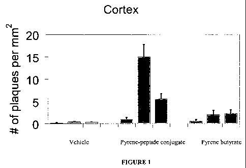
Figure 1 shows the number of A0 plaques detected per mm2 by vehicle,
peptide-agent pyrene conjugate, or pyrene butyrate administered intranasally
to
transgenic mice.
-3-
CA 02718860 2010-09-17
WO 2009/117041 PCT/US2009/000613
Figure 2 illustrates the correlation between A13 plaques detected in the
cortex
by intranasally administered conjugate (AD 185) fluorescence (-) versus
Thioflavin S
staining (A).
Figure 3 illustrates the correlation between Afl plaques detected in the
cortex
(Figure 3A) and hippocampus (Figure 3B) by intravenously administered
conjugate
(AD 185) fluorescence (-) versus Thioflavin S staining (.).
DETAILED DESCRIPTION
Before particular embodiments of the invention are described and
disclosed, it is to be understood that the particular materials, methods and
compositions described herein are presented only by way of examples, and are
not
limiting of the scope of the invention. The technical and scientific terms
used herein
have the meanings commonly understood by one of ordinary skill in the art to
which
the present invention pertains, unless otherwise defined. Publications and
other
materials setting forth known methodologies to which reference is made are
incorporated herein by reference in their entireties as though set forth in
full.
Standard reference works setting forth the general principles of recombinant
DNA technology include Sambrook, J., et al. (1989) Molecular Cloning: A
Laboratory Manual, 2d Ed., Cold Spring Harbor Laboratory Press, Planview,
N.Y.;
McPherson, M.J. Ed. (1991) Directed Mutagenesis: A Practical Approach, IRL
Pres:
Oxford; Jones, J. (1992) Amino Acid and Peptide Synthesis, Oxford Science
Publications, Oxford; Austen, B.M. and Westwood, O.M.R. (1991) Protein
Targeting
and Secretion, IRL Press, Oxford. Any suitable materials and/or methods known
to
those of ordinary skill in the art can be utilized in carrying out the present
invention.
However, preferred materials and methods are described. Materials, reagents
and the
like to which reference is made in the following description and examples are
obtainable from commercial sources, unless otherwise noted.
As used herein, the singular forms "a," "an," and "the" designate both the
singular and the plural, unless expressly stated to designate the singular
only.
-4-
CA 02718860 2010-09-17
WO 2009/117041 PCT/US2009/000613
The term "about" and the use of ranges in general, whether or not qualified b:
the term about, means that the number comprehended is not limited to the exact
number set forth herein, and is intended to refer to ranges substantially
within the
quoted range while not departing from the scope of the invention. As used
herein,
"about" will be understood by persons of ordinary skill in the art and will
vary to
some extent on the context in which it is used. If there are uses of the term
which are
not clear to persons of ordinary skill in the art given the context in which
it is used,
"about" will mean up to plus or minus 10% of the particular term.
As used herein "subject" denotes any animal in need of detection or
therapeutic treatment, including humans and domesticated animals, such as
cats, dog
swine, cattle, sheep, goats, horses, rabbits, and the like. "Subject" also
includes
animals used in research settings, including mice and other small mammals. A
typic
subject may be at risk of a neurological condition, disease or disorder or
suspected of
suffering from such a condition, or may be desirous of determining risk or
status witl
respect to a particular condition. As used herein, "therapeutic" treatment
includes the
administration of a therapeutic agent to treat an existing condition, to
prevent a
condition that the subject is at risk or developing, or for health
maintenance.
As used herein, the phrase "therapeutically effective amount" means that drul
dosage in a subject that provides the specific pharmacological response for
which the
drug is administered in a patient in need of such treatment. It is emphasized
that a
therapeutically effective amount will not always be effective in treating the
conditions/diseases described herein, even though such dosage is deemed to be
a
therapeutically effective amount by those of skill in the art.
As used herein, "peptide" refers to any polymer of two or more individual
amino acids (whether or not naturally occurring) linked via a peptide bond. As
used
herein, the term "peptide agent" includes peptides, proteins, and antibodies.
PeptideE
include fragments of full-length proteins, where fragments may include at
least 5
contiguous amino acids, at least 10 contiguous amino acids, at least 15
contiguous
-5-
CA 02718860 2010-09-17
WO 2009/117041 PCT/US2009/000613
amino acids, at least 20 contiguous amino acids, or at least 25 contiguous
amino acid
of the full-length protein. Peptides also include synthetic peptides.
As used herein, "conformation" or "conformational constraint" refers to the
presence of a particular protein conformation, for example, an a-helix,
parallel and
antiparallel n-strands, a leucine zipper, a zinc finger, etc. In addition,
conformationa
constraints may include amino acid sequence information without additional
structural information. As an example, "-C-X-X-C-" is a conformational
constraint
indicating that two cysteine residues must be separated by two other amino
acid
residues, the identities of each of which are irrelevant in the context of
this particular
constraint. A "conformational change" is a change from one conformation to
anothe
The term "An protein" is used herein to refer to all forms of the A(3 protein,
including A(334, A(337, A(338, AR40 and A(342.
"Recombinant proteins or peptides" refer to proteins or peptides produced by
recombinant DNA techniques, i.e., produced from cells, microbial or mammalian,
transformed by an exogenous recombinant DNA expression construct encoding the
desired protein or polypeptide. Proteins or peptides expressed in most
bacterial
cultures will typically be free of glycan. Proteins or peptides expressed in
yeast may
have a glycosylation pattern different from that expressed in mammalian cells.
As used herein, the term "naturally occurring" or "native" with reference to
peptide agent refer to agents (e.g., peptides, proteins and antibodies) that
are present
in the body or recovered from a source that occurs in nature. A native peptide
agent
may be modified either chemically or enzymatically, including post-
translational
modifications, including but not limited to, acetylation, glycosylation,
phosphorylation, lipid conjugation, acylation and carbonylation.
As used herein, the term "synthetic" with reference to a peptide agent
specific
that the agent is not naturally occurring, but may be obtained by other means
such as
chemical synthesis, biochemical methods, or recombinant methods.
-6-
CA 02718860 2010-09-17
WO 2009/117041 PCT/US2009/000613
The terms "analog," "fragment," "derivative," and "variant," when referring I
peptides herein mean analogs, fragments, derivatives, and variants of such
peptides
that retain substantially similar functional activity or substantially the
same biologicz
function or activity as the reference peptides, as described herein. An
"analog"
includes a pro-polypeptide that comprises the amino acid sequence of a
peptide.
A "fragment" is a portion of a peptide that retains substantially similar
functional activity or substantially the same biological function or activity
as the
reference peptide, as shown in in vitro assays disclosed herein.
A "derivative" includes all modifications to a peptide of this invention that
substantially preserve the functions disclosed herein and include additional
structure
and attendant function, e.g., PEGylated peptides or albumin fused peptides.
A "variant" includes peptides having an amino acid sequence sufficiently
similar to the amino acid sequence of a reference peptide. The term
"sufficiently
similar' means that the sequences have a common structural domain (e.g.,
sequence
homology) and/or common functional activity. For example, amino acid sequences
that comprise a common structural domain that is at least about 45%, at least
about
50%, at least about 55%, at least about 60%, at least about 65%, at least
about 70%,
least about 75%, at least about 80%, at least about 85%, at least about 90%,
at least
about 91%, at least about 92%, at least about 93%, at least about 94%, at
least about
95%, at least about 96%, at least about 97%, at least about 98%, at least
about 99%,
at least about 100%, identical are defined herein as sufficiently similar.
Variants
include peptides encoded by a polynucleotide that hybridizes to a complement
of a
polynucleotide encoding the reference polypeptide under stringent conditions.
Such
variants generally retain the functional activity of the reference peptides.
Variants
also include peptides that differ in amino acid sequence due to mutagenesis.
"Substantially similar functional activity" and "substantially the same
biological function or activity" each means that the degree of biological
activity is
within about 50% to 100% or more, within 80% to 100% or more, or within about
-7-
CA 02718860 2010-09-17
WO 2009/117041 PCT/US2009/000613
90% to 100% or more, of that biological activity demonstrated by the reference
peptide, when the biological activity of each peptide is determined by the
same
procedure or assay. For example, an analog or derivative of an may exhibit the
same
biological activity as the referent agent qualitatively, although it may
exhibit greater
or lesser activity quantitatively. The suitability of a given analog or
derivative of an
agent can be verified by routine screening methods to confirm that the analog
or
derivative exhibits an activity of interest that is substantially similar to
that of the
referent agent. An analog or derivative may possess additional structural
features
and/or exhibit additional functional properties, such as PEGylated agents,
which
comprise a PEG moiety and may exhibit a longer circulating half-life in vivo.
"Similarity" between two peptides is determined by comparing the amino
acid sequences. An amino acid of one polypeptide is similar to the
corresponding
amino acid of a second polypeptide if it is identical or a conservative amino
acid
substitution. Conservative substitutions include those described in Dayhoff,
M.O.,
ed., The Atlas of Protein Sequence and Structure 5, National Biomedical
Research
Foundation, Washington, D.C. (1978), and in Argos, P. (1989) EMBO J. 8:779-
785.
For example, amino acids belonging to one of the following groups represent
conservative changes or substitutions:
-Ala, Pro, Gly, Gln, Asn, Ser, Thr;
-Cys, Ser, Tyr, Thr;
-Val, Ile, Leu, Met, Ala, Phe;
-Lys, Arg, His;
-Phe, Tyr, Trp, His; and
-Asp, Glu.
Some aspects of the invention relate to the diagnosis and treatment of
disease,
and conditions associated with a specific structural state of a protein, such
as a
specific conformation or self-aggregative state of a protein. PCT application
PCT/US2007/016738 (WO 2008/013859) and U.S. Patent Application 11/828,953,
which disclose relevant embodiments, are incorporated herein by reference in
their
-8-
CA 02718860 2010-09-17
WO 2009/117041 PCT/US2009/000613
entireties. Some aspects of the invention provide conjugates and methods for
the in
vivo detection of proteins in a specific structural state, including misfolded
proteins
and self-aggregated proteins, such as those associated with disease states,
and
conjugates and methods for the treatment of those disease states. In some
embodiments, the proteins are associated with amyloidogenic diseases.
Proteins that are associated with human or animal disease when they adopt a
specific conformational or self-aggregated state are known in the art.
Examples of
such diseases includes amyloidogenic diseases, including Alzheimer's disease
(AD),
cerebral amyloid angiopathy (CAA), and cerebral vascular disease (CVD). As
used
herein, "amyloidogenic diseases" are diseases in which amyloid plaques or
amyloid
deposits are formed in the body. Amyloid formation is found in a number of
disorders, such as diabetes, AD, scrapie, bovine spongiform encephalopathy
(BSE),
Creutzfeldt-Jakob disease (CJD), chronic wasting disease (CWD), related
transmissible spongiform encephalopathies (TSEs).
A variety of diseases are associated with a specific structural form of a
proteii
(e.g., a "misfolded protein" or a self-aggregated protein), while the protein
in a
different structural form (e.g., a "normal protein") is not harmful. In many
cases, the
normal protein is soluble, while the misfolded protein forms insoluble
aggregates.
Examples of such insoluble proteins include prions in transmissible spongiform
encephalopathy (TSE); A(3-peptide in amyloid plaques of Alzheimer's disease
(AD),
cerebral amyloid angiopathy (CAA), and cerebral vascular disease (CVD); oc-
synuclein deposits in Lewy bodies of Parkinson's disease, tau in
neurofibrillary
tangles in frontal temporal dementia and Pick's disease; superoxide dismutase
in
amylotrophic lateral sclerosis; and huntingtin in Huntington's disease. See,
e.g.,
Glenner et al., J. Neurol. Sci. 94:1-28, 1989; Haan et al., Clin. Neurol.
Neurosurg.
92(4):305-310, 1990.
Often, these insoluble proteins form aggregates composed of non-branching
fibrils with the common characteristic of a (3-pleated sheet conformation. In
the CNIE
amyloid can be present in cerebral and meningeal blood vessels
(cerebrovascular
-9-
CA 02718860 2010-09-17
WO 2009/117041 PCT/US2009/000613
deposits) and in brain parenchyma (plaques). Neuropathological studies in
human
and animal models indicate that cells proximal to amyloid deposits are
disturbed in
their normal functions. See, e.g., Mandybur, Acta Neuropathol. 78:329-331,
1989;
Kawai et al., Brain Res. 623:142-146, 1993; Martin et al., Am. J. Pathol.
145:1348-
1381, 1994; Kalaria et al., Neuroreport 6:477-80, 1995; Masliah et al., J.
Neurosci.
16:5795-5811, 1996. Other studies additionally indicate that amyloid fibrils
may
actually initiate neurodegeneration. See, e.g., Lendon et al., J. Am. Med.
Assoc.
277:825-831, 1997; Yankner, Nat. Med. 2:850-852, 1996; Selkoe, J. Biol. Chem.
271:18295-18298, 1996; Hardy, Trends Neurosci. 20:154-159, 1997.
While the underlying molecular mechanism that results in protein misfolding
is not well understood, a common characteristic for all the above mentioned
neurological disorders is the formation of fibrils which come together to form
a
i3-sheet structure. Fibril formation and the subsequent formation of secondary
0-shee
structures associated with plaque deposits, occurs via a complex mechanism
involvin
a nucleation stage, in which monomers of the protein associate to form
fibrils,
followed by extension of the fibrils at each end. Thus, peptide, protein or
antibody
probes that are capable of disrupting fibril formation would prevent disease
progression and thus be of therapeutic importance. Additionally, agents
capable of
associating with a particular self-associating state of the diseased protein
are useful
diagnostic tools to detect and quantify a particular form of the misfolded
protein, as
well as provide insights to the progression of the disease. Thus, highly
selective
peptide agents capable of associating with specific proteins in a particular
state of
self-aggregation are useful, both as detection agents as well as for
therapeutic
applications.
A. Methods for Delivering Peptide Agents Across the BBB
Applicant has discovered that pharmaceutically relevant peptide agents, e.g.,
peptides, proteins and antibodies, conjugated to a pyrene carrier show an
enhanced
ability to cross the blood-brain barrier (BBB) when administered to a subject.
-10-
CA 02718860 2010-09-17
WO 2009/117041 PCT/US2009/000613
In one embodiment, there is provided a method for delivering a peptide agent
across the BBB that comprises administering to a subject a conjugate
comprising (i),
peptide agent and (ii) pyrene. In some embodiments, the peptide agent is a
peptide,
protein, or antibody. In some embodiments, the peptide agent is a detection
agent or
therapeutic agent. In specific embodiments, the peptide agent is a detection
agent
capable of identifying a target protein or structure (such as a specific
conformation of
state of self-aggregation) associated with a neurological condition. In other
embodiments, the peptide agent is a therapeutic agent useful in treating a
neurologica
condition. As used herein, "capable of identifying" means that the peptide
agent
selectively and preferentially binds to the target protein or structure.
The conjugate may be formulated in any composition suitable for
administration to a subject, such as a composition comprising the conjugate
and a
pharmaceutically acceptable carrier. The conjugate may be administered by any
suitable means, including by intranasal, intravenous, intraperitoneal,
intraarterial,
intramuscular, subcutaneous, oral, buccal, or transdermal, administration, and
may b
formulated accordingly. For example, the pharmaceutically acceptable carrier
may bE
a liquid, so that the composition is adapted for parenteral administration, or
may be
solid, i.e., a capsule shell plus vehicle, a tablet, a pill and the like,
formulated for oral
administration. Alternatively, the pharmaceutically acceptable carrier may be
in the
form of a nebulizable liquid or solid so that the composition is adapted for
inhalation
Pharmaceutically acceptable carriers are known in the art, and may include,
without
limitation, dissolution or suspension agents such as water or a naturally
occurring
vegetable oil like sesame, peanut, or cottonseed oil or a synthetic fatty
vehicle like
ethyl oleate or the like. Buffers, preservatives, antioxidants, binders,
excipients,
disintegrating agents, lubricants, sweetening agents and flavoring agents may
also be
included in the composition.
In the methods described herein, one or more conjugates comprising the same
or different detection agents, therapeutic agents, pyrene moities and/or
labels may be
used, with each conjugate provided in the same composition or in one or more
-11-
CA 02718860 2010-09-17
WO 2009/117041 PCT/US2009/000613
different compositions that may be administered simultaneously or sequentially
by tli
same route or by one or more different routes.
In some embodiments, the pyrene-conjugated peptide agent exhibits a
permeability across the BBB that is substantially greater than that of the non-
conjugated active agent, such as at least three, at least five, at least ten,
at least fifteen
at least twenty times greater, or more, than that of the non-conjugated active
agent.
One measure of permeability across the BBB is the amount of conjugate that
enters the brain relative to the amount that was injected and relative to the
amount the
enters other tissues (%IDI). In some embodiments, the pyrene-conjugate has an
octanol/water partition coefficient between 1-10.
It is believed that some carriers that are used for increasing the
permeability c
a peptide across the BBB also have the effect of increasing the half-life of
the peptide
carrier conjugate. For example, carriers that add a significant amount of
structural
size to the peptide-carrier conjugate may decrease the rate of degradation or
clearanc
of the peptide. The A1340 peptide, for example, under normal physiological
conditions is degraded in both the periphery and in the brain. However,
conjugates
using, for example, putrescine or OX26 as carriers increase the half life of
A040
dramatically. While an increased half-life may have some advantages, such as
contributing to an increase in concentration in the brain, it also may have
significant
disadvantages, such as an increase in non-specific localization in the brain.
This mal
be a particular concern if, for example, non-specifically localized conjugate
contributes to a high background that decreases the sensitivity and/or
selectivity of it
vivo imaging.
The conjugates described herein do not suffer from this drawback. For
example, experiments conducted with a conjugate comprising an A13 peptide
labeled
at both termini with pyrene showed that the conjugate was cleared 6 hours post-
administration, as determined by analysis of cerebrospinal fluid, which
revealed no
evidence of circulating conjugate.
-12-
CA 02718860 2010-09-17
WO 2009/117041 PCT/US2009/000613
The rate of localization and clearance or degradation of a conjugate can be
assessed experimentally, for example, by administering the conjugates to mice
and
sacrificing them for analysis at different times post-administration, such as
at time
periods including 2 minutes, 10 minutes, 30 minutes, 60 minutes, 1 hour, 2
hours, 3
hours, 4 hours, 5 hours, 6 hours, or longer, post-administration.
The non-toxicity of the conjugates can be verified experimentally, for
example, using in vitro assays and in vivo rodent toxicity studies that are
known in tI
art.
B. Peptide Agents
The nature of the peptide agent is not limited, other than comprising amino
acid residues. The peptide agent can be a synthetic or a naturally occurring
peptide,
including a variant or derivative of a naturally occurring peptide. The
peptide can be
a linear peptide, cyclic peptide, constrained peptide, or a peptidomimetic.
Methods
for making cyclic peptides are well known in the art. For example, cyclization
can b
achieved in a head-to-tail manner, side chain to the N- or C-terminus
residues, as we]
as cyclizations using linkers. The selectivity and activity of the cyclic
peptide
depends on the overall ring size of the cyclic peptide which controls its
three
dimensional structure. Cyclization thus provides a powerful tool for probing
progression of disease states, as well as targeting specific self-aggregation
states of
diseased proteins.
In some embodiments, the peptide agent specifically binds to a target protein
or structure associated with a neurological condition. In accordance with
these
embodiments, the invention provides agents useful for the selective targeting
of a
target protein or structure associated with a neurological condition, for
diagnosis or
therapy.
In some embodiments, the peptide agent is a peptide probe as described in
PCT application PCT/US2007/016738 (WO 2008/013859) and U.S. Patent
Application 11/828,953, the entire contents of which are incorporated herein
by
-13-
CA 02718860 2010-09-17
WO 2009/117041 PCT/US2009/000613
reference in their entirety. As described therein, such peptide probes may be
useful z
detection agents and/or as therapeutic agents. Exemplary peptide probes
described is
PCT application PCT/US2007/016738 (WO 2008/013859) and U.S. Patent
Application 11/828,953 include an amino acid sequence corresponding to a
region of
the target protein which undergoes a conformational shift from an alpha-
helical
conformation to a beta-sheet conformation, and the peptide probe itself
undergoes a
conformational shift from an alpha-helical conformation to a beta-sheet
conformatioi
but does not include the full-length sequence of the target protein. For
example, a
peptide probe may consist of at least 5, or from about 10 to about 25,
contiguous
amino acids from the target protein sequence, including at least 5, at least
10, up to
about 25 and up to about 50, such as 5 to 50, 10 to 50, 5 to 25 or 10 to 25
contiguous
amino acids from the target protein sequence. In some embodiments, the peptide
probe may undergo a conformational shift when contacted with a target protein
that i
in the beta-sheet conformation.
As described in PCT application PCT/US2007/016738 (WO 2008/013859)
and U.S. Patent Application 11/828,953, the peptide probes described therein
are
useful for detecting proteins in a sample or in vivo, and for detecting
proteins in a
specific structural state (e.g., a target structural state), such as a
specific conformatioi
or state of self-aggregation. For example, a peptide probe may be conjugated
to
pyrene such that it does not form excimers when the peptide probe is an alpha-
helix i
random coil conformation (or soluble state), but does form excimers when the
peptid
probe is in a beta-sheet conformation (or insoluble aggregated state). A
target
structural state may be associated with a disease while a different structural
state is
not associated with a disease. The target structural state may cause the
disease, may
be a factor in a symptom of the disease, may appear in a sample or in vivo as
a result
of other factors, or may otherwise be associated with the disease.
In some embodiments, the peptide agent comprises the amino acid sequence i
SEQ ID NO 34 of PCT application PCT/US2007/016738 WO 2008/013859) and U.I.
Patent Application 11/828,953. In some embodiments, the peptide agent
comprises
-14-
CA 02718860 2010-09-17
WO 2009/117041 PCT/US2009/000613
the amino acid sequence of SEQ ID NO:35, SEQ ID NO:36, SEQ ID NO:37, SEQ II
NO:38, or SEQ ID NO:45 of PCT application PCT/US2007/016738 WO
2008/013859) and U.S. Patent Application 11/828,953, which are useful in the
context of the detection and treatment of AD. In some embodiments, the peptide
agent is selected from SEQ ID NO:35, SEQ ID NO:36, SEQ ID NO:37, SEQ ID
NO:38, or SEQ ID NO:45 of WO 2008/013859. In other embodiments, the peptide
agent is other than SEQ ID NO 34, SEQ ID NO:35, SEQ ID NO:36, SEQ ID NO:37,
SEQ ID NO:38, or SEQ ID NO:45 of WO 2008/013859. In some embodiments, the
peptide is selected from SEQ ID NO:36 or SEQ ID NO:38 of WO 2008/013859. In
some embodiments, the peptide is other than SEQ ID NO:36 or SEQ ID NO:38 of
WO 2008/013859, including a peptide selected from SEQ ID NO 34, SEQ ID NO:35
SEQ ID NO:37, or SEQ ID NO:45 of WO 2008/013859 or another peptide. In some
embodiments, the peptide is SEQ ID NO:36 of WO 2008/013859. In some
embodiments, the peptide is other than SEQ ID NO:36 of WO 2008/013859,
including a peptide selected from SEQ ID NO 34, SEQ ID NO:35, SEQ ID NO:37, o
SEQ ID NO:45 of WO 2008/013859 or another peptide. In some embodiments, the
peptide is SEQ ID NO:38 of WO 2008/013859. In some embodiments, the peptide is
other than SEQ ID NO:38 of WO 2008/013859, including a peptide selected from
SEQ ID NO 34, SEQ ID NO:35, SEQ ID NO:37, or SEQ ID NO:45 of WO
2008/013859 or another peptide.
SEQ ID NO:1 (SEQ ID NO:34 of WO 2008/013859)
Val Val Ala Gly Ala Ala Ala Ala Gly Ala Val His Lys Leu Asn Thr Lys Pro Lys
Let
Lys His Val Ala Gly Ala Ala Ala Ala Gly Ala Val Lys
SEQ ID NO:2 (SEQ ID NO:35 of WO 2008/013859)
Leu Val Phe Phe Ala Glu Asp Val Gly Ser Asn Lys Gly Ala Ile Ile Gly Leu Met
SEQ ID NO:3 (SEQ ID NO:36 of WO 2008/013859)
Lys Leu Val Phe Phe Ala Glu Asp Val Gly Ser Asn Lys Gly Ala Ile Ile Gly Leu
Met
-15-
CA 02718860 2010-09-17
WO 2009/117041 PCT/US2009/000613
SEQ ID NO:4 (SEQ ID NO:37 of WO 2008/013859)
Leu Val Phe Phe Ala Glu Asp Val Gly Ser Asn Lys Gly Ala Ile Ile Gly Leu Met
Lys
SEQ ID NO:5 (SEQ ID NO:38 of WO 2008/013859)
Lys Leu Val Phe Phe Ala Glu Asp Val Gly Ser Asn Lys Gly Ala Ile Ile Gly Leu
Met
Lys
SEQ ID NO: 6 (SEQ ID NO:45 of WO 2008/013859)
Glu Val His His Gln Lys Leu Val Phe Phe Ala Glu Asp Val Gly Ser Asn Lys Gly Al
Ile Ile Gly Leu Met Val Gly Gly Val Val Ile Ala
In other embodiments, the peptide agent specifically binds to a target protein
or structure associated with other neurological conditions, such as stroke,
cerebrovascular disease, epilepsy, transmissible spongiform encephalopathy
(TSE);
A(3-peptide in amyloid plaques of Alzheimer's disease (AD), cerebral amyloid
angiopathy (CAA), and cerebral vascular disease (CVD); a-synuclein deposits in
Lewy bodies of Parkinson's disease, tau in neurofibrillary tangles in frontal
temporal
dementia and Pick's disease; superoxide dismutase in amylotrophic lateral
sclerosis;
and Huntingtin in Huntington's disease and benign and cancerous brain tumors
such
as glioblastoma's, pituitary tumors, or meningiomas.
In some embodiments, the peptide agent undergoes a conformational shift
other than the alpha-helical to beta-sheet shift discussed above, such as a
beta-sheet t
alpha-helical shift, an unstructured to beta-sheet shift, etc. Such peptide
agents may
undergo such conformational shifts upon interaction with target peptides or
structure
associated with a neurological condition.
In other embodiments, the peptide agent is an antibody that specifically binds
to a target protein or structure associated with a neurological condition,
such as a
target protein or structure (such as a specific conformation or state of self-
-16-
CA 02718860 2010-09-17
WO 2009/117041 PCT/US2009/000613
aggregation) associated with an amyloidogenic disease, such as the anti-
amyloid
antibody E610, and NG8. Other anti-amyloid antibodies are known in the art, as
are
antibodies that specifically bind to proteins or structures associated with
other
neurological conditions.
Other peptide detection agents include fluorescent proteins, such as Green
Flourescent Protein (GFP), streptavidin, enzymes, enzyme substrates, and other
peptide detection agents known in the art.
Exemplary peptide therapeutic agents include peptide macromolecules and
small peptides. For example, neurotrophic proteins are useful as peptide
agents in th
context of the methods described herein. Neurotrophic proteins include nerve
growth
factor (NGF), brain-derived neurotrophic factor (BDNF), neurotrophin-3 (NT-3),
neurotrophin-4 (NT-4), neurotrophin-5 (NT-5), insulin-like growth factors (IGF-
I ani
IGF-II), glial cell line derived neurotrophic factor (GDNF), fibroblast growth
factor
(FGF), ciliary neurotrophic factor (CNTF), epidermal growth factor (EGF), glia-
derived nexin (GDN), transforming growth factor (TGF-.alpha. and TGF-.beta.),
interleukin, platelet-derived growth factor (PDGF) and S 100(3 protein, as
well as
bioactive derivatives and analogues thereof.
Neuroactive peptides also include the subclasses of hypothalamic-releasing
hormones, neurohypophyseal hormones, pituitary peptides, invertebrate
peptides,
gastrointestinal peptides, those peptides found in the heart--such as atrial
naturetic
peptide, and other neuroactive peptides.
The subclass of hypothalamic releasing hormones includes as suitable
examples, thyrotropin-releasing hormones, gonadotropin-releasing hormone,
somatostatins, corticotropin-releasing hormone and growth hormone-releasing
hormone.
The subclass of neurohypophyseal hormones is exemplified by compounds
such as vasopressin, oxytocin, and neurophysins.
-17-
CA 02718860 2010-09-17
WO 2009/117041 PCT/US2009/000613
The subclass of pituitary peptides is exemplified by adrenocorticotropic
hormone, (3-endorphin, a-melanocyte-stimulating hormone, prolactin,
luteinizing
hormone, growth hormone, and thyrotropin.
Suitable invertebrate peptides are exemplified by FMRF amide, hydra head
activator, proctolin, small cardiac peptides, myomodulins, buccolins, egg-
laying
hormone and bag cell peptides.
Gastrointestinal peptides includes such neurologically active compounds sucl
as vasoactive intestinal peptide, cholecystokinin, gastrin, neurotensin,
methionineenkephalin, leucine-enkephalin, insulin and insulin-like growth
factors I
and II, glucagon, peptide histidine isoleucineamide, bombesin, motilin and
secretins.
Examples of other neuroactive peptides include angiotensin II, bradykinin,
dynorphin, opiocortins, sleep peptide(s), calcitonin, CGRP (calcitonin gene-
related
peptide), neuropeptide Y, neuropeptide Yy, galanin, substance K (neurokinin),
physalaemin, Kassinin, uperolein, eledoisin and atrial naturetic peptide.
Peptide agents also include proteins associated with membranes of synaptic
vesicles, such as calcium-binding proteins and other synaptic vesicle
proteins. The
subclass of calcium-binding proteins includes the cytoskeleton-associated
proteins,
such as caldesmon, annexins, calelectrin (mammalian), calelectrin (torpedo),
calpacti
I, calpactin complex, calpactin II, endonexin I, endonexin II, protein II,
synexin I; any
enzyme modulators, such as p65.
Other synaptic vesicle proteins include inhibitors of mobilization (such as
synapsin Ia,b and synapsin Ila,b), possible fusion proteins such as
synaptophysin, an(
proteins of unknown function such as p29, VAMP-1,2 (synaptobrevin), VAT I, rab
3A, and rab 3B.
Peptide agents also include a-, (3- and y-interferon, epoetin, Fligrastim,
Sargramostin, CSF-GM, human-IL, TNF and other biotechnology drugs.
-18-
CA 02718860 2010-09-17
WO 2009/117041 PCT/US2009/000613
Peptide agents also include peptides, proteins and antibodies obtained using
recombinant biotechnology methods.
Peptide agents also include "anti-amyloid agents" or "anti-amyloidogenic
agents," which directly or indirectly inhibit proteins from aggregating and/or
forminj
amyloid plaques or deposits and/or promotes disaggregation or reduction of
amyloid
plaques or deposits. Anti-amyloid agents also include agents generally
referred to in
the art as "amyloid busters" or "plaque busters." These include drugs which
are
peptidomimetic and interact with amyloid fibrils to slowly dissolve them.
"Peptidomimetic" means that a biomolecule mimics the activity of another
biologically active peptide molecule. "Amyloid busters" or "plaque busters"
also
include agents which absorb co-factors necessary for the amyloid fibrils to
remain
stable.
Anti-amyloid agents include antibodies and peptide probes, as described in
PCT application PCT/US2007/016738 (WO 2008/013859) and U.S. Patent
Application 11/828,953, the entire contents of which are incorporated herein
by
reference in their entirety. As described therein, a peptide probe for a given
target
protein specifically binds to that protein, and may preferentially bind to a
specific
structural form of the target protein. While not wanting to be bound by any
theory, i
is believed that binding of target protein by a peptide probe will prevent the
formatio
of higher order assemblies of the target protein, thereby preventing or
treating the
disease associated with the target protein, and/or preventing further
progression of th
disease. For example, binding of a peptide probe to a monomer of the target
protein
will prevent self-aggregation of the target protein. Similarly, binding of a
peptide
probe to a soluble oligomer or an insoluble aggregate will prevent further
aggregatio:
and protofibril and fibril formation, while binding of a peptide probe to a
protofibril
or fibril will prevent further extension of that structure. In addition to
blocking
further aggregation, this binding also may shift the equilibrium back to a
state more
favorable to soluble monomers, further halting the progression of the disease
and
alleviating disease symptoms.
-19-
CA 02718860 2010-09-17
WO 2009/117041 PCT/US2009/000613
Those skilled in the art will recognize that many of the peptide agents
described above as exemplary detection agents also are useful as therapeutic
agents,
and that many of the peptide agents described above as exemplary therapeutic
agents
also are useful as detection agents. Thus, these descriptors are in no way
limiting.
In some embodiments, the peptide agent is a variant of a peptide agent
described above, with one or more amino acid substitutions, additions, or
deletions,
such as one or more conservative amino acid substitutions, additions, or
deletions,
and/or one or more amino acid substitutions, additions, or deletions that
further
enhances the permeability of the conjugate across the BBB. For example amino
acid
substitutions, additions, or deletions that result in a more hydrophobic amino
acid
sequence may further enhance the permeability of the conjugate across the BBB.
C. Pyrene
The pyrene can be pyrene or any pyrene derivative or analog that, when
conjugated to a non-peptide agent improves the permeability of the agent
across the
BBB.
Pyrene consists of four fused benzene rings:
By "pyrene" deriviative or analog is meant a molecule comprising the four
fused benzene rings of pyrene, wherein one or more of the pyrene carbon atoms
is
substituted or conjugated to a further moiety. Exemplary pyrene derivatives
include
alkylated pyrenes, wherein one or more of the pyrene carbon atoms is
substituted wit
a linear or branched, substituted or unsubstituted, alkyl, alkenyl, alkynyl or
acyl
group, such as a CI-C20, linear or branched, substituted or unsubstituted
alkyl, alkeny
alkynyl or acyl group, where the group may be substituted with, for example, a
moiety including an 0, N or S atom (e.g., carbonyl, amine, sulfhydryl) or with
a
-20-
CA 02718860 2010-09-17
WO 2009/117041 PCT/US2009/000613
halogen. In some embodiments the pyrene derivative includes one or more free
carboxyl groups and/or one or more free amine groups, each of which may be
directl
attached to a pyrene carbon atom or attached to any position on a linear or
branched,
substituted or unsubstituted, alkyl, alkenyl, alkynyl or acyl group as
described above
such as being attached at a carbon atom that is separated from a pyrene carbon
by I c
more, such as 1 to 3, 1 to 5, or more, atoms. In some embodiments, the pyrene
is
substituted with one or more acetic acid moieties and/or one or more
ethylamine
moieties. In some embodiments, the pyrene derivative is substituted with a
single
methyl, ethyl, propyl or butyl group. In some embodiments, the pyrene is
substitute(
with a short chain fatty acid, such as pyrene butyrate. In another embodiment,
the
pyrene is conjugated to albumin, transferring or an Fc fragment of an
antibody. In
some embodiments, the substituent is attached to pyrene through a carbon-
carbon
linkage, amino group, peptide bond, ether, thioether, disulfide, or an ester
linkage.
Pyrene derivatives can be made by methods known in the art. For example,
substituted pyrenes may be used to attach fatty acids to the tetracyclic
scaffold.
Suitalbe reagents, including functionalized alkyl derivatives of pyrene, and
derivatizing reactions are known in the art. For example amino pyrene can be
reacte
with 1,4-butanedioic acid methyl ester to yield a butanoic acid derivative of
pyrene.
Alternatively, 1-thiocyanato pyrene can be reacted with 4-aminobuatnoic acid
methy
ester to yield a thio-substituted butanoic acid derivative of pyrene. Yet
other
alternative reactions include reacting pyrene boronic acid and a substituted
fatty acid
to yield fatty acid derivatives of pyrene.
In other embodiments, the pyrene derivative is PEGylated pyrene, i.e, pyrene
conjugated to polyethylene glycol (PEG). Such pyrene derivatives may exhibit a
longer circulating half-life in vivo. In other embodiments, the pyrene
derivative is
pyrene conjugated to albumin.
In some embodiments, the pyrene derivative exhibits reduced toxicity as
compared to pyrene. In some embodiments, the pyrene derivative exhibits an
increased circulating half-life in vivo as compared to pyrene, such as
PEGylated
-21-
CA 02718860 2010-09-17
WO 2009/117041 PCT/US2009/000613
pyrene discussed above. In some embodiments, the pyrene derivate exhibits even
greater increased permeability across the BBB as compared to pyrene, such as
albumin conjugated pyrene. In some embodiments, the pyrene derivative has an
octanol/water partition coefficient between 1-10.
D. Conjugates
The peptide agent may be conjugated to pyrene by any means known in the
art, including chemical (covalent) conjugation. In some embodiments, the
peptide
agent is directly conjugated to pyrene through a side chain residue. In one
embodiment the pyrene is conjugated to the peptide agent via the a-amino group
of a
lysine residue. Derivatives of pyrene, such as chloropyrene can be coupled to
the amino. group of lysine through palladium catalyzed cross-coupling
reactions. In othe
embodiments, the peptide agent is conjugated to pyrene through a linker.
Compound
used as linkers are well known in the art, and include optionally substituted
C1-C20
alkyl groups, alkanoic acids, alkenoic acids, alkynoic acids, alkoxide groups,
aminoalkanoic acids, alkyl amines, alkoxy groups, bifunctional imido esters,
glutaraldehyde, ethylene oxide polymers (PEG), optionally substituted aryl
groups,
alkynyl pyridyl, alkynyl bipyridyl, phthalic acid, malic acid and maleic acid,
N-hydroxysuccinimide esters, hetero-bifunctional reagents and group specific-
reactive agents such as the maleimido moiety, dithio moiety (SH) and
carbodiimide
moiety
Conjugates may be formed by chemical synthesis or bioengineering methods,
such as methods including expressing pyrene in living organisms together with
the
agent. Such bioengineering methods include direct engineering of synthetic
biological processes or evolution and screening for pyrene-agent conjugate
combinations.
In some embodiments, the peptide agent is conjugated to a single pyrene
moiety. In other embodiments, the peptide agent is conjugated to two or more
pyren,
moieties. When the peptide agent is conjugated to two or more pyrene moieties,
eacl
pyrene moiety may be conjugated to the agent (directly or through a linker).
-22-
CA 02718860 2010-09-17
WO 2009/117041 PCT/US2009/000613
In one embodiment the pyrene moiety is conjugated to the peptide agent at it,
N- or C-terminus. In another embodiment, the pyrene moiety is conjugated to
the
peptide agent at an internal (non-terminal) amino acid residue. In embodiments
with
two pyrene moieties, one pyrene moiety may be conjugated to each terminus of
the
peptide agent, one pyrene moiety may be conjugated to the N- or C-terminus and
the
other conjugated at an internal residue, or both may be conjugated at internal
residue
When more than two pyrene moieties are conjugated to a peptide agent, the
moieties
can be positioned at any permutation or combination of terminal and internal
residue
In some embodiments the pyrene moieties are conjugated in proximity to each
other,
while in others they are at spaced apart or distant positions on the peptide
agent. In
other embodiments, one or more pyrene moieties is conjugated (directly or
through a
linker) to one or more pyrene moieties, at least one of which is conjugated,
directly c
through a linker, to the peptide agent.
Regardless of the position(s) of the pyrene moiety(ies), the conjugate may
exhibit enhanced permeability of the agent across the BBB.
In some embodiments, the conjugates are labeled with pyrene such that they
are capable of forming pyrene excimers. That is, the peptide agents are
conjugated b
pyrene moieties in such a way as to permit excimer formation between pyrene
moieties conjugated to the same or different molecules of peptide agent, as
may be
desired. In accordance with these embodiments, two or more pyrene moieties may
b
conjugated to the same peptide agent molecule so as to permit excimer
formation by
interaction between pyrene moieties on the same peptide agent molecule, such
as ma
be associated, for example, with a specific conformation of the peptide agent.
Alternatively, the excimer formation may be due to interaction between pyrene
moieties on different peptide agent molecules, such as may be associated, for
example, with localization, binding and/or interaction between the peptide
agent
molecules.
In some embodiments different pyrene derivatives are used, at least one of
which includes one or more free carboxyl groups (such as an acetic acid
moietiy) an(
-23-
CA 02718860 2010-09-17
WO 2009/117041 PCT/US2009/000613
at least one of which includes one or more free amine groups (such as an
ethylamine
moiety), as discussed above. In accordance with this embodiment, interactions
between the free carboxyl group(s) on one pyrene derivative and the free amine
group(s) on another pyrene derivative may stabilize interactions between the
pyrene
derivatives, such as via the formation of a salt bridge, and may stabilize the
excimer
forming adducts and/or maximize excimer fluorescene. In accordance with these
embodiments, two different pyrene derivatives may be conjugated to the same
peptid
agent molecule, such as to stabilize excimer formation by interaction between
the
different pyrene derivatives on the same peptide agent molecule, such as may
be
associated, for example, with a specific conformation of the peptide agent.
Alternatively, one pyrene derivative may be conjugated to one peptide agent
molecu]
and a different pyrene derivative may be conjugated to a different peptide
agent
molecule, such as to stabilize excimer formation by interaction between the
different
peptide agent molecules, such as may be associated, for example, with
localization,
binding and/or interaction between the peptide agent molecules.
In some embodiments, the conjugate is labeled with a detectable label. For
example, the conjugate may comprise a peptide agent that is coupled or fused,
either
covalently or non-covalently, to a label. In embodiments where the peptide
agent is
detection agent, the detectable label may offer improved detection or
detection under
additional conditions. In embodiments where the peptide agent is a therapeutic
agen
the detectable label may offer detection in addition to the therapy offered by
the
therapeutic agent.
As used herein, a " detectable label" includes any moiety that can be detected
The specific label chosen may vary widely, depending upon the analytical
technique
to be used for analysis. The label may be complexed or covalently bonded at or
neat
the amino or carboxy end of the peptide agent, which may be endcapped with a
shod
hydrophobic peptide sequence. In some aspects of the invention, both the amino
anc
carboxy ends of the peptide agent are endcapped with small hydrophobic
peptides
ranging in size from about 1 to about 5 amino acids. These peptides may be
natural
-24-
CA 02718860 2010-09-17
WO 2009/117041 PCT/US2009/000613
synthetic, but are often natural (i.e., derived from the target protein). A
label may be
attached at or near the amino and/or carboxy end of the peptide, or at any
other
suitable position.
As used herein, a "detectable label" is a chemical or biochemical moiety
useful for labeling the conjugate. "Detectable labels" may include fluorescent
agent;
(e.g., fluorophores, fluorescent proteins, fluorescent semiconductor
nanocrystals),
phosphorescent agents, chemiluminescent agents, chromogenic agents, quenching
agents, dyes, radionuclides, metal ions, metal sols, ligands (e.g., biotin,
streptavidin
haptens, and the like), enzymes (e.g., beta-galactosidase, horseradish
peroxidase,
glucose oxidase, alkaline phosphatase, and the like), enzyme substrates,
enzyme
cofactors (e.g., NADPH), enzyme inhibitors, scintillation agents, inhibitors,
magnetic
particles, oligonucleotides, and other moieties known in the art.
Where the agent or label is a fluorophore, one or more characteristics of the
fluorophore may be used to assess the state of the labeled conjugate. For
example, tr
excitation wavelength of the fluorophore may differ based on whether the
conjugate
bound or free. In some embodiments, the emission wavelength, intensity, or
polarization of fluorescence also may vary based on the state of the
conjugate.
As used herein, a "fluorophore" is a chemical group that may be excited by
light to emit fluorescence or phosphorescence. A "quencher" is an agent that
is
capable of quenching a fluorescent signal from a fluorescent donor. A first
fluorophore may emit a fluorescent signal that excites a second fluorophore. A
first
fluorophore may emit a signal that is quenched by a second fluorophore. The
probes
disclosed herein may undergo fluorescence resonance energy transfer (FRET).
Fluorophores and quenchers may include the following agent (or fluorophore
and quenchers sold under the following tradenames): 1,5 IAEDANS; 1,8-ANS;
umbelliferone (e.g., 4-Methylumbelliferone); acradimum esters, 5-carboxy-2,7-
dichlorofluorescein; 5-Carboxyfluorescein (5-FAM); 5-
Carboxytetramethylrhodamir
(5-TAMRA) ; 5-FAM (5-Carboxyfluorescein); 5-HAT (Hydroxy Tryptamine) ; 5-
-25-
CA 02718860 2010-09-17
WO 2009/117041 PCT/US2009/000613
Hydroxy Tryptamine (HAT); 5-ROX (carboxy-X-rhodamine); 5-TAMRA (5-
Carboxytetramethylrhodamine); 6-Carboxyrhodamine 6G; 6-CR 6G; 6-JOE; 7-
Amino-4-methylcoumarin; 7-Aminoactinomycin D (7-AAD); 7-Hydroxy-4-
methylcoumarin; 9-Amino-6-chloro-2-methoxyacridine; ABQ; Acid Fuchsin; ACM.
(9-Amino-6-chloro-2-methoxyacridine); Acridine Orange; Acridine Red; Acridine
Yellow; Acriflavin; Acriflavin Feulgen SITSA; Alexa Fluor 350TM; Alexa Fluor
430TM; Alexa Fluor 488TM; Alexa Fluor 532TM; Alexa Fluor 546TM; Alexa Fluor
568TM; Alexa Fluor 594TM; Alexa Fluor 633TM; Alexa Fluor 647TM; Alexa Fluor
660TM; Alexa Fluor 680TM; Alizarin Complexon; Alizarin Red; Allophycocyanin
(APC); AMC; AMCA-S; AMCA (Aminomethylcoumarin); AMCA-X;
Aminoactinomycin D; Aminocoumarin; Aminomethylcoumarin (AMCA); Anilin
Blue; Anthrocyl stearate; APC (Allophycocyanin); APC-Cy7; APTS; Astrazon
Brilliant Red 4G; Astrazon Orange R; Astrazon Red 6B; Astrazon Yellow 7 GLL ;
Atabrine; ATTO-TAGTM CBQCA; ATTO-TAGTM FQ; Auramine; Aurophosphine C
Aurophosphine; BAO 9 (Bisaminophenyloxadiazole); Berberine Sulphate; Beta
Lactamase; BFP blue shifted GFP (Y66H); Blue Fluorescent Protein; BFP/GFP
FRET; Bimane; Bisbenzamide; Bisbenzimide (Hoechst); Blancophor FFG;
Blancophor SV; BOBOTM -1; BOBOTM -3; Bodipy 492/515; Bodipy 493/503; Bodip
500/5 10; Bodipy 505/515; Bodipy 530/550; Bodipy 542/563; Bodipy 558/568;
Bodipy 564/570; Bodipy 576/589; Bodipy 581/591; Bodipy 630/650-X; Bodipy
650/665-X; Bodipy 665/676; Bodipy FL; Bodipy FL ATP; Bodipy Fl-Ceramide;
Bodipy R6G SE; Bodipy TMR; Bodipy TMR-X conjugate ; Bodipy TMR-X, SE;
Bodipy TR; Bodipy TR ATP; Bodipy TR-X SE; BO-PROTM-1; BO-PROTM-3;
Brilliant Sulphoflavin FF; Calcein; Calcein Blue ; Calcium CrimsonTM; Calcium
Green; Calcium Orange; Calcofluor White; Carboxy-X-rhodamine (5-ROX); Cascad
B1ueTM; Cascade Yellow; Catecholamine; CCF2 (GeneBlazer); CFDA; CFP - Cyan
Fluorescent Protein; CFP/YFP FRET; Chlorophyll; Chromomycin A; CL-NERF
(Ratio Dye, pH); CMFDA; Coelenterazine f; Coelenterazine fcp; Coelenterazine
h;
Coelenterazine hcp; Coelenterazine ip; Coelenterazine n; Coelenterazine 0;
Coumarin Phalloidin; C-phycocyanine; CPM Methylcoumarin; CTC; CTC Formazar
Cy2TM; Cy3.1 8; Cy3.5TM; Cy3TM; Cy5.1 8 ; Cy5.5TM; Cy5TM; Cy7TM; Cyan GFP;
-26-
CA 02718860 2010-09-17
WO 2009/117041 PCT/US2009/000613
cyclic AMP Fluorosensor (FiCRhR); Dabcyl; Dansyl; Dansyl Amine; Dansyl
Cadaverine; Dansyl Chloride; Dansyl DHPE; Dansyl fluoride; DAPI; Dapoxyl;
Dapoxyl 2; Dapoxyl 3; DCFDA; DCFH (Dichlorodihydrofluorescein Diacetate);
DDAO; DHR (Dihydorhodamine 123); Di-4-ANEPPS; Di-8-ANEPPS (non-ratio);
DiA (4-Di-16-ASP); Dichlorodihydrofluorescein Diacetate (DCFH); DiD -
Lipophili
Tracer; DiD (DiIC18(5)); DIDS ; Dihydorhodamine 123 (DHR); DiI (DiIC18(3));
Dinitrophenol; DiO (DiOC18(3)); DiR; DiR (DiIC18(7)); DNP; Dopamine; DsRed;
DTAF; DY-630-NHS; DY-635-NHS; EBFP; ECFP; EGFP; ELF 97; Eosin;
Erythrosin; Erythrosin ITC ; Ethidium Bromide; Ethidium homodimer -1 (EthD-1);
Euchrysin; EukoLight; Europium (III) chloride; EYFP; Fast Blue; FDA; Feulgen
(Pararosaniline); FITC; Flazo Orange; Fluo-3; Fluo-4; Fluorescein (FITC);
Fluorescein Diacetate; Fluoro-Emerald; Fluoro-Gold (Hydroxystilbamidine);
Fluor-
Ruby; FluorX; FM 1-43TM; FM 4-46; Fura RedTM; Fura RedTM/Fluo-3; Fura-2; Fura-
2/BCECF; Genacryl Brilliant Red B; Genacryl Brilliant Yellow I OGF; Genacryl
Pin]
3G; Genacryl Yellow 5GF; GeneBlazer (CCF2); a fluorescent protein (e.g., GFP
(S65T); GFP red shifted (rsGFP); GFP wild type, non-UV excitation (wtGFP); GFP
wild type, UV excitation (wtGFP); and GFPuv); Gloxalic Acid ; Granular Blue;
Haematoporphyrin; Hoechst 33258; Hoechst 33342; Hoechst 34580; HPTS;
Hydroxycoumarin; Hydroxystilbamidine (FluoroGold); Hydroxytryptamine; Indo-1;
Indodicarbocyanine (DiD); Indotricarbocyanine (DiR); Intrawhite Cf; JC-1; JO-
JO-1
JO-PRO-I; Laurodan; LDS 751 (DNA); LDS 751 (RNA); Leucophor PAF;
Leucophor SF; Leucophor WS; Lissamine Rhodamine; Lissamine Rhodamine B ;
Calcein/Ethidium homodimer; LOLO-1; LO-PRO-1; Lucifer Yellow; luminol, Lyso
Tracker Blue; Lyso Tracker Blue-White; Lyso Tracker Green; Lyso Tracker Red;
Lyso Tracker Yellow; LysoSensor Blue; LysoSensor Green; LysoSensor
Yellow/Blue; Mag Green; Magdala Red (Phloxin B); Mag-Fura Red; Mag-Fura-2;
Mag-Fura-5; Mag-Indo-1; Magnesium Green; Magnesium Orange; Malachite Greer
Marina Blue; Maxilon Brilliant Flavin 10 GFF; Maxilon Brilliant Flavin 8 GFF;
Merocyanin; Methoxycoumarin; Mitotracker Green FM; Mitotracker Orange;
Mitotracker Red; Mitramycin ; Monobromobimane; Monobromobimane (mBBr-
GSH); Monochlorobimane; MPS (Methyl Green Pyronine Stilbene); NBD; NBD
-27-
CA 02718860 2010-09-17
WO 2009/117041 PCT/US2009/000613
Amine; Nile Red; NEDTM; Nitrobenzoxadidole; Noradrenaline; Nuclear Fast Red;
Nuclear Yellow; Nylosan Brilliant lavin E8G; Oregon Green; Oregon Green 488-X;
Oregon GreenTM; Oregon GreenTM 488; Oregon GreenTM 500; Oregon GreenTM 514;
Pacific Blue; Pararosaniline (Feulgen); PBFI; PE-Cy5; PE-Cy7; PerCP; PerCP-
Cy5.:
PE-TexasRed [Red 613]; Phloxin B (Magdala Red); Phorwite AR; Phorwite BKL;
Phorwite Rev; Phorwite RPA; Phosphine 3R; Phycoerythrin B [PE]; Phycoerythrin
I
[PE]; PKH26 (Sigma); PKH67; PMIA; Pontochrome Blue Black; POPO-1; POPO-3
PO-PRO-l; PO-PRO-3; Primuline; Procion Yellow; Propidium lodid (PI); PyMPO;
Pyrene; Pyronine; Pyronine B; Pyrozal Brilliant Flavin 7GF; QSY 7; Quinacrine
Mustard; Red 613 [PE-TexasRed]; Resorufin; RH 414; Rhod-2; Rhodamine;
Rhodamine 110 ; Rhodamine 123; Rhodamine 5 GLD; Rhodamine 6G; Rhodamine
B; Rhodamine B 200; Rhodamine B extra; Rhodamine BB; Rhodamine BG;
Rhodamine Green; Rhodamine Phallicidine; Rhodamine Phalloidine; Rhodamine
Red; Rhodamine WT ; Rose Bengal; R-phycocyanine; R-phycoerythrin (PE); RsGFI
S65A; S65C; S65L; S65T; Sapphire GFP; SBFI; Serotonin; Sevron Brilliant Red 2B
Sevron Brilliant Red 4G; Sevron Brilliant Red B; Sevron Orange; Sevron Yellow
L;
sgBFPTM; sgBFPTM (super glow BFP); sgGFPTM; sgGFPTM (super glow GFP); SITS;
SITS (Primuline); SITS (Stilbene Isothiosulphonic Acid); SNAFL calcein; SNAFL-
1
SNAFL-2; SNARF calcein; SNARFI; Sodium Green; SpectrumAqua;
SpectrumGreen; SpectrumOrange; Spectrum Red; SPQ (6-methoxy-N-(3-
sulfopropyl)quinolinium); Stilbene; Sulphorhodamine B can C; Sulphorhodamine G
Extra; SYTO 11 ; SYTO 12; SYTO 13; SYTO 14; SYTO 15; SYTO 16; SYTO 17;
SYTO 18; SYTO 20; SYTO 21; SYTO 22; SYTO 23; SYTO 24; SYTO 25; SYTO
40; SYTO 41; SYTO 42; SYTO 43; SYTO 44; SYTO 45; SYTO 59; SYTO 60;
SYTO 61; SYTO 62; SYTO 63; SYTO 64; SYTO 80; SYTO 81; SYTO 82; SYTO
83; SYTO 84; SYTO 85; SYTOX Blue; SYTOX Green; SYTOX Orange; TETTM;
Tetracycline; Tetramethylrhodamine (TRITC); Texas RedTM; Texas Red-XTM
conjugate; Thiadicarbocyanine (DiSC3); Thiazine Red R; Thiazole Orange ;
Thioflavin 5; Thioflavin S; Thioflavin TCN; Thiolyte; Thiozole Orange; Tinopol
CB
(Calcofluor White); TMR; TO-PRO-1; TO-PRO-3; TO-PRO-5; TOTO-1; TOTO-3;
TriColor (PE-Cy5); TRITC TetramethylRodaminelsoThioCyanate; True Blue;
-28-
CA 02718860 2010-09-17
WO 2009/117041 PCT/US2009/000613
TruRed; Ultralite; Uranine B; Uvitex SFC; VIC ; wt GFP; WW 781; X-Rhodamin
XRITC; Xylene Orange; Y66F; Y66H; Y66W; Yellow GFP; YFP; YO-PRO-1; YO-
PRO-3; YOYO-1; YOYO-3; and salts thereof.
Agents may include derivatives of fluorophores that have been modified to
facilitate conjugation to another reactive molecule. As such, agents may
include
amine-reactive derivatives such as isothiocyanate derivatives and/or
succinimidyl
ester derivatives of the agent.
In embodiments for in vivo detection, agents useful for in vivo detection can
be used. For example, agents useful for magnetic resonance imaging, such as
fluorine-18 can be used, as can chemiluminescent agents.
In one embodiment, the label is a PET or an MRI image contrast agent.
Although MRI was initially hoped to provide a means of making definitive
diagnose;
noninvasively, the addition of contrast agents in many cases improves the
sensitivity
and/or specificity towards the tissue being imaged. MRI contrast agents can
include
positive or negative agents. Positive agents generally include paramagnetic
molecule
or short-T1 relaxation agents, although the combination of the two are also
used.
Exemplars of paramagnetic, positive GI contrast agents include ferric
chloride, ferric
ammonium citrate, and gadolinium-DTPA (with and without mannitol). Short TI
relaxation time contrast agents include mineral oil, oil emulsions, and
sucrose
polyester. Diamagnetic agents are used as negative contrast agent, for
example, a
mixture of kaolin and bentonite. Another diamagnetic contrast agent is
suspension o
a barium sulfate. Additionally, perfluoro chemical agents, such as
Perfluoroctylbromide(PFOB) can also be used as a negative MRI contrast agent.
Superparamagnetic agents can be used as oral negative MRI contrast agents.
Compounds such as magnetite albumin microspheres, oral magnetic particles
(Nycomed A/S, Oslo, Norway), and superparamagnetic iron oxide (AMI121,
Advanced Magnetics, Cambridge, Mass.) are generally used. These compounds
contain small iron oxide crystals approximately 250 to 350 angstroms in
diameter an
are mixtures of Fe203 and Fe3O4.
-29-
CA 02718860 2010-09-17
WO 2009/117041 PCT/US2009/000613
In another embodiment, the agents is a radioactive agent. For example, the
agent may provide positron emission of a sufficient energy to be detected by
machines currently employed for this purpose. One example of such an entity
comprises oxygen- 15 (an isotope of oxygen that decays by positron emission).
Another example are compounds having fluorine-18 such as F-18 fluoro-L-dopa
(FDOPA), F- 18 fluorotyrosine (FTYR), fluorodeoxyglucose (FDG) as well as
compounds containing C11 atoms, (e.g., C-I 1 methionine (MET).
As noted above, the probes may be comprised in fusion proteins that also
include a fluorescent protein coupled at the N-terminus or C-terminus of the
probe.
The fluorescent protein may be coupled via a peptide linker as described in
the art
(U.S. 6,448,087; Wurth et al., J. Mol. Biol. 319:1279-1290 (2002); and Kim et
al., J.
Biol. Chem. 280:35059-35076 (2005), which are incorporated herein by reference
in
their entireties). In some embodiments, suitable linkers may be about 8-12
amino
acids in length. In further embodiments, greater than about 75% of the amino
acid
residues of the linker are selected from serine, glycine, and alanine
residues.
Detectable labels also include oligonucleotides. For example, the peptide
probes may be coupled to an oligonucleotide tag which may be detected by known
methods in the art (e.g., amplification assays such as PCR, TMA, b-DNA, NASBA,
and the like).
Where the agent or label is a fluorophore, one or more characteristics of the
fluorophore may be used to assess the state of the labeled conjugate. For
example, tt
excitation wavelength of the fluorophore may differ based on whether the
conjugate
bound or free. In some embodiments, the emission wavelength, intensity, or
polarization of fluorescence also may vary based on the state of the
conjugate.
E. In Vivo Detection With Peptide Conjugates
Also provided are in vivo detection (including in vivo imaging) methods for
detecting conjugate that has crossed the BBB and localized in the brain. As
used
-30-
CA 02718860 2010-09-17
WO 2009/117041 PCT/US2009/000613
herein, "localized in the brain" means has crossed the blood brain barrier,
and
includes localization in fluid surrounding the brain.
In one embodiment, the method comprises (a) administering to a subject a
conjugate comprising (i) a peptide detection agent and (ii) pyrene and (b)
detecting
conjugate that has localized in the brain of the subject. In some embodiments,
the
peptide detection agent specifically binds to a protein or structure localized
in the
brain, thereby providing selective targeting of the protein or structure. In
some
embodiments, the conjugate specifically binds to a protein or structure
localized in tl.
brain and associated with a neurological condition, such as misfolded A(3
protein or
A(3 plaques associated with Alzheimer's Disease, or other proteins or
structures
associated with other neurological conditions, as discussed above, thereby
providing
selective targeting of the protein or structure.
In another embodiment, the method comprises (a) administering to a subject
conjugate comprising (i) a peptide agent and (ii) pyrene, wherein the
conjugate is
labeled with a detectable label, and (b) detecting conjugate that has
localized in the
brain of the subject. In some embodiments, the conjugate specifically binds to
a
protein or structure localized in the brain, such as a protein or structure
associated
with a neurological condition, such as misfolded A(3 protein or A,(3 plaques
associates
with Alzheimer's Disease, or other proteins or structures associated with
other
neurological conditions, as discussed above, thereby providing selective
targeting of
the protein or structure.
For example, the detection agent or label may be a fluorophore, an MRI
contrast agent, ion emitter (PET), radioactive (scintillation counter), and
the like. Th
conjugate can be detected by means suitable for detecting the detection agent
or labe
such as Fourier transform infra-red, ultra-violet, MRI, PET, scintillation
counter, or
fluorescence, light scattering, fluorescence resonance energy transfer (FRET),
fluorescence quenching, and various chromatographic methods routinely used by
on(
of ordinary skill in the art.
-31-
CA 02718860 2010-09-17
WO 2009/117041 PCT/US2009/000613
In some embodiments, the detecting step includes detecting pyrene excimer
formation. An excimer is an adduct that is not necessarily covalent and that
is forme
between a molecular entity that has been excited by a photon and an identical
unexcited molecular entity. The adduct is transient in nature and exists until
it
fluoresces by emission of a photon. An excimer represents the interaction of
two
fluorophores that, upon excitation with light of a specific wavelength, emits
light at
different wavelength, which is also different in magnitude from that emitted
by eithe
fluorophor acting alone. It is possible to recognize an excimer (or the
formation of a
excimer) by the production of a new fluorescent band at a wavelength that is
longer
than that of the usual emission spectrum. An excimer may be distinguished from
fluorescence resonance energy transfer since the excitation spectrum is
identical to
that of the monomer. The formation of the excimer is dependent on the
geometric
alignment of the fluorophores and is heavily influenced by the distance
between ther.
In one embodiment, pyrene moieties are present at each terminus of the
peptide agent and excimer formation between fluorophores is negligible as long
as tl
overall peptide conformation is a-helix or random coil, but excimers are
formed whe
the peptide agent undergoes a structural change (such as a conformational
change)
such that the pyrene moieties are brought into proximity with each other.
Pyrene
moieties present at other positions on the peptide also may be useful in this
context,
long as excimer formation is conformation dependent. Further, the magnitude of
excimer formation is directly related to the amount of protein analyte
present. For
example, when the peptide agent is a peptide probe as described in PCT
application
PCT/US2007/016738 (WO 2008/013859) and U.S. Patent Application 11/828,953,
the peptide agent may undergo a conformation shift that leads to excimer
formation
when it comes into contact with or interacts with a target protein or
structure, such a:
an amyloid protein in a (3-sheet conformation or in a specific state of self-
aggregatiot
Thus, the methods of the present invention permit detection and in vivo
imaging of a
target protein or structure in the brain by detecting excimer formation.
-32-
CA 02718860 2010-09-17
WO 2009/117041 PCT/US2009/000613
The formation of excimers may be detected by a change in optical properties.
Such changes may be measured by known fluorimetric techniques, including UV,
IR
CD, NMR, or fluorescence, among numerous others, depending upon the
fluorophore
attached to the probe. The magnitude of these changes in optical properties is
directl
related to the amount of conjugate that has adopted the structural state
associated wit;
the change, and is directly related to the amount of target protein or
structure present.
The conjugates described herein also are useful in other in vivo detection
methods. For example, the conjugates can be used to detect a target protein or
structure (such as a specific conformation or state of self-aggregation) in
any other in
vivo site, such as any organ including the heart, lungs, liver, kidney, or any
tissue.
Specific areas of interest also may include vascular tissue or lymph tissue.
The
conjugates described herein also are useful in detecting and imaging a target
protein
or structure in intravial microscopy methods.
In some embodiments, conjugates comprising different fluorescent labels
(such as, for example, GFP) can be used with the pyrene conjugates in FRET
methodologies. Fluorescence resonance energy transfer (FRET) involves the
radiationless transfer of energy from a "donor" fluorophore to an
appropriately
positioned "acceptor" fluorophore. The distance over which FRET can occur is
limited to between 1-10 nm, and hence this technique is used to demonstrate
whether
two types of molecules, labeled with a donor-fluorophore and a receptor
fluorophore
occur within 10 nm of each other. Measuring FRET by confocal imaging enables
the
intracellular locations of the molecular interaction to be determined.
FRET can occur when the emission spectrum of a donor fluorophore
significantly overlaps (>30%) the absorption spectrum of an acceptor. The
combination of CFP and YFP labelled fusion proteins has been widely used for
FRE'
measurements in living cells. Other donor and acceptor fluorophore pairs which
havf
been used for FRET include CFP and dsRED, BFP and GFP, GFP or YFP and
dsRED, Cy3 and Cy5, Alexa488 and Alexa555, Alexa488 and Cy3, FITC and
Rhodamine (TRITC), YFP and TRITC or Cy3.
-33-
CA 02718860 2010-09-17
WO 2009/117041 PCT/US2009/000613
In some embodiments, a conjugate comprises a peptide labeled with a pyrene
moiety and another fluorophore, positioned such that FRET can occur when the
peptide adopts a specific conformation, such as a (3-sheet conformation, such
as may
occur when a peptide probe as described above interacts with a target protein
or
structure. Administration of such a conjugate to a subject permits the
detection of
localized conjugate by the detection of the FRET signal.
F. Therapy With Peptide Conjugates
Also provided are methods of treating neurological disorders that comprise
delivering a therapeutic agent across the BBB. In one embodiment, the method
comprises- (a) administering to a subject a conjugate comprising (i) a peptide
therapeutic agent and (ii) pyrene. In another embodiment, the conjugate is
labeled
with a detectable label, and the method further comprises detecting conjugate
that ha
localized in the brain of the subject. In some embodiments, the peptide
therapeutic
agent is an anti-amyloid agent. In some embodiments, the method comprises
administering a therapeutically effective amount of conjugate. In some
embodiment,
the conjugate specifically binds to a protein or structure localized in the
brain, such a
a protein or structure and associated with a neurological condition, such as
misfolded
A13 protein or A(3 plaques associated with Alzheimer's Disease, or other
proteins or
structures associated with other neurological conditions, as discussed above,
thereby
providing selective targeting of the protein or structure.
EXAMPLES
The following examples provide further illustration of the invention without
being limiting.
Example 1
The following illustrates the ability of peptide-pyrene conjugates to cross
the
BBB. Similar methodology can be used to confirm the suitability of a given
conjugate for use in accordance with the methods described herein, and/or to
confirm
-34-
CA 02718860 2010-09-17
WO 2009/117041 PCT/US2009/000613
that the conjugate exhibits enhanced permeability across the BBB as compared
to the
non-conjugated agent.
The following illustrates the ability of peptide agent conjugates to target A0
plaques (e.g., insoluble self-aggregates of A(3 protein associated with
Alheimer's
disease) in vivo. A peptide agent specific for A(3 corresponding to residues
16-35 of
the A0 protein (SEQ ID NO:3) with an added C-terminal lysine residue (e.g.,
SEQ II
NO:5) for conjugating pyrene, and labeled at each terminus with pyrene is
used.
SEQ ID NO:3:
Lys Leu Val Phe Phe Ala Glu Asp Val Gly Ser Asn Lys Gly Ala Ile Ile Gly Leu
Met
SEQ ID NO:5:
Lys Leu Val Phe Phe Ala Glu Asp Val Gly Ser Asn Lys Gly Ala Ile Ile Gly Leu
Met
Lys
In vivo studies use four homozygous hAPP751 SL transgenic 10 month old
mice and four littermate controls (siblings not carrying the transgene). The
labeled
peptide agent conjugate is administered intranasally, at 101td liquid per
administratioi
(at concentrations of from 0.1 to 2.0 mg/ml) with an administration interval
of a
planned half of an hour, adjusted according to the condition of the animal
after
treatment.
At the end of the treatment, mice are sacrificed and CSF and brains are
extracted. (All mice are sedated by standard inhalation anaesthesia,
Isofluran, Baxter
Cerebrospinal fluid is obtained by blunt dissection and exposure of the
foramen magnum. Upon exposure, a Pasteur pipette is inserted to the
approximate
depth of 0.3 - 1 mm into the foramen magnum. CSF is collected by suctioning
and
capillary action until flow fully ceases. CSF is immediately frozen and kept
at -80 C
until use.
-35-
CA 02718860 2010-09-17
WO 2009/117041 PCT/US2009/000613
After CSF sampling, the stomach, stomach content and the brains are rapidly
removed. Brains are hemisected, and the right hemisphere of all mice are
immersion
fixed in freshly produced 4% Paraformaldehyde/PBS (pH 7.4) for one hour at
room
temperature, and transferred to a 15% sucrose/PBS solution for 24 hours to
ensure
cryoprotection. Thereafter, brains are frozen in liquid isopentane on the next
day and
stored at -80 C until used for histological investigations. The other brain
half is
immediately shock frozen in liquid isopentane for future use.
Images are recorded from transgenic mice treated with the highest dose of
peptide agent conjugate and from control mice and from a transgenic vehicle
control
(e.g., the diluent used for the peptide agent conjugate) to confirm that the
peptide
agent conjugate crosses the blood-brain barrier (BBB), which it does.
To assess the specifity of staining by the peptide agent conjugate,
fluorescenc
is excited using a UV-2A and B-1 E filter of a microscope to detect probable
auto-
fluorescence in the lower spectrum. Fluorescent parts are recorded in the
consecutive
slice to ensure that impurity (e.g. dust) does not causes fluorescence.
Transgenic
slices are stained with ThioflavinS to assess plaque load.
As noted above, hAPP751 SL transgenic mice express hAPP in certain blood
vessels in the periphery of the brain. The peptide agent conjugate binds to
the amyloi
and agglomerates outside the blood vessel in the brain. In the nontransgenic
mice, the
peptide agent conjugate reaches the olfactory bulb, but does not bind to a
specifiable
morphological structure.
Example 2
The following example confirms the ability of the Afl peptide-agent conjugate
described above to selectively target Aj3 plaques in the brain after
intranasal
administration..
Three groups of three hAPP transgenic mice were treated with vehicle (10%
DMSO), the Al peptide-agent conjugate described above, or pyrene butyrate.
Mice
-36-
CA 02718860 2010-09-17
WO 2009/117041 PCT/US2009/000613
received three 10 l injections at 20 minute intervals over a one hour period.
Mice
were sacrificed 6 hours later and tissues were collected. Flourescence in
sagital
sections was performed using fixed frozen tissue and a UV-2A fileter-equipped
microscope. All plaque counts were performed on a digital images using Image-
Pro-
Plus software (Media Cybernetics, Inc., Bethesda, MD).
As seen in Figure 1, only the mice treated with conjugate ("Pyrene-peptide
conjugate") showed fluorescent labeling of Af3 plaques, while mice treated
with
vehicle or pyrene butyrate did not. The mouse in the conjugate-treated group
that
displayed only background levels of fluorescence contained almost no Af3
plaques as
determined by an anti-AO antibody (the 6E 10 antibody), or Thioflavin S (which
is
specific for amyloid plaques) staining. Figure 2 illustrates the correlation
between
conjugate fluorescence (ADI 85) and Thioflavin S staining. A positive
correlation
was found in both the hippocampus (data not shown) and cortex (plotted in
Figure 2)
with an r2=0.555 and p=0.005.
Sequential sagital brain sections were stained with either 6E10 antibody or
Thioflavin S and co-merged with fluorescent images from the conjugate-labeled
sections. These data showed that the conjugate fluorescence coincided with the
antibody and Thioflavin S plaque staining, further demonstrating the
specificity of flu
conjugate for A0 plaques.
Example 3
The following example confirms the ability the Af3 peptide-agent conjugate
described above to selectively target Af3 plaques in the brain after
intravenous
administration.
hAPP transgenic mice were administered the Af3 peptide-agent conjugate
described above intravenously at a dose of 30 mg/kg through the tail vein.
Mice wer
sacrificed at 6 hours after the administration of the conjugate, and brain
sections wer
prepared for imaging as described above. After a section was imaged for
conjugate
fluorescence, it was bleached of fluorescence and stained with a Thioflavin S
stain.
-37-
CA 02718860 2010-09-17
WO 2009/117041 PCT/US2009/000613
The data revealed a significant correlation between conjugate fluorescence (AD
185)
and Thioflavin S staining, in both the cortex (Figure 3A) and hippocampus
(Figure
3B).
It will be apparent to those skilled in the art that various modifications and
variations can be made in the practice of the present invention without
departing fron
the scope or spirit of the invention. Other embodiments of the invention will
be
apparent to those skilled in the art from consideration of the specification
and practic
of the invention. It is intended that the specification and examples be
considered as
exemplary only, with the true scope and spirit of the invention being
indicated by the
following claims.
-38-