Note: Descriptions are shown in the official language in which they were submitted.
CA 02718949 2016-09-12
WO 2009/114943
PCT/CA2009/000343
TITLE: Peptide Compositions for Cancer Treatment
Field of the Invention
The invention relates to peptides for use in reduction of cell proliferation
and
treatment of cancer, including metastatic cancer.
Background of the Invention
A 54 amino acid paralytic peptide named soricidin (NCB! accession no.
POC2C6) was discovered and isolated from the submaxilary saliva gland of
the Northern Short-tailed Shrew (Blarina brevicauda). A previous patent on
the use of soricidin in treating pain (US patent no. 7,119,168)
provided data that showed that this 54-mer
peptide caused paralysis and inhibited calcium uptake by two ovarian cancer
cell lines (US patent no. 7,273,850).
There remains a need for anti-cancer agents that do not have
paralytic activity.
One group of calcium ion channels implicated in cancer is the Transient
Receptor Potential (TRP) channels that are found across the invertebrates
.. and vertebrates. A member of the TRP super-family was named after it was
discovered that activates in the presence of vanilloids (capsaicin from hot
peppers for example) and are called Transient Receptor Potential Vanilloid.
The first four of these receptors tested (TRPV1, TRPV2, TRPV3 and TRPV4)
all responded to capsaicin and were also responsible for detecting changes in
temperature. The remaining two of the TRPV sub-family, TRPV5 and TRPV6
were found predominantly in epithelial type tissues and were responsible for
influx of calcium ion. TRPV5/6 were identified as responsible for import of
calcium into epithelial tissues of the intestine and hence uptake of calcium
from the diet. These channels were also shown to be present in a number of
.. other tissues in varying amounts, but most notably intestinal epithelial
cell,
kidney, placenta and pancreas. The expression of TRPV6 was measured as
highly elevated in human ovarian, prostate and mammary cancer, thyroid and
1
CA 02718949 2010-09-20
WO 2009/114943
PCT/CA2009/000343
colon tumors and in some known prostate cancer cell-lines. In prostate
cancer, TRPV6 was hugely up-regulated in carcinomas that have breached to
tissue wall and have begun to metastasize (Zhuang et al. 2002). TRPV6 is
therefore a potential target for cancer therapy. Accordingly, there is a
strong
need for compounds that block the activity of the overabundant TRPV6
channels in cancer cells.
More recently, the involvement of TRPV6 in cancer has been suggested to
activate a pro-survival/anti-apoptotic pathway in two cancer cell lines with
over-expressed TRPV6. In the prostate cancer cell line (LnCaP) intracellular
calcium concentrations, increased by greater levels of TRPV6, activate
nuclear factor of activated T-cells (NFAT; Lehen'kyi et al. 2007). Bolanz et
al.
(2008) have also shown the involvement of calcium-activated NFAT in
switching on anti-apoptotic genes in a human breast cancer cell line (T 47-D).
Bolanz et al. showed that reducing the production of TRPV6 by interfering
RNA seemed to relieve the apoptotic block. These reports further accentuate
the need for TRPV6 inhibitors in cancer treatment.
Summary of the Invention
The inventors have synthesized isolated peptides that provide calcium
channel inhibition activity and, in particular, TRPV6 calcium channel
inhibition
activity. The peptides are useful for treating cancer, including metastatic
cancer. Surprisingly, these compounds have sequence identity to a
continuous string of amino acids in soricidin but they have calcium channel
inhibition activity without paralytic activity. It was not previously known
that
the structure of soricidin that provided calcium channel inhibition activity
was
separate from the structure that caused paralytic activity. The peptides of
the
invention are optionally half the length of soricidin, or shorter. The
peptides of
the invention also unexpectedly have greater calcium channel inhibition
activity than soricidin in some cells. It was also determined that the
inhibition
activity increased in some cells as peptide length decreased.
2
CA 02718949 2010-09-20
WO 2009/114943
PCT/CA2009/000343
The peptides of the invention have other unexpected properties compared to
full-length soricidin, such as increased solubility, increased shelf stability
and
reduced antigenicity. In one embodiment, the invention relates to an isolated
peptide comprising all or part of the amino acid sequence: EGKLSSNDTE
GGLCKEFLHP SKVDLPR (SEQ ID NO: 1), wherein the peptide inhibits
calcium channel activity. The peptide is optionally KEFLHPSKVD LPR or HP
SKVDLPR. Another aspect of the invention relates to method of inhibiting
calcium uptake by a cancer cell, inducing cell apoptosis and/or preventing or
treating cancer, comprising administering to the cell all or part of a peptide
of
the invention, wherein the peptide inhibits calcium uptake by the cancer cell.
The cancer optionally comprises breast cancer, ovarian cancer, blood cancer,
brain cancer, retinal cancer, liver cancer, thyroid cancer, colon cancer,
prostate cancer, or endometrial cancer. The peptides of the invention inhibit
calcium channel activity, such as TRPV6 calcium channel activity. The
cancer optionally comprises a metastatic cancer, such as a metastatic cancer
located in a lymph node, lung tissue, kidney tissue or liver tissue.
Another embodiment of the invention relates to a pharmaceutical composition
comprising a peptide of the invention and a carrier. The invention includes
peptides of the invention for use in preparation of a medicament for
inhibiting
calcium channels in a cell, inducing apoptosis of a cancer cell and/or
treatment of cancer. The invention also includes peptides of the invention for
use in inhibiting calcium channels in a cell, inducing apoptosis of a cancer
cell
and/or treatment of cancer.
Other features and advantages of the present invention will become apparent
from the following detailed description. It should be understood, however,
that
the detailed description and the specific examples while indicating preferred
embodiments of the invention are given by way of illustration only, since
various changes and modifications within the spirit and scope of the invention
will become apparent to those skilled in the art from the detailed
description.
3
CA 02718949 2010-09-20
WO 2009/114943
PCT/CA2009/000343
Brief Description of the Drawings
Embodiments of the invention will be described in relation to the drawings in
which:
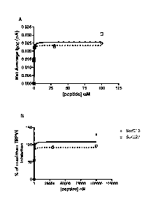
Figure 1 shows the net average change in calcium current (Isoc) through
TRPV6 channels as a function of peptide concentration (panel A). SorC13 is
represented by open circles and broken line. SorC27 is represented by open
squares and a solid line. Note that the positive numbers indicate inhibition
(net
current) of calcium current flowing into the cell. Panel B shows the percent
of
maximum inhibition of calcium current (Isoc) through TRPV6 channels as a
function of peptide concentration. SorC13 is represented by open circles and
broken line. SorC27 is represented by open squares and a solid line.
Figure 2 shows the effect of SorC27 on an ovarian cancer cell line (SKOV-3,
panel A) and a breast cancer cell line (MCF 7, panel B).
Figure 3 shows the effect of SorC13 on an ovarian cancer cell line (SKOV-3,
panel A) and a breast cancer cell line (T 47D, panel B).
Figure 4 shows a comparison of the induction of apoptosis by Paclitaxel (10
uM) and SorC13 (10 uM) in T 47D (panel A). Panel B shows a comparison of
Paclitaxel (10 uM) and SorC27 (10 uM) in MCF 7.
Figure 5 shows a comparison of the ratio of CAT treatment:CAT control (no
CAT) to the ratio of SorC13 treatment:SorC13 control (no SorC13) and
SorC27 treatment:SorC27 control (no SorC27) on SKOV3 (panel A). Panel B
shows the comparison of CAT and SorC13 treatments on Ca0V3. The
concentration of SorC13 was 100 uM; SorC27 was 10 uM. CAT = Paclitaxel
(10 uM) plus carboplatin (20 mM). The data points are the means of four
measurements of induction of apoptosis. The dotted line is a 1:1 ratio
obtained for no effect.
4
CA 02718949 2010-09-20
WO 2009/114943
PCT/CA2009/000343
Figure 6 shows a comparison of the ratio of CAT treatment:CAT control (no
CAT) to the ratio of SorC27 treatment:SorC27 control (no SorC27) on
OVCAR3. The concentrations of SorC27 were 10 uM (filled square) and 100
uM (empty square). CAT = Paclitaxel (10 uM) plus carboplatin (20 mM). The
data points are the means of four measurements of induction of apoptosis.
The dotted line is a 1:1 ratio obtained for no effect.
Figure 7 is a line drawing showing the location of the lymph nodes in the
mouse. Significant accumulation of SorC13-Cy5.5 and SorC27-Cy5.5 four
hours after i.v. injection of 100 ug of each of the labeled peptides into CD1
mice was observed at the following lymph nodes labeled in Figure 7: 1.
Superfacial cervical nodes; 4. Axillary nodes; 5. Branchial nodes; 8.
Mesenteric nodes; and 9. Inguinal nodes.
Figure 8 shows the distribution of Cy5.5 labeled SorC13 in CD1 mice 4 hours
after i.v. injection. The Y-axis is the percentage of total fluorescence
measured in all tissues.
Figure 9 shows the distribution of Cy5.5 labeled SorC27 in CD1 mice 4 hours
after i.v. injection. The Y-axis is the total fluorescence measured in each
tissue.
Figure 10 shows the distribution of Cy5.5 labeled SorC27 in CD1 mice after
i.v. injection over time after perfusion to wash out fluids. The Y-axis is the
percentage of total fluorescence measured in all tissues. The highest percent
uptake (of total fluorescence) of SorC27 was observed in liver, lung and
kidney. Lymph node is not shown because perfusion washes out lymph.
Panel A shows the distribution 4 hours after i.v. injection and Panel B 24
hours after i.v. injection.
Figure 11 shows degradation of SorC27 in human blood plasma from HPLC
analysis of 100 ul of plasma diluted in 100 ul of 2 M KCI, 94 ul injection.
The
5
CA 02718949 2010-09-20
WO 2009/114943
PCT/CA2009/000343
Y-axis indicates the amount (ug) of SorC27 in plasma prior to diluting with
KCI, Half-Life = 20.0 min, r = 0.87, p < 0.05. The data are mean SEM, n = 6.
Figure 12 shows results from an in vivo experiment with NOD/SCID mice
xenografted with human SKOV3 ovarian cancer tumors. Mice were injected
with either control saline, SorC27 or a mixture of Paclitaxel and Carboplatin
(CAT). SorC27 reduced the tumor volume significantly (p < 0.05) when
compared to a control saline injection and was not distinguishable from mice
treated with CAT, over 12 days when tumor size was normalized to body
weight.
Detailed Description of the Invention
The inventors have made isolated peptides that provide calcium channel
inhibition activity. In particular, the peptides have TRPV6 calcium channel
inhibition activity and block TRPV6 function in cancer cells that have over-
expressed TRPV6. The peptides are useful in methods of treating cancer,
such as breast cancer, ovarian cancer, blood cancer (leukemia), brain cancer,
retinal cancer, liver cancer, thyroid cancer, colon cancer, prostate cancer
and
endometrial cancer.
The peptides are also useful in methods of medical treatment of metastatic
cancer that has originated in a first tissue and spread to a secondary site,
such as a lymph node, liver, lung or kidney tissue.
In one embodiment, the peptide of the invention has the amino acid
sequence: HPSKVDLPR (called "SorC9"; amino acid nos. 19-27 of SEQ ID
NO:1). In another embodiment, the compound has the amino acid sequence
KEFLHPSKVDLPR (called "SorC13"; amino acid nos. 15-27 of SEQ ID NO:1).
In another embodiment, the compound comprises all or part of the amino acid
sequence EGKLSSNDTEGGLCKEFLHPSKVDLPR (called "SorC27"; SEQ ID
NO:1).
6
CA 02718949 2010-09-20
WO 2009/114943
PCT/CA2009/000343
The invention also includes an isolated nucleic acid encoding a peptide of the
invention, such as a nucleic acid encoding SEQ ID NO:1 or one of its
fragments described herein. The invention also relates to isolated antibodies
against a peptide of the invention. The antibody optionally selectively binds
a
peptide of the invention, but does not bind to soricidin.
Surprisingly, the compounds of the invention have sequence identity to part of
soricidin but have calcium channel inhibition activity without paralytic
activity.
It was not previously known that soricidin had two functional domains in its
structure. It was also unknown that peptides could be prepared that
separated the calcium channel inhibition activity from the paralytic activity.
The peptides of the invention are optionally half the length of soricidin, or
shorter. The peptides of the invention not only isolate and retain calcium
channel inhibition activity, they also unexpectedly have greater calcium
channel inhibition activity than soricidin in certain cell lines, such as OV
2008.
In certain cell lines, the inhibition activity also increased as peptide
length
decreased.
The surprising nature of this invention is emphasized by considering that,
while bifunctionally large proteins and enzymes are common in biological
systems, inherent bifunctionality is a very rare phenomenon in small peptides.
Reports in the literature of bifunctionality have typically resulted from
artificial
production, for example, where two different peptides have been chemically
fused (Anes et al. 2006; Yamamoto et al. 2008). The inventor determined that
the N-terminal of soricidin has a paralytic function and the C-terminal has a
calcium channel inhibitor function. Truncating soricidin at the N-terminal
successfully produced peptides that retain calcium channel inhibition activity
without paralytic activity.
The peptides of the invention have other unexpected properties compared to
full-length soricidin. For example, the peptides of the invention have
increased solubility, which allows smaller doses to be provided to achieve a
target peptide concentration in blood plasma. Based on relative solubilities,
7
CA 02718949 2010-09-20
WO 2009/114943
PCT/CA2009/000343
the dose volume of the peptides of the invention to achieve a target blood
concentration could be smaller than soricidin by at least 10-fold.
Additionally, the peptides have greater shelf-life stability and solution
stability
due to the presence of fewer sensitive disulfide bonds (soricidin has 3
disulfide bonds). The peptides of the invention optionally have no disulfide
bonds. An absence of disulfide bonds provides a very long shelflife during
storage under minimal refrigeration or at room temperature as a
solid. Additionally when the peptides of the invention with no disulfide bonds
are dissolved in water, cleavage of disulfide bonds cannot occur. Such
cleavage, in full-length soricidin, can render the peptide less active and
more
susceptible to the formation of inter-molecular S-S bonds. The formation of
inter-peptide S-S bonds also results in precipitation of soricidin. The
peptides
of the invention, such as SorC13 and SorC27, are typically stable in aqueous
solution at 8 C for at least 3 weeks with no change in purity as measured by
HPLC.
The peptides of the invention also avoid one of the major adverse effects of
pharmaceuticals, which is related to the ability to cross the central nervous
system protective barrier, the blood-brain barrier. The inability of the
peptides
of the invention to cross this protective barrier obviates the potential
toxicity in
the central nervous system.
The peptides of the invention advantageously have low toxicity. As shown in
Example 23, after in vivo i.v. injection into CD1 mice, SorC27 resulted in no
change in blood pressure or heart rate over a 1 hour measurement period.
There were no also neurological/behavioral changes over 72 hours. After in
vivo i.v. injection into CD1 mice, SorC13 caused a small spike (approximately
25%) in blood pressure in the first 15 min which disappeared by 1 hour.
There were no neurological/behavioral changes in the mice over 72
hours. Single intravenous injection of SorC13 or SorC27 into CD1 mice at
doses of 10 mg/kg, 100 mg/kg and 500 mg/kg resulted in no adverse events
over a 5-day post-injection observation period. Necropsy also showed no
8
CA 02718949 2010-09-20
WO 2009/114943
PCT/CA2009/000343
significant changes in all major organ systems. Furthermore, a multiple dose
of SorC27 at 400 mg/kg (i.p.) each day for 12 days in mice showed no
indication of toxicity.
The peptides of the invention, particularly the shorter peptides, such as
SorC13, are typically less antigenic than soricidin. Peptides having a number
of amino acids equal to or less than the empirical cutoff for antigenicity
(typically considered to be 13 amino acids for peptides in general) possess no
antigenicity.
As noted above, a peptide of the invention will have calcium channel activity
(i.e. the ability to partially or totally inhibit channel activity) without
paralytic
activity. The peptides having such activity are readily identified with any
known assays suitable for measuring i) calcium channel blocking and ii) lack
of paralytic activity. For example, in one embodiment, a peptide having
calcium channel inhibition activity is optionally identified by determining
that
the peptide reduces calcium channel activity by reducing (i.e., partially or
fully
inhibiting) the flow of calcium through calcium channels.
Calcium channel inhibition activity is optionally identified by a readily
available
cell line (e.g. human embryonic kidney cell lines- HEK, lymph node prostate
cancer cell, LnCaP) transfected with an expression vector for TRPV6. Such
transfected cells are readily aliquoted and stored (typically -80 C) until
required. This provides a standard transfected cell preparation to test for
inhibition of calcium ion uptake by the cells. Analysis of the calcium
movement into the cell lines to identify peptides of the invention that
provide
reduced calcium channel activity is optionally done by fluorometric
measurement (internalized F URA calcium ion probe) or by an
electrophysiological protocol involving patch clamping cells and measuring
calcium current in the presence or absence of a peptide. Optionally the
peptides have an equilibrium inhibition constant of less than: 1000 nM, 150
nM or 100 nM in a LnCaP cell model while not affecting the sciatic nerve
transmission of an action potential in a rat sciatic nerve model (i.e., no
9
CA 02718949 2010-09-20
WO 2009/114943
PCT/CA2009/000343
paralytic activity). For example, the equilibrium dissociation constant (Kd)
for
SorC27 is 140 nM and SorC13 is 100 nM in the LnCaP model (Fig. 6). Based
on a linear relationship with the number of amino acids, the Kd of SorC9 is
expected to be approximately 90 nM.
The isolated peptides are useful for a number of purposes. In one
embodiment, the invention includes methods of reducing cell proliferation by
administering a peptide of the invention to a cell, such as a cancer cell.
Reduction in cell proliferation is readily determined by contacting a peptide
with a proliferating cell, such as a cancer cell, in vitro or in vivo, and
determining whether the peptide reduces (i.e., fully or partially) inhibits
cell
proliferation. In another embodiment, the invention includes methods of
inducing apoptosis of a cell, such as a cancer cell, by administering a
peptide
of the invention to the cell. Reduction in cell proliferation is readily
determined
by contacting a peptide with a cell, such as a cancer cell, in vitro or in
vivo,
and determining whether the peptide induces apoptosis.
As noted above, the isolated peptides are useful for treating cancer. The
invention includes methods of treating cancer in a mammal, such as a human,
by administering an isolated peptide of the invention to the mammal.
Remarkably, in separate head-to-head tests against the well-known cancer
drug, Paclitaxel, and the cancer drug cocktail, Paclitaxel and carboplatin
("CAT cocktail"), two peptides of the invention, SorC13 and SorC27, provided
faster and more active induction of apoptosis in certain cell lines. This
superior effect has been shown against both breast cancer and ovarian
cancer. A typical treatment for breast cancer is Paclitaxel and a typical
treatment for ovarian cancers is the CAT cocktail. In other cell lines, the
performance of SorC13 and SorC27 was comparable to CAT.
The isolated peptide SorC27 has also been shown to be useful for treating
cancer in an in vivo xenograft mouse cancer model. As shown in Example 14
and Figure 12, in NOD/SCID mice xenografted with human ovarian cancer
tumors SorC27 reduced the tumor volume significantly (p < 0.05) when
CA 02718949 2010-09-20
WO 2009/114943
PCT/CA2009/000343
compared to the control situation (saline injection) and was not
distinguishable
from mice treated with CAT, over 12 days when tumor size was normalized to
body weight.
Without wishing to be bound by theory on the cause of the anti-cancer effect,
the inventor has determined, through experiments with SorC27 and SorC13,
that the peptides of the invention activate the apoptosis cascade through
caspase 3 and/or 7 in ten different cancer cells lines but not in two non-
cancerous cell lines.
Therefore, a peptide of the invention is optionally identified as having anti-
cancer activity if it causes induction of the enzymes caspase 3 and caspase 7
above the control cell levels of caspase 3 and caspase 7. Once these
enzymes are activated, cell death will occur. The activity of the caspase
enzymes in treated cells is readily measured and compared to control,
untreated cells.
Furthermore, as shown in Table 9 and Example 25, the presence of TRPV6
mRNA in a cell line corresponds with those cell lines that exhibit an
apoptotic
effect in response to the peptides of the present invention.
Metastatic Cancer
The peptides of the invention have anti-cancer activity against a cancer cell
in
a primary tumor site or a metastasized cancer cell in a secondary cancer site.
The peptides localize predominantly in lymph nodes, but also in lung, liver
and
kidney, which are common sites where metastatic cancer is located (Figure
7).
The peptides of the present application are useful in the treatment of
metastatic cancer, including a cancer that has spread to the lung, brain,
liver,
kidney, spleen and bone marrow. The peptides can be used alone or in a
pharmaceutical composition comprising a second anti-cancer agent.
11
CA 02718949 2010-09-20
WO 2009/114943
PCT/CA2009/000343
The peptides of the present application are particularly useful in treating
lymph node metastases. "Lymph node metastases" optionally include lung
cancer (Mujoomdar et al, 2007), gastric cancer, cervical carcinoma (Lyshchik
et al., 2007), vulvar carcinoma (Vernooij et al, 2007), endometrial cancer
(Aalders et al, 2007), head and neck squamous cell carcinoma (Veness et al.,
2007), esophagus and throat cancer, nasopharyngeal carcinoma (Ma et al.,
2007), gastrointestinal cancer (Wind et al., 2006), Gall bladder cancer, brain
cancer (Mujoomdar et al., 2007), thyroid cancer, breast cancer, ovarian
cancer, prostate cancer and colorectal cancer. The peptides of the invention
are therefore useful in treating cancer in a mammal at any of cancer stages I,
II, III or IV.
Peptides of the Invention
Isolated peptides comprising all or part of any of SEQ ID NO:1 and having
calcium channel inhibition activity (TRPV6 inhibition activity), without
paralytic
activity, are useful peptides for treatment of cancer. Amino acids may be
added to the peptides of the invention, but the isolated peptides, with amino
acids, are typically 35 or 30 amino acids or less, optionally less than: 27,
25,
20, 15 or 13 amino acids long, while optionally at least 9 amino acids long.
One can readily make longer peptides by adding a variety of additional amino
acids to the SorC27 sequence to make a peptide that could be up to, for
example, 30, 35, 40 or 45 amino acids long (eg. additional amino acids
corresponding to the soricidin amino acid sequence such as one or more of
the C-terminal amino acids (SILARPAELNTETCILEC SEQ ID NO:2), a
targeting sequence, or other amino acids).
The peptide optionally comprises, consists essentially of or consists of the
amino acid sequence: HPSKVDLPR (amino acid nos. 19-27 of SEQ ID NO:1),
KEFLHPSKVDLPR (amino acid nos. 15-27 of SEQ ID NO:1) or
EGKLSSNDTEGGLCKEFLHPSKVDLPR (SEQ ID NO:1). Optionally the
peptide comprises at least: 9, 10, 11, 12, 13, 14, 15, 16, 17, 18, 19 or 20
amino acids of SEQ ID NO:1. Optionally, the isolated peptide comprises at
least: 9, 10, 11, 15 or 18 amino acids of SEQ ID NO:1. Optionally, the peptide
12
CA 02718949 2010-09-20
WO 2009/114943
PCT/CA2009/000343
comprises a fragment of 9-13, 10-15, 15-20, 20-25 or 20-27 amino acids of
SEQ ID NO:1, wherein the peptide inhibits calcium channels, reduces cell
proliferation and does not have paralytic activity.
The peptides of the invention optionally also include analogs of the
aforementioned peptides. Analogs of the protein of the invention optionally
include, but are not limited to an amino acid sequence containing one or more
amino acid substitutions, insertions, deletions and/or mutations. Amino acid
substitutions may be of a conserved or non-conserved nature. Conserved
amino acid substitutions involve replacing one or more amino acids of the
peptides of the invention with amino acids of similar charge, size, and/or
hydrophobicity characteristics. When only conserved substitutions are made,
the resulting analog should be functionally equivalent. Non-
conserved
substitutions involve replacing one or more amino acids of the amino acid
sequence with one or more amino acids which possess dissimilar charge,
size, and/or hydrophobicity characteristics. The analog is optionally a
peptoid,
which is an N-substituted polyglycine with amino acid R groups attached at
the N atom.
One or more amino acid insertions are optionally introduced into the amino
acid sequences of the invention. Amino acid insertions consist of single
amino acid residues or sequential amino acids ranging for example from 2 to
15 amino acids in length.
Deletions consist of the removal of one or more amino acids, or discrete
portions from the amino acid sequence of the peptide. The deleted amino
acids may or may not be contiguous.
The peptides of the invention are readily prepared by chemical synthesis
using techniques well known in the chemistry of proteins such as solid phase
synthesis (Merrifield, 1964, J. Am. Chem. Assoc. 85:2149-2154) or synthesis
in homogenous solution (Houbenweyl, 1987, Methods of Organic Chemistry,
ed. E. Wansch, Vol. 15 I and II, Thieme, Stuttgart).
13
CA 02718949 2010-09-20
WO 2009/114943
PCT/CA2009/000343
Analogs of a protein of the invention are optionally prepared by introducing
mutations in a nucleotide sequence encoding the peptide. Mutations in
nucleotide sequences constructed for expression of analogs of a protein of
the invention preserve the reading frame of the coding sequences.
Furthermore, the mutations will preferably not create complementary regions
that could hybridize to produce secondary mRNA structures such as loops or
hairpins, which could adversely affect translation of the mRNA.
Mutations are optionally introduced at particular loci by synthesizing
oligonucleotides containing a mutant sequence, flanked by restriction sites
enabling ligation to fragments of the native sequence. Following ligation, the
resulting reconstructed sequence encodes an analog having the desired
amino acid insertion, substitution, or deletion.
Alternatively, oligonucleotide-directed site-specific mutagenesis procedures
are employed to provide an altered gene having particular codons altered
according to the substitution, deletion, or insertion required. Deletion or
truncation of a peptide of the invention is also readily achieved by utilizing
convenient restriction endonuclease sites adjacent to the desired deletion.
Subsequent to restriction, overhangs may be filled in, and the DNA re-ligated.
Exemplary methods of making the alterations set forth above are disclosed by
Sambrook et al (Sambrook J et al. 2000. Molecular Cloning: A Laboratory
Manual (Third Edition), Cold Spring Harbor Laboratory Press).
In addition, analogs of a protein of the invention are readily prepared by
chemical synthesis using techniques well known in the chemistry of proteins
such as solid phase synthesis (Merrifield, 1964, J. Am. Chem. Assoc.
85:2149-2154) or synthesis in homogenous solution (Houbenweyl, 1987,
Methods of Organic Chemistry, ed. E. Wansch, Vol. 15 I and II, Thieme,
Stuttgart). The peptides of the invention also include peptides having
sequence identity to a peptide of the invention, mutated peptides and/or
truncations thereof as described herein. Such peptides have amino acid
14
CA 02718949 2010-09-20
WO 2009/114943
PCT/CA2009/000343
sequences that correspond to nucleic acid sequences that hybridize under
stringent hybridization conditions (see discussion of stringent hybridization
conditions herein) with a probe used to obtain a peptide of the invention.
Peptides having sequence identity will often have the regions which are
characteristic of the protein.
Other useful peptides of the invention optionally comprise, consist
essentially
of or consist of an amino acid sequence with at least: 30%, 40%, 50%, 60%,
70%, 80%, 90% or 95% sequence identity to all or part of SEQ ID NO:1
described herein, wherein the peptide has calcium channel inhibition activity
and no paralytic activity and is useful for treatment of cancer. Sequence
identity is typically assessed by the BLAST version 2.1 program advanced
search (parameters as above; Altschul, S.F., Gish, W., Miller, W., Myers, E.W.
& Lipman, D.J. (1990) "Basic local alignment search tool." J. Mol. Biol.
215:403_410). BLAST is a series of programs that are available online
through the U.S. National Center for Biotechnology Information (National
Library of Medicine Building 38A Bethesda, MD 20894) The advanced Blast
search is set to default parameters. References for the Blast Programs
include: Altschul, S.F., Gish, W., Miller, W., Myers, E.W. & Lipman, D.J.
(1990) "Basic local alignment search tool." J. Mol. Biol. 215:403-410; Gish,
W.
& States, D.J. (1993) "Identification of protein coding regions by database
similarity search." Nature Genet. 3:266-272.; Madden, T.L., Tatusov, R.L. &
Zhang, J. (1996) "Applications of network BLAST server" Meth. Enzymol.
266:131-141; Altschul, S.F., Madden, T.L., Schaffer, A.A., Zhang, J., Zhang,
Z., Miller, W. & Lipman, D.J. (1997) "Gapped BLAST and PSI-BLAST: a new
generation of protein database search programs." Nucleic Acids Res.
25:3389-3402); Zhang, J. & Madden, T.L. (1997) "PowerBLAST: A new
network BLAST application for interactive or automated sequence analysis
and annotation." Genome Res. 7:649-656).
The present invention also includes a protein of the invention conjugated with
a selected protein, or a selectable marker protein to produce fusion proteins.
CA 02718949 2010-09-20
WO 2009/114943
PCT/CA2009/000343
Therapeutic Methods
The peptides of the invention, such as all or part of SEQ ID NO:1 described
herein, are useful for reducing cell proliferation, inducing cell apoptosis
and
preventing or treating cancer by administration of the peptide to a human.
The phrase "reducing cell proliferation" as used herein refers to slowing the
rate of proliferation of a cell as compared to the rate of proliferation of a
cell in
the absence of the substance.
The phrase "inducing cell apoptosis" as used herein refers to increasing the
rate of apoptosis of cells as compared to the rate of apoptosis of cells in
the
absence of the substance.
The term "effective amount" as used herein means an amount effective and at
dosages and for periods of time necessary to achieve the desired result (e.g.
optionally blocking calcium channel activity, reducing cell proliferation,
inducing apoptosis and/or preventing or treating cancer).
Administering a peptide or substance to a mammal includes both in vivo and
ex vivo administrations.
The term "a cell" as used herein includes a single cell as well as a plurality
or
population of cells. Administering a peptide or substance to a cell includes
both in vitro and in vivo administrations.
The phrase "reduce calcium channel activity" as used herein means that the
substance can result in a decrease in calcium channel activity as compared to
calcium channel activity in the absence of the substance.
The peptides of the invention strongly inhibit calcium uptake in cancer cells
particularly cancer cells in which the calcium uptake channel TRPV6 has
increased expression, such as in breast cancer, ovarian cancer, blood cancer,
brain cancer, retinal cancer, liver cancer, thyroid cancer, colon cancer,
16
CA 02718949 2010-09-20
WO 2009/114943
PCT/CA2009/000343
prostate cancer and endometrial cancer. The peptides of the invention, by
reducing calcium uptake, disrupt intracellular calcium essential for
proliferation
of normal and cancerous cells. Therefore, the invention includes the use of a
peptide of the invention for reducing cell proliferation, inducing apoptosis
and/or preventing or treating tumours and cancer in mammals (e.g. humans)
by administration of the peptide to the mammal.
As used herein, and as well understood in the art, "to treat" or "treatment"
is
an approach for obtaining beneficial or desired results, including clinical
results. Beneficial or desired clinical results can include, but are not
limited to,
alleviation or amelioration of one or more symptoms or conditions,
diminishment of extent of disease, stabilized (i.e., not worsening) state of
disease or disorder, preventing spread of disease or disorder, delay or
slowing of disease or disorder progression, amelioration or palliation of the
disease or disorder state, and remission (whether partial or total), whether
detectable or undetectable. "Treatment" can also mean prolonging survival as
compared to expected survival if not receiving treatment.
Pharmaceutical Compositions
The invention also includes the use of the peptides of the invention for
preparation of a medicament for treatment of cancer. The isolated peptides of
the invention are optionally formulated into a pharmaceutical composition for
administration to subjects in a biologically compatible form suitable for
administration in vivo. By "biologically compatible form suitable for
administration in vivo" is meant a form of the substance to be administered in
which any toxic effects are outweighed by the therapeutic effects. The
substances may be administered to living organisms including humans, and
animals.
Administration of a therapeutically active amount of pharmaceutical
compositions of the present invention is defined as an amount effective, at
dosages and for periods of time necessary to achieve the desired result. For
example, a therapeutically active amount of a substance may vary according
17
CA 02718949 2010-09-20
WO 2009/114943
PCT/CA2009/000343
to factors such as the disease state, age, sex, and weight of the individual,
and the ability of the substance to elicit a desired response in the
individual.
Dosage regimes may be adjusted to provide the optimum therapeutic
response. For example, several divided doses may be administered daily or
the dose may be proportionally reduced as indicated by the exigencies of the
therapeutic situation.
The peptide of the invention is preferably combined with other components
such as a carrier in a composition such as a pharmaceutical composition. The
compositions are useful when administered in methods of medical treatment
or prevention of cancer.
The pharmaceutical compositions can be administered to humans or animals
by a variety of methods including, but not restricted to topical
administration,
oral administration, aerosol administration, intratracheal instillation,
intraperitoneal injection, injection into the cerebrospinal fluid, intravenous
injection and subcutaneous injection. Dosages to be administered depend on
patient needs, on the desired effect and on the chosen route of
administration.
Nucleic acid molecules and peptides may be introduced into cells using in
vivo delivery vehicles such as liposomes. They may also be introduced into
these cells using physical techniques such as microinjection and
electroporation or chemical methods such as co-precipitation, pegylation or
using liposomes.
The pharmaceutical compositions are prepared by known methods for the
preparation of pharmaceutically acceptable compositions which can be
administered to patients, and such that an effective quantity of the nucleic
acid molecule or peptide is combined in a mixture with a pharmaceutically
acceptable vehicle. Suitable vehicles are described, for example in
Remington's Pharmaceutical Sciences (Remington's Pharmaceutical
Sciences, Mack Publishing Company, Easton, Pa., USA) or Handbook of
Pharmaceutical Additives (compiled by Michael and Irene Ash, Gower
Publishing Limited, Aldershot, England (1995). On this basis, the
18
CA 02718949 2010-09-20
WO 2009/114943
PCT/CA2009/000343
compositions include, albeit not exclusively, solutions of the substances in
association with one or more pharmaceutically acceptable vehicles or
diluents, and may be contained in buffered solutions with a suitable pH and/or
be iso-osmotic with physiological fluids. In this regard, reference can be
made
to U.S. Pat. No. 5,843,456.
On this basis, the pharmaceutical compositions optionally includes an active
compound or substance, such as a peptide or nucleic acid molecule, in
association with one or more pharmaceutically acceptable vehicles or
diluents, and contained in buffered solutions with a suitable pH and iso-
osmotic with the physiological fluids. The methods of combining the active
molecules with the vehicles or combining them with diluents are well known to
those skilled in the art. The composition optionally includes a targeting
agent
for the transport of the active compound to specified sites within tissue.
Preparation of Antibodies
Antibodies to the peptide are useful to identify the presence of the peptide
in a
test sample. Any method of labeling the antibody that would report on peptide
density/location would be useful (e.g. radioactively labeled peptide or
fluorescently tagged peptide). The antibody is typically a monoclonal antibody
or a polyclonal antibody. The antibodies are also valuable for immuno-
purification of peptides. For example, one may contact a biological sample
with the antibody under conditions allowing the formation of an immunological
complex between the antibody and a peptide recognized by the antibody and
detecting the presence or absence of the immunological complex whereby the
presence of the peptide of the invention is detected in the sample. The
invention also includes compositions preferably including the antibody, a
medium suitable for the formation of an immunological complex between the
antibody and a peptide recognized by the antibody and a reagent capable of
detecting the immunological complex to ascertain the presence of the
peptides of the invention or similar peptides.
19
CA 02718949 2016-09-12
WO 2009/114943
PCT/CA2009/000343
To recognize the peptides of the invention, one may generate antibodies
against a range of unique epitopes throughout the peptides.
Monoclonal and polyclonal antibodies are prepared according to the
description in this application and techniques known in the art. For examples
of methods of the preparation and uses of monoclonal antibodies, see U.S.
Pat. Nos. 5,688,681, 5,688,657, 5,683,693, 5,667,781, 5,665,356, 5,591,628,
5,510,241, 5,503,987, 5,501,988, 5,500,345 and 5,496,705.
Examples of the preparation and
uses of polyclonal antibodies are disclosed in U.S. Pat. Nos. 5,512,282,
4,828,985, 5,225,331 and 5,124,147.
The term "antibody" as used herein to include fragments thereof which also
specifically react with a peptide of the invention. Antibodies can be
fragmented using conventional techniques and the fragments screened for
utility in the same manner as described above. For example, F(ab')2
fragments can be generated by treating antibody with pepsin. The resulting
F(ab')2 fragment can be treated to reduce disulfide bridges to produce Fab'
fragments.
The invention also includes methods of using the antibodies, such as in
detection of receptors that bind to the peptides of the invention. For
example,
the invention includes a method for detecting the presence of a peptide of the
invention by: a) contacting a sample containing one or more peptides with an
antibody of the invention under conditions suitable for the binding of the
antibody to peptides with which it is specifically reactive; b) separating
unbound peptides from the antibody; and c) detecting antibody which remains
bound to one or more of the peptides in the sample.
Research Tool
The peptides of the invention are useful in research protocols to explore the
neuromuscular junction and ion channels. The ability to alter certain ion
CA 02718949 2010-09-20
WO 2009/114943
PCT/CA2009/000343
channels or classes of ion channels selectively provides another tool with
which to perturb the neuromuscular-junction in a predictable manner. This
identifies the role of susceptible peptide targets in neuromuscular functions
and processes. The invention includes a method of determining the response
of an ion channel to a paralytic peptide comprising contacting a channel or
cells comprising a channel with a peptide of the invention or a derivative
thereof and determining whether the channels transport ions or whether ion
transport (e.g. Ca2+) has been reduced.
The peptides of the invention inhibit TRPV6 channels and are readily tagged
with a label (fluorescent label, radioactive label, biotin label, etc.) to
identify
the TRPV6 channel in a cell or tissue, or to label cells or tissues that
express
large quantities of calcium channels. Accordingly, the invention relates to
methods of identifying a TRPV6 channel in a cell or tissue, comprising
contacting the cell or tissue with a peptide of the invention that has been
tagged with detectable label and detecting the peptide bound to TRPV6
channel in the cell or tissue.
Nucleic Acids
The peptides of the invention (including truncations, analogs, etc.) may be
prepared by chemical synthesis or by using recombinant DNA methods.
Accordingly, the invention includes nucleic acid molecules having a sequence
that encodes a peptide of the invention. These sequences are readily
incorporated according to procedures known in the art into an appropriate
expression vector that ensures good expression of the peptide. Expression
vectors include but are not limited to cosmids, plasmids, or modified viruses
(e.g., replication defective retroviruses, adenoviruses and adeno-associated
viruses), so long as the vector is compatible with the host cell used. The
expression "vectors suitable for transformation of a host cell", means that
the
expression vectors contain a nucleic acid molecule of the invention and
regulatory sequences, selected on the basis of the host cells to be used for
expression, which are operatively linked to the nucleic acid molecule.
21
CA 02718949 2010-09-20
WO 2009/114943
PCT/CA2009/000343
"Operatively linked" means that the nucleic acid is linked to regulatory
sequences in a manner that allows expression of the nucleic acid.
The invention therefore includes a recombinant expression vector of the
invention containing a nucleic acid molecule of the invention, or a fragment
thereof, and the necessary regulatory sequences for the transcription and
translation of the inserted peptide-sequence. Suitable regulatory sequences
are optionally derived from a variety of sources, including bacterial, fungal,
or
viral genes (For example, see the regulatory sequences described in
Goeddel, Gene Expression Technology: Methods in Enzymology 185,
Academic Press, San Diego, Calif. (1990). Selection of appropriate regulatory
sequences is dependent on the host cell chosen, and may be readily
accomplished by one of ordinary skill in the art. Examples of such regulatory
sequences include: a transcriptional promoter and enhancer or RNA
.. polymerase binding sequence, a ribosomal binding sequence, including a
translation initiation signal. Additionally, depending on the host cell chosen
and the vector employed, other sequences, such as an origin of replication,
additional DNA restriction sites, enhancers, and sequences conferring
inducibility of transcription may be incorporated into the expression vector.
It
will also be appreciated that the necessary regulatory sequences may be
supplied by the native compound and/or its flanking regions.
The invention further provides a recombinant expression vector comprising a
DNA nucleic acid molecule of the invention cloned into the expression vector
in an antisense orientation. These vectors are useful experimental systems to
study the peptides of the invention or its variants or to test antidotes. The
peptides may or may not be toxic to the host cells. They are also useful to
produce large amounts of the peptide.
The recombinant expression vectors of the invention may also contain a
selectable marker gene that facilitates the selection of host cells
transformed
or transfected with a recombinant molecule of the invention. Examples of
selectable marker genes are genes encoding a protein such as G418 and
22
CA 02718949 2010-09-20
WO 2009/114943
PCT/CA2009/000343
hygromycin which confer resistance to certain drugs, beta-galactosidase,
chloramphenicol acetyltransferase, or firefly luciferase. Transcription of the
selectable marker gene is monitored by changes in the concentration of the
selectable marker protein such as beta-galactosidase, chloramphenicol
acetyltransferase, or firefly luciferase. If the selectable marker gene
encodes
a protein conferring antibiotic resistance such as neomycin resistance
transformant cells can be selected with G418. Cells that have incorporated
the selectable marker gene will survive, while the other cells die. This makes
it
possible to visualize and assay for expression of recombinant expression
vectors of the invention and in particular to determine the effect of a
mutation
on expression and phenotype. It will be appreciated that selectable markers
can be introduced on a separate vector from the nucleic acid of interest.
The recombinant expression vectors may also contain genes which encode a
fusion moiety which provides increased expression of the recombinant
protein; increased solubility of the recombinant protein; and aid in the
purification of a target recombinant protein by acting as a ligand in affinity
purification. For example, a proteolytic cleavage site may be added to the
target recombinant protein to allow separation of the recombinant protein from
the fusion moiety subsequent to purification of the fusion protein.
Recombinant expression vectors can be introduced into host cells to produce
a transformed host cell. These cells are useful experimental systems.
Accordingly, the invention includes a host cell comprising a recombinant
expression vector of the invention. The term "transformed host cell" is
intended to include prokaryotic and eukaryotic cells which have been
transformed or transfected with a recombinant expression vector of the
invention. Prokaryotic cells can be transformed with nucleic acid by, for
example, electroporation or calcium-chloride mediated transformation. Nucleic
acid can be introduced into mammalian cells via conventional techniques
such as calcium phosphate or calcium chloride co-precipitation, DEAE-
dextran-mediated transfection, lipofectin, electroporation or microinjection.
Suitable methods for transforming and transfecting host cells can be found in
23
CA 02718949 2010-09-20
WO 2009/114943
PCT/CA2009/000343
Sambrook et al. (Molecular Cloning: A Laboratory Manual (Third Edition),
Cold Spring Harbor Laboratory Press), and other such laboratory textbooks.
Suitable host cells include a wide variety of prokaryotic and eukaryotic host
cells. For example, the peptides of the invention may be expressed in
bacterial cells such as E. coli, Pseudomonas, Bacillus subtillus, insect cells
(using baculovirus), yeast cells or mammalian cells. Other suitable host cells
can be found in Goeddel, Gene Expression Technology: Methods in
Enzymology 185, Academic Press, San Diego, Calif. (1991).
As an example, to produce peptides recombinantly, for example, E. coli can
be used using the T7 RNA polymerase/promoter system using two plasmids
or by labeling of plasmid-encoded proteins, or by expression by infection with
M13 Phage mGPI-2. E. coli vectors can also be used with Phage lambda
regulatory sequences, by fusion protein vectors (e.g. lacZ and trpE), by
maltose-binding protein fusions, and by glutathione-S-transferase fusion
proteins.
Alternatively, a peptide can be expressed in insect cells using baculoviral
vectors, or in mammalian cells using vaccinia virus. For expression in
mammalian cells, the cDNA sequence may be ligated to heterologous
promoters and introduced into cells, such as COS cells to achieve transient or
long-term expression. The stable integration of the chimeric gene construct
may be maintained in mammalian cells by biochemical selection, such as
neomycin and mycophoenolic acid.
The DNA sequence can be altered using procedures such as restriction
enzyme digestion, fill-in with DNA polymerase, deletion by exonuclease,
extension by terminal deoxynucleotide transferase, ligation of synthetic or
cloned DNA sequences, site-directed sequence alteration with the use of
specific oligonucleotides together with PCR. For example, one to five or five
to
ten amino acids or more may be removed or mutated.
24
CA 02718949 2010-09-20
WO 2009/114943
PCT/CA2009/000343
The DNA sequence or portions thereof, or a mini gene consisting of a DNA
with an intron and its own promoter, is introduced into eukaryotic expression
vectors by conventional techniques. These vectors permit the transcription of
the DNA in eukaryotic cells by providing regulatory sequences that initiate
and
enhance the transcription of the DNA and ensure its proper splicing and
polyadenylation. The endogenous gene promoter can also be used. Different
promoters within vectors have different activities that alter the level of
expression of the cDNA. In addition, certain promoters can also modulate
function such as the glucocorticoid-responsive promoter from the mouse
mammary tumor virus.
Some of the vectors listed contain selectable markers or neo bacterial genes
that permit isolation of cells by chemical selection. Stable long-term vectors
can be maintained in cells as episomal, freely replicating entities by using
regulatory elements of viruses. Cell lines can also be produced which have
integrated the vector into the genomic DNA. In this manner, the gene product
is produced on a continuous basis.
Vectors are introduced into recipient cells by various methods including
calcium phosphate, strontium phosphate, electroporation, lipofection, DEAE
dextran, microinjection, or by protoplast fusion. Alternatively, the cDNA can
be
introduced by infection using viral vectors.
Peptides of the invention are readily isolated from cells or tissues,
including
mammalian cells or tissues, in which the peptide is expressed.
The peptide is readily purified by conventional purification methods known to
those in the art, such as chromatography methods, high performance liquid
chromatography methods or precipitation.
For example, an anti-peptide antibody (as described herein) is readily used to
isolate a peptide, which is then purified by standard methods.
CA 02718949 2010-09-20
WO 2009/114943 PCT/CA2009/000343
The isolated peptides of the invention are also readily prepared by chemical
synthesis using techniques well known in the chemistry of proteins such as
solid phase synthesis (Merrifield, 1964, J. Am. Chem. Assoc. 85:2149-2154)
or synthesis in homogenous solution (Houbenweyl, 1987, Methods of Organic
Chemistry, ed. E. Wansch, Vol. 15 I and II, Thieme, Stuttgart).
EXAMPLES
The following examples illustrate embodiments of the invention and do not
limit the scope of the invention.
EXAMPLE 1: Separation of the calcium channel-inhibitor amino acid
sequence from soricidin
Sequences of amino acids from the carboxyl-terminal region of soricidin were
synthesized. These segments have been named SorC13 (which has
sequence identity to 13 amino acids at the C-terminal end of soridicin) and
SorC27 (which has sequence identity to 27 amino acid sequence at the C-
terminal end of soricidin). The amino acid sequences of SorC13 and SorC27
as well as some of their physical and physiological properties as are outlined
in Table 1. The half-lives of SorC13 and SorC27 (as shown in Example 22)
were lower than the half-life of 5or54.
Table 1: Summary Comparison of SorC54, SorC27 and SorC13
Property Sor54 SorC27 SorC13
No. of amino 54-mer 27-mer 13-mer
acids
Molecular weight 5806(3 disulfide bonds) 2957 (no disulfide bonds) 1566
(no disulfide
bonds)
Sequence NCB! accession no. EGKLSSNDTEGGLCKEF KIEFLH PSKVDLPR
POC2C6 LHPSKVDLPR
(SEQ ID NO:1)
pl 4.3 (measured) ¨5.6 (calculated) ¨8.3 (calculated)
Physical White solid White solid White solid
appearance
Solubility <20 mg/ mL >200 mg/mL >200 mg/mL
_(aqueous)
Purity > 95% >95% >95%
Stability in stable Very stable Very stable
aqueous solution
0-20 C
26
CA 02718949 2010-09-20
WO 2009/114943 PCT/CA2009/000343
Stability solid @ - stable Very stable Very stable
20 C
Stability solid @ unstable Very stable Very stable
Room Temp.
Detection limit via z 200 ng z 200 ng z 200 ng
direct HPLC
Physiology
Clearance route in To be determined. Rapid via kidney. Rapid via kidney.
iv mice @ 5
mg/kg
Cellular Target voltage-gated sodium TRPV6 TRPV6
channels and TRPV6
Physiol. effects in Paralytic Not paralytic Not paralytic
vivo (mealworm
larvae); 5mg/kg
Physiol. effects in Not tested. None observed over 72 hr 15% BP spike,
@
vivo (mice) @ 5 15 min,
stabilizes
mg/kg by 1 hr, no
observed effects up
to 72 hrs
-Half-life in rat >30 hour; binds to 21 min; doesn't bind to 58 min; doesn't
plasma plasma proteins plasma proteins bind to plasma
proteins
Half-life in human >30 hours; binds to 20 min; doesn't bind to 56.6 min;
doesn't
plasma plasma proteins plasma proteins bind to plasma
___________________________________________________________ proteins
Maximum Not tested No toxic effects No toxic effects
Tolerated Dose in
CD-1 mice by
single i.v. dose
10, 100 and 500
mg/kg doses
Repeated dose in Not tested No loss of body weight No loss of body
NOD/SCID mice compared to controls, weight
compared to
@ 400 mg/kg, no toxic effects noted controls, no
toxic
daily i.p. injection effects noted
for 12 days
"Very stable" in the above table means that there was no observable
degradation of the solid peptides over 6 months. As a solution in sterile
water, the peptides are typically stable for at least 3 weeks at 8 C and at -
20 C, are expected to be stable more than 1 year.
SorC27 and SorC13 do not exhibit paralytic activity. To show this, the
mealworm larva paralytic bioassay reported previously (US patent no.
7,119,168) was used. Animals from a mealworm colony were arbitrarily
selected and placed in groups (treatment and control). The animals were
weighed to allow calculation of equivalent doses. The treatment was with
27
CA 02718949 2010-09-20
WO 2009/114943
PCT/CA2009/000343
either SorC13 or SorC27 dissolved in buffered insect Ringer's solution. The
control was injection was an equivalent volume of insect Ringer's (NaCI;
10.40 g; KCl, 0.32 g; CaCl2, 0.48 g; NaHCO3 , 0.32 g dissolved in 1 L of
Millipore sterile water, pH 7.4). Peptide formulations were: SorC27 solutions,
prepared at 1.0 mg/100 uL insect Ringer's (10 ug/uL, 3.4 uM, MW = 2957);
and SorC13, prepared at 0.5 mg/100 uL (5 ug/uL, 3.4 uM, MW = 1566). The
doses were 50 ug SorC27 per 100 mg of animal mass and 25 ug SorC13 per
100 mg of animal mass. Peptide solutions and control saline were injected
dorsally at the fourth segment from the head, just under the integumen.
Animals were gently 'tail tweaked' with forceps to trigger the escape reflex
reaction and scored on time-to-effect, duration of effect and intensity of
effect.
None of the animals treated with SorC13 or SorC27 exhibited any noticeable
effects, while those animals treated with soricidin exhibited profound
paralysis.
Table 2 shows the comparison of the number of larvae paralyzed by injection
of the C-peptides compared to soricidin and to control saline injection. The
shrew peptide (soricidin) is profoundly paralytic in the mealworm larva model.
When the SorC27 and SorC13 peptides were injected into larvae at molar
equivalent doses (20 nanomole/100 mg larva) there was no evidence of
paralysis.
Table 2: Comparison of the activity of paralytic shrew soricidin with SorC13
and SorC27 derived from it. Animals were dosed at 20 nmole/100 mg mass.
Time (min) Control (saline) Soricidin SorC13 SorC27
0 0/4 4/4 0/4 0/4
2 0/4 4/4 0/4 0/4
0/4 4/4 0/4 0/4
EXAMPLE 2: SorC13 and SorC27 strongly inhibit calcium ion uptake
through the transient receptor potential (vallinoid) six (TRPV6)
An expression vector (pCAGS-IRES-hTRPV6b) (Bodding et al. 2003)
30 containing the nucleotide code for human TRPV6b was transfected into and
28
CA 02718949 2010-09-20
WO 2009/114943
PCT/CA2009/000343
expressed by human lymph node prostate cancer (LnCaP, ATCC CRL-1740).
Patch clamping with whole-cell current acquisition was used to measure the
calcium current through this channel. Cells were treated and calcium store
operated current (lsoc) was measured with either SorC13 (n = 90) or SorC27
(n = 30) in a closed-circuit perfusion for 3 min at each of cumulative
concentrations of 0 (baseline), 100 nM, 500 nM 1 uM, 30 uM and 100 uM.
Washout perfusion was done at 2 mL/min for 3 min. The positive control used
was a known store operated channel (SOC) inhibitor MRS-1845 (N-
propargylnitrendipene; available from Sigma Aldrich) at 5 uM and, in this
system, demonstrated inhibition of the calcium current. A summary of the
effects of SorC13 and SorC27 are shown in Figure 1. It is clear that both of
the C-peptides strongly inhibited the flow of calcium through the TRPV6
channel. Fitting the data to a hyperbolic function allowed calculation of the
concentration providing 50% inhibition of the SOC, for SorC13 (Is0c-50 = 0.10
uM) and SorC27 (Is0c-50 = 0.14 uM). Further, both C-peptides are very
strong inhibitors of calcium current through the TRPV6 channel with inhibition
constants in the 100 nM range.
EXAMPLE 3: SorC13 and SorC27 induce apoptosis in human breast and
ovarian cancer cell lines, particularly TRPV6 cancer cell lines
The peptides of the invention are useful for inducing apoptosis in cells in
vitro
and in vivo, particularly in cancer cells such as the cell lines listed in
Table 3.
The cell lines used to model human breast cancer and human ovarian cancer
were obtained from the American Type Culture Collection (ATCC) and grown
under the recommended conditions and in culture media recommended by
ATCC.
Culture of cancer cell lines
The cell lines were cultured under sterile conditions at 37 C, in a humidified
5% CO2 atmosphere. Culture media used are listed below for each cell line:
29
CA 02718949 2010-09-20
WO 2009/114943
PCT/CA2009/000343
T 47D was cultured in RPMI medium (Sigma-Aldrich) modified with 15% fetal
bovine serum, 2 mM L-glutamine, 1 mM sodium pyruvate, 0.1 mg/mL bovine
insulin and penicillin plus streptomycin mixture (50 ug/mL each).
MCF-7, MDA-MB-231, MDA-MB-415, MDA-MB-468 were grown in DMEM
modified with 10% (v/v) fetal bovine serum, 2 mM L-glutamine and 50 ug/mL
each of penicillin and streptomycin.
MCF-10A, MCF-12A were cultured in 50% DMEM plus 50% Ham's F12
modified with 2 mM L-glutamine, 1 mM sodium pyruvate, 0.01 mg/mL bovine
insulin, 500 ng/mL hydrocortisone, 50 ug/mL of penicillin and streptomycin.
The combined medium was supplemented with 5% (v/v) fetal bovine serum.
OVCAR-3 was cultured in RPM! 1640 medium supplemented with 1.0 mM
sodium pyruvate, 0.01 mg/mL insulin and 10% (v/v) fetal bovine serum.
SKOV-3 was cultured in McCoy's %A medium supplemented with 1.5 mM L-
glutamine, 2.2 g/L sodium bicarbonate and 10% (v/v) fetal bovine serum.
CAOV-3 was cultured in Dulbecco's Modified Eagle Medium (4.5 g/L glucose)
with 4 mM L-glutamine, 1.5 g/L sodium bicarbonate and 10% (v/v) fetal bovine
serum.
OV-90 was cultured in a 1:1 mixture of MCDB 105 medium and Medium 199
supplemented with 15% (v/v) fetal bovine serum.
HeyC2 was cultured in RPMI 1640 medium supplemented with 1.0 mM
sodium pyruvate, 0.01 mg/mL insulin and 10% (v/v) fetal bovine serum.
30
CA 02718949 2010-09-20
WO 2009/114943
PCT/CA2009/000343
Table 3: List of human breast and ovarian cancer cell lines tested for the
induction of apoptosis by SorC13 and/or SorC27
Cell Line ATCC number Description
Human Breast Cancer Lines _________________________________________________
MCF 7 HTB-22 Non-invasive mammary gland
epithelial adenocarcinoma
MDA-MB-231 HTB-26 Invasive mammary epithelial
adenocarcinoma
MDA-MB-415 HTB-128 Human mammary gland
adenocarcinoma
MDA-MB-468 HTB-132 Human mammary gland
adenocarcinoma
T 47D HTP-133 Human mammary gland ductal
carcinoma
Human Breast Non-Cancer Cell Lines
MCF-10A CRL-10317 non-tumorigenic mammary
gland epithelial
MCF-12A CRL-10782 Human mammary gland
epithelial, spontaneous
immortalization
Human Ovarian Cancer Cell Lines __________________________________________
Ca0V-3 HTB-75 Human ovarian adenocarcinoma
OVCAR-3 HTB-161 Human ovary epithelial
adenocarcinoma
OV-90 CRL-11732 Human ovarian malignant
papillary serous
adenocarcinoma
b.
SKOV-3 HTB-77 Human ovarian adenocarcinoma
HEY C-2 Human ovarian epithelial
cancer
MCF-7, MDA-MB-468, MCF-10A, MCF-12A and OVCAR-3 were tested for the
induction of apoptosis by both SorC13 and SorC27. T 47D and Ca0V-3 were
tested for the induction of apoptosis by SorC13. MDA-MB-231 and MDA-MB-
415 were tested for the induction of apoptosis by SorC27. The other cell lines
were tested with SorC13 and SorC27 using a similar methodology to show
that the C-peptides induce apoptosis.
Monitoring induction of apoptosis and cell viability
The levels of apoptotic induction and cell viability were determined by
multiplexing the measurements for single samples. CellTiter Blue and APO-
31
CA 02718949 2010-09-20
WO 2009/114943
PCT/CA2009/000343
ONE assay systems from Promega allows the determination of cell viability
correlated to cell number, and induction of apoptosis. This protocol uses a
fluorgenic peptide that contains a peptide recognition-sequence for caspase 3
and caspase 7, initiating proteolytic enzymes of the apoptosis cascade. The
CellTiter Blue reagent was added to wells in a 96-well plate containing cells
under test, immediately after addition of peptide solution or vehicle. About
5000 cells were plated into the wells the day before. At the chosen time (Day
1, Day 2 etc.) the fluorescence was measured at 590 nm (emission) after
excitation at 560 nm. The caspase 3/7 activity was measured (485 nm
Ex/527 nm Em) in the same wells after adding 120 uL of the Apo-ONE
reagent, freeze fracturing at -80 C for 1 hr, and incubating for 1 hr at room
temperature.
For this work, the concentrations of SorC13 and SorC27 used were 0 uM, 1
uM, 10 uM and 100 uM. The experiments extended over a 5 to 7 day period.
All measurements were done in quadruplicate with both a blank (no cells) and
control (vehicle, no peptide) used to correct the test measurements. The
time-response data, and the dose-response data were plotted as means
standard error of the mean. Any statistical comparisons were done with the
Student's t-test or by ANOVA analysis over the dose course. The level of
statistical significance was considered to be the 95% confidence interval
(i.e.,
p < 0.05).
Induction of apoptosis and attendant decrease was observed in cell viability
in
both breast and ovarian cancer cell lines. The effects of SorC27 on an ovarian
(SKOV-3) and breast (MCF 7) cancer cell line are shown in Figure 2.
Similarly, the effect of SorC13 on ovarian and breast cancer cell lines is
shown in Figure 3. From the large increases in the activities of the
caspase3/7 activity, it is clear that there is a time dependent effect and a
dose
dependent effect on the induction of apoptosis.
The observations of the effects of SorC13 on breast cancer cell lines are
summarized in Table 4 and show:
32
CA 02718949 2010-09-20
WO 2009/114943
PCT/CA2009/000343
= SorC13 induced apoptosis and decreased cell viability in three out of
four cancerous cell lines at statistically significant levels.
= SorC13 induced apoptosis in T 47D (Table 4, Figure 3B) at a
statistically significant level.
= SorC13 did not induce apoptosis in two controls, non-cancerous cell
lines (MCA 10A and MCA 12A).
The observations of the effects of SorC13 on ovarian cancer cell lines are
summarized in Table 5 and show:
= 3 ovarian cancer cell lines treated showed induction of the apoptotic
cascade significantly above the 'no treatment' control (positive:
Ca0V3, OVCAR3, SKOV3).
Table 4: The effect of SorC13 on induction of apoptosis in human breast
cancer cell lines. The table shows an Apoptotic Index (treatment
response/control response; Al = 1 when there is no effect). Also shown is an
indication of whether cell viability decreased (+) or not (-). Two non-
cancerous breast cell lines are included for comparison (MCF 10A and 12A).
Breast Concentration 1 Apoptosis Statistical Decline in
cancer cell of SorC13 (uM) Index significance cell viability
line (1 = no effect)
MB 468 _________ 1 2.7 ________ p <0.05
10 3.3 p < 0.05
100 ___________________________ 2.7 p < 0.05
T 47D __________ 1 2.0 p <0.05
10 2.8 p < 0.05
100 4.3 p < 0.05
MCF 7 1 1.2 p > 0.05
10 1.1 p > 0.05
________________ 100 _________ 2.4 p < 0.001
MCF 10A 100 1.0 __
MCF 12A 100 1.0
25
33
CA 02718949 2010-09-20
WO 2009/114943 PCT/CA2009/000343
Table 5: The effect of SorC13 on induction of apoptosis in human ovarian
cancer cell lines. The table shows an Apoptotic Index (treatment
response/control response; Al = 1 when there is no effect). Also shown is an
indication of whether cell viability decreased (+) or not (-).
_________________________
Ovarian Concentration Apoptosis Statistical Decline in cell
cancer cell of SorC13 (uM) Index significance viability
line (1 = no effect
Ca0V3
Day5 100 2.7 p < 0.05
OVCAR3
Day 4 100 1.4 p > 0.05
-Day 5 100 1.7 ___________________ p < 0.05
SKOV3
Day 4 100 __________ 2.2 p > 0.05
Day 5 ___________ 100 2.7 ___________________ p < 0.05
The observations of the effects of SorC27 on breast cancer cell lines are
summarized in Table 6 and show:
= 4 cancerous breast cancer cell lines treated with SorC27 showed a
significant apoptotic response in response to exposure to SorC27
(positive: MB 416, MB 468, MB 231, MCF 7).
= Non-cancerous breast cell lines (MCA 10A and MCA 12A) were not
affected by SorC27.
The observations of the effects of SorC27 on ovarian cancer cell lines are
summarized in Table 7 and show:
= 4 ovarian cancer cell lines (0V90, OVCAR3, SKOV3, HEYC2) showed
induction of apoptosis after treatment with SorC27. The effects ranged
from 1.9-fold to 6.3-fold greater than control condition of no treatment.
25
34
CA 02718949 2010-09-20
WO 2009/114943 PCT/CA2009/000343
Table 6: The effect of SorC27 on a number of human breast cancer cell lines.
The table shows an Apoptotic Index (treatment response/control response; Al
= 1 when there is no effect). Also shown is an indication of whether cell
viability decreased (+) or not (-). Two non-cancerous breast cell lines are
included for comparison (MCF 10A and 12A).
Breast Concentration Apoptosis Statistical Decline in
Adenocarcinoma of SorC27 Index significance cell
cell line M) (1 = no viability
effect)
MB 231
Day 3 10 1.5 p < 0.05
100 1.7 p <0.01
MCF 7 _______________________________________________________________
Day 2 10 1.0 p> 0.05
100 2.8 p <0.001
Day_ 3 10 2.9 p < 0.05
100 4.6 p <0.001
MB 415 ____________ 10 1.3 p<0.05
100 1.5 p < 0.01
MB 468 __
Day 3 10 1.1 _________________ p = 0.07
100 1.2 p < 0.01
I-MCF 10A 100 1.0
MCF 12A 100 1.0
Table 7: The effect of SorC27 on a number of human ovarian cancer cell
lines. The table shows an Apoptotic Index (treatment response/control
response; Al = 1 when there is no effect). Also shown is an indication of
whether cell viability decreased (+) or not (-).
Ovarian Concentration Apoptosis Statistical Decline in
cancer cell of SorC27 (uM) Index significance cell viability
line (1 = no effect) _________
HEY C2
Day 6 100 uM 1.4 p<0.05
Day 7 10 uM 2.2 p < 0.05
100 uM 2.8 p <0.05
OV90 ________________________________________________
L Day 2 100 uM _1.8 p = 0.003
L. Day 3 1 uM 11.2 p = 0.026
10 uM _________________________ 1.9 p = 0.001
100 uM 1.9 p = 0.001
OVCAR3
Day 3 10 uM 2.2 p = 0.0026
100 uM 2.2 p = 0.003
Day 5 10 uM 4.1 p < 0.0001
100 uM 6.3 p < 0.0001
CA 02718949 2010-09-20
WO 2009/114943
PCT/CA2009/000343
¨SKOV3
Day 2 10 uM 2.7 p = 0.0008
100 uM 1.5 p = 0.09
______ Day 3 1 uM 1.7 p = 0.0012
uM 2.4 p < 0.0001
100 uM 2.7 p < 0.0001
EXAMPLE 4: Analysis of SorC13 and SorC27 against Paclitaxel in the
induction of apoptosis in metastatic human breast cancer cell lines
5 To benchmark the effect of both C-type peptides to the gold-standard
treatment presently used against breast cancer (Paclitaxel), SorC13, SorC27
and Paclitaxel (10 uM) were compared. Both the APO-ONE assay (Promega)
and the CellTiter Blue were used on the panel of breast cancer cell lines. The
data were compared directly in terms of mean response and tested for
10 significant difference.
The cell lines used were T47D and MCF7 and were obtained from the
American Type Culture Collection (ATCC) and grown under the
recommended conditions and in culture media recommended by ATCC. The
cell lines were cultured under sterile conditions at 37 C, in a humidified 5%
CO2 atmosphere, using culture media as noted above.
The SorC-peptides had a profound effect when compared to Paclitaxel. As
illustrated in Figure 4, SorC13 was, in all cases, more effective at inducing
a
faster and more intense apoptotic response and, by this standard, more
effective than Paclitaxel. SorC27 was also, in all cases, more effective at
inducing a faster and more intense apoptotic cascade than Paclitaxel.
Because of recent findings that calcium flux through TRPV6 can initiate an
anti-apoptotic response in cancer cells, that is communicated through the
NFAT transcription factor circuit (Lehen'kyi et al. 2007) and that reduction
in
the amount of TRPV6 in the breast cancer cell line T 47D enhances the effect
of a tamoxifen, a taxol (Bolanz et al. 2008), co-treatment with either of the
C-
peptides (or a combination of them) with any of the taxols would result in
enhanced anti-cancer treatment.
36
CA 02718949 2010-09-20
WO 2009/114943
PCT/CA2009/000343
Comparison of SorC13 and Paclitaxel in the human breast cancer cell line T
47D and the comparison of SorC27 and Paclitaxel in the human breast cancer
cell line MCF 7 is illustrated in Figure 4. There was more rapid induction of
apoptosis by the SorC-peptides in these two cells lines, and greater intensity
of the apoptosis cascade. Similar effects of earlier and more intense
initiation
of the apoptotic cascade by the two SorC -peptides were observed in MB 468
and MB231.
EXAMPLE 5: Analysis of SorC13 and SorC27 with Paclitaxel in
enhancing the induction of apoptosis in metastatic human breast cancer
cell lines
To benchmark the effect of both C-type peptides with a gold-standard
treatment presently used against breast cancer (Paclitaxel), combinations of
SorC13 (10 and 100 uM), SorC27 (10 and 100 uM) and Paclitaxel (10 uM) are
compared in human breast cancer cell lines MCF7 (from ATCC HTB-22 and
T47D (from ATCC HTB-133). The cell lines are prepared as per ATCC
methods. Both the APO-ONE assay (Promega) and the CellTiter Blue are
used on the panel of breast cancer cell lines. The treatment groups are as
follows:
(n=8 per treatment)
1) No Treatment (Control)
2) Treat with Paclitaxel (10 uM)
3) Treat with 2 doses SorC13 (10, 100 uM)
4) Treat with 2 doses SorC27 (10, 100 uM)
5) Treat with single dose Paclitaxel (10 uM) and 2 doses SorC13 (10, 100 uM)
6) Treat with single dose Paclitaxel (10 uM) and 2 doses SorC27 (10, 100 uM)
The data are compared directly in terms of mean response and tested for
significant difference.
The combination of either SorC27 or SorC13 with Paclitaxel has a greater
effect inducing apoptosis in human breast cancer cell lines than either of the
37
CA 02718949 2010-09-20
WO 2009/114943
PCT/CA2009/000343
treatments alone. These results are measured as an Apoptotic Index
(treatment response/control response; Al = 1 when there is no effect) and by
determining whether cell viability decreased (+) or not (-).
EXAMPLE 6: Analysis of SorC13 and SorC27 against the
Paclitaxel/carboplatin cocktail in induction of apoptosis in metastatic
human ovarian cancer cell lines
To compare the effect of the two SorC-peptides against the gold-standard
treatment for ovarian cancers SorC13 or SorC27 was compared to treatment
with a carboplatin and taxol (CAT) cocktail (10 uM Paclitaxel, 20 mM
carboplatin) using the APO-ONE assay, which measures the combined
activities of caspase 3 and caspase 7. Because of the mixed treatment
(CAT), the effects were compared in terms of the ratio of treatment:control.
If
no effect was noted then the ratio = 1. Comparisons of the treatment:control
ratios were examined with the Student's t-test at a confidence level of 95%.
Ratios of larger than a value of unity indicated a positive effect. The
effects
are illustrated in Figure 5. A concentration effect of SorC27 on one of the
ovarian cell lines is presented in Figure 6. These Figures illustrate that:
= SorC27 (10 uM and 100 uM) produced apoptosis sooner and more
intensely than the CAT cocktail in SKOV3, OVCAR3 and was similar to
the CAT in Ca0V3 and 0V90.
= SorC13 produced earlier and more intense apoptosis cascade than the
CAT cocktail for SKOV3, Ca0V3, OVCa3 and 0V90.
EXAMPLE 7: Analysis of SorC13 or SorC27 with the
Paclitaxel/carboplatin cocktail in enhancing the induction of apoptosis
in metastatic human ovarian cancer cell lines
To compare the effect of the two C-peptides against the gold-standard
treatment for ovarian cancers SorC13 and SorC27 are compared to treatment
with a carboplatin and taxol (CAT) cocktail (10 uM Paclitaxel, 20 mM
carboplatin) using the APO-ONE assay. Two
human ovarian
adenocarcinoma tumor cell lines, SKOV3 (from ATCC HTB-77) and
NIH:OVCAR-3 (from ATCC HTB-161) are used and prepared as per ATCC
38
CA 02718949 2010-09-20
WO 2009/114943
PCT/CA2009/000343
methods. Because of the mixed treatment (CAT), the effects are compared in
terms of the ratio of treatment:control. If no effect is noted then the ratio
= 1.
Comparisons of the treatment:control ratios are examined. Ratios of larger
than a value of unity indicate a positive effect. The treatment groups are as
follows:
(n=8 per treatment)
1) No Treatment (Control)
2) Treat with Paclitaxel (10 uM)
3) Treat with 2 doses SorC13 (10, 100 uM)
4) Treat with 2 doses SorC27 (10, 100 uM)
5) Treat with single dose Paclitaxel (10 uM) and 2 doses SorC13 (10, 100 uM)
6) Treat with single dose Paclitaxel (10 uM) and 2 doses SorC27 (10, 100 uM)
The data are compared directly in terms of mean response. The combination
effects of SorC27 + Paclitaxel and SorC13 + Paclitaxel on one of the ovarian
cell lines show that:
= SorC27 (10 uM and 100 uM) in combination with CAT cocktail enhance
apoptosis sooner and more intensely than either treatment alone in
SKOV3 and NIH:OVCAR-3 ovarian cancer cell lines.
= SorC13 (10 uM and 100 uM) in combination with CAT cocktail enhance
apoptosis sooner and more intensely than either treatment alone in
SKOV3 and NIH:OVCAR-3 ovarian cancer cell lines.
These results are measured as an Apoptotic Index (treatment
.. response/control response; Al = 1 when there is no effect) and by
determining
whether cell viability decreased (+) or not (-).
EXAMPLE 8: Tissue distribution of SorC13 and SorC27
SorC13 and SorC27 were labeled with the near-infrared probe, Cy5.5.
SorC13 was labeled at lysine-1 and lysine-8 with the infrared fluorescent
probe cy5.5 through reaction with Cy5.5 NHS ester-activated process.
SorC27 was labeled at the single cysteine thiol with Cy5.5 maleamide-
activated reaction. The labeled peptides were purified with a combination of
39
CA 02718949 2010-09-20
WO 2009/114943
PCT/CA2009/000343
size exclusion chromatography and HPLC. The label, Cy5.5, fluoresces in the
infra-red region after excitation with a scanning laser. The low energy laser
is
able to penetrate the animal to about 1 cm and, thus, by scanning prone and
supine positions, the presence of the tagged peptides can be quantified in
three dimensions.
Cy5.5-labeled peptides were intravenously injected into CD1 mice (4 for each
compound) at 100 ug per animal in 100 uL, and animals were imaged live
using an optical imaging system, Optix eXplorer (GE Healthcare Systems) at
different time points (30 min, 90 min, 4 h). Some animals were observed at
24 hours after perfusion to remove blood (and lymph). The bio-distribution of
the labeled peptides in different organs and tissues were visualized and
relatively quantified by optical imaging analysis. This protocol allows for
visualization of the location of the labeled peptides and how the location
changes over time. Figure 7 shows the location of lymph nodes in the mouse.
Nodes that accumulated the labeled peptides are indicated by line 1
(superfacial cervical nodes), line 4 (axillary nodes), line 5 (brachial
nodes),
line 8 (mesenteric nodes) and line 9 (inguinal nodes). Figures 8, 9 and 10
show the amounts of labeled peptides in various organs ex vivo. Combined,
these experiments show that:
= Neither of the C-peptides moved across the blood-brain barrier.
= Tagged SorC13 and SorC27 localize predominantly in lymph nodes,
lung, liver and kidney.
= Tagged SorC13 and SorC27 were still detectable in these tissues after
perfusion at 24 hours.
= Measurement of the fluorescence life-time in various organs showed
that metabolism of labeled peptides appears to be in liver and kidney
as Cy5.5 has a shorted life-time than peptide/Cy5.5 adducts.
EXAMPLE 9: SorC13 and SorC27 induce apoptosis in human Non Small
Cell Lung Carcinoma (NSCLC) tumor cells
The effects of the C-peptides on metastasizing human lung carcinoma cells
are studied. The tissue distribution data described above indicated that the C-
CA 02718949 2010-09-20
WO 2009/114943
PCT/CA2009/000343
peptides accumulate in the lung and localize in the lymph nodes. In vitro
studies employing SorC13 and SorC27 are carried out in metastatic lymph
node-derived cell lines to determine their effect on Human Non-Small Cell
Lung Carcinoma (NSCLC) lines H1437 (from ATCC CRL-5872) and H2087
(from ATCC CRL-5922). Both of these cell lines are adenocarcinomas
derived from the metastaic site (lymph node) and from a stage 1 NSCLC lung
cancer. Major lymphatics of interest which are usefully treated with peptides
of the invention include superfacial cervical nodes, axillary nodes, brachial
nodes, mesenteric nodes and inguinal nodes.
For the study, the cell lines are grown under the recommended conditions and
in culture media recommended by ATCC. The cell lines are cultured under
sterile conditions at 37 C, in a humidified 5% CO2 atmosphere.
Both the APO-ONE assay and the CellTiter Blue (Promega) are used on the
cell lines to evaluate apoptosis and cell viability, respectively. The
treatment
groups are as follows:
1) No Treatment
2) Treat with 2 doses SorC13 (10 and 100 uM)
3) Treat with 2 doses SorC27 (10 and 100 uM)
The data are compared directly in terms of mean response. SorC27 and
SorC13 induce apoptosis in human metastatic NSCLC lines.
EXAMPLES 10-13: The effects of the C-peptides are evaluated in a
variety of human cancer cell lines
The effects of SorC13 and SorC27 treatment on a number of cancer cell lines
are tested in vitro. As described above, the C-peptides induced apoptosis in
both breast and ovarian cancer cell lines. The C-peptides also demonstrate
the ability to induce apoptosis and decrease cell viability in other cancer
cells
in vitro. In the following Examples 10-13, SorC13 and SorC27 are assessed
for their ability to induce apoptosis in a selected variety of human cancer
lines.
They include: PC3, human chronic myelogenous leukemia (CML) K-562,
41
CA 02718949 2010-09-20
WO 2009/114943
PCT/CA2009/000343
human acute myelogenous leukemia (AML) MV-4-11, Burkitt's Lymphoma cell
line Daudi.
For this work, the concentrations of SorC13 and SorC27 used are 0 uM, 10
uM and 100 uM. The experiments extend over a 5 to 7 day period. All
measurements are done in quadruplicate with both a blank (no cells) and
control (vehicle, no peptide) used to correct the test measurements. The
time-response data, and the dose-response data are plotted as means
standard error of the mean.
EXAMPLE 10: The C-peptides induce apoptosis in human prostate
cancer cell lines
The cell lines PC3 (from ATCC CRL-1435) and LnCaP clone FGC (from
ATCC CRL-1740), obtained from the American Type Culture Collection
(ATCC) and grown under the recommended conditions and in culture media
recommended by ATCC. The cell lines are cultured under sterile conditions
at 37 C, in a humidified 5% CO2 atmosphere.
Induction of apoptosis and attendant decrease is observed in cell viability in
the cancer cell lines. From the large increases in the activities of the
caspase3/7 activity, it is clear that there is a time dependent effect and a
dose
dependent effect on the induction of apoptosis.
The observations of the effects of SorC13 and SorC27 on the cell line are:
= SorC27 induces apoptosis and decreases cell viability;
= SorC13 induces apoptosis and decreases cell viability.
EXAMPLE 11: The C-peptides induce apoptosis in human Burkitt's
Lymphoma cell lines
The cell line Daudi (from ATCC CCL-213) is grown under the recommended
conditions and in culture media recommended by ATCC. The cell lines were
cultured under sterile conditions at 37 C, in a humidified 5% CO2 atmosphere.
42
CA 02718949 2010-09-20
WO 2009/114943
PCT/CA2009/000343
Induction of apoptosis and attendant decrease is observed in cell viability in
the cancer cell lines. From the large increases in the activities of the
caspase3/7 activity, it is clear that there is a time dependent effect and a
dose
dependent effect on the induction of apoptosis.
The observations of the effects of SorC13 and SorC27 on the cell line are:
= SorC27 induces apoptosis and decreases cell viability.
= SorC13 induces apoptosis and decreases cell viability.
EXAMPLE 12: The C-peptides induce apoptosis in Human chronic
myelogenous leukemia (CML) cell line, K-562
The cell line K-562 (from ATCC CCL-243) is grown under the recommended
conditions and in culture media recommended by ATCC. The cell lines were
cultured under sterile conditions at 37 C, in a humidified 5% CO2 atmosphere.
Induction of apoptosis and attendant decrease is observed in cell viability in
the cancer cell lines. From the large increases in the activities of the
caspase
3/7 activity, it is clear that there is a time dependent effect and a dose
dependent effect on the induction of apoptosis.
The effects of SorC13 and SorC27 on the cell line are:
= SorC27 induces apoptosis and decreases cell viability;
= SorC13 induces apoptosis and decreases cell viability.
EXAMPLE 13: The C-peptides induce apoptosis in Human acute
myelogenous leukemia (AML) cell line, MV- 4-11
The cell line MV-4-11 (from ATCC DRL-9591) is grown under the
recommended conditions and in culture media recommended by ATCC. The
cell lines were cultured under sterile conditions at 37 C, in a humidified 5%
CO2 atmosphere.
Induction of apoptosis and attendant decrease is observed in cell viability in
the cancer cell lines. From the large increases in the activities of the
43
CA 02718949 2010-09-20
WO 2009/114943 PCT/CA2009/000343
caspase3/7 activity, it is clear that there is a time dependent effect and a
dose
dependent effect on the induction of apoptosis.
The observations of the effects of SorC13 and SorC27 on the cell line are:
= SorC27 induces apoptosis and decreases cell viability;
= SorC13 induces apoptosis and decreases cell viability.
EXAMPLES 14-20: The SorC-peptides have anti-tumor activity in vivo
In vivo studies (Examples 14-20) employing the C-peptides in a xenograft
model are conducted to evaluate the response of xenografts of human cancer
cell lines in a rodent (mouse) model to treatment. The SorC peptides have
anti-tumor activity in vivo.
The cell lines used to model human breast cancer and human ovarian cancer
are obtained from the American Type Culture Collection (ATCC) and grown
under the recommended conditions and in culture media recommended by
ATCC. The cell types, ATCC catalogue number and cell line description are
listed in Table 8.
Table 8: Description of ATCC Cell Lines
NIH-OVCAR-3 The NIH:OVCAR-3 line was established in 1982 by T.C.
Hamilton, et
(ATCC HTB-161) al. from the malignant ascites of a patient with progressive
adenocarcinoma of the ovary.
The cell line is resistant to clinically relevant concentrations of
adriamycin, melphalan and cisplatin.
Xenograft models have been used to show that treatment with 17
beta estradiol induces progesterone receptors in this human ovarian
carcinoma.
NIH:OVCAR-3 is an appropriate model system in which to study drug
resistance in ovarian cancer, and the presence of hormone receptors
is useful for the evaluation of hormonal therapy (from ATCC HTB-
161).
TRPV6 mRNA is expressed (the trpv6 genes are turned on) in this
ovarian cell line.
SKOV3 SKOV3 cells from the malignant ascites of a patient with
progressive
(ATCC HTB-77) adenocarcinoma of the ovary.
This cell line is resistant to tumor necrosis factor and to several
cytotoxic drugs including diphtheria toxin, cis-platinum and
adriamycin (from ATCC HTB-77).
__________________ TRPV6 mRNA is expressed (the trpv6 genes are turned on)
in this
44
CA 02718949 2010-09-20
WO 2009/114943 PCT/CA2009/000343
ovarian cell line.
Daudi: Burkitt's The Daudi line was derived from a 16-year-old male with
Burkitt's
Lymphoma lymphoma by E. Klein and G. Klein in May, 1967.
(ATCC CCL-213) The cells are negative for beta-2-microglobulin.
They are positive for EBNA, VCA and Surface immunoglobulin
(sIg+).
The Daudi is a well characterized B lymphoblast cell line which has
been employed extensively in studies of mechanisms of
leukemogenesis. (from ATCC CCL-213)
Human chronic The continuous cell line K-562 was established by Lozzio and
Lozzio
myelogenous from the pleural effusion of a 53-year-old female with chronic
leukemia (CML) myelogenous leukemia in terminal blast crises. [PubMed:
163658].
cell line, K-562 Studies conducted by Anderson, et at., on the surface
membrane
properties showed that the K-562 was a human erythroleukemia line.
(ATCC CCL-243) The cell line is tumorigenic in nude mice. (Tumors developed
within
21 days at 100% frequency (5/5) in nude mice inoculated
subcutaneously with 107 cells) (from ATCC CCL-243).
Human acute
The MV4-11 cell line was established by Rovera and associates from
myelogenous the blast cells of a 10-year-old male with biphenotypic B-
leukemia (AML) myelomonocytic leukemia.
cell line, MV 4-11 This line was formerly designated ATCC HTB-189. (from ATCC
CRL-9591).
(ATCC CRL
9591) __________________________________________________________________
Human prostate LnCaP clone FGC was isolated in 1977 by J.S. Horoszewicz, et
at.,
cancer LnCaP
from a needle aspiration biopsy of the left supraclavicular lymph
clone FGC node of a 50-year-old Caucasian male (blood type B+) with
confirmed diagnosis of metastatic prostate carcinoma.
(ATCC CRL-
They attach only lightly to the substrate, do not become confluent
1740) and rapidly acidify the medium.
Growth is very slow.(from ATCC CRL-1740).
Human prostate The PC-3 was initiated from a bone metastasis of a grade IV
adenocarcinoma prostatic adenocarcinoma from a 62-year-old male Caucasian.
cell line PC3 Tumors developed within 21 days at 100% frequency (5/5) in
nude
mice inoculated subcutaneously with 10(7) cells.
(ATCC CRL-
The cells exhibit low acid phosphatase and testosterone-5-alpha
1435) reductase activities. (from ATCC CRL-1435).
Human breast Derived from a metastatic site as a pleural effusion.
cancer cell line The primary tumour derived from breast in a patient with
epithelial
MCF7 adenocarcinoma.
The MCF7 line retains several characteristics of differentiated
(ATCC HTB-22) mammary epithelium including ability to process estradiol via
cytoplasmic estrogen receptors and the capability of forming domes.
The cells express the WNT7B oncogene [PubMed: 81680881
Contains the Tx-4 oncogene.
Growth of MCF7 cells is inhibited by tumor necrosis factor alpha
(TNF alpha). Secretion of IGFBP's can be modulated by treatment
with anti-estro = ens. (ATCC# HTB-22)
CA 02718949 2010-09-20
WO 2009/114943 PCT/CA2009/000343
Human breast Derived from a metastatic site as a pleural effusion.
cancer cell line The primary tumour derived from breast in a patient with
epithelial
MDA-MB-231 adenocarcinoma. The cells express the WNT7B oncogene.
(ATCC HTB-26) MDA-MB-231 cells express low levels of HER2. (from ATCC HTB-
26).
EXAMPLE 14: Anti-Tumor Activity of SorC13 and SorC27 alone and in
combination with CAT (Carboplatin and Paclitaxel cocktail) Against
Human Ovarian Adenocarcinoma Tumor Cells in a Mouse Xenograft
Model
The human ovarian adenocarcinoma cell lines, SKOV3 (ATCC # HTB-77) and
NIH:OVCAR-3 (ATCC # HTB-161), are cultured in growth media prepared
with ATCC complete growth medium.
Cell Cultures
SKOV3 Cell Line
The base medium for the SKOV3 line is ATCC-formulated McCoy's 5a
Medium Modified (ATCC Catalog No. 30-2007). To make the complete growth
medium, the following components are added to the base medium: fetal
bovine serum to a final concentration of 10% at 37 C (air, 95%; carbon
dioxide (CO2),5%). The sub-culturing protocol of ATCC for SKOV3 cell
cultures is followed.
NIH-OVCAR-3 Cell Line
The base medium for the NIH-OVCAR-3 cell line is ATCC-formulated RPMI-
1640 Medium (ATCC Catalog No. 30-2001). To make the complete growth
medium the ATCC propagation protocol for NIH-OVCAR-3 cell cultures is
followed.
Cell Preparation
Cells that have cryo-preserved in liquid nitrogen are rapidly thawed at 37 C
and transferred to a tissue culture flask containing growth media and then
incubated at 37 C in a 5% CO2 incubator. To expand the cell line, cultures are
46
CA 02718949 2010-09-20
WO 2009/114943
PCT/CA2009/000343
passaged 1:2 to a density of 5 x 10⁶ cells/ml every three days by adding an
equal volume of fresh growth media. When the flasks reach a density of
approximately 10 x 106 cells/ml, the above passaging process is repeated
until sufficient cells are obtained for implantation into mice.
Seven to eight week old, female NOD/SCID mice were housed 4-5/cage in
micro-isolators, with a 12 hr/12 hr light/dark cycle, acclimated for at least
1
week prior to use and fed normal laboratory chow ad libitum. Studies were
conducted on animals that were between 8 and 12 weeks of age at the time of
tumor cell implantation.
Formulation Procedures
SorC13/SorC27
To formulate SorC13 and SorC27, stock solutions of the test article are
prepared by dissolving the appropriate amounts of the compound in
physiologically buffered saline (pH 7.0 to 7.4 at 25 C) to comprise a final
solution to deliver 10 and 100 mg/Kg mouse. Stock solutions are prepared
weekly, stored at -20 C and diluted fresh each day for dosing. All solutions
are filter sterilized with a 0.22 mu filter prior to further manipulation. The
filter
sterilizer units were pre-washed with a small volume of sterile distilled
water.
Carboplatin / Paclitaxel (CAT) Reference Solution
Stock solutions are prepared containing 20 mM and 10 uM of carboplatin and
Paclitaxel, respectively. Carboplatin and Paclitaxel are obtained from Sigma-
Aldrich. All solutions are filter sterilized with a 0.22 mu filter prior to
further
manipulation.
Implantation Procedures
Optionally, to implant NIH-OVCAR-3 or SKOV3 tumor cells into NOD/SCID
mice, cell cultures are centrifuged to pellet the cells, the supernatant is
aspirated, the cell pellet is resuspended in 10 ml of growth media and the
cell
number is determined using a hemocytometer. The cells are then washed in
appropriate media and re-suspended at a concentration of 5 x10⁷ cells/ml in
47
CA 02718949 2010-09-20
WO 2009/114943
PCT/CA2009/000343
media. Using a 27 gauge needle and 1 cc syringe, 0.1 ml of the cell
suspension is injected subcutaneously into the flanks of the mice. Tumors are
then permitted to develop in vivo until the majority reach 100-200 mm3 in
tumor volume, which typically requires 1-2 weeks following implantation.
These tumors are used to produce tissue subcutaneous implants into
NOD/SCID mice which after 2 days are treated with either the test peptides or
CAT.
The following groups are typically included:
1) Mice with tumor implants receiving vehicle;
2) Mice with tumor implants receiving the reference compound CAT;
3) Mice with tumor implants receiving dose 1 of SorC13;
4) Mice with tumor implants receiving dose 1 of SorC27;
5) Mice with tumor implants receiving dose 1 of SorC13 and CAT;
6) Mice with tumor implants receiving dose 1 of SorC27 and CAT.
Measurements
Animals with oblong, very small or large tumors are discarded, and only
animals carrying tumors that displayed consistent growth rates are selected
for studies. Tumor volumes (V) are calculated by caliper measurement using
standard measurements of the width (W), length (L) and thickness (T) of
tumors. Animals are randomized into treatment groups so that the median
tumor volumes of each group were similar at the start of dosing. % T/C
values, as a measure of efficacy, are determined where C refers to volume of
control or untreated tumor, and T refers to volume of treated tumor.
Treatment procedures
Animals are intraperitoneally (i.p.) injected with this formulation at 10 ml
per
kg body weight on a schedule.
Results
Treatment with a dose of 400 mg/kg body weight of SorC13 and SorC27
substantially decreases the growth rate of NIH-OVCAR-3 and SKOV3 cells in
48
CA 02718949 2010-09-20
WO 2009/114943
PCT/CA2009/000343
NOD/SCID mice. This effect is not associated with toxicity, as shown by
stable body weight and lack of stress symptoms (diarrhea, panting, ruffled
fur).
Experimental Results with SKOV3 cells and SorC27
SKOV3 cells were obtained from ATCC, cultured and expanded to suitable
numbers to establish xenografts in mice and then formulated into gel pellets
and grafted under the renal capsule of surrogate NOD/SCID mice in order to
establish the tumors. Tumors are then permitted to develop in vivo until the
majority reach 100-200 mm3 in tumor volume, which typically requires 1-2
weeks following implantation. Once xenografts are established, they are
harvested and cut into multiple uniform pieces. Sixteen to eighteen pieces
were implanted subcutaneously into each NOD/SCID mouse for
experimentation. After 2 days, the animals are treated with either the test
.. peptides or CAT or combinations.
Mice implanted with SKOV3 cells as detailed above received saline (control),
SorC27 (400 mg/kg), by i.p. injection every day for 12 days or CAT injections
one every week. The tumor volume and body weights were measured every
day. As well, general health of the animals was monitored every day. The
results shown in Figure 12 are presented in terms of tumor load normalized to
body weight. Both SorC27 and CAT treatments showed greatly reduced tumor
volume and were significantly different than Control. %T/C ratio (mean
tumour volume of treated/mean tumour volume of control) was 39.9%,
reflecting a 60.1% decrease in tumour growth. By Day 12 there was no
statistically significant difference between CAT and SorC27 at this dose.
EXAMPLE 15: Anti-Tumor Activity of SorC13 and SorC27 alone and in
combination with Paclitaxel Against Human Breast Cancer Cells in a
.. Mouse Xenog raft Model
49
CA 02718949 2010-09-20
WO 2009/114943
PCT/CA2009/000343
The human breast cancer cell lines, MCF7 (ATCC # HTB-22) and T47D
(ATCC # HTB-133), are cultured in growth media prepared with ATCC
complete growth medium.
Cell cultures, cell preparation, animal procedures, SorC13/SorC27 formulation
procedures, implantation procedures, measurements and treatment
procedures are as described in Example 14.
The following groups are typically included:
1) Mice with tumor implants receiving vehicle;
2) Mice with tumor implants receiving the reference compound Paclitaxel;
3) Mice with tumor implants receiving dose 1 of SorC13;
4) Mice with tumor implants receiving dose 1 of SorC27;
5) Mice with tumor implants receiving dose 1 of SorC13 and Paclitaxel;
6) Mice with tumor implants receiving dose 1 of SorC27 and Paclitaxel.
Formulation Procedures
Paclitaxel Reference Solution
Stock solutions are prepared containing 10 uM of Paclitaxel. Paclitaxel is
obtained from Sigma-Aldrich.
Results
Treatment with a dose of SorC13 or SORC27, optionally 200-400 mg/kg body
weight, substantially decreases the growth rate of MCF7 and T47D cells in
NOD/SCID mice, with a % TIC value of < 100%. This effect is not associated
with toxicity, as shown by reduced %T/C over the course of the study.
EXAMPLE 16: Anti-Tumor Activity of SORC13 and SORC27 Against
Human Chronic Myelogenous Leukemia (CML) Tumor Cells, [K-562
(ATCC #CCL-243)] in a Mouse Xenograft Model
The human chronic myelogenous leukemia (CML) cell line, K-562 (ATCC #
CCL-243) is obtained from the American Type Culture Collection. The cells
are cultured in growth media prepared with ATCC complete growth medium.
CA 02718949 2010-09-20
WO 2009/114943
PCT/CA2009/000343
Cell Cultures
The base medium for the K-562 line is ATCC-formulated lscove's Modified
Dulbecco's Medium, Catalog No. 30-2005. To make the complete growth
medium, the following components are added to the base medium: fetal
bovine serum to a final concentration of 10%. To make the complete growth
medium, the ATCC propagation protocol for K-562 cell cultures is be followed.
The following groups are typically included:
1) Mice with tumor implants receiving vehicle;
2) Mice with tumor implants receiving dose 1 of SORC13;
3) Mice with tumor implants receiving dose 1 of SORC27;
Cell preparation, animal procedures, SorC13/SorC27 formulation procedures,
implantation procedures, measurements and treatment procedures are as
described in Example 14.
Results
Treatment with a dose of SorC13 or SORC27, optionally 200-400 mg/kg body
weight, substantially decreases the growth rate of K-562 cells in NOD/SCID
mice, with a %T/C value of < 100. This effect is not associated with toxicity.
EXAMPLE 17: Anti-Tumor Activity of SorC13 and SorC27 Against Human
Prostate Cancer Tumor Cells, LnCaP clone FGC in a Mouse Xenograft
Model
The human prostate cancer cell line, LnCaP clone FGC (from ATCC CRL-
1740) is obtained from the American Type Culture Collection. The cells are
cultured in growth media prepared with ATCC complete growth medium.
Cell Cultures
The base medium for the LnCaP clone FGC line is ATCC-formulated.
Animal Procedures
51
CA 02718949 2010-09-20
WO 2009/114943
PCT/CA2009/000343
As described in Example 14
The following groups are typically included:
1) Mice with tumor implants receiving vehicle;
2) Mice with tumor implants receiving dose 1 of SorC13;
3) Mice with tumor implants receiving dose 1 of SorC27.
Cell preparation, SorC13/SorC27 formulation procedures, implantation
procedures, measurements and treatment procedures are as described in
Example 14.
Results
Treatment with a dose of SorC13 or SORC27, optionally 200-400 mg/kg body
weight, substantially decreases the growth rate of LnCaP cells in SCID mice,
with a VoT/C value of < 100. This effect is not associated with toxicity, as
shown by reduced /0T/C over the course of the study.
EXAMPLE 18: Anti-Tumor Activity of SorC13 and SorC27 Against Human
Breast Cancer Tumor Cells, MCF7 in a Mouse Xenog raft Model
The human breast cancer cell line, MCF-7 (from ATCC HTB-22) is obtained
from the American Type Culture Collection. The cells are cultured in growth
media prepared with ATCC complete growth medium.
Animal Procedures
As described in Example 14.
The following groups are typically included:
1) Mice with tumor implants receiving vehicle;
2) Mice with tumor implants receiving dose 1 of SorC13;
3) Mice with tumor implants receiving dose 1 of SorC27;
Cell culture, cell preparation, SorC13/SorC27 formulation procedures,
implantation procedures, measurements and treatment procedures are as
described in Example 14.
52
CA 02718949 2010-09-20
WO 2009/114943
PCT/CA2009/000343
Results
Treatment with a dose of SorC13 or SORC27, optionally 200-400 mg/kg body
weight, substantially decreases the growth rate of MCF7 cells in NOD/SCID
mice, with a %T/C value of < 100. This effect is not associated with toxicity,
as shown by reduced %T/C over the course of the study.
EXAMPLE 19: Anti-Tumor Activity of SorC13 and SorC27 Against Human
Breast Cancer Tumor Cells, MDA-MB-231 in a Mouse Xenograft Model
The human breast cancer cell line, MDA-MB-231 (from ATCC HTB-26) is
obtained from the American Type Culture Collection. The cells are cultured in
growth media prepared with ATCC complete growth medium.
Cell Cultures
The base medium for the MDA-MB-231 cell line is ATCC-formulated
Leibovitz's L-15 Medium, Catalog No. 30-2008. To make the complete growth
medium, the following components are added to the base medium: fetal
bovine serum to a final concentration of 10%. To make the complete growth
medium, the ATCC propagation protocol for MDA-MB-231 cell cultures is
followed.
Animal Procedures
As described in Example 14
The following groups are typically included:
1) Mice with tumor implants receiving vehicle;
2) Mice with tumor implants receiving dose 1 of SorC13;
3) Mice with tumor implants receiving dose 1 of SorC27;
Cell preparation, SorC13/SorC27 formulation procedures, implantation
procedures, measurements and treatment procedures are as described in
Example 14.
Results
Treatment with a dose of SorC13 or SORC27, optionally 200-400 mg/kg body
weight, substantially decreases the growth rate of MDA-MB-231 cells in SCID
53
CA 02718949 2010-09-20
WO 2009/114943
PCT/CA2009/000343
mice, with a %T/C value of < 100. This effect is not associated with toxicity,
as shown by reduced %T/C over the course of the study.
EXAMPLE 20: Anti-Tumor Activity of SorC13 and SorC27 Against Human
Acute Myelogenous Leukemia (AML) Tumor Cells, MV-4-11 in a Mouse
Xenograft Model
The human breast cancer cell line, MV-4-11 (from ATCC CRL-9591) is
obtained from the American Type Culture Collection. The cells are cultured in
growth media prepared with ATCC complete growth medium.
Cell Cultures
The base medium for the MV-4-11 cell line is ATCC-formulated Iscove's
Modified Dulbecco's Medium, Catalog No. 30-2005. To make the complete
growth medium, the following components are added to the base medium:
fetal bovine serum to a final concentration of 10%. To make the complete
growth medium, the ATCC propagation protocol for MV-4-11 cell cultures is
followed.
Animal Procedures
As described in Example 14.
The following groups are typically included:
1) Mice with tumor implants receiving vehicle;
2) Mice with tumor implants receiving dose 1 of SorC13;
3) Mice with tumor implants receiving dose 1 of SorC27;
Cell preparation, SorC13/SorC27 formulation procedures, implantation
procedures, measurements and treatment procedures are as described in
Example 14.
Results
Treatment with a dose of SorC13 or SORC27, optionally 200-400 mg/kg body
weight, substantially decreases the growth rate of MV-4-11cells in SCID mice,
54
CA 02718949 2010-09-20
WO 2009/114943
PCT/CA2009/000343
with a %T/C value of < 100. This effect is not associated with toxicity, as
shown by reduced %T/C over the course of the study.
EXAMPLE 21: Detection and detection limits of the C-peptides.
Reversed-phase HPLC was used to detect and quantify SorC13 and SorC27.
A Phenomenex Gemini, 5 u, C-18 reversed-phase column, (250 x 4.60 mm)
was used for analytical procedures. The solvent system was a gradient of
acetonitrile and water, each containing 0.1% v/v trifluoroacetic acid. HPLC
was conducted at a column temperature of 30.0 C. The solvent system was a
10% to 60% acetonitrile (0.1% TFA) gradient with 90% to 40% water (0.1%
TEA) over 40 minutes, at a flow rate of 1.0 mL/min. Detection was at 224 nm.
Retention times for the peptides were 11.5 min and 14.7 min for SorC13 and
SorC27 respectively.
The quantification and detection limits of SorC13 and SorC27 in water were
determined by sequentially diluting solutions containing known masses of
pure peptides and submitting to HPLC analysis. In the case of quantifying the
two C-peptides in rat or human plasma the process required pre-treatment to
eliminate proteolytic activity from the plasma samples. A combination of the
addition of Pefabloc (Sigma-Aldrich; a protease inhibitor) at 2 uL of 2 mM
Pefabloc per 100 uL solution and centrifugation in an Amicon YM-10
Centricon size-exclusion filter (MWCO 10 kDa) to remove proteolytic enzymes
provided a peptide-stable medium. All quantification experiments were done
in triplicate and analyzed by calculating the best-straight line equation at
the
95% confidence level.
Standard curves in all three media fit to a linear model within at the 95% (or
greater) confidence level. The lower detection limits of SorC13 and SorC27 in
all media under these conditions were at least 1 ug in a sample injection
volume of 94 uL. Decrease in detection limits for these peptides is easily
achieved with pre-analysis derivization for either colorimetric and
fluorometric
methods. Additionally, decreasing the wavelength of detection (e.g. 204 nm)
also decreases the detection limit of the peptides.
CA 02718949 2010-09-20
WO 2009/114943
PCT/CA2009/000343
EXAMPLE 22: Degradation rate of C-peptides in rat and human plasma
Rat plasma was obtained from blood taken from sacrificed rats (Sprague-
Dawley) and placed into heparinized glass vials to prevent clotting. Whole
blood was centrifuged at 8000xg for 5 minutes to spin down the cells leaving
the plasma as supernatant. Human whole blood samples from healthy
humans were taken in lithium-heparinized vacuum tubes. The samples were
placed into 2.0 mL microcentrifuge tubes and centrifuged at 8000xg for 5 min.
The plasma was carefully separated from red blood cells and used
immediately.
Determination of degradation rate of the C-peptides was done by measuring,
with the HPLC protocol, the amount of either SorC13 or SorC27 in dosed
plasma samples, after time. Sample tubes (500 uL) containing 2 uL of 2 mM
Pefabloc were previously prepared for sample delivery SorC13 (1.0 mg) or
SorC27 (2.0 mg) were dissolved in 1.0 mL of fresh plasma (37 C) and placed
in an incubating water bath at 37 C. A sample (100 uL) was taken
immediately and quenched in sample tubes containing 2 uL of 2 mM
Pefabloc, mixed vigorously and immediately frozen at -80 C. This initial
sample represented time zero for the run. At successive time intervals of 5
minutes, 100 uL samples were withdrawn, quenched in Pefabloc, mixed, and
frozen at -80 C until analysis.
The rat plasma degradation rate was determined for the plasma from three
different rats, while the rate of degradation in human plasma used six
separate plasma samples. The data used were the means ( SEM) and were
fitted to a simple one-phase exponential decay model to determine the half-
life of the two peptides. This also allowed the calculation of a static
degradation rate. Figure 11 is illustrative of the rate at which SorC27 was
degraded in human plasma. Similar analysis for the two peptides in both
plasma types showed:
= SorC27 degraded with a half-life of 21 min in rat plasma and 20 min in
human plasma
56
CA 02718949 2010-09-20
WO 2009/114943
PCT/CA2009/000343
= SorC13 degraded with a half-life of 58 min in rat plasma and 56.6 min
in human plasma
EXAMPLE 23: Toxicity studies of SorC13 and SorC27
Acute toxicity of SorC13 and SorC27
A single dose acute intravenous bolus injection toxicity study followed by a 5-
day observation period in CD-1 mice was performed as described below.
Three doses of each of SorC13 and SorC27 were used (10, 100 and 500
mg/Kg). Daily cage-side exams for 5 days with hourly exams for the first 4 hrs
post injection. As part of the analysis the following assessments were made:
body weights prior to dosing and at termination; all animals were necropsied
(including preparation of bone marrow smears); organ weights were taken of
all animals.
After in vivo i.v. injection into CD1 mice, SorC27 resulted in no change in
blood pressure or heart rate over a 1 hour measurement period. There were
no also neurological/behavioral changes over 72 hours. After in vivo i.v.
injection into CD1 mice, SorC13 caused a small spike (approximately 25%) in
blood pressure in the first 15 min which disappeared by 1 hour. There were
no neurological/behavioral changes in the mice over 72 hours. Single
intravenous injection of SorC13 or SorC27 into CD1 mice at doses of 10
mg/kg, 100 mg/kg and 500 mg/kg resulted in no adverse events over a 5 day
post-injection observation period. In this latter experiment, necropsy showed
no significant changes in all major organ systems. Also, a multiple dose of
SorC27 at 400 mg/kg (i.p.) each day for 12 days in mice showed no indication
of toxicity.
Repeated Dose Toxicity Study
In order to investigate repeated dose toxicity, SorC27 at 400 mg/kg
formulated in saline was injected (i.p.) into NOD/SCID mice every day for 12
days. The animals were observed cage-side each day and examined at the
end of the experimental period. Comparisons with control mice injected with
57
CA 02718949 2010-09-20
WO 2009/114943
PCT/CA2009/000343
saline alone showed no toxic effect, weight loss or change in organs on
necropsy.
EXAMPLE 24:The Pharmacokinetic (PK) profiles of SorC13 and SorC27
A single dose acute intravenous bolus injection PK study is conducted using
three doses of each of SorC13 and SorC27 (3 mg/Kg, 30 mg/Kg and 150
mg/Kg) using at least 3 mice per dosage group. Blood samples are taken at 6
time points: 5, 15 minutes, 1, 2 and 4 hrs post dose. Samples are then
assayed for the presence of SorC13 or SorC27.
Results
The combination of clearance rate from the blood compartment plus enzymic
degradation due to enzymes in plasma results in movement out of the blood
compartment with a half-life less than 30 minutes.
EXAMPLE 25: Comparison of the presence of TRPV6 mRNA with
apoptotic activity of C-peptides in different cell lines
The presence of the mRNA for TRPV6 was tested using RT-PCR in each of
the cell lines listed in Table 9. The TRPV6 primers (amplicon size ¨400 bp on
agar gel) used were 5'-CTGCCTATGGAGC1AAGTTCTG (forward primer) and
5'-TCAGATGTCATGGGCTCAAAG (reverse primer). The thermocycler
program used for the amplification reaction was: 94 C for 3 minutes; then,
repeated for 30 cycles: 94 C for 30 seconds, 53 C for 30 seconds, 72 C for
1min 30s 72 C for 7 minutes. The amplification reactions were monitored by
standard agar gel electrophoresis.
Results
Table 9 shows some a correspondence between the ability of the C-peptides
to induce apoptosis in various breast and ovarian cancer cell lines and the
presence of the mRNA for TRPV6. It is clear that those cell lines expressing
the trpv6 gene, are susceptible to C-peptide induced apoptosis.
58
CA 02718949 2016-09-12
WO 2009/114943
PCT/CA2009/000343
Table 9: Comparison of the expression of the trpv6 gene and induction of
apoptosis by SorC13 and SorC27
Cancer Type Cell Line TRPV6 mRNA Apoptosis induced
(RT-PCR) by C-peptides
Breast
MB 231 (+) (+)
MB 468 (+) ( )
T 47D (4-) ( )
HC01954 (+) (+)
MCF7 (+) ( )
MCF 10A (-) (-)
MCF 12A (-) (-)
Ovarian 1 _________
Ii ii L9VCAR3 (+) (+)
________________ OV C13 1j-j_ Not Tested
________________ SKOV-3 (+) (+)
OV 90 Not Tested (+)
OV 2008 (+) Not Tested
HEY C2 (-4-) (+)
Other SorC peptides of the invention, such as SorC9 (HPSKVDLPR), are
readily tested in protocols described in the above examples and shown to
have in vitro and in vivo activity for inhibition of calcium activity with no
paralytic activity. The peptides are also useful for preventing cell
proliferation,
inducing apoptosis and preventing or treating cancer
While the present invention has been described with reference to what are
presently considered to be the preferred examples, it is to be understood that
the invention is not limited to the disclosed examples. To the contrary, the
invention is intended to cover various modifications and equivalent
arrangements included within the spirit and scope of the appended claims.
20
59
CA 02718949 2010-09-20
WO 2009/114943
PCT/CA2009/000343
REFERENCES:
Agnes, R.S., Lee, Y.S., Davis, P., Ma, S.W., Badghisi, H., Porreca, F., Lai,
J.,
and Hruby, V.J. 2006. Structure-activity relationships of bifunctional
peptides
based on overlapping parmacophores and opioid and cholescytokinin
receptors. Journal of Medicinal Chemistry, 49: 2868-2875.
Peng, J., Zhuang, L., Berger, U.V., Adam, R.M., Willians, B.J., Brown, E.M.,
Hediger, M.A. and Freeman, M.R. 2001. CaT1 expression correlates with
tumor grade in prostate cancer. [note: CaT1 is now called TRPV6.]
Pigozzi, D., Ducret, T., Tajeddine, N., Gala, J.L., Tombai, B and Gailly, P.
2006, Calcium store contents control the expression of TRPC1, TRPC3 and
TRPV6 in LnCaP prostate cancer cell line. Cell Calcium, 39: 401-415.
Yamamoto, T., Nair, P. Vagner, J., Largent-Milnes, T. Davis, P., Ma, S.W.,
Navratilova, E., Moye, S., Tumati, S., Lai, J., Yamamura, H.I., Vanderah,
T.W., Porreca, F., and Hruby, V.J. 2008. A structure-activity relationship
study
of combinatorial synthetic approach of C-terminal modified bifunctional
peptides that are delt/mu opioid receptor agonists and neurokinin 1 receptor
antagonists. Journal of Medicinal Chemistry, epub, 12 Feb.
Zhuang, L., Peng, J., Tou, L., Takanaga, H., Adam, R. M. Hediger, M.A. and
Freeman, M.R. 2002. Calcium-selective ion channel, CaT1, is apically
localized in gastrointestinal tract epithelia and is aberrantly expressed in
human malignancies. Laboratory Investigation, 82: 1755-1764. [note: CaT1 is
now called TRPV6.]
Lyshchik A, Higashi T, Asato R, Tanaka S, Ito J, Hiraoka M, Insana MF, Brill
AB, Saga T, Togashi K. Cervical lymph node metastases: diagnosis at
sonoelastography--initial experience. Radiology. 2007 Apr;243(1):258-67.
Epub 2007 Feb 9.
Vernooij F, Sie-Go DM, Heintz AP. Lymph node recurrence following stage IA
vulvar carcinoma: two cases and a short overview of literature. Int J Gynecol
Cancer. 2007 Mar-Apr;17(2):517-20. Epub 2007 Feb 19. Review.
Aalders JG, Thomas G.Endometrial cancer--revisiting the importance of pelvic
and para aortic lymph nodes. Gynecol Oncol. 2007 Jan;104(1):222-31. Epub
2006 Nov 28. Review
Veness MJ, Porceddu S, PaIme CE, Morgan GJ. Cutaneous head and neck
squamous cell carcinoma metastatic to parotid and cervical lymph nodes.
Head Neck. 2007 Jul;29(7):621-31. Review.
Ma J, Liu L, Tang L, Zong J, Lin A, Lu T, Cui N, Cui C, Li L. Retropharyngeal
lymph node metastasis in nasopharyngeal carcinoma: prognostic value and
staging categories. Clin Cancer Res. 2007 Mar 1;13(5):1445-52.
CA 02718949 2010-09-20
WO 2009/114943
PCT/CA2009/000343
Mujoomdar A, Austin JH, Malhotra R, Powell CA, Pearson GD, Shiau MC,
Raftopoulos H. Clinical predictors of metastatic disease to the brain from non-
small cell lung carcinoma: primary tumor size, cell type, and lymph node
metastases. Radiology. 2007 Mar;242(3):882-8. Epub 2007 Jan 17.
Wind J, Lagarde SM, Ten Kate FJ, Ubbink DT, Bemelman WA, van Lanschot
JJ.A systematic review on the significance of extracapsular lymph node
involvement in gastrointestinal malignancies. Eur J Surg Oncol. 2007
May;33(4):401-8. Epub 2006 Dec 15. Review.
Mujoomdar A, Austin JH, Malhotra R, Powell CA, Pearson GD, Shiau MC,
Raftopoulos H. Clinical predictors of metastatic disease to the brain from non-
small cell lung carcinoma: primary tumor size, cell type, and lymph node
metastases. Radiology. 2007 Mar;242(3):882-8. Epub 2007 Jan 17.
Lehen'kyi V, Flourakis M, Skryma R, Prevarskaya N. TRPV6 channel controls
prostate cancer cell proliferation via Ca2+/NFAT-dependent pathways.
Oncogene. 2007. 26: 7380-7385.
Bolanz KA, Hediger MA, Landowski CP. The role of TRPV6 in breast
carcinogenesis. Molecular Cancer Therapy. 2008. 7(2): 271-279.
Bodding M, Fecher-Trost C, Flockerzi V. Store-operated Ca2+ Current and
TRPV6 Channels in Lymph Node Prostate Cancer Cells. J Biol Chem. 2003.
278 (51): 50872-50879.
35
61