Note: Descriptions are shown in the official language in which they were submitted.
CA 02720068 2012-10-23
Virtual Separation of Bound and Free Label in a Ligand Assay for Performing
Immunoassays of Biological Fluids, Including Whole Blood
BACKGROUND OF THE INVENTION
1. Technical Field
[0002] The present invention relates to the virtual detection, quantization
and
characterization of immunologically detected substances electronically in
human and
animal biological fluids such as whole blood, serum, plasma, urine, milk,
pleural and
peritoneal fluids, and semen, which detection, quantification and
characterization is
performed in a thin chamber on a quiescent fluid sample, said chamber having
at least
two parallel planar walls, at least one of which is transparent.
2. Background Information
[0003] This invention relates to the improvement in the performance of all
immunoassays that presently involve the physical separation of bound from free
analyte
by, instead, performing a virtual separation of bound and free optically
detected label in
a ligand assay, wherein the label is preferably a fluorescent label although
any optically
detectable and quantifiable label will suffice. The chambers for use in this
assay and the
instruments for measuring the analytes in these chambers are described in the
following
issued U.S. Patent Nos.: 6,929,953 issued to S. C. Wardlaw; 6,869,570 issued
to S. C.
Wardlaw; 6,866,823 issued to S. C. Wardlaw; and U.S. Patent Application
Publication
No. US 2007/0087442, to S. C. Wardlaw, published April 19, 2007.
[0004] Physical separation of bound from free analytes have, in the prior
art, been
accomplished by multiple means including but not limited to, adsorption of the
free label
by charcoal or talc, magnetic separation of beads containing either the bound
or unbound
analyte, adsorption of the bound labeled analyte by the container such as
antibodies
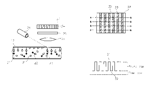
coupled to the wall of a test tube and the use of second precipitation
antibodies directed
CA 02720068 2010-09-29
WO 2009/124179
PCT/US2009/039280
against the analyte binding antibody followed by centrifugation as well as the
methods
described in the above noted patents and publications.
[0005] Some of the types of prior art physical separation of bound target
analyte
assays are described in the following U.S. Patents: 5,834,217; 5,776,710;
5,759,794;
5,635,362; 5,593,848; 5,342,790; 5,460,979; 5,480,778; and 5,360,719, all
issued to R. A.
Levine et al. In the aforementioned patents, the separation of bound from free
analyte is
performed by centrifugation, or other physical methods, such as decanting,
filtration, or
the like.
[0006] The prior art also describes a type of immunoassay, which is
called a
"homogeneous immunoassay". Homogeneous immunoassays do not require the
physical
separation of bound from non-bound, or free, analyte. The "separation of the
bound from
free" is accomplished by utilizing the steric interference of an enzyme by the
relatively
large antibody and quantifying the colored or fluorescent products of the
enzymatic
action. Additional methods of homogeneous assays utilize the fluorescent
quenching of
fluorophores to distinguish bound from free analyte. While these methods
greatly
simplify the performance of immunoassays, they are generally useful only for
high
concentrations of analytes with low molecular weight since the large molecular
weight of
target analytes such as proteins (e.g., insulin), growth hormones, and the
like will also
interfere with the enzyme and may affect quenching. Additionally immunoassays
of this
homogeneous type typically do not have the high sensitivity of standard
immunoassays.
[0007] It would be highly desirable to provide a ligand assay of a target
analyte
wherein the quantification of the target analyte is a virtual one which can be
performed
electronically thereby having the advantages of a homogeneous immunoassay
while
maintaining the sensitivity of standard immunoassays, as well as the ability
to have large
size target analytes such as hoiniones like insulin, growth hormone and the
like.
SUMMARY OF THE INVENTION
[0008] Immunoassays are used to analyze a wide range of analytes, such as
hormones in blood, etc. They work by the general technique of finding a
specific binder
which specifically binds to the target analyte being measured. A binder is
referred to
herein as a ligand. Ligands are defined herein as including, but not limited
to those
2
CA 02720068 2010-09-29
WO 2009/124179
PCT/US2009/039280
antibodies, lectins, aptimers, or naturally occurring substances, that are
operative to bind
a target analyte. The sample to be measured is admixed with the ligand which
is specific
to the target analyte, and a labeled version of the analyte to be measured. As
this mixture
is incubated, the labeled and unlabeled target analyte molecules compete for
binding sites
on the ligand. After a suitable period, the ligand is removed by any number of
ways, and
the label bound to the ligand is compared to the label which is unbound and
remains free
in the mixture. This bound/free ratio relates to the concentration of the
target analyte
originally in the sample, although either the bound or free label can give the
same
information. The use of the ratio allows the quality control check wherein the
total of
bound plus free is relatively constant if the volume is constant. This quality
control may
also be employed in the practice of this invention.
[0009] According to an aspect of the present invention, a method for
assaying a
biological fluid sample for a target analyte material that may be in the fluid
sample is
provided. The method utilizes a virtual separation of free and bound target
analyte
disposed within the fluid sample involving electronic scanning of the sample.
The
method involves placing the fluid sample in a test chamber having a
predetermined and
fixed height so as to produce a thin layer of the fluid sample in the chamber.
At least one
wall of the chamber is transparent, usually the top wall, so that the sample
can be
observed in the chamber. In certain cases both the top and bottom walls of the
chamber
are transparent. The height of the chamber (e.g., typically lu to 20011) can
vary
according to the application at hand. For example, when anticoagulated whole
blood is
being analyzed, a chamber height of 6 is advantageous because it creates a
monolayer of
red blood cells and interspersed plasma lacunae within the blood sample
[0010] The height of the fixed structure or ligand-coated bead optimally
should
be no less than one tenth of that of the chamber and ideally approaching the
height of the
chamber. The reason for this is that if the total amount of label (e.g.,
fluorophore)
present in the free state, surrounding the particle or structure to which the
label is bound,
is much greater than the amount of the lowest amount of bound label to be
detected, the
ability to accurately determine the amount of label bound to the bead or
structure is
diminished due to the influence of signal to noise ratios. Mathematically
there is no
limit to the height of the chamber but practical limits due to signal to noise
of the
CA 02720068 2010-09-29
WO 2009/124179
PCT/US2009/039280
detected label require a thin chamber and structures occupying at least ten
percent of the
volume of a cylinder drawn around the periphery of the structure and extending
from the
base to the tip of the chamber for optimal function. In examples where the
ligand is
adherent to the chamber top or bottom rather than a structure or bead, the
above ratios
apply, but the assay optimally should be formulated so that the cylindrical
volume above
the bound ligand area contains not more than ten times the lowest amount bound
to the
ligand area that is desired to be detected. This constraint can be diminished
by making
the chamber as thin as possible or by altering the stoichiometry of the
reaction.
[0011] The method of this invention can be used to test for drug
allergies or
allergen sensitivities in patients at the point of care. Drug allergies and
allergen
sensitivities are a common and important problem. It is expensive to the
patient and
society. Treating a penicillin allergic patient with a penicillin class drug,
for example,
can cause death or serious reactions. Penicillin is used in this discussion as
a
representative drug and because it is the drug type that is the most common
cause of
severe allergic reactions. The present invention is not limited to testing for
penicillin
allergies, and can be used to test for sensitivity to other drugs (e.g.,
antibiotics, muscle
relaxants, anesthetics, etc.) and allergens.
[0012] Penicillin is an inexpensive, effective and generally non-toxic
drug.
Patients who think they have a penicillin allergy, can be treated with another
less
microbial-targeted drug in view of the perceived allergy. Such replacement
drugs may
cause serious side effects in patients, however, and incur enormous costs to
the health
care system, since newer medications can be hundreds or thousands of times
more
expensive then penicillin drugs. Equally importantly are the costs to society
associated
with the increased development of drug resistant bacteria, viruses, or other
infectious
agents that occurs when broader spectrum drugs are used instead of drugs more
focused
on the target organism. It is, therefore, important to individual patients,
healthcare
providers, and society, to determine the presence or absence of drug allergies
or allergen
sensitivities by methods in addition to the history given by the patient. One
goal of this
invention is to detect the presence or absence of drug allergies and/or
allegen sensitivities
in a sample of a patient's whole blood or plasma.
[0013] It is well documented that many patients who claim to be allergic
to
4
CA 02720068 2010-09-29
WO 2009/124179
PCT/US2009/039280
penicillin are not allergic and similarly some patients who think that they
are not allergic
may have developed an allergy since their last exposure. There are many
reports that
about 80% of individuals who believe they are allergic to penicillin will in
fact tolerate
penicillin use, so for these patients the constraints on antibiotic choice,
potentially
resulting in less effective, more toxic and more expensive treatment, are
unnecessary.
[0014] Nowhere is the need for the ability to detect drug allergy more
needed
than at point of care encounters with the patient. Physicians about to
prescribe a
medication in their office, the emergency room or hospital do not have the
luxury of
waiting many hours or a day for the test to be perfouned either in vitro, or
by skin tests.
Skin testing may additionally expose the patient to risk of reaction to the
testing
substance and has the theoretical possibility of inducing allergy or
increasing it by an
anamnestic response. In vitro tests at present are complex, time consuming to
perform
and yield information to the physician long after it would be most useful.
Additionally
the allergenic nature of many drugs, including penicillin type drugs, may be
due to more
than one epitope and accurate testing would require testing for all common
epitopes
which may be the cause of the allergic response. RAST testing, well described
in the
literature, is generally performed on a limited number of test allergens and
their epitopes.
[0015] It is generally agreed that IgE mediated immune response is the
cause of
most severe allergic reaction including anaphylaxis, hives, intestinal
swelling with
diarrhea and respiratory obstruction due to swelling of airways. It is
suggested by some
experts that other immunoglobulin classes may also contribute to the allergic
response to
drugs but generally the allergic response to IgG and IgM mediated drug
allergies is not
life threatening and more likely to be a rash.
[0016] An advantage of the present invention is that it provides a means
to
perform, optimally at the point of care, a determination of the presence of
IgE or any
other immunoglobulin which has an affinity for one or more drugs that are or
may be
indicated for use in a given situation.
[0017] The label of choice is the use of a fluorophore that is easily
detected and
attached to the ligand. The present invention is not limited to using
fluorometric labels,
however. More than one color fluorophore may be used if it is desired to check
for the
presence or absence of more than one class of immunoglobulin that may become
attached
CA 02720068 2010-09-29
WO 2009/124179
PCT/US2009/039280
to the beads in the same chamber. Beads without the attached antigen are used
as
controls. The control beads can be chemically and geometrically similar to the
coated
beads, differing only in color or other means enabling their detection (e.g.,
fluorescence
or combinations of fluorescence dyes incorporated into their structure). The
control
beads provide a control so that the detection, for example of significant
fluorescent signal
from the fluorescent labeled antibody directed against the IgE that is
attached to the beads
containing a determinant (epitope) of the drug being tested as a potential
allergen, may be
compared to the signal that is present of similar beads not coated or bound to
the epitope.
Thus, nonspecific binding is controlled and will not result in a false
positive.
DESCRIPTION OF THE DRAWINGS
[0018] FIG. 1 is a schematic side view of a ligand-bearing surface which
may be
used to perfoini an immunoassay on a blood or other sample in accordance with
the prior
art.
[0019] FIG. 1(a) is a view similar to FIG. 1 but showing the surface
after the
sample has been washed away therefrom in accordance with the prior art.
[0020] FIG. 2 is a schematic side view similar to FIG. 1, but showing a
ligand-
bearing surface which may be used to perfotin a sandwich immunoassay on a
blood or
other sample in accordance with the prior art.
[0021] FIG. 2(a) is a view similar to FIG. 2 but showing the sample after
a second
label has been added to the sample in accordance with the prior art.
[0022] FIG. 2(b) is a view similar to FIG. 2(a) but showing the surface
after the
sample has been washed away therefrom in accordance with the prior art.
[0023] FIG. 3 is a plan view of a first embodiment of a sampling chamber
formed
in accordance with this invention which contains an anticoagulated whole blood
sample
to which blood sample ligand-bearing analyte-capturing particles have been
added.
[0024] FIG. 4 is a side sectional view of a portion of the sampling
chamber of
FIG. 3 which contains one of the ligand-coated target analyte-capturing
particles.
[0025] FIG. 5 is a plan view of a test chamber like that shown in FIG. 4,
showing
an area of the sample containing one of the ligand-coated target analyte-
capturing
particles and also showing another area of the sample which does not contain
one of the
6
CA 02720068 2010-09-29
WO 2009/124179
PCT/US2009/039280
ligand-coated target analyte -capturing particles but only contains the free
labeled target
analyte in the blood sample.
[0026] FIG. 6 is a fragmented cross-sectional view of a partially ligand
coated
target analyte capturing surface wherein portions of the surface are coated
with ligands
and other portions of the surface are not.
[0027] FIG. 7 is a sectional schematic view of a closed chamber having a
top
surface such as that shown in FIG. 6.
[0028] FIG. 8 is a plan view of the surface shown in FIG. 6.
[0029] FIG. 9 is a trace of the emissions from the ligand bands on the
capture
surface shown in FIGS. 6 and 8.
DETAILED DESCRIPTION OF THE INVENTION
[0030] Referring now to the drawings, FIGS. 1 and 1(a) illustrate a prior
art
competitive immunoassay (also referred to as an "equilibrium assay") which is
commonly used for analytes of low molecular weight, such as the thyroid
hormone,
thyroxin, where the numeral 1 denotes a surface to which a ligand 2, which is
specific to
the target analyte, is attached by any number of means well-known to the art.
Surface 1
may be a transparent wall of a glass or plastic tube or a particle. A solution
3 contains a
mixture of the unlabeled target analyte 4 (the unknown) and a labeled target
analyte 5.
After a period of time, which may be from minutes to hours, depending upon the
target
and the label, the labeled target analyte 5 and the unlabeled target analyte 4
will be in an
equilibrium with each other, wherein many, but generally not all, of the
ligand sites 2 will
be occupied with either a labeled target analyte 5 or unlabeled target analyte
4. At this
point (Fig la), the mixture 3 is separated from the ligand-bearing surface 1
in a manner
that preserves the labeled target analytes 5 which are bound to the ligand 2.
The labeled
target analytes 5 bound to the surface 1 are then measured (see FIG. la), and
the free
labeled target analytes may also be measured or may be calculated as: Total =
Free +
Bound, or Bound = Total - Free. The bound to free target analyte ratio is
inversely
related to the total target analyte amount in the sample.
[0031] FIGS. 2 - FIG. 2 (b) show a ligand assay often referred to as a
"sandwich"
assay, where two separate ligands are utilized. Surface 1 has ligand 2 bound
("bound
7
CA 02720068 2012-10-23
ligand") thereto in a similar manner described above, and the sample
containing the target
analyte 4 is introduced into the solution 3 and incubated with the surface 1.
Either
immediately, or after a suitable period of time, a separate labeled ligand 6
is introduced
into the solution, which labeled ligand 6 binds to a site on the target
analyte 4 which is
different than bound ligand 2 (Fig 2a). This, in effect, creates a "sandwich",
containing
the target analyte 4 in the center. The free labeled ligand 6 is then washed
off the surface
1 to leave the surface 1 covered with labeled sites (Fig 2b). The labeled
target analytes 4
bound to the surface 1 are then quantified, and the signal therefore is
directly proportional
to the amount of the target analyte 4 in the original sample. It is generally
recognized
that the sandwich assay is more precise and somewhat more accurate, but it can
only be
applied to target analyte molecules which have at least two different sites to
which
ligands can be bound.
[0032] In either of the above assays, the separation of the bound label
from the
free label is recognized as one of the challenging aspects of the procedure,
and often
requires one or more mechanically complex steps, such as centrifuging,
decanting,
washing, etc. As a result, instrumentation to automate these tests has been
relatively
complex, requiring multiple operations.
[0033] Aspects of the present invention, in contrast, provide a means of
"virtual
separation", wherein the bound and free label are not physically separated,
but rather
separated by a combination of test cell configuration and mathematical
manipulation of
the signals from different regions in the test cell. As a result, simplified
automated ligand
assay methods and apparatus can be performed.
[0034] According to aspects of the present invention, immunoassays or
ligand
assays are performed where the binder is a ligand or other a substance having
a high
affinity for the target analyte.
[0035] Assays according to the present invention can be performed, for
example,
using the sample containers and imaging instrument systems described in the
U.S. Patent
Publication Nos. 2007/0243117 and 2007/0087442 and U.S. Patent No. 6,866,823.
The
present assays are not limited to these chambers and imaging devices, however.
[0036] The term "immunoassay" as used in this disclosure and claims shall
mean
8
CA 02720068 2010-09-29
WO 2009/124179
PCT/US2009/039280
both antibody-based binding agents and non antibody-based binding agents.
Examples of
the latter include, but are not limited to, intrinsic factors for binding
vitamin B12, and
avidin for binding biotin-labeled targets or vice versa.
[0037] Under aspects of the present invention, a well-defined and
physically
circumscribed surface is provided to which the ligand is attached, and then
the signal
from the label bound to that surface is mathematically distinguished from that
of any
surrounding free label that may reside in solution. There are two general
cases which are
described as follows.
[0038] FIG. 3 is a plan view of a section of a specimen chamber assembly
40,
which chamber assembly 40 contains an anticoagulated whole blood sample. The
chamber assembly 40 includes upper and lower walls 7 (see FIG. 4), at least
one of which
is transparent. Preferably, both of the walls 7 are transparent. The chamber
assembly 40
includes spacer members 42 (see FIG. 3) which are randomly located inside of
the
chamber assembly 40. The spacer members 42 are preferably spherical and
determine
and control the height of the chamber assembly 40. In the case of assaying an
anticoagulated whole blood sample, spacer members 42 having a diameter of
about 61Lt
work particularly well. The blood sample which is contained in the chamber
assembly 40
will include individual red blood cells 44 and agglomerations of red blood
cells 46. The
blood sample also includes clear plasma lacunae areas 48 which do not contain
any
formed blood components. Finally, the blood sample also includes a plurality
of ligand-
coated target analyte-capturing particles 8 which are preferably in the form
of spheres.
The target analyte-capturing particles 8 are randomly distributed throughout
the blood
sample, and may be about 311 - 4 in diameter for a blood sample analysis, so
that they
can be easily detected in the blood sample.
[0039] FIG. 4 shows the structure of the chamber assembly 40 of FIG. 3.
The
chamber assembly 40 is bounded by top and bottom wall 7, at least one of which
must be
transparent. Within the chamber is a particle 8, whose surface is covered with
a ligand 9.
The particle 8 may be any shape as long as its volume can be determined, but
it is
preferably a sphere. The particle 8 may be of any material to which a ligand
can be
attached, such as glass, polystyrene, or the like. The particles are not
limited to any
particular diameter (e.g., 2 - 1000, and the diameter can vary depending on
the fluid
9
CA 02720068 2010-09-29
WO 2009/124179
PCT/US2009/039280
being assayed and the height of the chamber being used. The distance between
the walls
7 is typically not less than the diameter of the particle 8, but the upper
distance limit will
depend upon the nature of the particle 8.
[0040] A mixture 10 contains both a target analyte 11 and a labeled
target analyte
12 in a manner similar to that described in connection with FIG. 1 above.
After a suitable
period of incubation, the signals from the bound and free target analyte are
processed.
[0041] FIG. 5 is a top view of a test chamber assembly like that shown in
FIG. 4,
showing an undefined expanse 13 of the mixture 10. Within this expanse, the
total signal
from the label 12 is collected over a defined area 14, which area is not
limited to any
particular shape. The means of collection can be a fluorescence scanner, in
the case of a
fluorescent label, or a radio nucleotide scanner, in the case of a radio
label. The area is
chosen so that it includes at least one particle 8, with a known or measurable
diameter.
An adjacent defined area 16, not containing a particle, is also measured. The
signal from
area 16 represents that from the unbound label, since there are no binding
sites in that
location. The signal from area 14, however, has a signal from both the bound
and the free
label. The influences of each can be determined in a number of ways. If the
particle is
spherical, which is a preferred shape, its volume (Vp) can be calculated from
its diameter,
which can be measured with the same optical system that collects the signal
from the
label. The volume of the defined areas 14 (V14) and 16 (V16) can be readily
calculated
from their width and the chamber depth. Assuming that the chamber volumes
associated
with defined areas 14 and 16 are identical, the signal from the free label is
equal to that of
the signal from area 16 (S16). This means, that in the absence of signal from
the particle
(the bound label), the signal from area 14 (SO should be: Sf = S16 x (V14 -
Vp). Any
signal in excess of this amount is from the bound label (Sb): Sb= S14 - Sf. If
the volume
of the particle is de minimus compared to the volume within the area 14, then
the volume
correction is not necessary. What is determined is the average label signal
intensity per
pixel (or collective group of pixels) of the scans. The term pixel as used in
this
application may include the meaning of one or more adjacent pixels.
[0042] In a second, and most preferred embodiment, ligands are attached
to at
least one surface of the chamber itself FIG. 6 shows an (upper) transparent
chamber
surface 17, which may be glass or plastic, such as acrylic or polystyrene, to
which a
CA 02720068 2010-09-29
WO 2009/124179
PCT/US2009/039280
uniform coating of the ligands has been attached by any number of means well
known to
the art. After the uniform coating is formed, ligand are selectively removed
from one or
more regions 18, either by mechanical or chemical means, or by laser ablation,
consequently leaving active ligands in adjacent regions 19.
[0043] FIG. 7 shows this surface 17 as part of a thin chamber containing
mixture
20, comprising unlabeled target analyte 21 and labeled analyte 22. The chamber
is
preferably less than about 1 mm in height, and is most preferably less than
20011 (e.g., in
a range of 1 to 2004 As before, after a suitable period of time, the labeled
and
unlabeled analyte will reach equilibrium with the ligand, leaving a portion of
the labeled
analyte 23 bound to the surface, but only in the region where the ligand
remains. In the
case of a fluorescent label, the chamber surface 17 is illuminated with light
source 24 of
the appropriate wavelength to excite fluorescence in the label. Lens 25
collects the
fluorescent emissions, which are filtered by optical filter 26 and projected
onto an image
dissection device 27, which may be a charge couple device (CCD), complimentary
metal
oxide semiconductor (CMOS), or the like. Alternatively, the light source may
be a laser
which focuses a tiny, moving spot onto the chamber, and the light collecting
device 27
would be, in that case, a simple phototube or photomultiplier.
[0044] The net result of either process is shown in FIG. 8, which is a
schematic
top view of the chamber 28, where the active ligand 29 and ablated ligand 30
appear as a
series of vertical stripes. The scan lines from the apparatus of FIG. 7 are
represented by
the lines a-a. FIG. 9 is a representation of the waveform taken across the
scan lines a-a,
where the peaks 31 are the signal from the active ligand, and the valleys 32
are from the
inactive areas. Thus, the bound label concentration is represented by the
distance from
the peaks to the valleys, and the height of the valleys represents the free
label. The active
areas and inactive areas are not limited to any particular geometry.
[0045] In some embodiments, a chamber wall 17 can be used that is
sufficiently
flexible that it can be locally elastically deformed by subjecting it to a
relatively small
point load. The elastic nature of the chamber wall 17 allows a unique option
to capture
very weak "bound" signals. If the chamber wall 17 is compressed, such as by a
small
stylus just out of the imaged area, the free label 22 is expelled laterally
from the local
field of view, and thus its signal is markedly reduced. With this "background"
signal
11
CA 02720068 2010-09-29
WO 2009/124179
PCT/US2009/039280
reduced, very weak signals from bound label 23 can be detected.
[0046] Multiple analytes could be measured simultaneously if the labels
fluoresce
at different wavelengths, or if the ligand for analyte 1 were at a different
physical location
in the chamber from the ligand for analyte 2.
[0047] An example of a method according to the present invention method
includes performing an assay to determine whether a patient may be allergic to
one or
more drugs (e.g., antibiotics, including penicillin, etc.) or allergens. The
assay is
performed using a cartridge that has an analysis chamber containing a large
number (e.g.,
thousands) of antibiotic epitope coated beads and uncoated control beads. For
those
analyses directed toward more than one antibiotic epitope, each particular
antibiotic
epitope is matched with a particular type of bead for identification purposes.
The groups
of beads associated with different epitopes can be distinguished from one
another using
characteristics such as a bead color, size, shape, etc; e.g., epitope A is
coated on white
beads, epitope B is coated by red beads, etc. A small amount of sample (e.g.,
0.5 to 5
micro liters) of capillary or venous anticoagulated whole blood is deposited
in the
chamber (e.g., drawn into the chamber by capillary action) and upon closing
the chamber
the blood is directed into an area within the chamber containing the beads.
After
incubation for a first period of time (e.g., minutes to an hour)
immunoglobulin present
within the sample binds to those beads coated with a drug (or allergen) to
which the
immunoglobulin molecule has a specific affinity. Different immunoglobulin
molecules
present within the sample may have different affinities specific to different
drugs (or
allergens). The combined beads and blood sample is further mixed with one or
more
labeled antibodies directed against the immunoglobulin being tested (e.g.,
Immunoglobulin E ("IgE"), etc.) and allowed to incubate for a second period of
time
(e.g., seconds to minutes). A fluorophore may be tagged to the antibodies
directed
against the immunoglobulin being tested to create the "labeled antibody". The
sample is
then directed into the analysis chamber of the type described above. The
actual times
needed for incubation for the two steps can be empirically determined and will
likely
depend upon the avidity and concentration of the antibodies present. The
sample
disposed within the chamber is analyzed by collecting the signal from the
labeled
antibodies both free and bound in one or more of the manners described above.
If the
12
CA 02720068 2010-09-29
WO 2009/124179
PCT/US2009/039280
assay involves the determination of allergy susceptibility of more than one
drug, or
sensitivity to more than on allergen, the analysis will include distinguishing
the bound
labeled antibodies as a function of the different types of coated beads as
well. The bound
label represents those labeled antibodies that are bound to the immunoglobulin
being
tested, which immunoglobulin is bound to the particle coated with the drug (or
allergen)
with which the particular immunoglobulin particle has a specific affinity. The
amount of
label bound on a particle may be calculated by measuring the total signal of
the imaged
particle and subtracting the surrounding free signal in the immediate area
surrounding the
particle that is included in the image. Ratios of the amount of label on a
given class of
coated beads can be calculated by measuring labeled coated and uncoated beads
of the
same type. Thus, the determination of whether a sample contains immunoglobulin
molecules having an affinity for a given drug (or allergen) can be performed
by
practicing the present invention. In addition, simultaneous detection of an
allergy to
more than one drug (or sensitivity to more than one allergen) can be performed
under the
present invention using different types of detectable beads or particles, with
each type
coated with a different drug (or allergen). A single type particle (or bead)
may be used as
a control particle for all the drug allergy (allergen sensitivity) tests if
the particle is the
same in size and composition as the coated particles. If necessary, more than
one type of
control particle may be used with the size of the control particle matching
the size of the
drug or allergen coated particles to which it is being compared.
[0048] In some embodiments, after the second incubation (the one
containing the
labeled ligand) the method includes the step of adding a liquid containing no
label to the
sample containing unbound label disposed within the chamber, thereby leaving
primarily
the label attached to the immobilized beads or structures. The virtual
separation of
bound from free is subsequently performed as previously stated but the removal
of the
liquid containing the label can serve to increase sensitivity of the assay at
the expense of
complexity. Since the total capacity of the chamber and the amount of liquid
in the
chamber is in the range of less than one to several micro liters, the addition
of a label-
free fluid to the chamber in a substantial volume (e.g., tens of micro liters)
will remove
much of the fluid containing label and the remaining free label signal will be
removed by
the utilization of the virtual separation of bound from free process.
13
CA 02720068 2010-09-29
WO 2009/124179
PCT/US2009/039280
[0049] The above described methodology provides a novel and desirable
technique for determining the amount of bound and free labeled target analyte
within
areas of a chamber that contain ligands or are free of ligands specific to
that target analyte
within a sample, and thereby provides qualitative and quantitative information
relative to
the sample. In some instances, qualitative information such as knowing whether
the
target analyte is present or absent in the sample is sufficient information
for the analysis
at hand. An example of such an instance is the determination of whether a
specimen has
specific IgE directed against a given drug, when the absence of such IgE is
the normal
state. If more quantitative information is desired (e.g., the concentration of
the target
analyte in the sample), the obtained bound/free information may be used with a
standard
curve, which curve is empirically derived for the particular target analyte
and sample
being considered, to determine the quantitative information; e.g., the amount
of target
analyte within the sample. Standard curves operable to be used with all types
of
immunoassays are known and the present invention is not limited to any
particular
standard curve. Sample curves may be performed prior to or concurrently with
the assay
and the results stored on the instrument performing the analysis.
[0050] Although the invention has been shown and described with respect
to
specific detailed embodiments thereof, it will be understood by those skilled
in the art
that various changes in form and detail thereof may be made without departing
from the
spirit and the scope of the invention.
14